A new possibility for theoretical interpretation of electron transitions and electron states

Milestones in Electron Crystallography
Ludovic Renault, Hui-Ting Chou, Po-Lin Chiu, Rena M. Hill, Xiangyan Zeng, Bryant Gipson,Zi Yan Zhang, Anchi Cheng*, Vinzenz Unger**, and Henning StahlbergMolecular and Cellular Biology, University of California at Davis, 1 Shields Ave., Davis, CA 95616, USA
* Department of Cell Biology, The Scripps Research Institute, 1055 N. Torrey Pines Road, La Jolla, CA 02037,USA
** Department of Molecular Biophysics and Biochemistry, Yale University, PO Box 208024, New Haven, CT065200-8024, USA
SummaryElectron crystallography determines the structure of membrane embedded proteins in the two-dimensionally crystallized state by cryo-transmission electron microscopy imaging and computerstructure reconstruction. Milestones on the path to the structure are high-level expression, purificationof functional protein, reconstitution into two-dimensional lipid membrane crystals, high-resolutionimaging, and structure determination by computer image processing. Here we review the currentstate of these methods. We also created an Internet information exchange platform for electroncrystallography, where guidelines for the imaging and data processing method are maintained. Theserver (http://2dx.org) provides the electron crystallography community with a central informationexchange platform, which is structured in blog and Wiki form, allowing visitors to add comments ordiscussions. It currently offers a detailed step-by-step introduction to image processing with the MRCsoftware program. The server is also a repository for the 2dx software package, a user-friendly imageprocessing system for 2D membrane protein crystals.
Keywords2dx; electron crystallography; membrane proteins; image processing; 2D crystallization; electronmicroscopy
IntroductionMembrane proteins are of central importance for health and disease. They correspond to morethan 25% of predicted proteins and act as transporters, channels, receptors, and scaffoldingcomponents. Because of their strategic location, membrane proteins play critical roles in manyfunctions such as cell proliferation, nerve and muscle contraction, and regulation of bloodpressure, salt and water balance. Consequently, loss of function typically results in criticalphysiological effects such as neurological disorders, cancer, digestive diseases, heart failureetc. Because of their biological importance, membrane proteins represent the majority oftoday’s drug targets in pharmaceutical research and are the major targets for current drugsranging from Aspirin (cyclo-oxygenase) to Zoloft (serotonin transporter) [1]. Structuralbiology of membrane proteins is thus one of the most important fields of modern biology.Nevertheless, less than 300 structures of membrane proteins have been determined (see http://
Corresponding author: Henning Stahlberg, Molecular & Cellular Biology, Briggs Hall, College of Biological Sciences, University ofCalifornia at Davis, 1 Shields Ave., Davis, CA 95616, USA, Tel.: +1 (530) 752 8282, Fax: +1 (530) 752 3085, email:[email protected].
NIH Public AccessAuthor ManuscriptJ Comput Aided Mol Des. Author manuscript; available in PMC 2008 January 14.
Published in final edited form as:J Comput Aided Mol Des. 2006 ; 20(7-8): 519–527.
NIH
-PA Author Manuscript
NIH
-PA Author Manuscript
NIH
-PA Author Manuscript
blanco.biomol.uci.edu/Membrane_Proteins_xtal.html), about one third of which areconsidered as unique structures. These structures were determined mostly by X-ray diffraction(XRD), electron crystallography and nuclear magnetic resonance (NMR), to a resolutionsufficient to at least identify trans-membrane helices. Compared to more than 37,000 availablestructures of soluble proteins available in the Protein Data Bank (PDB, [2]), this low numberof determined membrane protein structures is in stark contrast to their biological importance.
Structure determination of membrane proteins – eukaryotic membrane proteins in particular -faces several technical hurdles. Difficulties in heterologous over-expression, non-denaturingdetergent solubilization and gentle purification limit the amount of functional membraneprotein sample that are available for structural studies. Nevertheless, in cases where it wassuccessful, structure determination by X-ray diffraction of three-dimensional (3D) crystals,nuclear magnetic resonance and cryo-electron microscopy (cryo-EM) of two-dimensional (2D)crystals revealed amazing structural concepts and mechanisms that nature employs to solvethe challenging tasks posed to these proteins. Recent highlights include the 1.35Å structure byXRD of the ammonium transporter AmtB [3], the structure of the waterchannel Aqp0 by cryo-EM at 1.9Å [4] and XRD at 2.2Å resolution [5], and the structure of Mistic [6] by NMR [7],to give only a few examples.
Over the past decade, electron crystallography has established itself as a viable alternative toX-ray crystallographic structure determination, especially of membrane proteins. While theresolution often is lower than that which can be obtained by XRD, electron crystallographytakes advantage of the fact that it analyses the structure of the protein embedded in a nativemembrane environment. Using this approach, atomic models for seven membrane proteins andtubulin were so far determined: BR [8], LHCII [9], AQP1 [10,11], nAChR [12], and AQP0[4,13], AQP4 [14], MGST1 [15], and Tubulin [16]. Several other membrane proteins classifiedas transporters, ion pumps, receptors and membrane bound enzymes have been studied byelectron crystallography at lower resolution allowing localization of secondary structure motifssuch as trans-membrane helices, and are likely to produce atomic models in the near future(e.g. [17–20]).
MilestonesStructural investigation of membrane proteins by electron crystallography involves milestonesas depicted in Figure 1. Before 2D crystals can be obtained identified targets have to be cloned,expressed and purified. Structural analysis by transmission electron microscopy firstlyestablishes a two-dimensional projection map of the membrane protein as seen from above themembrane. Combination of this map with data from tilted 2D crystal samples allows thereconstruction of the 3D map of the protein. At a resolution of 7 Å, alpha-helical transmembranesegments can be identified. At 4.5 Å the first densities for larger amino acid side-chains canbe recognized, and an atomic model can be proposed at this or better resolution. Figure 1 alsogives estimations for the required average time to reach the next milestone, as well as theexpected success rates for those steps. These values are estimates based on the current state ofthe technology. We estimate an average time of 15 months for the production of highly pure,homogeneous and active protein, followed by one year to grow well-ordered 2D crystals.Structure determination by an experienced laboratory would then require ~3 years, duringwhich time the quality of the 2D crystals would be further refined. Accumulative failure ratesimply that from 20 targeted membrane proteins one atomic structure can be expected after ~5 years. In comparison with structure determination by XRD, obtaining well-ordered 2Dcrystals has a higher likelihood and lower estimated required time. However, the structuredetermination from such 2D crystals is still a labor- and time-intensive procedure, where onlya handful of laboratories have so far demonstrated success. In the following we describe the
Renault et al. Page 2
J Comput Aided Mol Des. Author manuscript; available in PMC 2008 January 14.
NIH
-PA Author Manuscript
NIH
-PA Author Manuscript
NIH
-PA Author Manuscript
different steps of electron crystallography and then describe how we suggest accelerating theprocess and increasing the success rate.
Membrane protein production and purificationIdentified target sequences usually have to be cloned, inserted into a vector and over-expressedin a heterologous host. In the past the use of bacterial expression systems has been particularlyuseful. However, bacterial systems are frequently of little use for over-expression ofmammalian trans-membrane proteins, which require many post-translational modificationsand processing events for correct protein folding. The study of bacterial homologues asrepresentatives of human membrane proteins will be of limited use, as no bacterial or archaealhomologues have been identified for about 50% of human membrane proteins [21].
Several membrane proteins affect the function of the host cell, e.g. insertion of the mammalianmembrane proteins into bacterial cell membranes often results in cell death. Thus the searchfor a suitable expression system may be challenging [22]. Cell-free expression may be avaluable alternative route for highly cyto-toxic membrane proteins. Cell-free systems allowspecific labeling of the sample, and the resulting expression product usually does not needfurther purification [23,24]. However, cell-free synthesis systems generally lackposttranslational modification machinery, which may be disadvantageous in the case ofmammalian membrane proteins. Nevertheless, Klammt et al., (2004) could produce severalmg of membrane protein per ml of reaction volume of apparently correctly folded protein afterdissolving from a protein precipitate by the addition of detergents and lipids [25]. Recently thesame authors reported that the addition of detergents and lipids to the translation mix stabilizedsome of the proteins tested [26]. In addition to these approaches, bacterial expression ofeukaryotic membrane proteins can be facilitated by addition of the Mistic sequence to the targetmembrane protein sequence. Mistic is a small, highly charged protein from Bacillus subtilis,which facilitated bacterial expression and membrane insertion of several mammalianmembrane proteins [6,27].
The need to reside in a membrane environment requires membrane proteins to be amphiphilic.These physicochemical properties of membrane proteins necessitate the use of detergents forsolubilization from the membrane and for purification. Finding the best detergent for any givenmembrane protein is a matter of trial and error. Nevertheless, many membrane proteins can besolubilized and purified using a small collection of detergents [28]. The purification ofsolubilized membrane proteins uses similar techniques to those employed for soluble proteins.Chromatographic methodologies are widely used. In contrast to soluble proteins, membraneproteins may interact strongly with the column media, thus lowering purification efficiency.Purification steps that remove bound endogenous lipids from the solubilized membraneproteins may also induce conformational changes or loss of correct folding of the protein withsubsequent loss of function and may also result in protein precipitation [29,30]. The use ofpoly-His or FLAG tags facilitates purification on specific affinity columns, yet the overallefficiency tends to be lower. Specific cleavage sites can be inserted to remove unwanted tags.Yet in this case, compatibility of the chosen protease with detergents should be tested inadvance. Fortunately, well-ordered 2D crystals can also be grown in the presence of thepurification tags (e.g. [31]) thus making the addition of protease cleavage sites mostly optional.
Solubilization, purification, and crystallization may have different detergent requirements.Therefore, it is necessary to perform a detergent-screen for all three steps. Moreover, additionof solubilized lipids to the purification buffers may help stabilize the membrane protein duringthe purification, as indicated by the beneficial effect of addition of phospholipids in the 3Dcrystallization of lactose permease (LacY) (Guan & Kaback, personal communication, see also[32]).
Renault et al. Page 3
J Comput Aided Mol Des. Author manuscript; available in PMC 2008 January 14.
NIH
-PA Author Manuscript
NIH
-PA Author Manuscript
NIH
-PA Author Manuscript

The brief synopsis given in the previous paragraph emphasizes the intricacies involved inmembrane protein production for structural studies, regardless of the approach that is chosenfor the actual data collection. With this in mind, centralization of membrane protein productionis extremely beneficial to the membrane protein community by supplying pure protein forstructural analysis This approach is implemented by several centers supported by the proteinstructure initiative (PSI) of the National Institute of General Medicine and Sciences (NIGMS,http://www.nigms.nih.gov/Initiatives/PSI/Centers). For example, the Membrane ProteinExpression Center (MPEC, http://mpec.ucsf.edu) performs large-scale membrane proteinproduction that benefits structural studies by several methods and laboratories. Similarapproaches are underway in Canada (http://www.sgc.utoronto.ca/), Europe (e.g. E-MeP, http://www.e-mep.org/) and Japan (BIRC, http://unit.aist.go.jp/birc/index_e.html, [33]).
Two-dimensional membrane protein crystallizationThe most widely used approach to achieve 2D crystallization of membrane proteins is the slowremoval of the detergent from the protein-detergent micellar solution in the presence of addedsolubilized lipids at a low lipid-to-protein ratio, to allow reconstitution of the protein into anewly formed lipid bilayer (Figure 2). The goal is to reduce the detergent concentration tobelow its critical micellar concentration (cmc) for which precise knowledge of the startingdetergent concentration is required. Kaufmann et al. (2005) have recently presented a devicethat allows exact determination of detergent concentrations during purification andcrystallization trials using minimal sample quantities [34]. Here we briefly list and summarizethe key aspect of the different approaches that have been successfully used in the past togenerate 2D-crystals [28,35–38].
Dialysis—The dialysis method consists of dialyzing 20–100 μl samples against a largevolume of buffer. This method allows slow and controlled removal of the detergent, but isdifficult with low-cmc detergents, which require a longer dialysis time. As with 3D-crystallization, many factors affect the membrane reconstitution and two-dimensionalcrystallization, such as the choice of detergent, lipid, lipid-to-protein ratio, pH value, the kindsof salts, the salt concentrations, temperature, and other factors [36]. The use of a computer-controlled dialysis machine can reduce the dialysis time, and also allows a precise control ofthe temperature profile [35], which helps to make the entire process more reproducible. Still,fragile membrane proteins that do not withstand the presence of detergent for a long period oftime are difficult to crystallize with this method.
Dilution—The principle of the dilution method is to dilute the protein-lipid-detergent solutionto a detergent concentration below that of its cmc, thus allowing reconstitution of the proteininto the lipid bilayer. It has the advantage that the detergent concentration can be very quicklybrought to below its cmc. This method is also suited for low-cmc detergents [39,40].
Hydrophobic adsorption—The detergent in a protein-lipid-detergent mixture is removedby hydrophobic adsorption onto polystyrene beads, which usually can selectively bind thedetergent but not protein or lipids. This procedure allows detergent removal in a short periodof time, but the rate of removal cannot easily be controlled. This method can be used to removeboth high- and low-cmc detergents [41,42].
Lipid monolayer crystallization—This method is based on the specific interactionbetween the solubilized membrane protein and a lipid monolayer that covers an air/waterinterface. The lipid monolayer is usually composed of two different kinds of lipids, a ligandlipid for the protein and a dilution lipid. Lipids need to be chosen that can maintain themonolayer even in the presence of the detergent. The lipid film is spread at an air/water interfaceof an aqueous solution containing the protein to be crystallized. The lipid-protein complex
Renault et al. Page 4
J Comput Aided Mol Des. Author manuscript; available in PMC 2008 January 14.
NIH
-PA Author Manuscript
NIH
-PA Author Manuscript
NIH
-PA Author Manuscript

diffuses in the plane of the film, where the protein concentration is increased due to bindingto the surface. Once bound, the membrane proteins can be reconstituted into a phospholipidmembrane by adding lipids to the sub-phase and removing the detergent with the addition ofBiobeads. This approach requires very small amounts of protein, less than 1 μg per incubationtrial [43,44]. A similar approach was developed by Auer et al. (1999), who grew 2D crystalson an electron microscopy grid [45].
Crystal screening—As for three-dimensional membrane protein crystallization, 2Dcrystallization trials have to be screened for the presence of crystals. Due to the small size ofthe crystal this is so far done by transmission electron microscopy (TEM) imaging of negativelystained specimens. This step is labor intensive and monotonous, and represents a majorbottleneck in the structure determination pipeline, given sufficient supply of purified protein.The use of an automated imaging system such as Leginon [46] or AutoEM [47] will greatlybenefit crystal screening. The challenge for automatic systems lies in distinguishing 2D crystalsfrom other precipitates, protein-free lipid membranes and stain and other artifacts. 2D crystalscan have a multitude of shapes and appearances and are accompanied by a highly variablebackground. Nevertheless, the ability to calculate power spectra of recorded images is apowerful tool to assess crystallinity of a sample. Progress in automated crystal screeningsystems will greatly benefit systematic crystallization trials.
Electron microscopy data collectionOnce suitable 2D crystals have been produced, a cryo-EM sample preparation method has tobe established that maintains the high-resolution order of the 2D crystal and at the same timepresents the 2D crystals with good contrast in the electron microscope. Several samplepreparation methods are available and have to be adapted to the individual sample. 2D crystalsusually are adsorbed to a thin carbon film, covering an electron microscopy grid. The grid withthe crystals is then quick-frozen to liquid nitrogen temperature to vitrify the buffer and therebypreserve the membrane protein structure for cryo-EM imaging (e.g. [48]). Alternatively, thecrystal solution can be quick-frozen on a fenestrated carbon film grid, and the crystals are thenimaged in the vitrified ice over the holes of the carbon film [49]. However, the viscous (liquid)vitrified ice provides inferior stability, electrical conductivity and sample flatness than asupporting continuous carbon film can provide [50], which can only be partly compensated bysurface coating [51]. Sugar embedding is another sample preparation method that involvespartial drying of the carbon-film adsorbed membrane protein crystals in the presence of sugars.Tannic acid [9], trehalose [10,16,52] or glucose [53] can preserve the ultra-structure of most2D crystals while facilitating the sample preparation and reducing the amount of icecontamination on the frozen samples. Nevertheless, as far as we know the latter approacheshave not yet worked with any membrane protein that has large loops and/or termini residingoutside the membrane.
The three-dimensional structure of the membrane protein is reconstructed by combining imagesfrom tilted 2D crystal samples. Depending on the actual structure and the goal of the analysis,this requires the recording of cryo-EM images of flat 2D crystal samples at angles up to 60°or 70° of sample tilt. Unfortunately, the electron-beam/sample interaction complicates therecording of images of tilted samples, which frequently show anisotropic diffraction that ispoor in the direction perpendicular to the tilt axis. This beam-induced resolution loss wasattributed to buildup of positive charge during the electron exposure, sample rearrangementsunder the electron beam, and/or a drum-head movement of the sample during image recording[50,54,55]. This phenomenon reduces the efficiency in data collection and can void high-resolution data in almost all of the recorded images of tilted samples. The data collectionefficiency can be significantly enhanced by use of the SpotScanning data collection method[56–58] or by the sandwich sample preparation method [59]. The first consists of illuminating
Renault et al. Page 5
J Comput Aided Mol Des. Author manuscript; available in PMC 2008 January 14.
NIH
-PA Author Manuscript
NIH
-PA Author Manuscript
NIH
-PA Author Manuscript
only a small area of the sample, while the concentrated electron beam “jumps” over the tiltedsample and records a pattern of spots on one large image. This SpotScanning method reducesthe effect of the beam-induced image drift with the dimensions of the illuminated sample area.The sandwich sample preparation method embeds the 2D crystals between two symmetricallyarranged carbon films, thereby increasing the electrical conductivity and stability of the sampleunder the beam. Combination of both methods can further increase the efficiency of the datacollection.
Recorded cryo-EM images of the 2D crystals are so-called real-space images, which provideamplitudes and phases for the reconstruction of the structure of the membrane protein. Theresolution of such real-space images, however, is affected by sample vibration or movement,beam-induced sample- or image-drift as well as limited coherence of the electron microscopeunder high defocusing conditions. Similar to X-ray diffraction, the electron microscope canalso be used to record electron diffraction patterns from the 2D crystals. Such electrondiffraction patterns provide the precise amplitudes of the structure, but do not give immediateaccess to phase information. However, electron diffraction data collection is not sensitive tosample movement and does not significantly suffer from beam-induced effects onto the sample.Therefore, electron diffraction is an efficient way to collect high-resolution data as long ashighly ordered and flat 2D crystals of sufficient size (generally diameter > 1μm) are available[60]. As in XRD studies, the phases of the structure then have to be obtained by other means,for example from homologous protein models [4] or by phase extension from a lower resolution3D dataset [53]. A remarkable achievement from electron diffraction data collection is therecently solved 1.9 Å resolution 3D structure of Aquaporin-0 in the membrane-embedded state[4]. This map also allowed the building of an atomic model of the protein-surrounding lipidmembrane and the protein-lipid interactions.
Computer image processingExtensive computer image processing of the recorded electron crystallography data is neededto extract the structure factors (amplitudes and phases) for the membrane protein structure.Electron diffraction patterns are quantitatively evaluated and the diffraction intensities of thedifferent diffraction spots are integrated to yield the intensities of the diffracted electron rays.The square root of these intensities then gives the amplitudes for the structure reconstruction.The real-space images of 2D crystals have to be Fourier transformed to produce a pattern similarto the diffraction pattern. The computed complex Fourier transformation, however, containsamplitude and phase information. While the high-resolution content of the real-space imagesis likely affected by the microscope and sample stability limitations, the real-space image hasthe advantage that any 2D crystal defects can be recognized and computationally corrected,before the Fourier transformation is calculated. This “unbending” of the 2D crystal images wasfirst introduced by Henderson and Unwin [61], and results in significant sharpening of thediffraction peaks in the calculated Fourier transformation, after which the values for amplitudesand phases for the spots can be measured with much better precision.
Each real-space image or electron diffraction pattern contributes data along a plane in the three-dimensional Fourier space. Measurements from several images or diffraction pattern have tobe combined, merged and interpolated to obtain a coherent 3D dataset in Fourier space, whichcan then be used to calculate a 3D map of the membrane protein density. If this map hassufficiently high resolution, an atomic model of the protein structure can be built. Due to thefact that images or diffraction patterns of 2D crystal samples can only be collected at sampletilts up to 60 or 70 degrees tilt, a 3D dataset from electron crystallography is devoid ofinformation in the vertical direction perpendicular to the membrane plane and within a coneof at best 20–30° degrees opening angle to the vertical axis. This “missing cone” phenomenonis inherent to the method and is the reason why structures from electron crystallography have
Renault et al. Page 6
J Comput Aided Mol Des. Author manuscript; available in PMC 2008 January 14.
NIH
-PA Author Manuscript
NIH
-PA Author Manuscript
NIH
-PA Author Manuscript
a lower resolution in the vertical direction: All details in the structure are smeared out vertically,and thinner horizontal elements, for example the surface loops of helical transmembraneproteins, may become undetectable (see for example the lower panels in Figure 1). In addition,regions of the membrane protein crystal that are less-well ordered will also appear at lower oreven absent density in the 3D map. Less-well ordered regions are usually the surface loops ofa membrane protein crystal due to deformations caused by adsorption of the crystal to theunderlying carbon film. This is the reason why most electron crystallography structures do notshow densities for the helix-connecting loops.
The computer image processing of the recorded images or diffraction pattern is usually donewith the “MRC programs” for image processing [62]. Over many years a large set of programshas been written for processing images of two-dimensional crystals and electron diffractionpatterns, usually in Fortran-77 [8–10,63]. While this software collection offers a vast andinvaluable repertoire of tools for the processing of 2D crystal images, its usage involves a highamount of interactive time. An interesting new development is the IPLT software, the ImageProcessing Library and Toolbox [64]. Bsoft is another powerful software system that is partlyapplicable for 2D crystals [65].
Information exchange – 2dx.orgElectron crystallography so far lacks the community infrastructure that became available forX-ray diffraction through the CCP4 initiative [66]. While the MRC programs for electroncrystallography are a powerful and well-maintained software collection, the MRC softwareand electron crystallography in general so far did not have a detailed manual, a tutorial, a schoolor a dedicated workshop, or conferences. This made it extremely difficult for newcomers tostart using this method, since it usually required for a beginning student to be introduced to themethod and software by one of the handful of experts in the field.
To address this need for information exchange, we have created a web server 2dx.org to providethe community of electron crystallography with a central information exchange platform forelectron crystallography of membrane proteins. This server is maintained by the Stahlberglaboratory at UC Davis, and is based on a Zope platform (http://www.zope.org), running Plone(http://www.plone.org) and a ZWiki engine (http://www.zwiki.org). The 2dx.org serverintends to collect information about electron crystallography sample preparation, imaging andcomputer image processing. It should give the newcomer to the method all the informationneeded to quickly familiarize him/herself with the method. It should also serve the advancedelectron crystallography user as a central database for expert knowledge and informationexchange. Currently, the 2dx.org server contains a detailed step-by-step introduction to thephilosophy and usage of the MRC software programs, as well as documentation about the MRCsoftware and the conventions for 2D crystal image processing, which were contributed byVinzenz Unger (Yale University) and Anchi Cheng (Scripps Research Institute). The MRCdocumentation section includes the description of the functions and interfaces of the MRCprograms, a collection of tips and guidelines for the usage of these programs, as well asdefinitions of the involved file formats, the conventions for the tilt geometry and otherparameters. All manual or documentation pages of the 2dx.org server allow the visitors to addcomments, corrections or questions in form of an online blog at the end of each page.
We also produce a software system 2dx for the user-friendly image processing of electroncrystallography data [67]. 2dx provides a graphical user-interface (GUI) that guides the userthrough the required processing steps, displays processing parameters and processing resultsin a clearly structured way, and offers extensive help information and functions in all phasesof the image processing, see
Renault et al. Page 7
J Comput Aided Mol Des. Author manuscript; available in PMC 2008 January 14.
NIH
-PA Author Manuscript
NIH
-PA Author Manuscript
NIH
-PA Author Manuscript

Figure 3. The 2dx software is partly based on the MRC programs, and assists in the usage ofthe MRC software and the interpretation of the processing results. 2dx in addition offersoptionally a high level of automation, upto fully automatic processing of electroncrystallography data. While the current implementation of 2dx is based on the MRC software,it can equally well be used as user-friendly GUI for other backend processing packages, forexample for single particle processing tasks using the Spider software [68]. The 2dx softwareis available under the Gnu Public License (GPL), and is freely available as open source softwareon the 2dx.org web server. 2dx runs natively on Mac OSX and Linux/X11 (Linux, IRIX andother Unix variants). The 2dx GUI has an interactive help function (right-mouse click), whichoffers a context-sensitive direct link to the documentation on the 2dx.org web server. Thisallows the user throughout the different image processing steps a close interaction with themanual and documentation of the 2dx software, where the users can also make use of the onlinediscussion blog for every section of the manual, to directly pose questions or add commentsor suggestions or contribute their experiences or expert knowledge.
B. Hankamer from the University of Queensland, Australia, H. Stahlberg and R. Hill alsoorganize a bi-annual international workshop on electron crystallography of membrane proteins,of which the first took place at UC Davis from August 6–11, 2006 (see: http://2dx.org/workshop). This first electron crystallography workshop featured 23 speakers, and wasoversubscribed and limited to 20 students, showing the need in the community for informationexchange in electron crystallography. Also for the workshops, the 2dx.org web server is thecentral information exchange platform.
ConclusionsHere we briefly summarized the different steps of a structural study of membrane proteins byelectron crystallography. Each step presents difficulties, and improvements are required butwithin reach. For now, development of cell-free expression systems, dialysis machines,automated imaging systems, and a variety of different image processing software packages areunderway, thus accelerating the path to the structure. Electron crystallography is an excellentalternative to X-ray crystallography. Obtaining the structure of the membrane proteinembedded in a lipid bilayer instead of detergent molecules may better reflect the native structureof the membrane protein. Currently, atomic models for seven membrane proteins and twelvemedium resolution models for which no X-ray structure is available have so far beendetermined by this method. Information included in these medium resolution maps can becombined with other techniques such as homology modeling to gain insights into the structure-function relationships of more membrane proteins, not amenable to 3D crystallization [21,69]. To provide the community of electron crystallography with a platform for informationexchange, we have created a web server 2dx.org, where information about electroncrystallography sample preparation, imaging and computer image processing is maintained.The server hosts user manuals for image processing software, the software system 2dx, anddocumentation about a bi-annual workshop on electron crystallography of membrane proteins.
Acknowledgements
This work was in part supported by the NSF, grant number MCB-0447860 and by the NIH, grant number U54-GM074929. V Unger is supported by NIH grants GM66145 and GM071590. We thank Y. Fujiyoshi and J. Fethièrefor providing material for Figure 1.
References1. Russell RB, Eggleston DS. Nat Struct Biol 2000;7(Suppl):928–930. [PubMed: 11103989]2. Berman HM, Westbrook J, Feng Z, Gilliland G, Bhat TN, Weissig H, Shindyalov IN, Bourne PE. Nuc
Acids Res 2000;28:235–242.
Renault et al. Page 8
J Comput Aided Mol Des. Author manuscript; available in PMC 2008 January 14.
NIH
-PA Author Manuscript
NIH
-PA Author Manuscript
NIH
-PA Author Manuscript

3. Khademi S, O’Connell J 3rd, Remis J, Robles-Colmenares Y, Miercke LJ, Stroud RM. Science2004;305:1587–1594. [PubMed: 15361618]
4. Gonen T, Cheng Y, Sliz P, Hiroaki Y, Fujiyoshi Y, Harrison SC, Walz T. Nature 2005;438:633–638.[PubMed: 16319884]
5. Harries WE, Akhavan D, Miercke LJ, Khademi S, Stroud RM. Proc Natl Acad Sci U S A2004;101:14045–14050. [PubMed: 15377788]
6. Roosild TP, Greenwald J, Vega M, Castronovo S, Riek R, Choe S. Science 2005;307:1317–1321.[PubMed: 15731457]
7. Wüthrich K. Nat Struct Biol 1998;5 Suppl:492–495. [PubMed: 9665176]8. Henderson R, Baldwin JM, Ceska TA, Zemlin F, Beckmann E, Downing KH. J Mol Biol
1990;213:899– 929. [PubMed: 2359127]9. Kühlbrandt W, Wang DN, Fujiyoshi Y. Nature 1994;367:614–621. [PubMed: 8107845]10. Murata K, Mitsuoka K, Hirai T, Walz T, Agre P, Heymann JB, Engel A, Fujiyoshi Y. Nature
2000;407:599–605. [PubMed: 11034202]11. Ren G, Reddy VS, Cheng A, Melnyk P, Mitra AK. Proc Natl Acad Sci U S A 2001;98:1398–1403.
[PubMed: 11171962]12. Miyazawa A, Fujiyoshi Y, Unwin N. Nature 2003;424:949–955. [PubMed: 12827192]13. Gonen T, Sliz P, Kistler J, Cheng Y, Walz T. Nature 2004;429:193–197. [PubMed: 15141214]14. Hiroaki Y, Tani K, Kamegawa A, Gyobu N, Nishikawa K, Suzuki H, Walz T, Sasaki S, Mitsuoka K,
Kimura K, et al. J Mol Biol 2006;355:628–639. [PubMed: 16325200]15. Holm PJ, Bhakat P, Jegerschöld C, Gyobu N, Mitsuoka K, Fujiyoshi Y, Morgenstern R, Hebert H. J
Mol Biol. 2006in press16. Nogales E, Wolf SG, Downing KH. Nature 1998;391:199–203. [PubMed: 9428769]17. Hirai T, Heymann JA, Shi D, Sarker R, Maloney PC, Subramaniam S. Nat Struct Biol 2002;9:597–
600. [PubMed: 12118242]18. Tate CG, Ubarretxena-Belandia I, Baldwin JM. J Mol Biol 2003;332:229–242. [PubMed: 12946360]19. Kukulski W, Schenk AD, Johanson U, Braun T, de Groot BL, Fotiadis D, Kjellbom P, Engel A. J
Mol Biol 2005;350:611–616. [PubMed: 15964017]20. Schenk AD, Werten PJ, Scheuring S, de Groot BL, Müller SA, Stahlberg H, Philippsen A, Engel A.
J Mol Biol 2005;350:278–289. [PubMed: 15922355]21. Fleishman SJ, Unger VM, Ben-Tal N. Trends Biochem Sci 2006;31:106–113. [PubMed: 16406532]22. Grisshammer R. Biochim Biophys Acta 2003;1610:1.23. Baranov VI, Spirin AS. Meth Enzymol 1993;217:123–142. [PubMed: 8474328]24. Spirin AS, Baranov VI, Ryabova LA, Ovodov SY, Alakhov YB. Science 1988;242:1162–1164.
[PubMed: 3055301]25. Klammt C, Lohr F, Schafer B, Haase W, Dotsch V, Ruterjans H, Glaubitz C, Bernhard F. Eur J
Biochem 2004;271:568–580. [PubMed: 14728684]26. Klammt C, Schwarz D, Fendler K, Haase W, Dotsch V, Bernhard F. FEBS J 2005;272:6024–6038.
[PubMed: 16302967]27. Roosild TP, Vega M, Castronovo S, Choe S. BMC Struct Biol 2006;6:10. [PubMed: 16704729]28. Rigaud J, Chami M, Lambert O, Levy D, Ranck J. Biochim Biophys Acta 2000;1508:112–128.
[PubMed: 11090821]29. Abramson J, Smirnova I, Kasho V, Verner G, Kaback HR, Iwata S. Science 2003;301:610–615.
[PubMed: 12893935]30. Long SB, Campbell EB, Mackinnon R. Science 2005;309:897–903. [PubMed: 16002581]31. Braun, T.; Philippsen, A.; Borgnia, M.; Agre, P.; Kühlbrandt, W.; Engel, A.; Stahlberg, H. GLPF: A
structural variant of the aquaporin tetramer. In: Hohmann, S.; Nielsen, S., editors. Molecular biologyand physiology of water and solute transport. Kluwer Academic; New York: 2000. p. 13-22.
32. Guan L, Smirnova IN, Verner G, Nagamori S, Kaback HR. Proc Natl Acad Sci USA 2006;103:1723–1726. [PubMed: 16446422]
33. Kyogoku Y, Fujiyoshi Y, Shimada I, Nakamura H, Tsukihara T, Akutsu H, Odahara T, Okada T,Nomura N. Acc Chem Res 2003;36:199–206. [PubMed: 12641477]
Renault et al. Page 9
J Comput Aided Mol Des. Author manuscript; available in PMC 2008 January 14.
NIH
-PA Author Manuscript
NIH
-PA Author Manuscript
NIH
-PA Author Manuscript
34. Kaufmann TC, Engel A, Remigy HW. Biophys J 2006;90:310–317. [PubMed: 16214861]35. Jap BK, Zulauf M, Scheybani T, Hefti A, Baumeister W, Aebi U, Engel A. Ultramic 1992;46:45–84.36. Kühlbrandt W. Q Rev Biophys 1992;25:1–49. [PubMed: 1589568]37. Hasler L, Heymann JB, Engel A, Kistler J, Walz T. J Struct Biol 1998;121:162–171. [PubMed:
9615435]38. Ringler, P.; Heymann, BJ.; Engel, A. Two-dimensional crystallization of membrane proteins. In:
Baldwin, SA., editor. Membrane Transport. Oxford University Press; Oxford, UK: 2000. p. 229-268.39. Dolder M, Engel A, Zulauf M. FEBS Lett 1996;382:203–208. [PubMed: 8612753]40. Remigy HW, Caujolle-Bert D, Suda K, Schenk A, Chami M, Engel A. FEBS Lett 2003;555:160–
169. [PubMed: 14630337]41. Rigaud JL, Mosser G, Lacapere JJ, Olofsson A, Levy D, Ranck JL. J Struct Biol 1997;118:226–235.
[PubMed: 9169232]42. Hankamer B, Morris EP, Barber J. Nat Struct Biol 1999;6:560–564. [PubMed: 10360361]43. Levy D, Mosser G, Lambert O, Moeck GS, Bald D, Rigaud JL. J Struct Biol 1999;127:44–52.
[PubMed: 10479616]44. Levy D, Chami M, Rigaud JL. FEBS Lett 2001;504:187–193. [PubMed: 11532452]45. Auer M, Scarborough GA, Kühlbrandt W. J Mol Biol 1999;287:961–968. [PubMed: 10222203]46. Carragher B, Kisseberth N, Kriegman D, Milligan RA, Potter CS, Pulokas J, Reilein A. J Struct Biol
2000;132:33–45. [PubMed: 11121305]47. Zhang P, Borgnia MJ, Mooney P, Shi D, Pan M, O’Herron P, Mao A, Brogan D, Milne JL,
Subramaniam S. J Struct Biol 2003;143:135–144. [PubMed: 12972350]48. Ren G, Cheng A, Reddy V, Melnyk P, Mitra AK. J Mol Biol 2000;301:369–387. [PubMed: 10926515]49. Cyrklaff M, Kühlbrandt W. Ultramic 1994;55:141–153.50. Henderson R. Ultramic 1992;46:1–18.51. Brink J, Gross H, Tittmann P, Sherman MB, Chiu W. J Microsc 1998;191( Pt 1):67–73. [PubMed:
9723190]52. Kimura Y, Vassylyev DG, Miyazawa A, Kidera A, Matsushima M, Mitsuoka K, Murata K, Hirai T,
Fujiyoshi Y. Nature 1997;389:206–211. [PubMed: 9296502]53. Grigorieff N, Ceska TA, Downing KH, Baldwin JM, Henderson R. J Mol Biol 1996;259:393–421.
[PubMed: 8676377]54. Glaeser RM, Downing KH. Microsc Microanal 2004;10:790–796.55. Typke D, Downing KH, Glaeser RM. Microsc Microanal 2004;10:21–27. [PubMed: 15306063]56. Downing KH, Glaeser RM. Ultramicroscopy 1986;20:269–278. [PubMed: 3824680]57. Bullough P, Henderson R. Ultramic 1987;21:223–230.58. Downing KH. Science 1991;251:53–59. [PubMed: 1846047]59. Gyobu N, Tani K, Hiroaki Y, Kamegawa A, Mitsuoka K, Fujiyoshi Y. J Struct Biol 2004;146:325–
333. [PubMed: 15099574]60. Vonck J. Ultramic 2000;85:123–129.61. Henderson R, Unwin PN. Nature 1975;257:28–32. [PubMed: 1161000]62. Crowther RA, Henderson R, Smith JM. J Struct Biol 1996;116:9–16. [PubMed: 8742717]63. Unwin PN, Henderson R. J Mol Biol 1975;94:425–440. [PubMed: 1236957]64. Philippsen A, Schenk AD, Stahlberg H, Engel A. J Struct Biol 2003;144:4–12. [PubMed: 14643205]65. Heymann JB. J Struct Biol 2001;133:156–169. [PubMed: 11472087]66. Collaborative Computational Project, N. Acta Crystallog 1994;50:760–763.67. Gipson B, Zeng X, Zhang ZY, Stahlberg H. J Struct Biol. 2006in press68. Frank J, Radermacher M, Penczek P, Zhu J, Li Y, Ladjadj M, Leith A. J Struct Biol 1996;116:190–
199. [PubMed: 8742743]69. Sali A, Glaeser R, Earnest T, Baumeister W. Nature 2003;422:216–225. [PubMed: 12634795]
Renault et al. Page 10
J Comput Aided Mol Des. Author manuscript; available in PMC 2008 January 14.
NIH
-PA Author Manuscript
NIH
-PA Author Manuscript
NIH
-PA Author Manuscript

Abbreviations2D
two-dimensional
cmc critical micellar concentration
cryo-EM cryo-electron microscopy
NMR nuclear magnetic resonance
XRD X-ray diffraction
Renault et al. Page 11
J Comput Aided Mol Des. Author manuscript; available in PMC 2008 January 14.
NIH
-PA Author Manuscript
NIH
-PA Author Manuscript
NIH
-PA Author Manuscript

Figure 1.Milestones in the structure determination pipeline in electron crystallography (center). Ourestimates for the required time to reach the next milestone for one experienced researcher aregiven on the left, and a crude estimate of the thinning of the pipeline is given on the right. Thesevalues are rough estimates based on the current state of the technology. Pooling resources,high-throughput and automation approaches, and open knowledge exchange is likely tosignificantly accelerate and broaden the pipeline in the future.
Renault et al. Page 12
J Comput Aided Mol Des. Author manuscript; available in PMC 2008 January 14.
NIH
-PA Author Manuscript
NIH
-PA Author Manuscript
NIH
-PA Author Manuscript
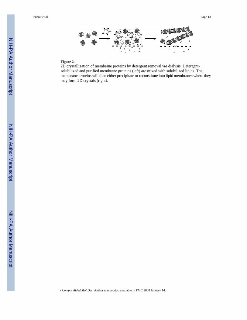
Figure 2.2D crystallization of membrane proteins by detergent removal via dialysis. Detergent-solubilized and purified membrane proteins (left) are mixed with solubilized lipids. Themembrane proteins will then either precipitate or reconstitute into lipid membranes where theymay form 2D crystals (right).
Renault et al. Page 13
J Comput Aided Mol Des. Author manuscript; available in PMC 2008 January 14.
NIH
-PA Author Manuscript
NIH
-PA Author Manuscript
NIH
-PA Author Manuscript
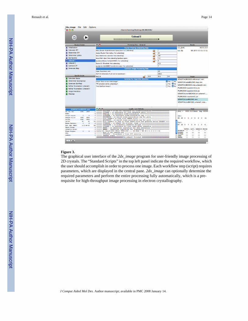
Figure 3.The graphical user interface of the 2dx_image program for user-friendly image processing of2D crystals. The “Standard Scripts” in the top left panel indicate the required workflow, whichthe user should accomplish in order to process one image. Each workflow step (script) requiresparameters, which are displayed in the central pane. 2dx_image can optionally determine therequired parameters and perform the entire processing fully automatically, which is a pre-requisite for high-throughput image processing in electron crystallography.
Renault et al. Page 14
J Comput Aided Mol Des. Author manuscript; available in PMC 2008 January 14.
NIH
-PA Author Manuscript
NIH
-PA Author Manuscript
NIH
-PA Author Manuscript
Copyright © 2022 FDOKUMEN