the esoteric nature of death preparations in taoism and their ...

British Homoeopathic Jounlal January 1997, Vol. 86, pp. 16-26
Effects of Podophyllum peltatum compounds in various preparations and dilutions on human neutrophil functions in vitro S. CHIRUMBOLO,* A. CONFORTI,** S. LUSSIGNOLI,* H. METELMANN,*** P. BELLAVITE*
Abstract Human blood neutrophil granulocytes (neutrophils) treated with Podophyllum peltatum L.-derived compounds exhibited an enhanced oxidative response to subsequent challenge with bacterial formyl peptides. This priming effect was concerned with superoxide anion (O2-) release (respiratory burst). The phenomenon was observed with a potentized preparation containing, among other things, podophyllum extract (Podophyllum compositum), with Podophyllum 4x (final concentration of active principle about 0.025 pg/ml), whereas enhancement of 0 2- release was not caused by homoeopathic Podophyllum 12x or other components of the complex homoeopathic preparation. Purified podophyllotoxin had the same effect at doses of 0.1-10 pg/ml, whereas doses higher than 100 pg/ml of podophyllotoxin inhibited the respiratory burst, so that pure toxin showed a typical bi-phasic dose-response curve. Similar effects were obtained with purified colchicine (1-1000 pg/ml), a microtubule-disrupting agent. No priming by a Podophyllum-derived compound was observed on neutrophils stimulated with 50 ng/ml phorbol ester. Further, both potentized podophyllum-derived compounds and pure podophyllotoxin-inhibited cellular adhesion to the serum-coated surface of culture microplates. These results show that low potencies of a drug extract have specific stimulating effects on the activation of neutrophil metabolism. The same stimulating effects are also caused by low doses of the active principle of the drug, which is an inhibitor when used at high doses.
KEVWORDS: Leukocyte function; Superoxide anion; Adhesion; Podophyllum peltatum L.; Podophyllotoxin; Colchicine; Hormesis.
Introduction Podophyllum belongs to a series of purgative drugs derived from the Berberidaceae (Podophyllum peltatum L.). Podophyllin, the raw extract containing the active principle podophyllotoxin, is drawn from the rhizome. Podophyllum has proved an irritant on patch testing 1 and a mixture of natural and semisynthetic (modified) lignan from Podo- phyllum emodi (proreside) has been used for many years in the treatment of rheumatoid arthritis, but its use was hampered by gastro- intestinal side-effects. 2 In addition podophyllum and podophyl lotoxin have been used in
*Institute of Clinical Chemistry and Microscopy, University of Verona; **Institute of Pharma- cology, University of Verona; ***Biologische Heilmittel Heel GmbH, Baden Baden.
dermatological conditions such as condylomata acuminata.3.4 Podophyllotoxin and its chemical derivatives like epipodophyl lo toxins (teniposide) affect cell division. As they interact with microtubules or with associated protein or modify tubulin GTP binding and also affect DNA, they have been widely investigated as anti-cancer drugs. 5, 6 The action ofpodophyllum derivatives on leukocytes has been studied principally as regards the effect of its main component, the podophyllotoxin and its deriva- tives. Its action is to inhibit lymphocyte proliferation, 7 probably because it affects micro- tubule dynamics 8 and DNA topoisomerases. 9 Podophyllotoxin molecular action could be ascribed to its chemical resemblance to colchicine and vinca alkaloids. These com- pounds have a significant effect on microtubule function.
16
Volume 86 January 1997 17
Podophyllum peltatum L. extracts (Bajiaolian) have been widely used in Chinese herbal medicine 10 and in homoeopathy for many symptoms related to dysentery, gas- troenteritis, proctitis, fevers and bronchitis. 11, 12 Clarke11 noted the irritant and inflammatory effects of podophyllum, with external applica- tion to the skin producing an intertrigo-like rawness; the dust of the powdered root getting into the eyes causes intense inflammation, ulceration and leukoma. Fevers of many kinds are treated with podophyllum.
Special interest in the drug arises from the fact that it is currently used in both conven- tional medicine (mainly for its anti-mitotic proper t ies) and in various potencies in homoeopathy (on a number of indications, including regula t ion of in f lammatory processes). The present study was designed to investigate the effect of potentized prepara- tions of podophyllum and its main active principle on a sensitive cellular model in our laboratory, exploring the metabolic and adhesion functions of human neutrophils exposed to drugs and stimulants. 13, 14 At present there is no evidence that PodophyUum peltatum L. derivatives act on phagocytes (neutrophils , monocytes , macrophages) , important cell types involved in the first line of immunolog ica l survei l lance (natural immunity) and in inflammatory reactions.
The functional parameters explored in this study are superoxide (02-) release and adhe- sion to se rum-coa ted plast ic surfaces . Superoxide is the fundamental product of oxidative metabolism in all phagocytes when their membrane interacts with specific partic- ulate (e.g. opsonized bacteria and yeast) or soluble (e.g. formyl peptides, phorbol esters, lectins) agents. Adhesion is a recently re- evaluated function of leukocytes that allows them to migrate outside the blood vessels, spread onto various surfaces and adhere to connective tissue structures. Measurement of these functional responses to various agonists in the absence and presence of drug com- pounds evaluates the impact drugs may have at cell level on a broad range of targets like mem- brane receptors, metabolic enzymes, anchoring proteins, signal transduction pathways and cytoskeleton structures.
To assess the effect of podophyllum compounds on these functions under various conditions of cell responsiveness, we also
carried out a series of assays on neutrophils pre-treated with tumour necrosis factor (TNF). This cytokine induces a pre-activated state (priming) in vitro, similar to the modifications occurring in diseases like endotoxaemia, severe shock, terminal cancer, etc.
We studied drug preparations made in physiological saline and thus fully compatible with cell viability. All the drug preparations were compared with matched control assays where the cells were incubated with the same physiological saline. As a first empirical approach, we looked for possible effects of commercially available complex homoeo- pathic (compound) preparations based on Podophyllum extracts. Then the effects of sin- gle components present in the same extract were investigated to identify possible active components and the dilutions at which the effect was present. Finally, various doses of pure podophyl lotoxin were tested on the described leukocyte functions.
As some pharmacological actions of podophyllotoxin on cell division are also exhib- ited by other alkaloids, 15 we used the same model system to test the effects of colchicine, an alkaloid of Colchicum autumnale, that is known to bind to microtubular proteins and cause the disappearance of fibrillar micro- tubules in granulocytes and other motile cells. 15
Materials and methods Reagents Podophyl lotoxin , colchicine, formyl-L- methionyl-u-leucyl-u-phenylalanine (fMLP, a synthetic bacterial tripeptide), phorbol-13 myristate-12 acetate (PMA, a phorbol ester), the cytokine tumour necrosis factor-alpha (TNF<t) were purchased from Sigma, St Louis, Mo, USA. Fe 3§ cytochrome c was purchased from Boehringer, Mannheim, Germany, foetal bovine serum (FBS) and 96 fiat bottom well microtitre plates from ICN Flow, Costa Mesa, Ca, USA, Percoll from Pharmacia, Uppsala, Sweden. Hank's balanced salt solution (HBSS) without calcium and magnesium was purchased from Gibco Ltd, Paisley, Scotland, human serum albumin (HSA) from Behring Institute, Marburg, Germany. Other materials and reagents were of the highest purity available. To avoid contamination which is a possible cause of artefacts or priming of neutrophils, sterile solutions and disposable plastic ware were used throughout, with experiments carried
18 British Homoeopathic Journal
out, whenever possible, under a laminar flow hood. Reagents were prepared and/or diluted using pyrogen-free water or 0.9% sodium chloride solutions.
Drugs Prepara t ions were made by Bio logische He i lmi t t e l Heel , G m b H , Baden Baden, Germany, according to the German Homoeo- pathic Pharmacopoeia 16 (GHP 1, method 3a), from the fresh rhizome of Podophyllum peltatum L. Starting from the crude extract (mother tincture), a series of 1:10 dilutions (indicated by x) in sterile physiological saline (0.9% NaC1), f o l l owed by v igo rous succussion, were prepared and provided in 2.2 ml glass vials. The plant material content of the mother tincture was 1/3, and had a density of 0.89 g/ml. The podophyllotoxin content was determined by HPLC analysis, using dichloromethane/methanol 98 + 2 as a solvent. It was 329 + 6 Rg/ml (n = 3) in the mother tincture and 0.1 Rg/ml in Podophyllum 4x. Considering the dilution of the drug to produce the assay mixture (see below), the concentra t ion of podophyl lo tox in in the leukocyte suspensions using Podophyllum compositum and Podophyllum 4x was approximately 0.025 lag/ml.
Podophyllum compositum was prepared by adding 22 pl of the following decimal dilutions in a final volume of 2.2 ml of physiological saline: Podophyllum 2x, Podophyllum 10x, Ignatia 3x, Acidum muriaticum 4x, Acidum muriaticum 10x, Mercurius sublimatus corrosivus 8x. According to these dilution procedures, the final doses in the Podophyllum compositum preparation corresponded to those conta ined in the fo l lowing dilutions: Podophyllum 4x, Podophyllum 12x, lgnatia 5x, Acidum muriaticum 6x, Acidum muriaticum 12x, Mercurius sublimatus corrosivus 10x. These doses were used to test the effect of individual components. Heel also provided the physiological saline for the dilution of drugs; the same physiological saline was used in parallel assays as control.
Solutions of pure podophyl lotoxin and colchicine obtained from Sigma were made in our laboratory by making serial 1:10 dilu- tions in sterile physiological saline (0.5 ml of the more concentrated solution plus 4.5 ml diluent), followed by vigorous succussion (6 seconds at 1,500 strokes/min using a Dyna
HV1 dynamizator) . All drug preparations were stored at +4 ~ and were brought to room temperature at least one hour before the experiment. Pre-incubation and assays were done at 37 ~
Preparation of neutrophils H u m a n neu t roph i l s were p repa red by centrifugation over 62/73% discontinuous Percoll gradients, using ethylene diamine te t ra -ace ta te an t i coagu la ted b lood f rom healthy donors or otherwise from buffy coats rich in leukocytes prepared by centrifugation of citrate anticoagulated whole blood) 3, x7
Cells were f inal ly suspended at a concentration of 2.67 x 10⁶ cells/ml in HBSS supplemented with 0.2% human serum albumin (buffer H-GA). Cell recovery was > 95% neutrophils and > 99% viable cells as judged by the trypan blue exclusion test. 10 minutes before starting the assay, cell suspensions were supplemented with 0.5 mmol/1 CaC12 and 1 mmol/1 MgC12 (buffer H-ACGM) and kept at 37 ~ Whenever indicated, cells were pre- treated with 1 ng/ml TNF-et for 15 minutes at 37 ~
Assay of superoxide release and adhesion Tests were performed in microtitre plates previously coated with inactivated bovine serum and washed. 13 The assays were done in a final volume of 150 lal, in triplicate for each experimental condition as follows. Human neutrophils (75 Rl/microwell) were incubated at 37 ~ for 15 minutes (dose-response assays) or for various times (time evolution assay) with 25 Rl/microwell of the test compound or with physiological saline (test compound solvent), as control. Soon after the pre-incubation period, 50 lal of an H-ACGM solution containing the stimulant and 450 lamol/1 of Fe3§ c were added to each microwell. Where indicat- ed, stimulation was done with 5 x 10 -7 M (final concentration) of fMLP or 50 ng/ml (final con- centration) of PMA. Incubation at 37 ~ was maintained for the indicated time (10-60 minutes). During this time superoxide anion (02-) release was measured by superoxide dismutase-inhibitable reduction of ferricy- tochrome c. Plates were transferred rapidly into a microplate reader (Reader 400, SLT Labs Ins t rumen t s ) and the reduct ion of cytochrome c was measured at 550 nm using 540 nm as reference wavelength and 0.037
Volume 86 January 1997
optical density units as the quenching coeffi- cient for I nmol of reduced cytochrome c.
For adhesion evaluation the microplates were transferred to an automatic washer (Easy Washer 2, SLT Labs Instruments) and subject- ed to 2 washing cycles with PBS Dulbecco at room temperature. The incubation time for the adhesion assay was 40 minutes, unless indicat- ed otherwise. Cells adhering to the microwell bottom were quantified by measuring membrane e n z y m e acid phospha tase , us ing 4-n i t ro- phenylphosphoric acid as substrate, with the percentage of adhesion calculated by referring to a s tandard curve obta ined with k n o w n numbers of neutrophils from the same subject.
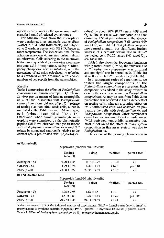
Results Table 1 summarizes the effect of Podophyllum compositum on human neutrophil 0 2- release. In vitro pre-treatment of human neutrophils at 37 ~ for 15 minutes with Podophyllum compositum alone did not affect O 2- release of resting (i.e. non-stimulated) cells, either in untreated cells (Table l a) and TNF<t treated cells (p r imed neut rophi l s ) (Table lb) . Otherwise, when human granulocyte neu- trophils were stimulated by the chemotactic peptide fMLP we observed that pre-treatment with Podophyllum compositum increased 0 2- release by stimulated neutrophils relative to the control (cells pre-treated with physiological
19
saline) by about 70% (8.47 versus 4.99 nmol 02- ). The increase was comparable to that caused by TNF-t~ pre-treatment in the absence of Podophyllum compositum (8.61 versus 4.99 nmol 02- , see Table 1). PodophyUum composi- tum caused a small, but significant further increase of superoxide release also in TNF-t~ pre-treated cells (10.27 versus 8.61 nmol O2-, see Table lb).
Table 1 also shows that following stimulation with phorbol esters (PMA), the increase due to Podophyllum compositum was minimal and not significant in normal cells (Table 1 a) as well as in TNF-t~ treated cells (Table lb).
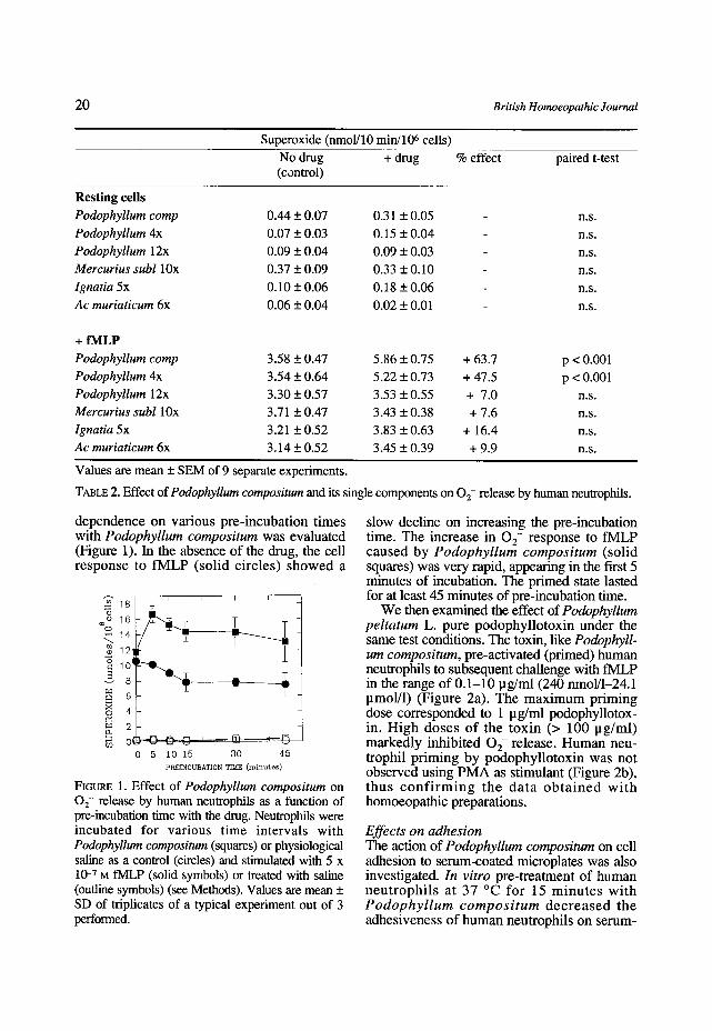
In a subsequent series of experiments, we t e s t ed the s ing le c o m p o n e n t s o f the Podophyllum compositum preparation. Each component was added to the assay mixture in exactly the same dose as used in PodophyUum compositum. As may be seen from Table 2, no preparation was observed to have a direct effect on resting cells, whereas a priming effect on fMLP-stimulated cells was observed on pre- treating the cells with Podophyllum 4x and Podophyllum compositum. Other components caused minor, non-significant stimulation of fMLP-activated neutrophils, suggesting that most if not all of the effect of Podophyllum compositum in this assay system was due to Podophyllum 4x.
The extent of the priming phenomenon in
a) Normal cells Superoxide (nmol/10 min/106 cells)
No drug + drug % effect paired t-test (control)
Resting (n = 5) 0.20 _+ 0.35 0.18 _+ 0.22 - 10.0 n.s. fMLP (n ~ 5) 4.99 _+ 1.26 8.47 _+ 1.77 + 69.7 p < 0.01 PMA (n = 3) 23.00 _+ 3.27 27.35 _+ 7.97 + 18.9 n.s.
b) TNF-treated cells Superoxide (nmol/10 min/106 cells)
No drug + drug % effect paired t-test (control)
Resting (n = 5) 1.28 + 0.95 1.67 -+ 1.5 + 30 n.s. fMLP (n = 5) 8.61 _+ 1.83 10.27 _+ 1.43 + 19.2 p < 0.05 PMA (n = 3) 20.97 + 1.48 24.14 _+ 3.53 + 15.1 n.s.
Values are mean _+ SD of the indicated number of experiments, fMLP = formyl-L-methionyl-L-leucyl-L- phenylalanine (a synthetic bacterial tripeptide); PMA = phorbol-13-myristate-12-acetate (a phorbol ester). TABLE 1. Effect of Podophyllum compositum on 02- release by human neutrophils.
20 British Homoeopathic Journal
Superoxide (nmol/10 min/106 cells)
No drug + drug % effect paired t-test (control)
Resting cells
Podophyllum comp 0.44 _+ 0.07 0.31 _+ 0.05 n.s.
Podophyllum 4x 0.07 + 0.03 0.15 + 0.04 n.s. Podophyllum 12x 0.09 + 0.04 0.09 + 0.03 n.s. Mercurius subl 10x 0.37 + 0.09 0.33 + 0.10 n.s.
Ignatia 5x 0.10 + 0.06 0.18 + 0.06 n.s. Ac muriaticum 6x 0.06 + 0.04 0.02 + 0.01 n.s.
+ fiVILP
Podophyllum comp 3.58 + 0.47 5.86 + 0.75 + 63.7 p < 0.001 Podophyllum 4x 3.54 + 0.64 5.22 + 0.73 + 47.5 p < 0.001 Podophyllum 12x 3.30 + 0.57 3.53 + 0.55 + 7.0 n.s. Mercurius subl 10x 3.71 + 0.47 3.43 + 0.38 + 7.6 n.s. Ignatia 5x 3.21 _+ 0.52 3.83 + 0.63 + 16.4 n.s. Ac muriaticum 6x 3.14 + 0.52 3.45 _+ 0.39 + 9.9 n.s.
Values are mean + SEM of 9 separate experiments.
TABLE 2. Effect of Podophyllum compositum and its single components on 02- release by human neutrophils.
dependence on various pre-incubation times with Podophyllum compositum was evaluated (Figure 1). In the absence of the drug, the cell response to fMLP (solid circles) showed a
--q'18 ~ 16
14 ~'~ 121
i1:1 ~ 6~
4
o<
" T
Z 0 5 10 15 30 4 5
PREINCUBATION TIME (minutes)
FIGURE 1. Effect of Podophyllum compositum on 02- release by human neutrophils as a function of pre-incubation time with the drug. Neutrophils were incubated for various time intervals with Podophyllum compositum (squares) or physiological saline as a control (circles) and stimulated with 5 x 10 -7 M fMLP (solid symbols) or treated with saline (outline symbols) (see Methods). Values are mean + SD of triplicates of a typical experiment out of 3 performed.
slow decline on increasing the pre-incubation time. The increase in 0 2- response to fMLP caused by Podophyllum compositum (solid squares) was very rapid, appearing in the first 5 mmutes of incubation. The primed state lasted for at least 45 minutes of pre-incubation time.
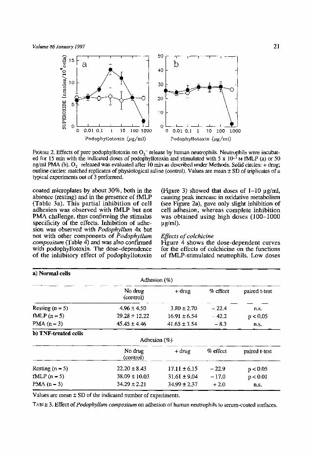
We then examined the effect of Podophyllum peltatum L. pure podophyllotoxin under the same test conditions. The toxin, like Podophyll- um compositum, pre-activated (primed) human neutrophils to subsequent challenge with fMLP in the range of 0.1-10 pg/ml (240 nmol/1-24.1 pmol/1) (Figure 2a). The maximum priming dose corresponded to 1 pg/ml podophyllotox- in. High doses of the toxin (> 100 lag/ml) markedly inhibited 0 2- release. Human neu- trophil priming by podophyllotoxin was not observed using PMA as stimulant (Figure 2b), thus c o n f i r m i n g the data o b t a i n e d wi th homoeopathic preparations.
Effects on adhesion The action of Podophyllum compositum on cell adhesion to serum-coated microplates was also investigated. In vitro pre-treatment of human neu t roph i l s at 37 ~ for 15 minu tes with Podophyllum compositum decreased the adhesiveness of human neutrophils on serum-
Volume 86 January 1997
" ~ i I I i i i .~15
a
N s
0 0.01 0.1 1 10 100 1000
Podophyllotoxin (/~g/ml)
~0 E i i I i I
b 4O
2O
10
ol 0 0.01 0.1 1 10 I00 1000
Podophyllotoxin (/zg/ml)
21
FIGURE 2. Effects of ptlre podophyllotoxin on 02- release by human neutrophils. Neutrophils were incubat- ed for 15 min with the indicated doses of podophyllotoxin and stimulated with 5 x 10 -7 M fMLP (a) or 50 ng/ml PMA (b). 02- released was evaluated after 10 min as described under Methods. Solid circles: + drug; outline circles: matched replicates of physiological saline (control). Values are mean + SD of triplicates of a typical experiments out of 3 performed.
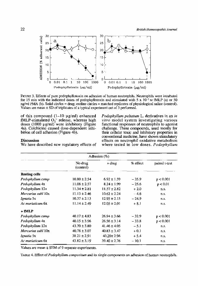
coated rnicroplates by about 30%, both in the absence (resting) and in the presence of fMLP (Table 3a). This part ial i nh ib i t ion of cell adhesion was observed with fMLP but not PMA challenge, thus confirming the stimulus specificity of the effects. Inhibition of adhe- sion was observed with Podophyllum 4x but not with other components of Podophyllum compositum (Table 4) and was also confirmed with podophyllotoxin. The dose-dependence of the inhibi tory effect of podophyllotoxin
(Figure 3) showed that doses of 1-10 pg/ml, causing peak increase in oxidative metabolism (see Figure 2a), gave only slight inhibition of cell adhesion, whereas complete inhibi t ion was obtained us ing high doses (100-1000 pg/ml).
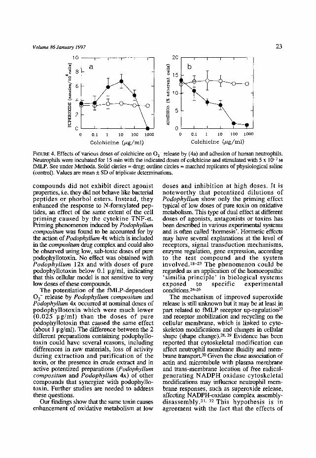
Effects of colchicine Figure 4 shows the dose-dependent curves for the effects of colchicine on the functions of fMLP-stimulated neutrophils. Low doses
a) Normal cells Adhesion (%)
No drug + drug % effect paired t-test (control)
Resting (n = 5) 4.96 _+ 4.50 3.80 + 2.70 - 22.4 n.s. IMLP (n = 5) 29.28 _+ 12.22 16.91 + 6.54 - 42.2 p < 0.05
PMA (n = 3) 45.45 + 4.46 41.65 + 3.54 - 8.3 n.s.
b) TNF-treated cells Adhesion (%)
No drug + drug % effect paired t-test (control)
Resting (n = 5) 22.20_+8.43 17.11 -+6.15 -22.9 p < 0.05 fMLP (n -- 5) 38.09 _+ 10.03 31.61 -+ 9.04 - 17.0 p < 0.01 PMA (n = 3) 34.29 _+ 2.21 34.99 + 2.37 + 2.0 n.s.
Values are mean + SD of the indicated number of experiments.
TABLE 3. Effect ofPodophyllum compositum on adhesion of human neutrophils to serum-coated surfaces.
22
3O
o 25
20i
10 z o
< 0 0
I l I I I I
\ . I I I I I I
0.01 0.i 1 I0 i00 iO00
Podophyllotoxin (/zg/rnl)
30
25
20
15
10
British Homoeopathic Journal
. . . . . I I I I I I
I I I I I I
0.01 0.i i I0 100 i000
Podophyllotoxin (Ng/ml)
FIGURE 3. Effects of pure podophyllotoxin on adhesion of human neutrophils. Neutrophils were incubated for 15 min with the indicated doses of podophyllotoxin and stimulated with 5 x 10 -7 M fMLP (a) or 50 ng/ml PMA (b). Solid circles = drug; outline circles = matched replicates of physiological saline (control). Values are mean + SD of triplicates of a typical experiment out of 3 performed.
o f t h i s c o m p o u n d ( 1 - 1 0 p g / m l ) e n h a n c e d f M L P - s t i m u l a t e d 0 2- r e l ease , w h e r e a s h i g h doses (1000 p g / m l ) w e r e i n h i b i t o r y (F igure 4a). C o l c h i c i n e c a u s e d d o s e - d e p e n d e n t inh i - b i t ion of cel l a d h e s i o n (F igure 4b).
D i s c u s s i o n W e h a v e desc r ibed n e w regu la to ry ef fec ts o f
Podophyllum peltatum L. de r i va t i ve s in an in vitro m o d e l s y s t e m i n v e s t i g a t i n g v a r i o u s func t iona l r e s p o n s e s o f n e u t r o p h i l s to agon i s t chal lenge. These c o m p o u n d s , used mos t ly for their cellular toxic and inhibi tory propert ies in convent ional medic ine , have s h o w n st imulatory e f f ec t s o n n e u t r o p h i l o x i d a t i v e m e t a b o l i s m w h e r e t e s t e d i n l o w d o s e s . Podophyl lum
Adhesion (%)
No drug + drug % effect paired t-test (control)
Resting cells
Podophyllum comp 10.80 + 2.54 6.92 + 1.59 - 35.9 p < 0.001
Podophyllum 4x 11.08 + 2.57 8.24 + 1.99 - 25.6 p < 0.01
Podophyllum 12x 11.34 + 2.81 11.57 + 2.82 + 2.0 n.s.
Mercurius subl 10x 11.13 + 2.46 10.62 + 2.24 - 4.6 n.s.
Ignatia 5x 10.37 + 2.13 12.95 + 2.15 - 24.9 n.s.
Ac muriaticum 6x 11.14 _+ 2.49 12.05 + 2.01 + 8.1 n.s.
+ fMLP
Podophyllum comp 40.17 + 4.83 26.94 + 3.66 - 32.9 p < 0.001
Podophyllum 4x 40.15 + 3.96 26.56 + 3.14 - 33.8 p < 0.001
Podophyllum 12x 43.70 + 3,80 41.46 _+ 4.05 - 5.1 n.s.
Mercurius subl 10x 40.78 _+ 3.07 40.83 + 3.47 + 0.1 n.s.
lgnatia 5x 38.21 _+ 2.91 40.28_+ 2.96 + 5.4 n.s.
Ac muriaticum 6x 43.82 _+ 3.19 39.40 _+ 2.76 - 10.1 n.s.
Values are mean _+ SEM of 9 separate experiments.
TABLE 4. Effect of Podophyllum compositum and its single components on adhesion of human neutrophils.
Volume 86 January 1997
10
~ 8
6 "d
~ 4 s x ~ 2
o~ 0
I I I I I
a
O . l 1 1 0 1 0 0 1000
C o l c h i c i n e ( / z g / / m l )
2 0 , I I I I I
0 f I ~ 0 0 . 1 1 1 0 1 0 0 1 0 0 0
C o l c h i c i n e ( / zg / / rn l )
23
FIeURE 4. Effects of various doses of colchicine on 0 2- release by (4a) and adhesion of human neutrophils. Neutrophils were incubated for 15 min with the indicated doses of colchicine and stimulated with 5 x 10 -7 M fMLP. See under Methods. Solid circles = drug; outline circles = matched replicates Of physiological saline (control). Values are mean + SD of triplicate determinations.
compounds did not exhibit direct agonist properties, i.e. they did not behave like bacterial pept ides or phorbol esters. Instead, they enhanced the response to N-formylated pep- tides, an effect of the same extent of the cell p r iming caused by the cytokine TNF-t~. Priming phenomenon induced by PodophyUum compositum was found to be accounted for by the action of PodophyUum 4x which is included in the compositum drug complex and could also be observed using low, sub-toxic doses of pure podophyllotoxin. No effect was obtained with Podophyllum 12x and with doses of pure podophyllotoxin below 0.1 pg/ml, indicating that this cellular model is not sensitive tq very low doses of these compounds.
The potentiation of the fMLP-dependent 0 2- release by Podophyllum compositum and PodophyUum 4x occurred at nominal doses of podophyl lotoxin which were much lower (0.025 pg /ml ) than the doses of pure podophyllotoxin that caused the same effect (about 1 pg/ml). The difference between the 2 different preparations containing podophyllo- toxin could have several reasons, including differences in raw materials, loss of activity during extract ion and purif icat ion of the toxin, or the presence in crude extract and in active potentized preparations (Podophyllum compositum and Podophyllum 4x) of other compounds that synergize with podophyllo- toxin. Further studies are needed to address these questions.
Our findings show that the same toxin causes enhancement of oxidative metabolism at low
doses and inhibi t ion at high doses. It is notewor thy that potent ized dilutions of Podophyllum show only the priming effect typical of low doses of pure toxin on oxidative metabolism. This type of dual effect at different doses of agonists, antagonists or toxins has been described in various experimental systems and is often called 'hormesis'. Hormetic effects may have several explanations at the level of receptors, signal transduction mechanisms, enzyme regulation, gene expression, according to the test compound and the sys tem invo lvedJ 8-23 The phenomenon could be regarded as an application of the homoeopathic ' s imi l ia p r inc ip le ' in biological sys tems exposed to specif ic exper imenta l conditions.24-26
The mechanism of improved superoxide release is still unknown but it may be at least in part related to fMLP receptor up-regulation 27 and receptor mobilization and recycling on the cellular membrane, which is linked to cyto- skeleton modifications and changes in cellular shape (shape change). 28, 29 Evidence has been reported that cytoskeletal modification can affect neutrophil membrane fluidity and mem- brane transport. 3~ Given the close association of actin and microtubule with plasma membrane and trans-membrane location of free radical- generat ing N A D P H oxidase cytoskele ta l modifications may influence neutrophil mem- brane responses, such as superoxide release, affecting NADPH-oxidase complex assembly- disassembly.31.32 This hypothes is is in agreement with the fact that the effects of
24 British Homoeopathic Journal
podophyUotoxin are shared also by colchicine, a wel l -known agent that affects cel lular microtubule dynamics in a variety of cells including granulocytes.15
We have shown that podophy l lum compounds and colchicine inhibited neutrophil adhesion, and in our assay system this distin- guished their effect from the priming effects of TNF-t~, which did not inhibit but slightly stimulated leukocyte adhesion. It is likely that, as a microtubule disrupting agent, podophyllum derivatives could inhibit neutrophil adhesion on serum-coated microplates because cell flat- tening on the serum protein-coated microwell can imply mic ro tubu le po lymer i za t i on . However, the effect on cell adhesiveness was only partial when potentized preparations or low doses of podophyllotoxin were used, and it was stimulant (fMLP)-specific, indicating that it was not due to cell toxicity but to fine regulation of this function. Adhesion is a very complex phenomenon involving a series of receptors and in t race l lu lar t ransduct ion controls (gating systems) that make this function par t icular ly subject to fine regnlation.13, 23, 33, 34 In fact, in order to perform their functions and in particular chemotactic migration into tissue exudates, neutrophils have to adhere and detach alternatively from their anchoring surfaces, according to the different doses of exogenous (bacterial) or endogenous (inflammatory) factors.
Whether priming of oxidative metabolism and inhibition of adhesion are expressions of the same or different biochemical mechanisms caused by podophyllum and podophyllotoxin (microtubule/fMLP receptor interaction, mem- brane fluidity increase or, even, to intracellular messengers variations) is an intriguing prob- lem. It has been previously shown that adhesion to specific surfaces may represent a negative signal affecting the metabolic responses 35 and this is confirmed at least in part by our time- evolution data (Figure 1). It is therefore possible to suggest that in our experimental conditions a certain degree of microtubule disassembly caused by low doses of podophyllotoxin or colchicine stimulates the neutrophil metabolic response by down-regulating the negative sig- nal provided by adhesion. Certainly, further insights about the molecular mechanism of priming and the possible action of microtubule disrupting compounds on the intracellular signal transduction may complete the picture and give
promising new clues to these plant compounds. Apart from intrinsic biological interest the
phenomenon may have pharmaco log ica l impl ica t ions . Del iver ing a high dose of podophyllotoxin in a local tissue, one would expect the compound to have an inhibitory effect at the point of injection or application. This is, for instance, the ant i -mitot ic or cy to tox ic e f f ec t ob ta ined with top ica l application of 2-5 mg/ml podophyllotoxin against condylomata acuminata. 3, 4 On the other hand, cons ider ing the t issues at a certain distance from the application point which are exposed to low doses of the toxin, a stimulatory effect on oxidative metabolism of phagocytic cells, and therefore an increase in their resistance, could be envisaged as a possible mechanism of action of the drug.
Showing an in vitro effect of a potentized drug demonstrates that it has a precise cellular target, in this case at the level of a fundamental cell involved in the inflammatory process. However, as we have already pointed out in a previous paper, 14 a similar finding is far from representing the explanation of the action of classical homoeopathic medicines that are prescribed--often at potencies much higher than those shown to be effective in vitro--- according to similarity of symptoms. The identification of a cellular mechanism does not rule out the possibility that other different cellular or systemic levels of organization are modulated by the same drug when administered in vivo. In this context, our data have to be regarded as a small piece of a complex picture.
Acknowledgements We thank Dr Adr iana Car lucc io and Dr Alessandro Orlandini for helpful suggestions. This work was supported by grants f rom Heel (Baden Baden), Guna (Milano) and Un ive r s i ty of Verona . We thank Kent Homeopa th ic / IDEAnet for supplying the Zizia Materia Medica program.
References 1 Wantke F, Dammer CM, Gotz M, Jarisch R.
Podophyllum: an irritant on patch testing. Contact Dermatitis 1993; 29: 274.
2 Larsen A, Petersson I, Svensson B. Podophyllum derivatives (CPH82) compared with placebo in the treatment of rheumatoid arthritis. Br J Rheumatol 1989; 28: 124.
3 Syed TA, Lundin S, Ahmad SA. Topical 0.3%
Volume 86 January 1997 25
and 0.5% podophyllotoxin cream for self- t reatment of condylomata acuminata in women. A placebo-controlled, double-blind study. Dermatology 1994; 189: 142.
4 von Krogh G, Szpack E, Andersson M, Bergelin I. Self-treatment using 0.25%-0.50% podophyllotoxin-ethanol solutions against penile condylomta acuminata: a placebo- controlled comparative study. Genitourin Med 1994; 70: 105.
5 Rivera GK. The epipodophyllotoxin teniposide in therapy for childhood acute lymphocytic leukemia. J Clin Onc 1988; 6: 191.
6 Bjorkholm M. Etoposide and teniposide in the t reatment of acute leukemia. Med Oncol Tumor Pharmacoter 1990; 7: 3.
7 Mansson B, Geborek P, Hallberg T, Sturfelt G. Inhibition of Con A-induced proliferat ive response in human lymphocytes by podophyl- lotoxin and a detergent, Tween 80. Clin Exper Rheumatol 1988; 6: 405.
8 Schilstra MJ, Martin SR, Bayley PM. The effect of podophyllotoxin on microtubule dynamics. JBiol Chem 1989; 264: 8827.
9 Long BH, Stringfellow DA. Inhibitors of topoisomerase II: structure-activity relation- ships and mechanism of action of podophyllin congeners. Adv Enzyme Regul 1988; 27: 223.
10 Chang LW, Yang CM, Chen CF, Deng JF. Experimental podophyllotoxin (Bajiaolian) poi- soning: 11. Effects on the liver, intestine, kidney, pancreas and testis. Biomed Environ Sci 1992; 5: 293.
11 Clarke JH. Dictionary o f Practical Materia Medica. Vol. III, reprint edition. New Delhi: B. Jain Publishers 1990.
12 Boericke O. Materia Medica with Repertory. Augmented and Revised Edition. New Delhi: B. Jain Publishers 1991.
13 Bellavi te P, Chirumbolo S, Mansoldo C, Gandini G, Dri P. Simultaneous assay for oxidative metabolism and adhesion of human neutrophils: evidence for correlations and dis- sociations of the two responses. J Leukocyte Biol 1992; 51: 329.
14 Chirumbolo S, Signorini A, Bianchi I, Lippi G, Bellavite P. Effects of homoeopathic prepara- tions of organic acids and of minerals on the oxidative metabolism of human neutrophils. Br Horn J 1993; 82: 227.
15 Goodman Gilman A, Goodman LS, Gilman A. The Pharmacological Basis of Therapeutics. New York: MacMillan 1980.
16 German Homoeopathic Pharmacopoeia. Trans
AR Meuss. Berlin: Deutscher Apotheker Verlag/ British Homoeopathic Association 1991/3.
17 Metcalf JA, Gallin Jl, Nauseef WM, Root RK. Laboratory Manual of Neutrophil Function. New York: Raven Press 1986.
18 Towsend JF, Luckey TD. Hormoligosis in pharmacology. JAMA 1960; 173: 128.
19 Furst A. Hormetic effects in pharmacology: pharmacological inversions as prototypes for hormesis. Health Phys 1987; 52: 527.
20 Calabrese EJ, McCarthy ME, Kenyon E. The occurrence of chemically induced hormesis. Health Phys 1987; 52: 531.
21 Boxenbaum H, Neafsey PJ, Fournier DJ. Hormesis , Gomper tz funct ions, and r isk assessment. Drug Metabol Rev 1988; 19: 195.
22 Bellavite P, Lippi G, Signorini A, Andrioli G, Chirumbolo S. Nonlinear dose-dependent metabolic and adhesive responses of human neutrophils to chemotact ic agents. In: Omeomed92 (Bornoroni C, ed.). p. 135.
. Bologna: Editrice Compositori 1993. 23 Bellavite P, Chirumbolo S, Santonastaso C,
Biasi D, Luss ignol i S, Andr io l i G. Dose-dependence of the various functional responses of neutrophils to formylpeptides. Activation, regulation, and inverse effects according to the agonist dose and cell condi- tion. In: Signals and Images (Bastide M, ed.). Dordrecht: Kluwer Acad Publ (in press).
24 Oberbaum M, Cambar J. Hormesis: dose- dependent reverse effects of low and very low doses. In: Ultra High Dilution (Endler PC, Schulte J, eds.), p. 5. Dordrecht: Kluwer Acad Publ 1994.
25 Van Wijk R, Wiegant FAC. Cultured Mammalian Cells in Homeopathy Research. The Similia Principle in Self-recovery. Utrecht: Utrecht University Press 1994.
26 Bellavite P, Signorini A. Homeopathy: a Frontier in Medical Science. Experimental Studies and Theoret ical Foundat ions , Berkeley: North Atlantic Books 1995.
27 Zimmerli W, Seligman B, Gallin JI. Exudation primes human and guinea pig neutrophils for subsequent responsiveness to the chemotactic peptide N-formylmethyonylleucylphenylalanine and increases complement component C3bi receptor expression. J Clin Invest 1986; 77: 925.
28 Sklar LA, Omann GM, Painter RG. Relationship of actin polymerization and depolymerization to light scattering in human neutrophils: depen- dence on receptor occupancy and intracellular Ca++. J CellBiol 1985; 101:1161.
26
29 Jesaitis AJ, Tolley JO, Allen RA. Receptor- cytoskeleton interaction and membrane traffic may regulate chemoattractant-induced superox- ide production in human granulocytes. J Biol Chem 1986; 261: 13662.
30 Wright SE, Hines LH, White JC. Effect of the lipophilic anticancer drug teniposide (VM-26) on membrane transport. Chem Biol Interact 1990; 75: 31.
31 Nauseef WM, Volpp BD, McCormick S, Leidal KG, Clark RA. Assembly of the neu- trophil respiratory burst oxidase. Protein kinase C promotes cytoskeletal and membrane associ- ation of cytosolic membrane components. J Biol Chem 1991; 266: 5911.
32 Woodman RC, Ruedi JM, Jesai t is AJ, Okamura N, Quinn MT, Smith RM, Curnutte JT, Babior BM. Respiratory burst oxidase and
British Homoeopathic Journal
three of four oxidase-related polypeptides are associated with the cytoskeleton of human neutrophils. J Clin Invest 1991; 87: 1345.
33 Bellavite P, Chirumbolo S, Lippi G, Andrioli G, Bonazzi L, Ferro I. Dual effects of formylpeptides on the adhesion of endotoxin- primed human neutrophils . Cell Biochem Funct 1993; 11: 231.
34 Bellavite P, Carletto A, Biasi D, Caramaschi P, Poli F, Suttora F, Bambara LM. Studies of skin- window exudate human neutrophils: complex patterns of adherence to serum-coated surfaces in dependence on fMLP doses. Inflammation 1994; 18: 571.
35 Hoffs te in ST, Gennaro DE, Manzi , RM. Surface contact inhibits neutrophil superoxide generation induced by soluble stimuli. Lab Invest 1985; 52: 515.
Address for correspondence Professor P. Bellavite Istituto di Chimica e Microscopia Clinica Universith di Verona Ospedale Policl inico-Laboratorio 37134 Verona Italy
Copyright © 2022 FDOKUMEN