HIF-1α regulates bone formation after osteogenic mechanical loading
Upload
independentCategory
view
0download
0
Author's personal copy
Engineering of large osteogenic grafts with rapid engraftment capacity using
mesenchymal and endothelial progenitors from human adipose tissue
Sinan Güven a, Arne Mehrkens a, Franziska Saxer a, Dirk J. Schaefer a, Roberta Martinetti b, Ivan Martin a,*,Arnaud Scherberich a
aDepartments of Surgery and of Biomedicine, University Hospital Basel, Hebelstrasse 20, CH-4031 Basel, Switzerlandb Fin-Ceramica Faenza S.p.A, Via Ravegnana, 186, 48018 Faenza, Italy
a r t i c l e i n f o
Article history:
Received 7 March 2011
Accepted 23 April 2011
Available online 24 May 2011
Keywords:
Mesenchymal stem cell
Bone tissue engineering
Bioreactor
Bone graft
Adipose tissue
Endothelial cell
a b s t r a c t
We investigated whether the maintenance in culture of endothelial and mesenchymal progenitors from
the stromal vascular fraction (SVF) of human adipose tissue supports the formation of vascular structures
in vitro and thereby improves the efficiency and uniformity of bone tissue formation in vivo within
critically sized scaffolds. Freshly-isolated human SVF cells were seeded and cultured into hydroxyapatite
scaffolds (1 cm-diameter, 1 cm-thickness) using a perfusion-based bioreactor system, which resulted in
maintenance of CD34þ/CD31þ endothelial lineage cells. Monolayer-expanded isogenic adipose stromal
cells (ASC) and age-matched bone marrow stromal cells (BMSC), both lacking vasculogenic cells, were
used as controls. After 5 days in vitro, SVF-derived endothelial and mesenchymal progenitors formed
capillary networks, which anastomosed with the host vasculature already 1 week after ectopic nude rat
implantation. As compared to BMSC and ASC, SVF-derived cells promoted faster tissue ingrowth, more
abundant and uniform bone tissue formation, with ossicles reaching a 3.5 mm depth from the scaffold
periphery after 8 weeks. Our findings demonstrate that maintenance of endothelial/mesenchymal SVF
cell fractions is crucial to generate osteogenic constructs with enhanced engraftment capacity. The single,
easily accessible cell source and streamlined, bioreactor-based process makes the approach attractive
towards manufacturing of clinically relevant sized bone substitute grafts.
� 2011 Elsevier Ltd. All rights reserved.
1. Introduction
Bone loss following traumatic, neoplastic or degenerative events
often requires the availability of large structural grafts, up to few
cubic centimeters in size [1]. The gold standard of bone substitutes,
namely autologous bone, is generally associated with relevant
morbidity at the donor site [2], is available in limited amounts, and
most often is not feasibly shaped into large implants of defined
sizes and shapes [3]. The engineering of osteogenic grafts by the
association of osteoprogenitor cells with porous scaffold materials
may in principle overcome the limitations above, but has yet to face
several challenges to demonstrate a reproducible clinical benefit
[4]. Among the most critical limitations of engineered osteogenic
constructs, especially those in a large size, is their reduced
efficiency of engraftment. By ‘engraftment’ we refer here to the
implant penetration by host tissue and connection with host blood
vessels, ultimately regulating the efficiency and spatial uniformity
of bone tissue formation. Indeed, when cylindrical scaffolds of up to
20 mm diameter were uniformly seeded with bone marrow
stromal cells (BMSC) and implanted in an experimental rabbit
model, bone tissue formation was restricted to the outer 1.8 mm
region after 12 weeks, and the vast majority of the scaffold space
was empty or necrotic [5]. In order to improve and accelerate
engraftment of engineered tissues, the attractive strategy to
co-culture vasculogenic cells along with the tissue-specific
progenitors was successfully validated in the context of muscle
regeneration [6]. Several studies have reported the feasibility of
co-culturing osteoprogenitor and endothelial cells within 3D scaf-
folds and have characterized the functional crosstalk between the
two cell types [7]. However, it is yet still to be demonstrated that
this strategy can effectively enhance engraftment of large engi-
neered, osteogenic constructs [8].
We have previously shown that progenitor cells freshly isolated
from human adipose tissue, typically referred to as stromal
* Corresponding author. Tissue Engineering, Institute for Surgical Research and
Hospital Management, University Hospital Basel, Hebelstrasse 20, CH-4031 Basel,
Switzerland. Tel.: þ41 61 265 2384; fax: þ41 61 265 3990.
E-mail address: [email protected] (I. Martin).
Contents lists available at ScienceDirect
Biomaterials
journal homepage: www.elsevier .com/locate/biomater ia ls
0142-9612/$ e see front matter � 2011 Elsevier Ltd. All rights reserved.
doi:10.1016/j.biomaterials.2011.04.064
Biomaterials 32 (2011) 5801e5809

Author's personal copy
vascular fraction (SVF) cells, can be used to generate osteogenic
grafts with intrinsic vasculogenic capacity if seeded and cultured
for 5 days within hydroxyapatite porous scaffolds by using
a perfusion-based bioreactor system [9]. The study critically
established that human SVF-derived cells are effectively competent
in generating frank bone tissue in vivo, even in the absence of
exogenous osteoinductive signals. Moreover, the findings indicated
that the endothelial lineage fraction within SVF cells retained the
ability to form in vivo functional blood vessels, connected with the
host vasculature [9]. In the present work, we first aimed at char-
acterizing the phenotype, function and structural organization of
the endothelial and mesenchymal progenitors from SVF during
in vitro culture. We then used human SVF cells to engineer criti-
cally sized grafts and hypothesized that the presence of endothelial
progenitors could enhance the construct engraftment and unifor-
mity of bone tissue formation upon ectopic implantation into nude
rats.
2. Materials and methods
2.1. Cell isolation and culture
Adipose tissue, in the form of liposuction or excision samples, was obtained
from 15 healthy donors following informed consent and according to a protocol
approved by the local ethical committee (EKBB, Ref. 78/07). All donors were females
between 20 and 65 years of age. Finely minced excision samples or liposuction
samples were digested with 0.075% collagenase type II (355 U/mg, Worthington,
Lakewood, NJ, USA) for 60e90 min at 37 �C. After centrifugation at 190 g for 10 min,
the lipid-rich layer was discarded and the cellular pellet was washed once
with phosphate buffered saline (PBS, Gibco, Invitrogen, Grand Island, NY, US). Red
blood cells were lysed by incubation for 2 min in a solution of 0.15 M ammonium
chloride, 1 mM potassium hydrogen carbonate (both Merck, Darmstadt, Germany)
and 0.1 mM EDTA (Fluka Analytical, SigmaeAldrich Chemie GmbH, Buchs,
Switzerland). The resulting SVF cells were then resuspended in complete medium
(CM), consisting of a-MEM supplemented with 10% of fetal bovine serum (FBS), 1%
HEPES, 1% Sodium pyruvate and 1% of Penicillin-Streptomycin-Glutamin (100 )
solution (all from Gibco), stained with crystal violet (Sigma) and counted in
a Neubauer chamber. For monolayer expansion, SVF cells were seeded at a density
of 2 103 cells/cm2 onto tissue culture plates, cultured in CM supplemented with
5 ng/mL FGF-2 (R&D Systems) and serially replated in new dishes at a density of
3 103 cells/cm2 when reaching subconfluence. The expanded adipose-derived
cells will be hereafter referred to as adipose stromal cells (ASC), to make
a distinction from the population of freshly derived SVF cells. Human bone marrow-
derived mesenchymal stromal cells (BMSC) were isolated and expanded from bone
marrow aspirates of 3 donors (2 males of age 45 and 46 and one female of age 21) as
previously described [10] and used as reference for a standard osteoprogenitor cell
population.
2.2. Cell characterization and sorting
The number of clonogenic cells, generally referred to as colony-forming unit-
fibroblasts (CFU-f), was determined by plating 5 102 SVF cells per 78 cm2
petri dishes. Cells were cultured for 10e14 days in CM containing 5 ng/mL FGF-2.
After fixation with 4% formalin for 10 min and staining with crystal violet, the
colonies were counted and normalized to the number of plated cells. The
phenotype of SVF cells and ASC was determined by cytofluorimetric analysis with
fluorochrome-conjugated antibodies to human CD105 (from AbD Serotec, Oxford,
United Kingdom), CD90, CD31, CD34, CD146, CD45, CD14 or CD73. Isotype IgGs
were used as control (all from BD Biosciences). Cells in suspension were incubated
for 40 min with the different antibodies at 4 �C in CM, washed with PBS, resus-
pended in FACS buffer (PBS, 0.5% Human Serum Albumin, 0.5 mM EDTA) and
analyzed with a FACSCalibur flow cytometer (BD Biosciences, USA). In some
experiments, SVF cells were separated by using a FACS sorter (BD Influx cell sorter,
BD Biosciences USA) based on staining intensity levels for human CD34 (APC-
conjugated) and CD31 (FITC-conjugated). Osteoblastic differentiation capacity of
SVF cells, ASC and BMSC was assessed by 3 weeks’ culture in CM supplemented
with 10 mM b-glycerophosphate and 0.05 M ascorbic acid (osteogenic medium,
OM). Cells cultured with CM containing FGF-2 were used as a control. Deposition
of mineralized matrix was detected by alizarin red staining, as previously
described [9].
2.3. Assessment of cell vasculogenic capacity in vitro
The in vitro formation of vascular structures was tested by seeding unfractioned
SVF cells, sorted CD34þ/CD31" or CD34þ/CD31þ populations from SVF cells, or ASC
onto (i) Matrigel! (BD Biosciences)-coated culture plates, (ii) rapid-prototyped grid-
shaped poly(L-lactide-co-glycolide) (PLGA) scaffolds (2mm 2mmwith a thickness
of 1 mm, kindly provided by the Institute for Material Research andMacromolecular
Chemistry, Freiburg, Germany), or (iii) hydroxapatiteebased Engipore! scaffolds
(Finceramica-Faenza, Faenza, Italy). Seeded constructs were cultured for 5e6 days in
CM supplementedwith 5 ng/ml FGF-2. Capillary network formation onMatrigel was
observed with a phase contrast microscope. Constructs were analysed by scanning
electron microscopy (SEM) or by fluorescence microscopy. For SEM, cell-seeded
constructs were fixed overnight at 4 �C with 4% formaldehyde and washed with
PBS. Samples were gradually dehydrated with 30-50-70-90-100% ethanol, coated
with gold and imaged with a Philips XL 30 ESEM microscope. For fluorescence
microscopy, constructs were stained with a PE-conjugated anti-human CD31 anti-
body (from BD Biosciences) for 40 min at 4 �C, washed with PBS and observed using
an IX50 fluorescence microscope (Olympus, Japan). Some constructs were assessed
by confocal laser scanning microscopy (LSM710, Zeiss, Germany), following
combined staining with anti-human CD31 (DakoCytomation, Denmark) and anti-
human desmin (AbCam, UK) antibodies, followed by Alexa-conjugated series
secondary antibodies (Invitrogen, Molecular Probes, USA) and with DAPI for nuclear
structures.
2.4. Generation and analysis of large size ceramic-based scaffolds
Hydroxyapatite scaffolds (Engipore!, Finceramica-Faenza, Faenza, Italy) in
the form of large, porous cylinders (1 cm diameter 1 cm thickness) were placed
into custom produced chambers of a previously developed perfusion-based
bioreactor system [11] and wetted with culture medium prior to cell seeding.
1.2 107 SVF cells or 3 106 either ASC or BMSC were resuspended in CM
supplemented with dexamethasone, ascorbic acid and FGF-2 and perfused in
alternate directions at a flow rate of 1 ml/min through the scaffold pores for 5
days, as previously described [9]. In order to assess the cell seeding homogeneity
and viability, some constructs were stained by methyl-tetrazolium (MTT, Sigma)
either at day 1 or at day 5, i.e. at the time of the in vivo implantation, according to
the manufacturer’s instructions. In order to extract cells, some constructs were
perfused for 5 min with a 0.15% collagenase solution inside the perfusion
bioreactor, rinsed with PBS and perfused with trypsin-EDTA for 5 min. Retrieved
cells were assessed cytofluorimetrically for the expression of CD31, CD34, CD45,
CD90, CD105 and CD146 as described above. In order to assess osteogenic, vas-
culogenic and engraftment capacity, some constructs were implanted into nude
rats as described below.
2.5. In vivo studies
Cell-seeded ceramic cylinders or fibrin hydrogels, generated as described above,
were implanted subcutaneously in nude rats (RNu/Nu Charles-River, Germany) as
previously described [9]. At the indicated time points, the animals were sacrificed by
inhalation of CO2. Maintenance of animals, surgical procedures and sacrificing
methods were all performed in agreement with Swiss legislation and according to
protocol approved by the local veterinary office (“Kantonales Veterinäramt Basel-
Stadt”, permission #2167). Harvested ceramic-based constructs were bisected along
the vertical axis, fixed overnight with 1.5% paraformaldehyde, subjected to a slow
decalcification process by incubation in a solution of 7% w/v EDTA (Fluka Analytical,
SigmaeAldrich, Buchs, Switzerland), 10% w/v sucrose (SigmaeAldrich) at 37 �C on
an orbital shaker for 7e10 days and paraffin embedded.
2.6. Histological analysis
Histological sections (7 mm thick) of explanted constructs were first stained with
hematoxylin/eosin to quantitatively assess the distribution of the newly formed
tissue and of the bone ossicles. Briefly, microscopic pictures of the central sections of
the full size constructs were taken. For tissue distribution, the distance from the
surface of the construct to the deepest part of the tissue was measured. At least 6
independent constructs per experimental group were analysed, performing 10
measurements per construct cross section. For the quantification of bone tissue
formation, the number of pores exhibiting characteristic ossicle structures was
counted and normalized to the total number of pores in the sections. In addition, the
shortest distances from the deepest region of every pore containing bone tissue to
the scaffold periphery were measured and their distribution plotted. In order to
identify cells of human origin in the large constructs implanted in rats, sections were
stained for human-specific Alu sequences by using the ZytoFast CMV chromogenic
in situ hybridization (CISH) Kit (Zytovision GmbH, Bremerhaven, Germany). Human
and rat skin sections were used as positive and negative controls, respectively.
Immunostaining for bone sialoprotein (BSP) was performed by using an anti-human
BSP antibody (A4232.1/A4232.2; Immundiagnostik AG, Bensheim, Germany)
followed by incubationwith ABC-alkaline phosphatase complex kit (Dako, Glostrup,
Denmark). The presence of blood vessels of human origin was assessed by using
a biotin-conjugated anti-human CD34-specific antibody (Chemicon, Temecula,
USA), as previously described [9].
S. Güven et al. / Biomaterials 32 (2011) 5801e58095802
Author's personal copy
3. Results
3.1. Characterization of freshly-isolated SVF cells and in vitro
expanded ASC
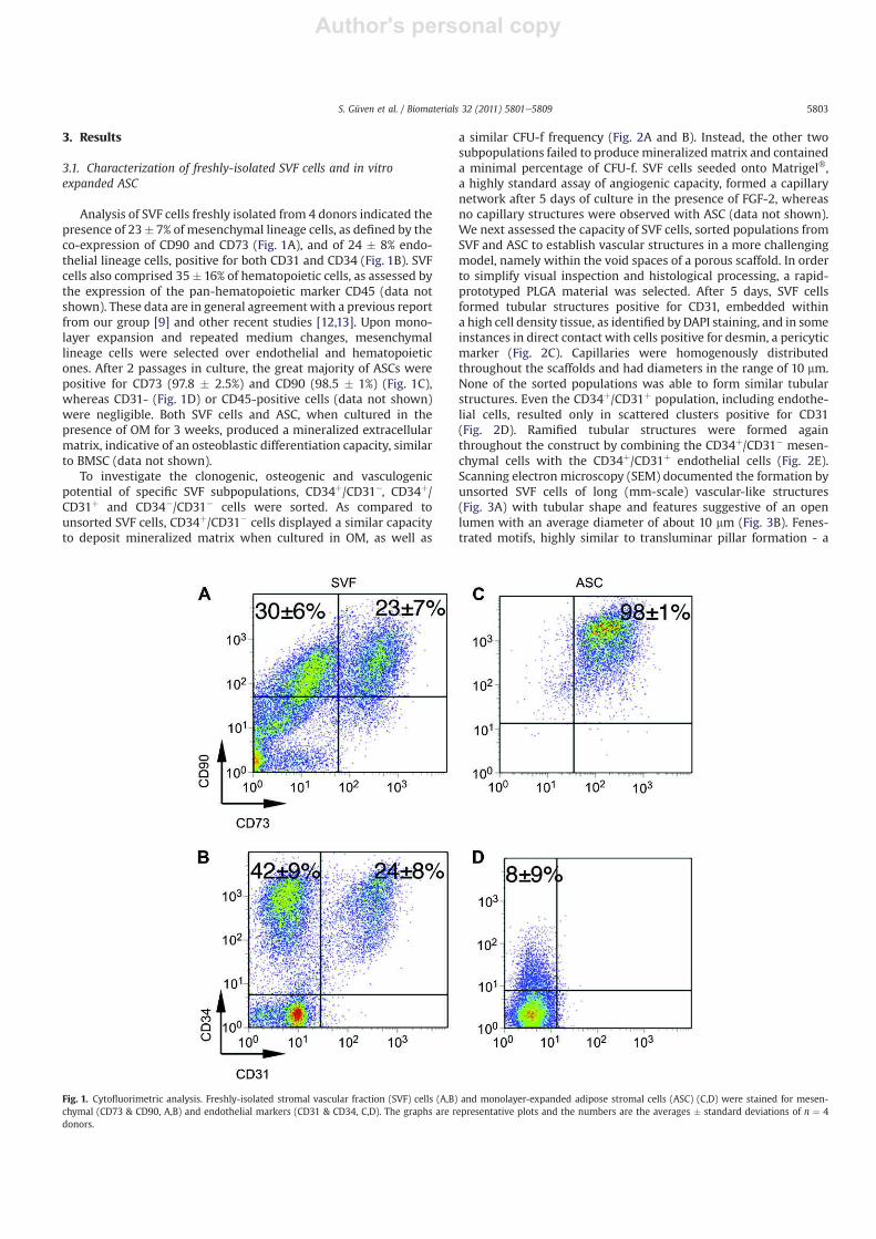
Analysis of SVF cells freshly isolated from 4 donors indicated the
presence of 23� 7% of mesenchymal lineage cells, as defined by the
co-expression of CD90 and CD73 (Fig. 1A), and of 24 � 8% endo-
thelial lineage cells, positive for both CD31 and CD34 (Fig. 1B). SVF
cells also comprised 35� 16% of hematopoietic cells, as assessed by
the expression of the pan-hematopoietic marker CD45 (data not
shown). These data are in general agreement with a previous report
from our group [9] and other recent studies [12,13]. Upon mono-
layer expansion and repeated medium changes, mesenchymal
lineage cells were selected over endothelial and hematopoietic
ones. After 2 passages in culture, the great majority of ASCs were
positive for CD73 (97.8 � 2.5%) and CD90 (98.5 � 1%) (Fig. 1C),
whereas CD31- (Fig. 1D) or CD45-positive cells (data not shown)
were negligible. Both SVF cells and ASC, when cultured in the
presence of OM for 3 weeks, produced a mineralized extracellular
matrix, indicative of an osteoblastic differentiation capacity, similar
to BMSC (data not shown).
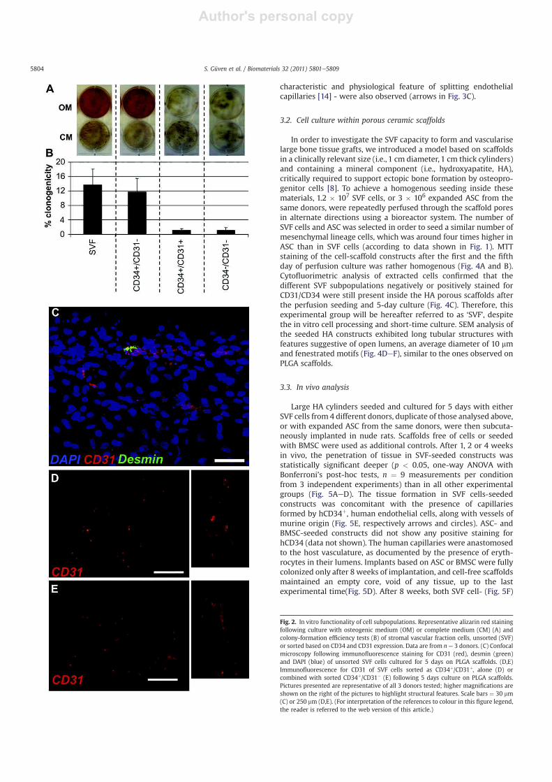
To investigate the clonogenic, osteogenic and vasculogenic
potential of specific SVF subpopulations, CD34þ/CD31!, CD34þ/
CD31þ and CD34!/CD31! cells were sorted. As compared to
unsorted SVF cells, CD34þ/CD31! cells displayed a similar capacity
to deposit mineralized matrix when cultured in OM, as well as
a similar CFU-f frequency (Fig. 2A and B). Instead, the other two
subpopulations failed to producemineralizedmatrix and contained
a minimal percentage of CFU-f. SVF cells seeded onto Matrigel!,
a highly standard assay of angiogenic capacity, formed a capillary
network after 5 days of culture in the presence of FGF-2, whereas
no capillary structures were observed with ASC (data not shown).
We next assessed the capacity of SVF cells, sorted populations from
SVF and ASC to establish vascular structures in a more challenging
model, namely within the void spaces of a porous scaffold. In order
to simplify visual inspection and histological processing, a rapid-
prototyped PLGA material was selected. After 5 days, SVF cells
formed tubular structures positive for CD31, embedded within
a high cell density tissue, as identified by DAPI staining, and in some
instances in direct contact with cells positive for desmin, a pericytic
marker (Fig. 2C). Capillaries were homogenously distributed
throughout the scaffolds and had diameters in the range of 10 mm.
None of the sorted populations was able to form similar tubular
structures. Even the CD34þ/CD31þ population, including endothe-
lial cells, resulted only in scattered clusters positive for CD31
(Fig. 2D). Ramified tubular structures were formed again
throughout the construct by combining the CD34þ/CD31! mesen-
chymal cells with the CD34þ/CD31þ endothelial cells (Fig. 2E).
Scanning electron microscopy (SEM) documented the formation by
unsorted SVF cells of long (mm-scale) vascular-like structures
(Fig. 3A) with tubular shape and features suggestive of an open
lumen with an average diameter of about 10 mm (Fig. 3B). Fenes-
trated motifs, highly similar to transluminar pillar formation - a
Fig. 1. Cytofluorimetric analysis. Freshly-isolated stromal vascular fraction (SVF) cells (A,B) and monolayer-expanded adipose stromal cells (ASC) (C,D) were stained for mesen-
chymal (CD73 & CD90, A,B) and endothelial markers (CD31 & CD34, C,D). The graphs are representative plots and the numbers are the averages � standard deviations of n ¼ 4
donors.
S. Güven et al. / Biomaterials 32 (2011) 5801e5809 5803
Author's personal copy
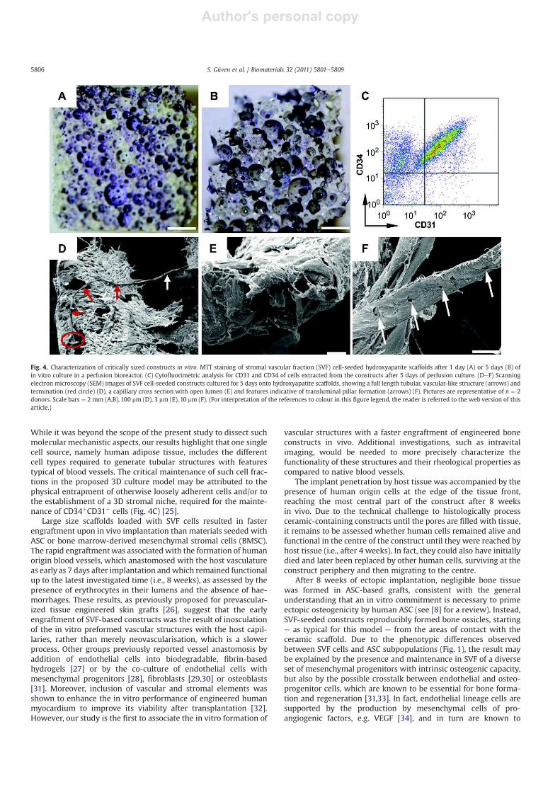
characteristic and physiological feature of splitting endothelial
capillaries [14] - were also observed (arrows in Fig. 3C).
3.2. Cell culture within porous ceramic scaffolds
In order to investigate the SVF capacity to form and vascularise
large bone tissue grafts, we introduced a model based on scaffolds
in a clinically relevant size (i.e., 1 cm diameter, 1 cm thick cylinders)
and containing a mineral component (i.e., hydroxyapatite, HA),
critically required to support ectopic bone formation by osteopro-
genitor cells [8]. To achieve a homogenous seeding inside these
materials, 1.2 � 107 SVF cells, or 3 � 106 expanded ASC from the
same donors, were repeatedly perfused through the scaffold pores
in alternate directions using a bioreactor system. The number of
SVF cells and ASC was selected in order to seed a similar number of
mesenchymal lineage cells, which was around four times higher in
ASC than in SVF cells (according to data shown in Fig. 1). MTT
staining of the cell-scaffold constructs after the first and the fifth
day of perfusion culture was rather homogenous (Fig. 4A and B).
Cytofluorimetric analysis of extracted cells confirmed that the
different SVF subpopulations negatively or positively stained for
CD31/CD34 were still present inside the HA porous scaffolds after
the perfusion seeding and 5-day culture (Fig. 4C). Therefore, this
experimental group will be hereafter referred to as ‘SVF’, despite
the in vitro cell processing and short-time culture. SEM analysis of
the seeded HA constructs exhibited long tubular structures with
features suggestive of open lumens, an average diameter of 10 mm
and fenestrated motifs (Fig. 4DeF), similar to the ones observed on
PLGA scaffolds.
3.3. In vivo analysis
Large HA cylinders seeded and cultured for 5 days with either
SVF cells from 4 different donors, duplicate of those analysed above,
or with expanded ASC from the same donors, were then subcuta-
neously implanted in nude rats. Scaffolds free of cells or seeded
with BMSC were used as additional controls. After 1, 2 or 4 weeks
in vivo, the penetration of tissue in SVF-seeded constructs was
statistically significant deeper (p < 0.05, one-way ANOVA with
Bonferroni’s post-hoc tests, n ¼ 9 measurements per condition
from 3 independent experiments) than in all other experimental
groups (Fig. 5AeD). The tissue formation in SVF cells-seeded
constructs was concomitant with the presence of capillaries
formed by hCD34þ, human endothelial cells, along with vessels of
murine origin (Fig. 5E, respectively arrows and circles). ASC- and
BMSC-seeded constructs did not show any positive staining for
hCD34 (data not shown). The human capillaries were anastomosed
to the host vasculature, as documented by the presence of eryth-
rocytes in their lumens. Implants based on ASC or BMSC were fully
colonized only after 8 weeks of implantation, and cell-free scaffolds
maintained an empty core, void of any tissue, up to the last
experimental time(Fig. 5D). After 8 weeks, both SVF cell- (Fig. 5F)
Fig. 2. In vitro functionality of cell subpopulations. Representative alizarin red staining
following culture with osteogenic medium (OM) or complete medium (CM) (A) and
colony-formation efficiency tests (B) of stromal vascular fraction cells, unsorted (SVF)
or sorted based on CD34 and CD31 expression. Data are from n ¼ 3 donors. (C) Confocal
microscopy following immunofluorescence staining for CD31 (red), desmin (green)
and DAPI (blue) of unsorted SVF cells cultured for 5 days on PLGA scaffolds. (D,E)
Immunofluorescence for CD31 of SVF cells sorted as CD34þ/CD31þ, alone (D) or
combined with sorted CD34þ/CD31" (E) following 5 days culture on PLGA scaffolds.
Pictures presented are representative of all 3 donors tested; higher magnifications are
shown on the right of the pictures to highlight structural features. Scale bars ¼ 30 mm
(C) or 250 mm (D,E). (For interpretation of the references to colour in this figure legend,
the reader is referred to the web version of this article.)
S. Güven et al. / Biomaterials 32 (2011) 5801e58095804

Author's personal copy
and ASC-seeded constructs (data not shown) displayed a homoge-
nous staining for Alu sequences, indicating that the implanted cells
survived. Moreover, analysis of the SVF cell-based explants at the
deepest front of tissue formation over time indicated the presence
of Alu positive cells and of human endothelial cells, which reached
the construct core after 4 weeks (Supplementary Figure 1, left and
central columns).
After 8 weeks, constructs seeded with SVF cells from the 4
donors displayed formation of bone tissue within 18.5 � 7.5% of the
pores (Fig. 6AeC), with the bone matrix progressively filling the
pore space starting from the area in contact with the ceramic
material. Bone formation had a typical structure/cell morphology
previously reported for small-scale constructs [9] and was accom-
panied by a temporally increasing amount of areas positively
stained for BSP (Supplementary Figure 1, right column). Constructs
seededwith ASCwere void of any bone tissue, with the exception of
one ossicle found in a peripheral pore of one construct (Fig. 6C,D).
Cell-free scaffolds, as expected, did not exhibit evidences of bone
tissue (Fig. 6E). BMSC-seeded constructs showed relevant bone
formation but in a significantly lower percentage of pores
(p ¼ 0.038, T-test, n ¼ 6 measurements from at least 3 independent
experiments) than SVF-seeded ones (Fig. 6C) and strictly limited to
the most peripheral part of the constructs (Fig. 6F,G). The distri-
bution of bone formation inside SVF- and BMSC-seeded constructs
as a function of the distance from the outer surface quantitatively
confirmed the morphological observations. In fact, bone tissue
formed by BMSC was confined within 1.5 mm from the outer
periphery, whereas SVF-seeded constructs exhibited bone forma-
tion up to a 3.5 mm depth from the surface (Fig. 6H).
4. Discussion
In this study, we have demonstrated that in vitro generation of
organized vascular structures by SVF cells from human adipose
tissue critically requires the cooperation of both endothelial and
mesenchymal progenitor fractions. We further identified that a 3D
model based on a porous scaffold and perfusion bioreactor system
allowed maintaining in culture both SVF subpopulations, as
opposed to the standard 2D culture in Petri dishes. The system
resulted in the engineering of large 3D osteogenic and vasculogenic
grafts with increased efficiency of engraftment and ultimatelymore
homogenous bone tissue formation after ectopic implantation in
nude rats.
The presence of vasculogenic cells within the adipose tissue-
derived SVF fraction was first proposed by Miranville et al. [15]
and thereafter confirmed by different groups [16e18], describing
the presence of a CD34þ/CD31" cell populationwith characteristics
of endothelial progenitors and the capacity to rescue hindlimb
ischemia. We have also recently identified that human SVF cells
include endothelial lineage populations which can form functional
blood vessels when ectopically implanted in nude mice [9,19,20]. In
the present study, sorting of SVF cells based on both CD31 and CD34
allowed to determine that (i) the clonogenic mesenchymal
progenitors predominantly reside in the CD34þCD31" fraction, and
(ii) the formation of tubular vascular structures requires the avail-
ability of both CD34þCD31" and CD34þCD31þ cells. Together with
the perivascular detection of mural cells positive for desmin and
negative for CD31 (Fig. 2C), these results suggest that the
CD34þCD31" fraction includes not only osteoprogenitor cells,
capable to produce mineralized matrix, but also cells with a peri-
cytic function, necessary for stabilization of newly formed endo-
thelial capillaries [21]. These data are also in accordance with
a recent study showing that ASC co-cultured with primary endo-
thelial cells from umbilical cord can establish vascular-like struc-
tures in vitro [22] and with the general recognition that putative
mesenchymal stem cells from various tissues derive and share the
function of pericytic/mural cells [23]. Alternatively, organized
vascular structures in our model could have been generated by the
endothelial differentiation of CD34þCD31" cells, triggered by
factors (e.g., CXCL12) produced by capillary endothelial cells [24].
Fig. 3. Scanning electron microscopy characterization of vascular structures. Stromal
vascular fraction (SVF) cells were seeded and cultured for 5 days on PLGA scaffolds. (A)
Full length tubular, vascular-like structure (arrows) and termination (red circle).
(B) Sectioned capillary at the tip of the tubular structure, suggestive of an open lumen.
(C) Detail of the wall of a capillary, with structures indicative of transluminal pillar
formation in the capillaries (arrows). Pictures are representative of n ¼ 2 donors. Scale
bars ¼ 80 mm (A), 3 mm (B, C). (For interpretation of the references to colour in this
figure legend, the reader is referred to the web version of this article.)
S. Güven et al. / Biomaterials 32 (2011) 5801e5809 5805
Author's personal copy
While it was beyond the scope of the present study to dissect such
molecular mechanistic aspects, our results highlight that one single
cell source, namely human adipose tissue, includes the different
cell types required to generate tubular structures with features
typical of blood vessels. The critical maintenance of such cell frac-
tions in the proposed 3D culture model may be attributed to the
physical entrapment of otherwise loosely adherent cells and/or to
the establishment of a 3D stromal niche, required for the mainte-
nance of CD34þCD31þ cells (Fig. 4C) [25].
Large size scaffolds loaded with SVF cells resulted in faster
engraftment upon in vivo implantation than materials seeded with
ASC or bone marrow-derived mesenchymal stromal cells (BMSC).
The rapid engraftment was associatedwith the formation of human
origin blood vessels, which anastomosed with the host vasculature
as early as 7 days after implantation andwhich remained functional
up to the latest investigated time (i.e., 8 weeks), as assessed by the
presence of erythrocytes in their lumens and the absence of hae-
morrhages. These results, as previously proposed for prevascular-
ized tissue engineered skin grafts [26], suggest that the early
engraftment of SVF-based constructs was the result of inosculation
of the in vitro preformed vascular structures with the host capil-
laries, rather than merely neovascularisation, which is a slower
process. Other groups previously reported vessel anastomosis by
addition of endothelial cells into biodegradable, fibrin-based
hydrogels [27] or by the co-culture of endothelial cells with
mesenchymal progenitors [28], fibroblasts [29,30] or osteoblasts
[31]. Moreover, inclusion of vascular and stromal elements was
shown to enhance the in vitro performance of engineered human
myocardium to improve its viability after transplantation [32].
However, our study is the first to associate the in vitro formation of
vascular structures with a faster engraftment of engineered bone
constructs in vivo. Additional investigations, such as intravital
imaging, would be needed to more precisely characterize the
functionality of these structures and their rheological properties as
compared to native blood vessels.
The implant penetration by host tissue was accompanied by the
presence of human origin cells at the edge of the tissue front,
reaching the most central part of the construct after 8 weeks
in vivo. Due to the technical challenge to histologically process
ceramic-containing constructs until the pores are filled with tissue,
it remains to be assessed whether human cells remained alive and
functional in the centre of the construct until they were reached by
host tissue (i.e., after 4 weeks). In fact, they could also have initially
died and later been replaced by other human cells, surviving at the
construct periphery and then migrating to the centre.
After 8 weeks of ectopic implantation, negligible bone tissue
was formed in ASC-based grafts, consistent with the general
understanding that an in vitro commitment is necessary to prime
ectopic osteogenicity by human ASC (see [8] for a review). Instead,
SVF-seeded constructs reproducibly formed bone ossicles, starting
e as typical for this model e from the areas of contact with the
ceramic scaffold. Due to the phenotypic differences observed
between SVF cells and ASC subpopulations (Fig. 1), the result may
be explained by the presence and maintenance in SVF of a diverse
set of mesenchymal progenitors with intrinsic osteogenic capacity,
but also by the possible crosstalk between endothelial and osteo-
progenitor cells, which are known to be essential for bone forma-
tion and regeneration [31,33]. In fact, endothelial lineage cells are
supported by the production by mesenchymal cells of pro-
angiogenic factors, e.g. VEGF [34], and in turn are known to
Fig. 4. Characterization of critically sized constructs in vitro. MTT staining of stromal vascular fraction (SVF) cell-seeded hydroxyapatite scaffolds after 1 day (A) or 5 days (B) of
in vitro culture in a perfusion bioreactor. (C) Cytofluorimetric analysis for CD31 and CD34 of cells extracted from the constructs after 5 days of perfusion culture. (DeF) Scanning
electron microscopy (SEM) images of SVF cell-seeded constructs cultured for 5 days onto hydroxyapatite scaffolds, showing a full length tubular, vascular-like structure (arrows) and
termination (red circle) (D), a capillary cross section with open lumen (E) and features indicative of transluminal pillar formation (arrows) (F). Pictures are representative of n ¼ 2
donors. Scale bars ¼ 2 mm (A,B), 100 mm (D), 3 mm (E), 10 mm (F). (For interpretation of the references to colour in this figure legend, the reader is referred to the web version of this
article.)
S. Güven et al. / Biomaterials 32 (2011) 5801e58095806

Author's personal copy
Fig. 5. Characterization of critically sized constructs in vivo. Hematoxylin-Eosin stained sections of constructs retrieved after 1 week implantion in nude rats and based on
hydroxyapatite scaffolds without cells (A), seeded with expanded adipose stromal cells (ASC) (B) or seeded and cultured with freshly-isolated stromal vascular fraction (SVF) cells
(C). (D) Graph summarizing the kinetics of tissue penetration in implanted constructs seeded with the indicated cell types, using bone marrow stromal cells (BMSC) as controls.
Asterisks (*) indicate statistically significant differences between SVF cells and all other experimental groups at the indicated time point. (E) Immunostaining for human-specific
CD34 of a representative SVF cell-seeded construct 1 week after in vivo implantation. Arrows show blood vessels of human origin with erythrocytes in the lumens, whereas dashed
lines show host (rat) blood vessels. (F) In situ hybridization for ALU sequences specific for human cells of a representative SVF cell-seeded construct 8 weeks after in vivo
implantation. Dashed lines delineate the contour of the cross section. The inset shows ALU staining in individual cells at higher magnification. All data are representative of 3 donors
for BMSC and ASC, and 4 donors for SVF. Scale bars ¼ 2 mm (A, B, C, F) or 25 mm (E).
S. Güven et al. / Biomaterials 32 (2011) 5801e5809 5807

Author's personal copy
secrete key osteogenic factors like BMP-2 and BMP-4 [35]. The
maintenance of an endothelial cell population from SVF cells in the
3D perfusion cultures and the formation of a prevascular network
could thus have contributed to the osteogenic commitment of the
mesenchymal CD34þ/CD31 cell fraction. Importantly, as compared
to SVF-seeded constructs, those based on typical and well-
accredited osteogenic cells, namely BMSC, displayed lower
amounts of bone formation, due to the spatial restriction to the
outer regions. The deeper production of bone tissue by SVF cells
was consistent with the more efficient penetration of host tissue
and associated vessels, and allowed for the production of bone
tissue up to 3.5 mm from the periphery of the construct. Over the
206 bones found in the human body, the one with largest contin-
uous dimension is the human femur shaft, with an integral bone
thickness of 7e10 mm [36]. Therefore, the proposed approach
could overcome the challenge of osteogenic tissue engraftment for
virtually any bone defect. Obviously, this will require further vali-
dation at an orthotopic site of an immunocompetent model, where
biomechanical and inflammation/immune-related aspects can
better mirror a clinical case scenario, although the species speci-
ficity of SVF biology may introduce a relevant bias. The process
implementation using a streamlined bioreactor system, bypassing
the labor-intensive phase of monolayer expansion and reducing the
culture time, provides the basis for standardized, automated and
possibly cost-effective graft manufacturing [37].
5. Conclusion
Our findings support the concept that vascular progenitors
derived from human SVF cells accelerate the engraftment of criti-
cally sized osteogenic constructs, ultimately improving the effi-
ciency and uniformity of bone tissue formation. The use of a single,
easily available and abundant cell source for both osteogenic and
vasculogenic progenitors offers a practical and clinically attractive
approach.
Acknowledgments
We acknowledgefinancial support by the Swiss National Science
Foundation (Grant 310030-120432) to A.S. We are grateful to Dr
Mülhaupt (Freiburg-In-Breisgau, Germany) for kindly supplying the
rapid-prototyped grid-shaped poly(L-lactide-co-glycolide) (PLGA)
scaffolds. We would like to thank Ms. Rosaria Santoro (Basel,
Switzerland) for the design of the up-scaled perfusion bioreactor
chambers used in this study and Dr. Anna Marsano (Basel,
Switzerland) for general advice and assistance in the statistical
analysis.
Appendix. Supplementary material
Supplementary data associated with this article can be found, in
the online version, at doi:10.1016/j.biomaterials.2011.04.064.
Fig. 6. Bone formation within critically sized constructs. Hematoxylin-Eosin stained
sections of constructs retrieved after 8 weeks implantation in nude rats and based on
hydroxyapatite scaffolds seeded with stromal vascular fraction (SVF) cells (A,B),
expanded adipose stromal cells (ASC) (D), no cells (E), or bone marrow stromal cells
(BMSC) (F,G). (C) Quantification of the percentage of pores containing bone tissue in
the constructs seeded with the indicated cell source. (H) Distribution of pores con-
taining bone tissue plotted as a function of the distance from the scaffold surface for
constructs generated by SVF cells or BMSC. All data are representative of 3 donors for
BMSC and ASC, and of 4 donors for SVF. Scale bars ¼ 2 mm (A, D, E, F) or 200 mm (B, G).
S. Güven et al. / Biomaterials 32 (2011) 5801e58095808

Author's personal copy
References
[1] Giannoudis PV, Dinopoulos H, Tsiridis E. Bone substitutes: an update. Injury2005;36:S20e7.
[2] Vail TP, Urbaniak JR. Donor-site morbidity with use of vascularizedautogenous fibular grafts. J Bone Joint Surg Am 1996;78:204e11.
[3] Holister SJ. Porous scaffold design for tissue engineering. Nat Mater 2005;4:518e24.
[4] Meijer GJ, de Bruijn JD, Koole R, van Blitterswijk CA. Cell-based bone tissueengineering. PLoS Med 2007;4:e9.
[5] Scheufler O, Schaefer DJ, Jaquiery C, Braccini A, Wendt DJ, Gasser JA, et al.Spatial and temporal patterns of bone formation in ectopically pre-fabricated,autologous cell-based engineered bone flaps in rabbits. J Cell Mol Med 2008;12:1238e49.
[6] Levenberg S, Rouwkema J,MacdonaldM,GarfeinES, KohaneDS,DarlandDC, et al.Engineeringvascularised skeletalmuscle tissue.Nat Biotechnol2005;23:879e84.
[7] Rouwkema J, Westerweel PE, Boer J, Verhaar MC, van Blitterswijk CA. The useof endothelial progenitor cells for prevascularized bone tissue engineering.Tissue Eng-A 2009;15:2015e27.
[8] Scherberich A, Müller AM, Schäfer DJ, Banfi A, Martin I. Adipose tissue-derivedprogenitors for engineering osteogenic and vasculogenic grafts. J Cell Physiol2010;225:348e53.
[9] Scherberich A, Galli R, Jaquiery C, Farhadi J, Martin I. Three-dimensionalperfusion culture of human adipose tissue-derived endothelial andosteoblastic progenitors generates osteogenic constructs with intrinsicvascularization capacity. Stem Cells 2007;25:1823e9.
[10] Frank O, Heim M, Jakob M, Barbero A, Schaefer D, Bendik I, et al. Real-timequantitative RT-PCR analysis of human bone marrow stromal cells duringosteogenic differentiation in vitro. J Cell Biochem 2002;85:737e46.
[11] Wendt D, Marsano A, Jakob M, Heberer M, Martin I. Oscilating perfusion of cellsuspensions through three-dimensional scaffolds enhances cell seedingefficiency and uniformity. Biotechnol Bioeng 2003;84:205e14.
[12] Suga H, Eto H, Aoi N, Kato H, Araki J, Doi K, et al. Adipose tissue remodelingunder ischemia: death of adipocytes and activation of stem/progenitor cells.Plast Reconstr Surg 2010;126:1911e23.
[13] Zimmerlin L, Donnenberg VS, Pfeifer ME, Meyer EM, Péault B, Rubin JP, et al.Stromal vascular progenitors in adult human adipose tissue. Cytometry 2010;77A:22e30.
[14] Djonov V, Baum Burri PH. Vascular remodeling by intussusceptiveangiogenesis. Cell Tissue Res 2003;314:107e17.
[15] Miranville A, Heeschen C, Sengenes C, Curat CA, Busse R, Bouloumié A.Improvement of postnatal neovascularization by human adipose tissuederived stem cells. Circulation 2004;110:349e55.
[16] Planat-Benard V, Silvestre JS, Cousin B, André M, Nibbelink M, Tamarat R, et al.Plasticity of human adipose lineage cells toward endothelial cells. Circulation2004;109:656e63.
[17] Cao Y, Sun Z, Liao L, Meng Y, Han Q, Zhao RC. Human adipose tissue-derivedstem cells differentiate into endothelial cells in vitro and improve postnatalneovascularization in vivo. Biochem Biophys Res Commun 2005;332:370e9.
[18] Sumi M, Sata M, Toya N, Yanaga K, Ohki T, Nagai R. Transplantation of adiposestromal cells, but not mature adipocytes, augments ischemia-induced angio-genesis. Life Sci 2007;80:559e65.
[19] Müller AM, Davenport M, Verrier S, Droeser R, Alini M, Bocelli-Tyndall C, et al.Platelet lysateasa serumsubstitute for2Dstatic and3Dperfusioncultureof stromalvascular fraction cells from human adipose tissue. Tissue Eng-A 2009;15:869e75.
[20] Müller AM, Mehrkens A, Schäfer DJ, Jaquiery C, Güven S, Lehmicke M, et al.Towards an intraoperative engineering of osteogenic and vasculogenic graftsfrom the stromal vascular fraction of human adipose tissue. Eur Cell Mater2010;19:127e35.
[21] Traktuev DO, Clauss SM, Li J, Kolonin M, Arap W, Pasqualini R, et al.A population of multipotent CD34-positive adipose stromal cells sharepericyte and mesenchymal surface markers, reside in a periendotheliallocation and stabilize endothelial networks. Circ Res 2008;102:77e85.
[22] Verseijden F, Posthumus van Sluijs SJ, Pavljasevic P, Hofer SO, van Osch GJ,Farrell E. Adult human bone marrow- and adipose tissue-derived stromal cellssupport the formation of prevascular-like structures from endothelial cellsin vitro. Tissue Eng-A 2010;16:101e14.
[23] Crisan M, Yap S, Casteill L, Chen CW, Corselli M, Park TS, et al. A perivascularorigin for mesenchymal stem cells in multiple human organs. Cell Stem Cell2008;3:301e13.
[24] Sengenes C, Miranville A, Maumus M, de Barros S, Busse R, Bouloumié A.Chemotaxis and differentiation of human adipose tissue CD34þ/CD31 progenitor cells: Role of stromal derived factor e 1 released by adipose tissuecapillary endothelial cells. Stem Cells 2007;25:2269e76.
[25] Di Maggio N, Piccinini E, Jaworski M, Trumpp A, Wendt DJ, Martin I. Towardmodeling the bone marrow niche using scaffold-based 3D culture systems.Biomaterials 2011;32:321e9.
[26] Tremblay PL, Hudon V, Berthod F, Germain L, Auger FA. Inosculation oftissue-engineered capillaries with the host’s vasculature in a reconstructedskin transplanted on mice. Am J Transplant 2005;5:1002e10.
[27] Montano I, Schiesti C, Schneider J, Pontiggia L, Luginbühl J, Biedermann T,et al. Formation of human capillaries in vitro: the engineering of prevascu-larized matrices. Tissue Eng-A 2010;16:269e82.
[28] Seebach C, Henrich D, Kahling C, Wilhelm K, Tami AE, Alini M, et al.Endothelial progenitor cells and mesenchymal stem cells seeded ontobeta-TCP granules enhance early vascularisation and bone healing in a criticalsize bone defect in rats. Tissue Eng-A 2010;16:1961e70.
[29] Chen X, Aledia AS, Ghajar CM, Griffith CK, Putnam AJ, Hughes CC, et al.Prevascularization of a fibrin based tissue construct accelerates the formationof functional anastomosis with host vasculature. Tissue Eng-A 2009;15:1363e71.
[30] Chen X, Aledia AS, Popson SA, Him L, Hughes CC, George SC. Rapid anasto-mosis of endothelial progenitor cell-derived vessels with host vasculature ispromoted by a high density of cotransplanted fibroblasts. Tissue Eng-A 2010;16:585e94.
[31] Unger RE, Ghanaati S, Orth C, Sartoris A, Barbeck M, Halstenberg S, et al. Therapid anastomosis between prevascularized networks on silk fibroin scaffoldsgenerated in vitro with cocultures of human microvascular endothelial andosteoblast cells and the host vasculature. Biomaterials 2010;31:6959e67.
[32] Stevens KR, Kreutziger KL, Dupras SK, Korte FS, Regnier M, Muskheli V, et al.Physiological function and transplantation of scaffold-free and vascularizedhuman cardiac muscle tissue. Proc Natl Acad Sci 2009;106:16568e73.
[33] Grellier M, Bordenave L, Amedee J. Cell-to-cell communication betweenosteogenic and endothelial lineages: implications for tissue engineering.Trends Biotechnol 2009;10:562e71.
[34] Clarkin CE, Emery RJ, Pitsillides AA, Wheeler-Jones CP. Evaluation ofVEGF-mediated signaling in primary human cells reveals a paracrine actionfor VEGF in osteoblast-mediated crosstalk to endothelial cells. J Cell Physiol2008;214:537e44.
[35] Smadja DM, Bièche I, Silvestre JS, Germain S, Cornet A, Laurendeau I, et al.Bone morphogenetic proteins 2 and 4 are selectively expressed by lateoutgrowth endothelial progenitor cells and promote neoangiogenesis.Arterioscler Thromb Vasc Biol 2008;28:2137e43.
[36] Shipman P, Walker A, Bichell D. The human skeleton. London: HarvardUniversity Press; 1985. p. 3.
[37] Martin I, Riboldi SA, Jakob M, Wendt D. SnapShot: Bioreactors systems intissue engineering (TE) & regenerative medicine (RM). Biomaterials 2010;31:3114e5.
S. Güven et al. / Biomaterials 32 (2011) 5801e5809 5809
Copyright © 2022 FDOKUMEN