c-fos expression precedes osteogenic differentiation of cartilage cells in vitro
11
c-fos Expression Precedes Osteogenic Differentiation of Cartilage Cells In Vitro Ellen I. Closs, A. Beatrice Murray,* Jtrg Schrnidt, Annemarie Schfn, Volker Erfle, and P. Giinter Strauss Gesellschaft ffir Strahlen- und Umweltforschung-Abteilung for Molekulare Zellpathologie; *Gesellschaft for Strahlen- und Umweltforschung-Institut for Pathologie, Ingolsthdter Landstr. 1, D-8042 Neuherberg, Federal Republic of Germany Abstract. We have investigated the temporal pattern of expression of c-fos in cartilage ceils in mouse man- dibular condyles. During in vitro cultivation, the pro- genitor cells in this organ differentiate to osteoblasts, and hypertrophic chondrocytes start to show features indicative of osteogenic differentiation. Prior to these processes we observed two distinct patterns of c-fos expression. High, transient c-fos expression was found in the entire tissue within 30 min of culture. This type of c-fos expression appeared to result from mechanical forces applied during dissection. The second type of c-fos expression appeared in individual cells in the zone of hypertrophic chondrocytes. A varying number of formerly quiescent chondrocytes expressed high lev- els of c-fos mRNA after between 30 min and 10 d in culture, with a peak in the number of cells between days 1 and 3. c-fos expression in these cartilage cells was followed by DNA replication and expression of genes typifying osteoblastic differentiation. After 7 d in culture, groups of cells with the typical ultrastruc- tural features of osteoblasts, and surrounded by an osteoid-like matrix, were observed in single chondrocyte-type lacunae, suggesting division of chon- drocytes and differentiation to osteoblasts. The data suggest that c-fos may play a crucial role in the per- turbation of determined pathways of skeletoblast differentiation and in the regulation of endochondral bone formation. T HE protooncogene c-fos is a multifaceted gene which is expressed during growth, differentiation, and devel- opment in various experimental systems (for review see MOiler and Verma, 1984; Verma and Graham, 1987). Much of the data on the role of the c-fos protein remains cor- relative, but recent studies have shown that c-fos can act as a transcriptional regulator (Setoyama et al., 1986; Distel et al., 1987; Chiu et al., 1988; Lech et al., 1988; Schtnthal et al., 1988) and that it is required in cell proliferation (Holt et al., 1986; Nishikura and Murray, 1987; Riabowol et al., 1988). The potential of c-fos to interfere with normal bone development (Rfither et al., 1987) and to induce bone tumors in c-fos transgenic mice (Rfither et al., 1989), and the detec- tion of c-fos expression in mesodermal web tissue, growth plate of fetal bone, and the developing hip (Dony and Gruss, 1987; Sandberg et al., 1988; Togni et al., 1988; Heckl and Wagner, 1989), indicate a specific role of c-fos in the regula- tion and differentiation of skeletal cells. The mandibular condyle of the newborn mouse is a power- ful model for studying skeletoblast differentiation in vitro. It is composed of distinct zones containing cartilage cells at different stages of differentiation. An outer layer of mesen- chyme-like cells, the perichondrium, surrounds a zone of Ellen I. Closs' present address is Howard Hughes Medical Institute, Divi- sion of Hematology/Department of Medicine, Brigham and Women's Hos- pital, Harvard Medical School, Boston, MA 02115. progenitor cells comprising an apical and lateral sheath, which in turn encompasses an area of young chondroblasts and hy- pertrophic chondrocytes. During in vitro cultivation, cells in the progenitor zone undergo osteogenic differentiation and form new bone (Silbermann et al., 1983, 1986). In vitro infection with the v-fos bearing Finkel-Biskis-Reilly murine osteosarcoma virus (FBR MSV) results in the formation of an osteosarcoma-like lesion with a morphological resem- blance to FBR MSV-induced osteosarcomas in mice (Schmidt et al., 1986, 1989; Silbermann et al., 1987; Closs, 1989). Recent studies have shown that during culture of mandibu- lar condyles not only do cells in the progenitor zone differen- tiate to osteoblasts, but also that mature cartilage cells start to express genes typifying bone cell differentiation (Strauss et al., 1990). In the following sections we describe the tem- poral pattern of c-fos expression which precedes the expres- sion of bone cell characteristic genes, cell proliferation, and the appearance of osteoblast-like cells in the zone of hyper- trophic chondrocytes. Materials and Methods 1Issue Culture Mandibular condyles were dissected from the mandibles of newborn NMRI mice (from the breeding colony of the GSF). The explants were freed from adherent muscle, ligaments, and mandibular bone, transferred onto collagen © The Rockefeller University Press, 0021-9525/90/09/1313/11 $2.00 The Journal of Cell Biology, Volume 111, September 1990 1313-1323 1313 on February 21, 2014 jcb.rupress.org Downloaded from Published September 1, 1990
-
Upload
independent -
Category
Documents
-
view
3 -
download
0
Transcript of c-fos expression precedes osteogenic differentiation of cartilage cells in vitro

c-fos Expression Precedes Osteogenic Differentiation of Cartilage Cells In Vitro Ellen I. Closs, A. Beatr ice Murray,* J t r g Schrnidt, Annemar ie Schfn , Volker Erfle, and P. Giinter Strauss
Gesellschaft ffir Strahlen- und Umweltforschung-Abteilung for Molekulare Zellpathologie; * Gesellschaft for Strahlen- und Umweltforschung-Institut for Pathologie, Ingolsthdter Landstr. 1, D-8042 Neuherberg, Federal Republic of Germany
Abstract. We have investigated the temporal pattern of expression of c-fos in cartilage ceils in mouse man- dibular condyles. During in vitro cultivation, the pro- genitor cells in this organ differentiate to osteoblasts, and hypertrophic chondrocytes start to show features indicative of osteogenic differentiation. Prior to these processes we observed two distinct patterns of c-fos expression. High, transient c-fos expression was found in the entire tissue within 30 min of culture. This type of c-fos expression appeared to result from mechanical forces applied during dissection. The second type of c-fos expression appeared in individual cells in the zone of hypertrophic chondrocytes. A varying number of formerly quiescent chondrocytes expressed high lev-
els of c-fos mRNA after between 30 min and 10 d in culture, with a peak in the number of cells between days 1 and 3. c-fos expression in these cartilage cells was followed by DNA replication and expression of genes typifying osteoblastic differentiation. After 7 d in culture, groups of cells with the typical ultrastruc- tural features of osteoblasts, and surrounded by an osteoid-like matrix, were observed in single chondrocyte-type lacunae, suggesting division of chon- drocytes and differentiation to osteoblasts. The data suggest that c-fos may play a crucial role in the per- turbation of determined pathways of skeletoblast differentiation and in the regulation of endochondral bone formation.
T HE protooncogene c-fos is a multifaceted gene which is expressed during growth, differentiation, and devel- opment in various experimental systems (for review
see MOiler and Verma, 1984; Verma and Graham, 1987). Much of the data on the role of the c-fos protein remains cor- relative, but recent studies have shown that c-fos can act as a transcriptional regulator (Setoyama et al., 1986; Distel et al., 1987; Chiu et al., 1988; Lech et al., 1988; Schtnthal et al., 1988) and that it is required in cell proliferation (Holt et al., 1986; Nishikura and Murray, 1987; Riabowol et al., 1988). The potential of c-fos to interfere with normal bone development (Rfither et al., 1987) and to induce bone tumors in c-fos transgenic mice (Rfither et al., 1989), and the detec- tion of c-fos expression in mesodermal web tissue, growth plate of fetal bone, and the developing hip (Dony and Gruss, 1987; Sandberg et al., 1988; Togni et al., 1988; Heckl and Wagner, 1989), indicate a specific role of c-fos in the regula- tion and differentiation of skeletal cells.
The mandibular condyle of the newborn mouse is a power- ful model for studying skeletoblast differentiation in vitro. It is composed of distinct zones containing cartilage cells at different stages of differentiation. An outer layer of mesen- chyme-like cells, the perichondrium, surrounds a zone of
Ellen I. Closs' present address is Howard Hughes Medical Institute, Divi- sion of Hematology/Department of Medicine, Brigham and Women's Hos- pital, Harvard Medical School, Boston, MA 02115.
progenitor cells comprising an apical and lateral sheath, which in turn encompasses an area of young chondroblasts and hy- pertrophic chondrocytes. During in vitro cultivation, cells in the progenitor zone undergo osteogenic differentiation and form new bone (Silbermann et al., 1983, 1986). In vitro infection with the v-fos bearing Finkel-Biskis-Reilly murine osteosarcoma virus (FBR MSV) results in the formation of an osteosarcoma-like lesion with a morphological resem- blance to FBR MSV-induced osteosarcomas in mice (Schmidt et al., 1986, 1989; Silbermann et al., 1987; Closs, 1989).
Recent studies have shown that during culture of mandibu- lar condyles not only do cells in the progenitor zone differen- tiate to osteoblasts, but also that mature cartilage cells start to express genes typifying bone cell differentiation (Strauss et al., 1990). In the following sections we describe the tem- poral pattern of c-fos expression which precedes the expres- sion of bone cell characteristic genes, cell proliferation, and the appearance of osteoblast-like cells in the zone of hyper- trophic chondrocytes.
Materials and Methods
1Issue Culture
Mandibular condyles were dissected from the mandibles of newborn NMRI mice (from the breeding colony of the GSF). The explants were freed from adherent muscle, ligaments, and mandibular bone, transferred onto collagen
© The Rockefeller University Press, 0021-9525/90/09/1313/11 $2.00 The Journal of Cell Biology, Volume 111, September 1990 1313-1323 1313
on February 21, 2014
jcb.rupress.orgD
ownloaded from
Published September 1, 1990

t Figure 1. Schematic representation of morphological changes in mandibular condyles during cultivation in vitro. (a) Fresh explant. A distinct layer of mesenchyme-like perichondrium cells encom- passes the zone of progenitor cells. In culture these cells undergo osteogenic differentiation and form de novo osteoid. Young chon- droblasts and hypertrophic chondrocytes are localized in the core and at the basal part of the explant. The site of dissection from the mandible is indicated. (b) Condyle after 14 d in culture. Advanced stage of osteogenic differentiation and osteoid formation encircling the area of hypertrophic chondrocytes. Osteoblast-like cells have migrated into the underlying collagen sponge and form ectopic nodules ofosteoid. Osteoid at the basal part of the explant is formed by cells originating from the progenitor zone which extends along the dissection line.
sponges (Colla-Tec, Plainsboro, NJ), and cultured in BGJ/b containing 10% FCS as described (Sehmidt et al., 1986, 1989). The condyles were removed from the culture medium at selected times, and washed with ice-cold PBS. Freshly obtained condyles were processed directly after dissection. For RNA preparation, the condyles were frozen in liquid nitrogen and stored at -120°C immediately after removal from the collagen sponges. For in situ hybridizations, condyles and underlying collagen sponges were placed in a drop of embedding medium (Reicbert and Jung, Nubloch, FRG), frozen in liquid nitrogen, and stored at -80°C until cryosectioning.
Effect of Serum Factors and Mechanical Stress Experiments were performed to determine whether the peak level of c-fos expression at 30 min after start of the culture resulted from mechanical stress applied during dissection of the tissue, or stimulation by serum added to the culture medium.
Serum effect was tested in condyles which were cultured in the absence of FCS for 6 or 18 h. Serum<10%) was added to the medium, and the cul- tures were kept for an additional 0.5-6 h. The condyles cultured in serum- free medium showed the same high c-fos expression after a 30-rain incuba- tion period as did condyles cultured in serum-containing medium. Addition of serum (10%) at 6 or 18 h after start of the serum-free cultures was not followed by any increase in c-fos expression within the next 2 h. Similarly, c-myc expression was not affected by the addition of serum to the culture medium (data not shown).
In another experiment, mechanical stress was applied to the condyles to simulate the process of dissection. After 6 h of culture in medium contain- ing 10% FCS, i.e., at a time when c-fos RNA expression had returned to the low level present throughout the rest of the culture period (Fig. 3), in- dividual condyles were carefully pinched with forceps several times, avoid- ing tissue injury. The condyles were cultured for a further 60 min before slot-blot analysis.
DNA Probes
The following probes were used for slot-blot analysis and in situ hybridiza- tion: p ~, Hac-69A (human actin; Moos and Gallwitz, 1983), pH R28-1 (hu- man ribosomal RNA; Erickson et al., 1981), pfos 1 (v-fos, FBJ; Curran et al., 1982), and pSV c-myc 1 (murine c-myc; Land et al., 1983; Hf677 [hu-
man procd(I) collagen]; Chu et ai., 1982). Only the probe-specific inserts were 32p labeled to a specific activity of 5 x 10⁸ cpm//zg DNA using a ran- dom primer labeling kit (Amersham Buchler GmBH, Braunschweig, FRG). For in situ hybridizations the length of the probes was determined by alka- line agarose gel electrophoresis. The average probe size was adjusted to ,'~500 bp by radiolytic degradation.
Slot-Blot Analysis Frozen condyles from the same time points were pooled and homogenized in the presence of guanidinium isothiocyanate buffer (Chirgwin et al., 1979) in liquid nitrogen. Total RNA was prepared by ultracentrifugation of thawed homogenate through a CsC1 cushion (Chirgwin et al., 1979). Total RNA yield from one condyle was 0.5-1.5 #g. For slot-blot analysis, the RNA was denatured in 2.5 M formaldehyde, 6xSSC for 15 min at 60°C. 5 #g of total RNA was applied to a Gene Screen Plus membrane (Du Pont Co., Wilming- ton, DE) using a Minifold II apparatus (Schleicher and Schuell, Dassel, FRG). The filters were hybridized as described (Sch6n et al., 1986). The amount of transferred RNA was estimated by hybridization of the filters with 32p-labeled human actin or rRNA sequences.
In Situ Hybridization In situ hybridization was carried out by a modification of the procedures de- scribed by Hafen et al. (1983), Wolf et al. (1984), and Descbepper et al. (1986). 8-t~m sections were cut on a cryostat at -20°C and spread on pre- treated slides (Brahic and Haase, 1978). The sections were dried on a hot plate for 5 rain at 500C, fixed in freshly prepared 4% formaldehyde (in 0.1 M phosphate buffer) for 30 min at room temperature, washed in PBS and deionized H20, dehydrated in ethanol, air dried, and stored at -80°C. Before hybridization, the sections were incubated in 0,2 M HCI at room temperature for 20 min, incubated in 2xSSC (0.3 M NaCI, 0.03 M sodium citrate) for 30 min at 70°C, and treated with 1 mg/ml of proteinase K for 15 rain at 370C. The sections were fixed again in formaldehyde, dehydrated in ethanol, and air dried. Prehybridization was performed at 450C for 3-4 h with 10 ml of 50% formamide, 0.6 M NaC1, 10 mM Tris HCI (pH 7.4), 1 mM EDTA, 1 mg/ml t-RNA, 0.1 mg/ml polyadenylate, 1 mg/ml salmon sperm DNA, and Denhard's solution (0.02% polyvinylpyrrolidone, 0.02% Ficoll, 0.1% BSA). Hybridization was performed in 10 ml of the same solu- tion containing 1 x 10⁶ cpm of 32p-labeled DNA (specific activity 5 × 10 s cpm/t~g DNA, the DNA was denatured by boiling for 10 rain and chilled on ice) at 45°C for 24 h. After hybridization the slides were washed three times (5 h each) in formamide buffer (50% formamide, 0.6 M NaCI, 10 mM Tris-HCl, pH 7.4, 1 mM EDTA) at room temperature tbllowed by washing in 2 ×SSC and in 0.2 xSSC for 30 rain each at 450C. The slides were rinsed with ethanol ammonium acetate (70-90%/0.3 M), and air dried. Autoradi- ography was performed by dipping the slides in Kodak NTB-2 emulsion (Eastman Kodak Co., Rochester, NY) diluted 1:2 with distilled water at 45°C. The slides were air dried at room temperature for 30 min and exposed at 4°C in a dry chamber. Slides were developed with Kodak D-19 developer for 8 rain, rinsed briefly with 3 % acetic acid, and fixed in Tetanal Superfix (Tetanal; Norderstedt, FRG) for 20 rain. The slides were counterstained with haematoxylin eosin.
p H]Thymidine Labeling Condyles were cultured in the presence of 2 /~Ci/ml of [3H]thymidine (specific activity 117 Ci[4.3 MBq]/mMol; Amersham Buchler GmBH) for the last 2 h of the culture period. Thereafter, the cultures were prepared as for EM (see below). 1-~m sections were cut and autoradingraphy was per- formed as described for in situ hybridization. Sections were counterstained with toluidine blue.
EM
Cultures were washed in PBS, fixed in 4% glutaraldehyde followed by 1% osmium, dehydrated in alcohol, and flat-embedded in epon. Thin sections were stained with uranyl acetate and lead citrate and viewed in a Zeiss EM 10 CR electron microscope.
Autoradiography was performed by covering thin sections mounted on nickel grids with a thin film of Ilford L.4 emulsion (Cheshire, UK) from a loop. Sections were exposed for 1-2 wk at 4°C in a dry chamber. The grids were developed with Kodak A-19 developer and fixed in Tetanal su- perfix. Prepared grids were contrasted and viewed as described above.
The Journal of Cell Biology, Volume 111, 1990 1314
on February 21, 2014
jcb.rupress.orgD
ownloaded from
Published September 1, 1990
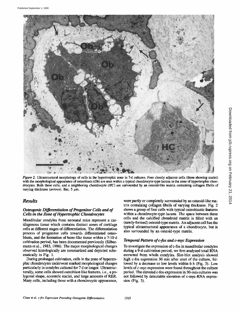
Figure 2. Ultrastructural morphology of ceils in the hypertrophic zone in 7-d cultures. Four closely adjacent cells (three showing nuclei) with the morphological appearance of osteoblasts (Ob) are seen within a typical chondroeyte-type lacuna in the zone of hypertrophic chon- drocytes. Both these cells, and a neighboring chondrocyte (HC) are surrounded by an osteoid-like matrix containing collagen fibrils of varying thickness (arrows). Bar, 5 tim.
Results
Osteogenic Differentiation of Progenitor Cells and of Cells in the Zone of Hypertrophic Chondrocytes Mandibular condyles from neonatal mice represent a car- tilagenous tissue which contains distinct zones of cartilage cells at different stages of differentiation. The differentiation process of progenitor cells towards differentiated osteo- blasts, and the formation of bone-like tissue within a 7-10-d cultivation period, has been documented previously (Silber- mann et al., 1983, 1986). The major morphological changes observed histologically are summarized and depicted sche- matically in Fig. 1.
During prolonged cultivation, cells in the zone of hypertro- phic chondrocytes underwent marked morphological changes particularly in condyles cultured for 7 d or longer. Ultrastruc- turally, some cells showed osteoblast-like features, i.e., a po- lygonal shape, eccentric nuclei, and large amounts of RER. Many cells, including those with a chondrocytic appearance,
were partly or completely surrounded by an osteoid-like ma- trix containing collagen fibrils of varying thickness. Fig. 2 shows a group of four cells with typical osteoblastic features within a chondrocyte-type lacuna. The space between these cells and the calcified chondroid matrix is filled with an (newly-formed) osteoid-type matrix. An adjacent cell has the typical ultrastructural appearance of a chondrocyte, but is also surrounded by an osteoid-type matrix.
Temporal Pattern of c-fos and c-myc Expression To investigate the expression of c-fos in mandibular condyles during a 9-d cultivation period, we first analyzed total RNA extracted from whole condyles. Slot-blot analysis showed high c-fos expression 30 min after start of the culture, fol- lowed by a decrease to low levels within 6 h (Fig. 3). Low levels of c-myc expression were found throughout the culture period. The elevated c-fos-expression in 30-min cultures was not followed by detectable elevation of c-myc-RNA expres- sion (Fig. 3).
Closs et al. c-los Expression Preceding Osteogenic Differentiation 1315
on February 21, 2014
jcb.rupress.orgD
ownloaded from
Published September 1, 1990

Figure 3. Expression of c-fos and c-myc. Total RNA was isolated from condyles cultured for the time indicated. RNA was bound to a filter by the slot-blot procedure and sequentially hybridized to actin-, fos-, and myc-specific probes. The probes were stripped from the filter completely after each hybridization.
We performed in situ hybridizations on sections of man- dibular condyles to determine the localization of the c-fos and c-myc expressing cells, c-fos expression was not found in any cells in condyles that had been frozen immediately af- ter dissection (Fig. 4, a and b). In contrast, a strong signal was found in condyles that had been cultured for 30 min (Fig. 4 c). The hybridization signal was distributed over the entire tissue. The intensity of the signal corresponded with the cell density in the different zones: i.e., it was highest in the area of progenitor cells. Individual cells expressing c-fos were not prominent.
Similar to the pattern observed by slot-blot analysis, the diffuse type of c-fos expression decreased after 2 h of culture and did not reappear during prolonged cultivation (Fig. 4, d-h). However, high levels of c-fos RNA were found in an increasing number of individually identifiable cells in the basal part of the condyle, i.e., the area of hypertrophic chon- drocytes (Fig. 4, c-h). This type of c-fos expression was first observed in a few cells along the basal border as early as 30 min after start of the culture. The number of cells showing high expression peaked between 1 and 3 d, at a time when the diffuse type of c-fos expression was no longer detected (Fig. 4, d-h). With increased time of culture the area of c-fos expressing cells extended towards the zone of chondroblasts. After 3 d the number of hypertrophic chondrocytes express- ing c-fos decreased, and only a few c-fos-positive ceils were found after day 10.
Distinct c-myc expression was not found in fresh explants (Fig. 5, a and b) or in condyles cultured for 30 min (Fig. 5 c); it was first observed 2 h after start of the culture in a few cells located in the basal area (Fig. 5 d). During culture the number of c-myc-expressing cells increased and the area containing c-myc positive cells extended further into the zone of hypertrophic chondrocytes (Fig. 5 e). In contrast to c-fos, the highest number of c-myc expressing cells was ob- served between 16 and 24 h of culture (Fig. 5, e and f ) . At this time, c-myc expressing cells were also found in the peri- chondrium. No significant c-myc expression was found in the condylar tissue in the late phase of the cultures (Fig. 5, g and h). c-myc RNA could also be observed in some of the cells that had migrated into the underlying collagen sponge (data not shown).
FCS did not affect the level of c-fos or c-myc RNA (see Materials and Methods). However, c-fos expression was ob- served after manipulation of condyles in culture simulating mechanical stress applied to the condyles in the process of dissection. Slot-blot analysis showed the same high c-fos ex- pression 1 h after mechanical manipulation as observed 1 h after dissection (Fig. 6).
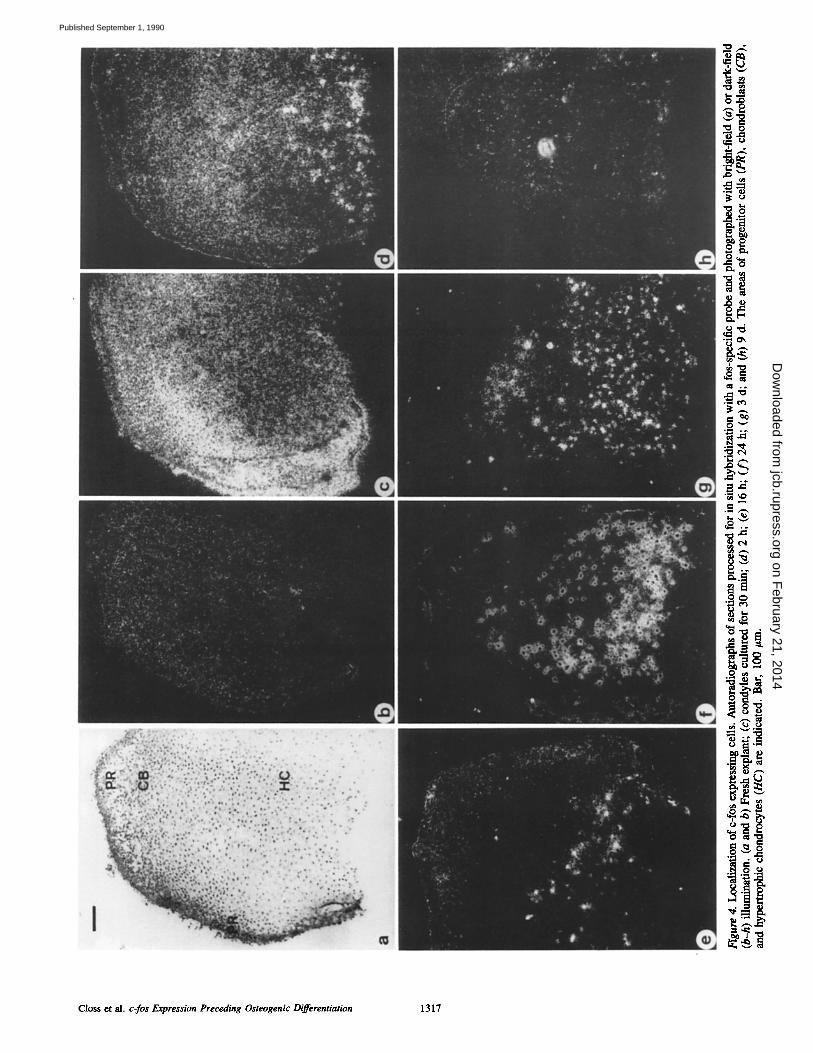
c-fos Expression Is Followed by Cell Proliferation and the Expression of Osteoblast Marker Genes To examine whether c-fos activation in hypertrophic chon- drocytes was followed by DNA replication and cell prolifera- tion, we determined [3H]thymidine incorporation into cel- lular DNA by autoradiography (Fig. 7). After incubation of freshly obtained explants with [3H]thymidine for 2 h, la- beled nuclei were found in cells in the progenitor zone but not in the zone of hypertrophic chondrocytes (Fig. 7, a and e). After 24 h, labeled cells were found in the zone of hyper- trophic chondrocytes (Fig. 7 f ) as well as in the perichon- drium and progenitor zone (Fig. 7 b). The number of labeled cells in the zone of hypertrophic chondrocytes increased fur- ther by day 2 (Fig. 7 g). The number of labeled cells de- creased in the hypertrophic zone after prolonged culture, whereas the number of labeled cells in the progenitor zone remained constant (Fig. 7, d and h). A consistent finding in 7-d cultures was the appearance of label in cells that had migrated into the underlying sponge (Fig. 7 h). Electron mi- croscope autoradiography confirmed the uptake of [3H]thy- midine by the nuclei of mature chondrocytes in areas show- ing signs of early calcification (Fig. 8).
One of the earliest marker genes expressed at high levels during differentiation of osteoblasts (Yoon et al., 1988) is collagen type I; it is also expressed in chondroprogenitor cells (Castagnola et al., 1988), In situ hybridization on sec- tions of freshly obtained condyles showed expression of col- lagen type I in cells in the progenitor zone but not in hyper- trophic chondrocytes (Fig. 9 a). In cells of the hypertrophic zone, collagen type I expression was first found in the basal area of the condyle after 2 h in culture (Fig. 9 b). After 3 d of culture, collagen type I expression was found throughout the area of hypertrophic chondrocytes (Fig. 9 c).
Discussion
The mandibular condyles of newborn mice contain undiffer- entiated progenitor cells, young chondroblasts, and mature
The Journal of Cell Biology, Volume 111, 1990 1316
on February 21, 2014
jcb.rupress.orgD
ownloaded from
Published September 1, 1990
° ~
~~ .~
g'g
.~ ; ~
~.~
Closs et al. c-fos Expression Preceding Osteogenic Differentiation 1317
on February 21, 2014
jcb.rupress.orgD
ownloaded from
Published September 1, 1990

!i D
~S ~'qD
° ~
~z
~ ° ~
. ~
The Journal of Cell Biology, Volume 111, 1990 1318
on February 21, 2014
jcb.rupress.orgD
ownloaded from
Published September 1, 1990

Figure 6. Mechanical stimula- tion of c-fos expression. Slot- blot analysis of RNAs isolated from fresh explants (0), from condyles cultured for 1 and 6 h, and from condyles that were pinched after cultivation for 6 h and cultured for an additional 60 min. The filter was hybridized sequentially to probes specific for ribosomal RNA and c-fos.
chondrocytes. Recent studies have shown that during in vitro cultivation both progenitor cells and hypertrophic chondro- cytes exhibit de novo expression of genes that are charac- teristically expressed during bone cell differentiation (Strauss et al., 1990), indicating a switch from chondrogenic to os- teogenic differentiation. We have now investigated the ex- pression of c-fos and c-myc protooncogenes which precedes this change in gene expression, and the subsequent ultra- structural appearance of the cells.
Two different patterns of c-fos expression were observed during culture of the mandibular condyles: a short term tran- sient expression in cells of the entire tissue, and a longer- lasting high expression in individual cells in the zone of hypertrophic chondrocytes. The transient c-fos expression between 30 min and 2 h of the culture apparently resulted from the mechanical stress applied to the tissue during dis- section. This type of fos expression could be induced ex- perimentally at a later time in culture by mechanical treat- ment simulating the dissection procedure. Thus, mechanical stress applied to condylar tissue seems to exert an effect similar to that of heat shock or wounding (Angel et al., 1985; Verrier et al., 1986; Gubits and Fairhurst, 1988).
A different type of c-fos expression was observed only in hypertrophic chondrocytes. Individual cells showed high levels of c-fos RNA which could not be attributed to mechan- ical or serum stimulation, the maximum number of cells ap- pearing between day 1 and day 3 of culture. The results indi- cate that this expression of c-fos was associated with two separate effects: entry of cells into the cell cycle, and a change in gene expression.
High c-fos mRNA levels appear to be a prerequisite for quiescent cells to reenter the cell cycle (Nishikura and Mur- ray, 1987; Riabowol et al., 1988). A number of observations indicate that the high c-fos expression in cells in the zone of hypertrophic chondrocytes was associated with onset of DNA replication and cell division. (a) c-fos expression preceded c-myc expression, a sequence that seems to be indicative for the induction of proliferation (Miiller et al., 1984a; Kelly et al., 1983; Kaczmarek et al., 1985; Colletta et al., 1986). (b) The highest number of [3H]thymidine-labeled cells was ob- served on day 2, following the maximum c-fos expression on
day 1. (c) The appearance of closely associated groups of cells within single chondrocyte-type lacunae in the hyper- trophic zone of 7-d cultures is strongly suggestive of preced- ing cell division.
A number of authors have shown that c-fos can act as a transcriptional regulator with a direct effect on gene expres- sion, separate from the effects associated with the initiation of a cell cycle (Setoyama et al., 1986; Distel et al., 1987; Chiu et al., 1988; Lech et al., 1988; Sch6nthal et al., 1988). Some differentiating cell types show raised levels of c-fos mRNA for extended periods (Gonda and Metcalf, 1984; Miiller et al., 1983, 1984b, 1985; Mitchell et al., 1985; Cur- ran and Morgan, 1985; Kruijer et al., 1986; Greenberg et al., 1985; Rtither et al., 1985). The pattern of c-fos expres- sion is different from that in growth factor-stimulated cells, in which c-fos is downregulated within a short period of time. The pattern of c-fos expression in the hypertrophic chondro- cytes was similar to that observed in other differentiating cells as mentioned above. Equally the number of c-fos ex- pressing hypertrophic chondrocytes was larger than that of c-myc expressing cells, and c-fos expression was observed for a longer period of time. We have shown that cells in this zone of hypertrophic chondrocytes start to express de novo collagen type I osteonectin, alkaline phosphatase, and bone gla protein, genes typical of cells of the osteogenic lineage, within the first 3 d of culture (this study, and Strauss et al., 1990). Both c-fos expression and expression of bone marker genes were seen at a time when the zone of hypertrophic chondrocytes contains a homogeneous population of mature cartilage cells in characteristic lacunae formed by a variably calcified matrix. Neither we nor other investigators have ever observed other cell types by light or EM within this zone within the first 3 d of culture (Lewinson and Silbermann, 1982; Silbermann et al., 1983, 1986). Taken together these results indicate that the expression of c-fos in the hyper- trophic chondrocytes was associated with the subsequent change in gene expression.
The de novo expression of genes typifying cells of the os- teogenic lineage, which follows the high expression of c-fos in hypertrophic chondrocytes, suggests that c-fos may con- trol the lineage specificity of skeletal cells. Support for this hypothesis comes from experimental induction of chondro- osseous neoplasms by v-fos or c-fos. Newborn mice infected with FBR MSV and FBJ MSV, both carrying the fos on- cogene, as well as mice transplanted with in vitro FBR MSV- infected mandibular condyles, develop sarcomas which are characterized by various degrees of osseochondrous differ- entiation (Finkel and Biskis, 1966; Finkel et al., 1973; Ward and Young, 1976; Schmidt et al., 1986; Silbermann et al., 1987). Furthermore, c-fos transgenic mice develop primar- ily slowly progressing benign skeletal lesions (Riither et al., 1987) and later osteochondrosarcomas (RiJther et al., 1989). These heterogeneous tumours comprise populations of cells of both the chondrogenic and osteogenic lineages. In both these cases the v-fos oncogene and the c-fos protooncogene interfere specifically with the differentiation pathway of skel- etoblasts.
c-fos expression was not observed in the perichondrium and progenitor zone except at the start of culture as a result of mechanical stress. The transient peak of c-myc expressing cells found in the perichondrium on day 1 coincided with the beginning of enhanced proliferation activity of these cells
Closs et al. c-fos Expression Preceding Osteogenic Differentiation 1319
on February 21, 2014
jcb.rupress.orgD
ownloaded from
Published September 1, 1990

Fo
Figu
re 7
. [3H
]thy
mid
ine u
ptak
e in
man
dibu
lar
cond
yles
. A
utor
adio
grap
hs o
f man
dibu
lar
cond
yles
(se
mith
in e
pon
sect
ions
) pul
se-l
abel
ed w
ith [
3Hlth
ymid
ine f
or th
e la
st 2
h o
f the
cul
ture
pe
riod
. (a
and
e)
Fres
h ex
plan
t; (b
and
f),
cond
yles
cul
ture
d fo
r 24
h; (
c an
d g)
for
2 d
; (d
and
h)
for 7
d.
(a-d
) Z
one
of ap
ical
pro
geni
tor
cells
; (e
-h)
zone
of h
yper
trop
hic
chon
droc
ytes
. l-
#m e
pon
sect
ions
, tol
uidi
ne b
lue.
Bar
, 10
0 #m
.
on February 21, 2014jcb.rupress.orgDownloaded from
Pub
lishe
d S
epte
mbe
r 1,
199
0
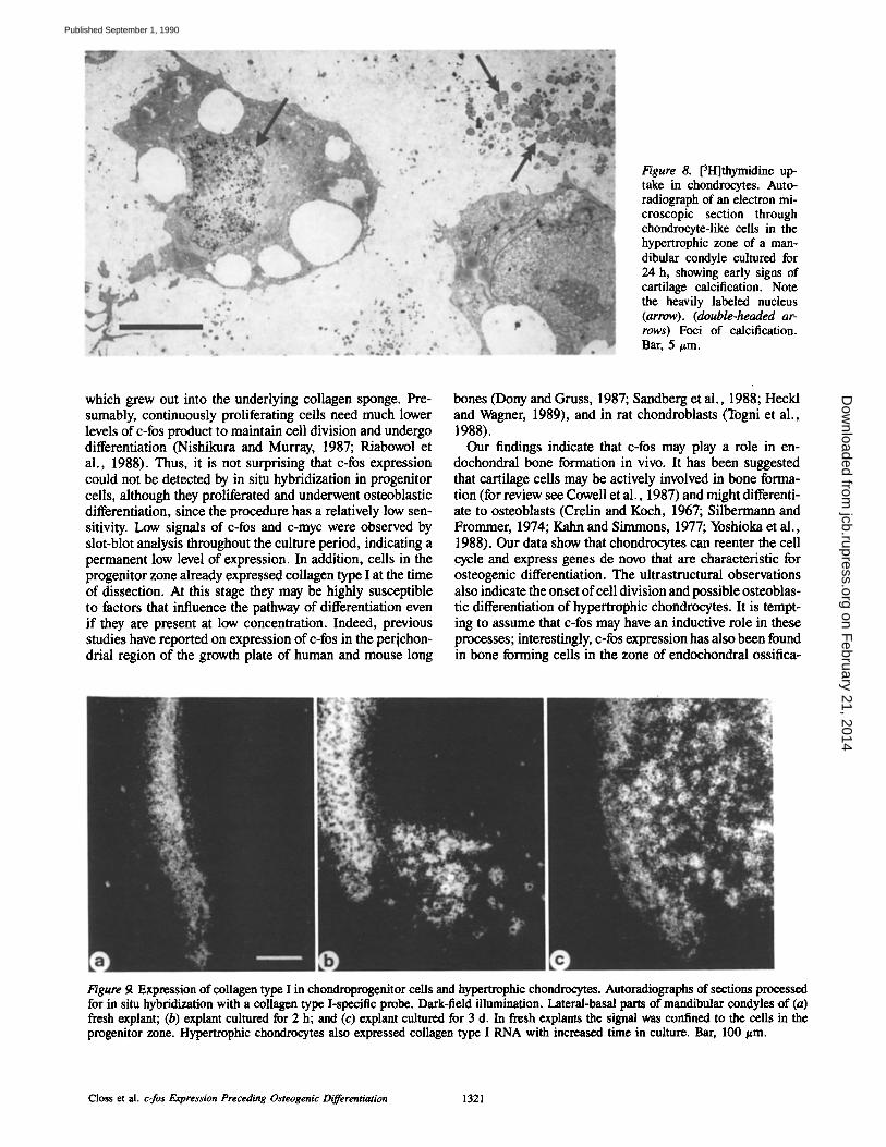
Figure 8. [3H]thymidine up- take in chondrocytes. Auto- radiograph of an electron mi- croscopic section through chondrocyte-like cells in the hypertrophic zone of a man- dibular condyle cultured for 24 h, showing early signs of cartilage calcification. Note the heavily labeled nucleus (arrow). (double-headed ar- rows) Foci of calcification. Bar, 5/~m.
which grew out into the underlying collagen sponge. Pre- sumably, continuously proliferating cells need much lower levels of c-fos product to maintain cell division and undergo differentiation (Nishikura and Murray, 1987; Riabowol et al., 1988). Thus, it is not surprising that c-fos expression could not be detected by in situ hybridization in progenitor cells, although they proliferated and underwent osteoblastic differentiation, since the procedure has a relatively low sen- sitivity. Low signals of c-fos and c-myc were observed by slot-blot analysis throughout the culture period, indicating a permanent low level of expression. In addition, cells in the progenitor zone already expressed collagen type I at the time of dissection. At this stage they may be highly susceptible to factors that influence the pathway of differentiation even if they are present at low concentration. Indeed, previous studies have reported on expression of c-fos in the perichon- drial region of the growth plate of human and mouse long
bones (Dony and Gruss, 1987; Sandberg et al., 1988; Heckl and Wagner, 1989), and in rat chondroblasts (Togni et al., 1988).
Our findings indicate that c-fos may play a role in en- dochondral bone formation in vivo. It has been suggested that cartilage cells may be actively involved in bone forma- tion (for review see Cowell et al., 1987) and might differenti- ate to osteoblasts (Crelin and Koch, 1967; Silbermann and Frommer, 1974; Kahn and Simmons, 1977; Yoshioka et al., 1988). Our data show that chondrocytes can reenter the cell cycle and express genes de novo that are characteristic for osteogenic differentiation. The ultrastructural observations also indicate the onset of cell division and possible osteoblas- tic differentiation of hypertrophic chondrocytes. It is tempt- ing to assume that c-fos may have an inductive role in these processes; interestingly, c-fos expression has also been found in bone forming cells in the zone of endochondral ossifica-
Figure 9. Expression of collagen type I in chondroprogenitor ceils and hypertrophic chondrocytes. Autoradiographs of sections processed for in situ hybridization with a collagen type I-specific probe. Dark-field illumination. Lateral-basal parts of mandibular condyles of (a) fresh explant; (b) explant cultured for 2 h; and (c) explant cultured for 3 d. In fresh explants the signal was confined to the cells in the progenitor zone. Hypertrophic chondrocytes also expressed collagen type I RNA with increased time in culture. Bar, 100 #m.
Closs et al. c-los Expression Preceding Osteogenic Differentiation 1321
on February 21, 2014
jcb.rupress.orgD
ownloaded from
Published September 1, 1990

tion (Heckl and Wagner, 1989). Further investigations are in progress to clarify the regulatory effects of c-fos on bone for- mation.
We gratefully acknowledge the excellent technical assistance of R. Baier, F. Heinemann, L. Rieke, H. Schulze, and B. Zimmermann.
This work was supported by the Deutsche Forschungsgemeinschaft (SFB 324), by the Wilhelm-Sander-Stiftung (Nr. 88.012.1), and by the Eu- ropean Atomic Energy Commission. (B-I-6-0080-D [B]).
Received for publication 5 March 1990 and in revised form 22 May 1990.
References
Angel, P., H. J. Rahmsdorf, A. P6ting, and P. Herrlich. 1985. C-fos mRNA levels in primary human fibroblasts aRer arrest in various stages of the cell cycle. Cancer Cells (Cold Spring Harbor). 3:315-319.
Brahic, M., and A. T. Haase. 1978. Detection of viral sequences of low reitera- tion frequency by in situ hybridization. Proc. Natl. Acad. Sci. USA. 75: 6125-6129.
Castagnola, P., B. Dozin, G. Moro, and R. Cancedda. 1988. Changes in the expression of collagen genes show two stages in chondrocytes differentiation in vitro. J. Cell Biol. 106:461-467.
Chirgwin, J. M., A. E. Przybyla, R. J. McDonald, and W. J. Putter. 1979. Isolation of biologically active ribonucleic acid from sources enriched in ribonuclease. Biochemistry. 18:5294-5299.
Chin, R., W. J. Boyle, J. Meed, T. Smeal, T. Hunter, and M. Karin. 1988. The c-fos protein interacts with c-jun/AP-1 to stimulate transcription of AP-1 responsive genes. Cell. 54:541-552.
Chu, M. L., J. C. Myers, M. P. Bernard, J. F. Ding, and F. Ramirez. 1982. Cloning and characterization of five overlapping cDNAs specific for the hu- man pro(x 1 (I) collagen chain. Nucleic Acids Res. 10:5925-5934.
Closs, E. I. Osteogene Differenzierung und neoplastiche Transformation von Knorpelgewebe in vitro. Ph.D. thesis. University of Munich, Munich. 1989. 196 pp.
Colletta, G., A. M. Cirafici, and G. Vecchio. 1986. Induction of the c-fos on- cogene by thyrotropic hormone in rat thyroid cells in culture. Science (Wash. DC). 233:458--461.
Cowell, H. R., E. B. Hunziker, and L. Rosenberg. 1987. The role of hyper- trophic chondrocytes in endochondral ossification and in the development of secondary centers of ossification. J. Bone Jr. Surg. Am. Vol. 69-A: 159-161.
Crelin, E. S., andW. E. Koch. 1967. An antoradiographic study ofchondrocyte transformation into chondroclasts and osteocytes during bone formation in vitro. Anat. Rec. 158:473-484.
Curran, T., and J. P. Morgan. 1985. Superinduction of c-fos by nerve growth factor in the presence of peripherally active benzodiazepines. Science (Wash. DC). 229:1265-1268.
Curran, T., G. Peters, C. Van Beveren, N. M. Teich, and I. M. Verma. 1982. FBJ murine osteosarcoma virus: identifcation and molecular cloning of bio- logically active proviral DNA. J. Virol. 44:674-682.
Deschepper, C. F., S. H. Mellon, F. Cumin, J. D. Baxter, and W. F. Ganong. 1986. Analysis by immunocytochemistry and in sito hybridization of renin and its mRNA in kidney, testis, adrenal, and pituitary of the rat. Proc. Natl. Acad. Sci. USA. 83:7552-7556.
Distel, R. J., H.-S. Ro, B. S. Rosen, D. L. Groves, and B. M. Spiegelman. 1987. Nucleoprntein complexes that regulate gene expression in adipocyte differentiation: direct participation of c-fos. Cell. 49:835-844.
Dony, C., and P. Gruss. 1987. Proto-oncogene c-fos expression in growth regions of fetal bone and mesodermal web tissue. Nature (Load.). 328:711- 714.
Erickson, J. M., C. L. Rushford, D. J. Dorney, G. N. Wilson, and R. D. Schmickel. 1981. Structure and variation of human ribosomal DNA: molec- ular analysis of cloned fragments. Gene. 16:1-9.
Finkel, M. P., and B. O. Biskis. 1986. Virus induction of osteosarcomas in n'rice. Science (Wash. DC). 151:698-701.
Finkel, M. P., C. A. Reilly Jr., B. O. Biskis, and I. L. Greco. 1973. Bone tumour viruses. In Bone, certain aspects of neoplasia. C. H. G. Price, and F. G. M. Ross, editors. Butterworth's, London. 353-364.
Gonda, T. J., and D. Metcaif. 1984. Expression of myb, myc and fos prnto- oncogenes during the differentiation of a murine myeloid leukaemia. Nature (Load.). 310:249-251.
Grcenberg, M. E., L. A. Greene, and E. B. Ziff. 1985. Nerve growth factor and epidermal growth factor induce rapid transient changes in proto- oncogene transcription in PC12 cells. J. Biol. Chem. 260:14101-14110.
Gubits, R. M., and J. L. Fairhurst. 1988. c-fos mRNA levels are increased by the cellular stressors, heat shock and sodium arsenite. Oncogene. 3:163- 168.
Hafen, E., M. Levine, R. L. Garber, and W. J. Gehring. 1983. An improved in situ hybridization method for the detection of cellular RNAs in drosophila tissue sections and its application for localizing transcripts of the homeotic antennapedia gane complex. EMBO (Fur. Mol. Biol. Organ.) J. 2:617-623.
Heckl, K., and E. F. Wagner. 1989. In situ analysis ofc-fos expression in trans-
genic mice. In Current Communications in Molecular Biology. Molecular Genetics of Early Drosophila and Mouse Development. M. R. Capeochi, editor. Cold Spring Harbor Laboratory, Cold Spring Harbor, NY. 117-129.
Holt, J. T., T. Venkat Gopal, A. D. Moulton, and A. W. Nienhuis. 1986. In- ducible production of c-los antisense RNA inhibits 3T3 cell proliferation. Proc. Natl. Acad. Sci. USA. 83:4794-4798.
Kacznmrek, L., L K. Hyland, R. Watt, M. Rosenberg, and R. Baserga. 1985. Microinjected c-myc as a competence factor. Science (Wash. DC). 228: 1313-1315.
Kahn, A. L, and D. J. Simmons, 1977. Chondrocyte-to-osteocyte transforma- tion in grafts in penchondrium-free epiphyseal cartilage. Clin. Orthop. Rel. Res, 129:299-304.
Kelly, K., B. H. Cochran, C. D. Stiles, and P. Leder. 1983. Cell-specific regu- lation of the c-myc gene by lymphocyte mitogens and platelet-derived growth factor. Cell. 35:603-610.
Krnijer, W., H. Skelly, F. Botteri, H. v. d. Pntten, J. Barber, I. Verma, and J. Leffert. 1986. Proto-oocogene expression in regenerating liver is simu- lated in cultures of primary adult rat hepatocytes. J. Biol. Chem. 261: 7929-7933.
Land, H., L. F. Parada, and R. A. Weinberg. 1983. Cellular oncogenes and multistep carcinogenesis. Science (Wash. DC). 222:771-778.
Lech, K., K. Anderson, and R. Brent. 1988. DNA-bound fos proteins activate transcription in yeast. Cell. 52:179-184.
Lewinson, D., and M. Silbermann. 1982. Landmarks in chondrocyte differenti- ation and maturation as envisaged by changes in the distribution of calcium complexes: an ultrastrucmral study. Metub. Bone Dis. & Rel. Res. 4:143- 150.
Marks, S. C., and S. N. Popoff. 1988. Bone cell biology: the regulation of de- velopment, structure, and function in the skeleton. Am. J. Anat. 183:1-144.
Mitchell, R. L., L. Zokas, R. D. Schreiber, and I. M. Verma. 1985. Rapid in- duction of the expression of proto-oncogene los during human monocytic differentiation. Cell. 40:209-217.
Moos, M., and D. Gallwitz. 1983. Structure of two human O-actin-related processed genes one of which is located next to a simple repetitive sequence. EMBO (Fur, Mol. Biol. Organ.) J. 2:757-761.
Miiller, R., and I. M. Verma. 1984. Expression of cellular oncogenes. Carr. Top. MicrobioL Immunol. I12:73-I15.
Miiller, R., I. M. Verma, and E. D. Adamson. 1983. Expression of c-onc genes: c-los transcripts accumulate to high levels during development of mouse placenta, yolk sac and amnion. EMBO (Eur. Mol. Biol, Organ.) J. 2:679-684.
Miiller, R., R. Bravo, J. Burckhardt, and T. Curran. 1984a. Induction of c-los gene and protein by growth factors precedes activation of c-myc. Nature (Load.). 312:716-720.
Mfiller, R., D. Miiller, and L. Guilbert. 1984b. Differential expression of c-fos in hematopoietic cells: correlation with differentiation of monomyelocytic cells in vitro. EMBO (Eur. Mol. Biol. Organ.) J. 3:1887-1890.
Mfiller, R., T. Curran, D. M(iller, and L. Guilbert. 1988. Induction of c-los during myelomonocytic differentiation and macrophage proliferation. Na- ture (Load.). 314:546-548.
Nishikura, K., and J. M. Murray. 1987. Antisenso RNA of proto-oncogene c-fos blocks renewed growth of quiescent 3T3 cells. Mol. Cell. Biol. 7: 639-649.
Riabowol, K. T., R. J. Vosatka, E. B. Ziff, N. J. Lamb, and J. R. Feramisco. 1988. Microinjection of fos-specific antibodies blocks DNA synthesis in fibroblast cells. Mol. Cell. Biol. 8:1670-1676.
Riither, U., E. F. Wagner, and R. Miiller. 1985. Analysis of the differentiation- promoting potential of inducible c-fos genes introduced into embryonal car- cinoma cells. EMBO (Eur. MoL Biol. Organ.) J. 4:1775-1781.
Riither, U., C. Garber, D. Komitowski, R. Miiller, and E. F. Wagner. 1987. Deregulated c-fos expression interferes with normal bone development in transgeoic mice. Nature (Load.). 325:412--416.
Riither, U., D. Komitowski, F. R. Schubert, and E. F. Wagner. 1989. C-fos expression induces bone tumors in transgenic mice. Oncogene. 4:861-865.
Sandberg, M., T. Vuorio, H. Hirvonen, K. Alitalo, and E. Vuorio. 1988. En- hanced expression of TGF-B and c-fos mRNAs in the growth plates of de- veloping human long bones. Development (Camb.). 102:461-470.
Schmidt, J., E. Livne, V. Erfe, W. G6ssner, and M. Silbermann. 1986. Mor- phology and in vivo growth characteristics of an atypical murine proliferative osseous lesion induced in vitro. Cancer Res. 46:3090-3098.
Schmidt, J., E. I. Closs, E. Livne, V. Erfle, and M. Silbermann. 1989. Bio- chemical characterization of virus-induced osteosarcoma-like osseous lesion in vitro. Calcif. ~ssue Int. 45:232-242.
Sch6n, A., L. Michiels, M..Ianowski, J. Merregaert, and V. Erfle. 1986. Ex- pression of protooneogenes in murine osteosarcomas. Int. J. Cancer. 38: 67-74.
Sch6nthal, A., P. Herrlich, H. J. Rahmsdoff, and H. Ponta. 1988. Requirement for fos gene expression in the transcriptional activation of collagenase by other oncogenes and pborbol esters. Cell. 54:325-334.
Setoyama, C., R. Frunzio, G. Lian, M. Mudryj, and B. Crnmbrugge. 1986. Transcriptional activation encoded by the v-fos gene. Proc. Natl. Acad. Sci. USA. 83:3213-3217.
S ilbermann, M., and J. Frommer. 1974. Electron microscopy of the secondary cartilage in mandibular condyle in the mouse. Acta. Anat. 90:330-346.
Silbermann, M., D. Lewinson, H. Gonen, M. A. Lizarbe, and K. yon der
The Journal of Cell Biology, Volume 111, 1990 1322
on February 21, 2014
jcb.rupress.orgD
ownloaded from
Published September 1, 1990
Mark. 1983. In vitro transformation of chondroprogenitor cells into osteo- blasts and the formation of new membrane bone. Anat. Rec. 206:373-383.
Silbermann, M., H. Reddi, A. Hand, and R. Leapman. 1986. In vitro differenti- ation of skeletoblasts and osteoblasts and the development of new bone. In Cell Mediated Calcification and Matrix Vesicles. Ali Y, editor. Elsevier, Amsterdam. 285-290.
Silbermann, M., J. Schmidt, E. Livne, K. yon der Mark, and V. Erfle. 1987. In vitro induction of osteosarcoma-like lesion by transformation of differen- tiating skeletal precursor cells with FBR murine osteosarcomas virus. Calcif Tissue Int. 41:208-217.
Strauss, P. G., E. I. Closs, J. Schmidt, and V. Erfle. 1990. Gene expression during osteogenic differentiation in mandibular condyles in vitro. J. Cell Biol. 110:1369-1378.
Togni de, P., H. Niman, V. Raymond, P. Sawchenko, and I. M. Verma. 1988. Detection of fos protein during osteogenesis by monoclonal antibodies. Mol. Cell. Biol. 8:2251-2256.
Verma, I. M., and W. R. Graham. 1987. The fos oncogene. Adv. CancerRes. 49:29-52.
Verrier, B., D. Miiller, R. Bravo, and R. Miiller. 1986. Wounding a fibroblast monolayer results in the rapid induction of the c-los proto-oncogene. EMBO (Fur. Mol. Biol. Organ.)J. 5:913-917.
Ward, J. M., and D. M. Young. 1976. Histogenesis and morphology of peri- osteal sarcomas induced by FBJ virus in NIH swiss mice. Cancer Res. 36: 3985-3992.
Wolf, H,, M. Hans, and E. Wilmes. 1984. Persistence of Epstein-Barr virus in the parotid gland. J. Virol. 51:795-798.
Yoon, K., S. J. C. Rutledge, R. F. Buenega, and G. Redan. 1988. Characteriza- tion of rat osteocalcin gene: stimulation of promoter activity by 1,25- dihydroxyvitamin D3. Biochemistry. 27:8521-8526.
Yoshioka, C., and T. Yagi. 1988. Electron microscopic observations on the fate of hypertrophic chondrocytes in condylar cartilage of rat mandible. J. Craniofacial Genet. Dev. Biol. 8:253-264.
Closs et al. c-fos Expression Preceding Osteogenic Differentiation 1323
on February 21, 2014
jcb.rupress.orgD
ownloaded from
Published September 1, 1990