Imaging Spontaneous Tumorigenesis: Inflammation Precedes Development of Peripheral NK Tumors
31
doi:10.1182/blood-2008-07-166462 Prepublished online October 29, 2008; Lee Ratner Dan Rauch, Shimon Gross, John Harding, Stefan Niewiesk, Michael Lairmore, David Piwnica-Worms and of peripheral NK tumors Imaging spontaneous tumorigenesis: Inflammation precedes development (1561 articles) Lymphoid Neoplasia Articles on similar topics can be found in the following Blood collections http://bloodjournal.hematologylibrary.org/site/misc/rights.xhtml#repub_requests Information about reproducing this article in parts or in its entirety may be found online at: http://bloodjournal.hematologylibrary.org/site/misc/rights.xhtml#reprints Information about ordering reprints may be found online at: http://bloodjournal.hematologylibrary.org/site/subscriptions/index.xhtml Information about subscriptions and ASH membership may be found online at: digital object identifier (DOIs) and date of initial publication. the indexed by PubMed from initial publication. Citations to Advance online articles must include final publication). Advance online articles are citable and establish publication priority; they are appeared in the paper journal (edited, typeset versions may be posted when available prior to Advance online articles have been peer reviewed and accepted for publication but have not yet Copyright 2011 by The American Society of Hematology; all rights reserved. 20036. the American Society of Hematology, 2021 L St, NW, Suite 900, Washington DC Blood (print ISSN 0006-4971, online ISSN 1528-0020), is published weekly by For personal use only. by guest on November 28, 2013. bloodjournal.hematologylibrary.org From For personal use only. by guest on November 28, 2013. bloodjournal.hematologylibrary.org From
-
Upload
independent -
Category
Documents
-
view
0 -
download
0
Transcript of Imaging Spontaneous Tumorigenesis: Inflammation Precedes Development of Peripheral NK Tumors

doi:10.1182/blood-2008-07-166462Prepublished online October 29, 2008;
Lee RatnerDan Rauch, Shimon Gross, John Harding, Stefan Niewiesk, Michael Lairmore, David Piwnica-Worms and of peripheral NK tumorsImaging spontaneous tumorigenesis: Inflammation precedes development
(1561 articles)Lymphoid Neoplasia �Articles on similar topics can be found in the following Blood collections
http://bloodjournal.hematologylibrary.org/site/misc/rights.xhtml#repub_requestsInformation about reproducing this article in parts or in its entirety may be found online at:
http://bloodjournal.hematologylibrary.org/site/misc/rights.xhtml#reprintsInformation about ordering reprints may be found online at:
http://bloodjournal.hematologylibrary.org/site/subscriptions/index.xhtmlInformation about subscriptions and ASH membership may be found online at:
digital object identifier (DOIs) and date of initial publication. theindexed by PubMed from initial publication. Citations to Advance online articles must include
final publication). Advance online articles are citable and establish publication priority; they areappeared in the paper journal (edited, typeset versions may be posted when available prior to Advance online articles have been peer reviewed and accepted for publication but have not yet
Copyright 2011 by The American Society of Hematology; all rights reserved.20036.the American Society of Hematology, 2021 L St, NW, Suite 900, Washington DC Blood (print ISSN 0006-4971, online ISSN 1528-0020), is published weekly by
For personal use only. by guest on November 28, 2013. bloodjournal.hematologylibrary.orgFrom For personal use only. by guest on November 28, 2013. bloodjournal.hematologylibrary.orgFrom

1
Imaging Spontaneous Tumorigenesis:
Inflammation Precedes Development of Peripheral NK Tumors
Dan Rauch,1 Shimon Gross,2 John Harding,1 Stefan Niewiesk,3 Michael Lairmore,3,4 David
Piwnica-Worms,2,5 and Lee Ratner1,6
1Department of Medicine, Division of Molecular Oncology, 2Molecular Imaging Center,
Mallinckrodt Institute of Radiology, 5Department of Molecular Biology and Pharmacology,
Washington University School of Medicine, St. Louis, MO 63110.
3College of Veterinary Medicine, 4Center for Retrovirus Research, Department of Veterinary
Biosciences, The Ohio State University, Columbus OH 43210.
6Correspondence should be addressed to L.R. at [email protected]
Mailing address: 660 S. Euclid, Campus Box 8069, St. Louis, MO 63110.
Phone: (314) 362-8836, Fax: (314) 747-2797
Running title: Inflammation precedes NK Tumors
Blood First Edition Paper, prepublished online October 29, 2008; DOI 10.1182/blood-2008-07-166462
Copyright © 2008 American Society of Hematology
For personal use only. by guest on November 28, 2013. bloodjournal.hematologylibrary.orgFrom
2
Abstract
Early events in tumor development are spontaneous, microscopic, and affected by the
microenvironment. We developed a mouse model of spontaneous lymphoma in which malignant
transformation is coupled with light emission that can be detected non-invasively using
bioluminescent imaging. The human T-cell leukemia virus type 1 transcriptional transactivator Tax, is
an oncogene sufficient to produce lymphoma in transgenic animal models. Using the granzyme B
promoter to restrict Tax expression to the mature NK/T cell compartment, we have reproduced many
elements of HTLV-associated adult T-cell leukemia/lymphoma. Tax activates signaling cascades
associated with transformation, inflammation, and tumorigenesis. Here we report that Tax mediated
activation of luciferase in LTR-LUC mice serves as a reporter for imaging these processes in vivo.
Using BLI, we discovered that microscopic intraepithelial lesions precede the onset of peripheral
subcutaneous tumors, tumorigenesis progresses through early reversible stages, and Tax is sufficient
for inducing tumors. Based on these findings, we propose that Tax expression in activated
lymphocytes initiates a cascade of events that leads to NK/T cell recruitment, activation, and
transformation. The use of BLI expands our ability to interrogate the role of Tax in tumorigenesis in
vivo, and has made the association of inflammation with tumor initiation amenable for study.
For personal use only. by guest on November 28, 2013. bloodjournal.hematologylibrary.orgFrom
3
Introduction
Early detection of malignant tumors and identification of events that precede tumorigenesis are
critical in cancer treatment and prevention strategies. Malignant transformation involves successive
accumulation of genetic events that breach anticancer defense mechanisms and confer growth
advantages to the mutated cell, events that are spontaneous and microscopic 1. Within the host,
transformation is also affected by the tumor microenvironment. Limitation in detecting these
microscopic spontaneous events in vivo has restricted the utility of animal models in the interrogation
of spontaneous tumorigenesis 2. To overcome these limitations, we developed a mouse model of
spontaneous lymphoma in which malignant transformation is coupled with light emission that can be
detected non-invasively using bioluminescent imaging (BLI).
The Tax oncogene from human T-cell leukemia virus type 1 (HTLV-1), the causative agent of
adult T-cell leukemia/lymphoma (ATLL) and HTLV-1 associated myelopathy/tropical spastic
paraparesis (HAM/TSP), is a potent transcriptional trans-activator capable of promoting cellular
transformation 3. In the context of HTLV-1 replication, Tax promotes viral gene expression by
activating the 5’ long terminal repeat (LTR) of the genome. Tax activity also results in growth
advantages normally acquired over time by malignant cells 4. Tax triggers cell growth and prevents
apoptosis by activating pathways that signal through the cAMP responsive element binding
transcription factor (CREB), nuclear factor kappa B (NFkB), and serum response factor (SRF).
Inhibition of p53 and dysregulation of other checkpoint controls by Tax leads to genetic instability
accelerating mutagenesis 5. Tax restricts retinoblastoma protein (Rb) activity disengaging
mechanisms designed to inhibit cell growth and activates telomerase allowing limitless replication
potential 6. Placing the Tax oncogene under the human granzyme B promoter (GZB-TAX) restricts its
expression to activated T and NK cells 7. Mice carrying the GZB-TAX transgene develop large
granular lymphocytic (LGL) leukemia/lymphoma, osteolytic disease, neutrophilia, and splenomegaly
during the first year of life 8,9. Transformed cells derived from peripheral tumors are most often
CD16hi, IL-2 independent NK cells 10.
For personal use only. by guest on November 28, 2013. bloodjournal.hematologylibrary.orgFrom
4
To couple spontaneous tumorigenesis and bioluminescence imaging (BLI), we hypothesized
that transgenic mice in which firefly luciferase is regulated by the HTLV-1 LTR should emit light in
response to Tax expression. Here we report the production of LTR-LUC transgenic mice, confirm
that double transgenic (TAX-LUC) mice recapitulate the original tumor model, and demonstrate that
bioluminescence in these mice serves as a novel reporter for spontaneous tumorigenesis in vivo.
Using bioluminescence to monitor Tax-mediated transformation, we discovered that i) microscopic
intraepithelial lesions precede the onset of peripheral subcutaneous tumors, ii) tumorigenesis in this
model progresses through early reversible stages, iii) Tax expression is sufficient for initiating
tumorigenesis in vivo, but is not required for tumor maintenance, and iv) primary tumor cells retain
bioluminescent properties in tumor allograft models. Use of BLI dramatically expands our ability to
interrogate the role of Tax in all aspects of tumorigenesis in vivo, and has made an important
connection between wounding, inflammation, and tumor initiation in this model amenable for future
study.
For personal use only. by guest on November 28, 2013. bloodjournal.hematologylibrary.orgFrom

5
Methods
Cells
B5Luc cells were derived from B5 cells (primate kidney fibroblasts) transfected with LTRLuc,
selected in G418 (400μg/ml), and cloned by limiting dilution 11. Co-culture of B5Luc cells with
irradiated 293T cells transfected with a replication competent HTLV-1 molecular clone that expresses
Tax (pACH) causes cell fusion, infection of B5Luc cells, and activation of LTRLuc by Tax 12. Mirus
TransIt reagent was used in all transfections according to manufacturer’s instructions (Fisher, St
Louis, MO). CD4+ CD16/32+ cell lines subsequently derived from transplanted bioluminescent
tumors have been stable in culture for more than twelve months.
Transgenic Mice
The LTRLuc plasmid consists of the 0.7 Kb XhoI-HindIII 5’LTR fragment of HTE-1 and the
luciferase open reading frame from PGL-3 (Promega) cloned into the XhoI-NotI vector backbone of
pEGFP-1 (Clontech, Mountain View, CA) 13. The XhoI-AflII fragment of LTRLuc was used to generate
two independent transgenic founders with indistinguishable bioluminescence characteristics 14. GZB-
TAX mice in which HTLV-1 Tax is regulated by the 5’ flanking region (positions -1170 to +36) of the
human granzyme B gene have been previously described 8. Mice were housed under pathogen free
conditions and animal protocols were approved by the Animal Studies Committee in accordance with
the guidelines of the Washington University School of Medicine.
For personal use only. by guest on November 28, 2013. bloodjournal.hematologylibrary.orgFrom
6
Flow Cytometry
Cell suspensions derived from organs or tumors were stained with FITC-conjugated FcγR II/III
antibodies (clone 2.4G2; BD Pharmingen) for 30 minutes at 4oC and analyzed on a FACScan (Becton
Dickenson, Franklin Lakes, NJ) 8.
Imaging
The IVIS100 imaging system (Xenogen) was used to image bioluminescence in vivo in
anesthetized mice (isoflurane inhalation) 15. Standard imaging parameters included D-luciferin dose,
50 mg/ml i.p (500 mg/kg body weight); exposure time, 300 sec; binning, 4; f/stop, 1; no optical filter.
Areas of the body covered by hair required shaving or depiliation prior to imaging. X-ray images were
obtained in a Kodak in-vivo imaging system 4000MM (exposure time: 60 sec; aperture: 2.8; focus:
12.5 mm; field of view: 60 mm). Color scale unless otherwise indicated is x104 photons/sec/cm2/sr.
Histology
Histology and tumor cell characterization in GZB-TAX tumors was described previously 8,9,16-18
Malignant LGL cells are distinguishable by morphological features including size, granularity, shape,
and nuclear morphology, they typically constitute approximately 10% of tumor cells, and constitutively
express high levels of CD16.
Tissues were fixed in 4% paraformaldehyde (sections including bone were decalcified in 0.5 M
EDTA for 14 days), and embedded in paraffin for serial sectioning. Paraffin embedded mouse tissue
was cut at 4 microns and placed on positively charged slides. Slides with specimens were then
placed in a 60oC oven for 1 hour, cooled, deparaffinized and rehydrated through xylene graded
ethanol solutions to water. All slides were quenched for 5 minutes in a 3% hydrogen peroxide
solution in water to block endogenous peroxidase. Antigen retrieval was performed by a heat method
in which specimens were placed in Dako’s target retrieval solution, pH 6.1 for 25 minutes at 94oC
using a vegetable steamer and cooled for 15 minutes in solution. Slides were then placed on a Dako
For personal use only. by guest on November 28, 2013. bloodjournal.hematologylibrary.orgFrom
7
Autostainer immunostaining system for use with immunohistochemistry. Slides were initially blocked
for endogenous biotin before the addition of biotinylated primary antibody. The biotinylated primary
antibody solution was incubated for 1 hour. Next a labeled streptavidin was applied and incubated for
30 minutes. Lastly, the slides were developed with DAB chromogen. Slides were then
counterstained in hematoxylin, dehydrated through graded ethanol solutions and coverslipped.
Stained sections were visualized with a Nikon Eclipse E400 microscope and digital images were
obtained using a Magnafire camera and software (Optronics, Muskogee, OK).
For personal use only. by guest on November 28, 2013. bloodjournal.hematologylibrary.orgFrom

8
Results
Previously, we described a transgenic model for ATLL in which Tax is expressed in activated T
and NK cells under the regulation of the human granzyme B promoter (GZB-TAX) 8. To use Tax
expression as a biomarker for malignancy, we developed a second transgenic line in which firefly
luciferase expression is regulated by a Tax-inducible promoter, the HTLV-1 LTR . The LTR contains
three Tax responsive elements, which are 21 nucleotide repeats homologous to the consensus cAMP
responsive element (CRE) (Fig.1A) 19. Tax activates viral transcription by recruiting CREB and CREB-
binding proteins CBP and p300 to the U3 region of the viral 5’LTR 20. In transient transfection
experiments in human (Fig. 1B) and mouse cell lines (Fig 1C) luciferase expression from the LTR-
LUC construct was induced 50-100 fold by Tax. In these experiments Tax-independent basal LTR
activity also increased relative to transgene copy number. Tax-independent HTLV-1 LTR activity is
mediated by Sp1, AP1, and C/EBP families of transcription factors 21-24 can be induced by apoptosis
or stress responses 25-28 and is repressed by HDAC1 and ATFx 29,30. However, when stably
integrated in a primate cell line, the LTRLUC transgene has low basal activity and 100-200 fold
inducibility by retrovirally expressed Tax (Fig. 1D-E). Similarly, in both founder LTR-LUC transgenic
mice, basal luciferase activity was undetectable by BLI, and no pathology was associated with the
transgene.
When GZB-TAX mice were bred with LTR-LUC mice, luciferase was expressed in tumors of
the double transgenic (TAX-LUC) offspring and was detectable by non-invasive BLI (Fig. 2A).
Bioluminescent tumors arising in TAX-LUC mice, like the original GZB-TAX mice, occurred at
subcutaneous sites (Fig. 2B), and were often associated with osteolytic lesions (Fig. 2C). Primary
and disseminated tumors contain an admixture of inflammatory cells and malignant large granular
lymphocytes that can be distinguished by size, morphology, and surface expression of CD16, also
included CD16lo neutrophils and CD16- lymphocytes (Fig. 2D). Tax protein was detectable by
immunohistochemistry in large granular lymphocytes in primary tumors and involved organs (Fig. 2E)
9. As in the original model, complete and differential counts of peripheral blood of TAX-LUC mice (Fig
For personal use only. by guest on November 28, 2013. bloodjournal.hematologylibrary.orgFrom

9
2F) revealed an increase in total white blood cell count (32,000/µl in the example shown), dramatic
neutrophilia (78% of nucleated cells), polychromasia in the erythrocyte series, and an increase in the
number of circulating LGLs many of which contain multinucleated or multi-lobulated nuclei (arrows,
Fig. 2F)8. Finally, the presence of the LTR-LUC transgene did not affect the onset or penetrance of
Tax-mediated tumorigenesis in TAX-LUC animals (Fig. 2G). Taken together, these data establish
that bioluminescence correlated with spontaneous tumorigenesis in TAX-LUC mice, and the spatially
and temporally dynamic molecular events that precede spontaneous tumor development in this model
in-vivo were unaffected by introduction of the LTR-LUC transgene.
Since the granzyme B promoter is active in mature T and NK cells, Tax expression and
subsequent luciferase activity in bioluminescent tumors originates from transformed cells or from
normal cells activated within, or recruited to, the tumor microenvironment. While cell lines derived
from GZB-TAX tumors constitutively express NFκB and are highly resistant to apoptosis, transformed
cells constitute the minority of cells in the primary tumor in vivo and express very little Tax in culture
10,16. To determine if bioluminescence is a property of the transformed cells within a primary tumor,
cell homogenates from TAX-LUC tumors were injected subcutaneously into beige / nude / xid (BNX)
mice. We found that cells derived from primary TAX-LUC tumors when transferred to allograft
recipients propagated into tumors composed of Tax-expressing LGLs (Fig. 3A), spread to liver, lung,
spleen, and bone (Fig. 3B), and expressed luciferase that was observable by BLI (Fig. 3C). These
results indicate i) that luciferase activity was a property of the malignant LGL cell population in TAX-
LUC mice and ii) that BLI can be used to identify primary tumor tissue and monitor growth and
dissemination of transplanted LGL tumors in allograft models.
Many factors can affect bioluminescence in vivo, including availability of the D-luciferin and co-
factors required for the luciferase reaction, depth and pigmentation of the tissue being imaged, and
the number and transcriptional activity of the cells carrying the bioluminescent reporter 2. We
observed heterogeneity in photon output among tumors over time (Fig 4A) which correlated with the
level of Tax expression (Fig 4B), even though both tumors persist and maintain a malignant
For personal use only. by guest on November 28, 2013. bloodjournal.hematologylibrary.orgFrom
10
phenotype. These results directly correlated bioluminescence with Tax activity in tumor cells,
identified Tax as a dynamic variable during tumor development, and suggest that constitutive Tax
activity is not necessary to maintain a malignant phenotype.
Tax is essential for HTLV-1 replication and development of ATLL, yet in HTLV-1 infected
individuals viral replication and gene expression is tightly restricted. In ATLL patients, Tax expression
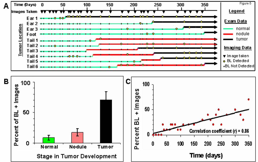
is often low or undetectable. Utilizing TAX-LUC mice for observing Tax expression during tumor
development, we conducted physical exams and obtained bioluminescent images on a cohort of
animals over the course of a year (Fig 5A). Physical exam data was divided into three categories;
normal, nodule, and tumor. Normal tissue had no visual or palpable mass. Nodules, which only
developed in mice carrying the Tax transgene, were most clearly defined on the tail as small (<5mm),
palpable, chronically inflamed masses, that arose rapidly but did not progress. Over time,
approximately 50% of nodules resolved, 30% developed into malignant tumors, and 20% remained
unchanged for the duration of the study. Tumors were defined as masses exhibiting consistent and
aggressive growth and often involved tissue damage and necrosis. Imaging data was obtained at
time points indicated (▼) and bioluminescent regions ( ) had a defined focus clearly distinguishable
from background noise with photon flux greater than 3X10³ photons/sec/cm2/sr. Of 10 lesions that
spontaneously developed during the study (Fig. 5A), only one (Ear 1) was bioluminescent at every
time point after inception. In seven lesions (all except Ear 1, Foot, and Tail 1) bioluminescence was
punctuated by intervals lacking luciferase activity, nine of the lesions (all except Tail 3) exhibited a
bioluminescent signature prior to aggressive tumor growth, and in two cases (Tail 1 and Tail 2)
nodules developed but did not progress into tumors during the course of the study. While
bioluminescence was most frequently observable in tumors, luciferase activity in normal tissue and
nodules was stochastic and infrequent (Fig. 5B). The frequency of bioluminescence in TAX-LUC
mice increased over time as tumors developed with a high degree of variability at any given time point
(Fig. 5C). The bioluminescence profiles in these data suggest that Tax expression is dynamic. Tax
For personal use only. by guest on November 28, 2013. bloodjournal.hematologylibrary.orgFrom

11
expression is restricted but not permanently extinguished during tumorigenesis and can be used as a
predictive reporter for spontaneous tumor development in TAX-LUC mice.
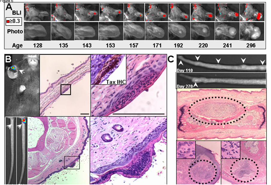
Development of preventative therapies requires identifying the events that promote malignant
transformation and precede tumorigenesis in vivo. The ability to non-invasively detect a
bioluminescent signature weeks or months before a palpable nodule or visible tumor was detectable
allowed us to investigate events that precede and promote tumor initiation, and temporally and
spatially predict tumor onset. As described above, in almost all cases, small and often transient foci
of bioluminescence in normal tissue preceded the onset of tumorigenesis, and one example is shown
in Fig. 6A. Microscopic examination of tissue sections revealed that the loci of bioluminescence
preceding tumorigenesis correspond to regions involving Tax expression within superficial
inflammatory lesions within the epidermis (Fig 6B). These epidermal lesions preceded the
development of palpable subcutaneous nodules, and are composed primarily of an admixture of
neutrophils and lymphocytes. As described above, these nodules resolve more often than they
develop into a tumor, and Fig 6C shows examples of such lesions (Fig. 6C). These findings rendered
the earliest and previously inaccessible stages of tumorigenesis amenable to further study, revealed
a previously unknown and variable pattern of oncogene expression within spontaneous tumors, and
represent the first evidence that the initiation of spontaneous peripheral tumors in TAX-LUC animals
begins with inflammation associated with a microscopic lesion.
Discussion
Bioluminescence imaging has emerged as a sensitive and reliable tool for obtaining dynamic
molecular information in vivo 15. Sampling errors and variability inherent in assays that require
invasive procedures can easily mask subtle events, and increase the number of subjects necessary
for statistical significance. Using a subject as it own control, imaging allows detection of systemic
changes, identifies regions of interest to guide invasive procedures, and reveals unanticipated
variability within a cohort over time. While these advantages of BLI have been well established in
For personal use only. by guest on November 28, 2013. bloodjournal.hematologylibrary.orgFrom
12
experiments involving engraftment of bioluminescent cell lines, they remain largely untapped in
animal models that develop spontaneous tumors.
Sufficient to transform primary cells in culture and produce a variety of malignancies in
transgenic animal models, Tax activates NFκB and its downstream targets, promotes cell proliferation
and inhibits cell-cycle arrest, genetic stability, and apoptosis 3. Using part of the human granzyme B
promoter to restrict Tax expression to the mature T and NK cell compartment, we have been able to
reproduce many elements of the lymphoma phenotype seen in humans with ATLL 8. Tumors that
spontaneously arise in these animals during their first year of life contain malignant lymphocytes that
constitutively express NFκB and NFκB target genes including interleukin 6 (IL-6), IL-10, granulocyte
macrophage colony stimulation factor (GM-CSF), and interferon gamma (IFN-γ) 31. Our hypothesis
proposed that the tight regulation of Tax by the granzyme B promoter, the exquisite sensitivity of
bioluminescence imaging, and the unique properties of the HTLV-1 LTR would enable us to use Tax
to promote malignant transformation and provide a means of detection in vivo.
We have shown that introduction of the LTR-LUC transgene did not affect disease parameters
in the TAX transgenic mice, malignant LGLs within primary tumors were bioluminescent, and
bioluminescence correlated with Tax activity. While signaling pathways, surface markers, growth
properties, and cytokine production in the LGL cell lines derived from these animals have been
examined, several important questions regarding the earliest events in tumorigenesis remain
unresolved 32. What determines when and where a tumor will form? Is Tax expressed prior to and
during tumorigenesis, and is it required for tumor maintenance? Is it possible to identify precancerous
stages during tumorigenesis? How does the tumor microenvironment affect tumorigenesis in vivo?
With Tax-Luc mice and bioluminescence imaging we have begun addressing these questions. We
discovered a bioluminescent signature preceding tumorigenesis and corresponding to microscopic
epithelial lesions. This unprecedented glimpse into the initiation events that promote tumorigenesis in
this model is the first evidence that these Tax tumors are nucleated by wounds. Grossman et al.
were the first to suggest that injury to epithelial cells could start an inflammatory response that
For personal use only. by guest on November 28, 2013. bloodjournal.hematologylibrary.orgFrom

13
recruits and activates LGLs, production of Tax, and further exacerbation of inflammation through
activation of NFκB 10. Inflammation is emerging as an important promoter of many cancers including
lymphoma, with NFκB serving as a master regulator 33. Chronic inflammation, a promoter of
transformation perpetuated by Tax, explains the correlation seen between wounds and tumors, the
occurrence of inflammatory nodules in GZB-TAX and TAX-LUC mice, and the preponderance of
neutrophils at all stages of tumorigenesis 34.
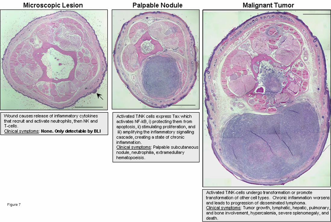
Based on these findings, we propose that Tax expression in activated lymphocytes transforms
wounds into a chronically inflamed state, initiating a cascade of events that lead to NK cell
recruitment, activation, and Tax-mediated transformation (Fig. 7). Initially, CD16+ NK cells, which
predominate in the peripheral blood, are recruited to acutely inflamed peripheral lesions along with
macrophages, neutrophils, and lymphocytes 35. Activation of the cells results in Tax expression and
subsequent luciferase activity in TAX-LUC animals. The inflammatory process either resolves or
expands into a palpable subcutaneous nodule which may subsequently resolve, remain stable, or
progress into an aggressive tumor. We propose that nodules that do not expand into invasive
tumors, either do not contain transformed cells or contain cells undergoing transformation that were
subsequently restricted by another mechanism. The angiostatic effect of IFN-γ retards tumor growth
in this model, and may be one such mechanism of restriction 18. Alternatively, the loss of Tax
expression during transformation could serve as a mechanism of restriction. Although
bioluminescence precedes and is associated with tumorigenesis our imaging data would suggest that
in the majority of tumors, Tax is not constitutively expressed. In fact, even in individuals suffering
from ATLL, Tax activity is variable and often undetectable 3. Paradoxically, while Tax promotes
immortalization and transformation, it also promotes the expression of IFN-gamma and TNF-alpha, it
is angiostatic, it leads to tumor restriction and necrosis, under some conditions Tax can lead to cell
cycle arrest, and Tax promotes CTL lysis of cells in which it is expressed 3,36-38. The loss of Tax
expression and bioluminescence in some tumors indicates that once transformation has occurred,
Tax activity is not necessary to maintain the malignant phenotype.. Extracellular Tax protein has also
For personal use only. by guest on November 28, 2013. bloodjournal.hematologylibrary.orgFrom
14
been reported to mediate T-cell activation and proliferation 39,40. This activity is mediated through
uptake of Tax and activation of gene expression. Thus, paracrine release of Tax from inflammatory
lesions could contribute to tumor initiation and maintenance in GZB-Tax mice, and would be
detectable by BLI.
Although ATLL is a disease of human CD4+ T-cells and TAX-LUC mice develop NK leukemia
and lymphoma, the mechanisms by which Tax transforms human and murine lymphocytes are of
sufficient similarity to allow TAX-LUC mice to recapitulate many aspects of ATLL including NFkB
activation, delayed onset, osteolytic bone disease, hypercalcemia, hepatomegaly, splenomegaly, and
pulmonary involvement. Moreover, acute, chronic, and smoldering forms of ATLL all present with
skin lesions as a predominant characteristic of disease. While several TAX transgenic mouse models
have been previously described, and many develop inflammatory diseases, GZB-TAX-LUC animals
uniquely model the link between inflammation and tumorigenesis 41-43. In models in which Tax is
driven by the HTLV-1 LTR, it is expressed in a variety of tissues including fibroblasts, skeletal muscle,
bone, peripheral nerves, brain, spleen, thymus, and skin 44-50. These various models developed
fibroblast tumors, neurofibromas, skeletal abnormalities, and inflammatory conditions, but none of
these models recapitulated human ATLL. Tax expression in other models has been driven from the
IgG promoter/enhancer 51, CD4 promoter 43, and Thy-1 promoter 52. While these mice also develop
inflammatory disorders, thymic atrophy, and non-hematopoeitic malignancies, they did not lead to
leukemia or lymphoma. Two recent mouse models, in which Tax is expressed from the Lck promoter
in thymocytes and/or peripheral T-cells, develop ATLL-like diseases 42,53. Central to the contributions
of TAX-LUC mice to the literature of ATLL animal models is the ability to detect Tax activity in vivo,
using bioluminescent imaging to identify the microenvironment involved in Tax-mediated malignant
transformation. Identifying cellular and molecular factors that contribute to these events may provide
new molecular targets for prevention of ATLL in HTLV-1 positive individuals.
We also propose that BLI and the TAX-LUC animal model could more broadly be used to
investigate a range of signaling events associated with inflammation and tumor development.
For personal use only. by guest on November 28, 2013. bloodjournal.hematologylibrary.orgFrom
15
Inflammation promotes many forms of lymphoma including those caused by Helicobacter pylori,
Epstein Barr virus, human herpesvirus 8, and hepatitis c virus. 54-56. Several forms of lymphoma with
a non-infectious etiology, like Hodgkin’s, mantle cell, and diffuse large B cell lymphomas, are also
associated with inflammation or constitutive NFκB activation 57,58 We are currently using the TAX-
LUC mouse model as a tool to pursue the role of T-cell activation in tumorigenesis, the effect of
mediators of T and NK cell development on Tax-mediated transformation, the nature of the
inflammatory response associated with the onset of peripheral tumors, and the means by which tumor
suppressors modulate Tax activity.
Acknowledgements
We are grateful to N. Campbell, D. Blanden, S. Mitra-Kaushik, D. Link, R. Kopan, and T.
Tsukahara for excellent advice and technical assistance. We thank D. Novak, J. Weber, M. Colonna,
and K. Weilbaecher for helpful discussion and critical reading of the manuscript. This research was
supported by grants from the National Institutes of Health to M.L. (CA10073), D.P-W. (CA94056),
and L.R (CA10521 and CA63417).
Author Contributions. All authors designed research and analyzed data. D.R., J.H., and S.G
performed the research. S.N., D.P-W., L.R., M.L, contributed reagents and analytic tools. DR
assembled the figures and wrote the paper.
Author Disclosure. The authors declare no competing financial interests.
For personal use only. by guest on November 28, 2013. bloodjournal.hematologylibrary.orgFrom

16
References
1. Hanahan D, Weinberg RA. The hallmarks of cancer. Cell. 2000;100:57-70.
2. Gross S, Piwnica-Worms D. Spying on cancer: molecular imaging in vivo with genetically
encoded reporters. Cancer Cell. 2005;7:5-15.
3. Matsuoka M, Jeang K-T. Human T-cell leukaemia virus type 1 (HTLV-1) infectivity and cellular
transformation. Nature Reviews in Cancer. 2007;7:270-280.
4. Azran I, Schavinsky-Khrapunsky Y, Aboud M. Role of Tax protein in human T-cell leukemia
virus type-1 leukemogenicity. Retrovirology. 2004;1:20-43.
5. Tabakin-Fix Y, Azran I, Schavinky-Khrapunsky Y, Levy O, Aboud M. Functional inactivation of
p53 by human T-cell leukemia virus type 1 Tax protein: mechanisms and clinical implications.
Carcinogenesis. 2006;27:673-681.
6. Kehn K, Fuente CL, Strouss K, et al. The HTLV-I Tax oncoprotein targets the retinoblastoma
protein for proteasomal degradation. Oncogene. 2005;20:525-540.
7. Hanson R, Sclar G, Kanagawa O, Ley T. The 5'-flanking region of the human CGL-
1/Granzyme B gene targets expression of a reporter gene to activated T-lymphocytes in transgenic
mice. Journal of Biological Chemistry. 1991;266:24433-24438.
8. Grossman WJ, Kimata JT, Wong F-H, Zutter M, Ley TJ, Ratner L. Development of leukemia in
mice transgenic for the tax gene of human T-cell leukemia virus type I. Proceedings of the National
Academy of Sciences, USA. 1995;92:1057-1062.
9. Gao L, Dheng H, Zhao H, Hirbe A, Ratner L, Weilbaecher K. HTLV-1 Tax transgenic mice
develop spontaneous osteolytic bone disease prevented by osteoclast inhibiton. Blood.
2005;106:4294-4302.
10. Grossman W, Ratner L. Cytokine expression in human T-cell leukemia virus tax transgenic
leukemia. Blood. 1997;90:783-794.
For personal use only. by guest on November 28, 2013. bloodjournal.hematologylibrary.orgFrom
17
11. Derse D, Mikovits J, Polivanova M, Felber BK, Ruscetti F. Virions released from cells
transfected with a molecular clone of human T-cell leukemia virus type I give rise to primary and
secondary infections of T cells. Journal of Virology. 1995;69:1907-1912.
12. Kimata JT, Ratner L. Temporal regulation of viral and cellular gene expression during HTLV-I
mediated lymphocyte immortalization. Journal of Virology. 1991;65:4398-4407.
13. Delamarre L, Pique C, Pham D, Tursz T, Dokhelar M-C. Identification of functional regions in
the human T-cell leukemia virus type 1 SU glycoprotein. Journal of Virology. 1994;68:3544-3549.
14. Robek MD, Ratner L. Immortalization of T-lymphocytes by human T-cell leukemia virus type 1
tax mutants with different trans-activating phenotypes. Journal of Virology. 1999;73:4856-4865.
15. Luker GD, Sharma V, Piwnica-Worms D. Visualizing protein-protein interactions in living
animals. Methods. 2003;29:110-122.
16. Bernal-Mizrachi L, Lovly CM, Ratner L. The role of nuclear factor kB-1 and -2-mediated
resistance to apoptosis in lymphomas. Proceedings of the National Academy of Science, USA.
2006;103:9220-9225.
17. Mitra-Kaushik S, Harding J, Hess J, Ratner L. Effects of the proteasome inhibitor, PS-341, on
tumor growth in HTLV-1 Tax transgenic mice and Tax tumor transplants. Blood. 2004;104:802-809.
18. Mitra-Kaushik S, Harding J, Hess J, Schreiber R, Ratner L. Enhanced tumorigenesis in HTLV-
1 Tax transgenic mice deficient in interferon gamma. Blood. 2004;104:3305-3311.
19. Yin MJ, Paulssen EJ, Seeler JS, Gaynor RB. Protein domains involved in both in vivo and in
vitro interactions between human T-cell leukemia virus type I tax and CREB. Journal of Virology.
1995;69:3420-3432.
20. Robek M, Ratner L. Role of Tax-CBP interaction in HTLV-1 immortalization. Journal of
Virology. 2000;74:11988-11992.
21. Grant C, Jain P, Nonnenmacher M, et al. AP-1-directed human T cell leukemia virus type 1
viral gene expression during monocytic differentiation. Journal of Leukocyte Biology. 2006;80:640-
650.
For personal use only. by guest on November 28, 2013. bloodjournal.hematologylibrary.orgFrom
18
22. Grant C, Nonnemacher M, Jain P, et al. CAAT/enhancer-binding proteins modulate human T
cell leukemia virus type 1 long terminal repeat activation. Virology. 2006;348:354-369.
23. Langlois M, Audet B, Legault E, et al. Activation of HTLV-I gene transcription by protein
tyrosine phosphatase inhibitors. . Virology. 2004;329:395-411.
24. Livengood JA, Nyborg JK. The high affinity Sp1 binding site in the HTLV-1 promoter
contributes to Tax-independent basal expression. Nucleic Acids Research. 2004;32:2839-2837.
25. Schavinsky-Khrapunsky Y, Gold E, Ben-Aroya Z, Torgeman A, Aboud M, Huleihel M.
Activation of HTLV-I long terminal repeat by apoptosis inducing agents: mechanism and implications
for HTLV-I pathogenicity. International Journal of Molecular Medicine. 2003;11:3-11.
26. Torgeman A, Ben-Aroya Z, Grunspan A, et al. Activation of HTLV-I long terminal repeat by
stress-inducing agents and protection of HTLV-I-infected T-cells from apoptosis by the viral tax
protein. . Experimental Cell Research. 2001;15:169-179.
27. Torgeman A, Mor-Vaknin N, Aboud M. Sp1 is involved in a protein kinase C-independent
activation of human T cell leukemia virus type I long terminal repeat by 12-O-tetradecanoylphorbol-
13-acetate. Virology. 1999;254:279-287.
28. Torgeman A, MorVaknin N, Zelin E, et al. Sp1-p53 heterocomplex mediates activation of
HTLV-I long terminal repeat by 12-O-tetradecanoylphorbol-13-acetate that is antagonized by protein
kinase C. . Virology. 2001;281:10-20.
29. Forgacs E, Gupta SK, Kerry JA, Semmes OJ. The bZIP transcription factor ATFx binds human
T-cell leukemia virus type 1 (HTLV-1) Tax and represses HTLV-1 long terminal repeat-mediated
transcription. . Journal of Virology. 2005;79:6932-6937.
30. Lu H, Pise-Masison CA, Linton R, et al. Tax relieves transcriptional repression by promoting
histone deacetylase 1 release from the human T-cell leukemia virus type 1 long terminal repeat. .
Journal of Virology. 2004;78:6735-6743.
For personal use only. by guest on November 28, 2013. bloodjournal.hematologylibrary.orgFrom
19
31. Portis T, Harding JC, Ratner L. The contribution of NF kB activity to spontaneous proliferation
and resistance to apoptosis in human T-cell leukemia virus type 1 (HTLV-1) tax-induced tumors.
Blood. 2001;98:1200-1208.
32. Lairmore M, Silverman L, Ratner L. Animal models for HTLV-1 transformation. Oncogene.
2005;24:6005-6015.
33. Karin M. Nuclear factor-kappaB in cancer development and progression. Nature.
2006;441:431-436.
34. Peloponese JM, Yeung ML, Jeang KT. Modulation of nuclear factor-kappaB by human T cell
leukemia virus type 1 Tax protein: implications for oncogenesis and inflammation. Immunology
Research. 2006;34:1-12.
35. Moretta A, Marcenaro E, Parolini S, Feriazzo G, Moretta L. NK cells at the interface between
innnate and adaptive immunity. Cell Death and Differentiation. 2008;15:226-233.
36. Kuo YL, Giam CZ. Activation of the anaphase promoting complex by HTLV-1 tax leads to
senescence. . EMBO Journal. 2006;25:1741-1752.
37. Liu B, Merling R, Xia Z, Giam CZ. Human T-cell leukemia virus type 1 infection leads to arrest
in the G1 phase of the cell cycle. . Journal of Virology. 2008;82:8442-8455.
38. Tripp A, Banerjee P, Sieburg M, Planelles V, Li F, Feuer G. Induction of cell cycle arrest by
human T-cell lymphotropic virus type 1 Tax in hematopoietic progenitor (CD34+) cells: modulation of
p21cip1/waf1 and p27kip1 expression. . Journal of Virology. 2005;79:14069-14078.
39. Jain P, Mostoller K, Flaig KE, et al. Identification of human T cell leukemia virus type 1 tax
amino acid signals and cellular factors involved in secretion of the viral oncoprotei. Journal of
Biological Chemistry. 2007;282:34581-34593.
40. Marriott SJ, Trinh D, Brady JN. Activation of interleukin-2 receptor alpha expression by
extracellular HTLV-I Tax1 protein: a potential role in HTLV-I pathogenesis. . Oncogene. 1992;7:1749-
1755.
For personal use only. by guest on November 28, 2013. bloodjournal.hematologylibrary.orgFrom
20
41. Grossman W, Ratner L. Transgenic mouse models for HTLV-1 infection. Journal of the
Acquired Immune Deficiency Syndrome and Human Retroviruses. 1996;13 (Suppl 1):S162-S169.
42. Kwon H, Ogle L, Benitez B, et al. Lethal cutaneous disease in transgenic mice conditionally
expressing type I human T cell leukemia virus Tax. Journal of Biological Chemistry. 2005;280:35713-
35722.
43. Habu K, Nakayama-Yamada J, Asano M, et al. The human T cell leukemia virus type I-tax
gene is responsible for the development of both inflammatory polyarthropathy resembling rheumatoid
arthritis and noninflammatory ankylotic arthropathy in transgenic mice. . Journal of Immunology.
1999;162:2956-2963.
44. Bieberich CJ, King CM, Tinkle BT, Jay G. A transgenic model of transactivation by the Tax
protein of HTLV-I. . Virology. 1993;196:309-318.
45. Coscoy L, Gonzalez-Dunia D, Chirinian-Swan S, Brahic M, Ozden S. Analysis of the
expression directed by two HTLV-I promoters in transgenic mice. . Journal of Neurovirology.
1996;2:336-344.
46. Gonzalez-Dunia D, Grimber G, Briand P, Brahic M, Ozden S. Tissue expression pattern
directed in transgenic mice by the LTR of an HTLV-I provirus isolated from a case of tropical spastic
paraparesis. . Virology. 1992;187:705-710.
47. Green JE, Hinrichs SH, Vogel J, Jay G. Exocrinopathy resembling Sjögren's syndrome in
HTLV-1 tax transgenic mice. . Nature. 1989;341:72-74.
48. Inoue JL, Seiki M, Taniguchi T, Tsuru S, Yoshida M. Induction of interleukin 2 receptor gene
expression by p40x encoded by human T-cell leukemia virus type 1. EMBO Journal. 1986;5:2883-
2888.
49. Nerenberg MI, Wiley CA. Degeneration of oxidative muscle fibers in HTLV-1 tax transgenic
mice. . American Journal of Pathology. 1989;135:1025-1033.
50. Ruddle NH, Li CB, Horne WC, et al. Mice transgenic for HTLV-I LTR-tax exhibit tax expression
in bone, skeletal alterations, and high bone turnover. . Virology. 1993;197:196-204.
For personal use only. by guest on November 28, 2013. bloodjournal.hematologylibrary.orgFrom
21
51. Benvenisty N, Ornitz DM, Bennett GL, et al. Brain tumours and lymphomas in transgenic mice
that carry HTLV-I LTR/c-myc and Ig/tax genes. . Oncogene. 1992;7:2399-2405.
52. Shinohara T. HTLV-I tax mediated activation of cellular genes in transgenic mice. J Hokkaido
Igaku Zasshi. 1991;66:534-543.
53. Hasegawa H, Sawa H, Lewis MJ, et al. Thymus-derived leukemia-lymphoma in mice
transgenic for the Tax gene of human T-lymphotropic virus type I. Nature Medicine. 2006;12:466-472.
54. Lu H, Ouyang W, Huang C. Inflammation, a key event in cancer development. Molecular
Cancer Research. 2006;4:221-233.
55. Coussens L, Werb Z. Inflammation and cancer. Nature. 2002;420:860-867.
56. Balkwill F, Mantovani A. Inflammation and cancer: back to Virchow? Lancet. 2001;357:539-
545.
57. Karin M, Cao Y, Greten FR, Li ZW. NF-kappaB in cancer: from innocent bystander to major
culprit. Nature Reviews in Cancer. 2002;2:301-310.
58. Pikarsky E, Porat RM, Stein I, et al. NF kB functions as a tumor promote in inflammation-
associated cancer. Nature. 2004;431:461-466.
Figure Legends
Figure 1: Luciferase activity is inducible by Tax. A) Schematic representation of the transgenic
construct used. Tax activates viral transcription by recruiting CRE binding transcription factor (CREB)
to the U3 region of the 5’LTR. Luciferase activity in lystates from B) 293T cells and C) NIH3T3 cells 2
days after co-transfection with 100 ng of CMV-Tax and indicated amounts of LTRLuc plasmid. D)
Luciferase activity from lysates of B5Luc cells, (B5 cells containing stably integrated LTRLuc), co-
cultured with indicated numbers of irradiated HTLV-1 infected (Tax expressing) cells or with E) 105
For personal use only. by guest on November 28, 2013. bloodjournal.hematologylibrary.orgFrom
22
irradiated HTLV-1 infected cells for 3 weeks. Data represents the average of 2 experiments in
duplicate.
Figure 2: TAX-LUC mice spontaneously develop bioluminescent tumors. A) BLI of TAX-LUC
mice with peripheral tumors on the nose, ear, foot, and tail. Color scale for A) and C) = 104
photons/sec/cm2/sr. B) H/E stained section of a bioluminescent inflammatory tail nodule (arrow). Bar
= 1 mm. C) Co-registration of photograph, BLI, and X-ray images. D) FACS histogram of a tumor
homogenate. E) IHC on sequential sections of a bioluminescent tumor (white inset) and liver
involvement (red inset with black circle) stained with Tax or pre-bleed serum. Bar = 50 µm. F)
Wright/Giemsa stained peripheral blood smear from an animal with advanced lymphoma. Arrows
indicate multinucleated LGLs. H) Rate of tumor development in TAX-LUC (n=11) and GZB-TAX (n=9)
mice.
Figure 3: Bioluminescence is a property of transformed LGL cells. Homogenate from a primary
bioluminescent tumor implanted into and passaged in Beige/Nude/Xid (BNX) mice. A) Tax IHC on
sections of BNX tumors. B) IHC on sequential sections of a bioluminescent lung tumor stained with
H/E, Tax serum, or pre-bleed serum. C). BLI of tumors in BNX mice confirmed that luciferase activity
transferred with the malignant cell population. Bar = 20 µm. Color scale x104 photons/sec/cm2/sr.
Data shown are representative of allografts in 23 mice injected with cells from fresh or frozen primary
tumor tissue.
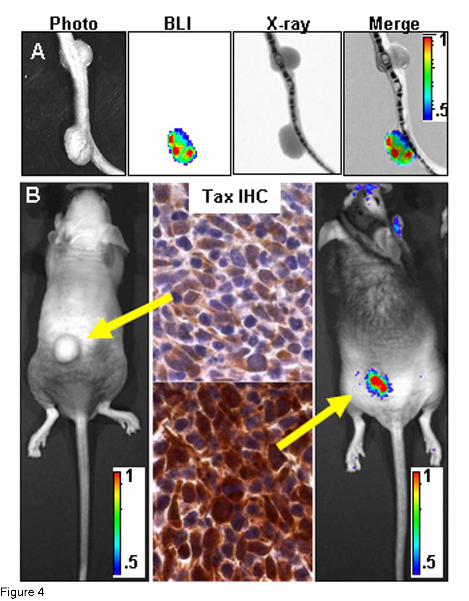
Figure 4: Bioluminescence correlates with Tax expression.
A) Co-registration of photograph, BLI, and X-ray images on tail tumors with differing bioluminescent
properties. B) Comparison of bioluminescence and TAX expression on tumors arising in two different
BNX animals injected with homogenate from a single bioluminescent primary TAX-LUC tumor.
For personal use only. by guest on November 28, 2013. bloodjournal.hematologylibrary.orgFrom
23
Arrows indicate correspondence between tumor BLI and TAX IHC. Color scale x104
photons/sec/cm2/sr.
Figure 5: Stochastic bioluminescence correlates with spontaneous tumorigensis. A) Summary
of physical exam and bioluminescent imaging data from 10 spontaneous lesions that developed in a
cohort of TAX-LUC animals over the period of a year. B) Frequency with which bioluminescence is
associated with various stages in tumor development. C) Frequency of bioluminescence within the
cohort over time increases as animals develop spontaneous tumors.
Figure 6: Bioluminescence identifies early stages in tumor development. Serial images of the
development of a spontaneous ear tumor in a TAX-LUC animal. Color indicates flux ≥ 0.3x10⁴
photons/sec/cm2/sr. B) Bioluminescence (arrow) generated by D-luciferin on an otherwise
unremarkable ear or tail juxtaposed to corresponding histology. H/E, Bar = 100 µm. Inset is TAX IHC
on a serial section. C) Palpable nodules that develop then resolve on the tails of TAX-LUC animals.
Histological sections through the nodules consistently involve inflammatory admixture but presence of
malignant LGLs is variable.
Figure 7: Model overview of tumorigenesis in TAX-LUC mice. Schematic summary of the
proposed mechanism of tumorigenesis and stages associated with bioluminescence. Photographs of
H/E stained tail sections representing stages of inflammation-induced tumorigenesis in this model.
Bar = 1 mm.
For personal use only. by guest on November 28, 2013. bloodjournal.hematologylibrary.orgFrom

For personal use only. by guest on November 28, 2013. bloodjournal.hematologylibrary.orgFrom

For personal use only. by guest on November 28, 2013. bloodjournal.hematologylibrary.orgFrom

For personal use only. by guest on November 28, 2013. bloodjournal.hematologylibrary.orgFrom
For personal use only. by guest on November 28, 2013. bloodjournal.hematologylibrary.orgFrom
For personal use only. by guest on November 28, 2013. bloodjournal.hematologylibrary.orgFrom
For personal use only. by guest on November 28, 2013. bloodjournal.hematologylibrary.orgFrom
For personal use only. by guest on November 28, 2013. bloodjournal.hematologylibrary.orgFrom