Extranodal NK/T-cell lymphoma: diagnosis and treatment cues
7
Review Article Extranodal NK/T-cell lymphoma: diagnosis and treatment cues Ritsuro Suzuki 1 * , Kengo Takeuchi 2 , Koichi Ohshima 3 and Shigeo Nakamura 4 1 Department of HSCT Data Management, Nagoya University, School of Medicine, Nagoya, Japan 2 Department of Pathology, The Cancer Institute of Japanese Foundation for Cancer Research, Tokyo, Japan 3 Department of Pathology, Kurume University, School of Medicine, Fukuoka, Japan 4 Department of Pathology, Nagoya University, School of Medicine, Nagoya, Japan *Correspondence to: Ritsuro Suzuki, Department of HSCT Data Management, Nagoya University, School of Medicine, 1-1-20 Daiko-Minami, Higashi-ku, Nagoya, 461-0047 Japan. E-mail: [email protected] Abstract Extranodal NK/T-cell lymphoma, nasal type (ENKL) is mostly endemic to East Asia. It predominantly occurs in the nasal or paranasal areas and less frequently in the skin. Most of the tumours show NK-cell, but rarely T-cell, phenotypes. The Epstein–Barr virus (EBV) genome can be usually detected in lymphoma cells. Geographic localization of ENKL matches the endemic distribution of EBV, suggesting that EBV plays an important role in lymphomagenesis. Originally, NK-cell and T-cell types were believed to present the same clinicopathologic characteristics, but recent data suggest more aggressive characteristics for the NK-cell phenotype. Although ENKL is sensitive to radiotherapy, it shows a poorer response to chemotherapeutic agents than other lymphomas due to expression of p-glycoprotein. Therefore, new therapeutic approaches must be considered. Several new clinical trials are now being conducted in East Asia. Copyright # 2008 John Wiley & Sons, Ltd. Keywords: natural killer cell; azurophilic granule; Epstein–Barr virus; CD56; cytotoxic molecule Received: 19 June 2007 Revised: 9 December 2007 Accepted: 31 December 2007 Introduction Extranodal NK/T-cell lymphoma (ENKL), nasal type most frequently affects the nose and paranasal area [1–3]. The immunophenotype of the lymphoma cells mostly reflects that of NK-cells, but sometimes is also characteristic of T-cells. In some cases differential diagnosis is difficult when only using paraffin embedded specimens. Therefore at present, the diagnostic term ‘NK/T-cell lymphoma’ is used. It should however be noted that no ‘NK/T-cell’ actually exists. This type of lymphoma shows a marked geographic preference for East Asia and Latin America. The incidence is also different within the endemic areas; in Asia, the rates of occurrence are: 3.3% in Japan [4], 5% in Taiwan [5], 6% in Hong Kong [6] and 8% in Korea [7]. In this review, we summarize the disease characteristics of ENKL of nasal type with special emphasis on diagnostic pitfalls. Ontogeny of NK-cells NK-cells were first defined as a functional subset of lymphocytes that mediate major histocompatibility complex-nonrestricted cytotoxicity [8]. They were later recognized to have large granular lymphocyte (LGL) morphology, germline configurations of T-cell receptor (TCR) and immunoglobulin genes and a surface CD3 (sCD3)-negative and CD56-positive phenotype [9]. From these findings, NK-cells are now regarded as a third lineage of lymphocytes that is distinct from T- and B-cells. Because NK-cells develop from T/NK bi-potential common progenitors (Figure 1) [10,11], they share many similarities with T-cells, particularly with cytotoxic T-cells. Therefore, the phenotypes of NK-cell and T-cell lymphoma/leukae- mia also have much in common, which makes it difficult to perform differential diagnosis [12–14]. Pathological transformation of NK-cells Myeloid antigen-positive T/NK bi-potential progenitors are believed to develop by transformation into myeloid/NK cell precursor acute leukaemia [15,16]. NK-cell lineage committed progenitors are also hypothesized to transform to blastic NK-cell lymphoma (BNKL) or precursor NK- cell acute lymphoblastic leukaemia/lymphoma (NK-ALL) [17,18]. Previously, CD4-positive and CD4-negative types of BNKL/NK-ALL were identified [18]. Although there remain several controversies regarding CD56 expression and dendritic cell lineage, the CD4 þ CD56 þ type of this tumour is somehow related to plasmacytoid dendritic cells or to the monocytic lineage [19,20], The CD4-negative type probably represents the true BNKL/NK-ALL. Two mature NK-cell neoplasms, ENKL [1] and aggressive NK-cell leukaemia [18d], are transformed from functionally Hematological Oncology Hematol Oncol 2008; 26: 66–72 Published online 19 February 2008 in Wiley InterScience (www.interscience.wiley.com) DOI: 10.1002/hon.847 Copyright ß 2008 John Wiley & Sons, Ltd.
-
Upload
independent -
Category
Documents
-
view
1 -
download
0
Transcript of Extranodal NK/T-cell lymphoma: diagnosis and treatment cues

Hematological OncologyHematol Oncol 2008; 26: 66–72Published online 19 February 2008 in Wiley InterScience
(www.interscience.wiley.com) DOI: 10.1002/hon.847Review Article
Extranodal NK/T-cell lymphoma:diagnosis and treatment cues
Ritsuro Suzuki1*, Kengo Takeuchi2, Koichi Ohshima3 and Shigeo Nakamura4
1Department of HSCT Data Management, Nagoya University, School of Medicine, Nagoya, Japan2Department of Pathology, The Cancer Institute of Japanese Foundation for Cancer Research, Tokyo, Japan3Department of Pathology, Kurume University, School of Medicine, Fukuoka, Japan4Department of Pathology, Nagoya University, School of Medicine, Nagoya, Japan
*Correspondence to:Ritsuro Suzuki, Department ofHSCT Data Management,Nagoya University, School ofMedicine, 1-1-20 Daiko-Minami,Higashi-ku, Nagoya, 461-0047Japan.E-mail:[email protected]
Copyright � 2008 John Wiley & Son
Abstract
Extranodal NK/T-cell lymphoma, nasal type (ENKL) is mostly endemic to East Asia. Itpredominantly occurs in the nasal or paranasal areas and less frequently in the skin. Mostof the tumours show NK-cell, but rarely T-cell, phenotypes. The Epstein–Barr virus (EBV)genome can be usually detected in lymphoma cells. Geographic localization of ENKLmatches the endemic distribution of EBV, suggesting that EBV plays an important role inlymphomagenesis. Originally, NK-cell and T-cell types were believed to present the sameclinicopathologic characteristics, but recent data suggest more aggressive characteristicsfor the NK-cell phenotype. Although ENKL is sensitive to radiotherapy, it shows apoorer response to chemotherapeutic agents than other lymphomas due to expressionof p-glycoprotein. Therefore, new therapeutic approaches must be considered. Several newclinical trials are now being conducted in East Asia. Copyright # 2008 John Wiley &Sons, Ltd.
Received: 19 June 2007Revised: 9 December 2007
Keywords: natural killer cell; azurophilic granule; Epstein–Barr virus; CD56; cytotoxicmolecule
Accepted: 31 December 2007Introduction
Extranodal NK/T-cell lymphoma (ENKL), nasal type most
frequently affects the nose and paranasal area [1–3]. The
immunophenotype of the lymphoma cells mostly reflects
that of NK-cells, but sometimes is also characteristic of
T-cells. In some cases differential diagnosis is difficult
when only using paraffin embedded specimens. Therefore
at present, the diagnostic term ‘NK/T-cell lymphoma’ is
used. It should however be noted that no ‘NK/T-cell’
actually exists. This type of lymphoma shows a marked
geographic preference for East Asia and Latin America.
The incidence is also different within the endemic areas; in
Asia, the rates of occurrence are: 3.3% in Japan [4], 5%
in Taiwan [5], 6% in Hong Kong [6] and 8% in Korea [7].
In this review, we summarize the disease characteristics of
ENKL of nasal type with special emphasis on diagnostic
pitfalls.
Ontogeny of NK-cells
NK-cells were first defined as a functional subset of
lymphocytes that mediate major histocompatibility
complex-nonrestricted cytotoxicity [8]. They were later
recognized to have large granular lymphocyte (LGL)
morphology, germline configurations of T-cell receptor
(TCR) and immunoglobulin genes and a surface CD3
s, Ltd.
(sCD3)-negative and CD56-positive phenotype [9]. From
these findings, NK-cells are now regarded as a third lineage
of lymphocytes that is distinct from T- and B-cells.
Because NK-cells develop from T/NK bi-potential common
progenitors (Figure 1) [10,11], they share many similarities
with T-cells, particularly with cytotoxic T-cells. Therefore,
the phenotypes of NK-cell and T-cell lymphoma/leukae-
mia also have much in common, which makes it difficult to
perform differential diagnosis [12–14].
Pathological transformation of NK-cells
Myeloid antigen-positive T/NK bi-potential progenitors
are believed to develop by transformation into myeloid/NK
cell precursor acute leukaemia [15,16]. NK-cell lineage
committed progenitors are also hypothesized to transform
to blastic NK-cell lymphoma (BNKL) or precursor NK-
cell acute lymphoblastic leukaemia/lymphoma (NK-ALL)
[17,18]. Previously, CD4-positive and CD4-negative types
of BNKL/NK-ALL were identified [18]. Although there
remain several controversies regarding CD56 expression
and dendritic cell lineage, the CD4þ CD56þ type of this
tumour is somehow related to plasmacytoid dendritic cells
or to the monocytic lineage [19,20], The CD4-negative
type probably represents the true BNKL/NK-ALL. Two
mature NK-cell neoplasms, ENKL [1] and aggressive
NK-cell leukaemia [18d], are transformed from functionally

Figure 1. Ontogeny of NK-cells and transformation to NK-cell malignancies. NK-cells are differentiated from stem cells throughmyeloid-antigen positive NK/T bi-potential progenitors and lineage-committed progenitors. Myeloid/NK cell precursor acuteleukaemia is transformed from the myeloid antigen-positive progenitor. Blastic NK-cell lymphoma and precursor NK-cell lymphoblasticleukaemia are derived from a relatively mature, NK-cell lineage committed progenitor. Two mature NK-cell neoplasms, aggressiveNK-cell leukaemia and extranodal NK-cell lymphoma, nasal type, are transformed from mature NK-cells
Extranodal NK/T-cell lymphoma 67
mature NK-cells. Aggressive NK-cell leukaemia is a
distinct leukaemic form of mature NK-cell malignancy
with frequent hepatosplenic involvement [21,22]. Although
these two diseases share many features, several clinico-
pathologic and phenotypic differences have been reported
[23]. Therefore, aggressive NK-cell leukaemia remains as
a distinct disease entity in the forthcoming World Health
Organization classifications. A summary of the clinico-
pathologic characteristics of NK-cell lineage neoplasms is
given in Table 1.
Clinical characteristics of extranodalNK-cell lymphoma, nasal type
The nose and paranasal area including the upper aero-
digestive tract contains the origin of more than 80% of
extranodal NK-cell lymphomas, nasal type. Macroscopic
findings by nasal endoscope are shown in Figure 2. Initial
complaints of ENKL, nasal type include local symptoms
such as nasal obstruction, discharge and bleeding. There-
after, as the disease extends, necrosis, swellingor bony
destruction of the nasal area develops. However, such
extreme local progressions are currently rare because of
early disease recognition and reference to specialized
physicians. The skin is the second most frequent organ of
origin, accounting for approximately 10% of cases [19].
Cases originating from the liver and/or spleen account
for 5% of ENKLs, nasal type. More rare organs of onset
include the lung, gastrointestinal tract, kidney, pancreas,
testis and brain. Nasal lymphomas more frequently present
as a localized disease (ratio 4:1), whereas lymphomas at
Copyright � 2008 John Wiley & Sons, Ltd.
other sites are more frequently detected at an advanced
stage (ratio 2:3) [24–35]. Because this lymphoma
essentially presents an extranodal origin, clinical stage III
is rare and most of the advanced stage cases are in stage IV.
Some cases show long-term limitation to the original site.
However, once the tumour develops outside the original
site, the disease rapidly progresses and disseminates.
Fever, haemophagocytosis and disseminated intravascular
coagulation are not rare in this situation. Several cases of
stage IV or aggressive NK-cell leukaemia could not be
treated because of the progression of the disease and poor
status of the patient [36].
Diagnosis of extranodal NK-celllymphoma, nasal type
Diagnosis of ENKL, nasal-type is based on histopathologic
examination of biopsy specimens, but is sometimes
difficult because of the existence of wide necrosis around
the tumour (Figure 3A) that is characterized by expression
of Fas and Fas ligand on the tumour cells [37]. Selection of
appropriate sites for biopsy is important for prompt
diagnosis, as are repeated approaches in case the speci-
mens only include necrotic tissue.
Histologically, tumour cells from ENKL generally show
angiocentric growth pattern (Figure 3A) [1,38]. The growth
pattern is such a notable feature of this lymphoma that the
diagnostic term used to be ‘angiocentric lymphoma’ [39].
In clinical practice, sampling error can prevent recognition
of angiocentricity; therefore this finding is currently not
mandatory for diagnosis [38]. The presence of cucumber-
Hematol Oncol 2008; 26: 66–72
DOI: 10.1002/hon
Table
1.
Clin
icopat
holo
gic
char
acte
rist
ics
of
NK
-cel
llin
eage
neo
pla
sms
Myeloid/N
Kcell
precursoracute
leukaemia
Blastic
NK-cell
lymphoma/Precursor
NK-celllymphoblastic
leukaemia
AggressiveNK-cell
leukaemia
ExtranodalNK
celllymphoma,nasaltype
Lim
itedstage
Advancedstage
Morpho
logy
Blastic
Blastic
LGL
LGL
Azurophilic
granule
��
þþ
Lymphno
deinvolvem
ent
þþ
þ�
þ/�
Extranodalinvolvem
ent
Bone
marrow,blood,mediastinum
Skin,bone
marrow
Bone
marrow,blood,liver,spleen
Nose,skin
Nose,skin,bone
marrow,blood
B-sym
ptom
Rare
Rare
Frequent
Rare
Frequent
Surfacemarker
CD7þ,CD33þ,CD34þ,CD56þ
CD4þ/�
,CD7þ,CD56þ,TdTþ
CD2þ,CD16þ,CD56þ
CD2þ,cyCD3þ,CD56þ
EBV
��
þ/�
þClinicalcourse
Aggressive
Aggressive
Aggressive
Sometimes
indolent
Aggressive
Therapy
AMLchem
otherapy
Chemotherapyforlympho
idneoplasm
sNostandardtherapy
Radiotherapyfollowed
by
chem
otherapy
Nostandardtherapy
Prognosis
Relapse
isfrequent,andthe
prognosisispoor.
Relapse
isfrequent,andthe
prognosisispoor.
Verypoor
Fair
Verypoor
LGL,
larg
egr
anula
rly
mphocy
te;EB
V,Epst
ein–B
arr
viru
s;A
ML,
acute
mye
loid
leuka
emia
.
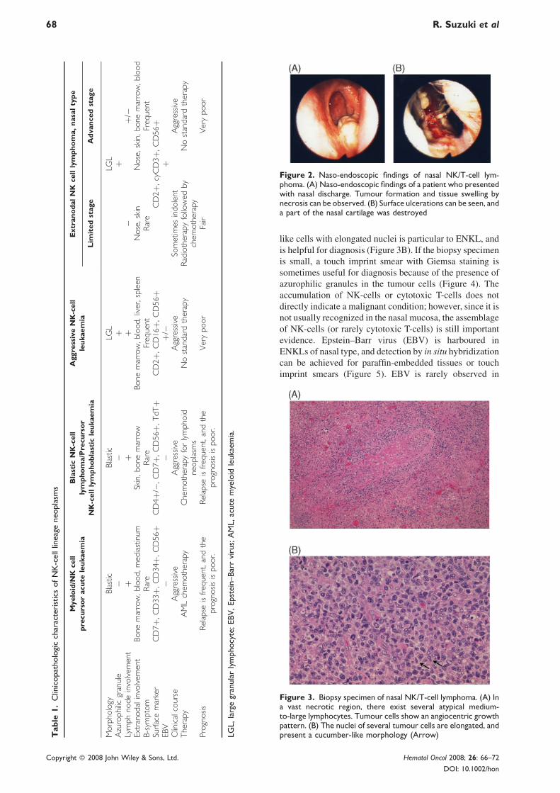
Figure 2. Naso-endoscopic findings of nasal NK/T-cell lym-phoma. (A) Naso-endoscopic findings of a patient who presentedwith nasal discharge. Tumour formation and tissue swelling bynecrosis can be observed. (B) Surface ulcerations can be seen, anda part of the nasal cartilage was destroyed
Figure 3. Biopsy specimen of nasal NK/T-cell lymphoma. (A) Ina vast necrotic region, there exist several atypical medium-to-large lymphocytes. Tumour cells show an angiocentric growthpattern. (B) The nuclei of several tumour cells are elongated, andpresent a cucumber-like morphology (Arrow)
68 R. Suzuki et al
Copyright � 2008 John Wiley & Sons, Ltd.
like cells with elongated nuclei is particular to ENKL, and
is helpful for diagnosis (Figure 3B). If the biopsy specimen
is small, a touch imprint smear with Giemsa staining is
sometimes useful for diagnosis because of the presence of
azurophilic granules in the tumour cells (Figure 4). The
accumulation of NK-cells or cytotoxic T-cells does not
directly indicate a malignant condition; however, since it is
not usually recognized in the nasal mucosa, the assemblage
of NK-cells (or rarely cytotoxic T-cells) is still important
evidence. Epstein–Barr virus (EBV) is harboured in
ENKLs of nasal type, and detection by in situ hybridization
can be achieved for paraffin-embedded tissues or touch
imprint smears (Figure 5). EBV is rarely observed in
Hematol Oncol 2008; 26: 66–72
DOI: 10.1002/hon

Figure 4. Touch imprint smear of ENKL. There are manyatypical NK-cells with prominent azurophilic granules
Extranodal NK/T-cell lymphoma 69
lymphocytes residing in normal or inflammatory nasal
mucosa or adjacent tissue; therefore detection is particu-
larly important for specimens that mostly consist of
necrotic tissue.
For ENKLs, histopathologic diagnosis of bone marrow
involvement is occasionally difficult. Detection of EBV is
also helpful in this situation [40]. Recently, the prognostic
significance of such occult or minute involvement has been
shown for early stage patients [41]. Routine examination of
bone marrow involvement by using EBV in situ hybrid-
ization is now recommended.
Immunophenotype of extranodal NK-celllymphoma, nasal type
Phenotypic markers expressed in ENKL include CD2,
cytoplasmic CD3 (cyCD3), CD7 and CD56, which also
represent the phenotype of normal NK-cells [25,42,43].
Cytotoxic molecules such as TIA-1, granzyme B and
perforin, are also positive in ENKL [37,44]. Table 1 shows
the differential diagnosis of mature NK-cell tumours. If
lymphoma cells are negative for these cytotoxic molecules
and show a T-cell phenotype, diagnosis of another type of
T-cell lymphoma should be considered. For differentiation
of NK-cell from T-cell lymphoma, expression of sCD3,
CD5 or TCRs on the lymphoma cells can be evaluated
[31,43,45], in addition to the rearrangement of TCR genes.
However, routine diagnostic use of these procedures
is sometimes difficult and unavailable. Previously, the
igure 5. Epstein–Barr virus small RNA (EBER) in-situ hybrid-ation (ISH). Because lymphoma cells harbour the EBV, they areositive for EBER-ISH
Fizp
Copyright � 2008 John Wiley & Sons, Ltd.
clinical features and prognosis of true T-cell nasal lym-
phoma were regarded to be similar to that of NK-cell type,
resulting in the adoption of the term ‘NK/T-cell lymphoma’.
It should be noted, however, that this nomenclature falsely
suggests existence of ‘NK/T-cells’. Another point to be
noted is that the use of the term ‘NK/T’ is restricted to
lymphomas occurring in the nasal/paranasal area, and is
not applied to lymphomas originated from extra-nasal
sites. Differential diagnosis from other types of T-cell
lymphomas is therefore required for the extra-nasal type of
extranodal NK-cell lymphoma.
Recently, studies with large numbers of patients
showed that the prognosis of nasal NK-cell lymphoma
is significantly poorer than that of nasal ‘T-cell’ lymphoma
[29,46]. Differential diagnosis of NK-cell lymphoma from
genuine T-cell lymphoma may therefore be required in
the future. A search for a diagnostic marker is therefore
warranted.
Diagnostic pitfalls for extranodal NK/T-cell lymphoma
Because CD56 is also expressed in a part of acute myeloid
leukaemia (AML) [47], differential diagnosis of NK-cell
malignancies from CD56-positive AML is occasionally
difficult, particularly for those with extramedullary or
cutaneous involvement. CD4þ CD56þ haematodermic
neoplasm also frequently shows cutaneous/subcutaneous
involvement [18,19]. In this context, CD56-positive AML
and CD4þ CD56þ haematodermic neoplasm are unex-
ceptionally negative for EBV, which is useful for the
differential diagnosis. Positive EBV status is thus required
for the diagnosis of NK/T-cell lymphomas.
Treatment of extranodalNK/T-cell lymphoma
Limited stages
For limited stages of usual aggressive non-Hodgkin
lymphoma, three to four courses of a chemotherapy
regimen that includes anthracycline, such as CHOP,
supplemented with involved field irradiation is regarded
as the standard therapy [48]. However, for nasal NK/T-cell
lymphoma, the overall 5-year survival rate using this
strategy is less than 50% [49,50]. Reasons include the
expression of the multidrug-resistant p-glycoprotein in
NK/T-cell lymphoma cells [51,52]. P-glycoprotein actively
exports doxorubicin and vincristine, which are the main
components of CHOP chemotherapy. Radiotherapy
remains effective but cannot prevent recurrence of the
disease outside the radiation field. The overall 5-year
survival rate therefore remains limited to 40–50% when
using radiotherapy alone [31,53,54].
Ribrag et al. treated eight patients in the limited stage of
nasal NK/T-cell lymphoma with radiotherapy followed by
chemotherapy and reported an excellent result (10 years
overall survival: 100%) [55]. They have concluded that a
sufficient dose of radiotherapy immediately after diagnosis
Hematol Oncol 2008; 26: 66–72
DOI: 10.1002/hon

70 R. Suzuki et al
is desirable for treatment of this disease. At present,
radiotherapy followed by chemotherapy is regarded as a
standard strategy for limited stage ENKL [31,56].
Yamaguchi et al. also reported excellent control of the
disease by simultaneous chemoradiotherapy using radi-
ation therapy and DeVIC chemotherapy (RT-DeVIC) [50].
Based on this finding, the Japanese Clinical Oncology
Group has conducted a phase I/II study of RT-DeVIC
chemoradiotherapy. The study is now closed with sufficient
numbers of patients registered. Its results are anticipated.
Advanced stages
The prognosis of advanced stage ENKL, nasal type, as well
as that of aggressive NK-cell leukaemia, is extremely poor
when using any chemotherapeutic regimen [22]. Aviles
et al. from Mexico reported the utility of sandwich
chemoradiotherapy, which consisted of three courses of
cyclophosphamide, methotrexate, etoposide and dexa-
methasone (CMED), radiotherapy and additional three
courses of CMED. The 5-year overall survival rate using
this method was reported as 65% [57]. This was an
excellent result, but the reported toxicities were surpris-
ingly low despite the relatively high dose of chemother-
apeutic drugs used. Therefore, confirmation through
replication is required.
Recently, several reports from East Asia suggest the
efficacy of L-asparaginase for treatment of mature NK/
T-cell lymphoma [58–60]. L-asparaginase is an enzyme
that digests serum L-asparagine and acts as an anti-tumour
agent through asparagine starvation of tumours with
low expression levels of asparagine synthetase [61,62].
Because L-asparaginase specifically acts on lymphoid
cells, myelosuppression by L-asparaginase is minimal.
L-asparaginase has long been regarded as a key drug for
paediatric acute lymphoblastic leukaemia. A Chinese
group treated nasal NK/T-cell lymphoma patients refrac-
tory to CHOP-like chemotherapy with a chemotherapy
regimen that consisted of L-asparaginase, vincristine and
dexamethasone supplemented by local radiotherapy. They
reported good results with a 5-year overall survival rate of
55.6% [63]. Likewise, L-asparaginase is effective for this
type of lymphoma but has many adverse reactions such as
haemostatic complications, allergy and pancreatitis. These
findings suggest a need for the establishment of safe
and effective chemotherapeutic regimens. The NK-cell
Tumour Study Group is now conducting clinical studies of
a novel L-asparaginase-containing chemotherapy for initial
stage IV, relapsed or refractory NK/T-cell leukaemia/
lymphoma [36]. This regimen is termed SMILE, and
consists of methotrexate, ifosfamide, etoposide, steroid
and L-asparaginase. A phase I dose finding study has been
completed [64] and we are now designing a subsequent
phase II study.
Haematopoietic stem celltransplantation (HSCT)
Because the prognosis of ENKL is poor, there exist several
reports of upfront autologous HSCT. In large-scale reports
Copyright � 2008 John Wiley & Sons, Ltd.
from Japan and Korea, long-term survival ranges from
50 to 70% [65–67]. However, retrospective analysis might
be biased by patient selection. Prospective clinical trials
are thus warranted before concluding that autologous
HSCT is effective for ENKL.
On the other hand, allogeneic HSCT can also be applied
for the treatment and is the only curative strategy for
advanced stage or nonremission patients. Two large-scale
analyses from Japan included high-risk patients and
reported a long-term survival rate ranging from 30 to
40% [66,68]. The second study included patients who
received reduced intensity stem cell transplants (RIST),
and both reports indicated the absence of late recurrence at
2 years post-transplantation. These findings suggest the
curative potential of allogeneic HSCT, but patient selection
bias is also possible. Since many types of stem cell sources
are now utilized for HSCT including cord blood and
mismatched donors, further accumulation of data and
prospective evaluations are also required.
Clinical significance of theEpstein–Barr virus
It is well-known that patient sera from EBV-positive
malignancies contain fragmented viral DNA [69,70].
Measurement of the circulating viral DNA load in
peripheral blood is useful for diagnosis, monitoring and
prognostication of the disease. However, detection is
sometimes misunderstood as the presence of viral particle
itself; rather, the detected DNA is derived from dead
tumour cells. For these reasons, most detected fragments
are less than 500 bp in length, and longer fragments or the
entire EBV genome are never detected [71]. EBV-DNA
can therefore be used as a marker to predict the tumour
burden [72,73], but prediction can potentially be affected
by the presence of EBV unrelated to the lymphoma. There
are several choices of source tissue for analysis including
plasma, total blood and mononuclear cells, and each choice
represents a different outcome [74]. The significance of
the viral load in peripheral blood and the choice of source
tissue used for analysis should be examined prospectively.
Conclusion
Several new insights have been recently developed for
extranasal NK/T-cell lymphoma, nasal type. Diagnosis is
thus becoming easier. However, the prognosis is particu-
larly poor in both the limited and advanced stages.
Appropriate therapeutic strategies should be explored by
prospective studies.
Acknowledgements
The authors are grateful to Drs. Kazuo Oshimi (Juntendo Uni-
versity School of Medicine), Junji Suzumiya (Fukuoka University
Chikushi Hospital), Motoko Yamaguchi (Mie University School
of Medicine) and Koji Izutsu (University of Tokyo) for critical
reading of the manuscript.
Hematol Oncol 2008; 26: 66–72
DOI: 10.1002/hon

Extranodal NK/T-cell lymphoma 71
References
1. Chan JKC, Jaffe ES, Ralfkiaer E. Extranodal NK/T-cell lym-
phoma, nasal type. In World Health Organization Classification
of Tumors. Pathology and Genetics of Tumours of Haematopoie-
tic and Lymphoid Tissues, Jaffe ES, Harris NL, Stein H, Vardi-
man JW (eds). IARC Press: Lyon, France, 2001; 204–207.
2. Jaffe ES. Classification of natural killer (NK) cell and NK-like
T-cell malignancies. Blood 1996; 87: 1207–1210.
3. Oshimi K. Leukemia and lymphoma of natural killer lineage
cells. Int J Hematol 2003; 78: 18–23.
4. Lymphoma Study Group of Japanese Pathologists. The World
Health Organization classification of malignant lymphomas in
Japan: incidence of recently recognized entities. Pathol Int 2000;
50: 696–702.
5. Chen CY, Yao M, Tang JL, et al. Chromosomal abnormalities of
200 Chinese patients with non-Hodgkin’s lymphoma in Taiwan:
with special reference to T-cell lymphoma. Ann Oncol 2004; 15:
1091–1096.
6. Au WY, Ma SY, Chim CS, et al. Clinicopathologic features and
treatment outcome of mature T-cell and natural killer cell
lymphomas diagnosed according to the World Health Organiz-
ation classification scheme: a single center experience of ten
years. Ann Oncol 2005; 16: 206–214.
7. Ko YH, Kim CW, Park CS, et al. REAL classification of
malignant lymphomas in the Republic of Korea: incidence of
recently recognized entities and changes in clinicopathologic
features. Cancer 1998; 83: 806–812.
8. Hercend T, Schmidt RE. Characteristics and uses of natural killer
cells. Immunol Today 1998; 9: 291–293.
9. Robertson MJ, Ritz J. Biology and clinical relevance of human
natural killer cells. Blood 1990; 76: 2421–2438.
10. Sanchez MJ, Muench MO, Roncarolo MG, Lanier LL, Phillips
JH. Identification of a common T/natural killer cell progenitor in
human fetal thymus. J Exp Med 1994; 180: 569–576.
11. Shibuya A, Nagayoshi K, Nakamura K, Nakauchi H. Lympho-
kine requirement for the generation of natural killer cells from
CD34þ hematopoietic progenitor cells. Blood 1995; 85:
3538–3546.
12. Ishii Y, Yamanaka N, Ogawa K, et al. Nasal T-cell lymphoma as a
type of so-called ‘‘lethal midline granuloma’’. Cancer 1982; 50:
2336–2344.
13. Chan JK, Ng CS, Lau WH, Lo ST. Most nasal/nasopharyngeal
lymphomas are peripheral T-cell neoplasms. Am J Surg Pathol
1987; 11: 418–429.
14. Ng CS, Chan JK, Lo ST. Expression of natural killer cell markers
in non-Hodgkin’s lymphomas. Hum Pathol 1987; 18: 1257–
1262.
15. Suzuki R, Yamamoto K, Seto M, et al. CD7þ and CD56þ
myeloid/natural killer cell precursor acute leukemia: A distinct
hematolymphoid disease entity. Blood 1997; 90: 2417–2428.
16. Suzuki R, Murata M, Kami M, et al. Prognostic significance of
CD7þ CD56þ phenotype and chromosome 5 abnormalities for
acute myeloid leukemia M0. Int J Hematol 2003; 77: 482–489.
17. Suzuki R, Nakamura S. Malignancies of natural killer (NK) cell
precursor: myeloid/NK cell precursor acute leukemia and blastic
NK cell lymphoma/leukemia. Leuk Res 1999; 23: 615–624.
18. Suzuki R, Nakamura S, Suzumiya J, et al. Blastic natural killer
cell lymphoma/leukemia (CD56-positive blastic tumor): prog-
nostication and categorization according to anatomic sites of
involvement. Cancer 2005; 104: 1022–1031.
19. Feuillard J, Jacob MC, Valensi F, et al. Clinical and biologic
features of CD4þ CD56þ malignancies. Blood 2002; 99:
1556–1563.
20. Petrella T, Comeau MR, Maynadie M, et al. ‘Agranular CD4þCD56þ hematodermic neoplasm’ (blastic NK-cell lymphoma)
originates from a population of CD56þ precursor cells related to
plasmacytoid monocytes. Am J Surg Pathol 2002; 26: 852–862.
21. Chan JKC, Wong KF, Jaffe ES, Ralfkiaer E. Aggressive NK-cell
leukemia. In World Health Organization classification of tumors.
Copyright � 2008 John Wiley & Sons, Ltd.
Pathology and genetics of tumours of haematopoietic and lym-
phoid tissues, Jaffe ES, Harris NL, Stein H, Vardiman JW (eds).
IARC Press: Lyon, France, 2001; 198–200.
22. Suzuki R, Suzumiya J, Nakamura S, et al. Aggressive natural
killer (NK)-cell leukemia revisited: large granular lymphocyte
leukemia of cytotoxic NK cells. Leukemia 2004; 18: 763–770.
23. Suzuki R, Suzumiya J, Nakamura S, Yamaguchi M, Kawa K,
Oshimi K. Natural killer (NK)-cell neoplasms: aggressive
NK-cell leukemia and extranodal NK-cell lymphoma, nasal type.
Ann Oncol 2005; 16 (Suppl. 5): v129–v130 [Abstract # 315].
24. Liang R, Todd D, Chan TK, et al. Treatment outcome and
prognostic factors for primary nasal lymphoma. J Clin Oncol
1995; 13: 666–670.
25. Emile J-F, Boulland M-L, Haioun C, et al. CD5- CD56þ T-cell
receptor silent peripheral T-cell lymphomas are natural killer cell
lymphomas. Blood 1996; 87: 1466–1473.
26. Nakamura S, Katoh E, Koshikawa T, et al. Clinicopathologic
study of nasal T/NK-cell lymphoma among the Japanese. Pathol
Int 1997; 47: 38–53.
27. Kwong YL, Chan ACL, Liang R, et al. CD56þ NK lymphomas:
clinicopathological features and prognosis. Br J Haematol 1997;
97: 821–829.
28. Logsdon MD, Ha CS, Kavadi VS, Cabanillas F, Hess MA, Cox
JD. Lymphoma of the nasal cavity and paranasal sinuses:
improved outcome and altered prognostic factors with combined
modality therapy. Cancer 1997; 80: 477–488.
29. Cheung MMC, Chan JK, Lau WH, et al. Primary non-Hodgkin’s
lymphoma of the nose and nasopharynx: clinical features, tumor
immunophenotype, and treatment outcome in 113 patients.
J Clin Oncol 1998; 16: 70–77.
30. Ko YH, Ree HJ, Kim WS, Choi WH, Moon WS, Kim SW.
Clinicopathologic and genotypic study of extranodal nasal-type
natural killer/T-cell lymphoma and natural killer precursor lym-
phoma among Koreans. Cancer 2000; 89: 2106–2116.
31. Li CC, Tien HF, Tang JL, et al. Treatment outcome and pattern of
failure in 77 patients with sinonasal natural killer/T-cell or T-cell
lymphoma. Cancer 2004; 100: 366–375.
32. Chim CS, Ma SY, Au WY, et al. Primary nasal natural killer cell
lymphoma: long-term treatment outcome and relationship
with the international prognostic index. Blood 2004; 103:
216–221.
33. You JY, Chi KH, Yang MH, et al. Radiation therapy versus
chemotherapy as initial treatment for localized nasal natural
killer (NK)/T-cell lymphoma: a single institute survey in Taiwan.
Ann Oncol 2004; 15: 618–625.
34. Kim TM, Park YH, Lee SY, et al. Local tumor invasiveness is
more predictive of survival than International Prognostic Index in
stage IE/IIE extranodal NK/T-cell lymphoma, nasal type. Blood
2005; 106: 3785–3790.
35. Lee J, Suh C, Park YH, et al. Extranodal natural killer T-cell
lymphoma, nasal-type: a prognostic model from a retrospective
multicenter study. J Clin Oncol 2006; 24: 612–618.
36. Suzuki R. Leukemia and lymphoma of natural killer cells. J Clin
Exp Hematop 2005; 45: 51–70.
37. Ohshima K, Suzumiya J, Shimazaki K, et al. Nasal T/NK cell
lymphomas commonly express perforin and Fas ligand: import-
ant mediators of tissue damage. Histopathology 1997; 31:
444–450.
38. Jaffe ES, Chan JKC, Su I-J, et al. Report of the workshop on nasal
and related extranodal angiocentric T/natural killer cell lympho-
mas. definitions, differential diagnosis, and epidemiology. Am J
Surg Pathol 1996; 20: 103–111.
39. Harris NL, Jaffe ES, Stein H, et al. A revised Europea-
n-American classification of lymphoid neoplasms: a proposal
from the International Lymphoma Study Group. Blood 1994; 84:
1361–1392.
40. Wong KF, Chan JKC, Cheung MMC, So JC. Bone marrow
involvement by nasal NK cell lymphoma at diagnosis is uncom-
mon. Am J Clin Pathol 2001; 115: 266–270.
41. Lee J, Suh C, Huh J, et al. Effect of positive bone marrow EBV in
situ hybridization in staging and survival of localized extranodal
Hematol Oncol 2008; 26: 66–72
DOI: 10.1002/hon

72 R. Suzuki et al
natural killer/T-cell lymphoma, nasal-type. Clin Cancer Res
2007; 13: 3250–3254.
42. Suzumiya J, Takeshita M, Kimura N, et al. Expression of adult
and fetal natural killer cell markers in sinonasal lymphomas.
Blood 1994; 83: 2255– 2260.
43. Yamaguchi M, Ohno T, Oka K, et al. Discordant reaction of Leu4
and rabbit anti-human CD3 epsilon in sinonasal ‘T’-cell lym-
phoma. Int J Hematol 1993; 59: 25–30.
44. Mori N, Yatabe Y, Oka K, et al. Expression of perforin in nasal
lymphoma. Am J Pathol 1996; 149: 699–705.
45. Chan JKC, Tsang WY, Pau MY. Discordant CD3 expression in
lymphomas when studied on frozen and paraffin sections. Hum
Pathol 1995; 26: 1139–1143.
46. Kim GE, Koom WS, Yang WI, et al. Clinical relevance of three
subtypes of primary sinonasal lymphoma characterized by
immunophenotypic analysis. Head Neck 2004; 26: 584–593.
47. Seymour JF, Pierce SA, Kantarjian HM, Keating MI, Estey EH.
Investigation of karyotypic, morphologic and clinical features in
patients with acute myeloid leukemia blast cells expressing the
neural cell adhesion molecule (CD56). Leukemia 1994; 8:
823–826.
48. Miller TP, Dahlberg S, Cassady JR, et al. Chemotherapy alone
compared with chemotherapy plus radiotherapy for localized
intermediate- and high-grade non-Hodgkin’s lymphoma. N Engl
J Med 1998; 339: 21–26.
49. Kim WS, Song SY, Ahn YC, et al. CHOP followed by involved
field radiation: is it optimal for localized nasal natural killer/
T-cell lymphoma? Ann Oncol 2001; 12: 349–352.
50. Yamaguchi M, Ogawa S, Nomoto Y, et al. Treatment outcome of
nasal NK-cell lymphoma: a report of 12 consecutively-diagnosed
cases and a review of the literature. J Clin Exp Hematop 2001;
41: 93–99.
51. Yamaguchi M, Kita K, Miwa H, et al. Frequent expression of
P-glycoprotein/MDR1 by nasal T-cell lymphoma cells. Cancer
1995; 76: 2351–2356.
52. Egashira M, Kawamata N, Sugimoto K, Kaneko T, Oshimi K.
P-glycoprotein expression on normal and abnormally expanded
natural killer cells and inhibition of P-glycoprotein function by
cyclosporin A and its analogue, P SC83. Blood 1999; 93:
599–606.
53. Kim GE, Cho JH, Yang WI, et al. Angiocentric lymphoma of the
head and neck: patterns of systemic failure after radiation treat-
ment. J Clin Oncol 2000; 18: 54–63.
54. Isobe K, Uno T, Tamaru J, et al. Extranodal natural killer/T-cell
lymphoma, nasal type: the significance of radiotherapeutic
parameters. Cancer 2006; 106: 609–615.
55. Ribrag V, Ell Hajj M, Janot F, et al. Early locoregional high-dose
radiotherapy is associated with long-term disease control in
localized primary angiocentric lymphoma of the nose and naso-
pharynx. Leukemia 2001; 15: 1123–1126.
56. Li YX, Yao B, Jin J, et al. Radiotherapy as primary treatment for
stage IE and IIE nasal natural killer/T-cell lymphoma. J Clin
Oncol 2006; 24: 181–189.
57. Aviles A, Neri N, Fernandez R, Calva A, Huerta-Guzman J,
Nambo MJ. Nasal NK/T-cell lymphoma with disseminated dis-
ease treated with aggressive combined therapy. Med Oncol 2003;
20: 13–17.
Copyright � 2008 John Wiley & Sons, Ltd.
58. Nagafuji K, Fujisaki T, Arima F, Ohshima K. L-asparaginase
induced durable remission of relapsed nasal NK/T-cell lym-
phoma after autologous peripheral blood stem cell transplan-
tation. Int J Hematol 2001; 74: 447–450.
59. Obama K, Tara M, Niina K. L-asparaginase-based induction
therapy for advanced extranodal NK/T-cell lymphoma. Int J
Hematol 2003; 78: 248–250.
60. Matsumoto Y, Nomura K, Kanda-Akano Y, et al. Successful
treatment with Erwinia L-asparaginase for recurrent natural
killer/T cell lymphoma. Leuk Lymphoma 2003; 44: 879–882.
61. Pinheiro JP, Boos J. The best way to use asparaginase in child-
hood acute lymphatic leukaemia–still to be defined? Br J Hae-
matol 2004; 125: 117–127.
62. Pui CH, Evans WE. Treatment of acute lymphoblastic leukemia.
N Engl J Med 2006; 354: 166–178.
63. Yong W, Zheng W, Zhang Y, et al. L-asparaginase-based regimen
in the treatment of refractory midline nasal/nasal-type T/NK-cell
lymphoma. Int J Hematol 2003; 78: 163–167.
64. Yamaguchi M, Suzuki R, Kwong YL, et al. Phase I study of
SMILE chemotherapy for advanced-stage or relapsed/refractory
extranodal NK/T-cell lymphoma/leukemia. Cancer Sci 2008 (in
press).
65. Au WY, Lie AK, Liang R, et al. Autologous stem cell trans-
plantation for nasal NK/T-cell lymphoma: a progress report on its
value. Ann Oncol 2003; 14: 1673–1676.
66. Suzuki R, Suzumiya J, Nakamura S, et al. Hematopoietic stem
cell transplantation for natural killer-cell lineage neoplasms.
Bone Marrow Transplant 2006; 37: 425–431.
67. Kim HJ, Bang SM, Lee J, et al. High-dose chemotherapy with
autologous stem cell transplantation in extranodal NK/T-cell
lymphoma: a retrospective comparison with non-transplantation
cases. Bone Marrow Transplant 2006; 37: 819–824.
68. Murashige N, Kami M, Kishi Y, et al. Allogeneic haematopoietic
stem cell transplantation as a promising treatment for natural
killer-cell neoplasms. Br J Haematol 2005; 130: 561–567.
69. Harabuchi Y, Yamanaka N, Kataura A, et al. Epstein–Barr virus
in nasal T-cell lymphomas in patients with lethal midline gran-
uloma. Lancet 1990; 335: 128–130.
70. Chan JK, Yip TT, Tsang WY, et al. Detection of Epstein–Barr
viral RNA in malignant lymphomas of the upper aerodigestive
tract. Am J Surg Pathol 1994; 18: 938–946.
71. Chan KCA, Zhang J, Chan ATC, et al. Molecular characteriz-
ation of circulating EBV DNA in the plasma of nasopharyngeal
carcinoma and lymphoma patients. Cancer Res 2003; 63:
2028–2032.
72. Lei KIK, Chan LYS, Chan W-Y, Johnson PJ, Dennis Lo YM.
Diagnostic and prognostic implications of circulating cell-free
Epstein–Barr virus DNA in natural killer/T-cell lymphoma. Clin
Cancer Res 2002; 8: 29–34.
73. Au WY, Pang A, Choy C, Chim CS, Kwong YL. Quantification
of circulating Epstein–Barr virus (EBV) DNA in the diagnosis
and monitoring of natural killer cell and EBV-positive lympho-
mas in immunocompetent patients. Blood 2004; 104: 243–249.
74. Stevens SJC, Pronk I, Middeldorp JM. Toward standardization of
Epstein–Barr virus DNA load monitoring: unfractionated whole
blood as preferred clinical specimen. J Clin Microbiol 2001; 39:
1211–1216.
Hematol Oncol 2008; 26: 66–72
DOI: 10.1002/hon