Dendritic cells, monocytes and macrophages: a unified nomenclature based on ontogeny

NK cell activity in tuberculosis is associated with impaired CD11aand ICAM-1 expression: a regulatory role of monocytes in NK
activation
Introduction
A protective immune response against Mycobacterium
tuberculosis depends on interferon (IFN)-c production by
CD4+ and CD8+ T cells.1 However, the early production
of IFN-c by cells of the innate immune response at
inflammatory sites can regulate innate resistance by acti-
vating phagocytic cells and priming antigen-presenting
cells (APCs) for interleukin (IL)-12 production, thus sha-
ping adaptive immunity towards the T helper type 1
(Th1) response necessary for elimination of many intra-
cellular pathogens. Natural killer (NK) cells are critical
components of the innate immune response that lack
expression of the T-cell receptor (TCR)–CD3 complex
and surface immunoglobulins but express CD56 antigen.
NK cells are characterized by potent cytotoxic activity
against tumours, virus-infected cells and intracellular
parasites.2 Resting NK cells circulate in the blood and,
once activated, they are able to migrate to and infiltrate
sites of infection where target cells are localized. The
interaction of NK with target cells is mediated by various
cell surface molecules, some involved in cell adhesion,
others activating the NK cytolytic programme, and oth-
ers inhibiting this activation by negative signalling.2 The
best studied activation receptor is the FccRIII (CD16)
molecule through which NK cells mediate antibody-
dependent cellular cytotoxicity (ADCC) against target
cells coated with immunoglobulin G (IgG).2 Although
not restricted to NK cells, integrins [CD11a/CD18, lym-
phocyte function-associated antigen (LFA)-2 and LFA-3]
Pablo Schierloh,1 Mercedes
Aleman,1 Noemı Yokobori,1
Leandro Alves,2 Nicolas Roldan,2
Eduardo Abbate,2 Marıa del C.
Sasiain1 and Silvia de la Barrera1
1Immunology Department, Institute of Haemo-
tology Research (IIHema), National Academy
of Medicine and 2Pneumonology Division,
Hospital F. J. Muniz, Buenos Aires, Argentina
doi:10.1111/j.1365-2567.2005.02259.x
Received 6 April 2005; revised 22 July 2005;
accepted 18 August 2005.
Correspondence: Dr Marıa del Carmen
Sasiain, IIHema. Inmunolog’a, Academia
Nacional de Medicina, Pacheco de Melo
3081, 1425 Buenos Aires, Argentina.
Email: [email protected]
Senior author: Dr M. Sasiain
Summary
Although the role of natural killer (NK) cells in mycobacterial infections
is unclear, it has been postulated that they contribute to protective immu-
nity through the production of interferon (IFN)-c. In this study, we
evaluate the effect of interleukin (IL)-10, IL-15 and IL-18 on NK lytic
activity through the expression of CD16, CD11a and CD69 molecules and
the induction of IFN-c production in patients with tuberculosis (TB) and
healthy individuals (N). Our results showed an impairment of NK lytic
activity and a gradual down-regulation of costimulatory and adhesion
molecules on NK cells which were dependent on the severity of the dis-
ease. NK lytic activity was increased by exogenous IL-15 and IL-18 in
both TB and N, and by neutralization of endogenous IL-10 only in TB;
IL-15 and IL-18 increased CD69 receptor expression, while anti-IL-10 up-
regulated CD16 and CD11a expression in TB. Mycobacterium tuberculosis
reduced the number of intracellular adhesion molecule (ICAM)-1+ CD14+
cells, but in the presence of IL-15, IL-18 and anti-IL-10 its expression was
up-regulated. In cells from TB patients, the observed effects of IL-15 and
IL-18 on NK function were not dependent on IL-10 modulation of the
surface expression of activator/adhesion molecules. In the absence of
monocytes, IL-10 activated NK cells, suggesting an indirect effect on their
function. Furthermore, in TB patients the depletion of monocytes
increased the production of IFN-c by NK cells. Therefore, monocytes
from TB patients regulated the NK function involving IL-10 which,
through an indirect mechanism, led to the down-regulation of costimula-
tory/adhesion molecules and/or IFN-c production.
Keywords: natural killer cells; cytotoxicity; receptors; monocytes; tuber-
culosis
� 2005 Blackwell Publishing Ltd, Immunology, 116, 541–552 541
IMMUNOLOGY OR IG INAL ART ICLE

which bind to intracellular adhesion molecule (ICAM)-1
(or CD54), ICAM-2 and ICAM-3 ligands have also been
implicated in NK cell adhesion to target cells, degranula-
tion and cytokine production.3,4 Also, the CD69 mole-
cule, which is rapidly acquired following activation and
belongs to the family of C-type lectin receptors bearing
strong similarity to the NK receptor CD94,5,6 has been
implicated in the cytotoxic activity and costimulation of
cytokine production of activated NK cells.7 It is unclear
which mechanisms contribute to the priming phase of
NK cell activation, but these cells can be rapidly activa-
ted in the periphery by chemokines in conjunction with
IL-2 and macrophage or dendritic cell (DC)-derived cyto-
kines such as IL-12, IL-15 and IL-18.8,9
Bacterial products activate macrophages to produce IL-
12, IL-15 and IL-18, playing a central role in the type 1
cytokine response and NK activity.8,10,11 NK cell cytokine
production is induced by IL-12 in synergy with IL-15 and
IL-18,8,12,13 which stimulates IFN-c production by NK
cells via different intracellular pathways.13–15 Considering
that protection against intracellular pathogens is critically
dependent on the function of NK cells at early stages of
the immune response and on type-1 cells at later stages,
the aim of the present study was to investigate the
costimulatory molecules involved in M. tuberculosis-
induced NK function through IFN-c production in
patients with tuberculosis. The role of IL-15 and IL-18
was also investigated. Our results show that, in patients
with tuberculosis, monocytes regulate NK activity.
Materials and methods
Patients
A total of 42 male patients with pulmonary tuberculosis
(TB) were included in the study. Patients were diagnosed
by the presence of recent clinical symptoms of TB, a posit-
ive sputum smear test for acid-fast bacilli confirmed by
a positive culture of TB bacilli and an abnormal chest
radiography. Informed consent was obtained from patients
according to the Hospital Francisco J. Muniz Ethics Com-
mittee. All patients had active TB and were under multi-
drug treatment at the time of study (0–15 days).
Pulmonary disease was classified according to the extent
and type of X-ray findings as mild (M) or advanced (A)
TB according to the American Tuberculosis Society cri-
teria. Patients with diabetes, chronic renal failure or malig-
nant diseases, as well as those who tested positive for
human immunodeficiency virus (HIV) or other con-
current infectious diseases, were excluded. Patients were
classified into two groups: (i) patients with moderate
tuberculosis (M-TB; n ¼ 22; age range 25–55 years) and
(ii) patients with advanced tuberculosis (A-TB; n ¼ 20,
age range 22–60 years). Fourteen healthy individuals (age
range 25–60 years) were included as controls.
Mononuclear cells
Peripheral blood mononuclear cells (PBMC) were iso-
lated from heparinized blood by Ficoll-Hypaque gra-
dient centrifugation16 and then suspended in RPMI 1640
(Gibco Laboratory, New York, NY) containing gentamy-
cin (85 lg/ml) and 15% heat-inactivated fetal calf serum
(FCS) (Gibco) (complete medium).
Monocyte-depleted PBMC
In order to obtain monocyte-depleted cell suspensions,
PBMC (5 · 106 cells/well) were incubated on the bot-
toms of 24-well Falcon plates for 2 hr at 37� to allow
cells to adhere to the plastic (85–95% of monocytes).
After removal of adherent cells, non-adherent lympho-
cytes were washed and suspended in complete medium.
CD14+ monocyte-depleted cell suspensions were
obtained by magnetic methods by treatment of PBMC
with anti-CD14 monoclonal antibody (Immunotech,
Marseille, France) for 30 min at 4� and then with goat
anti-mouse IgG-coated beads (Dynal, Oslo, Norway).
Monocyte-depleted cell suspensions (both adherent-
depleted and CD14-depleted populations) were suspen-
ded in complete medium, ensuring that the proportion
of each lymphocyte subset was the same as in total cul-
tured PBMC in order to allow comparison of NK lytic
activities. The proportion of contaminated monocytes in
cell suspensions depleted by adherence was 1–2% and
that in cell suspensions depleted by magnetic methods
was 0�5–0�6%.
Antigen
The c-irradiated M. tuberculosis H37-Rv strain used in this
study was kindly provided by Dr J. T. Belisle (Colorado
University, Denver, CO). Mycobacteria were suspended in
phosphate-buffered saline (PBS) free of pyrogen, sonicated
and adjusted at a concentration of 1 · 108 bacteria/ml.
PBMC culture
PBMC (2 · 106 cells/ml) or monocyte-depleted PBMC
(1�9 · 106 cells/ml) were cultured in Falcon 2063 tubes
(Becton Dickinson, Lincoln, NJ) at 37� in a humidified 5%
CO2 atmosphere, in complete medium with or without
M. tuberculosis (1 · 106 bacteria/ml), IL-15 (10 ng/ml),
IL-18 (20 ng/ml), IL-10 (10 ng/ml) or a monoclonal anti-
body specific for human IL-10 (10 ng/ml) (Peprotech,
Rocky Hill, NJ). After 24 hr of incubation, M. tuberculosis-
stimulated and/or cytokine-treated and control cells were
washed three times with RPMI 1640, suspended in com-
plete medium (2 · 106 cells/ml) and tested for cytotoxic
activity, expression of surface antigens and IFN-c produc-
tion by flow cytometry. An isotype-matched non-relevant
542 � 2005 Blackwell Publishing Ltd, Immunology, 116, 541–552
P. Schierloh et al.
control IgG1/j antibody for anti-IL10 was also tested and
was found to have no significant effect on cytotoxicity,
cytokine production or surface antigen expression.
Cytotoxic assay
Freshly isolated (ex vivo) and cultured PBMC or mono-
cyte-depleted PBMC were added in triplicate at different
effector to target cell ratios, in a final volume of 200 ll, to51Cr-labelled K562 target cells (5 · 103 cells seeded into
each well of 96-well microtitre plates; Coming incorpo-
rated (NY) Corning, USA). Plates were centrifuged at 50 g
for 5 min and incubated at 37� in 5% CO2 for 4 hr. After
centrifugation at 200 g for 5 min, 100 ll of supernatant
was removed from each well. The radioactivity of superna-
tants and pellets was measured in a gamma counter.
Results were expressed as percentage of cytotoxicity (%
Cx):
% Cx ¼ ½ðc.p.m. experimental release
� c.p.m. spontaneous releaseÞ=ðc.p.m. total release
� c.p.m. spontaneous release� � 100;
where c.p.m. is counts per minute. The radioactivity
released from target cells incubated with complete med-
ium alone was considered to represent spontaneous
release. It ranged from 5 to 10%.
Results expressed in lytic units (LU) for 107 effector
cells were calculated by defining 1 LU of cytotoxic activity
as the number of effector cells required to lyse 30% of
K562 target cells. These cell numbers were readily
obtained from the dose–response curves.
Immunofluorescence analysis
Expression of CD16, CD11a and CD69 on CD3– CD56+
lymphocytes and ICAM-1 on CD14+ cells. In order to
evaluate the expression of CD16, CD11a and CD69 anti-
gens on CD3– CD56+ NK cells, freshly isolated (ex vivo),
cultured or monocyte-depleted PBMC were incubated for
30 min at 4� with the following anti-human antibodies:
Cy5PE-CD3 (eBioscience, San Diego, CA), PE-CD56
(Immunotech), FICT-CD16 (Ancell, Bayport, MN), FICT-
CD11a (Caltag, Burlingam, CA) or FICT-CD69 (Ancell).
ICAM-1 was evaluated in control and M. tuberculosis-
stimulated PBMC with or without IL-15, IL-18 and anti-
IL-10 for 18 hr and stained with PE-CD54 (eBioscience)
and FITC-CD14 (Ancell). PECy5-, FITC- or PE-labelled-
isotype matched antibodies were also tested to evaluate
non-specific staining. Stained cells were analysed by flow
cytometry by acquiring 10 000–20 000 events, and gates
were set to forward and side-scatter to exclude cell debris
and apoptotic cells. Results are expressed as the percent-
age of positive cells or mean fluorescence intensity
(MFI).
Measurement of IFN-c+ cells
To determine the expression of intracytoplasmic IFN-c in
control and M. tuberculosis-stimulated CD3– CD56+ cells,
PBMC or monocyte-depleted PBMC were cultured for
24 hr with or without M. tuberculosis and/or IL-15 or
IL-18. Brefreldin A (5 lg/ml; Sigma, St Louis, MO) was
added for the final 4 hr to block IFN-c secretion. Cells
were washed, and 5 · 105 cells suspended in 100 ll of
PBS-azide were incubated with anti-CD3 and anti-CD56
MoAb (Ancell) for 15 min at room temperature. There-
after, the cells were fixed according to the manufacturer’s
instructions (IntraPrepTM permeabilization reagent; Immu-
notech), washed with PBS supplemented with 1% FCS
and 0�01% azide (PBS-FCS-azide) and suspended in
100 ll of PBS-FCS-azide. Fluorescein-conjugated antibody
for IFN-c (Caltag) was added together with 100 ll of per-meabilizing solution (IntraPrepTM) and incubated for
30 min at 4�, washed with PBS-FCS-azide, suspended in
IsoflowTM (Becton Dickinson) and analysed by flow cyto-
metry. 20 000 events were acquired for each sample and
results are expressed as the percentage of IFN-c-positivecells in the CD3– CD56+ population.
Statistics
Comparisons of TB and N were performed using Stu-
dent’s t-test. Cytoxicity values or flow cytometry data
obtained from the different subsets of effector cells of
each individual were compared using the Wilcoxon
signed rank test.
Results
CD3- CD56+ cells in patients with tuberculosisshowed reduced cytotoxicity against K562 target cells
Human NK cells are defined phenotypically by their expres-
sion of CD56 and their lack of expression of CD3, and two
subsets of NK cells with different levels of CD56 expression
have recently been identified.17 The majority of cytotoxic
human NK cells have low expression of CD56 (CD56dim)
and high expression of Fcc receptor III (CD16+), and have
potent cytotoxic activity against tumour cells and cells
infected with virus or bacteria. A minor population of NK
cells, expressing the CD56bright CD16dim or CD56bright
CD16– phenotype, produce cytokines following activation
of monocytes. Therefore, we analysed the proportions of
CD3– CD56+ CD16+/– cells and of these two subsets of cells
(CD3– CD56dim CD16+ and CD3– CD56bright CD16dim/–)
in peripheral blood mononuclear cells (PBMC) and 24-hr-
cultured PBMC from M-TB and A-TB patients and healthy
(N) controls by flow cytometry. As shown in Table 1, a lar-
ger proportion of CD3– CD56+ CD16+/– cells was observed
in recently isolated (ex vivo) and M. tuberculosis-stimulated
� 2005 Blackwell Publishing Ltd, Immunology, 116, 541–552 543
Monocyte regulation of NK activation in tuberculosis

PBMC from TB patients, which also showed an increased
proportion of the CD3– CD56dim CD16+ subset relative to
N controls. Moreover, M. tuberculosis stimulation did not
modify the proportion of either subset in TB patients or N
controls. Thus, M. tuberculosis stimulation did not mod-
ify the percentage of CD3– CD56dim CD16+ or CD3–
CD56bright CD16– cells found in PBMC from TB patients
and N controls.
To assess whether the larger number of CD56dim
CD16+ cells could be related to higher cytotoxic activity,
ex vivo, control and M. tuberculosis-stimulated PBMC
were tested for their ability to lyse the NK-sensitive cell
line K562. As shown in Table 1, the lowest lytic activity
was observed in cells from A-TB patients, while in
M-TB patients cytotoxicity was slightly diminished with
respect to the N controls. In addition, in TB patients,
NK lytic activity did not correlate with the days of treat-
ment (data not shown). M. tuberculosis induced an
increase in lytic activity in M-TB patients and N con-
trols but not in A-TB patients. Thus, the high propor-
tion of CD56dim CD16+ cells observed in TB patients
was associated with a progressive loss of NK lytic activity
as the disease became more severe. Furthermore, NK
cells from A-TB patients were not able to respond to
M. tuberculosis stimulation as did cells from M-TB and
N individuals.
Low cytotoxic activity in A-TB patients was relatedto low expression of adhesion/activation moleculeson NK cells
The activation of NK cells required for target cell lysis is
mediated by a balance of inhibitory and activatory NK
receptors as well as various adhesion and costimulatory
molecules.2 Among these molecules, the CD16 antigen is
one of the most extensively studied activating receptors in
the signalling of NK cells. Signalling via CD16 results in
the production of cytokines and several chemokines, and
causes degranulation of NK cells.18 In addition, it is
known that conjugate formation between NK and target
cells is a prerequisite for target cell lysis and is critically
dependent on engagement of the b2 integrin LFA-1 to its
ligands on APCs.19 Although constitutively expressed on
few cell types, CD69 is rapidly acquired following in vitro
activation.6 Thus, we determined by flow cytometry the
percentages and expression levels of CD16, CD11a and
CD69 molecules on CD3– CD56+ cells stimulated or not
stimulated with M. tuberculosis for 18 hr.
As shown in Table 2, no differences in CD56 expres-
sion between TB patients and N controls were observed,
but the expression of the CD16 molecule was reduced in
CD3– CD56+ cells from A-TB patients compared with
those from M-TB patients and N controls. Furthermore,
M. tuberculosis induced a slight down-regulation in CD16
expression in A-TB patients. The CD11a molecule was
expressed in NK cells from patients and N controls
(100% in all groups), although at low levels in cells from
A-TB patients, and was not modified by M. tuberculosis
stimulation. However, similar percentages of CD69+
NK cells were found in control PBMC cultures from
TB patients and N individuals [% CD69+ cells in
CD3– CD56+ cells; mean ± standard error of the mean
(SEM): M-TB: 28 ± 4; A-TB: 18 ± 4; N: 24 ± 3]. How-
ever, M. tuberculosis induced the activation of the CD69
receptor in NK cells from M-TB and N controls (%
CD69+ NK cells; mean ± SEM: M-TB: 46 ± 5, P < 0�05;N: 67 ± 7, P < 0�05) but not in A-TB patients (28 ± 5).
Furthermore, the lowest expression of CD69 was observed
in control and M. tuberculosis-stimulated NK cells from
A-TB patients (Table 2). Taken together, these results
show a gradual down-regulation of costimulatory and
adhesion molecules on NK cells correlated with the sever-
ity of the disease, which could explain the impairment in
lytic activity.
Table 1. Percentage and lytic activity of CD3– CD56+ cells from patients with tuberculosis (TB) and healthy controls (N)
M-TB A-TB N
Ex vivo Control M.tb Ex vivo Control M.tb Ex vivo Control M.tb
% total NK cells 20 ± 3* 21 ± 2* 20 ± 2* 17 ± 3* 17 ± 3 18 ± 3* 7 ± 2 8 ± 2 8 ± 3
% CD56dim CD16+ 16 ± 1 15 ± 2* 15 ± 3* 18 ± 2* 18 ± 3* 18 ± 2* 8 ± 2 8 ± 2 8 ± 2
% CD56bright CD16– 0�6 ± 0�2 0�7 ± 0�1 0�6 ± 0�2 1�0 ± 0�5 1�3 ± 0�5 1�0 ± 0�5 0�7 ± 0�1 0�6 ± 0�1 0�6 ± 0�1NK Cx (LU30) 43 ± 21 45 ± 31 58 ± 4*1 24 ± 22,3 25 ± 32,3 29 ± 31,3 57 ± 2 58 ± 4 71 ± 5*
Peripheral blood mononuclear cells (PBMC) were isolated from 22 patients with moderate tuberculosis (M-TB) and 20 patients with advanced
tuberculosis (A-TB) and 14 healthy controls (N). The percentages of CD3– CD56+ CD16+/–, CD56dim CD16+ and CD56bright CD16– cells from
ex vivo and 24-hr-cultured PBMC in complete medium (control) or Mycobacterium tuberculosis (M.tb) were determined. The cytotoxic assay was
performed employing K562 as target cells at different effector to target cell ratios, and lytic units (LU) for each individual were calculated.
Results are expressed as mean ± standard error of the mean (SEM). Statistic differences: M. tuberculosis-stimulated PBMC versus control PBMC:
*P < 0�05; patients versus N: 1P < 0�05, 2P < 0�02; A-TB versus M-TB: 3P < 0�05.NK, natural killer; Cx, cytotoxicity.
544 � 2005 Blackwell Publishing Ltd, Immunology, 116, 541–552
P. Schierloh et al.

IL-18 and IL-15 enhanced NK cell activity andup-regulated the expression of CD69, CD16 andCD11a molecules on NK cells from TB patients
It is well known that NK cells constitutively express sev-
eral monocyte cytokine receptors, including those for
IL-10, IL-12, IL-15 and IL-18, and thus receive some of
the earliest activation signals from monocytes during the
innate immune response. As IL-15 and IL-18 induce NK
lysis and IFN-c production by NK cells,8,13,14 we analysed
their role in the regulation of NK cell activation in TB
patients. Thus, IL-15 and IL-18 were added to PBMC sti-
mulated or not stimulated with M. tuberculosis for 18 hr
and their lytic activity was tested. As shown in Table 3,
addition of IL-15 or IL-18 enhanced NK activity in con-
trol or M. tuberculosis-stimulated PBMC from M-TB
patients and N controls. However, IL-15 but not IL-18
increased NK activity in A-TB patients.
Given that IL-15 and IL-18 increased NK activity, we
investigated whether the observed effect was also related
to the modulation of CD69, CD16 and CD11a molecules
on CD3– CD56+ cells. Therefore, PBMC were stimulated
Table 3. Effect of interleukin (IL)-15 and IL-18 on CD16, CD69 and CD11a molecules and natural killer (NK) cell lysis
PBMC from
PBMC
incubated with % cytotoxicity % CD69+ NK cells
MFI
CD16 CD11a
M-TB – 23 ± 3 32 ± 5 234 ± 45 245 ± 53
IL-15 38 ± 2* 43 ± 6 235 ± 53 245 ± 37
IL-18 35 ± 3 43 ± 6 238 ± 45 251 ± 49
M.tb 33 ± 2 48 ± 5 255 ± 43 265 ± 37
M.tb + IL-15 48 ± 3** 77 ± 3* 281 ± 34 307 ± 43
M.tb + IL-18 42 ± 2* 65 ± 3* 284 ± 36 285 ± 43
– 12 ± 2 17 ± 3 141 ± 27 140 ± 26
A-TB IL-15 22 ± 3* 25 ± 4 133 ± 32 165 ± 45
IL-18 17 ± 2 19 ± 4 135 ± 29 168 ± 36
M.tb 16 ± 2 20 ± 3 134 ± 20 109 ± 26
M.tb + IL-15 32 ± 2* 83 ± 4* 195 ± 21* 190 ± 35*
M.tb + IL-18 23 ± 3 45 ± 3* 163 ± 34 168 ± 26*
– 31 ± 3 22 ± 2 254 ± 56 256 ± 52
N IL-15 41 ± 3* 31 ± 3 247 ± 48 260 ± 48
IL-18 46 ± 4* 25 ± 4 250 ± 53 250 ± 47
M.tb 46 ± 4 65 ± 9 268 ± 63 246 ± 28
M.tb + IL-15 62 ± 4* 74 ± 3 269 ± 49 250 ± 43
M.tb + IL-18 59 ± 4* 70 ± 5 273 ± 61 259 ± 41
Peripheral blood mononuclear cells (PBMC) from 14 patients with moderate tuberculosis (M-TB), 16 patients with advanced
tuberculosis (A-TB) and 12 healthy controls (N) were incubated with or without Mycobacterium tuberculosis (M.tb), in the presence
or absence of interleukin (IL)-15 or IL-18, for 24 hr and then used as effector cells (E) against K562 target cells (T) (E:T ratio
40 : 1) and the percentage of cytotoxicity was determined. CD16 and CD11a expression [mean fluorescence intensity (MIF)] and
the percentage of CD69+ on CD3– CD56+ cells were also evaluated by flow cytometry. Results are expressed as mean ± standard
error of the mean (SEM). Statistical differences: PBMC + IL-15 or IL-18 versus PBMC without cytokines: *P < 0�05, **P < 0�02.
Table 2. Expression of CD56, CD16, CD11a and CD69 molecules on CD3– CD56dim natural killer (NK) cells from patients with tuberculosis
(TB) and healthy controls (N)
PBMC from
CD56 CD16 CD11a CD69
Control M.tb Control M.tb Control M.tb Control M.tb
M-TB 305 ± 25 299 ± 31 265 ± 34 291 ± 41 271 ± 17 250 ± 48 134 ± 26* 189 ± 26
A-TB 292 ± 29 281 ± 18 132 ± 22*,2 110 ± 23*,2 139 ± 38*,1 109 ± 26**,1 75 ± 6***,2 75 ± 5***,3
N 282 ± 37 258 ± 40 254 ± 62 268 ± 62 256 ± 22 236 ± 17 189 ± 20 238 ± 21
Peripheral blood mononuclear cells (PBMC) from 22 patients with moderate tuberculosis (M-TB), 20 patients with advanced tuberculosis
(A-TB) and 14 healthy controls (N) were cultured without or with Mycobacterium tuberculosis (control and M.tb, respectively) for 24 hr and the
expression of CD56, CD16, CD11a and CD69 molecules was determined. Results are expressed as mean fluorescence intensity (MIF), and the
mean ± standard error of the mean (SEM) is shown. Statistical differences: patients versus N: *P < 0�05, **P < 0�02, ***P < 0�005; A-TB versus
M-TB: 1P < 0�05, 2P < 0�02, 3P < 0�005.
� 2005 Blackwell Publishing Ltd, Immunology, 116, 541–552 545
Monocyte regulation of NK activation in tuberculosis

with M. tuberculosis in the presence or absence of IL-15
or IL-18 for 18 hr, and cell surface expression of CD69,
CD16 and CD11a on CD3– CD56+ cells was examined by
flow cytometry. As shown in Table 3, the proportion of
CD69+ NK cells in TB patients was increased by M. tuber-
culosis stimulation in the presence of IL-15 and IL-18. In
addition, in cells from A-TB patients, IL-15 modified
CD16 and CD11a expression, while IL-18 up-regulated
only the expression of the CD11a molecule. However,
neither IL-15 nor IL-18 modified these molecules in N
control cells. Taken together, these results suggest that
these cytokines modulate the activation of NK cells in TB
patients.
IL-10 down-regulated NK lytic activity and theexpression of CD16, CD11a and CD69
It has been demonstrated that lipopolysaccharide (LPS)-
stimulated monocytes can down-regulate the proliferation
of CD3– CD56+ NK cells through IL-10 secretion.20 In
addition, increased numbers of CD14+ IL-10+ cells in
response to M. tuberculosis stimulation and low expres-
sion of costimulatory molecules on monocytes from A-TB
patients have been demonstrated.21 Thus, to investigate
the role of IL-10 in NK activation, PBMC were incubated
with IL-10 (TB patients or N controls) or anti-IL-10 (TB
patients) for 18 hr and their lytic activity was tested. As
shown in Table 4, exogenous IL-10 inhibited control and
M. tuberculosis-induced NK activity in N controls. In con-
trast, the neutralization of endogenous IL-10 enhanced
NK cytotoxicity in TB patients, suggesting a role of IL-10
in NK function.
In order to investigate whether the low expression of
CD69, CD16 and CD11a molecules on NK cells could be
ascribed to IL-10 production by cells from A-TB patients,
anti-IL-10 was added to PBMC cultures from four A-TB
patients and, after 18 hr, the cells were tested for surface
expression of those molecules. As shown in Table 5, the
neutralization of endogenous IL-10 enhanced the expres-
sion of CD16 and CD11a molecules, whereas the number
of CD69+ cells increased in M. tuberculosis-stimulated NK
cells. In the presence of M. tuberculosis, the addition of
IL-15 increased the number of CD69+ NK cells, and nei-
ther IL-18 nor the neutralization of IL-10 modified the
proportion of CD69+ NK cells or the expression of CD16
and CD11a molecules. Overall, these results indicate that
the effects of IL-15 and IL-18 on NK cells from A-TB
patients are not dependent on IL-10 modulation of the
surface expression of activator/adhesion molecules.
M. tuberculosis down-regulates ICAM-1+ CD14+ cells
Considering that CD11a (LAF-1) is an adhesion molecule
implicated as an early signal for NK cytotoxicity,4,19 and
given that its expression is down-regulated in A-TB
patients, we evaluated whether its ligand on monocytes,
ICAM-1, may be modulated by M. tuberculosis and/or IL-
15 or IL-18, or by neutralization of IL-10. Thus, PBMC
from six TB patients (two M-TB and four A-TB) were
incubated at different PBMC:M. tuberculosis ratios (1 : 1,
1 : 3 and 1 : 5) in the presence or absence of IL-15, IL-18
and anti-IL-10 for 18 hr, and ICAM-1 expression was
determined on CD14+ cells. As shown in Fig. 1(a), the
number of ICAM-1+ CD14+ cells decreased by between
Table 4. Interleukin (IL)-10 modulated natural killer (NK) cell cyto-
toxicity
PBMC treated with
% cytotoxicity
M-TB A-TB N
– 23 ± 3 12 ± 2 31 ± 3
IL-10 19 ± 2 10 ± 2 19 ± 5*
anti-IL-10 27 ± 5 16 ± 2 –
M.tb 33 ± 2 16 ± 2 46 ± 4
M.tb + IL-10 22 ± 4 15 ± 3 30 ± 4*
M.tb + anti-IL-10 46 ± 1* 42 ± 3* –
Peripheral blood mononuclear cells (PBMC) from 10 patients with
moderate tuberculosis (M-TB), 12 patients with advanced tuberculo-
sis (A-TB) and seven healthy controls (N) were incubated with or
without Mycobacterium tuberculosis (M.tb) in the presence or absence
of interleukin (IL)-10 or anti-IL-10 for 18 hr and then used as effec-
tor cells against K562 target cells (E:T ratio 40 : 1). Results are
expressed as the percentage of cytotoxicity [mean ± standard error
of the mean (SEM)]. Statistical differences: IL-10/anti-IL-10-treated
cells versus untreated cells: *P < 0�05.
Table 5. Neutralization of interleukin (IL)-10 modified stimulatory
molecules on CD3– CD56+ cells from patients with advanced tuber-
culosis (A-TB)
PBMC incubated with
MFI
% CD69+ cellsCD16 CD11a
– 178 ± 27 190 ± 48 14 ± 5
anti-IL-10 232 ± 30* 315 ± 40* 14 ± 6
M.tb 165 ± 36 162 ± 31 16 ± 5
M.tb + anti-IL-10 206 ± 47* 290 ± 46* 30 ± 6*
M.tb + IL-15 219 ± 50 250 ± 35 56 ± 6
M.tb + IL-15 + anti-IL-10 204 ± 31 310 ± 57 55 ± 6
M.tb + IL-18 171 ± 36 250 ± 38 25 ± 5
M.tb + IL-18 + anti-IL-10 166 ± 26 270 ± 46 28 ± 6
Peripheral blood mononuclear cells (PBMC) from four patients with
advanced tuberculosis (A-TB) were incubated in complete medium
(–) or Mycobacterium tuberculosis (M.tb) in the presence or absence
of interleukin (IL)-15 or IL-18, with or without anti-IL-10, for
24 hr and cell surface expression of CD16, CD69 or CD11a on
CD3– CD56+ cells was analysed by flow cytometry. Mean fluores-
cence intensity (MFI) for CD16 and CD11a molecules and the per-
centage of CD69+ cells are shown. Results are expressed as mean ±
standard error of the mean (SEM). Statistical differences:
PBMC + anti-IL10 versus PBMC without anti-IL-10: *P < 0�05.
546 � 2005 Blackwell Publishing Ltd, Immunology, 116, 541–552
P. Schierloh et al.

25 and 40% at the 1 : 1 ratio and was not modified by
higher bacterial concentrations. In addition, the number
of ICAM-1+ CD14+ cells and ICAM-1 expression were
increased by IL-15 or IL-18 or by neutralization of IL-10
in the presence of M. tuberculosis (Fig. 1b). Taken
together, these results suggest that the decrease in the
number of ICAM-1+ CD14+ cells produced by M. tuber-
culosis may modulate NK activation and lytic activity by
affecting adhesion to NK cells.
NK cell activity is down-regulated by monocytesin TB patients
Given that monocytes play an important role in NK acti-
vation, not only through the release of cytokines but also
by signalling through costimulatory molecules, we investi-
gated whether monocytes could modulate M. tuberculosis-
induced NK activation. Thus, PBMC depleted of
monocytes were stimulated with M. tuberculosis in the
presence or absence of IL-15, IL-18 and IL-10 for 18 hr
and tested for NK activity and CD16, CD11a and CD69
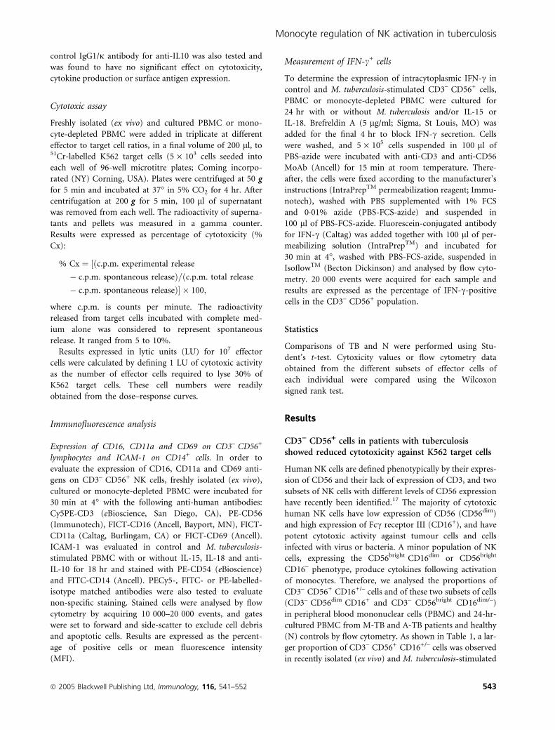
expression. As shown in Fig. 2, an increase in M. tubercu-
losis-induced NK lytic activity in cells from TB patients
was detected by depletion of monocytes, and furthermore,
in the presence of IL-15 or IL-18, an up-regulation of NK
function was observed in cells from A-TB patients. How-
ever, M. tuberculosis-induced NK function in N controls
was not affected by monocyte depletion. Taken together,
these results suggest a regulatory role of monocytes in NK
activation in TB patients.
As shown in Fig. 3, monocyte depletion did not modify
the proportion of CD69+ NK cells in control cells from TB
patients or healthy individuals. However, M. tuberculosis
stimulation in the absence of monocytes induced a
75(a)
(b)
50
25
00
100 1000
750
250
0
** *
* **
500
75
25
0
% IC
AM
-1+ C
D14
+
% IC
AM
-1+ C
D14
+
ICA
M-1
-MF
I
ControlMtbMtb + IL-15Mtb + IL-18Mtb + anti-IL-10
50
1 : 1 1 : 3 1 : 5PBMC:Mtb ratio
Figure 1. Mycobacterium tuberculosis (Mtb) regulated the number of
intracellular adhesion molecule (ICAM)-1+ CD14+ cells and ICAM-1
expression. (a) Peripheral blood mononuclear cells (PBMC) from
two patients with moderate tuberculosis (M-TB) and four patients
with advanced tuberculosis (A-TB) were incubated for 18 hr at dif-
ferent PBMC to M. tuberculosis ratios and the percentage of ICAM-1+
CD14+ cells was determined. Results are expressed as mean ± stand-
ard error of the mean (SEM). Statistical differences: PBMC +
M. tuberculosis versus non-stimulated PBMC: *P < 0�01. (b) PBMC
from six A-TB patients were incubated in complete medium
(control) or M. tuberculosis in the presence or absence of interleukin
(IL)-15, IL-18 or anti-IL-10 for 18 hr. The percentage of
ICAM-1+ CD14+ cells and ICAM-1 expression [mean fluorescence
intensity (MFI)] were then analysed by flow cytometry. Results are
expressed as mean ± SEM. Statistical differences: PBMC + IL-15,
IL-18 or anti-IL-10 versus PBMC without cytokines or anti-IL-10:
*P < 0�05.
70M-TB
A-TB
N
*
*
**
6050
403020100
% c
ytot
oxic
ity
706050
403020100
% c
ytot
oxic
ity
706050
403020100
% c
ytot
oxic
ity
Control Mtb Mtb + lL-15 Mtb + lL-18
Control Mtb Mtb + lL-15 Mtb + lL-18
Control Mtb Mtb + lL-15 Mtb + lL-18
Figure 2. Monocytes from patients with tuberculosis (TB) down-
regulated natural killer (NK) activity. Peripheral blood mononuclear
cells (PBMC) (grey bar) or monocyte-depleted mononuclear cells
(black grey bar) from 12 patients with moderate TB (M-TB),
15 patients with advanced TB (A-TB) and 12 healthy individuals (N)
were cultured in complete medium (control), Mycobacterium tuber-
culosis (Mtb) alone or M. tuberculosis plus interleukin (IL)-15 or IL-
18 for 24 hr and tested for their lytic activity against K562 target
cells. Results are expressed as percentage of cytotoxicity [mean ±
standard error of the mean (SEM)]. Statistical differences: % cyto-
toxicity from monocyte-depleted cells versus % cytotoxicity from
PBMC: *P < 0�05.
� 2005 Blackwell Publishing Ltd, Immunology, 116, 541–552 547
Monocyte regulation of NK activation in tuberculosis

decrease in the number of CD69+ NK cells from N con-
trols and an increase in that from A-TB patients, and did
not modify that from M-TB patients. Furthermore, while
IL-15 had no effect on the number of CD69+ NK cells in
TB and N controls in the absence of monocytes, the addi-
tion of IL-18 increased the percentage of CD69+ NK cells
and enhanced the expression of the CD16 molecule in
M. tuberculosis-stimulated cells from A-TB patients.
To assess whether IL-10 directly modulates the activa-
tion of NK cells, PBMC were depleted of CD14+ cells by
magnetic methods and stimulated or not stimulated with
M. tuberculosis in the presence or absence of IL-10 for
18 hr. Thereafter, the percentage of CD69+ cells (as a
measurement of activation) and CD16 expression on
CD3– CD56+ cells were determined by flow cytometry. As
shown in Fig. 4, while in the presence of monocytes exo-
genous IL-10 down-regulated the activation of NK cells in
M-TB patients and N controls, it had no effect on NK
activation in the absence of monocytes. In addition,
CD16 expression was not modified in NK cells by the
depletion of CD14+ cells (data not shown). With regard
to NK function, M. tuberculosis-induced NK lytic activity
was increased by monocyte depletion in M-TB patients
[PBMC (mean ± SEM): 33 ± 5; CD14-depleted: 49 ± 6,
n ¼ 3] and A-TB patients (PBMC: 10 ± 2; CD14-deple-
ted: 24 ± 5, n ¼ 3). Moreover, the addition of IL-10 to
monocyte-depleted PBMC increased NK lytic activity in
cells from M-TB patients (PBMC: 29 ± 4; CD14-depleted:
50 ± 6, n ¼ 3) and did not modify that in cells from
A-TB patients (PBMC: 10 ± 2; CD14-depleted: 16 ± 6,
n ¼ 3). Overall, these data indicate that IL-10 does not
exert a direct effect on NK activation, suggesting an indi-
rect effect through the down-regulation of costimulatory
molecules on monocytes necessary for the activation of
NK cells.
Numbers of M. tuberculosis-induced IFN-c+ NK cellswere increased by IL-15 and IL-18 in TB patients
In the light of the known roles of IL-15 and IL-18 in the
induction of IFN-c production by NK cells, we tested
whether the observed effect of these monokines on NK
activity could be ascribed to IFN-c production. For this
purpose, PBMC from TB patients and N controls were
stimulated or not stimulated with M. tuberculosis for
1009080706050403020100
400
300
200
100
0
400
300
200
100
0
300*
*
200
100
0
300
200
100
0
CD
16 M
FI
CD
11a
MF
I100
M-TB
MTB
A-TB
ATB
MTB ATB
N
9080706050403020100
100
**
*9080706050403020100
Contro
lM
tb
Mtb
+ lL
-15
Mtb
+ lL
-18
Contro
lM
tb
Mtb
+ lL
-15
Mtb
+ lL
-18
Contro
lM
tb
Mtb
+ lL
-15
Mtb
+ lL
-18
Contro
lM
tb
Mtb
+ lL
-15
Mtb
+ lL
-18
Contro
lM
tb
Mtb
+ lL
-15
Mtb
+ lL
-18
Contro
lM
tb
Mtb
+ lL
-15
Mtb
+ lL
-18
Contro
lM
tb
Mtb
+ lL
-15
Mtb
+ lL
-18
% C
D69
+ c
ells
Figure 3. Monocytes down-regulated activation molecules on natural
killer (NK) cells from patients with advanced tuberculosis (A-TB).
Peripheral blood mononuclear cells (PBMC) (grey bar) and mono-
cyte-depleted mononuclear cells (grey black bar) from 10 patients
with moderate tuberculosis (M-TB), 13 patients with advanced tuber-
culosis (A-TB) and eight healthy controls (N) were incubated in com-
plete medium (control), Mycobacterium tuberculosis or M. tuberculosis
plus interleukin (IL)-15 or IL-18 for 24 hr. The expression of CD16,
CD69 and CD11a molecules on CD3– CD56+ cells was then deter-
mined by flow cytometry. Results are expressed as mean fluorescence
intensity (MFI) for CD16 and CD11a molecules and percentage of
CD69+ cells [mean ± standard error of the mean (SEM)]. Statistical
differences: monocyte-depleted versus PBMC: *P < 0�05; **P < 0�01.
1009080706050403020100
PBMC Mo-depleted PBMC Mo-depleted PBMC Mo-depleted
% C
D69
cel
ls
M-TB A-TB N
*
Figure 4. Effect of interleukin (IL)-10 in the monocyte-depleted
(Mo-depleted) CD69+ CD3– CD56+ population. Peripheral blood
mononuclear cells (PBMC) or monocyte-depleted mononuclear cells
from six patients with moderate tuberculosis (M-TB), six patients
with advanced tuberculosis (A-TB) and five healthy individuals (N)
were cultured in complete medium (white bar), Mycobacterium
tuberculosis (grey black bar) or M. tuberculosis + IL-10 (grey bar) for
24 hr and the percentage of CD69+ on CD3– CD56+ cells was evalu-
ated by flow cytometry. Results are expressed as mean ± standard
error of the mean (SEM). Statistical differences: N-PBMC (Mtb vs
Mtb + IL-10): *P < 0�05.
548 � 2005 Blackwell Publishing Ltd, Immunology, 116, 541–552
P. Schierloh et al.

24 hr and IFN-c production by CD3– CD56+ cells was
analysed by flow cytometry. As shown in Table 6, no dif-
ferences in IFN-c production by non-stimulated (control)
NK cells from TB patients and N controls were observed.
However, upon M. tuberculosis stimulation, an increase in
the proportion of IFN-c+ CD3– CD56+ cells was detected
for M-TB patients and N controls, while for A-TB
patients no such increase was detected. Considering that
IL-15 and IL-18 increased the number of CD69+ cells,
up-regulated expression of the CD16 molecule and
enhanced NK function in both PBMC and monocyte-
depleted PBMC from A-TB patients, we assessed whether
these findings could be ascribed to enhanced IFN-c pro-
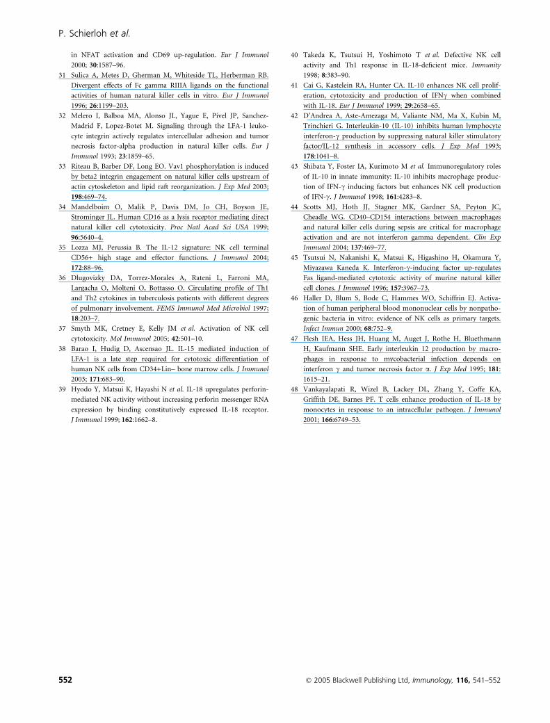
duction. As shown in Fig. 5, the proportion of IFN-c+
NK cells upon M. tuberculosis stimulation was increased
by IL-15, even in the presence of monocytes, while IL-18
produced no such effect. However, IL-18 up-regulated the
number of NK cells producing IFN-c in M. tuberculosis-
induced monocyte-depleted PBMC. Taken together, these
data indicate that monocytes have a down-regulatory
effect on IFN-c-producing NK cells from A-TB patients.
Discussion
Although the role of NK cells in mycobacterial infections
is unclear, in vitro studies have demonstrated that periph-
eral blood NK cells may contribute to protective immu-
nity through the production of IFN-c to maintain the
frequency of M. tuberculosis-responsive CD8+ IFN-c+ cells
and expand their cytotoxic activity.22,23 In addition, NK
cells mediate early killing of intracellular M. tuberculosis
via granule-independent mechanisms.24
Our study demonstrating a higher proportion of NK cells
in TB patients is in contrast with data previously repor-
ted.25,26 However, the decrease in lytic activity observed in
NK cells from TB patients is in accordance with the
results of studies employing either M. tuberculosis-infected
macrophages or the K562 cell line as target cells.25,26
Moreover, a major reduction in NK lytic activity is
observed in severe pulmonary involvement, indicating that
the microenvironment might affect NK activation and/or
function. In vitro studies have demonstrated that human
NK cells can be activated either by mycobacterium
(M. bovis) bacille Calmette–Guerin (BCG)27 or by lysed
human monocytes infected with live M. bovis BCG28 or
M. tuberculosis.29 The ability of NK cells to interact with
target cells depends on the expression of cell surface recep-
tors and adhesion molecules capable of binding to ligands
on target cells. Herein, we have demonstrated that
M. tuberculosis stimulates NK cells from healthy individ-
uals, as shown by high expression of CD69 antigen and
increases in natural cytotoxicity and IFN-c production.
However, M. tuberculosis stimulation of NK cells from TB
Table 6. Mycobacterium tuberculosis-induced interferon (IFN)-c+
natural killer (NK) cells
PBMC from
% IFN-c+ NK cells
Control PBMC M.tb-stimulated PBMC
M-TB (n ¼ 5) 1�0 ± 0�5 5�8 ± 0�8*A-TB (n ¼ 5) 0�4 ± 0�3 2�5 ± 0�9N (n ¼ 5) 0�6 ± 0�3 7�5 ± 0�7*
Peripheral blood mononuclear cells (PBMC) from patients with
moderate tuberculosis (M-TB) and advanced tuberculosis (A-TB)
and healthy controls (N) were incubated in complete medium (con-
trol) or with M. tuberculosis (M.tb) for 24 hr and then the percent-
age of IFN-c+ cells on CD3– CD56+ cells was evaluated by flow
cytometry. Results are expressed as mean ± standard error of the
mean (SEM). Statistical differences: control versus M. tuberculosis-
stimulated PBMC: *P < 0�05.
Control
Mtb
Mtb + IL-15
PE
-CD
56
PBMC
0·1%
3·3%
6·8%
3·7% 9·3%
10·4%
7·2%
Mo-depleted PBMC
10010
110
210
310
4
100 101 102 103 104
10010
110
210
310
4100 101 102 103 104 10
010
110
210
310
4
100 101 102 103 104
10010
110
210
310
4
100 101 102 103 104
10010
110
210
310
4
100 101 102 103 10410010
110
210
310
4
100 101 102 103 104
10010
110
210
310
4
100 101 102 103 104
Mtb + IL-18
FITC-IFN-γ
Figure 5. Monocytes from patients with advanced tuberculosis
(A-TB) modulated interferon (IFN)-c+ production by CD3– CD56+
cells. Peripheral blood mononuclear cells (PBMC) and monocyte-
depleted mononuclear cells (Mo-depleted) from three A-TB patients
were incubated in complete medium (control), Mycobacterium tuber-
culosis (Mtb), or M. tuberculosis plus interleukin (IL)-15 or IL-18 for
24 hr and IFN-c production by CD3– CD56+ cells was determined
by flow cytometry. Results are expressed as the percentage of positive
cells (data in the panels). A representative example is shown. FITC,
fluorescein isothiocyanate.
� 2005 Blackwell Publishing Ltd, Immunology, 116, 541–552 549
Monocyte regulation of NK activation in tuberculosis

patients showed impairment of their function, with the
severity of pulmonary disease, with low numbers of CD69+
NK cells and an absence of M. tuberculosis-induced lysis
and IFN-c production, as was observed in A-TB patients.
CD69 is a costimulatory molecule transiently expressed on
activated lymphocytes,6 and the mechanism regulating its
expression on NK cells is obscure. It has been demonstrated
that CD69 is up-regulated after activation of CD16, LFA-1/
CD11a, IL-2 or IFN-a receptors which signal through pro-
tein tyrosin kinase (PTK) phosphorylation and Vav/extra-
cellular regulated kinase (Vav/ERK).7,30,31 Binding of the
b2-integrin LFA-1 on NK cells to ICAM-1 on target cells
initiates triggering signal transduction pathways3,4,32 and,
furthermore, LFA-1 ligation costimulates the CD16 recep-
tor for cytokine production.33 In addition to the role of
CD16 in the killing of target cells by NK cells, its cross-link-
ing initiates signalling events inducing granule exocytosis
and cytokine production.18,34 Herein, we have demonstra-
ted that the expression of CD11a and CD16, molecules
implicated in the lysis of target cells, is down-regulated in
cells from A-TB patients and, furthermore, that M. tubercu-
losis reduces the numbers of ICAM-1+ CD14+ cells in TB
patients. However, in the presence of inflammatory cyt-
okines or neutralization of IL-10, the number of ICAM-1+
CD14+ cells and ICAM-1 expression are enhanced, even in
the presence of M. tuberculosis, along with an increase in
NK activity. Given that conjugate formation between NK
and target cells is a prerequisite for target cell lysis and is
critically dependent on engagement of CD11a with its lig-
ands on APCs, ICAM-1 regulation on monocytes from TB
patients could be a mechanism used by M. tuberculosis to
reduce NK activity.3,4,19,21,32 The finding that the expres-
sion of CD11a and CD16 molecules was associated in our
study with the severity of the disease suggests that, in TB
patients, the expression of costimulatory/activatory mole-
cules and their biological functions could be conditioned
by circulating cytokines and by those secreted upon
M. tuberculosis stimulation by accessory cells.35,36 There-
fore, the low expression of CD11a on NK cells, together
with ICAM-1 down-regulation on monocytes by M. tuber-
culosis, may contribute to the impairment of NK activity
in TB patients by diminishing the adhesiveness necessary
for triggering signalling pathways downstream of CD11a/
ICAM engagement. Although, to date, no receptor has been
shown to be either necessary or sufficient in signalling for
NK cell cytotoxicity, other receptors on NK cells, such as
NKG2D and NKp46, and their ligands on target cells could
be involved in the regulation of NK cell effector function,
indicating a possible redundancy of activation receptors
and activation pathways.2,37 In this context, natural cyto-
toxicity receptors may also be involved in the impairment
of NK activity, as demonstrated for NKp46 in TB
patients.25
It is well known that cytokines produced by accessory
cells play an essential role in NK development and
activation. IL-15 promotes in vitro NK cell development,
mediates LFA-1 expression,38 and enhances the lytic activ-
ity and proliferation of resting NK cells as well as the
production of cytokines and chemokines.13 While IL-18
increases the lytic activity of NK cells by up-regulating
perforin-dependent cytotoxic pathways and Fas ligand
(FasL) expression,39,40 the role of IL-10 in NK activation
is not clear.41,42 Our results showed that, in effect, IL-15
enhanced M. tuberculosis-induced NK cytotoxicity in
healthy individuals, inducing CD69 expression on NK
cells just as M. tuberculosis does. However, IL-15 also
up-regulated the expression of CD16 and CD11a on
M. tuberculosis-stimulated NK cells from A-TB patients,
in addition to increasing NK cytotoxicity in both groups
of TB patients in association with a high number of
CD69+ NK cells. In contrast to IL-15, IL-18 did not mod-
ify NK cytotoxicity in A-TB patients but slightly up-regu-
lated the expression of CD69 and CD11a molecules. IL-10
neutralization in PBMC from TB patients increased
M. tuberculosis-induced NK cytotoxicity and up-regulated
CD16 and CD11a expression, suggesting that endogenous
IL-10 could inhibit NK activation and function through
the down-regulation of ICAM-1 on monocytes. Therefore,
the increase in costimulatory molecules could enhance the
efficiency of NK lysis by increasing the interaction with
target cells and triggering granule exocytosis.
IL-10 can boost IFN-c production by isolated NK
cells,41,43 but in the presence of APCs it inhibits IFN-c pro-
duction.20,42 In this context, our data for monocyte-deple-
ted populations demonstrated that M. tuberculosis directly
triggered the activation of NK cells from both TB patients
and healthy controls, as previously reported with patho-
genic and non-pathogenic bacteria.27,44 Moreover, in the
absence of monocytes, M. tuberculosis-induced NK cell
activity was increased in TB patients while in N controls it
was not. In addition, CD69 and CD16 expression on
M. tuberculosis-stimulated NK cells was also increased in
cells from A-TB patients, and IL-18 up-regulated their
expression while IL-15 did not, demonstrating the different
roles of these two cytokines in NK function.38–40 Further-
more, in TB patients, the modulatory effect of monocytes
was also observed in the production of IFN-c by M. tuber-
culosis-stimulated NK cells, where the lowest levels detected
in A-TB patients were increased by the depletion of mono-
cytes. Although IFN-c is not required for NK cytotoxicity
and does not mediate the killing of intracellular M. tuber-
culosis,24,38 it may contribute to switching the immune
response towards type 1 by enhancing the production of
IL-12 and IL-18 in response to M. tuberculosis.47,48 Taken
together, these results confirm a regulatory role of mono-
cytes in NK cell activation and function in TB patients.
In conclusion, we have demonstrated that monocytes
from TB patients regulate NK function involving IL-10,
which acts through an indirect mechanism leading to
down-regulation of costimulatory/adhesion molecules
550 � 2005 Blackwell Publishing Ltd, Immunology, 116, 541–552
P. Schierloh et al.

and/or the IFN-c production necessary for the activation of
NK cells. Therefore, our findings demonstrate an underly-
ing role of monocytes in NK function in TB patients.
Acknowledgements
We thank the medical staff of the Division Tisio-
neumonologıa, Hospital F. J. Muniz for their substantial
help in providing clinical samples from patients with
tuberculosis. This work was supported by grants from the
Agencia Nacional de Promocion Cientıfica y Tecnologica
(ANPCyT, 05-14060) and CONICET.
References
1 Kaufmann SH. How can immunology contribute to the control
of tuberculosis? Nat Rev Immunol 2001; 1:20–30.
2 Moretta L, Bottino C, Pende D, Mingari MC, Blassoni R,
Moretta A. Human natural killer cells: their origin, receptors
and function. Eur J Immunol 2002; 32:1205–11.
3 Perez OD, Mitcheli D, Jager G, Nolan G. LFA-1 signaling
through p44/42 is coupled to perforin degranulation in
CD56+CD8+ natural killer cells. Blood 2004; 104:1083–93.
4 Barber DF, Faure M, Long EO. LFA-1 contributes an early signal
for NK cell cytotoxicity. J Immunol 2004; 173:3653–9.
5 Ziegler SF, Ramsdell F, Hjerrild KA et al. Molecular characteri-
zation of the early activation CD69: a type II membrane glyco-
protein related to a family of natural killer cell activation
antigens. Eur J Immunol 1993; 23:1643–8.
6 Sancho D, Gomez M, Sanchez-Madrid F. CD69 is an immuno-
regulatory molecule induced following activation. Trends Immu-
nol 2005; 26:136–40.
7 Borrego F, Pena J, Solana R. Regulation of CD69 expression on
natural killer cells: differential involvement of protein kinase C
and protein tyrosine kinases. Eur J Immunol 1993; 23:1039–43.
8 Fehniger TA, Shah MH, Turner MJ et al. Differential cytokine and
chemokine gene expression by human NK cells following activa-
tion with IL-18 or IL-15 in combination with IL-12: implications
for the innate immune response. J Immunol 1999; 162:4511–20.
9 Gerosa F, Baldani-Guerra B, Nisii C, Marchesini V, Carra G,
Trinchieri G. Reciprocal activating interaction between Natural
Killer cells and dendritic cells. J Exp Med 2002; 195:327–33.
10 Fehninger TA, Caligiuri M. Interleukin 15: biology and relevance
to human disease. Blood 2001; 97:14–32.
11 Nakanishi K, Yoshimoto T, Tsutsui H, Okamura H. Interleukin-
18 is a unique cytokine that stimulates both Th1 and Th2
responses depending on its cytokine milieu. Cytokine Growth
Factor Rev 2001; 12:53–72.
12 Chan SH, Perussia B, Gupta JW et al. Induction of interferon cproduction by natural killer cell stimulatory factor: characteriza-
tion of the responder cells and synergy with other inducers.
J Exp Med 1991; 173:869–79.
13 Carson WE, Giri JG, Lindemann MJ et al. Interleukin (IL) 15 is a
novel cytokine that activates human natural killer cells via compo-
nents of the IL-2 receptor. J Exp Med 1994; 180:1395–403.
14 Okamura H, Kashiwamura S, Tsutsui H, Yoshimoto T,
Nakanishi K. Regulation of interferon-c production by IL-12
and IL-18. Curr Opin Immunol 1998; 10:159–64.
15 Carson WE, Ross ME, Baiocchi RA, Marien MJ, Boiani N,
Grabstein K, Caligiuri MA. Endogenous production of interleu-
kin 15 by activated human monocytes is critical for optimal pro-
duction of interferon-c by natural killer cells in vitro. J Clin
Invest 1995; 96:2578–82.
16 Boyum A. Isolation of mononuclear cells and granulocytes from
human blood. Scand J Clin Invest 1968; 97:77–89.
17 Cooper MA, Fehniger TA, Turner SC, Chen KS, Ghaheri BA,
Ghayur T, Carson WE, Caligiuri MA. Human natural killer cells.
a unique innate immunoregulatory role for the CD56 (bright)
subset. Blood 2001; 97:3146–51.
18 Milella M, Gismondi A, Roncaioli P, Bisogno L, Palmieri G,
Frati L, Cifone MG, Santoni A. CD16 cross-linking induces
both secretory and extracellular signal-regulated kinase (ERK)-
dependent cytosolic phospholipase A2 (PLA2) activity in human
natural killer cells. involvement of ERK, but not PLA2, in CD16-
triggered granule exocytosis. J Immunol 1997; 158:3148–54.
19 Helander TS. Adhesion in NK cell function. Curr Top Microbiol
Immunol 1998; 230:89–98.
20 Goodier MR, Londei M. Lypopolysaccharide stimulates the pro-
liferation of human CD56+CD3– NK cells: a regulatory role of
monocytes and IL-10. J Immunol 2000; 165:139–47.
21 de la Barrera S, Aleman M, Musella R, Schierloh P, Pasquinelli V,
Garcia V, Abbate E, del Sasiain MC. IL-10 down-regulates
costimulatory molecules on Mycobacterium tuberculosis-pulsed
macrophages and impairs the lytic activity of CD4 and CD8 CTL
in tuberculosis patients. Clin Exp Immunol 2004; 138:128–38.
22 Fehniger TA, Cooper MA, Nuovo GJ, Cella M, Facchetti F,
Colonna M, Caligiuri MA. CD56bright natural killer cells are
present in human lymph nodes and are activated by T cell-
derived IL-2: a potential new link between adaptive and innate
immunity. Blood 2003; 101:3052–7.
23 Vankayalapati R, Klucar P, Wizel B, Weiz SE, Samtem B, Safi H,
Shams H, Barnes PF. NK cells regulate CD8+ T cell effector
function in response to an intracellular pathogen. J Immunol
2004; 172:130–7.
24 Brill KJ, Li Q, Larkin R, Canaday DH, Kaplan DR, Boom WH,
Silver RF. Human natural killer cells mediate killing of intracel-
lular Mycobacterium tuberculosis H37Rv via granule-independent
mechanisms. Infect Immun 2001; 69:1755–65.
25 Vankayalapati R, Wizel B, Weis SE et al. The NKp46 receptor
contributes to NK cell lysis of mononuclear phagocytes infected
with an intracellular bacterium. J Immunol 2002; 168:3451–7.
26 Nirmala R, Narayanan PR, Mathew R, Maran M, Deivanayagam
CN. Reduced NK activity in pulmonary tuberculosis patients
with/without HIV infection. Identifying the defective stage and
studying the effect of interleukins on NK activity. Tuberculosis
2001; 81:343–52.
27 Esin S, Batoni G, Pardini M et al. Functional characterization of
human natural killer cells responding to Mycobacterium bovis
bacille Calmette-Guerin. Immunology 2004; 112:143–52.
28 Molloy A, Meyn PA, Smith KD, Kaplan G. Recognition and
destruction of Bacillus Calmette-Guerin-infected human mono-
cytes. J Exp Med 1993; 177:1691–8.
29 Denis M. Interleukin-12 (IL-12) augments cytolytic activity of
natural killer cells toward Mycobacterium tuberculosis-infected
human monocytes. Cell Immunol 1994; 156:529–36.
30 Villaba M, Hernandez J, Deckert M, Tanaka Y, Altman A. Vav
modulation of the Ras/MEK/ERK signaling pathway plays a role
� 2005 Blackwell Publishing Ltd, Immunology, 116, 541–552 551
Monocyte regulation of NK activation in tuberculosis
in NFAT activation and CD69 up-regulation. Eur J Immunol
2000; 30:1587–96.
31 Sulica A, Metes D, Gherman M, Whiteside TL, Herberman RB.
Divergent effects of Fc gamma RIIIA ligands on the functional
activities of human natural killer cells in vitro. Eur J Immunol
1996; 26:1199–203.
32 Melero I, Balboa MA, Alonso JL, Yague E, Pivel JP, Sanchez-
Madrid F, Lopez-Botet M. Signaling through the LFA-1 leuko-
cyte integrin actively regulates intercellular adhesion and tumor
necrosis factor-alpha production in natural killer cells. Eur J
Immunol 1993; 23:1859–65.
33 Riteau B, Barber DF, Long EO. Vav1 phosphorylation is induced
by beta2 integrin engagement on natural killer cells upstream of
actin cytoskeleton and lipid raft reorganization. J Exp Med 2003;
198:469–74.
34 Mandelboim O, Malik P, Davis DM, Jo CH, Boyson JE,
Strominger JL. Human CD16 as a lysis receptor mediating direct
natural killer cell cytotoxicity. Proc Natl Acad Sci USA 1999;
96:5640–4.
35 Lozza MJ, Perussia B. The IL-12 signature: NK cell terminal
CD56+ high stage and effector functions. J Immunol 2004;
172:88–96.
36 Dlugovizky DA, Torrez-Morales A, Rateni L, Farroni MA,
Largacha O, Molteni O, Bottasso O. Circulating profile of Th1
and Th2 cytokines in tuberculosis patients with different degrees
of pulmonary involvement. FEMS Immunol Med Microbiol 1997;
18:203–7.
37 Smyth MK, Cretney E, Kelly JM et al. Activation of NK cell
cytotoxicity. Mol Immunol 2005; 42:501–10.
38 Barao I, Hudig D, Ascensao JL. IL-15 mediated induction of
LFA-1 is a late step required for cytotoxic differentiation of
human NK cells from CD34+Lin– bone marrow cells. J Immunol
2003; 171:683–90.
39 Hyodo Y, Matsui K, Hayashi N et al. IL-18 upregulates perforin-
mediated NK activity without increasing perforin messenger RNA
expression by binding constitutively expressed IL-18 receptor.
J Immunol 1999; 162:1662–8.
40 Takeda K, Tsutsui H, Yoshimoto T et al. Defective NK cell
activity and Th1 response in IL-18-deficient mice. Immunity
1998; 8:383–90.
41 Cai G, Kastelein RA, Hunter CA. IL-10 enhances NK cell prolif-
eration, cytotoxicity and production of IFNc when combined
with IL-18. Eur J Immunol 1999; 29:2658–65.
42 D’Andrea A, Aste-Amezaga M, Valiante NM, Ma X, Kubin M,
Trinchieri G. Interleukin-10 (IL-10) inhibits human lymphocyte
interferon-c production by suppressing natural killer stimulatory
factor/IL-12 synthesis in accessory cells. J Exp Med 1993;
178:1041–8.
43 Shibata Y, Foster IA, Kurimoto M et al. Immunoregulatory roles
of IL-10 in innate immunity: IL-10 inhibits macrophage produc-
tion of IFN-c inducing factors but enhances NK cell production
of IFN-c. J Immunol 1998; 161:4283–8.
44 Scotts MJ, Hoth JJ, Stagner MK, Gardner SA, Peyton JC,
Cheadle WG. CD40–CD154 interactions between macrophages
and natural killer cells during sepsis are critical for macrophage
activation and are not interferon gamma dependent. Clin Exp
Immunol 2004; 137:469–77.
45 Tsutsui N, Nakanishi K, Matsui K, Higashino H, Okamura Y,
Miyazawa Kaneda K. Interferon-c-inducing factor up-regulates
Fas ligand-mediated cytotoxic activity of murine natural killer
cell clones. J Immunol 1996; 157:3967–73.
46 Haller D, Blum S, Bode C, Hammes WO, Schiffrin EJ. Activa-
tion of human peripheral blood mononuclear cells by nonpatho-
genic bacteria in vitro: evidence of NK cells as primary targets.
Infect Immun 2000; 68:752–9.
47 Flesh IEA, Hess JH, Huang M, Auget J, Rothe H, Bluethmann
H, Kaufmann SHE. Early interleukin 12 production by macro-
phages in response to mycobacterial infection depends on
interferon c and tumor necrosis factor a. J Exp Med 1995; 181:
1615–21.
48 Vankayalapati R, Wizel B, Lackey DL, Zhang Y, Coffe KA,
Griffith DE, Barnes PF. T cells enhance production of IL-18 by
monocytes in response to an intracellular pathogen. J Immunol
2001; 166:6749–53.
552 � 2005 Blackwell Publishing Ltd, Immunology, 116, 541–552
P. Schierloh et al.
Copyright © 2022 FDOKUMEN