monocytes in cancer
13
ORIGINAL ARTICLE Monocytes and Macrophages in Cancer: Development and Functions David M. Richards & Jan Hettinger & Markus Feuerer Received: 8 October 2012 / Accepted: 15 October 2012 / Published online: 24 November 2012 # Springer Science+Business Media Dordrecht 2012 Abstract Monocytes and tumor-associated macrophages are part of the myeloid family, a group of hematopoietic derived cells. Monocytes are direct precursors of hematopoietic stem cell-derived macrophages. After their recruitment into the tu- mor tissue, they can differentiate into tumor-associated macro- phages, a very heterogeneous cell population in terms of phenotype and pro-tumor function, supporting tumor initiation, local progression and distant metastasis. Therefore, targeting monocytes and macrophages is a promising immunotherapeu- tic approach. This review will focus on the development of monocytes as macrophage precursors, the functions of tumor- associated macrophages and the possibility of interfering with tumor development and progression by targeting these myeloid cells. Keywords Tumor-associated macrophages . Monocytes . Myeloid cells . Immunotherapy Introduction The mononuclear phagocyte system (MPS) represents a body–wide, specialized system of different phagocytic cell types derived from bone marrow and yolk sac progenitors [1, 2]. Monocytes (Mo) and macrophages (MΦ) are essen- tial components of the MPS and critically involved in central processes of the MPS such as innate immune func- tions, support of adaptive immunity and tissue homeostasis [1, 3]. Ilya Mechnikov, a bacteriologist, first described macro- phages more than a century ago. They colonize every organ in the body and, based on the location, some specialized macrophages have received specific names, e.g., Kupffer cells in the liver, microglia in the neuronal tissue and osteo- clasts in the bone. They have specific functions under tissue homeostasis including phagocytosis of apoptotic cells, growth factor production and angiogenesis induction [4, 5]. In response to inflammation, they are needed for patho- gen clearance, followed by a phase of tissue repair and wound healing that promotes restoration of homeostasis [5]. Monocytes are precursors of resident macrophages. Differentiation occurs upon entering the tissue and is under the control of local environmental cues [6, 7]. As a second origin, it has recently been reported that yolk sac-derived cells can constitute a different line of macrophages in adult mice. Their development occurs independently of mono- cytes derived from adult hematopoietic stem cells (HSC) and the transcription factor Myb [2]. The tumor tissue comprises a very large and diverse set of myeloid cell lineages. This review will focus on mono- cytes and tumor-associated macrophages (TAMs), their dif- ferentiation, function and strategies to target them for immunotherapy. Steady-State Development of Monocytes Monocytes are found in the blood and spleen, but their largest reservoir under homeostatic conditions is the bone marrow, the primary site of monocyte generation [3, 8]. David M. Richards and Jan Hettinger equally contributed. D. M. Richards : J. Hettinger : M. Feuerer (*) Immune Tolerance, Tumor Immunology Program, German Cancer Research Center (DKFZ), Im Neuenheimer Feld 280, 69120 Heidelberg, Germany e-mail: [email protected] Cancer Microenvironment (2013) 6:179–191 DOI 10.1007/s12307-012-0123-x
Transcript of monocytes in cancer

ORIGINAL ARTICLE
Monocytes and Macrophages in Cancer: Developmentand Functions
David M. Richards & Jan Hettinger & Markus Feuerer
Received: 8 October 2012 /Accepted: 15 October 2012 /Published online: 24 November 2012# Springer Science+Business Media Dordrecht 2012
Abstract Monocytes and tumor-associated macrophages arepart of the myeloid family, a group of hematopoietic derivedcells. Monocytes are direct precursors of hematopoietic stemcell-derived macrophages. After their recruitment into the tu-mor tissue, they can differentiate into tumor-associated macro-phages, a very heterogeneous cell population in terms ofphenotype and pro-tumor function, supporting tumor initiation,local progression and distant metastasis. Therefore, targetingmonocytes and macrophages is a promising immunotherapeu-tic approach. This review will focus on the development ofmonocytes as macrophage precursors, the functions of tumor-associated macrophages and the possibility of interfering withtumor development and progression by targeting these myeloidcells.
Keywords Tumor-associated macrophages . Monocytes .
Myeloid cells . Immunotherapy
Introduction
The mononuclear phagocyte system (MPS) represents abody–wide, specialized system of different phagocytic celltypes derived from bone marrow and yolk sac progenitors[1, 2]. Monocytes (Mo) and macrophages (MΦ) are essen-tial components of the MPS and critically involved in
central processes of the MPS such as innate immune func-tions, support of adaptive immunity and tissue homeostasis[1, 3].
Ilya Mechnikov, a bacteriologist, first described macro-phages more than a century ago. They colonize every organin the body and, based on the location, some specializedmacrophages have received specific names, e.g., Kupffercells in the liver, microglia in the neuronal tissue and osteo-clasts in the bone. They have specific functions under tissuehomeostasis including phagocytosis of apoptotic cells,growth factor production and angiogenesis induction [4,5]. In response to inflammation, they are needed for patho-gen clearance, followed by a phase of tissue repair andwound healing that promotes restoration of homeostasis[5]. Monocytes are precursors of resident macrophages.Differentiation occurs upon entering the tissue and is underthe control of local environmental cues [6, 7]. As a secondorigin, it has recently been reported that yolk sac-derivedcells can constitute a different line of macrophages in adultmice. Their development occurs independently of mono-cytes derived from adult hematopoietic stem cells (HSC)and the transcription factor Myb [2].
The tumor tissue comprises a very large and diverse setof myeloid cell lineages. This review will focus on mono-cytes and tumor-associated macrophages (TAMs), their dif-ferentiation, function and strategies to target them forimmunotherapy.
Steady-State Development of Monocytes
Monocytes are found in the blood and spleen, but theirlargest reservoir under homeostatic conditions is the bonemarrow, the primary site of monocyte generation [3, 8].
David M. Richards and Jan Hettinger equally contributed.
D. M. Richards : J. Hettinger :M. Feuerer (*)Immune Tolerance, Tumor Immunology Program,German Cancer Research Center (DKFZ),Im Neuenheimer Feld 280,69120 Heidelberg, Germanye-mail: [email protected]
Cancer Microenvironment (2013) 6:179–191DOI 10.1007/s12307-012-0123-x
Still, monocytes are also produced by extra-medullary he-matopoiesis in the spleen and increase under inflammatoryconditions such as atherosclerosis and cancer as recentlyshown [9, 10]. In the bone marrow, monocytes are generatedfrom HSC in a sequential process of differentiation andcommitment steps [3, 11], collectively called monopoiesis(Fig. 1). Monopoiesis is tightly regulated by microenviron-mental cues, modulating gene expression in developingcells and leading to the often irreversible, phenotypic andfunctional changes associated with hematopoietic differen-tiation. Although the key role of cytokines, such as macro-phage colony-stimulating factor (M-CSF), granulocyte-macrophage colony-stimulating factor (GM-CSF) andinterleukin-3 (IL-3), within this developmental niche hasbeen intensively studied [12–14], the impact of other nichecomponents, like immobilized factors on stromal cells orextracellular matrix (ECM) components, on monopoiesisremains still unclear. Early-phase monopoiesis proceedsvia distinct proliferative progenitor stages, such as commonmyeloid progenitors (CMP) and granulocyte/macrophage
progenitors (GMP) [15], to the macrophage dendritic cellprogenitor (MDP) [12, 16], which serves as a commonprecursor for monocytes, macrophages and dendritic cells(DCs). During late-phase monopoiesis, the MDP finallygives rise to monocytes (Fig. 1) [12, 16]. These bonemarrow-derived monocytes have lost their proliferative poten-tial and can be sub-divided by expression of chemokine recep-tors and additional cell surface markers into “classical”Ly6Chigh and “non-classical” Ly6Clow monocytes [11, 17].However, the two subsets can also be distinguished by func-tions such as the patrolling behavior found in Ly6low but notLy6Chigh monocytes [18]. As these phenotypical and functionaldifferences are found in both human and murine monocytesubsets, evolutionary conservation of monocyte heterogeneityhas been suggested [19–22]. The developmental relationshipbetween the monocyte subsets is still not completely under-stood [11]. Previous studies have demonstrated that Ly6Chigh
monocytes shuttle between the blood and bone marrow andeventually lose expression of Ly6C, suggesting a linear con-version model of Ly6Chigh into Ly6Clow monocytes within the
HSC CMP GMP MDP Ly6Chigh Mo Ly6Clow Mo
Bone Marrow
Spleen Blood
Healthy Tissues
Ly6Chigh Mo Ly6Clow Mo
Mo-MMo-DC
Yolk Sac-DerivedProgenitor
YS-M
PeripheralTissues
Ly6Chigh Mo Ly6Clow Mo
inflammatoryMo-DC
M-MDSC
Inflamed Tissue(Non-Cancerous)
inflammatoryMo-M
TAM
Disease(Immunity and Tumor Progression)
Cancerous Tissue
Steady-State(Development and Homeostasis)
Fetus
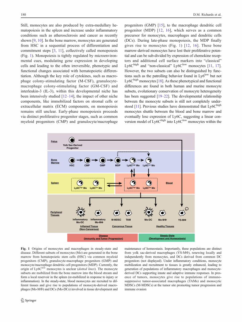
Fig. 1 Origins of monocytes and macrophages in steady-state anddisease. Different subsets of monocytes (Mo) are generated in the bonemarrow from hematopoietic stem cells (HSC) via common myeloidprogenitors (CMP), granulocyte-macrophage progenitors (GMP) andmonocyte/macrophage dendritic cell progenitors (MDP). Currently, theorigin of Ly6Clow monocytes is unclear (dotted lines). The monocytesubsets are mobilized from the bone marrow into the blood stream andform a local reservoir in the spleen (re-mobilized in response to injury orinflammation). In the steady-state, blood monocytes are recruited to dif-ferent tissues and give rise to populations of monocyte-derived macro-phages (Mo-MΦ) and DCs (Mo-DCs) involved in tissue development and
maintenance of homeostasis. Importantly, these populations are distinctfrom yolk sac-derived macrophages (YS-MΦ), renewing locally andindependently from monocytes, and DCs derived from common DCprogenitors (not displayed). Under inflammatory conditions, monocytemobilization and recruitment to tissues is greatly enhanced, leading togeneration of populations of inflammatory macrophages and monocyte-derived DCs supporting innate and adaptive immune responses. In pres-ence of tumors, monocytes give rise to populations of immuno-suppressive tumor-associated macrophages (TAMs) and monocyticMDSCs (M-MDSCs) at the tumor site promoting tumor progression andimmune evasion
180 D.M. Richards et al.
bone marrow (Fig. 1) [23]. Alternatively, a divergent modelis possible in which the monocyte subsets arise indepen-dently from each other from a common progenitor, how-ever supporting evidence for this model is limited [11].Novel transgenic mouse models with subset-specificdefects, such as the recently described transcription factorNr4a1-deficient mouse which lacks most of the Ly6Clow
monocytes [24], as well as novel subset-specific moleculartargets, e.g. the recently identified microRNA miR-146a[25], will be of great value for answering these and furtherquestions.
Development of Monocytes in Cancer
In cancer, many characteristics of myeloid cells, such asmigratory and functional properties, are altered, but whetherthe principal developmental processes in the bone marroware also affected is currently under investigation. Recently,Pittet and colleagues revealed alterations in myeloid devel-opment in a model of lung adenocarcinoma driven by acti-vation of K-Ras and inactivation of p53 [10]. In this model,early hematopoietic progenitors (such as the GMP) relo-cated to the spleen during tumor progression and locallyproduced monocytes and granulocytes that contributed totumor growth. Thus, the bone marrow partially outsourcesthe production of myeloid cells to extra-medullary sites incancer (and potentially also in other inflammatory diseases).The findings of Pittet and colleagues are reminiscent ofmyeloid-derived suppressor cells (MDSCs) [26–28]. MDSCs,comprising monocytic and granulocytic subpopulations, are
potent immune suppressors in various pathological conditions(like cancer and other inflammatory diseases) and promisingtargets for upcoming therapies (as reviewed by Dr. Umanskyand colleagues in this issue and [29]). Previous reviews havediscussed developmental aspects of MDSCs and suggestedthe generation of monocytic MDSCs (M-MDSCs) in bonemarrow without a monocytic intermediate stage from a sepa-rate developmental branch in monopoiesis [27, 28]. Usuallyidentified as CD11b+ Ly6C+ Ly6G− cells in mice, M-MDSCsyet share a common phenotype and morphology with normalLy6Chigh monocytes (Table 1) [26, 27]. Furthermore, thelimited marker panel used for M-MDSC identification andthe use of antibodies recognizing less specific markers like Gr-1 (clone: RB6-8C5), a shared epitope of Ly6C and Ly6G [30,31], make distinguishing M-MDSC from normal monocyteschallenging. Thus, M-MDSCs are currently defined by theirsuppressive activity, which is absent in normal monocytes[27]. This definition may result in an image that reflectsmonocytes and their macrophage progeny, which acquiredimmuno-suppressive properties in response to tumor-derivedfactors, rather than a separate developmental branch fromdefined monocyte/macrophage progenitors (such as theGMP or MDP) (Fig. 1). Although tumors produce factorsinvolved in several aspects of monocyte/macrophage biology(such as GM-CSF) [27], the current evidence supporting aconcept of M-MDSC development without a monocyticintermediate is limited and would benefit from compre-hensive fate-mapping approaches. The identification ofadditional markers or progenitors specific for M-MDSCs(compared to normal monocytes) will certainly help elucidat-ing this issue.
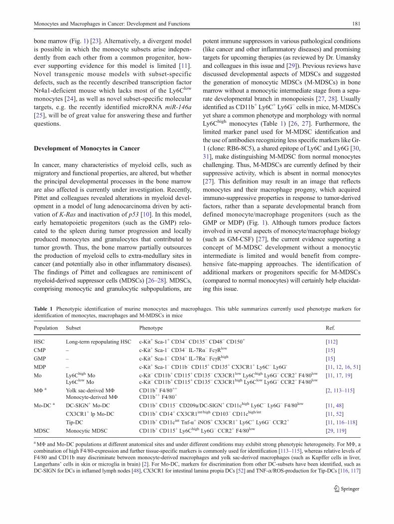
Table 1 Phenotypic identification of murine monocytes and macrophages. This table summarizes currently used phenotype markers foridentification of monocytes, macrophages and M-MDSCs in mice
Population Subset Phenotype Ref.
HSC Long-term repopulating HSC c-Kit+ Sca-1+ CD34− CD135− CD48− CD150+ [112]
CMP – c-Kit+ Sca-1− CD34− IL-7Rα− FcγRlow [15]
GMP – c-Kit+ Sca-1− CD34+ IL-7Rα− FcγRhigh [15]
MDP – c-Kit+ Sca-1− CD11b− CD115+ CD135+ CX3CR1+ Ly6C− Ly6G- [11, 12, 16, 51]
Mo Ly6Chigh Mo c-Kit− CD11b+ CD115+ CD135− CX3CR1low Ly6Chigh Ly6G− CCR2+ F4/80low [11, 17, 19]Ly6Clow Mo c-Kit− CD11b+ CD115+ CD135− CX3CR1high Ly6Clow Ly6G− CCR2− F4/80low
MΦ a Yolk sac-derived MΦ CD11b+ F4/80++ [2, 113–115]Monocyte-derived MΦ CD11b++ F4/80+
Mo-DC a DC-SIGN+ Mo-DC CD11b+ CD115− CD209a/DC-SIGN+ CD11chigh Ly6C− Ly6G− F4/80low [11, 48]
CX3CR1+ lp Mo-DC CD11b+ CD14+ CX3CR1int/high CD103− CD11chigh/int [11, 52]
Tip-DC CD11b+ CD11cint Tnf-α+ iNOS+ CX3CR1+ Ly6C+ Ly6G− CCR2+ [11, 116–118]
MDSC Monocytic MDSC CD11b+ CD115+ Ly6Chigh Ly6G− CCR2+ F4/80low [29, 119]
aMΦ and Mo-DC populations at different anatomical sites and under different conditions may exhibit strong phenotypic heterogeneity. For MΦ, acombination of high F4/80-expression and further tissue-specific markers is commonly used for identification [113–115], whereas relative levels ofF4/80 and CD11b may discriminate between monocyte-derived macrophages and yolk sac-derived macrophages (such as Kupffer cells in liver,Langerhans’ cells in skin or microglia in brain) [2]. For Mo-DC, markers for discrimination from other DC-subsets have been identified, such asDC-SIGN for DCs in inflamed lymph nodes [48], CX3CR1 for intestinal lamina propia DCs [52] and TNF-α/ROS-production for Tip-DCs [116, 117]
Monocytes and Macrophages in Cancer: Development and Functions 181

Mobilization and Recruitment of Monocytes to TargetSites
Monocytes are mobilized from the bone marrow and spleenin response to chemotactic signals and recruited to targettissues guiding their further differentiation [32]. These mi-gration processes have been recently reviewed in detail [32].Due to distinct expression of chemokine receptors (such asCCR2 and CX3CR1), the monocyte subsets have differentmigratory properties [19]. A major role in mobilization andrecruitment of Ly6Chigh monocytes, under both homeostaticand pathological conditions, has been attributed to the che-mokine receptor CCR2 (CD192), bound by the chemokinesCCL2 and CCL7 [32–34]. CCL2 (monocyte chemotacticprotein-1 or MCP-1) is expressed by many nucleated celltypes in response to pro-inflammatory or microbial stimuliand the role of the CCL2-CCR2-axis in recruitment ofLy6Chigh monocytes to sites of inflammation has been dem-onstrated for various pathological conditions (such as infec-tion, atherosclerosis and diabetes) [32–36].
In cancer, CCL2 was identified as the first tumor-derivedfactor (TDF) that induced chemotaxis in monocytes [37].Produced by tumor cells and the associated stromal cells,CCL2 is currently acknowledged as the major tumor-derived factor responsible for recruiting circulating mono-cytes from the blood into a variety of mouse and humantumors [28, 38]. Interestingly, recent work from Qian andcolleagues demonstrated that metastatic tumor cells andtarget-site stromal cells also produce CCL2 [39]. Using amammary tumor model, they observed that a sub-populationof CCR2-expressing inflammatory monocytes was recruitedto the target-site and promoted extravasation, establishmentand persistent growth of the tumor metastasis [39].
Tumor cells and the associated stromal network also pro-duce additional molecules that are traditionally involved inmonocyte recruitment to, and differentiation in, inflammatorysites. These TDFs include a number of chemokines, includingCCL3, CCL4, CCL5 and CXCL12, and various growth fac-tors, including M-CSF, transforming growth factor-α (TGF-α), fibroblast growth factor (FGF), Angiopoietin-2 (Ang-2,critical for Tie2-expressing monocytes) and vascular endothe-lial growth factor (VEGF) [26, 28, 40, 41].
Development and Functions of Macrophages
Following tissue recruitment, monocytes are polarized bythe local microenvironment and differentiate into residentmacrophages under the control of multiple tissue-specificfactors [6, 7]. This differentiation process is accompanied byvast phenotypic, functional and morphological changes,establishing a spectrum of diverse macrophage populationsthroughout the body [6, 7]. Notably, monocyte-derived
macrophages are phenotypically and functionally distinctfrom yolk sac-derived macrophages, which represent a ma-jor fraction of F4/80+ macrophages in some tissues [2]. Asrecently demonstrated by Geissmann and colleagues, theseyolk sac-derived macrophages develop independently of thetranscription factor Myb, renew locally and persist in tissuesin adult mice independently from HSC in contrast tomonocyte-derived macrophages [2] (Fig. 1). In the future,delineation between the functions of monocyte-derived ver-sus yolk sac-derived macrophages, under steady-state anddisease conditions, may be of great value for the field, as itmay help to understand the diversity of macrophages andhold therapeutic potential.
Tissue macrophages display enormous functional and phe-notypical plasticity in response to changing microenvironmen-tal stimuli [42, 43]. Under steady-state conditions,macrophages support either homeostatic processes or trophicprocesses for tissue development/remodeling in developing orhealing tissues [4, 5]. The mechanisms involved in this supportinclude phagocytosis, growth factor production, angiogenesisand degradation of ECM components by secreted MMPs(matrix metallo-proteinases) [4, 5]. In response to immunogen-ic signals (such as Toll-like receptor (TLR) signals or inflam-matory cytokines), the functional properties of macrophagesare polarized towards processes needed for immunity andpathogen defense. These include pathogen phagocytosis, re-lease of cytotoxic ROS/RNS (reactive oxygen/nitrogen spe-cies), production of pro-inflammatory cytokines and majorhistocompatibility complex (MHC) class II-mediated antigenpresentation [5]. Usually, these immuno-stimulatory processesare followed by a phase of tissue remodeling and restoration ofhomeostatic conditions. Interestingly, macrophage polarizationrequires some transcriptional regulators that are also involvedin monopoiesis. This was recently shown in Nr4a1-deficientmouse models yielding pro-inflammatory macrophages duringatherosclerosis [44].
According to their polarization status, tissue macro-phages were originally categorized on a linear scale frompro-inflammatory M1-macrophages (classically activated)to anti-inflammatory M2-macrophages (alternatively acti-vated) as the two extreme ends of the scale [45–47].Although still commonly used, and useful for conceptualunderstanding, the applicability of the M1/M2-classificationis limited due to its oversimplification of the functionaldiversity of macrophages [6, 45]. More recently, alternativeclassification systems have been suggested that are based onmacrophage function and relative contribution to tissue de-velopment, tissue remodeling and immune responses, thusreflecting the functional plasticity of macrophages in re-sponse to different stimuli [6].
Although established as precursors of macrophages, mono-cytes can develop into cells with more robust antigen-presentation capacity compared to macrophages [1, 3].
182 D.M. Richards et al.
Recently, these cells were termed monocyte-derived DCs(Mo-DCs) to discriminate them from classical and plasmacy-toid DCs which are derived from a separate progenitor[48–51]. These Mo-DCs were characterized in particular un-der inflammatory conditions and include Tip-DCs (TNF-α-and iNOS-producing DCs), DC-SIGN+ DCs and CX3CR1+
lamina propia DCs of the small intestine [23, 48, 52, 53](Fig. 1). The biology of DCs and their relationship to mono-cytes have been discussed elsewhere in detail [3, 11, 54, 55].
Tumor-Associated Macrophages
Tumor-derived factors attract circulating monocytes into thetumor tissue where they differentiate into macrophages(Fig. 1). In cancer, it is becoming clear that TAMs, likeother members of the myeloid family, are incredible hetero-geneous and depending on the tissue and type of tumor, thestage of tumor progression and location within the tumortissue, different subpopulations of TAMs may differ consid-erably in terms of function and phenotype [27, 56].
A recent study, using multiple mouse tumor models, dem-onstrated that multiple subpopulations of TAMs (and/or po-tentially monocyte-derived transition stages) could beconsistently identified based on their expression of surfacemarkers such as Ly6C and MHC [56]. These sub-populationslocalized to different regions of the tumor microenvironment(such as hypoxic and normoxic regions) and had distinctfunctions. In addition, the relative frequencies of these subsetschanged as the tumor microenvironment changed during dis-ease progression [27, 56].
These results highlight a potential reason for the dynamic(and often seemingly contradictory) nature of the TAM func-tion in cancer. Historically, a high TAM density has beenassociated with poor clinical outcome [57]. Although the ma-jority of animal and human data support the idea that TAMspromote tumor growth, there are exceptions that seem to de-pend on the tumor location and the stage of tumor develop-ment. For example, TAM infiltration has been shown tocorrelate with poor prognosis in breast, cervical, and bladdercancer but there is conflicting evidence in cancer of the pros-tate, lung and brain [57]. Furthermore, more recent research hassuggested that not the number, but the phenotype of TAMs ismore informative for patient prognosis. In this regard, increaseddensity of M2-like TAMs is a marker of bad prognosis, where-as increased density of M1-like TAMs is a marker of betterprognosis [57–61]. Thus, different macrophage subsets or po-larization states are critical for cancer patient prognosis.
Functions of Tumor-Associated Macrophages
TAMs can influence nearly every stage of tumor develop-ment and progression. They have a wide range of anti-tumor
functions, including production of cytotoxic factors, phago-cytosis of tumor cells, especially metastatic cells, and, re-cently described, participation in cancer immuno-editing[62, 63]. In that regard, new therapies, such as targetingthe CD47-signal regulatory protein-α (SIRPα) signalingpathway, are being investigated that aim to activate andexpand these functions. TAMs also have a wide range ofpro-tumor functions. In the pre-tumor stage, high productionof inflammatory mediators can contribute to neoplastictransformation. In more established tumors, TAMs producefactors that contribute to tumor growth, angiogenesis, im-mune suppression, and tissue remodeling. In addition,TAMs can promote metastatic spread of cancer cells [26,47, 64, 65]. In this review, we will focus on new insights inthe pro-tumor functions of TAMs and describe how under-standing these functions can benefit anti-tumor therapy.
Inflammation-Induced Genetic Alterations and Instability
It is becoming increasingly clear that chronic unresolvedinflammation is the underlying cause of many types ofcancer [63, 64, 66]. Mantovani and colleagues have calledthis “smoldering” inflammation the seventh addition toHanahan and Weinberg’s “Hallmarks of Cancer” [67, 68].In sites of chronic unresolved inflammation, macrophagesinitially triggered by a pathogen or tissue stress, recruitmonocytes that develop into additional inflammatory mac-rophages, producing cytokines and chemokines propagatingand amplifying the inflammatory cascade [66]. Activatedmacrophages, located in the sub-epithelial spaces, can con-tribute to genetic mutations in the adjacent epithelial cellsthrough the production of DNA-damaging reactive nitrogenand oxygen species [69–71]. Yet, if DNA repair fails andthese mutations accumulate and stabilize, neoplasia candevelop and lead to the production of a new wave ofTDFs, including M-CSF and CCL2, which will influencethe recruitment of more monocytes/macrophages.
Production of Trophic Growth Factors and Promotionof Angiogenesis
During the next stage of tumor development, recruited mono-cytes differentiate into macrophage subpopulations, unlikethose found in the acute inflammatory environment, whichsupport tissue remodeling and thereby promote tumor growth.This change in polarization state, exemplified by the M1/M2macrophage classification system, plays a key role in the pro-tumoral versus anti-tumoral functions of TAMs and, as such,is of high importance for tumor therapy [63].
TAMs are well-documented producers of trophic andactivating factors that directly promote the proliferationand survival of tumor and stromal cells [66]. This includes,among many others, endothelial growth factor (EGF), FGF,
Monocytes and Macrophages in Cancer: Development and Functions 183
VEGF, platelet-derived growth factor (PDGF), and trans-forming growth factor-β (TGF-β) [28, 66, 72]. In addition,TAMs secrete proteolytic enzymes that degrade the ECMand facilitate the diffusion of growth factors in the tumormicroenvironment [40, 66].
One of the most important roles for macrophages in tumorgrowth seems to be in promoting angiogenesis. Tumor devel-opment begins with an “avascular phase” characterized by alimited number of cells and acquisition of nutrients by simplediffusion. As the tumor becomes larger, the metabolicdemands increase and a more developed vascular infrastruc-ture is required. This transition to this “vascular phase”,termed the “angiogenic switch”, is stimulated bymacrophagesin the tumor [63, 73, 74]. Of note, it might be a very standardphysiological reaction of macrophages to respond to tissuehypoxia and initiate, or support, angiogenesis and not at allspecific to the tumor environment. Although most TAM pop-ulations have been shown to play a role in vascular growth, theTie2-expressing monocyte/macrophages (TEMs) seem to be acritical promoter of tumor angiogenesis [28, 75–77]. TEMsare a small subset of tumor-associated myeloid cells charac-terized by the expression of the Ang-2 receptor Tie2 [41, 77,78]. They derive from circulating Tie2-expressing monocytes,which are recruited into the hypoxic areas of solid tumors byhypoxia-induced, endothelial-derived chemotactic factors,such as Ang-2 and CXCL12 (the CXCR4 ligand) [76–78].
Promotion of Metastasis by Monocytesand Tumor-Associated-Macrophages
TAMs play a role in multiple stages of tumor metastasis,from promoting tumor cell escape from the primary site tofacilitating tumor cell arrival and establishment at distantsites. The association between monocytes/macrophages andtumor metastasis has been established by extensive clinicalcorrelations as well as genetic experiments where certainmyeloid cell types have been eliminated [40, 63, 79, 80].Recent results, particularly from new advances in multi-photon imaging, have begun to shed light on the specificmechanisms of monocyte/macrophage-mediated metastasis.
Migration and Invasion of Cancer Cells
After detachment from the primary tumor, tumor cells beginthe process of migration/invasion that brings them intocontact with blood or lymph vessels. Wyckoff and col-leagues elegantly demonstrated that interactions betweenmacrophages and tumor cells facilitate their simultaneousmigration through the primary tumor [81]. They concludedthat tumor-derived factors, like M-CSF, stimulate macro-phage migration and production of EGF, which then acti-vates tumor cell migration. In addition, inhibition of either
M-CSF or EGF signaling blocked migration of both celltypes [81].
Macrophages also produce proteases that facilitate the es-cape of tumor cells from the ECM at the tumor border througha process called invasion [63]. At specific stages of tumordevelopment, especially at the transition to metastatic disease,macrophages can be found at locations of basement-membrane breakdown suggesting that tumors could exploitthe normal matrix remodeling capacities of macrophages [80].
Although monocytes/macrophages play an important rolein the migration and invasion of solid tumors, their role inhematopoietic tumors is not as well understood. Recently,Lee and colleagues utilized in vitro assays to examine therole of monocytes/macrophages in the progression of humanB-cell precursor acute lymphoblastic leukemia (BCP-ALL)[82]. They observed that BCP-ALL “conditioned” mono-cytes as well as BCP-ALL patient-derived monocytes wereable to promote migration and invasive capacity of BCP-ALL cells in a CXCL10-dependent manner [82].
Intravasation of Cancer Cells
TAMs also enhance the ability of tumor cells to enter the bloodvessels, a process called intravasation. Intravasation efficiencycorrelates with migration/invasion capability and density ofmacrophages at the tumor-vascular interface [63, 80].Intravital multi-photon microscopy showed that tumor cellintravasation occurs through clusters of macrophages locatedon the abluminal side of the vessels [83]. Recently,Zervantonakis and colleagues described a three-dimensionalmicro-fluidic model to test tumor cell intravasation and endo-thelial barrier permeability [84]. They reported that macro-phage production of tumor necrosis factor-α (TNF-α)resulted in endothelial barrier impairment and a higher intra-vasation rate.
Extravasation of Cancer Cells and Establishment of LocalMetastasis
The tumor microenvironment and TAMs both play crucialroles in the initiation of metastasis at the primary tumor site.However, the functions of monocytes/macrophages at themetastatic site have remained largely unknown [27].Recently it was shown that unique monocyte/macrophagesub-populations are recruited to extravasating pulmonarymetastatic cells in an animal model of breast cancer metas-tasis [85]. These populations were required for extravasationas well as establishment and proliferation of the metastasis.
Qian and colleagues extended these earlier findings bydemonstrating that the Gr1+ CCR2+ inflammatory monocytesub-population is preferentially recruited to pulmonary metas-tases but not to primary mammary tumors in mice [39]. An exvivo whole-lung imaging system showed that CCL2 produced
184 D.M. Richards et al.
by both metastatic tumor cells and target-site stromal cells wascritical for this recruitment. In addition, the sub-population ofCCR2-expressing monocytes enhanced the extravasation ofthe tumor cells through the production of VEGF-A [39].
Even after successful extravasation into potential meta-static sites, the vast majority of tumor cells are killed in theunfriendly stromal microenvironment. Chen and colleaguesused a mouse metastatic breast cancer model to investigatetumor cell infiltration and survival in the leukocyte-rich mi-croenvironment of the lungs [86]. They found that vascularcell adhesion molecule-1 (VCAM-1 or CD106) is aberrantlyexpressed on lung metastatic breast cancer cells. VCAM-1,generally expressed by vascular endothelial cells, binds to theα4β1 integrin (also called Very Late Antigen-4 or VLA-4), ahetero-dimer composed of CD49d (α4) and CD29 (β1).VCAM-1 expression facilitates the tethering of metastasis-associated macrophages to the tumor cells in an α4-integrin-dependent manner. These interactions result in VCAM-1 clus-tering and activation of downstream signals that lead to pro-tection from pro-apoptotic cytokines, such as TRAIL, in thestromal microenvironment, thus supporting cancer cell sur-vival and establishment of local metastasis [86].
Therapeutic Targeting of Monocytes and Macrophagesin Cancer
Having discussed that monocytes and TAMs have multiplepro-tumor functions as well as an anti-tumor repertoire, we
want to highlight some findings on how to exploit this foranti-tumor therapy (Fig. 2). It is important to note that someof the following reports may describe targeting of mono-cytes, TAMs and/or MDSCs equally due to a lack of spec-ificity. A more comprehensive view on MDSC-specificstudies has recently been published [27].
Cytotoxic Compounds for Monocyte/MacrophageDepletion
Therapies based on using cytotoxic compounds for monocyte/macrophage depletion are loosely based on their general char-acteristics including location, proliferation, and phagocyticability. Their ability to target proliferating cells has made thesecompounds attractive general chemotherapeutic agents. Onlyrecently has their ability to specifically deplete monocytes andmacrophages been investigated. Trabectedin (or Yondelis), atetrahydroisoquinoline alkaloid produced by the marine tuni-cate Ecteinascidia turbinate, was discovered in a large anti-cancer screen of plant and marine material by the NationalCancer Institute performed in the 1950s and 1960s. It is aDNA minor groove binder that blocks cell cycle and interfereswith inducible gene transcription in a selective manner.Trabectedin was recently registered in Europe for the treatmentof soft tissue sarcoma and ovarian cancer and is currently in anumber of clinical trials for other types of cancer [87]. Inaddition to its anti-neoplastic activity, Trabectedin was shownto selectively deplete circulating monocytes in a small group oftumor patients, while other cells (including neutrophils and
Mo TAM Licensing PhagocytosisBlocking Recruitment
Cytokine-Based Reprograming
Functional ModificationReduction of Pool Size
Blocking Development
Agonist StimulationDeletion
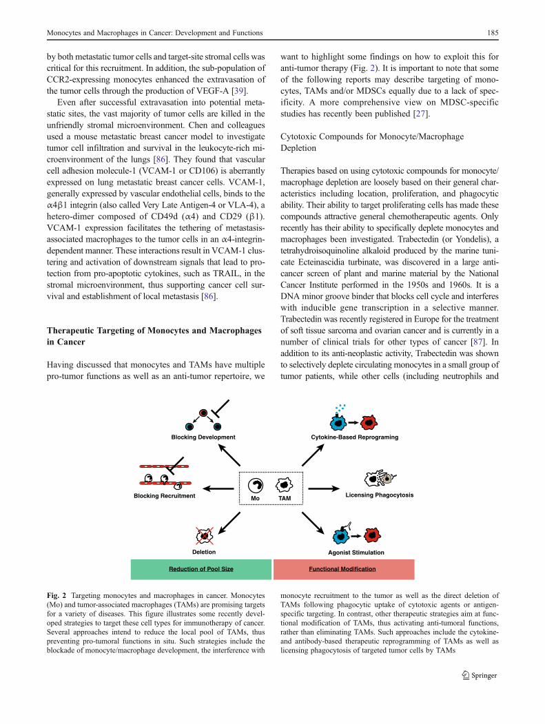
Fig. 2 Targeting monocytes and macrophages in cancer. Monocytes(Mo) and tumor-associated macrophages (TAMs) are promising targetsfor a variety of diseases. This figure illustrates some recently devel-oped strategies to target these cell types for immunotherapy of cancer.Several approaches intend to reduce the local pool of TAMs, thuspreventing pro-tumoral functions in situ. Such strategies include theblockade of monocyte/macrophage development, the interference with
monocyte recruitment to the tumor as well as the direct deletion ofTAMs following phagocytic uptake of cytotoxic agents or antigen-specific targeting. In contrast, other therapeutic strategies aim at func-tional modification of TAMs, thus activating anti-tumoral functions,rather than eliminating TAMs. Such approaches include the cytokine-and antibody-based therapeutic reprogramming of TAMs as well aslicensing phagocytosis of targeted tumor cells by TAMs
Monocytes and Macrophages in Cancer: Development and Functions 185

lymphocytes) were significantly less sensitive to the drug [88].Furthermore, sub-cytotoxic doses of Trabectedin inhibited bothin vitro and ex vivo differentiation of monocytes to macro-phages. Bisphosphonates are another class of anti-neoplasticcompounds being tested for their ability to deplete macro-phages in vivo. They are the primary treatment of bone metas-tases, secondary to several tumor types and have direct effectson different cancer cell lines [89]. To selectively target macro-phages, the drugs are encapsulated into liposomes, which arethen specifically endocytosed and degraded by macrophages.For example, clodronate-encapsulated liposomes have beenused for in vivo depletion of macrophages and have shownsubsequent reduction of tumor growth in a variety of tumormodels [63, 90].
Antigen-Specific Targeting of Tumor-AssociatedMacrophages
Antigen-specific tumor-targeting strategies have been widelyinvestigated and developed for clinical use. They have beenused to target, and generally kill, certain tumor cells as well asblocking the factors that promote recruitment or drive tumorprogression [91]. Unfortunately, the tremendous heterogene-ity of macrophage populations and redundancy of cell-surfacemarkers has made it difficult to use adaptive immune-basedtechniques, such as cytotoxic T cells (CTLs) or monoclonalantibodies, to specifically target TAMs. However, recent com-prehensive gene and protein expression analyses have identi-fied potential TAM-specific profiles that could be used forantigen-specific therapies [56, 92]. For example, DNA vac-cines against MHC class I-restricted epitopes derived fromlegumain were developed to generate TAM-specific CTLs.Legumain is a member of the asparaginyl endopeptidasefamily and is a stress protein highly over-expressed byTAMs in the tumor microenvironment. These vaccines in-duced a robust CD8+ T cell response against TAMs andprotected mice from lethal tumor cell challenges by suppres-sion of angiogenesis, tumor growth, and metastasis [28, 92].In addition, Movahedi and colleagues generated single-domain antibodies (sdAb) that are specific for the macrophagemannose receptor (MMR or CD206), which they found to behighly expressed on certain TAM subpopulations [56]. TheMMR-specific sdAb were able to bind TAMs isolated frommultiple tumor types ex vivo as well as in vivo followingintravenous injection of sdAb [93]. These pre-clinical resultssuggest that antigen-specific targeting of TAMs is possible,although it remains to be seen if this will translate to effectivetherapy.
Targeting Monocyte Recruitment
High densities of M2-like TAMs in tumors are often asso-ciated with poor clinical outcomes and TAM depletion
strategies are successful at improving clinical outcomes.Therefore, blocking migration of monocytes into the tumortissue could be an effective, and potentially less harmful,therapeutic strategy. As previously mentioned, a number oftumor-derived factors and their receptors, including CCL2,M-CSF and CXCL12 play important roles in recruitingmonocytes into tumor tissue.
Using a CCL2-specific monoclonal antibody, Qian andcolleagues inhibited the tumor recruitment of monocytes,decreased metastases and increased survival in a mammarytumor transgenic mouse system [39]. In addition, Leuschnerand colleagues targeted monocyte recruitment to tumorsusing siRNA-mediated silencing of the chemokine receptorCCR2 in EL4 lymphoma and CT26 colon carcinoma mod-els in mice, which resulted in decreased TAM accumulationand tumor growth [94].
M-CSF is critical for recruitment and differentiation ofmonocytes, and thus an important target for therapy. Manycomplementary strategies have been used to demonstratethat blocking this pathway significantly reduces monocyteinfiltration, tumor growth and metastasis in a variety oftumor models. This includes targeting both M-CSF and M-CSFR with antisense-RNA, siRNA, monoclonal antibodiesand chemical signaling inhibitors [27, 63, 95–97].
The CXCL12-CXCR4 pathway has been shown to par-ticipate in recruiting monocytes to tumor tissues. For exam-ple, interrupting this pathway with a CXCR4 antagonist issufficient to significantly reduce recruitment of monocytesand inhibit tumor growth in multiple tumor models [27, 98,99]. Chemokines promote monocyte recruitment into tissue,in part, by promoting changes in integrin affinity and aviditythat increases attachment to endothelial cells bordering tu-mor tissue. In a subcutaneous Lewis lung carcinoma model,CXCL12 was shown to activate the α4β1 integrin on mono-cytes, increase integrin clustering and monocyte attachmentto VCAM-1-expressing endothelial cell monolayers in vitro[98]. The importance of the α4β1 integrin in monocyterecruitment to tumor tissue was confirmed by using anα4–specific blocking antibody and an α4–specific small-molecule inhibitor to decrease monocyte infiltration andtumor growth [98]. These advancements in understandingthe mechanisms behind CXCL12-CXCR4-mediated recruit-ment will provide additional targets for therapy.
Therapeutic Reprogramming of Tumor-AssociatedMacrophages
The numerous uniquely polarized sub-populations of TAMsfound in different stages (and locations) of tumor develop-ment are potential targets for developing therapies that in-terfere with (or “reprogram”) macrophage differentiation.Manipulation of macrophage polarization is an especiallybroad topic that has been more comprehensively reviewed
186 D.M. Richards et al.
[27, 28]. In this review, we will highlight some of the morerecently described and further developed therapeutic ideasand give examples of two distinct techniques, monoclonalantibodies to surface-expressed proteins and recombinantcytokines, used to reprogram macrophages.
Beatty and colleagues used a human clinical trial and amouse model to test if CD40 activation could reverse immunesuppression and promote the anti-tumor T cell responses inpancreatic ductal adenocarcinoma (PDA) [100]. They tested ahumanized CD40 agonist antibody (in combination with gem-citabine chemotherapy) in a small group of patients withadvanced PDA and observed tumor regressions in somepatients. Unexpectedly, tumor regression did not correlatewith lymphocyte infiltration in primary lesions. In fact, furtherexperiments in the mouse model found that tumor regressionrequired macrophages but not T cells (or gemcitabine) [100].They observed that treatment with the CD40 agonist antibodyresulted in reprogramming of TAMs, including up-regulationof MHC class II and the costimulatory molecule CD86, accu-mulation in tumor tissue and ability to lyse tumor cells exvivo. These results demonstrated a novel mechanism forCD40-mediated tumor elimination involving TAMs [100].
IL-12 is an important cytokine that is involved in lympho-cyte and macrophage polarization and differentiation.Intravenous treatment of tumor-bearing mice with recombi-nant IL-12 has been shown to induce tumor regression andTAM conversion from a pro- to anti-tumoral phenotype [27,101]. The following two examples are interesting becausethey describe IL-12-mediated TAM reprogramming using aunique cytokine delivery method. Kerkar and colleagues gen-erated IL-12-secreting, tumor-specific CD8+ T cells in theB16 mouse model of established melanoma. By adoptivelytransferring these tumor-derived antigen-specific T cells, theywere able to deliver IL-12 directly into the tumor tissue. Theyfound that anti-tumor activity depended on the ability of themyeloid-derived cells, but not lymphocytes or NK cells, torespond to IL-12. In addition, this T cell-delivered IL-12resulted in significant reprogramming of multiple myeloid-derived cell populations, including MDSCs, macrophages,and DCs within the tumor [102]. Chmielewski and colleaguestook a different approach by engineering cytotoxic T cellswith a chimeric antigen receptor (CAR) with specificity fora tumor-associated antigen. These cells release an inducibleIL-12 (iIL12) upon CAR engagement in the tumor tissue[103]. Adoptive transfer of these CAR-iIL12 T cells resultedin an accumulation of activated macrophages within the tumortissue that was critical to the anti-tumor response [103].
TNF-α is also an important cytokine involved in lympho-cyte and macrophage polarization. Like the previous exam-ples, this report is particularly interesting because of thedelivery method. In a recent study, Johansson and colleaguesengineered TNF-α with a tumor vasculature-targeting peptideto accurately deliver specific doses of the cytokine into mice
carrying pancreatic neuro-endocrine tumors [104]. They dem-onstrated that intravenous injections of low-dose TNF-α en-hanced anti-tumor immunotherapy not by destroying thetumor vasculature, like intra-tumor interferon-γ (IFN-γ), butinstead by increasing vascular stability and vessel perfusion.This vascular remodeling was mediated, in part, by TAMs,reprogrammed towards a more inflammatory anti-tumor pro-file [104].
Licensing Phagocytosis
CD47, a cell surface protein in the immunoglobulin super-family, is expressed in the majority of normal tissues. Itserves as an anti-phagocytic signal by binding to SIRPα, aprotein expressed on macrophages and dendritic cells [105].A number of recent reports have described how tumor cellsuse this pathway to avoid phagocytosis. Tumor cells wereshown to constitutively up-regulate CD47 expression inmultiple tumor entities and animal models. Furthermore,over-expression of CD47 was a poor prognostic factor andcorrelated with increased pathogenicity [106–110]. Treatmentof solid or hematopoietic tumors with either CD47-specificblocking antibodies or SIRPα-fusion proteins resulted in anincrease in macrophage-dependent phagocytosis of tumorcells and a corresponding decrease in tumor size and numberof metastases [107–111]. Importantly, there did not seem to bea significant increase in the phagocytosis of normal cells invitro or in vivo.
Concluding Remarks
Targeting the myeloid immune cell compartment in tumors isa promising approach for immunotherapy. Three cell typesmay be of special interest here: monocytes, TAMs andMDSCs. Currently, two main strategies are investigated: re-duction of the pool size and functional modification of thesemyeloid cells (Fig. 2). The number of tumor infiltratingmono-cytes and macrophages can be reduced by controlling thedevelopment, recruitment and survival of these cells. Sincetheir half-life is relatively short, interfering with generationand differentiation may have great potential. Several tools areavailable to modify the cellular functions. The overall goal isto activate the anti-tumoral potential of these myeloid cells.This can be achieved either by agonistic antibodies targetingsurface structures such as CD40 or by reprogramming thecytokine profile of the cells. Another interesting approach isto license the phagocytosis potential by disrupting the CD47-SIRPα axis. Multiple manipulation strategies have been men-tioned in this review and many more have been tested, butunfortunately, could not be discussed in-depth here.
In summary, targeting myeloid cells for immunotherapy isvery encouraging, but whether a monotherapy will be
Monocytes and Macrophages in Cancer: Development and Functions 187
sufficient or a combination with adoptive immunotherapy orother strategies will be more effective remains to bedetermined.
Acknowledgments This work was supported by a grant from theHelmholtz Association of German Research Centers (HGF; HZ-NG-505)to M.F. and J.H. was supported by a HGF-PhD-fellowship. The authorsdeclare no competing financial interests.
References
1. van Furth R, Cohn ZA (1968) The origin and kinetics of mono-nuclear phagocytes. J Exp Med 128:415–435
2. Schulz C, Gomez PE, Chorro L, Szabo-Rogers H, Cagnard N,Kierdorf K, Prinz M, Wu B, Jacobsen SE, Pollard JW, FramptonJ, Liu KJ, Geissmann F (2012) A lineage of myeloid cellsindependent of Myb and hematopoietic stem cells. Science336:86–90
3. Auffray C, Sieweke MH, Geissmann F (2009) Blood monocytes:development, heterogeneity, and relationship with dendritic cells.Annu Rev Immunol 27:669–692
4. Pollard JW (2009) Trophic macrophages in development anddisease. Nat Rev Immunol 9:259–270
5. Schafer M, Werner S (2008) Cancer as an overhealing wound: anold hypothesis revisited. Nat Rev Mol Cell Biol 9:628–638
6. Mosser DM, Edwards JP (2008) Exploring the full spectrum ofmacrophage activation. Nat Rev Immunol 8:958–969
7. Gordon S, Taylor PR (2005) Monocyte and macrophage hetero-geneity. Nat Rev Immunol 5:953–964
8. Swirski FK, Nahrendorf M, Etzrodt M, Wildgruber M, Cortez-Retamozo V, Panizzi P, Figueiredo JL, Kohler RH, ChudnovskiyA, Waterman P, Aikawa E, Mempel TR, Libby P, Weissleder R,Pittet MJ (2009) Identification of splenic reservoir monocytes andtheir deployment to inflammatory sites. Science 325:612–616
9. Robbins CS, Chudnovskiy A, Rauch PJ, Figueiredo JL, Iwamoto Y,Gorbatov R, Etzrodt M, Weber GF, Ueno T, van Rooijen N,Mulligan-Kehoe MJ, Libby P, Nahrendorf M, Pittet MJ, WeisslederR, Swirski FK (2012) Extramedullary hematopoiesis generates Ly-6C(high) monocytes that infiltrate atherosclerotic lesions. Circulation125:364–374
10. Cortez-Retamozo V, Etzrodt M, Newton A, Rauch PJ,Chudnovskiy A, Berger C, Ryan RJ, Iwamoto Y, Marinelli B,Gorbatov R, Forghani R, Novobrantseva TI, Koteliansky V,Figueiredo JL, Chen JW, Anderson DG, Nahrendorf M,Swirski FK, Weissleder R, Pittet MJ (2012) Origins of tumor-associated macrophages and neutrophils. Proc Natl Acad Sci U SA 109:2491–2496
11. Geissmann F, Manz MG, Jung S, Sieweke MH, Merad M, Ley K(2010) Development of monocytes, macrophages, and dendriticcells. Science 327:656–661
12. Fogg DK, Sibon C, Miled C, Jung S, Aucouturier P, Littman DR,Cumano A, Geissmann F (2006) A clonogenic bone marrowprogenitor specific for macrophages and dendritic cells. Science311:83–87
13. Wiktor-Jedrzejczak WW, Ahmed A, Szczylik C, Skelly RR(1982) Hematological characterization of congenital osteopetro-sis in op/op mouse. Possible mechanism for abnormal macro-phage differentiation. J Exp Med 156:1516–1527
14. Valledor AF, Borras FE, Cullell-Young M, Celada A (1998)Transcription factors that regulate monocyte/macrophage differ-entiation. J Leukoc Biol 63:405–417
15. Akashi K, Traver D, Miyamoto T, Weissman IL (2000) A clono-genic common myeloid progenitor that gives rise to all myeloidlineages. Nature 404:193–197
16. Auffray C, Fogg DK, Narni-Mancinelli E, Senechal B, Trouillet C,Saederup N, Leemput J, Bigot K, Campisi L, Abitbol M, Molina T,Charo I, Hume DA, Cumano A, Lauvau G, Geissmann F (2009)CX3CR1+ CD115+ CD135+ common macrophage/DC precursorsand the role of CX3CR1 in their response to inflammation. J ExpMed 206:595–606
17. Ziegler-Heitbrock L, Ancuta P, Crowe S, Dalod M, Grau V, HartDN, Leenen PJ, Liu YJ, MacPherson G, Randolph GJ,Scherberich J, Schmitz J, Shortman K, Sozzani S, Strobl H,Zembala M, Austyn JM, Lutz MB (2010) Nomenclature ofmonocytes and dendritic cells in blood. Blood 116:e74–e80
18. Auffray C, Fogg D, Garfa M, Elain G, Join-Lambert O, Kayal S,Sarnacki S, Cumano A, Lauvau G, Geissmann F (2007)Monitoringof blood vessels and tissues by a population of monocytes withpatrolling behavior. Science 317:666–670
19. Geissmann F, Jung S, Littman DR (2003) Blood monocytes consistof two principal subsets with distinct migratory properties.Immunity 19:71–82
20. Passlick B, Flieger D, Ziegler-Heitbrock HW (1989) Identificationand characterization of a novel monocyte subpopulation in humanperipheral blood. Blood 74:2527–2534
21. Cros J, Cagnard N, Woollard K, Patey N, Zhang SY, Senechal B,Puel A, Biswas SK, Moshous D, Picard C, Jais JP, D’Cruz D,Casanova JL, Trouillet C, Geissmann F (2010) Human CD14dimmonocytes patrol and sense nucleic acids and viruses via TLR7and TLR8 receptors. Immunity 33:375–386
22. Ingersoll MA, Spanbroek R, Lottaz C, Gautier EL, FrankenbergerM, Hoffmann R, Lang R, Haniffa M, Collin M, Tacke F, HabenichtAJ, Ziegler-Heitbrock L, Randolph GJ (2010) Comparison of geneexpression profiles between human and mouse monocyte subsets.Blood 115:e10–e19
23. Varol C, Landsman L, Fogg DK, Greenshtein L, Gildor B, MargalitR, Kalchenko V, Geissmann F, Jung S (2007) Monocytes give riseto mucosal, but not splenic, conventional dendritic cells. J ExpMed204:171–180
24. Hanna RN, Carlin LM, Hubbeling HG, Nackiewicz D, GreenAM, Punt JA, Geissmann F, Hedrick CC (2011) The transcriptionfactor NR4A1 (Nur77) controls bone marrow differentiation andthe survival of Ly6C- monocytes. Nat Immunol 12:778–785
25. Etzrodt M, Cortez-Retamozo V, Newton A, Zhao J, Ng A,Wildgruber M, Romero P, Wurdinger T, Xavier R, GeissmannF, Meylan E, Nahrendorf M, Swirski FK, Baltimore D,Weissleder R, Pittet MJ (2012) Regulation of monocyte function-al heterogeneity by miR-146a and Relb. Cell Rep 1:317–324
26. Sica A, Bronte V (2007) Altered macrophage differentiation andimmune dysfunction in tumor development. J Clin Invest 117:1155–1166
27. Gabrilovich DI, Ostrand-Rosenberg S, Bronte V (2012)Coordinated regulation of myeloid cells by tumours. Nat RevImmunol 12:253–268
28. Sica A, Porta C, Morlacchi S, Banfi S, Strauss L, Rimoldi M,Totaro MG, Riboldi E (2012) Origin and functions of tumor-associated myeloid cells (TAMCs). Cancer Microenviron 5:133–149
29. Gabrilovich DI, Nagaraj S (2009) Myeloid-derived suppressor cellsas regulators of the immune system. Nat Rev Immunol 9:162–174
30. Daley JM, Thomay AA, Connolly MD, Reichner JS, Albina JE(2008) Use of Ly6G-specific monoclonal antibody to depleteneutrophils in mice. J Leukoc Biol 83:64–70
31. Fleming TJ, Fleming ML, Malek TR (1993) Selective expressionof Ly-6G on myeloid lineage cells in mouse bone marrow. RB6-8C5 mAb to granulocyte-differentiation antigen (Gr-1) detectsmembers of the Ly-6 family. J Immunol 151:2399–2408
188 D.M. Richards et al.
32. Shi C, Pamer EG (2011) Monocyte recruitment during infectionand inflammation. Nat Rev Immunol 11:762–774
33. Shi C, Jia T, Mendez-Ferrer S, Hohl TM, Serbina NV, Lipuma L,Leiner I, Li MO, Frenette PS, Pamer EG (2011) Bone marrowmesenchymal stem and progenitor cells induce monocyte emigra-tion in response to circulating toll-like receptor ligands. Immunity34:590–601
34. Serbina NV, Pamer EG (2006) Monocyte emigration from bonemarrow during bacterial infection requires signals mediated bychemokine receptor CCR2. Nat Immunol 7:311–317
35. Martin AP, Rankin S, Pitchford S, Charo IF, Furtado GC, Lira SA(2008) Increased expression of CCL2 in insulin-producing cellsof transgenic mice promotes mobilization of myeloid cells fromthe bone marrow, marked insulitis, and diabetes. Diabetes57:3025–3033
36. Tacke F, Alvarez D, Kaplan TJ, Jakubzick C, Spanbroek R,Llodra J, Garin A, Liu J, Mack M, van Rooijen N, Lira SA,Habenicht AJ, Randolph GJ (2007) Monocyte subsets differen-tially employ CCR2, CCR5, and CX3CR1 to accumulate withinatherosclerotic plaques. J Clin Invest 117:185–194
37. Bottazzi B, Polentarutti N, Acero R, Balsari A, Boraschi D,Ghezzi P, Salmona M, Mantovani A (1983) Regulation of themacrophage content of neoplasms by chemoattractants. Science220:210–212
38. Pollard JW (2004) Tumour-educated macrophages promote tu-mour progression and metastasis. Nat Rev Cancer 4:71–78
39. Qian BZ, Li J, Zhang H, Kitamura T, Zhang J, Campion LR,Kaiser EA, Snyder LA, Pollard JW (2011) CCL2 recruits inflam-matory monocytes to facilitate breast-tumour metastasis. Nature475:222–225
40. Joyce JA, Pollard JW (2009) Microenvironmental regulation ofmetastasis. Nat Rev Cancer 9:239–252
41. De Palma M, Naldini L (2009) Tie2-expressing monocytes(TEMs): novel targets and vehicles of anticancer therapy?Biochim Biophys Acta 1796:5–10
42. Krausgruber T, Blazek K, Smallie T, Alzabin S, Lockstone H,Sahgal N, Hussell T, Feldmann M, Udalova IA (2011) IRF5promotes inflammatory macrophage polarization and TH1-TH17 responses. Nat Immunol 12:231–238
43. Stout RD, Jiang C, Matta B, Tietzel I, Watkins SK, Suttles J (2005)Macrophages sequentially change their functional phenotype inresponse to changes in microenvironmental influences. J Immunol175:342–349
44. Hanna RN, Shaked I, Hubbeling HG, Punt JA, Wu R, Herrley E,Zaugg C, Pei H, Geissmann F, Ley K, Hedrick CC (2012)NR4A1 (Nur77) deletion polarizes macrophages toward an in-flammatory phenotype and increases atherosclerosis. Circ Res110:416–427
45. Gordon S, Martinez FO (2010) Alternative activation of macro-phages: mechanism and functions. Immunity 32:593–604
46. Biswas SK, Mantovani A (2010) Macrophage plasticity andinteraction with lymphocyte subsets: cancer as a paradigm. NatImmunol 11:889–896
47. Sica A, Larghi P, Mancino A, Rubino L, Porta C, Totaro MG,Rimoldi M, Biswas SK, Allavena P, Mantovani A (2008)Macrophage polarization in tumour progression. Semin Cancer Biol18:349–355
48. Cheong C, Matos I, Choi JH, Dandamudi DB, Shrestha E,Longhi MP, Jeffrey KL, Anthony RM, Kluger C, Nchinda G,Koh H, Rodriguez A, Idoyaga J, Pack M, Velinzon K, Park CG,Steinman RM (2010) Microbial stimulation fully differentiatesmonocytes to DC-SIGN/CD209(+) dendritic cells for immune Tcell areas. Cell 143:416–429
49. Naik SH, Sathe P, Park HY, Metcalf D, Proietto AI, Dakic A,Carotta S, O’Keeffe M, Bahlo M, Papenfuss A, Kwak JY, Wu L,ShortmanK (2007) Development of plasmacytoid and conventional
dendritic cell subtypes from single precursor cells derived in vitroand in vivo. Nat Immunol 8:1217–1226
50. Onai N, Obata-Onai A, Schmid MA, Ohteki T, Jarrossay D, ManzMG (2007) Identification of clonogenic common Flt3 + M-CSFR+ plasmacytoid and conventional dendritic cell progenitors inmouse bone marrow. Nat Immunol 8:1207–1216
51. Liu K, Victora GD, Schwickert TA, Guermonprez P, MeredithMM, Yao K, Chu FF, Randolph GJ, Rudensky AY, NussenzweigM (2009) In vivo analysis of dendritic cell development andhomeostasis. Science 324:392–397
52. Varol C, Vallon-Eberhard A, Elinav E, Aychek T, Shapira Y,Luche H, Fehling HJ, Hardt WD, Shakhar G, Jung S (2009)Intestinal lamina propria dendritic cell subsets have differentorigin and functions. Immunity 31:502–512
53. Rivollier A, He J, Kole A, Valatas V, Kelsall BL (2012)Inflammation switches the differentiation program of Ly6Chimonocytes from antiinflammatory macrophages to inflammatorydendritic cells in the colon. J Exp Med 209:139–155
54. Merad M, Manz MG (2009) Dendritic cell homeostasis. Blood113:3418–3427
55. Shortman K, Naik SH (2007) Steady-state and inflammatorydendritic-cell development. Nat Rev Immunol 7:19–30
56. Movahedi K, Laoui D, Gysemans C, Baeten M, Stange G, Vanden Bossche J, Mack M, Pipeleers D, In’t Veld P, De Baetselier P,Van Ginderachter JA (2010) Different tumor microenvironmentscontain functionally distinct subsets of macrophages derived fromLy6C(high) monocytes. Cancer Res 70:5728–5739
57. Bingle L, Brown NJ, Lewis CE (2002) The role of tumour-associated macrophages in tumour progression: implications fornew anticancer therapies. J Pathol 196:254–265
58. Nakayama Y, Nagashima N, Minagawa N, Inoue Y, Katsuki T,Onitsuka K, Sako T, Hirata K, Nagata N, Itoh H (2002)Relationships between tumor-associated macrophages and clinico-pathological factors in patients with colorectal cancer. AnticancerRes 22:4291–4296
59. Lin EY, Pollard JW (2007) Tumor-associated macrophagespress the angiogenic switch in breast cancer. Cancer Res67:5064–5066
60. Ryder M, Ghossein RA, Ricarte-Filho JC, Knauf JA, Fagin JA(2008) Increased density of tumor-associated macrophages isassociated with decreased survival in advanced thyroid cancer.Endocr Relat Cancer 15:1069–1074
61. Ohri CM, Shikotra A, Green RH, Waller DA, Bradding P (2009)Macrophages within NSCLC tumour islets are predominantly ofa cytotoxic M1 phenotype associated with extended survival. EurRespir J 33:118–126
62. O’Sullivan T, Saddawi-Konefka R, Vermi W, Koebel CM, ArthurC, White JM, Uppaluri R, Andrews DM, Ngiow SF, Teng MW,Smyth MJ, Schreiber RD, Bui JD (2012) Cancer immunoeditingby the innate immune system in the absence of adaptive immu-nity. J Exp Med 209:1869–1882
63. Qian BZ, Pollard JW (2010) Macrophage diversity enhancestumor progression and metastasis. Cell 141:39–51
64. Mantovani A, Sica A (2010) Macrophages, innate immunity andcancer: balance, tolerance, and diversity. Curr Opin Immunol22:231–237
65. Tan KL, Scott DW, Hong F, Kahl BS, Fisher RI, Bartlett NL,Advani RH, Buckstein R, Rimsza LM, Connors JM, Steidl C,Gordon LI, Horning SJ, Gascoyne RD (2012) Tumor-associatedmacrophages predict inferior outcomes in classical Hodgkin lym-phoma: a correlative study from the E2496 Intergroup trial. Blood[Epub ahead of print]
66. Mantovani A, Allavena P, Sica A, Balkwill F (2008) Cancer-related inflammation. Nature 454:436–444
67. Hanahan D, Weinberg RA (2000) The hallmarks of cancer. Cell100:57–70
Monocytes and Macrophages in Cancer: Development and Functions 189

68. Hanahan D, Weinberg RA (2011) Hallmarks of cancer: the nextgeneration. Cell 144:646–674
69. Meira LB, Bugni JM, Green SL, Lee CW, Pang B, BorenshteinD, Rickman BH, Rogers AB, Moroski-Erkul CA, McFaline JL,Schauer DB, Dedon PC, Fox JG, Samson LD (2008) DNAdamage induced by chronic inflammation contributes to coloncarcinogenesis in mice. J Clin Invest 118:2516–2525
70. DiNapoli MR, Calderon CL, Lopez DM (1996) The alteredtumoricidal capacity of macrophages isolated from tumor-bearing mice is related to reduce expression of the induciblenitric oxide synthase gene. J Exp Med 183:1323–1329
71. Balkwill F, Charles KA, Mantovani A (2005) Smoldering andpolarized inflammation in the initiation and promotion of malig-nant disease. Cancer Cell 7:211–217
72. Lin EY, Li JF, Bricard G, Wang W, Deng Y, Sellers R, PorcelliSA, Pollard JW (2007) Vascular endothelial growth factorrestores delayed tumor progression in tumors depleted of macro-phages. Mol Oncol 1:288–302
73. Bergers G, Benjamin LE (2003) Tumorigenesis and the angio-genic switch. Nat Rev Cancer 3:401–410
74. Lin EY, Li JF, Gnatovskiy L, Deng Y, Zhu L, Grzesik DA, QianH, Xue XN, Pollard JW (2006) Macrophages regulate the angio-genic switch in a mouse model of breast cancer. Cancer Res66:11238–11246
75. De Palma M, Venneri MA, Roca C, Naldini L (2003) Targetingexogenous genes to tumor angiogenesis by transplantation ofgenetically modified hematopoietic stem cells. Nat Med 9:789–795
76. De PalmaM, Venneri MA, Galli R, Sergi SL, Politi LS, SampaolesiM, Naldini L (2005) Tie2 identifies a hematopoietic lineage ofproangiogenic monocytes required for tumor vessel formation anda mesenchymal population of pericyte progenitors. Cancer Cell8:211–226
77. Venneri MA, De Palma M, Ponzoni M, Pucci F, Scielzo C, ZonariE, Mazzieri R, Doglioni C, Naldini L (2007) Identification ofproangiogenic TIE2-expressing monocytes (TEMs) in humanperipheral blood and cancer. Blood 109:5276–5285
78. Murdoch C, Tazzyman S, Webster S, Lewis CE (2007)Expression of Tie-2 by human monocytes and their responsesto angiopoietin-2. J Immunol 178:7405–7411
79. Lin EY, Nguyen AV, Russell RG, Pollard JW (2001) Colony-stimulating factor 1 promotes progression of mammary tumors tomalignancy. J Exp Med 193:727–740
80. Condeelis J, Pollard JW (2006) Macrophages: obligate partnersfor tumor cell migration, invasion, and metastasis. Cell 124:263–266
81. Wyckoff J, Wang W, Lin EY, Wang Y, Pixley F, Stanley ER, GrafT, Pollard JW, Segall J, Condeelis J (2004) A paracrine loopbetween tumor cells and macrophages is required for tumor cellmigration in mammary tumors. Cancer Res 64:7022–7029
82. Lee Y, Chittezhath M, Andre V, Zhao H, Poidinger M, Biondi A,D’Amico G, Biswas SK (2012) Protumoral role of monocytes inhuman B-cell precursor acute lymphoblastic leukemia: involve-ment of the chemokine CXCL10. Blood 119:227–237
83. Wyckoff JB, Wang Y, Lin EY, Li JF, Goswami S, Stanley ER,Segall JE, Pollard JW, Condeelis J (2007) Direct visualization ofmacrophage-assisted tumor cell intravasation in mammarytumors. Cancer Res 67:2649–2656
84. Zervantonakis IK, Hughes-Alford SK, Charest JL, Condeelis JS,Gertler FB, Kamm RD (2012) Three-dimensional microfluidicmodel for tumor cell intravasation and endothelial barrier func-tion. Proc Natl Acad Sci U S A 109:13515–13520
85. Qian B, Deng Y, Im JH, Muschel RJ, Zou Y, Li J, Lang RA,Pollard JW (2009) A distinct macrophage population mediatesmetastatic breast cancer cell extravasation, establishment andgrowth. PLoS One 4:e6562
86. Chen Q, Zhang XH, Massague J (2011) Macrophage binding toreceptor VCAM-1 transmits survival signals in breast cancer cellsthat invade the lungs. Cancer Cell 20:538–549
87. Allavena P, Mantovani A (2012) Immunology in the clinic reviewseries; focus on cancer: tumour-associated macrophages: undis-puted stars of the inflammatory tumour microenvironment. ClinExp Immunol 167:195–205
88. Allavena P, Signorelli M, Chieppa M, Erba E, Bianchi G,Marchesi F, Olimpio CO, Bonardi C, Garbi A, Lissoni A, deBraud F, Jimeno J, D’Incalci M (2005) Anti-inflammatory prop-erties of the novel antitumor agent yondelis (trabectedin): inhibi-tion of macrophage differentiation and cytokine production.Cancer Res 65:2964–2971
89. Morgan G, Lipton A (2010) Antitumor effects and anticancerapplications of bisphosphonates. Semin Oncol 37(Suppl 2):S30–S40
90. Gazzaniga S, Bravo AI, Guglielmotti A, van Rooijen N, MaschiF, Vecchi A, Mantovani A, Mordoh J, Wainstok R (2007)Targeting tumor-associated macrophages and inhibition ofMCP-1 reduce angiogenesis and tumor growth in a human mel-anoma xenograft. J Invest Dermatol 127:2031–2041
91. Zafir-Lavie I, Michaeli Y, Reiter Y (2007) Novel antibodies asanticancer agents. Oncogene 26:3714–3733
92. Lewen S, Zhou H, Hu HD, Cheng T, Markowitz D, Reisfeld RA,Xiang R, Luo Y (2008) A Legumain-based minigene vaccinetargets the tumor stroma and suppresses breast cancer growthand angiogenesis. Cancer Immunol Immunother 57:507–515
93. Movahedi K, Schoonooghe S, Laoui D, Houbracken I, WaelputW, Breckpot K, Bouwens L, Lahoutte T, De Baetselier P, Raes G,Devoogdt N, Van Ginderachter JA (2012) Nanobody-based tar-geting of the macrophage mannose receptor for effective in vivoimaging of tumor-associated macrophages. Cancer Res 72:4165–4177
94. Leuschner F, Dutta P, Gorbatov R, Novobrantseva TI, DonahoeJS, Courties G, Lee KM, Kim JI, Markmann JF, Marinelli B,Panizzi P, Lee WW, Iwamoto Y, Milstein S, Epstein-Barash H,Cantley W, Wong J, Cortez-Retamozo V, Newton A, Love K,Libby P, Pittet MJ, Swirski FK, Koteliansky V, Langer R,Weissleder R, Anderson DG, Nahrendorf M (2011) TherapeuticsiRNA silencing in inflammatory monocytes in mice. NatBiotechnol 29:1005–1010
95. Aharinejad S, Paulus P, Sioud M, Hofmann M, Zins K, Schafer R,Stanley ER, Abraham D (2004) Colony-stimulating factor-1blockade by antisense oligonucleotides and small interferingRNAs suppresses growth of human mammary tumor xenograftsin mice. Cancer Res 64:5378–5384
96. Paulus P, Stanley ER, Schafer R, Abraham D, Aharinejad S(2006) Colony-stimulating factor-1 antibody reverses chemore-sistance in human MCF-7 breast cancer xenografts. Cancer Res66:4349–4356
97. DeNardo DG, Brennan DJ, Rexhepaj E, Ruffell B, Shiao SL,Madden SF, Gallagher WM, Wadhwani N, Keil SD, Junaid SA,Rugo HS, Hwang ES, Jirstrom K, West BL, Coussens LM (2011)Leukocyte complexity predicts breast cancer survival and function-ally regulates response to chemotherapy. Cancer Discov 1:54–67
98. Schmid MC, Avraamides CJ, Foubert P, Shaked Y, Kang SW,Kerbel RS, Varner JA (2011) Combined blockade of integrin-alpha4beta1 plus cytokines SDF-1alpha or IL-1beta potently inhib-its tumor inflammation and growth. Cancer Res 71:6965–6975
99. Yang L, Huang J, Ren X, Gorska AE, Chytil A, Aakre M,Carbone DP, Matrisian LM, Richmond A, Lin PC, Moses HL(2008) Abrogation of TGF beta signaling in mammary carcino-mas recruits Gr-1 + CD11b + myeloid cells that promote metas-tasis. Cancer Cell 13:23–35
100. Beatty GL, Chiorean EG, Fishman MP, Saboury B, TeitelbaumUR, Sun W, Huhn RD, Song W, Li D, Sharp LL, Torigian DA,
190 D.M. Richards et al.
O’Dwyer PJ, Vonderheide RH (2011) CD40 agonists alter tumorstroma and show efficacy against pancreatic carcinoma in miceand humans. Science 331:1612–1616
101. Watkins SK, Egilmez NK, Suttles J, Stout RD (2007) IL-12rapidly alters the functional profile of tumor-associated andtumor-infiltrating macrophages in vitro and in vivo. J Immunol178:1357–1362
102. Kerkar SP, Goldszmid RS, Muranski P, Chinnasamy D, Yu Z,Reger RN, Leonardi AJ, Morgan RA, Wang E, Marincola FM,Trinchieri G, Rosenberg SA, Restifo NP (2011) IL-12 triggers aprogrammatic change in dysfunctional myeloid-derived cellswithin mouse tumors. J Clin Invest 121:4746–4757
103. Chmielewski M, Kopecky C, Hombach AA, Abken H (2011) IL-12 release by engineered T cells expressing chimeric antigenreceptors can effectively Muster an antigen-independent macro-phage response on tumor cells that have shut down tumor antigenexpression. Cancer Res 71:5697–5706
104. Johansson A, Hamzah J, Payne CJ, Ganss R (2012) Tumor-targeted TNFalpha stabilizes tumor vessels and enhances activeimmunotherapy. Proc Natl Acad Sci U S A 109:7841–7846
105. Chao MP, Weissman IL, Majeti R (2012) The CD47-SIRPalphapathway in cancer immune evasion and potential therapeuticimplications. Curr Opin Immunol 24:225–232
106. Jaiswal S, Jamieson CH, Pang WW, Park CY, Chao MP, MajetiR, Traver D, van Rooijen N, Weissman IL (2009) CD47 isupregulated on circulating hematopoietic stem cells and leukemiacells to avoid phagocytosis. Cell 138:271–285
107. Majeti R, Chao MP, Alizadeh AA, Pang WW, Jaiswal S, GibbsKD, van Rooijen N, Weissman IL (2009) CD47 is an adverseprognostic factor and therapeutic antibody target on human acutemyeloid leukemia stem cells. Cell 138:286–299
108. Chao MP, Tang C, Pachynski RK, Chin R, Majeti R, WeissmanIL (2011) Extranodal dissemination of non-Hodgkin lymphomarequires CD47 and is inhibited by anti-CD47 antibody therapy.Blood 118:4890–4901
109. Edris B, Weiskopf K, Volkmer AK, Volkmer JP, Willingham SB,Contreras-Trujillo H, Liu J, Majeti R, West RB, Fletcher JA, BeckAH, Weissman IL, van de Rijn M (2012) Antibody therapy target-ing the CD47 protein is effective in a model of aggressive metastaticleiomyosarcoma. Proc Natl Acad Sci U S A 109:6656–6661
110. Willingham SB, Volkmer JP, Gentles AJ, Sahoo D, Dalerba P,Mitra SS, Wang J, Contreras-Trujillo H, Martin R, Cohen JD,
Lovelace P, Scheeren FA, Chao MP, Weiskopf K, Tang C,Volkmer AK, Naik TJ, Storm TA, Mosley AR, Edris B, SchmidSM, Sun CK, Chua MS, Murillo O, Rajendran P, Cha AC, ChinRK, Kim D, Adorno M, Raveh T, Tseng D, Jaiswal S, Enger PO,Steinberg GK, Li G, So SK, Majeti R, Harsh GR, van de Rijn M,Teng NN, Sunwoo JB, Alizadeh AA, Clarke MF, Weissman IL(2012) The CD47-signal regulatory protein alpha (SIRPa) interactionis a therapeutic target for human solid tumors. Proc Natl Acad Sci U SA 109:6662–6667
111. Theocharides AP, Jin L, Cheng PY, Prasolava TK, Malko AV, HoJM, Poeppl AG, van Rooijen N, Minden MD, Danska JS, DickJE, Wang JC (2012) Disruption of SIRPalpha signaling in macro-phages eliminates human acute myeloid leukemia stem cells inxenografts. J Exp Med 209:1883–1899
112. Wang LD, Wagers AJ (2011) Dynamic niches in the originationand differentiation of haematopoietic stem cells. Nat Rev MolCell Biol 12:643–655
113. Hume DA, Perry VH, Gordon S (1984) The mononuclear phago-cyte system of the mouse defined by immunohistochemical local-isation of antigen F4/80: macrophages associated with epithelia.Anat Rec 210:503–512
114. Morris L, Graham CF, Gordon S (1991) Macrophages in haemo-poietic and other tissues of the developing mouse detected by themonoclonal antibody F4/80. Development 112:517–526
115. Hume DA, Ross IL, Himes SR, Sasmono RT, Wells CA, Ravasi T(2002) The mononuclear phagocyte system revisited. J LeukocBiol 72:621–627
116. Serbina NV, Salazar-Mather TP, Biron CA, Kuziel WA, PamerEG (2003) TNF/iNOS-producing dendritic cells mediate innateimmune defense against bacterial infection. Immunity 19:59–70
117. Narni-Mancinelli E, Campisi L, Bassand D, Cazareth J, GounonP, Glaichenhaus N, Lauvau G (2007) Memory CD8+ T cellsmediate antibacterial immunity via CCL3 activation of TNF/ROI+phagocytes. J Exp Med 204:2075–2087
118. Serbina NV, Jia T, Hohl TM, Pamer EG (2008) Monocyte-mediateddefense against microbial pathogens. Annu Rev Immunol 26:421–452
119. Peranzoni E, Zilio S, Marigo I, Dolcetti L, Zanovello P,Mandruzzato S, Bronte V (2010) Myeloid-derived suppressorcell heterogeneity and subset definition. Curr Opin Immunol22:238–244
Monocytes and Macrophages in Cancer: Development and Functions 191