Impaired NLRP3 inflammasome activity during fetal development regulates IL-1β production in human...
34
Impaired NLRP3 inflammasome activity during fetal development regulates IL-1β production in human monocytes Ashish A. Sharma 1,2,* P, Roger Jen 1,2,* , Bernard Kan 1,2 , Abhinav Sharma 1 , Elizabeth Marchant 1,2 P, Anthony Tang 1,2 , Izabelle Gadawski 3 , Christof Senger 3 , Amanda Skoll 4 , Stuart E. Turvey 1,2,5 , Laura M. Sly 1,2,5 , Hélène C.F. Côté 3 and Pascal M. Lavoie 1,2,5 1 Child & Family Research Institute, Vancouver BC, Canada 2 Department of Experimental Medicine, University of British Columbia, Vancouver BC, Canada 3 Department of Pathology and Laboratory Medicine, University of British Columbia, Vancouver BC, Canada 4 Department of Obstetrics, University of British Columbia, Vancouver BC, Canada 5 Department of Pediatrics, University of British Columbia, Vancouver BC, Canada Keywords: Neonate; Interleukin-1 beta (IL-1β); Inflammasome; Toll-like receptor; Human Corresponding author: Dr. Pascal Lavoie, Child & Family Research Institute, 4th floor Translational Research Building, 950 West 28th Avenue, Vancouver BC V5Z 4H4 Fax: +1-604-875-3106 e-mail: [email protected] * These authors contributed equally to this work
-
Upload
independent -
Category
Documents
-
view
3 -
download
0
Transcript of Impaired NLRP3 inflammasome activity during fetal development regulates IL-1β production in human...

Impaired NLRP3 inflammasome activity during fetal development regulates IL-1β
production in human monocytes
Ashish A. Sharma1,2,*P, Roger Jen1,2,*, Bernard Kan1,2, Abhinav Sharma1, Elizabeth Marchant1,2
P,
Anthony Tang1,2, Izabelle Gadawski3, Christof Senger3, Amanda Skoll4, Stuart E. Turvey1,2,5, Laura M.
Sly1,2,5, Hélène C.F. Côté3 and Pascal M. Lavoie1,2,5
1Child & Family Research Institute, Vancouver BC, Canada
2Department of Experimental Medicine, University of British Columbia, Vancouver BC, Canada
3Department of Pathology and Laboratory Medicine, University of British Columbia, Vancouver BC,
Canada
4Department of Obstetrics, University of British Columbia, Vancouver BC, Canada
5Department of Pediatrics, University of British Columbia, Vancouver BC, Canada
Keywords: Neonate; Interleukin-1 beta (IL-1β); Inflammasome; Toll-like receptor; Human
Corresponding author:
Dr. Pascal Lavoie, Child & Family Research Institute, 4th floor Translational Research Building, 950
West 28th Avenue, Vancouver BC V5Z 4H4
Fax: +1-604-875-3106
e-mail: [email protected]
*These authors contributed equally to this work

2
Abbreviations: ASC: Apoptosis-associated speck-like protein containing a CARD; FLICA:
Fluorescent-Labeled Inhibitor of Caspase-1 Activation; FMO: Fluorescence minus one
3
Abstract
Interleukin-1β (IL-1β) production is impaired in cord blood monocytes. However, the mechanism
underlying this developmental attenuation remains unclear. Here, we analyzed the extent of variability
within the Toll-like receptor (TLR)/NLRP3 inflammasome pathways in human neonates. We show
that immature low CD14-expressing/CD16pos monocytes predominate before 33 weeks of gestation,
and that these cells lack production of the pro-IL-1β precursor protein upon LPS stimulation. In
contrast, high levels of pro-IL-1β are produced within high CD14-expressing monocytes, although
these cells are unable to secrete mature IL-1β. The lack of secreted IL-1β in these monocytes parallels
a reduction of TLR-mediated NLRP3 induction resulting in a lack of caspase-1 activity before 29
weeks of gestation, whereas expression of the ASC protein and function of the P2X7 receptor are
preserved. Our analyses also reveal a strong inhibitory effect of placental infection on LPS/ATP-
induced caspase-1 activity in cord blood monocytes. Lastly, secretion of IL-1β in preterm neonates is
restored to adult levels during the neonatal period, indicating rapid maturation of these responses after
birth. Collectively, our data highlight important developmental mechanisms regulating IL-1β responses
early in gestation, in part due to a downregulation of TLR-mediated NLRP3 expression. Such
mechanisms may serve to limit potentially damaging inflammatory responses in a developing fetus.

4
Introduction
Newborns are at high risk of infections, in part due to attenuated innate immune defenses [1]. The
cytokine interleukin-1β (IL-1β) is an important inflammatory mediator in response to infections [2].
Mice lacking IL-1β display impaired acute phase and pyrogenic responses [3], and increased
susceptibility to pathogens commonly encountered in the neonatal period [4, 5]. In contrast, high levels
of IL-1β in a fetus can result in autoimmune organ damage [2], as well as lethal metabolic disturbances
including severe weight loss and hypoglycemia [6]. Together, these data illustrate the evolutionary
importance of a tight regulation of IL-1β in order to avoid inflammation-mediated organ damage,
neurological injury [7] or even premature birth in a developing human [2, 8].
Monocytes are primarily responsible for the production of IL-1β in circulating blood [9, 10].
Three subsets predominate in humans: ‘Classical’ monocytes express CD14, but lack expression of the
immunoglobulin receptor CD16. These CD14high/CD16neg monocytes make up the majority of
monocytes in peripheral adult blood, and have been proposed to be capable of producing high amounts
of IL-10 [11]. In contrast, the smaller, non-classical CD14low/CD16pos monocytes express high levels
of the antigen-presenting molecule HLA-DR [12], as well as cytokine TNF-α [13]. This latter subset
has been proposed to play an important immediate responder ‘proinflammatory’ role in sepsis [11, 14,
15]. Finally, a third subset of monocytes expressing high levels of CD14, as well as CD16
(CD14high/CD16pos) may play a more intermediate role in inflammation [16].
Production of IL-1β differs from that of most other inflammatory cytokines as it requires a dual
signal for its activation [2]. First, intracellular pro-IL-1β is produced following stimulation of pattern-
recognition receptors (PRRs) such as the Toll-like receptors (TLRs). Second, in order to be functional
pro-IL-1β requires proteolytic cleavage, predominantly by caspase-1, a component of the NLRP3
inflammasome multi-protein complex, resulting in secretion of mature, biologically active IL-1β [17].
Activation of the NLRP3 inflammasome can be triggered by local mediators of host cell damage in
5
vivo (e.g. free radicals, DNA or adenosine triphosphate: ATP, through the purinoceptor P2X7) [18].
Earlier studies showed that IL-1β production is markedly reduced in human cord blood monocytes in
response to endotoxin (LPS) [19, 20]. Caspase-1 processing was also reduced in term neonates [21].
However, the functional consequence of this reduced caspase-1 processing on neonatal IL-1β
responses has never been functionally characterized.
In order to understand the molecular basis for the regulation of IL-1β secretion in monocytes in
humans at an early stage in life, we analyzed the developmental variability in the TLR and NLRP3
inflammasome pathways in neonates born late in gestation. Given the need for a tight regulation of IL-
1β responses in order to avoid major metabolic disturbances in the fetus, we hypothesized that the
NLRP3 inflammasome is regulated in a discrete manner before the term of gestation.
Results
Immature monocytes in cord blood before the term of gestation
Data are greatly lacking regarding the ontogeny of monocyte subsets in humans. To address this, we
first analyzed subsets of monocytes in preterm, term and adult subjects, using standard flow cytometry
gating strategy, as defined previously [22] (figure 1A). Expectedly, cumulative proportions of
monocytes were generally preserved in cord blood relative to blood mononuclear cells (BMC) (figure
1B). Also, monocytes from all three age groups displayed comparable expression of the myeloid
markers CD33 and CD11c, whereas expression of HLA-DR was reduced in preterm cord blood
(Supporting Information figure 1). Notably, we observed a predominance of CD14low/CD16pos
monocytes in preterm cord BMC (figure 1C), and this subset of monocytes did not express any
detectable intracellular pro-IL-1β upon LPS stimulation, suggesting that they represent an immature
form (figure 1D).

6
In contrast, when testing the functional capacity of the cord blood high CD14-expressing subset of
monocytes, both CD16pos and CD16neg subsets expressed levels of intracellular pro-IL-1β comparable
(if not higher than) to that of adult peripheral blood (figure 1D). In these cells, IL-1β secretion was
also profoundly reduced at decreasing gestation, as confirmed on a per cell basis, and was not restored
following co-activation of the inflammasome (figure 2A). To exclude a contribution of low CD14-
expressing monocytes to IL-1β secretion, negatively purified monocytes were similarly assayed, and
yielded identical results (not shown). This lack of mature IL-1β secretion at decreasing gestation was
also observed regardless of the TLR engaged (figure 2B). Altogether, these data demonstrate a
profound impairment of IL-1β production in high-CD14-expressing preterm monocytes despite
intracellular detection of pro-IL-1β.
Reduced TLR-induced IL-1β response in high CD14-expressing preterm monocytes
As shown above, pro-IL-1β was detectable in high CD14-expressing preterm monocytes. To better
understand the molecular basis for the developmental inability of monocytes to secrete IL-1β, we
examined whether expression of the IL1B transcript was induced following LPS stimulation. Indeed,
expression of the IL1B gene was significantly reduced at lower gestation (figure 3A). In parallel
experiments, secretion of IL-1β in whole blood was also markedly impaired indicating that the
gestational age-dependent attenuation in IL-1β secretion is not due to a lack of serum factor(s)
excluded during the BMC purification (figure 3B). Treatment of cells with cycloheximide prior to LPS
stimulation also completely abrogated secretion of IL-1β, consistent with a de novo requirement for
protein expression following LPS challenge, in order to produce mature IL-1β in all three age groups
(figure 3C). Moreover, production of the inflammasome-independent cytokine IL-6 was similarly
impaired, consistent with a hyporesponsive response intrinsic to the TLR component of the
TLR/inflammasome pathway in cord blood monocytes (Supporting Information figure 2C).

7
Adult-like production of pro-IL-1β in high CD14-expressing preterm neonatal monocytes
To further validate the production of pro-IL-1β in high CD14-expressing cells, despite their lack of IL-
1β secretion, we used a combination of western blot (full uncut western blots are provide in
Supporting Information figure 3), flow cytometry and ELISA experiments. Western blot
experiments confirmed a reduction in secreted IL-1β in culture supernatants after LPS/ATP stimulation
(figure 4A). Western blot experiments also confirmed the marginal reduction in pro-IL-1β in LPS-
stimulated preterm monocytes, compared to term and adult monocytes (figure 4A), after normalization
to β-actin (figure 4B). However, because western blots are not ideal for quantifying proteins, we
combined this method with the more quantitative flow cytometry detection. Importantly, when
combining age groups and stimulation conditions in parallel experiments, we confirmed a high
correlation between the amount of precursor pro-IL-1β protein detected in purified monocytes by
western blot, and the pro-IL-1β protein detected by flow cytometry (Spearman’s coefficient r = 0.88;
p<0.0001; figure 4C). With this result, we also demonstrate that our flow cytometry is indeed
detecting mainly pro-IL-1β in precursor form. Then, using flow cytometry, we confirmed that LPS-
induced preterm monocytes can produce levels of pro-IL-1β comparable to adults stratifying preterm
subjects by gestational age categories (figure 4D, E), despite a complete lack of secreted IL-1β at the
lower gestational ages tested (figure 4F). These results confirm that pro-IL-1β accumulates
intracellularly and is unable to be secreted in preterm cord blood monocytes.
Lack of NLRP3 induction in preterm monocytes following TLR stimulation
We next asked whether the activity of the NLRP3 inflammasome was impaired in preterm cord blood
monocytes. First, we determined whether both pro-caspase-1 (figure 5A, B) and the upstream P2X7
receptor, whose activation is required for assembly of the NLRP3 inflammasome, were expressed in
comparable levels in high CD14-expressing preterm, term and adult monocytes; and this was the case

8
(figure 5C). In addition, secretion of IL-1β by preterm monocytes was not restored using nigericin,
which bypasses the need for activation of the P2X7 receptor for IL-1β production [23], suggesting that
the developmental limitation in production of this cytokine lies downstream of the purinoceptor
(figure 5D). Others have shown that NLRP3 expression is inducible following TLR stimulation and its
upregulation is necessary in order to activate caspase-1 and to produce mature IL-1β [24]. When
analyzing preterm cord blood monocytes, TLR-induced NLRP3 upregulation was substantially
impaired upon activation of cells with LPS at lower gestation; this finding was evidenced using real-
time PCR, where induction of NLRP3 after LPS versus unstimulated was significantly greater in adults
compared to preterm subjects (figure 5E). Results were also validated at the protein level in western
blot (figure 5F). In comparison, expression of ASC was maintained (figure 5F). These results
suggested that the TLR-mediated induction of NLRP3 in high CD14-expressing monocytes is limiting
activation of the caspase-1 NLRP3 inflammasome during gestation. Furthermore, our data provide a
novel regulatory mechanism potentially limiting IL-1β responses downstream of TLR/NF-κB
activation and pro-IL-1β protein expression, during gestation.
Developmental lack of caspase-1 activity early in the third trimester of gestation
We use flow microscopy to determine the impact of the developmental lack of TLR-mediated NLRP3
induction on the activity of the caspase-1 (Supporting Information figure 4). In parallel experiments,
LPS/ATP-mediated activation of caspase-1 was well correlated with IL-1β secretion in BMC culture
supernatants in all three age groups (Spearman’s coefficient r = 0.69; p<0.0001; figure 6A). On the
other hand, in preterm neonates there was no correlation between LPS-mediated production of pro-IL-
1(beta) and LPS/ATP-mediated IL-1(beta) secretion. IL-1β secretion remained low even at high levels
of intracellular pro-IL-1β, suggesting a lack of caspase-1 activity (figure 6B). To confirm this
hypothesis, we quantified both production of intracellular pro-IL-1β and activity of caspase-1 on a per-
9
cell basis, gating on high CD14-expressing monocytes. In term neonates, peak caspase-1 activity was
comparable to adult levels. However, it became significantly impaired below 29 weeks of gestation,
reaching undetectable levels at 24 to <27 weeks of gestation (figure 6C, D). Overall, our results show
that activation of caspase-1 is profoundly limited in high CD14-expressing monocytes from preterm
cord blood, leading to accumulation of pro-IL-1β at lower gestation.
Placental infection contributing to developmental lack of inflammasome activity during gestation
In the next series of experiments, we sought to identify clinical factors that may impact the activity of
caspase-1 in human cord blood monocytes. To do so, we measured caspase-1 activation in a larger
group of preterm neonates (n = 21). Upon examination of factors commonly associated with
prematurity (i.e. chorioamnionitis, twin delivery, use of antenatal corticosteroids), we found
significantly lower caspase-1 activity in monocytes from preterm subjects in presence of histological
chorioamnionitis (figure 7A), whereas the former was not affected by twin delivery or use of antenatal
corticosteroids (p>0.2; details of cases provided in Supporting Information table 1). In regression
analyses, the association between caspase-1 activity and chorioamnionitis in preterm subjects remained
statistically significant after adjusting for gestational age, indicating an independent effect (r2 = 0.69;
p<0.0001). Our results implicate a role for fetal infection regulating activity of the NLRP3
inflammasome in utero.
Production of IL-1β rapidly matures after birth in preterm neonates
Finally, we asked how long does the developmental attenuation in IL-1β secretion persists after birth?
To this end, we directly examined the most downstream functional outcome of IL-1β secretion as the
main outcome, on peripheral blood of preterm neonates (mean±SD: 15±11 [range 7-35] days of
postnatal age). Responses obtained similarly in adults were used as a reference. In neonates born

10
between 24 and 29 weeks of gestation, secretion of IL-1β, upon ATP/LPS stimulation, was
heterogeneous but similar to adults during the neonatal period (figure 7B). These data suggest that the
developmental impairment in IL-1β secretion is restored after birth.
Discussion
Earlier studies have established that cord blood monocytes are impaired in their ability to produce IL-
1β upon endotoxin stimulation [20]. However, due to significant ethical challenges in studying
neonatal subjects, the developmental aspects, as well as the mechanism(s) regulating IL-1β responses
in humans remained unexplored at this early stage of life. In this study, we systematically investigated
main rate-limiting components along the TLR and inflammasome pathways leading to the processing
and secretion of IL-1β. The reduction in IL-6, IL1B gene and NLRP3 gene expression suggest a general
decrease in TLR signaling (signal 1) due to a gestational age-dependent immaturity of transduction
pathway(s) or deficient TLR expression, as reported [25]. However, our results implicate an additional
level of regulation in the inflammasome due to a lack of NLRP3/caspase-1 activation (signal 2).
Overall, our data provide novel insights into key mechanisms regulating IL-1β responses during fetal
life. To our knowledge, our study is the first to functionally assess the inflammasome developmentally
in human neonates.
During development, monocytes appear in circulating blood as early as the first trimester of
gestation. Our findings that cord blood CD16pos monocytes are abundant in late (third trimester)
gestation are consistent with data from others [26, 27]. Yet, we provide new evidence for a functional
immaturity of monocytes at this stage. High expression of CD16 on monocytes has been shown to
correlate with increased clearance of pathogens at the site of infection [11-13, 15], which may help
compensate for the lack of IL-1β in the early neonatal period. On the other hand, the lack of LPS-
mediated pro-IL-1β response in CD14low monocytes potentially represents an early developmental
11
stage at which monocyte IL-1β responses are dispensable. Of note, CD14 is required for TLR4
signaling, which may, in part, explain the lack of response of CD14low monocytes to LPS [28, 29]. Our
data may indicate potential developmental preferences for certain responses at the expense of others, in
order to maximize survival of the fetus.
Recently, processing of the caspase-1 enzyme was shown to be reduced in term neonates,
although without a functional characterization of the inflammasome activity it was impossible to
determine its impact on the production of IL-1β [21]. In fact, adult-like levels of caspase-1 activity
were detected in term neonates in our study, suggesting that the reduction in caspase-1 cleavage
previously reported in term neonates is functionally negligible [21]. A key finding of our research is
that preterm monocytes express levels of the pro-IL-1β precursor protein similar to adult monocytes
upon TLR stimulation, despite a marked reduction in IL1B gene expression. Previous studies have
shown dissociation between pro-IL-1β protein expression such that protein levels can be maintained in
human monocytes over important variations in IL1B transcript levels due to posttranscriptional
regulatory events [30, 31]. This characteristic likely warrants an additional level of suppression of IL-
1β responses through a regulation of caspase-1, as shown in our study.
Indeed, excessive inflammation can have major adverse health consequences in developing
fetuses [1]. In sheep, intra-amniotic endotoxin causes an increase in proinflammatory gene expression
in the lungs [32]. This strong inflammatory response parallels an increase in local inflammatory cell
infiltration, resulting in IL-1-mediated organ injury [32]. Hence, responses triggered by microbial
pathogens during fetal life, may cause tissue damage and oxidative stress which can be equally
damaging during critical phases of organ development [2]. The harmful effects of an excessive
production of IL-1β at this age is also evidenced from humans with rare mutations causing over-
activation of the NLRP3 inflammasome as the Neonatal-onset multisystem inflammatory disease
(NOMID). Subjects with this condition often develop severe arthritis, chronic meningitis leading to
12
neurologic damage [33]. Because NLRP3 is a key player in the regulation of caspase-1-mediated IL-1β
secretion from multiple different sources [34], centrally limiting induction of this protein provides a
unifying mechanism through which fetuses can potentially limit cellular IL-1β responses to a variety of
stimuli. Indeed caspase-1 is not solely activated through the NLRP3 inflammasome. However, its
induction is critical for maximal IL-1β secretion early on (<12 hours) [35, 36]. Caspase-8 is also
known to be involved in the late, non-canonical processing of pro-IL-1β, although its contribution to
TLR-mediated responses is more marginal [37, 38]. More recently, cathepsin C has also been
implicated in processing of pro-IL-1β in mice in vivo, but whether this is also true of humans’ remains
is unclear [39].
Another key result of our study is the demonstration that preterm neonates’ ability to produce
IL-1β was restored to adult levels quickly after birth, within 2 weeks of age indicating a rapid
maturation of this pathway during the neonatal period. Based on previous observations, we speculate
that the high production of placental-derived prostaglandins play an important role in suppressing IL-
1β responses before birth [40]. Our finding that chorioamnionitis negatively regulates caspase-1
activity is also of clinical relevance, as delivery in conditions of high prematurity is frequently
associated with infections [41]. In mice, chronic inflammation triggers the ubiquitin-mediated targeted
degradation of the NLRP3 complex through autophagy [42, 43]. Fetal control over the inflammasome
activity in presence of infection has, to the best of our knowledge, not been previously reported in
humans. Again, given the potentially harmful effect of IL-1β in a fetus, inhibition of this cytokine in
the context of an infection in utero may represent an evolutionary advantage [44, 45].
In conclusion, we identify a central developmental role for NLRP3 in regulating IL-1β during
gestation, and potentially also after birth. A better understanding of the mechanisms regulating
inflammatory responses in early life may provide critical insights into our understanding of the

13
dysregulation of IL-1β pathways in human diseases as well as also shed important light towards a
therapeutic exploitation of these mechanisms in prevention of neonatal sepsis.

14
Materials and methods
Recruitment of human subjects and blood sample collection
Cord blood was collected from either healthy neonates born at term or from preterm neonates born
between 24 and 32 weeks of gestation at the Children’s & Women’s Health Centre of British Columbia
(C&W). Peripheral blood was collected from healthy adult volunteers and preterm neonates 1 to 28
days of age. Due to ethical limitations, peripheral blood was not obtained from term neonates for
comparison of postnatal responses. All blood samples were collected in sodium heparin-anticoagulated
Vacutainers (BD Biosciences) and processed within 2 hours of collection. Chorioamnionitis was
determined by a blind examination of micro-dissection slides by a medical pathologist, and was
defined as maternal stage 1 or greater [46]. Informed consent was obtained for all subjects. Our study
was approved by the C&W Research Ethics Board.
Cell purification, stimulation and cytokine detection
Peripheral or cord BMCs were cultured as described [47]. High CD14-expressing monocytes were
purified from fresh BMCs by positive selection using EasySepTM for Human Monocyte (StemCell,
Canada). Each aliquot of purified monocytes was analyzed by flow cytometry using a CD14-PeCy7-
conjugated antibody to ensure >95% enrichment. Purified BMC (5.0 x 10⁵ cells/well) or monocytes
(5.0 x 10⁵ cells/well) were stimulated for 5 hours with or without lipopolysaccharides (LPS, 10
ng/mL,), with or without ATP (5 mM, MP Biomedical), or nigericin (30 μM, Invivogen) added during
the last hour of culture, as indicated. These conditions were chosen based on pilot experiments where
we determined that pro-IL-1β cytokine expression started as early as 1 hour and peaked ∼5 hours after
stimulation in all three age groups (data not shown). Secreted IL-1β experiments comparing age groups
were also carried out at 24 hours in order to exclude kinetic effects (Supporting Information figure
1A, B). PAM-CSK (Pam3Cys-SKKKK, TLR1/2 agonist) and R-FSL-1 (TLR2/6 agonist) were
15
obtained from EMC microcollection (catalog#L2000 and L7022, respectively). LPS (TLR4 agonist)
and R848 (TLR7/8 agonist) were obtained from InvivoGen. Following stimulation, cells were stained
for flow cytometry analyses as described [47]. IL-1β and IL-6 in culture supernatants were quantified
in batches by Enzyme-Linked ImmunoSorbent Assay (ELISA, eBioscience), in duplicate with
coefficients of variability <20% (not shown).
Detection of intracellular IL-1β production and caspase-1 activation
Subsets of monocytes were characterized by staining fresh BMCs (stimulated or controls) with
fluorescent-conjugated monoclonal antibodies against the cell surface markers CD14 (PE-Cy7), CD16
(eFluor 450), CD33 (PE; all from eBioscience), HLA-DR (PerCp-Cy5.5, BD Biosciences), CD33 (PE,
eBioscience), CD11c (AF 700, BD Pharmingen), a rabbit polyclonal antibody against the P2X7-
receptor (FITC; Alomone Labs, Israel), or against intracellular IL-1β (FITC- or Alexa fluor-647-
conjugated, eBioscience). In preliminary experiments, caspase-1 activation peaked within 60 min of
ATP stimulation in preterm, term and adult monocytes; therefore, all caspase-1 activation was assayed
after no more than one hour in order to maximize response but minimize apoptosis (not shown). For
detection of intracellular IL-1β, cells were stained using staining buffer (eBioscience). For caspase-1
activation (expressed as percentage activated cells), cells were stained using the FITC-conjugated
Fluorescent-Labeled Inhibitor of Caspase-1 Activity (FLICA) Z-YVAD (ABD Serotec) as described
[48] (Supporting Information figure 4). Data were acquired immediately after staining of cells on an
LSR-IITM flow cytometer (Becton Dickenson) or on an ImageStreamX imaging instrument (Amnis
Corporation). For data acquisition, standard voltage settings were set for each experiment using cells,
including fluorescence-minus-one (FMO) controls. Signals were compensated using single color
positive and negative control CompBeads (BD Biosciences). Flow cytometry data were analyzed with
FlowJo vX.0.7 (TreeStar Inc., OR) for Windows (Microsoft, WA).
16
Real-time PCR experiments
Primer sequences used for PCR amplification are provided as Supporting Information data. For NLRP3
gene expression, experiments were carried out in CD14-magnetic column purified monocytes. For
IL1B gene expression, because expression of the IL-1β protein was only significantly detected within
high CD14-expressing monocytes and due to often limited obtainable blood volumes, expression
experiments were carried out in whole blood. In this case whole blood was mixed 1:1 with RPMI
medium. Following stimulation, samples (cell pellets) were immediately frozen (dry ice) and stored at
-80°C for batch analyses. Cycloheximide (10 μg/mL, Sigma Aldrich) was added at the beginning of
stimulation as indicated. Upon analysis, mRNA was extracted using the TRIzol LS method
(Invitrogen), followed by an RNeasy column cleanup step (Qiagen). DNAse (Qiagen) I-treated RNA
was reverse transcribed into cDNA using the High Capacity cDNA RT kit (Applied Biosystems). Real-
time PCR was performed on a LightCycler® 480 System (Roche), using the LightCycler® SYBR
Green I Master (Roche). All gene amplifications were performed in triplicates. Sample cDNA copy
numbers were calculated based on the standard curve generated by serial dilutions of DNA plasmids
containing each gene studied, using the second derivative amplification threshold values (Ct). For
NLRP3, real-time PCR was performed on a ViiA 7 System (Applied Biosystems). PCR efficiencies
were similar (>1.95) for all genes. Inter-replicate coefficients of variability were <10%. Gene
expression levels were normalized to that of β-actin (IL1B:ACTB). Similar results were obtained
whether normalizing gene expression to ACTB or GAPDH (not shown).
Western blots experiments
Following incubation, cells (5.0 x 10⁵ cells) were washed once with PBS and lysed in 10% SDS with
β-mercaptoethanol (Sigma Aldrich) followed by immediate boiling at 95°C for 2 min before storage at

17
-80°C. Samples were run on SDS-PAGE electrophoresis, wet-transferred onto polyvinylidene fluoride
(PVDF) membranes (BioRad) that were then blocked as described [48]. After blocking, membranes
were incubated with 1:1 000 dilution (5% bovine serum albumin in TBS-T) of primary antibodies
against cleaved (Cell Signaling), whole IL-1β (Santa Cruz), caspase-1 (Cell Signaling), NLRP3
(Adipogen), ASC (Adipogen) or β-actin (Cell Signaling; used at 1:2 000 dilution) overnight at 4°C.
After 3 washes, the blots were probed with anti-mouse goat IgG (LI-COR) or anti-rabbit donkey IgG
(LI-COR) secondary antibodies (1:10 000 in TBS-T), as indicated, conjugated with fluorescent dyes
(IRDye 680 or IRDye 800 CW; LI-COR Biosciences), for 1 hour at room temperature. Blots were
imaged using the LI-COR Odyssey infrared imaging system as described [48].
Statistical analyses
A two-sided Mann-Whitney U Test was used for all group comparisons, assuming a non-parametric
distribution. In univariate analyses of clinical factors associated with caspase-1 activity, we included
21 subjects with a minimum of 6 subjects per group (clinical characteristics of subjects have been
provided in Supporting Information table 1) for binary variables (i.e. exposure to antenatal
corticosteroids, multiple gestation, histological chorioamnionitis) to provide 80% power (p<0.05). For
multivariate analyses of factors influencing caspase-1 activity, adjustment for the following
covariables: presence of maternal or fetal chorioamnionitis stage one or greater, gestational age was
performed using SPSS Statistics 20 (IBM, CA), with ‘caspase-1 activity’ (%-positive cells) as the
dependent variable.
Acknowledgements
We are grateful to the families who participated, to Nadine Lusney, Kristi Finlay Alice van Zanten and
Nicole Farrell, as well as the BC Women’s Hospital delivery room staff and members of the Research
18
in Advanced Fetal diagnosis and Therapy (RAFT) Group for their continuing support with recruitment
of human research subjects for this study. This research was funded by a Canadian Institutes of Health
Research grant (MOP-110938 to PML). AAS was supported by a Child & Family Research Institute
(CFRI) Graduate Studentship. PML is supported by a Clinician-Scientist Award from the CFRI and a
Career Investigator Award from the Michael Smith Foundation for Health Research (MSFHR). SET
holds the Aubrey J. Tingle Professorship in Pediatric Immunology and a Clinical Research Scholar
Award from the MSFHR. LMS holds a Biomedical Research Scholar Award from the MSFHR.
Conflict of interest
The authors declare no financial or commercial conflict of interest.

19
References
1 Sharma, A. A., Jen, R., Butler, A. and Lavoie, P. M., The developing human preterm neonatal
immune system: A case for more research in this area. Clin Immunol 2012. 145: 61-68.
2 Dinarello, C. A., Immunological and inflammatory functions of the interleukin-1 family. Annu Rev
Immunol 2009. 27: 519-550.
3 Zheng, H., Fletcher, D., Kozak, W., Jiang, M., Hofmann, K. J., Conn, C. A., Soszynski, D.,
Grabiec, C., Trumbauer, M. E., Shaw, A. and et al., Resistance to fever induction and impaired
acute-phase response in interleukin-1 beta-deficient mice. Immunity 1995. 3: 9-19.
4 Miller, L. S., Pietras, E. M., Uricchio, L. H., Hirano, K., Rao, S., Lin, H., O'Connell, R. M.,
Iwakura, Y., Cheung, A. L., Cheng, G. and Modlin, R. L., Inflammasome-mediated production
of IL-1beta is required for neutrophil recruitment against Staphylococcus aureus in vivo. J Immunol
2007. 179: 6933-6942.
5 Vonk, A. G., Netea, M. G., van Krieken, J. H., Iwakura, Y., van der Meer, J. W. and Kullberg,
B. J., Endogenous interleukin (IL)-1 alpha and IL-1 beta are crucial for host defense against
disseminated candidiasis. J Infect Dis 2006. 193: 1419-1426.
6 Oguri, S., Motegi, K., Iwakura, Y. and Endo, Y., Primary role of interleukin-1 alpha and
interleukin-1 beta in lipopolysaccharide-induced hypoglycemia in mice. Clin Diagn Lab Immunol
2002. 9: 1307-1312.
7 Favrais, G., van de Looij, Y., Fleiss, B., Ramanantsoa, N., Bonnin, P., Stoltenburg-Didinger,
G., Lacaud, A., Saliba, E., Dammann, O., Gallego, J., Sizonenko, S., Hagberg, H., Lelievre, V.
and Gressens, P., Systemic inflammation disrupts the developmental program of white matter. Ann
Neurol 2011. 70: 550-565.
8 Gotz, F., Dorner, G., Malz, U., Rohde, W., Stahl, F., Poppe, I., Schulze, M. and Plagemann, A.,
Short- and long-term effects of perinatal interleukin-1 beta-application in rats. Neuroendocrinology
1993. 58: 344-351.
20
9 Allen, J. N., Herzyk, D. J., Allen, E. D. and Wewers, M. D., Human whole blood interleukin-1-
beta production: kinetics, cell source, and comparison with TNF-alpha. J Lab Clin Med 1992. 119:
538-546.
10 Hsi, E. D. and Remick, D. G., Monocytes are the major producers of interleukin-1 beta in an ex
vivo model of local cytokine production. J Interferon Cytokine Res 1995. 15: 89-94.
11 Frankenberger, M., Sternsdorf, T., Pechumer, H., Pforte, A. and Ziegler-Heitbrock, H. W.,
Differential cytokine expression in human blood monocyte subpopulations: a polymerase chain
reaction analysis. Blood 1996. 87: 373-377.
12 Passlick, B., Flieger, D. and Ziegler-Heitbrock, H. W., Identification and characterization of a
novel monocyte subpopulation in human peripheral blood. Blood 1989. 74: 2527-2534.
13 Ziegler-Heitbrock, H. W., Strobel, M., Kieper, D., Fingerle, G., Schlunck, T., Petersmann, I.,
Ellwart, J., Blumenstein, M. and Haas, J. G., Differential expression of cytokines in human blood
monocyte subpopulations. Blood 1992. 79: 503-511.
14 Ancuta, P., Liu, K. Y., Misra, V., Wacleche, V. S., Gosselin, A., Zhou, X. and Gabuzda, D.,
Transcriptional profiling reveals developmental relationship and distinct biological functions of
CD16+ and CD16- monocyte subsets. BMC Genomics 2009. 10: 403.
15 Fingerle, G., Pforte, A., Passlick, B., Blumenstein, M., Strobel, M. and Ziegler-Heitbrock, H.
W., The novel subset of CD14+/CD16+ blood monocytes is expanded in sepsis patients. Blood
1993. 82: 3170-3176.
16 Wong, K. L., Yeap, W. H., Tai, J. J., Ong, S. M., Dang, T. M. and Wong, S. C., The three
human monocyte subsets: implications for health and disease. Immunol Res 2012. 53: 41-57.
17 Mariathasan, S. and Monack, D. M., Inflammasome adaptors and sensors: intracellular regulators
of infection and inflammation. Nat Rev Immunol 2007. 7: 31-40.
18 Sutterwala, F. S., Ogura, Y., Szczepanik, M., Lara-Tejero, M., Lichtenberger, G. S., Grant, E.
P., Bertin, J., Coyle, A. J., Galan, J. E., Askenase, P. W. and Flavell, R. A., Critical role for

21
NALP3/CIAS1/Cryopyrin in innate and adaptive immunity through its regulation of caspase-1.
Immunity 2006. 24: 317-327.
19 Strunk, T., Prosser, A., Levy, O., Philbin, V., Simmer, K., Doherty, D., Charles, A., Richmond,
P., Burgner, D. and Currie, A., Responsiveness of human monocytes to the commensal bacterium
Staphylococcus epidermidis develops late in gestation. Pediatr Res 2012. 72: 10-18.
20 Dinarello, C. A., Shparber, M., Kent, E. F., Jr. and Wolff, S. M., Production of leukocytic
pyrogen from phagocytes of neonates. J Infect Dis 1981. 144: 337-343.
21 Philbin, V. J., Dowling, D. J., Gallington, L. C., Cortes, G., Tan, Z., Suter, E. E., Chi, K. W.,
Shuckett, A., Stoler-Barak, L., Tomai, M., Miller, R. L., Mansfield, K. and Levy, O.,
Imidazoquinoline Toll-like receptor 8 agonists activate human newborn monocytes and dendritic
cells through adenosine-refractory and caspase-1-dependent pathways. J Allergy Clin Immunol
2012. 130: 195-204 e199.
22 Appleby, L. J., Nausch, N., Midzi, N., Mduluza, T., Allen, J. E. and Mutapi, F., Sources of
heterogeneity in human monocyte subsets. Immunol Lett 2013. 152: 32-41.
23 Munoz-Planillo, R., Kuffa, P., Martinez-Colon, G., Smith, B. L., Rajendiran, T. M. and
Nunez, G., K(+) efflux is the common trigger of NLRP3 inflammasome activation by bacterial
toxins and particulate matter. Immunity 2013. 38: 1142-1153.
24 Mehta, V. B., Hart, J. and Wewers, M. D., ATP-stimulated release of interleukin (IL)-1beta and
IL-18 requires priming by lipopolysaccharide and is independent of caspase-1 cleavage. J Biol
Chem 2001. 276: 3820-3826.
25 Sadeghi, K., Berger, A., Langgartner, M., Prusa, A. R., Hayde, M., Herkner, K., Pollak, A.,
Spittler, A. and Forster-Waldl, E., Immaturity of infection control in preterm and term newborns
is associated with impaired toll-like receptor signaling. J Infect Dis 2007. 195: 296-302.
22
26 Sohlberg, E., Saghafian-Hedengren, S., Bremme, K. and Sverremark-Ekstrom, E., Cord blood
monocyte subsets are similar to adult and show potent peptidoglycan-stimulated cytokine responses.
Immunology 2011. 133: 41-50.
27 Currie, A. J., Curtis, S., Strunk, T., Riley, K., Liyanage, K., Prescott, S., Doherty, D., Simmer,
K., Richmond, P. and Burgner, D., Preterm infants have deficient monocyte and lymphocyte
cytokine responses to group B streptococcus. Infect Immun 2011. 79: 1588-1596.
28 Levy, E., Xanthou, G., Petrakou, E., Zacharioudaki, V., Tsatsanis, C., Fotopoulos, S. and
Xanthou, M., Distinct roles of TLR4 and CD14 in LPS-induced inflammatory responses of
neonates. Pediatr Res 2009. 66: 179-184.
29 Rallabhandi, P., Bell, J., Boukhvalova, M. S., Medvedev, A., Lorenz, E., Arditi, M., Hemming,
V. G., Blanco, J. C., Segal, D. M. and Vogel, S. N., Analysis of TLR4 polymorphic variants: new
insights into TLR4/MD-2/CD14 stoichiometry, structure, and signaling. J Immunol 2006. 177: 322-
332.
30 Schindler, R., Clark, B. D. and Dinarello, C. A., Dissociation between interleukin-1 beta mRNA
and protein synthesis in human peripheral blood mononuclear cells. J Biol Chem 1990. 265: 10232-
10237.
31 Herzyk, D. J., Allen, J. N., Marsh, C. B. and Wewers, M. D., Macrophage and monocyte IL-1
beta regulation differs at multiple sites. Messenger RNA expression, translation, and post-
translational processing. J Immunol 1992. 149: 3052-3058.
32 Kallapur, S. G., Nitsos, I., Moss, T. J., Polglase, G. R., Pillow, J. J., Cheah, F. C., Kramer, B.
W., Newnham, J. P., Ikegami, M. and Jobe, A. H., IL-1 mediates pulmonary and systemic
inflammatory responses to chorioamnionitis induced by lipopolysaccharide. Am J Respir Crit Care
Med 2009. 179: 955-961.
33 Huttenlocher, A., Frieden, I. J. and Emery, H., Neonatal onset multisystem inflammatory
disease. J Rheumatol 1995. 22: 1171-1173.

23
34 Zhou, R., Tardivel, A., Thorens, B., Choi, I. and Tschopp, J., Thioredoxin-interacting protein
links oxidative stress to inflammasome activation. Nat Immunol 2009. 11: 136-140.
35 Kuida, K., Lippke, J. A., Ku, G., Harding, M. W., Livingston, D. J., Su, M. S. and Flavell, R.
A., Altered cytokine export and apoptosis in mice deficient in interleukin-1 beta converting enzyme.
Science 1995. 267: 2000-2003.
36 Li, P., Allen, H., Banerjee, S., Franklin, S., Herzog, L., Johnston, C., McDowell, J., Paskind,
M., Rodman, L., Salfeld, J. and et al., Mice deficient in IL-1 beta-converting enzyme are
defective in production of mature IL-1 beta and resistant to endotoxic shock. Cell 1995. 80: 401-
411.
37 Maelfait, J., Vercammen, E., Janssens, S., Schotte, P., Haegman, M., Magez, S. and Beyaert,
R., Stimulation of Toll-like receptor 3 and 4 induces interleukin-1beta maturation by caspase-8. J
Exp Med 2008. 205: 1967-1973.
38 Gross, O., Poeck, H., Bscheider, M., Dostert, C., Hannesschlager, N., Endres, S., Hartmann,
G., Tardivel, A., Schweighoffer, E., Tybulewicz, V., Mocsai, A., Tschopp, J. and Ruland, J.,
Syk kinase signalling couples to the Nlrp3 inflammasome for anti-fungal host defence. Nature
2009. 459: 433-436.
39 Kono, H., Orlowski, G. M., Patel, Z. and Rock, K. L., The IL-1-dependent sterile inflammatory
response has a substantial caspase-1-independent component that requires cathepsin C. J Immunol
2012. 189: 3734-3740.
40 Kunkel, S. L., Chensue, S. W. and Phan, S. H., Prostaglandins as endogenous mediators of
interleukin 1 production. J Immunol 1986. 136: 186-192.
41 Goldenberg, R. L., Culhane, J. F., Iams, J. D. and Romero, R., Epidemiology and causes of
preterm birth. Lancet 2008. 371: 75-84.

24
42 Shi, C. S., Shenderov, K., Huang, N. N., Kabat, J., Abu-Asab, M., Fitzgerald, K. A., Sher, A.
and Kehrl, J. H., Activation of autophagy by inflammatory signals limits IL-1beta production by
targeting ubiquitinated inflammasomes for destruction. Nat Immunol 2012. 13: 255-263.
43 Harris, J., Hartman, M., Roche, C., Zeng, S. G., O'Shea, A., Sharp, F. A., Lambe, E. M.,
Creagh, E. M., Golenbock, D. T., Tschopp, J., Kornfeld, H., Fitzgerald, K. A. and Lavelle, E.
C., Autophagy controls IL-1beta secretion by targeting pro-IL-1beta for degradation. J Biol Chem
2011. 286: 9587-9597.
44 Kramer, B. W. and Jobe, A. H., The clever fetus: responding to inflammation to minimize lung
injury. Biol Neonate 2005. 88: 202-207.
45 Bird, S., Zou, J., Wang, T., Munday, B., Cunningham, C. and Secombes, C. J., Evolution of
interleukin-1beta. Cytokine Growth Factor Rev 2002. 13: 483-502.
46 Redline, R. W., Faye-Petersen, O., Heller, D., Qureshi, F., Savell, V. and Vogler, C., Amniotic
infection syndrome: nosology and reproducibility of placental reaction patterns. Pediatr Dev Pathol
2003. 6: 435-448.
47 Lavoie, P. M., Huang, Q., Jolette, E., Whalen, M., Nuyt, A. M., Audibert, F., Speert, D. P.,
Lacaze-Masmonteil, T., Soudeyns, H. and Kollmann, T. R., Profound lack of interleukin (IL)-
12/IL-23p40 in neonates born early in gestation is associated with an increased risk of sepsis. J
Infect Dis 2010. 202: 1754-1763.
48 Tang, A., Sharma, A., Jen, R., Hirschfeld, A. F., Chilvers, M. A., Lavoie, P. M. and Turvey, S.
E., Inflammasome-mediated IL-1beta production in humans with cystic fibrosis. PLoS One 2012. 7:
e37689.

25
Figures legends
26
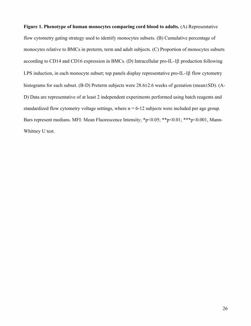
Figure 1. Phenotype of human monocytes comparing cord blood to adults. (A) Representative
flow cytometry gating strategy used to identify monocytes subsets. (B) Cumulative percentage of
monocytes relative to BMCs in preterm, term and adult subjects. (C) Proportion of monocytes subsets
according to CD14 and CD16 expression in BMCs. (D) Intracellular pro-IL-1β production following
LPS induction, in each monocyte subset; top panels display representative pro-IL-1β flow cytometry
histograms for each subset. (B-D) Preterm subjects were 28.6±2.6 weeks of gestation (mean±SD). (A-
D) Data are representative of at least 2 independent experiments performed using batch reagents and
standardized flow cytometry voltage settings, where n = 6-12 subjects were included per age group.
Bars represent medians. MFI: Mean Fluorescence Intensity; *p<0.05; **p<0.01; ***p<0.001, Mann-
Whitney U test.
27
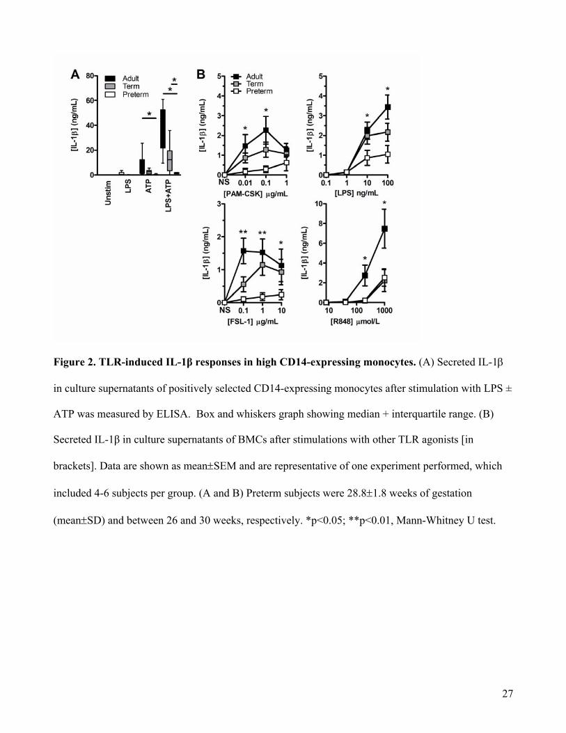
Figure 2. TLR-induced IL-1β responses in high CD14-expressing monocytes. (A) Secreted IL-1β
in culture supernatants of positively selected CD14-expressing monocytes after stimulation with LPS ±
ATP was measured by ELISA. Box and whiskers graph showing median + interquartile range. (B)
Secreted IL-1β in culture supernatants of BMCs after stimulations with other TLR agonists [in
brackets]. Data are shown as mean±SEM and are representative of one experiment performed, which
included 4-6 subjects per group. (A and B) Preterm subjects were 28.8±1.8 weeks of gestation
(mean±SD) and between 26 and 30 weeks, respectively. *p<0.05; **p<0.01, Mann-Whitney U test.

28
Figure 3. TLR-induced cytokine gene and protein expression are impaired in preterm neonates.
(A) Kinetics of IL1B mRNA expression following LPS stimulation was measured by real-time PCR.
Data represent the ratio of IL1B to ACTB (β-actin) transcript copy numbers of experiments performed
on whole blood samples in duplicates (solid lines = LPS stimulation; dashed lines = unstimulated
cells). (B) Kinetics of IL-1β protein secretion in culture supernatants from subjects simultaneously
tested in (A) was measured by ELISA. Levels of statistical significance were only indicated for
comparisons with preterm subjects. (A and B) Data are shown as mean ± SEM (n = 6 subjects per
group) and are representative of 2 independent experiments performed. Preterm subjects for this figure
were 28.7±1.8 weeks of gestation (mean±SD). (C) Box and whiskers graph showing median +
interquartile range of LPS-induced IL-1β secretion with or without cycloheximide (CH) added at the
beginning of stimulation. IL-1β concentration was measured by ELISA. Preterm subjects were
28.8±1.8 weeks of gestation. Data are shown as mean ± SD (n=4-6 subjects per group) and are
representative of one experiment performed. *p<0.05; **p<0.01, Mann-Whitney U test.
29
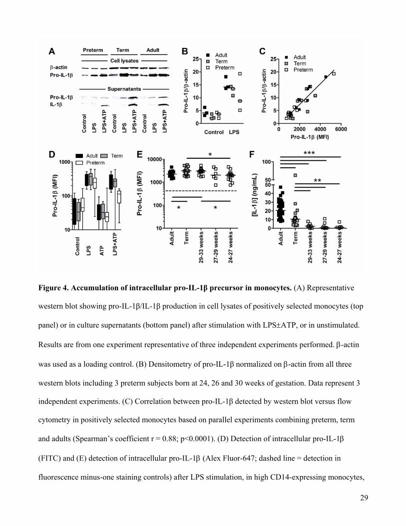
Figure 4. Accumulation of intracellular pro-IL-1β precursor in monocytes. (A) Representative
western blot showing pro-IL-1β/IL-1β production in cell lysates of positively selected monocytes (top
panel) or in culture supernatants (bottom panel) after stimulation with LPS±ATP, or in unstimulated.
Results are from one experiment representative of three independent experiments performed. β-actin
was used as a loading control. (B) Densitometry of pro-IL-1β normalized on β-actin from all three
western blots including 3 preterm subjects born at 24, 26 and 30 weeks of gestation. Data represent 3
independent experiments. (C) Correlation between pro-IL-1β detected by western blot versus flow
cytometry in positively selected monocytes based on parallel experiments combining preterm, term
and adults (Spearman’s coefficient r = 0.88; p<0.0001). (D) Detection of intracellular pro-IL-1β
(FITC) and (E) detection of intracellular pro-IL-1β (Alex Fluor-647; dashed line = detection in
fluorescence minus-one staining controls) after LPS stimulation, in high CD14-expressing monocytes,

30
by flow cytometry. (F) Secreted levels of IL-1β (ELISA) after LPS/ATP stimulation in preterm
subjects (BMCs) categorized by gestational age. (D-F) Data are representative of at least 3 independent
experiments performed, where up to12-25 subjects per group were included. Bars represent medians.
MFI: Mean Fluorescence Intensity; *p<0.05; **p<0.01; ***p<0.001, Mann-Whitney U test.

31
Figure 5. Impaired NLRP3 induction in preterm neonatal monocytes. (A) Representative western
blot of pro-caspase-1 (Pro-Casp-1) processing into active caspase-1 (Casp-1) after LPS±ATP in
preterm, term and adult positively selected CD14-expressing monocytes. Results are from one
experiment representative of three independent experiments performed. β-actin was used as a loading
control. (B) Ratio of pro-caspase-1 over β-actin band intensity as detected by western blot in three
independent experiments, including preterm subjects born at 24, 26 and 30 weeks of gestation. (C)
Expression of P2X7R on high CD14-expressing monocytes after LPS induction; Representative flow

32
cytometry histogram (lower panel in C); MFI: Mean Fluorescence Intensity. (D) IL-1β secretion
(ELISA) after LPS±ATP or nigericin.(E) Real time PCR analysis of NLRP3 mRNA expression (fold-
change compared to unstimulated) after stimulation of positively selected CD14-expressing monocytes
with or without LPS; Data is normalized against ACTB. Data is shown as mean ±SEM and is
representative of one real-time PCR experiment performed in triplicates, including 8 adults, 8 term and
6 preterm subjects born between 24 to <28 weeks of gestation. (F) Representative western blot
showing protein validation of NLRP3 and ASC expression following LPS stimulation in positively
selected CD14-expressing monocytes (from a preterm neonate 27 weeks of gestation). Results are from
one experiment representative of three independent experiments performed (left panel). β-actin was
used as a loading control. Densitometry of NLRP3 (middle panel) and ASC (right panel) quantification
by western blots (normalized on β-actin) in positively selected CD14-expressing monocytes with or
without LPS. Data represent 3 independent experiments performed. Bars represent medians, n = 3
different subjects per group. *p<0.05; **p<0.01; ***p<0.001, Mann-Whitney U test, except for (E)
where an ANOVA was used with a posttest Bonferroni correction.
33
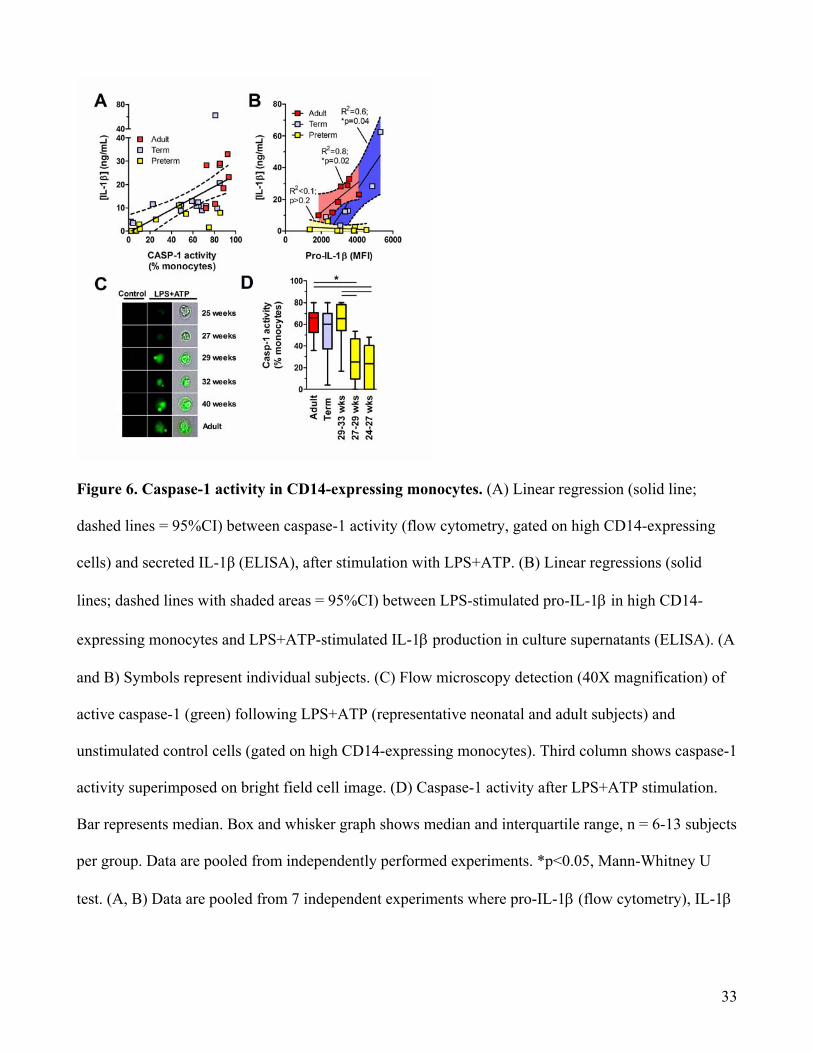
Figure 6. Caspase-1 activity in CD14-expressing monocytes. (A) Linear regression (solid line;
dashed lines = 95%CI) between caspase-1 activity (flow cytometry, gated on high CD14-expressing
cells) and secreted IL-1β (ELISA), after stimulation with LPS+ATP. (B) Linear regressions (solid
lines; dashed lines with shaded areas = 95%CI) between LPS-stimulated pro-IL-1β in high CD14-
expressing monocytes and LPS+ATP-stimulated IL-1β production in culture supernatants (ELISA). (A
and B) Symbols represent individual subjects. (C) Flow microscopy detection (40X magnification) of
active caspase-1 (green) following LPS+ATP (representative neonatal and adult subjects) and
unstimulated control cells (gated on high CD14-expressing monocytes). Third column shows caspase-1
activity superimposed on bright field cell image. (D) Caspase-1 activity after LPS+ATP stimulation.
Bar represents median. Box and whisker graph shows median and interquartile range, n = 6-13 subjects
per group. Data are pooled from independently performed experiments. *p<0.05, Mann-Whitney U
test. (A, B) Data are pooled from 7 independent experiments where pro-IL-1β (flow cytometry), IL-1β

34
(ELISA) and caspase-1 activity (flow cytometry) were measured in parallel. MFI: Mean Fluorescence
Intensity.
Figure 7. Intra-uterine and postnatal maturation in inflammasome activity. (A) Caspase-1
activity, in monocytes (gated on high CD14-expressing cells; using FLICA), of preterm subjects with
or without histological chorioamnionitis. Caspase-1 activity was measured after stimulation of BMCs
with LPS+ATP. For this panel, preterm subjects were 28.2±1.0 and 29.5±2.1 weeks of gestational age
(mean±SD) with and without chorioamnionitis, respectively (flow cytometry). (B) IL-1β secretion in
whole peripheral blood stimulated with a supra-saturating dose of LPS (100 ng/mL) (ELISA). The
blood for these studies was obtained from preterm subjects born 24 to <29 weeks of gestation (white
squares), sampled at 7 to 28 days of age, compared to healthy adults. Data are pooled from 5
independent experiments performed. Symbols represent individual subjects, bar represents median, n =
5-10 subjects per group ***p<0.001, Mann-Whitney U test.