Defects in mitochondrial clearance predispose human monocytes to interleukin-1β hyper-secretion
14
Defects in Mitochondrial Clearance Predispose Human Monocytes to Interleukin-1 Hypersecretion Received for publication, November 27, 2013, and in revised form, December 15, 2013 Published, JBC Papers in Press, December 19, 2013, DOI 10.1074/jbc.M113.536920 Robert van der Burgh ‡ , Lotte Nijhuis ‡ , Kalliopi Pervolaraki ‡ , Ewoud B. Compeer ‡ , Lieneke H. Jongeneel ‡ , Marielle van Gijn § , Paul J. Coffer ‡¶ , Michael P. Murphy , Pier G. Mastroberardino** 1 , Joost Frenkel ‡ , and Marianne Boes ‡2 From the ‡ Department of Pediatric Immunology and Infectious Diseases, University Medical Center Utrecht, Wilhelmina Children’s Hospital, 3584 EA Utrecht, The Netherlands, the Departments of ¶ Cell Biology and § Genetics, University Medical Center Utrecht, 3584 EA Utrecht, The Netherlands, the Medical Research Council Mitochondrial Biology Unit, Wellcome Trust, Cambridge, CB2 0XY United Kingdom, and the **Department of Genetics, Erasmus Medical Center, 3015 GE Rotterdam, The Netherlands Background: Periodic fever syndromes are caused by deregulation of interleukin-1 release. Results: Defective autophagy leads to accumulation of damaged mitochondria in monocytes. Conclusion: Mitochondrial components in the cytosol cause priming of monocytes for interleukin-1 release. Significance: The molecular mechanism behind deregulated cytokine secretion provides new clues for intervention. Most hereditary periodic fever syndromes are mediated by deregulated IL-1 secretion. The generation of mature IL-1 requires two signals: one that induces synthesis of inflam- masome components and substrates and a second that activates inflammasomes. The mechanisms that mediate autoinflamma- tion in mevalonate kinase deficiency, a periodic fever disease characterized by a block in isoprenoid biosynthesis, are poorly understood. In studying the effects of isoprenoid shortage on IL-1 generation, we identified a new inflammasome activation signal that originates from defects in autophagy. We find that hypersecretion of IL-1 and IL-18 requires reactive oxygen species and is associated with an oxidized redox status of monocytes but not lymphocytes. IL-1 hypersecretion by monocytes involves decreased mitochondrial stability, release of mitochondrial content into the cytosol and attenuated autophagosomal degradation. Defective autophagy, as estab- lished by ATG7 knockdown, results in prolonged cytosolic retention of damaged mitochondria and increased IL-1 secre- tion. Finally, activation of autophagy in healthy but not meval- onate kinase deficiency patient cells reduces IL-1 secretion. Together, these results indicate that defective autophagy can prime monocytes for mitochondria-mediated NLRP3 inflam- masome activation, thereby contributing to hypersecretion of IL-1 in mevalonate kinase deficiency. Periodic fever syndromes are characterized by inflammation that occurs in the absence of apparent infection or high titer autoantibodies (1, 2). In most periodic fever syndromes, the generalized inflammation is driven by IL-1 generated through proteolytic cleavage by the NLRP3 inflammasome. An addi- tional feature associated with inflammation is the production of reactive oxygen species (ROS) 3 (3, 4). During infection, ROS produced by NADPH-oxidase subunits at the plasma mem- brane are beneficial because ROS in phagosomes contribute to the killing of intracellular pathogens (5). In several inflamma- tory diseases, including periodic fever syndromes, ROS levels are increased in the cytosol and contribute to pathology (6). ROS play roles as intracellular second messengers and have immediate effects on the intracellular redox status. In TNF receptor 1-associated periodic syndrome (TRAPS), for exam- ple, mitochondria-derived ROS facilitate inflammatory cyto- kine production (7). Mitochondria are believed to be the main source of ROS in several autoinflammatory disorders (8). ROS are normally generated within mitochondria as byprod- ucts of oxidative phosphorylation, but when liberated in the cytosol ROS can facilitate activation of the NLRP3 inflam- masome (9 –11). Normally, mitochondrial contents, including ROS and mitochondrial DNA (mtDNA) are prevented from reaching the cytosol because damaged mitochondria are swiftly neutralized by autophagy (12). During autophagy cytosolic con- stituents are enclosed within a double-layered lipid membrane vesicle geared to fuse with lysosomes for degradation and recy- cling of the internal contents (12). Impaired autophagy may interfere with mitochondrial turnover and stimulate NLRP3 activation and IL-1 release, as was shown in murine sepsis models (11), indicating the possibility of a link between defec- tive mitochondrial clearance and autoinflammatory disease. Furthermore, in mice, mitochondria-derived ROS contributes to the activation of the NLRP3-inflammasome-mediated acti- vation of caspase-1 by LPS and ATP (11). We studied the potential contribution of disordered mito- chondrial biology in to IL-1 mediated inflammation in the Author’s Choice—Final version full access. 1 Supported by The Netherlands Genomics Initiative Grant NGI/NWO 05040202 and Marie Curie Grant IRG 247918. 2 Supported by a grant from the WKZ-Fonds Foundation. To whom corre- spondence should be addressed: Dept. of Pediatric Immunology, Univer- sity Medical Center Utrecht, Wilhelmina Children’s Hospital, 3584 EA Utrecht, The Netherlands. Tel.: 31-88-75-53328; Fax: 31-88-75-55350; E-mail: [email protected]. 3 The abbreviations used are: ROS, reactive oxygen species; MKD, mevalonate kinase deficiency; TRAPS, TNF receptor 1-associated periodic syndrome; mtDNA, mitochondrial DNA; TMRM, tetramethyl rhodamine methyl ester; TPP, decyltriphenylphosphonium; PBMC, peripheral blood mononuclear cell; KD, knockdown; mtROS, mitochondrial ROS; DPI, diphylene iodonium; GGPP, geranylgeranylpyrophosphate; HO-1, heme oxygenase-1; Baf A1, bafilomycin A1; H 2 DCFDA, 2,7-dichlorodihydrofluorescein diacetate. THE JOURNAL OF BIOLOGICAL CHEMISTRY VOL. 289, NO. 8, pp. 5000 –5012, February 21, 2014 Author’s Choice © 2014 by The American Society for Biochemistry and Molecular Biology, Inc. Published in the U.S.A. 5000 JOURNAL OF BIOLOGICAL CHEMISTRY VOLUME 289 • NUMBER 8 • FEBRUARY 21, 2014 by guest on September 25, 2016 http://www.jbc.org/ Downloaded from
Transcript of Defects in mitochondrial clearance predispose human monocytes to interleukin-1β hyper-secretion

Defects in Mitochondrial Clearance Predispose HumanMonocytes to Interleukin-1� HypersecretionReceived for publication, November 27, 2013, and in revised form, December 15, 2013 Published, JBC Papers in Press, December 19, 2013, DOI 10.1074/jbc.M113.536920
Robert van der Burgh‡, Lotte Nijhuis‡, Kalliopi Pervolaraki‡, Ewoud B. Compeer‡, Lieneke H. Jongeneel‡,Marielle van Gijn§, Paul J. Coffer‡¶, Michael P. Murphy�, Pier G. Mastroberardino**1, Joost Frenkel‡,and Marianne Boes‡2
From the ‡Department of Pediatric Immunology and Infectious Diseases, University Medical Center Utrecht, Wilhelmina Children’sHospital, 3584 EA Utrecht, The Netherlands, the Departments of ¶Cell Biology and §Genetics, University Medical Center Utrecht,3584 EA Utrecht, The Netherlands, the �Medical Research Council Mitochondrial Biology Unit, Wellcome Trust, Cambridge, CB2 0XYUnited Kingdom, and the **Department of Genetics, Erasmus Medical Center, 3015 GE Rotterdam, The Netherlands
Background: Periodic fever syndromes are caused by deregulation of interleukin-1� release.Results: Defective autophagy leads to accumulation of damaged mitochondria in monocytes.Conclusion: Mitochondrial components in the cytosol cause priming of monocytes for interleukin-1� release.Significance: The molecular mechanism behind deregulated cytokine secretion provides new clues for intervention.
Most hereditary periodic fever syndromes are mediated byderegulated IL-1� secretion. The generation of mature IL-1�requires two signals: one that induces synthesis of inflam-masome components and substrates and a second that activatesinflammasomes. The mechanisms that mediate autoinflamma-tion in mevalonate kinase deficiency, a periodic fever diseasecharacterized by a block in isoprenoid biosynthesis, are poorlyunderstood. In studying the effects of isoprenoid shortage onIL-1 � generation, we identified a new inflammasome activationsignal that originates from defects in autophagy. We find thathypersecretion of IL-1� and IL-18 requires reactive oxygenspecies and is associated with an oxidized redox status ofmonocytes but not lymphocytes. IL-1� hypersecretion bymonocytes involves decreased mitochondrial stability, releaseof mitochondrial content into the cytosol and attenuatedautophagosomal degradation. Defective autophagy, as estab-lished by ATG7 knockdown, results in prolonged cytosolicretention of damaged mitochondria and increased IL-1� secre-tion. Finally, activation of autophagy in healthy but not meval-onate kinase deficiency patient cells reduces IL-1� secretion.Together, these results indicate that defective autophagy canprime monocytes for mitochondria-mediated NLRP3 inflam-masome activation, thereby contributing to hypersecretion ofIL-1� in mevalonate kinase deficiency.
Periodic fever syndromes are characterized by inflammationthat occurs in the absence of apparent infection or high titerautoantibodies (1, 2). In most periodic fever syndromes, thegeneralized inflammation is driven by IL-1� generated throughproteolytic cleavage by the NLRP3 inflammasome. An addi-
tional feature associated with inflammation is the production ofreactive oxygen species (ROS)3 (3, 4). During infection, ROSproduced by NADPH-oxidase subunits at the plasma mem-brane are beneficial because ROS in phagosomes contribute tothe killing of intracellular pathogens (5). In several inflamma-tory diseases, including periodic fever syndromes, ROS levelsare increased in the cytosol and contribute to pathology (6).ROS play roles as intracellular second messengers and haveimmediate effects on the intracellular redox status. In TNFreceptor 1-associated periodic syndrome (TRAPS), for exam-ple, mitochondria-derived ROS facilitate inflammatory cyto-kine production (7). Mitochondria are believed to be the mainsource of ROS in several autoinflammatory disorders (8).
ROS are normally generated within mitochondria as byprod-ucts of oxidative phosphorylation, but when liberated in thecytosol ROS can facilitate activation of the NLRP3 inflam-masome (9 –11). Normally, mitochondrial contents, includingROS and mitochondrial DNA (mtDNA) are prevented fromreaching the cytosol because damaged mitochondria are swiftlyneutralized by autophagy (12). During autophagy cytosolic con-stituents are enclosed within a double-layered lipid membranevesicle geared to fuse with lysosomes for degradation and recy-cling of the internal contents (12). Impaired autophagy mayinterfere with mitochondrial turnover and stimulate NLRP3activation and IL-1� release, as was shown in murine sepsismodels (11), indicating the possibility of a link between defec-tive mitochondrial clearance and autoinflammatory disease.Furthermore, in mice, mitochondria-derived ROS contributesto the activation of the NLRP3-inflammasome-mediated acti-vation of caspase-1 by LPS and ATP (11).
We studied the potential contribution of disordered mito-chondrial biology in to IL-1� mediated inflammation in the
Author’s Choice—Final version full access.1 Supported by The Netherlands Genomics Initiative Grant NGI/NWO
05040202 and Marie Curie Grant IRG 247918.2 Supported by a grant from the WKZ-Fonds Foundation. To whom corre-
spondence should be addressed: Dept. of Pediatric Immunology, Univer-sity Medical Center Utrecht, Wilhelmina Children’s Hospital, 3584 EAUtrecht, The Netherlands. Tel.: 31-88-75-53328; Fax: 31-88-75-55350;E-mail: [email protected].
3 The abbreviations used are: ROS, reactive oxygen species; MKD, mevalonatekinase deficiency; TRAPS, TNF receptor 1-associated periodic syndrome;mtDNA, mitochondrial DNA; TMRM, tetramethyl rhodamine methyl ester;TPP, decyltriphenylphosphonium; PBMC, peripheral blood mononuclearcell; KD, knockdown; mtROS, mitochondrial ROS; DPI, diphylene iodonium;GGPP, geranylgeranylpyrophosphate; HO-1, heme oxygenase-1; Baf A1,bafilomycin A1; H2DCFDA, 2�,7�-dichlorodihydrofluorescein diacetate.
THE JOURNAL OF BIOLOGICAL CHEMISTRY VOL. 289, NO. 8, pp. 5000 –5012, February 21, 2014Author’s Choice © 2014 by The American Society for Biochemistry and Molecular Biology, Inc. Published in the U.S.A.
5000 JOURNAL OF BIOLOGICAL CHEMISTRY VOLUME 289 • NUMBER 8 • FEBRUARY 21, 2014
by guest on September 25, 2016
http://ww
w.jbc.org/
Dow
nloaded from
human monogenetic periodic fever disorder, mevalonatekinase deficiency (MKD), which gives rise to the hyper-IgD andperiodic fever syndrome. MKD is caused by loss of function ofmevalonate kinase, an enzyme in the mevalonate pathwayinvolved in cholesterol and non-sterol isoprenoid synthesis.The defect seriously impairs isoprenoid biosynthesis, resultingin reduced prenylation of proteins, particularly of some smallGTPases. Individuals suffering from MKD experience recur-ring fever episodes that are to a large extent mediated by IL-1�(13). In this study, we aimed to clarify underlying mechanismsthat cause hypersecretion of IL-1�. We show that impaired iso-prenoid biosynthesis interferes with autophagy, attenuates theactivity of the mitochondrial respiratory chain, and increasesthe release of mtDNA into the cytosol, thereby fostering anoxidized cytosolic milieu, which ultimately leads to hypersecre-tion of IL-1�.
MATERIALS AND METHODS
Reagents—Simvastatin, geranylgeranylpyrophosphate, ATP,N-acetyl cysteine, diphenylene iodonium, apocynin, and bafilo-mycin A1 were purchased from Sigma-Aldrich. MitoTrackerGreen, MitoTracker Deep Red, MitoSOX, tetramethyl rhoda-mine methyl ester (TMRM), and H2DCFDA were purchasedfrom Invitrogen. A Cyto-ID autophagy detection kit was pur-chased from Enzo Life Sciences. LPS (Escherichia coli EH 100)was obtained from Alexis Biochemical. MitoQ and decyltriph-enylphosphonium (TPP) were synthesized as described (7).Simvastatin was hydrolyzed to its bioactive form as describedpreviously (14). ATP solution was made in RPMI 1640 and wasbuffered to a pH of 7.5.
Patient Samples—Patients were children between the ages of2 and 16 years with hyper-IgD periodic fever syndrome causedby compound heterozygous mutations affecting both alleles ofmevalonate kinase. All had residual mevalonate kinase activi-ties between 0.1 and 8.5% of healthy controls. At scheduledoutpatient visits, patients who were afebrile and well under-went routine blood analysis. The ethical committee of the Uni-versity Medical Center Utrecht approved the use of residualmaterial for this study. After informed consent was obtainedfrom parents and from patients 12 years and older, residualmaterial from routine blood tests was used to obtain peripheralblood mononuclear cells (PBMCs). PBMCs from patients andhealthy donors were isolated using Ficoll density gradient.PBMC fraction was washed twice in RPMI supplemented with2% FBS and used immediately.
Cell Cultures—THP-1 and HEK293T cells were both cul-tured in RPMI 1640 supplemented with 1% glutamine, antibi-otics (penicillin, streptomycin), and 10% FBS. Simvastatintreatment of cells was 48 h prior to the start of the experimentand at a concentration of 10 �M unless stated otherwise in thefigure legends.
Mitochondrial Damage, Potential, and Superoxide Mea-surements—Cells were washed once in PBS and resuspended inRPMI (without phenol red and without FBS) and appropriateprobe. Staining concentrations were 50 nM MitoTracker Green,50 nM MitoTracker Deep Red, 20 nM TMRM, and 5 �M
MitoSOX. Cells were incubated in the dark for 30 min at 37 °C.Cells were centrifuged (500 � g for 5 min) and suspended in
RPMI without phenol red with 10% FBS. Cells were kept in thedark until measurement on FACS CANTO-II. Analysis wasdone with FACS Diva software.
Oxygen Consumption and Glycolysis Measurements—Oxy-gen consumption rate and glycolysis were measured using theSeahorse XFe24 extracellular flux analyzer (Seahorse Biosci-ences) according to the manufacturer’s instructions. THP-1cells were bound to the well using BD Cell-TAK coating. Coat-ing of the wells was done according to the manufacturer’sinstructions.
RNA Isolation and Quantification—RNA was isolated by dis-solving cell pellets in TRIpure (RnD) and following manufac-turer’s protocols. Isolated RNA was converted to cDNA usingiScript (Bio-Rad) according to manufacturer’s instructions.Detection was done with CF-96 (Bio-Rad) using SYBR green(Bio-Rad), 100 ng of cDNA was used per reaction. The primersused were heme oxygenase-1 (HO-1) forward, 5�-TCAGGCA-GAGGGTGATAGAAG-3�; HO-1 reverse, 5�-TTGGTGTCA-TGGGTCAGC-3�; ATG7 forward, 5�-CAGTTTGCCCCTTT-TAGTAGTGC-3�; ATG7 reverse, 5�-CCTTAATGTCCTTGG-GAGCTTCA-3�; B2M forward, 5�-CCAGCAGAGAATGGAAA-GTC-3�; B2M reverse, 5�-GATGCTGCTTACATGTCTCG-3�; GAPDH forward, 5�-GTCGGAGTCAACGGATT-3�; andGAPDH reverse, 5�-AAGCTTCCCGTTCTCAG-3�.
ATG7 shRNA Knockdown—Two different short hairpin RNAsequences (Sigma-Aldrich) in a lentiviral vector (MISSIONpLKO.1-puro) were used to make lentiviral particles. THP-1cells were infected twice and then selected for puromycinresistance. ATG7 knockdown (KD) was tested with quantita-tive PCR for ATG7. Hairpin sequences were ATG7 KD1,5�-CCGGGCCTGCTGAGGAGCTCTCCATCTCGAGATG-GAGAGCTCCTCAGCAGGCTTTTT-3�; ATG7 KD2, 5�-CCG-GGCTTTGGGATTTGACACATTTCTCGAGAAATGTGTC-AAATCCCAAAGCTTTTT-3�; and control (scrambled), 5�-CCGGCAACAAGATGAAGAGCACCAACTCGAGTTGGT-GCTCTTCATCTTGTTGTTTTT-3�. KD efficiency was det-ermined by quantitative PCR.
Cytosolic Mitochondrial DNA Measurement—Protocol wasadapted slightly from Ref. 15. Cultured cells (1.5 � 107) werewashed twice in ice-cold PBS and then homogenized with aDounce homogenizer in ice-cold 100 mM Tris-HCl (pH 7.4)containing 0.25 M sucrose, 1 mM EDTA, and protease inhibitormix. Samples were centrifuged (700 � g at 4 °C) for 10 min, andthe supernatant was collected and kept on ice. Protein contentwas determined by BCA protein assay. Samples were normal-ized to volume and protein concentration and centrifuged(10,000 � g at 4 °C) for 30 min. Supernatant was collected, and200 �l was used to isolate DNA with DNA blood and tissue kit(Qiagen). Mitochondrial DNA copy number was determinedby quantitative PCR for cytochrome b. The following primerswere used: CYTB forward, 5�-GCCTATATTACGGATCA-TTTCTCTACT-3�; and CYTB reverse, 5�-GCCTATGAAG-GCTGTTGCTATAGT-3�.
Glutathione Measurement—Glutathione and glutathionedisulfide were colorimetrically measured as described previ-ously (9). For glutathione disulfide, 2.5 times more sample wasused to achieve accurate readings. Ratio measurements werecorrected accordingly.
Defective Mitochondrial Clearance in MKD
FEBRUARY 21, 2014 • VOLUME 289 • NUMBER 8 JOURNAL OF BIOLOGICAL CHEMISTRY 5001
by guest on September 25, 2016
http://ww
w.jbc.org/
Dow
nloaded from

Antioxidant Capacity Measurement—Cells were centrifugedand resuspended in staining mix (RPMI without phenol redwith 10% FBS, 10 �M H2DCFCA) and incubated in the dark for15 min at 37 °C. Cells were immediately transferred to six FACStubes, and the fluorescence was measured. After the first fluo-rescent measurements, hydrogen peroxide was added to thedifferent tubes at a final concentration of 0, 5, 15, 45, 135, and150 �M, respectively. Tubes were briefly vortexed and incu-bated for 4 min covered from light and measured two moretimes in consecutive order. Analysis was performed by calcu-lating the rate of fluorescence increase over time per sample(slope). The ratio of the fluorescence increase (slopes) at thedifferent H2O2 concentrations over the background slope(without H2O2) provides a measure of antioxidant capacity (rel-ative ratio). This allows the comparison of measurements byremoving technical variation.
Cytokine Measurements—Cells were centrifuged (500 � g for5 min) and plated in 96-well plates in triplicates (2.0 � 105
cells/well in 200 �l). Inhibitors were added, followed by 1 h ofincubation at 37 °C. Next LPS (200 ng/ml) was added, andsupernatants were collected after 4 h and stored at �80 °C untilmeasurement. Cytokine concentrations were determined byMulitplex bead analysis.
Immunoblot Analysis—Cells were washed twice in PBS andthen resuspended in Laemmli buffer and boiled for 10 min.Samples were then aliquoted and stored at �20 °C until use.Protein content was determined with BCA assay, and sampleswere diluted to 1 �g/�l. 5% (v/v) �-mercaptoethanol was addedto the samples, and they were separated on 12% SDS-PAGE gel,followed by transfer to PVDF-FL membrane. 5% dried nonfatmilk was used for blocking followed by primary antibody incu-bation (overnight 4 °C, 0.5% milk in TBS-T), three washes, andsecondary antibody incubation (1 h at room temperature, 0.5%milk in TBS-T). Detection was done with ECL. Antibodies used:anti-p62 (Santa Cruz; sc-28359), anti-LC3 (Nanotools, 0231-100/LC3-5F10), anti-actin (Santa Cruz; sc-1616), and anti-HSP90 (Cell Signaling Technology; 4875S). We performedquantification of signal on blots using ImageJ (National Insti-tutes of Health).
Confocal Analysis—HEK 293T cells were seeded in 24-wellculture plates on 1.5-mm glass coverslips precoated with poly-L-lysine solution (0.1 (w/v) in H2O; Sigma-Aldrich) for 30 minat 37 °C. Plates washed twice in PBS, after which cells wereplated at 40% confluency. After 24 h, bafilomycin A1 was added(final concentration, 10 nM; 4 h at 37 °C). The coverslips werewashed twice in PBS and fixed (3.7% paraformaldehyde(Merck), 10 min at room temperature). Coverslips weremounted using Mowiol solution containing DAPI. Autophago-somes were counted in a semiautomated manner using Meta-morph software (Molecular Devices). LC3-positive regions ofinterests were derived from binarized images obtained bythresholding immunofluorescence pictures, using the samethreshold for all the samples. The number of cells was derivedfrom the number of nuclei.
PBMCs were plated on 8-well Lab-Tek� II Chamber Slidescoated with Cell-Tak (BD Biosciences) at a density of 1 � 106
cells/ml. After incubation with rapamycin, the cells werewashed twice with PBS and stained with Cyto-ID detection
agent according to the manufacturer’s instructions. Next cellswere washed twice with RPMI 1640 without phenol red, sup-plemented with 0.2% (v/v) BSA (Roche Applied Science) and 10mM HEPES, and live cell imaging was performed on a ZeissLSM710 confocal microscope equipped with a live cell chamberdevice to maintain 37 °C and 5% CO2 condition duringexperiments.
THP-1 cells were washed with PBS and stained in RPMI(without) phenol red with 100 nM MitoTracker Green and 150nM MitoTracker Red for 30 min at 37 °C. Cells were washed andplated on WillCo wells coated with Cell-Tak (BD Biosciences)in RPMI (without) phenol red and 10% FBS. All images wereobtained with 1.3� optical zoom using Plan-Apochromat 63�1.40 oil DIC M27 objective on a Zeiss LSM710 and processedusing Zen 2009 software (Zeiss Enhanced Navigation).
Statistics—The error bars shown represent S.E. unless statedotherwise in the figure legends. Statistical test between twovariables was done using the Mann-Whitney test. In the figures,one asterisk (*) indicates a p value of �0.05, and two asterisks(**) indicate a p value of �0.01.
RESULTS
Altered Redox State in MKD Model Monocytes Exhibiting IL-1�Hypersecretion—Mouse studies support that mitochondrialROS (mtROS) can mediate the activation of NRLP3 inflam-masomes (9, 11, 14). We hypothesized that IL-1� hypersecre-tion by human monocytic cells in analogy involves increasedcytosolic levels of mtROS. To test this hypothesis, we usedfreshly isolated monocytes from healthy individuals or patientssuffering from MKD and THP-1 cells that were rendered sim-ilarly isoprenoid-deficient because of 48-h treatment with sim-vastatin. Simvastatin inhibits the HMG-CoA reductase, therate-limiting enzyme in the mevalonate pathway, leading to ashortage of isoprenoids. The inclusion of simvastatin in LPS-treated monocytic cultures mimics the IL-1� hypersecretionphenotype seen in the autoinflammatory syndrome MKD(18 –22).
To verify the involvement of ROS in our model, we stimu-lated simvastatin-treated THP-1 cells with LPS for 4 h in thepresence of three different indiscriminate ROS inhibitors(1 h preincubation): Apocynin, diphylene iodonium (DPI),and N-acetylcysteine. Generalized ROS inhibition normalizedIL-1� and IL-18 secretion (Fig. 1A). To next assess the involve-ment of altered cytosolic redox status in our model, we com-pared glutathione concentrations in reduced and oxidized form(SH/SS ratio), in simvastatin-treated and control THP-1 mono-cytic cells. Simvastatin treatment lowers the SH/SS ratio by�50%, reflecting a shift in redox balance toward a more oxi-dative state. Inclusion of the geranylgeranylpyrophosphate(GGPP) transferase inhibitor recapitulates this reduction, sug-gesting that the decrease in SH/SS ratio is caused by a lack ofnon-sterol isoprenoids (Fig. 1B). Finally, oxidative stress causesthe up-regulation of HO-1 mRNA levels (23). Indeed, simvas-tatin-treated cells express more HO-1 mRNA. This up-regula-tion can be partly rescued by the addition of the generalizedROS scavenger N-acetylcysteine (Fig. 1C). To investigatewhether the increased oxidative burden is caused by increasedROS production or, alternatively, by decreased antioxidant
Defective Mitochondrial Clearance in MKD
5002 JOURNAL OF BIOLOGICAL CHEMISTRY VOLUME 289 • NUMBER 8 • FEBRUARY 21, 2014
by guest on September 25, 2016
http://ww
w.jbc.org/
Dow
nloaded from

capacity, we designed a new assay. In this assay we testedwhether a lack of non-sterol isoprenoids impedes the ability tocounter acute oxidative stress, in simvastatin-treated and non-treated cells. We added the general ROS-sensitive fluorescentprobe H2DCFDA to cells and exposed them to increasing con-
centrations of H2O2. Cells that have diminished capacity toclear oxidative stress exhibit a sharper increase in fluorescencein response to H2O2. The background measurements (which isessentially ROS production) show donor variation, but there isno indication that simvastatin-treated cells have increased ROS
simvastatinNAC -
+ ++
--
HO
-1 m
RN
A (fo
ld in
duct
ion)
0 20 40 60 80 100
untreated
simvastatin
GGTI
ratio ([GSH]/[GSSG])
*
****
A B
HC MKD
ratio
at 1
50 μ
M H
2O2
(incr
ease
ver
sus
back
grou
nd) monocytes
ratio
at 1
50 μ
M H
2O2
(incr
ease
ver
sus
back
grou
nd) lymphocytesG
F
HC MKD
0.9
1.0
1.1
1.2
1.3
1.4
1.5
C
simvastatinuntreated
**
rela
tive
ratio
(1
50 μ
M H
2O2)
apocynin (μM)DPI (μM)NAC (mM)
---
IL-1
β (p
g/m
L)
0
250
500
750
1000
200--
500--
-10-
-30-
--
25
H*
**
0
10
20
30
0
5
10
15
20
0
2
4
6
8*
IL-18
IL-1
8 (p
g/m
l)
0
500
1000
1500
2000IL-1β
0 200 400 600 8000
50
100
150
200 HCMKD
0 200 400 600 8000
200
400
600
800
1000
fluor
esce
nce
(AU
)
fluor
esce
nce
(AU
)
time (sec) time (sec)
background with 150 μM peroxide
0
5
10
15
20HCMKD
peroxide (μM) 0 5 15 45 135 150
rela
tive
ratio
0 200 400 600 8000
20
40
60
80
100
0 200 400 600 8000
50
100
150
200
250
0
2
4
6
time (sec) time (sec)
background with 150 μM peroxide
fluor
esce
nce
(AU
)
fluor
esce
nce
(AU
)
HCMKD
peroxide (μM) 0 5 15 45 135 150
HCMKD
D
E
rela
tive
ratio
apocynin (μM)DPI (μM)NAC (mM)
---
200--
500--
-10-
-30-
--
25
FIGURE 1. Altered redox state in periodic fever syndrome model cells and patient monocytes. A, general ROS inhibition leads to decreased IL-1� (left panel)and IL-18 (right panel) secretion in simvastatin-treated cells when stimulated with LPS. The values are representative of four independent experiments. B, ratioof reduced versus oxidized glutathione in THP-1 cells. Simvastatin treatment leads to 50% reduction of ratio, as does the GGPP transferase inhibitor, indicatinga more oxidized intracellular environment. The values are averages of five independent experiments. C, oxidative stress indicated by mRNA up-regulation ofHO-1 (normalized to �2M). Simvastatin treatment increases oxidative stress and up-regulates HO-1. This can be partly rescued by co-incubation with theantioxidant N-acetylcysteine (NAC). The values are averages of three independent experiments. D and E, example of antioxidant capacity measurements of asingle patient with its coupled healthy control. Gating on monocytes (D) and lymphocytes (E) was done using size and granularity. On the backgroundmeasurements, the slopes are similar, indicating equal ROS production. F, simvastatin treatment leads to reduced antioxidant capacity in THP-1 cells. Shownis the ratio of the fluorescence increase with peroxide treatment divided by the background increase. A higher ratio is increased fluorescence and thus a lowercapacity to prevent the probe from becoming oxidized. Each line represents an independent experiment. G, antioxidant capacity of blood monocytes fromhealthy controls (HC) versus MKD patients. The monocytes from patients have less capacity to clear oxidative stress. H, the difference in antioxidant capacity isspecific for monocytes as MKD lymphocytes have a similar antioxidant capacity as healthy controls.
Defective Mitochondrial Clearance in MKD
FEBRUARY 21, 2014 • VOLUME 289 • NUMBER 8 JOURNAL OF BIOLOGICAL CHEMISTRY 5003
by guest on September 25, 2016
http://ww
w.jbc.org/
Dow
nloaded from

production. The average slope of each sample is calculated anddivided by the slope of the background (0 �M peroxide). Thisnormalizes for the difference in ROS production between indi-viduals (Fig. 1, D and E). Despite increased expression of HO-1,simvastatin-treated cells have impaired ability to clear acuteoxidative stress, indicating diminished antioxidant capacity(Fig. 1F). Together, these data suggest that impaired non-sterolisoprenoid output is associated with a more oxidative cytosolicmilieu because of diminishing antioxidant capacity and notbecause of increased ROS production.
To confirm these findings in actual patient cells, we isolatedPBMC from seven MKD patients that did not experience feverat the time of sampling, each paired with a healthy control, aswell as two additional healthy controls. We stained fresh PBMCwith the H2DCFDA probe and gated on either monocytes orlymphocytes based on cell size and granularity features, usingflow cytometry. MKD patient monocytes have a significantlyreduced antioxidant capacity compared with monocytes fromhealthy control individuals (Fig. 1G). In contrast, lymphocytesfrom MKD patients and controls show comparable antioxidantresponses (Fig. 1H). We conclude that isoprenoid-deficientmonocytic cells have reduced antioxidant capacity.
Isoprenoid Deficiency Raises Mitochondrial MembranePotential—Our data thus far suggest that isoprenoid deficiencycauses prolonged exposure to oxidative stress. We hypothe-sized that mitochondria, a source of ROS, might have reducedintegrity in MKD, allowing leakage of ROS from the mitochon-dria. To address this possibility, we stained THP-1 cells with afluorescent mitochondrial probe that reports the relativeamount of mitochondria (MitoTracker Green) and, as a mea-sure of mitochondrial damage, a mitochondrial probe that issensitive to the mitochondrial inner transmembrane potential(MitoTracker Deep Red) (24). Damaged mitochondria losemembrane potential and lose MitoTracker Deep Red staining.Treatment with simvastatin resulted in an increase in trans-membrane potential, as shown by increase in MitoTrackerDeep Red staining, whereas the MitoTracker Green fluores-cence was unchanged (Fig. 2, A–C, and gating strategy in E).Thus, the lack of isoprenoids leads to increased mitochondrialpotential. Earlier studies had shown that an increase in mito-chondrial transmembrane potential is associated withincreased ROS production (25, 26). However, this work wasdone on isolated mitochondria. Other reports suggest that theregulation of mitochondrial potential can also be independentof ROS (27). To confirm the increase in mitochondrial potentialin isoprenoid-deficient monocytes, we used a different probe,TMRM. Measurements with TMRM supported our Mito-Tracker results: simvastatin treatment increases mitochondrialpotential. This is partially rescued by inclusion of GGPP, andtherefore, an increase in mitochondrial potential is mediated bythe lack of non-sterol isoprenoids (Fig. 2D). Although on aver-age MitoTracker Deep Red fluorescence increased, the numberof cells with intact MitoTracker Green fluorescence that lostMitoTracker Deep Red fluorescence altogether increased3-fold, from 0.7% in untreated cells to 2.2% in simvastatin-treated THP-1 cells (Fig. 2E). This implies that the percentageof living cells that harbored damaged mitochondria increased3-fold, as. Together, these data suggest that inhibition of pro-
tein isoprenylation is associated with increased mitochondrialtransmembrane potential and a decrease in mitochondrialstability.
Damaged Mitochondria Accumulate in the Cytosol of Isopre-noid-deficient Monocytes—We next investigated whether theincrease in mitochondrial transmembrane potential is due to anincrease in mitochondrial energy metabolism. To this end, wemeasured oxygen consumption and glycolysis rate. First basalrespiration was measured, followed by addition of the ATPaseinhibitor oligomycin to block respiration. Next, the uncouplerFCCP was added to induce maximum respiration, followed bythe electron transport chain inhibitors antimycin A and rote-none (complex III and complex I, respectively) to completelyabolish mitochondrial respiration. Simvastatin treatmentcaused a lower basal energy metabolism than that found inuntreated cells, when assayed for either oxygen consumption orglycolysis rate (Fig. 3A, left and right panels, respectively). How-ever, the regulation of oxygen consumption was similar underall conditions.
To assay for additional changes in mitochondria that mayassociate with the high transmembrane potential, we next visu-alized mitochondria in MitoTracker-stained THP-1 cells usingconfocal microscopy. The majority of simvastatin-treated cellsexhibited mitochondria that were elongated, whereas inuntreated cells, mitochondria appeared predominantly as smalldots (Fig. 3B). To confirm that the elongation is due to preny-lation and not to target effects of simvastatin, we repeated theexperiment with the geranylgeranyl transferase inhibitor. Thisresulted in mitochondrial elongation, although not to the sameextent. Earlier work showed that elongated mitochondria areassociated with induction of autophagy (28) and that elongationof mitochondria modulates metabolic efficiency (29). Further-more, isoprenoid deficiency caused by simvastatin treatmentcauses cells to proliferate more slowly. Taken together, isopre-noid deficiency in THP-1 cells causes both an increase in mito-chondrial potential and mitochondrial elongation. It is possiblethat the mitochondrial elongation is responsible for theincrease in membrane potential; however, this would need fur-ther investigation.
Mitochondrial components play an important role in prim-ing immune responses (6, 7, 14). Because we observed mito-chondrial irregularity and an increased proportion of cells har-boring depolarized mitochondria, we hypothesized thatisoprenoid-deficient monocytes contain released internalmitochondrial constituents. We therefore investigated thepresence of mtDNA into the cytosol, by isolation of cytosolicfractions of cells and measured in these fractions the content ofmtDNA (by quantitative PCR, assay for cytochrome b for whichthe coding sequence is located exclusively on mtDNA). Weconsistently found higher levels of mtDNA in cytosolic frac-tions in simvastatin-treated cells, both before (Fig. 3D) and afterLPS/ATP stimulation (Fig. 3E), although the latter was not sig-nificantly different. The effect of simvastatin was pronounced,inducing nearly half the amount of mtDNA release seen in cellstreated with the potent mitochondrial toxin DPI, which wasused as a positive control (Fig. 3F). Raised mtDNA could beeither actively released by the mitochondria or alternativelycould be a consequence of defective autophagy-mediated clear-
Defective Mitochondrial Clearance in MKD
5004 JOURNAL OF BIOLOGICAL CHEMISTRY VOLUME 289 • NUMBER 8 • FEBRUARY 21, 2014
by guest on September 25, 2016
http://ww
w.jbc.org/
Dow
nloaded from

ance of damaged mitochondria. In mice, mtROS are necessaryfor the active release of mtDNA (11). To test whether themtDNA is actively secreted by the mitochondria in humancells, we took a similar approach as Nakahira et al. (11) andco-incubated untreated and simvastatin-treated cells with themitochondria-targeted antioxidant MitoQ, which is a ubiqui-none targeted to the mitochondria by the triphenylphospho-nium lipophillic cation. The non-antioxidant mitochondria tar-geting moiety TPP was used as a control (30). Both compoundsshow similar and even increased release of mtDNA into thecytosol (Fig. 3G), indicating that mtROS is not necessary tocause the mtDNA release in our culture system.
Accumulation of Defective Mitochondria Is Due to ImpairedAutophagy—Can defects in autophagy of mitochondria predis-pose human monocytes to IL-1� hypersecretion? Autophagy iscontrolled by multiple pathways, including those that are medi-ated by small GTPases. Because small GTPases are isopreny-lated proteins, defects in the mevalonate pathway can lead tounprenylated GTPases (18, 31). We proposed that defects inisoprenylation may cause impairment of autophagy. We testedwhether isoprenylation is required for generation of LC3�-ma-ture autophagosomal membranes, using LC3-GFP fusion pro-teins expressed in HEK 293T cells (32). LC3 is a marker forautophagosomal membranes. Initially, it is an 18-kDa protein
mitotracker green
coun
tA B
simvastatin -- + +GGPP +- + -
fluor
esce
nce
mito
track
er g
reen
(AU
)
0
1000
2000
3000
simvastatin -- + +GGPP +- + -
fluor
esce
nce
mito
track
er d
eep
red
(AU
)
0
5000
10000
15000
0
2
4
6
ratio
(mito
track
er d
eep
red/
mito
track
er g
reen
)
simvastatin -- + +GGPP +- + -
C
0
1000
2000
3000
4000
fluor
esce
nce
TMR
M (A
U)
simvastatin -- + +GGPP +- + -
D
mitotracker deep red
coun
t
mitotracker green
mito
track
er d
eep
red
untreated SimvastatinE
simvastatin + -
loss
of m
itoch
ondr
ial
pot
entia
l (%
of l
ive
cells
)0.0
0.5
1.0
1.5
2.0
2.5 *
FSC
SS
C
live gate
0.595% 2.67%
FIGURE 2. Mitochondrial potential is altered in isoprenoid-deficient cells. A, top panel, total mitochondrial mass as determined with MitoTracker Green inTHP-1 cells under different conditions. Bottom panel, bar graph representation of data. Gating was done as shown in E, only top gate was used for potentialmeasurements. B, top panel, mitochondrial potential determined by MitoTracker Deep Red staining. The potential is clearly increased in simvastatin treatment.Bottom panel, bar graph representation of data. C, ratio of mitochondrial potential over mitochondrial mass to more accurately determine the potentialdifferences per unit of mitochondrial mass. Statin treatment leads to increased mitochondrial potential, which can be partially rescued by the addition ofexogenous GGPP. A–C show the data of a representative experiment of five independent experiments, each consisting of triplicate measurements. D, stainingof THP-1 cells with TMRM confirms MitoTracker data, the increased potential of mitochondria with simvastatin treatment, and dependence on isoprenoidavailability. The data are representative of three independent experiments shown. E, increasing amounts of cells lose mitochondrial potential with simvastatintreatment as determined combined MitoTracker Green and Deep Red staining. The amount of cells (gated on live cells) losing mitochondrial potential is tripledby statin treatment. Representative plots are shown of four independent experiments. The bar graph shows the averages and S.E. of four independentexperiments.
Defective Mitochondrial Clearance in MKD
FEBRUARY 21, 2014 • VOLUME 289 • NUMBER 8 JOURNAL OF BIOLOGICAL CHEMISTRY 5005
by guest on September 25, 2016
http://ww
w.jbc.org/
Dow
nloaded from

(LC3-I), which is matured to a 16-kDa form (LC3-II) when it isincorporated in the membranes. LC3-GFP cells were treatedwith simvastatin and/or the late stage autophagy inhibitor bafi-lomycin A1 (Baf A1). Baf A1 single treatment causes an accu-
mulation of autophagosomes, whereas addition of simvastatintreatment to Baf A1 counteracts Baf A1-mediated autophago-some buildup (Fig. 4A). Thus, simvastatin treatment inhibitsautophagosome formation as measured by LC3 autophago-
0 1000 2000 3000 4000 50000
100
200
300 untreatedGGPPsimvastatinsimvastatin + GGPP
oxyg
en c
onsu
mpt
ion
rate
(pM
oles
/min
)
extra
cellu
lar a
cidi
ficat
ion
rate
(mpH
/min
)
oligomycin FCCPantimycin Arotenone oligomycin
time (sec) time (sec)
simvastatin + -
mt D
NA
(fol
d in
crea
se)
simvastatin + -
LPS + ATP
DPI
mt D
NA
(fol
d in
crea
se)
mt D
NA
(fol
d in
crea
se)
simvastatinTPPMitoQ
---
+--
-+-
++-
--+
+-+
G
mt D
NA
(fol
d in
crea
se)
0.0
0.5
1.0
1.5
2.0
0
50
100
untreated simvastatin
simvastatin
normal mitochondriaelongated mitochondria
+ -
% o
f cel
ls
+ -
*
0 500 1000 1500 2000 25000
20
40
60
0
1
2
3
4
0.0
0.5
1.0
1.5
2.0*
0
1
2
3
4
*
GGTI
A
ED F
B
C
0
50
100
GGTI + -
% o
f cel
ls
*
normal mitochondriaelongated mitochondria
Defective Mitochondrial Clearance in MKD
5006 JOURNAL OF BIOLOGICAL CHEMISTRY VOLUME 289 • NUMBER 8 • FEBRUARY 21, 2014
by guest on September 25, 2016
http://ww
w.jbc.org/
Dow
nloaded from

somal membrane incorporation. This defect is caused specifi-cally by defective isoprenylation because exogenously addedGGPP rescues the phenotype.
To further analyze a possible role for autophagy in IL-1�hypersecretion by isoprenoid-deficient monocytes, we treatedTHP-1 cells with simvastatin (48 h) and prepared whole celllysates for LC3 and p62 protein analysis by Western blot.LC3-II levels represent the induction of autophagy, whereasp62 degradation is a marker for successful completion ofautophagy. Simvastatin, Baf A1 and their combined treatmentled to increased levels of LC3-II in THP-1 cells (Fig. 4B), con-firming earlier data that statins can induce autophagy (33, 34)and suggesting that the induction of autophagy is not inhibitedby simvastatin. Levels of p62 were induced by Baf A1 treatment,as expected, because Baf A1 inhibits autophagosomal turnoverof p62 and p62-associated protein aggregates (Fig. 4C). Statintreatment also caused an increase in p62, suggesting that statininhibits autophagy at a stage between LC3-I/LC3-II conversionand successful completion of autophagy. Combined Baf A1 andsimvastatin treatment resulted in a further increase in p62. Toconfirm that the increase in both p62 and LC3 is also isopre-noid-dependent, we repeated these experiments in the pres-ence of exogenously added GGPP, to rescue its deficiencycaused by simvastatin treatment. We conclude that inhibitionof protein isoprenylation causes defective autophagosomaldegradation in THP-1 cells.
We demonstrated that mtDNA release is not mediated bymtROS but wished to confirm that the mtROS released bydefective mitochondria do not inhibit autophagy, supportingour hypothesis that lack of prenylation is the key factor in inhib-ited autophagy. Therefore, we specifically inhibited mtROSwith the mitochondria-targeted antioxidant MitoQ, with TPPused as a control compound. We measured the protein levels ofLC3 and p62 in monocytic cells. Again, MitoQ and TPP treat-ment did not change LC3 and p62 levels, negating a role formtROS in the control of autophagy, with or without simvastatinpretreatment (Fig. 4D). This was confirmed in HEK293T LC3-GFP cells where the addition of MitoQ had no effect autopha-gosme formation (not shown). Antioxidant capacity of theMitoQ reagent itself was intact.4 We conclude the accumula-tion of damaged mitochondria originates from a defect inautophagy and leads to the release of mitochondrial compo-nents. Inhibition of mtROS cannot prevent the damage to mito-chondria nor rescue the autophagy defect in these cells.
IL-1� Secretion Involves Both Cytosolic Release of Mitochon-drial Components and Defective Autophagy—Is the release ofmitochondrial components into the cytosol necessary for IL-1�release? The selective inhibition of mtROS using MitoQ coun-teracts proinflammatory cytokine secretion, in the autoinflam-matory disease TRAPS (7). We used MitoQ in simvastatin-treated THP-1 monocytes to determine whether mtROS arerequired for LPS-induced IL-1� secretion. In cells treated withMitoQ, the IL-1� secretion upon LPS stimulation was unaf-fected (Fig. 5A). We conclude that ROS, but not mitochondrialROS, are involved in the IL-1� secretion in isoprenoid defi-ciency or MKD.
Oxidized mtDNA serves as activating ligand for the NLRP3inflammasome (14). Because we observed that isoprenoiddepletion significantly increased mtDNA levels in the cytosol(Fig. 3C), we hypothesized that the mtDNA is oxidized andactivates the NLRP3 inflammasome, thereby increasing IL-1�secretion. The binding of oxidized mtDNA to NLRP3 and sub-sequent inflammasome activation can be competitively inhib-ited by oxidized nucleotides that do not induce inflammasomeactivation (14). We incubated simvastatin-treated THP-1 cellsfor 1 h with either oxidized deoxyguanosine or regular deox-yguanosine, followed by 4 h of LPS stimulation. We found thatoxidized deoxyguanosine significantly reduced the amount ofsecreted IL-1� compared with both vehicle and deoxyguanos-ine control (Fig. 5B). Thus, mtDNA release contributes to theIL-1� hypersecretion via the NLRP3 inflammasome. Havingshown that isoprenoid shortage causes both IL-1� generationand impaired autophagy, we hypothesized that isoprenoidshortage causes IL-1� generation through impaired autophagy.We therefore transduced THP-1 monocytic cells with shRNAKD constructs for the autophagy protein ATG7 or a scrambledcontrol. We generated two different THP-1 lines with 50 and20% KD efficiency (Fig. 5C). Cells were stimulated with LPS andATP, and the level of secreted IL-1� was measured (Fig. 5D). Asexpected, both KD lines secreted more IL-1� than the scram-bled control, with the 50% KD secreting more than the 20% KD.This confirmed that impairment of autophagy enhances IL-�secretion, irrespective of isoprenoid biosynthesis. To finally con-firm that the defective autophagy is responsible for the accumula-tion of damaged mitochondria, we induced mitochondrial damagein the ATG7 KD and control THP-1 cells as described (4-h DPI (15�M) treatment (35)) and measured the fraction of cells that losemitochondrial potential in ATG7 KD and control cells. Loss ofpotential was gauged by flow cytometry using cell staining withMitoTracker Green and MitoTracker Deep Red. Using the samegating strategy as for Fig. 2E, we observed that ATG7 KD samples
4 R. van der Burgh, L. Nijhuis, K. Pervolaraki, E. B. Compeer, L. H. Jongeneel, M.van Gijn, P. J. Coffer, M. P. Murphy, P. G. Mastroberardino, J. Frenkel, and M.Boes, unpublished results.
FIGURE 3. Altered metabolism and release of mitochondrial components in isoprenoid-deficient cells. A, oxygen consumption and glycolysis. Simvas-tatin-treated THP-1 cells have reduced metabolism, which is partly rescued by GGPP. Representative picture shown of three independent experiments. B,THP-1 cells are stained with MitoTracker Green and Deep Red and checked for mitochondrial morphology with confocal microscopy. Mitochondria in simv-astatin-treated cells have a more elongated morphology. Representative pictures are shown. The bar graph consists of over 250 cells scored per condition; theerror bars indicate variance in scoring by three different observers. C, to confirm that mitochondrial elongation is mediated by lack of isoprenylation, THP-1 cellswere treated with GGPP transferase inhibitor (GGTI) (10 �M). This also leads to significant mitochondrial elongation, although not as strong as with simvastatin.The bar graph consists of over 150 cells scored per condition; the error bars indicate variance in scoring by three different observers. D, amount of mitochondrialDNA in the cytosolic fraction of THP-1 cells. Simvastatin treatment leads to an accumulation of mtDNA in the cytosol. The data are from four independentexperiments combined. E, the accumulation of mitochondrial DNA is independent of cell activation. The data are the averages of two experiments shown. F,simvastatin does not induce the amount of cytosolic mtDNA as seen with DPI. The data are from four independent experiments combined. G, the release ofmtDNA is not caused by ROS generated in the mitochondria. Co-incubation with mitochondrial ROS scavenger MitoQ cannot prevent the accumulation ofmtDNA in the cytosol. TPP is used as control for mitochondrial targeting group in MitoQ. The averages of two independent experiments are shown.
Defective Mitochondrial Clearance in MKD
FEBRUARY 21, 2014 • VOLUME 289 • NUMBER 8 JOURNAL OF BIOLOGICAL CHEMISTRY 5007
by guest on September 25, 2016
http://ww
w.jbc.org/
Dow
nloaded from
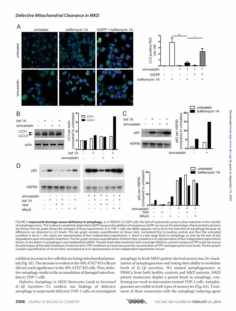
exhibit an increase in live cells that are losing mitochondrial poten-tial (Fig. 5E). The increase is evident in the 50% ATG7 KD cells yetdid not reach significance in the 20% ATG7 KD cells. Thus, defec-tive autophagy results in the accumulation of damaged mitochon-dria in THP-1 cells.
Defective Autophagy in MKD Monocytes Leads to IncreasedIL-1� Secretion—To confirm our findings of defectiveautophagy in isoprenoid-deficient THP-1 cells, we investigated
autophagy in fresh MKD patient-derived monocytes, by visual-ization of autophagosomes and testing their ability to modulatelevels of IL-1� secretion. We stained autophagosomes inPBMCs from both healthy controls and MKD patients. MKDpatient monocytes display a partial block in autophagy, con-firming our work in simvastatin-treated THP-1 cells. Autopha-gosomes are visible in both types of monocytes (Fig. 6A). Treat-ment of these monocytes with the autophagy-inducing agent
A
untre
ated
sim
vast
atin
bafilomycin 1A GGPP + bafilomycin 1Auntreated
0
1
2
3
4
5
simvastatin -GGPP -
bafilomycin 1A -
--+
+--
+-+
++-
+++
LC3
posi
tive
RO
I p
er c
ell
simvastatinbaf 1A -
-+-
-+
++
LC3-I
actin
simvastatinbaf 1AC
p62
HSP90
LC3-II
B--
+-
-+
++
simvastatinbaf 1ATPPMitoQ
----
-+--
--+-
-++-
---
-+-
+
--
+
--
+
-
+
-
+
-
+
-- + - + - +
+ ++ ++ +
p62
HSP90
D
simvastatin - +
p62
per H
SP
90(c
ompa
red
to u
ntre
ated
)
0
1
2
3
4LC3-ILC3-II
simvastatinbaf 1A -
-+-
-+
++
LC3
per a
ctin
(com
pare
d to
unt
reat
ed)
0
10
20
30untreatedbafilomycin 1A
simvastatin - - - + + +TPP - + -- - +
MitoQ - - - - ++
p62
per H
SP
90(c
ompa
red
to u
ntre
ated
)0
5
10
15
20
untreatedbafilomycin 1A
** *
FIGURE 4. Isoprenoid shortage causes deficiency in autophagy. A, in HEK293 LC3-GFP cells, the lack of isoprenoids causes a clear reduction in the numberof autophagosomes. This is almost completely dependent GGPP because the addition of exogenous GGPP can rescue the phenotype. Representative picturesare shown; the bar graph shows the averages of three experiments. B, in THP-1 cells, the defect appears not to be in the induction of autophagy because nodifferences are observed in LC3 levels. The bar graph contains quantification of shown blot, normalized first to loading control, and then the untreatedcondition is set to 1; the values are representative of four independent experiments. C, there is a late stage block in autophagy, as seen by the lack of p62degradation upon simvastatin treatment. The bar graph contains quantification of shown blot, treated as in B, representative of four independent experimentsshown. D, the defect in autophagy is not mediated by mtROS. The p62 levels after treatment with scavenger MitoQ or control compound TPP could not rescuethe phenotype of the statin treatment. Protein levels in TPP conditions are lower because the concentration of TPP used approaches toxic levels. The bar graphscontains quantification of shown blot, normalized as in B, representative of two independent experiments shown.
Defective Mitochondrial Clearance in MKD
5008 JOURNAL OF BIOLOGICAL CHEMISTRY VOLUME 289 • NUMBER 8 • FEBRUARY 21, 2014
by guest on September 25, 2016
http://ww
w.jbc.org/
Dow
nloaded from

rapamycin does not modify the number of cells with autopha-gosomes or autophagosomes per cell. However, there is a cleardifference in the IL-1� secretion between healthy controls andpatient cells treated with rapamycin. In healthy controls, prein-cubation of PBMCs with rapamycin (250 nM) 1 h prior to LPSstimulation (4 h) significantly reduces IL-1� secretion (p �0.01, n � 5). In contrast, the reduction of IL-1� secretion inrapamycin-treated MKD PBMCs did not reach significance(n � 3; Fig. 6B, ns). This indicates that inducing autophagycounteracts IL-1� cytokine secretion in healthy controls. Thisis in line with previous studies done in mice, where rapamycin-treated mice had lower IL-1� serum levels after LPS challenge(36). In MKD patients, where autophagy is already defective,inducing autophagy has limited effect. Taken together, our datasuggest that inflammasome activation in MKD, can involvedamaged mitochondria in monocytes. Because of defects inautophagy, these damaged mitochondria are not effectivelycleared, resulting in accumulation of mitochondrial compo-nents in the cytosol. These components, left present in the cyto-sol when autophagy is defective, prime monocytes in MKD forIl-1� hypersecretion.
DISCUSSION
We here addressed a role for autophagy in the developmentof autoinflammation in the periodic fever syndrome, MKD.Autophagy is a cellular process for the turnover of damaged
proteins and organelles, including mitochondria (12). It hadbeen known that both insufficient and excessive autophago-somal degradation is harmful for the cell. Autophagy istherefore strictly regulated by several signaling pathwaysthat are inhibitory (serine-threonine kinase mammaliantarget of rapamycin and class I PI3Ks) or that activateautophagy (class III PI3Ks) (38). Isoprenylation can modu-late the induction of autophagy as was suggested from exper-iments in which statin treatment-induced autophagy (33, 39,40). However, dissimilarities in statin dosage and treatmentprotocols complicate the interpretation of these studies.Also, the induction was not seen in all models, suggestingthat statin-induced autophagy is cell type-specific. Ourresults here show a direct role for isoprenylation in theinduction and completion of autophagy in monocytic cells.We show that autophagy controls IL-1� release throughremoval of mitochondrial components that otherwise stim-ulate NLRP3 inflammasome activation. A strong geneticlink was recently described between the autophagy geneATG16L1 and susceptibility to chronic inflammation inCrohn disease (41, 42). ATG16L1 suppresses IL-1� signalingby promoting the degradation of autophagosomal p62 (43).In addition, a recent study by Bachetti et al. (21) showed thatin TRAPS, a mutant version of TNF receptor 1 causes inhi-bition of autophagy, leading to autoinflammatory disease. In
A
veh. TPP MitoQ
IL-1
β (%
of v
eh.)
B
0
50
100
150
IL-1
β (%
of s
cr.)
***
veh. dG 8-OH-dG
IL-1
β (%
of v
eh.)
C
scr. ATG7KD-1
ATG7KD-2
fold
ATG
7 m
RN
Avs
scr
.
ED
0.0
0.5
1.0
1.5
***
0
100
200
300
400
0
50
100
150
scr. ATG7KD-1
ATG7KD-2
% in
crea
se o
f liv
e ce
lls w
ithlo
ss o
f mito
cond
rial p
oten
tial
scr. ATG7KD-1
ATG7KD-2
*
0
100
200
300
400
500
FIGURE 5. IL-1� secretion in isoprenoid-deficient cells involves inflammasome activation by oxidized mitochondrial DNA and defective autophagy. A,IL-1� secretion in MKD model is not mediated directly by mtROS. Despite MitoQ pretreatment, it did not ameliorate IL-1� secretion. B, inflammasome activationis mediated by oxidized mtDNA. Incubation of simvastatin-treated cells with oxidized nucleotides prior to stimulation leads to decreased IL-1� secretion,whereas non-oxidized nucleotide has no effect. The averages of three independent experiments are shown. C, knockdown of ATG7 by two virally stablytransduced shRNAs in THP-1 cells. The amount of mRNA for ATG7 was normalized to GAPDH and compared with scrambled control cells. D, IL-1� secretion ofATG7 KD cells after stimulation with LPS and ATP. IL-1� secretion is clearly increased in cells with impaired autophagy. This was done without simvastatinpretreatment to isolate the effect of defective autophagy. The averages of three experiments are shown. E, defective autophagy leads to accumulation ofdamaged mitochondria. ATG7 KD cells were either treated with DPI or left untreated, followed by staining with MitoTracker Green and Deep Red. Cells weregated as done for Fig. 2E. When autophagy is defective, induction of mitochondrial stress leads to accumulation of damaged mitochondria. The bar graph withaverages of four independent experiments is shown. veh., vehicle; scr., scrambled.
Defective Mitochondrial Clearance in MKD
FEBRUARY 21, 2014 • VOLUME 289 • NUMBER 8 JOURNAL OF BIOLOGICAL CHEMISTRY 5009
by guest on September 25, 2016
http://ww
w.jbc.org/
Dow
nloaded from

this current study, we provide experimental proof that gen-eral autophagic dysfunction enhances IL-1� release.
Although the effect of isoprenoid deficiency on autophagy isclear, there are still some unresolved issues. Despite our bestefforts, we have not been able to explain the increased mito-chondrial potential associated with isoprenoid deficiency. Thiseffect is highly reproducible yet seems contradictory with thelow oxygen metabolism. It is possible that the elongation of themitochondria, associated with increased efficiency, leads to ahigher potential even with lower oxygen consumption rates. Toclarify this issue, mitochondrial elongation should be inducedwithout the starvation effect of simvastatin. Unfortunately, weare unable to do so in our model system.
Recent work shows a role for ROS in the development of theIL-1�-driven autoinflammatory disorders (7, 16, 44). Initially,NADPH-oxidase-derived ROS production was also implicatedin IL-1� release in the periodic fever cryopyrin-associated per-iodic syndrome, but analyses of chronic granulomatous diseasepatients discounted a role for NADPH-oxidases in these disor-ders (16, 17). The source of ROS appeared mitochondrial (i.e.,mtROS) rather than derived from the NADPH-oxidases at theplasma membrane, as was supported by work in cells fromchronic granulomatous disease patients that have defectiveNADPH-dependent ROS caused by mutations in p47phox (16).A publication on TRAPS showed that specific inhibition ofmtROS release alleviates cytokine secretion (6). Our own datahere show that monocytes of MKD patients exhibit defectiveantioxidant responses, whereas MKD lymphocytes respondedsimilarly to healthy control cells when exposed to acute redoxstress. Although we can inhibit cytokine secretion using generalROS inhibitors, the specific inhibition of mtROS had no effecton the cytokine secretion. The question that remains is what isthe source of ROS that mediates the hypersecretion? It is pos-sible that the hypersecretion of IL-1� is mediated by mtROSand that the (membrane-bound) MitoQ is unable to inhibit allROS escaping from compromised mitochondria. However,additional research will be needed to confirm or discount thispossibility. The notion that ROS play a role in statin-induced
IL-1� generation has been recently supported by similar find-ings in simvastatin- and fluvastatin-treated human monocytesand murine macrophages (24).
Under conditions of oxidative stress, thioredoxin-interactingprotein (TXNIP) associates to NLRP3, as was shown in humanTHP-1 cells. The presence of ROS in the cytosol was therebydirectly linked to inflammasome activation and secretion ofbioactive IL-1� (9). Our work supports the earlier reportedrequirement for cytosolic ROS in IL-1� release by monocytes(7, 9, 44) and adds that mitochondrial alterations and defects inautophagy increase mtDNA into the cytosol. Our data therebyplace autophagy upstream of mtROS release and NLRP3inflammasome activation. The defective autophagy cannotclear damaged mitochondria, which leads to release of mtROSand mtDNA in the cytosol, which in turn can activate theNLRP3 inflammasome. We observed that increased IL-1�release is associated with reduced mitochondrial activity (i.e.,attenuated respiratory chain activity) and mtROS and mtDNAcytosolic release. The cytosolic presence of oxidized mtDNArelease contributes directly to IL-1� generation, which sup-ports earlier work showing that cytosolic mtDNA directly stim-ulates NLRP3 inflammasomes in mouse macrophages (11).Recent work shows the formation of autophagosomes at con-tact sites between mitochondria and endoplasmic reticulum,further corroborating a role for autophagosomes in clearance ofmitochondrial constituents (37). Mouse macrophages that aredeficient in autophagy proteins beclin-1 and LC3B secreteincreased levels of IL-1� upon NLRP3 inflammasome activa-tion using LPS/ATP combined treatment (11). Mouse macro-phages that lack mtDNA because of culture in the presenceethidium bromide exhibit reduced caspase-1 activity and atten-uation of respiratory chain activity and ROS production. Thegeneration of mtROS was placed as an upstream mechanism inthe release of IL-1�. However, NLRP3 inflammasome stimulimay differ in their subcellular source of ROS (i.e., crystallineparticles such as asbestos and silica may induce NLRP3 inflam-masome activation through ROS production by NADPH-oxi-dase (9, 45)). Accordingly, mito-TEMPO, which inhibits
AMKDHC
B
rapamycin + -- +MKDHC
IL-1
β se
cret
ion
(% o
f unt
reat
ed)
050
60
80
100**
*ns
0
20
40
60
80
100
rapamycin + -- +MKDHC
% o
f cel
l con
tain
ing
auto
phag
osom
es
FIGURE 6. Primary MKD cells respond differently to autophagy stimulus. A, freshly isolated PBMCs from a healthy donor and a MKD patient were stainedwith an autophagy probe. Samples were checked for number of cells with autophagosomes and autophagosomes per cell, but no significant differences wereobserved, largely because of significant variation in healthy controls (HC). Representative pictures show autophagosomes in both HC and MKD patients. Thebar graph shows quantification of the percentage of monocytes containing one or more autophagosomes (healthy controls, n � 3; patients, n � 2). B, PBMCsfrom healthy donors and MKD patients were stimulated for 1 h with rapamycin (250 nM) followed by 4 h of LPS (200 ng/ml) stimulation. Inducing autophagyby rapamycin leads to a decrease of IL-1� secretion in healthy controls of approximately 40%, whereas in MKD patients the reduction is less than 20%. Thisindicates that inducing autophagy has less of an effect in IL-1� secretion in MKD patients. *, p � 0.05; **, p � 0.01.
Defective Mitochondrial Clearance in MKD
5010 JOURNAL OF BIOLOGICAL CHEMISTRY VOLUME 289 • NUMBER 8 • FEBRUARY 21, 2014
by guest on September 25, 2016
http://ww
w.jbc.org/
Dow
nloaded from

mtROS release, did not inhibit IL-1� secretion induced by LPSand monosodium urate (11). Our own findings in human cellssupport these data, because our block of mtROS release usingMitoQ did not inhibit IL-1� release.
Defective autophagy had earlier been linked to IL-1� release;depletion of autophagy proteins promotes NLRP3-mediatedIL-1� release via the release of mtDNA and ROS in mousemacrophages (11). Moreover, Mycobacterium tuberculosis-in-fected ATG7 KD macrophages secrete more IL-1� (46). In per-iodic fever disorders, a role for defective autophagy had not yetbeen established. We here show, for the first time, the complexinterplay of autophagy and mitochondrial function and howimbalance of this interplay leads to excessive IL-1� generation,in a well established model of the autoinflammatory diseaseMKD. Furthermore, our data with fresh primary PBMC fromMKD patients confirm the data found in the MKD model sys-tem. Together, our results support that defective autophagyplays a role in the pathogenesis of this autoinflammatory dis-ease and possibly that of other periodic fever syndromes as well.
Acknowledgments—We kindly thank S. Tooze for providing theHEK293T LC3-GFP cells. We thank members of the Boes laboratoryfor helpful discussions. The Seahorse Extracellular Flux Analyzer waspurchased with the generous contribution of the Dorpmans-WigmansStichting.
REFERENCES1. Masters, S. L., Simon, A., Aksentijevich, I., and Kastner, D. L. (2009) Hor-
ror autoinflammaticus. The molecular pathophysiology of autoinflamma-tory disease. Annu. Rev. Immunol. 27, 621– 668
2. McDermott, M. F., Aksentijevich, I., Galon, J., McDermott, E. M.,Ogunkolade, B. W., Centola, M., Mansfield, E., Gadina, M., Karenko, L.,Pettersson, T., McCarthy, J., Frucht, D. M., Aringer, M., Torosyan, Y.,Teppo, A. M., Wilson, M., Karaarslan, H. M., Wan, Y., Todd, I., Wood, G.,Schlimgen, R., Kumarajeewa, T. R., Cooper, S. M., Vella, J. P., Amos, C. I.,Mulley, J., Quane, K. A., Molloy, M. G., Ranki, A., Powell, R. J., Hitman,G. A., O’Shea, J. J., and Kastner, D. L. (1999) Germline mutations in theextracellular domains of the 55 kDa TNF receptor, TNFR1, define a familyof dominantly inherited autoinflammatory syndromes. Cell 97, 133–144
3. Tschopp J. (2011) Mitochondria. Sovereign of inflammation? Eur. J. Im-munol. 41, 1196 –1202
4. Carta, S., Castellani, P., Delfino, L., Tassi, S., Venè, R., and Rubartelli, A.(2009) DAMPs and inflammatory processes. The role of redox in the dif-ferent outcomes. J. Leukocyte Biol. 86, 549 –555
5. Huang, J., Canadien, V., Lam, G. Y., Steinberg, B. E., Dinauer, M. C.,Magalhaes, M. A., Glogauer, M., Grinstein, S., and Brumell, J. H. (2009)Activation of antibacterial autophagy by NADPH oxidases. Proc. Natl.Acad. Sci. U.S.A. 106, 6226 – 6231
6. Naik, E., and Dixit, V. M. (2011) Mitochondrial reactive oxygen speciesdrive proinflammatory cytokine production. J. Exp. Med. 208, 417– 420
7. Bulua, A. C., Simon, A., Maddipati, R., Pelletier, M., Park, H., Kim, K. Y.,Sack, M. N., Kastner, D. L., and Siegel, R. M. (2011) Mitochondrial reactiveoxygen species promote production of proinflammatory cytokines and areelevated in TNFR1-associated periodic syndrome (TRAPS). J. Exp. Med.208, 519 –533
8. Savic, S., Dickie, L. J., Battellino, M., and McDermott, M. F. (2012) FamilialMediterranean fever and related periodic fever syndromes/autoinflam-matory diseases. Curr. Opin. Rheumatol. 24, 103–112
9. Zhou, R., Tardivel, A., Thorens, B., Choi, I., and Tschopp, J. (2010) Thi-oredoxin-interacting protein links oxidative stress to inflammasome acti-vation. Nat. Immunol. 11, 136 –140
10. Zhou, R., Yazdi, A. S., Menu, P., and Tschopp, J. (2011) A role for mito-chondria in NLRP3 inflammasome activation. Nature 469, 221–225
11. Nakahira, K., Haspel, J. A., Rathinam, V. A., Lee, S. J., Dolinay, T., Lam,H. C., Englert, J. A., Rabinovitch, M., Cernadas, M., Kim, H. P., Fitzgerald,K. A., Ryter, S. W., and Choi, A. M. (2011) Autophagy proteins regulateinnate immune responses by inhibiting the release of mitochondrial DNAmediated by the NALP3 inflammasome. Nat. Immunol. 12, 222–230
12. He, C., and Klionsky, D. J. (2009) Regulation mechanisms and signalingpathways of autophagy. Annu. Rev. Genet. 43, 67–93
13. Bodar, E. J., Kuijk, L. M., Drenth, J. P., van der Meer, J. W., Simon, A., andFrenkel, J. (2011) On-demand anakinra treatment is effective in meval-onate kinase deficiency. Ann. Rheum. Dis. 70, 2155–2158
14. Shimada, K., Crother, T. R., Karlin, J., Dagvadorj, J., Chiba, N., Chen, S.,Ramanujan, V. K., Wolf, A. J., Vergnes, L., Ojcius, D. M., Rentsendorj, A.,Vargas, M., Guerrero, C., Wang, Y., Fitzgerald, K. A., Underhill, D. M.,Town, T., and Arditi, M. (2012) Oxidized mitochondrial DNA activatesthe NLRP3 inflammasome during apoptosis. Immunity 36, 401– 414
15. Rahman, I., Kode, A., and Biswas, S. K. (2006) Assay for quantitative de-termination of glutathione and glutathione disulfide levels using enzy-matic recycling method. Nat. Protoc. 1, 3159 –3165
16. van de Veerdonk, F. L., Smeekens, S. P., Joosten, L. A., Kullberg, B. J.,Dinarello, C. A., van der Meer, J. W., and Netea, M. G. (2010) Reactiveoxygen species-independent activation of the IL-1� inflammasome incells from patients with chronic granulomatous disease. Proc. Natl. Acad.Sci. U.S.A. 107, 3030 –3033
17. van Bruggen, R., Köker, M. Y., Jansen, M., van Houdt, M., Roos, D., Kui-jpers, T. W., and van den Berg, T. K. (2010) Human NLRP3 inflammasomeactivation is Nox1– 4 independent. Blood 115, 5398 –5400
18. Kuijk, L. M., Beekman, J. M., Koster, J., Waterham, H. R., Frenkel, J., andCoffer, P. J. (2008) HMG-CoA reductase inhibition induces IL-1� releasethrough Rac1/PI3K/PKB-dependent caspase-1 activation. Blood 112,3563–3573
19. Kuijk, L. M., Mandey, S. H., Schellens, I., Waterham, H. R., Rijkers, G. T.,Coffer, P. J., and Frenkel, J. (2008) Statin synergizes with LPS to induceIL-1� release by THP-1 cells through activation of caspase-1. Mol. Immu-nol. 45, 2158 –2165
20. Mandey, S. H., Kuijk, L. M., Frenkel, J., and Waterham, H. R. (2006) A rolefor geranylgeranylation in interleukin-1� secretion. Arthritis Rheum. 54,3690 –3695
21. Bachetti, T., Chiesa, S., Castagnola, P., Bani, D., Di Zanni, E., Omenetti, A.,D’Osualdo, A., Fraldi, A., Ballabio, A., Ravazzolo, R., Martini, A., Gattorno,M., and Ceccherini, I. (2013) Autophagy contributes to inflammation inpatients with TNFR-associated periodic syndrome (TRAPS). Ann.Rheum. Dis. 72, 1044 –1052
22. Liao, Y. H., Lin, Y. C., Tsao, S. T., Yang, A. J., Huang, C. T., Huang, K. C.,and Lin, W. W. (2013) HMG-CoA reductase inhibitors activate caspase-1in human monocytes depending on ATP release and P2X7 activation.J. Leukocyte Biol. 93, 289 –299
23. Paine, A., Eiz-Vesper, B., Blasczyk, R., and Immenschuh, S. (2010) Signal-ing to heme oxygenase-1 and its anti-inflammatory therapeutic potential.Biochem. Pharmacol. 80, 1895–1903
24. Tal, M. C., Sasai, M., Lee, H. K., Yordy, B., Shadel, G. S., and Iwasaki, A.(2009) Absence of autophagy results in reactive oxygen species-dependentamplification of RLR signaling. Proc. Natl. Acad. Sci. U.S.A. 106,2770 –2775
25. Korshunov, S. S., Skulachev, V. P., and Starkov, A. A. (1997) High protonicpotential actuates a mechanism of production of reactive oxygen speciesin mitochondria. FEBS Lett. 416, 15–18
26. Valdez, L. B., Zaobornyj, T., and Boveris A. (2006) Mitochondrial meta-bolic states and membrane potential modulate mtNOS activity. Biochim.Biophys. Acta 1757, 166 –172
27. Woo, I. S., Jin, H., Kang, E. S., Kim, H. J., Lee, J. H., Chang, K. C., Park, J. Y.,Choi, W. S., and Seo, H. G. (2011) TMEM14A inhibits N-(4-hydroxyphe-nyl)retinamide-induced apoptosis through the stabilization of mitochon-drial membrane potential. Cancer Lett. 309, 190 –198
28. Gomes, L. C., Di Benedetto, G., and Scorrano, L. (2011) Essential aminoacids and glutamine regulate induction of mitochondrial elongation dur-ing autophagy. Cell Cycle. 10, 2635–2639
29. Gomes, L. C., Di Benedetto, G., and Scorrano, L. (2011) During autophagymitochondria elongate, are spared from degradation and sustain cell via-
Defective Mitochondrial Clearance in MKD
FEBRUARY 21, 2014 • VOLUME 289 • NUMBER 8 JOURNAL OF BIOLOGICAL CHEMISTRY 5011
by guest on September 25, 2016
http://ww
w.jbc.org/
Dow
nloaded from

bility. Nat. Cell Biol. 13, 589 –59830. Saulson, S. S. (1992) Kerr Insta-Char activated charcoal product. Am. J.
Emerg. Med. 10, 9731. Henneman, L., Schneiders, M. S., Turkenburg, M., and Waterham, H. R.
(2010) Compromized geranylgeranylation of RhoA and Rac1 in meval-onate kinase deficiency. J. Inherit. Metab. Dis. 33, 625– 632
32. Chan, E. Y., Kir, S., and Tooze, S. A. (2007) siRNA screening of the kinomeidentifies ULK1 as a multidomain modulator of autophagy. J. Biol. Chem.282, 25464 –25474
33. Parikh, A., Childress, C., Deitrick, K., Lin, Q., Rukstalis, D., and Yang W.(2010) Statin-induced autophagy by inhibition of geranylgeranyl biosyn-thesis in prostate cancer PC3 cells. Prostate 70, 971–981
34. Yang, P. M., Liu, Y. L., Lin, Y. C., Shun, C. T., Wu, M. S., and Chen, C. C.(2010) Inhibition of autophagy enhances anticancer effects of atorvastatinin digestive malignancies. Cancer Res. 70, 7699 –7709
35. Li, N., Ragheb, K., Lawler, G., Sturgis, J., Rajwa, B., Melendez, J. A., andRobinson, J. P. (2003) DPI induces mitochondrial superoxide-mediatedapoptosis. Free Radic. Biol. Med. 34, 465– 477
36. Harris, J., Hartman, M., Roche, C., Zeng, S. G., O’Shea, A., Sharp, F. A.,Lambe, E. M., Creagh, E. M., Golenbock, D. T., Tschopp, J., Kornfeld, H.,Fitzgerald, K. A., and Lavelle, E. C. (2011) Autophagy controls IL-1� se-cretion by targeting pro-IL-1� for degradation. J. Biol. Chem. 286,9587–9597
37. Hamasaki, M., Furuta, N., Matsuda, A., Nezu, A., Yamamoto, A., Fujita,N., Oomori, H., Noda, T., Haraguchi, T., Hiraoka, Y., Amano, A., andYoshimori, T. (2013) Autophagosomes form at ER-mitochondria contactsites. Nature 495, 389 –393
38. Mizushima, N., Levine, B., Cuervo, A. M., and Klionsky, D. J. (2008) Au-tophagy fights disease through cellular self-digestion. Nature 451,1069 –1075
39. Sane, K. M., Mynderse, M., Lalonde, D. T., Dean, I. S., Wojtkowiak, J. W.,Fouad, F., Borch, R. F., Reiners, J. J., Jr., Gibbs, R. A., and Mattingly, R. R.(2010) A novel geranylgeranyl transferase inhibitor in combination with
lovastatin inhibits proliferation and induces autophagy in STS-26TMPNST cells. J. Pharmacol. Exp. Ther. 333, 23–33
40. Araki, M., Maeda, M., and Motojima, K. (2012) Hydrophobic statins in-duce autophagy and cell death in human rhabdomyosarcoma cells by de-pleting geranylgeranyl diphosphate. Eur. J. Pharmacol. 674, 95–103
41. Hampe, J., Franke, A., Rosenstiel, P., Till, A., Teuber, M., Huse, K., Al-brecht, M., Mayr, G., De La Vega, F. M., Briggs, J., Günther, S., Prescott,N. J., Onnie, C. M., Häsler, R., Sipos, B., Fölsch, U. R., Lengauer, T., Platzer,M., Mathew, C. G., Krawczak, M., and Schreiber, S. (2007) A genome-wideassociation scan of nonsynonymous SNPs identifies a susceptibility vari-ant for Crohn disease in ATG16L1. Nat. Genet. 39, 207–211
42. Rioux, J. D., Xavier, R. J., Taylor, K. D., Silverberg, M. S., Goyette, P., Huett,A., Green, T., Kuballa, P., Barmada, M. M., Datta, L. W., Shugart, Y. Y.,Griffiths, A. M., Targan, S. R., Ippoliti, A. F., Bernard, E. J., Mei, L., Nicolae,D. L., Regueiro, M., Schumm, L. P., Steinhart, A. H., Rotter, J. I., Duerr,R. H., Cho, J. H., Daly, M. J., and Brant, S. R. (2007) Genome-wide associ-ation study identifies new susceptibility loci for Crohn disease and impli-cates autophagy in disease pathogenesis. Nat. Genet. 39, 596 – 604
43. Lee, J., Kim, H. R., Quinley, C., Kim, J., Gonzalez-Navajas, J., Xavier, R., andRaz, E. (2012) Autophagy Suppresses Interleukin-1� (IL-1�) Signaling byActivation of p62 Degradation via Lysosomal and Proteasomal Pathways.J. Biol. Chem. 287, 4033– 4040
44. Tassi, S., Carta, S., Delfino, L., Caorsi, R., Martini, A., Gattorno, M., andRubartelli, A. (2010) Altered redox state of monocytes from cryopyrin-associated periodic syndromes causes accelerated IL-1� secretion. Proc.Natl. Acad. Sci. U.S.A. 107, 9789 –9794
45. Dostert, C., Pétrilli, V., Van Bruggen, R., Steele, C., Mossman, B. T., andTschopp, J. (2008) Innate immune activation through Nalp3 inflam-masome sensing of asbestos and silica. Science 320, 674 – 677
46. Kleinnijenhuis, J., Oosting, M., Plantinga, T. S., van der Meer, J. W.,Joosten, L. A., Crevel, R. V., and Netea, M. G. (2011) Autophagy modulatesthe Mycobacterium tuberculosis-induced cytokine response. Immunology134, 341–348
Defective Mitochondrial Clearance in MKD
5012 JOURNAL OF BIOLOGICAL CHEMISTRY VOLUME 289 • NUMBER 8 • FEBRUARY 21, 2014
by guest on September 25, 2016
http://ww
w.jbc.org/
Dow
nloaded from

Mastroberardino, Joost Frenkel and Marianne BoesH. Jongeneel, Marielle van Gijn, Paul J. Coffer, Michael P. Murphy, Pier G.
Robert van der Burgh, Lotte Nijhuis, Kalliopi Pervolaraki, Ewoud B. Compeer, Lieneke Hypersecretionβ
Defects in Mitochondrial Clearance Predispose Human Monocytes to Interleukin-1
doi: 10.1074/jbc.M113.536920 originally published online December 19, 20132014, 289:5000-5012.J. Biol. Chem.
10.1074/jbc.M113.536920Access the most updated version of this article at doi:
Alerts:
When a correction for this article is posted•
When this article is cited•
to choose from all of JBC's e-mail alertsClick here
http://www.jbc.org/content/289/8/5000.full.html#ref-list-1
This article cites 46 references, 18 of which can be accessed free at
by guest on September 25, 2016
http://ww
w.jbc.org/
Dow
nloaded from