Identification of new populations of chicken natural killer (NK) cells
9
Developmental and Comparative Immunology 34 (2010) 759–767 Contents lists available at ScienceDirect Developmental and Comparative Immunology journal homepage: www.elsevier.com/locate/dci Identification of new populations of chicken natural killer (NK) cells Christine A. Jansen a,∗ , Peter M. van de Haar a , Daphne van Haarlem a , Peter van Kooten a , Sjaak de Wit b , Willem van Eden a , Birgit C. Viertlböck c , Thomas W. Göbel c , Lonneke Vervelde a a Department of Infectious Diseases and Immunology, Faculty of Veterinary Medicine, Utrecht University, Yalelaan 1, 3584 CL Utrecht, The Netherlands b GD Animal Health Service, Arnsbergstraat 7, 7400 AA Deventer, The Netherlands c Institute for Animal Physiology, Department of Veterinary Sciences, Ludwig Maximilians University, Veterinarstrasse 13, Munich 80539, Germany article info Article history: Received 21 January 2010 Received in revised form 12 February 2010 Accepted 12 February 2010 Available online 4 March 2010 Keywords: NK-cells Chickens Markers CD107 abstract Natural killer (NK) cell activity is conserved throughout vertebrate development, but characterization of non-mammalian NK-cells has been hampered by the absence of specific mAbs for these cells. Monoclonal antibodies were generated against in vitro IL-2 expanded sorted CD3−CD8+ peripheral blood lymphocytes, previously described to contain chicken NK-cells. Screening of embryonic and adult splenocytes with hybridoma supernatants resulted in five candidate NK markers. Activation of chicken NK-cells with PMA/Ionomycin or with the NK target cell-line LSCC-RP9 resulted in increased expression of CD107 (LAMP-1) and a newly developed flow cytometry based cytotoxicity assay showed that NK-cells were able to kill target cells. Combining NK markers with functional assays indicated that marker positive cells showed NK-cell function. In conclusion, we generated new monoclonal antibodies and developed two functional assays which will enhance our understanding of the role of NK-cells in healthy and diseased chickens. © 2010 Elsevier Ltd. All rights reserved. 1. Introduction Natural killer (NK) cells play an important role in the early defence against intracellular pathogens like viruses, bacteria and intracellular parasites [1,2]. Initially NK-cells were thought to kill any cell that did not express self-major histocompatibility com- plex (MHC) class I proteins, the so called missing self-hypothesis [3,4]. Now it is widely appreciated that NK-cells express both acti- vating and inhibiting receptors, and that the balance between these signals determines NK-cell activation [2,5]. In addition to their clas- sical role as killers, recently more regulatory functions of NK-cells have been described. Both human and mouse studies suggest that NK-cells may influence the adaptive immune response by the inter- action with dendritic cells (DC) or by the production of cytokines [6,7] and a recent study suggests a helper role for NK-cells in elicit- ing a functional CD8+ T-cell response in the absence of CD4+ T-cell help [8]. Abbreviations: CFSE, 5,6-carboxyfluorescein diacetate succinimidyl ester; CHIRc, hicken Ig-like receptor; ConA, concanavalin A; IBV, infectious bronchitis virus; IEL, intestinal epithelial lymphocytes; LRC, leukocyte receptor region; mAb, monoclonal antibody; MFI, mean fluorescent intensity; NCR, natural cytotoxicity receptors; NK-cell, natural killer cell; PBL, peripheral blood lymphocytes; PMA, phorbol 12- myristate 13-acetate. ∗ Corresponding author. Tel.: +31 30 253 4345; fax: +31 30 253 3555. E-mail address: [email protected] (C.A. Jansen). In humans, NK-cells have been defined as a population of lym- phocytes that lack cell surface expression of CD3 and do express the adhesion molecule CD56 (NCAM) [9,10]. These CD3−CD56+ lym- phocytes can be divided into a population of CD56 bright cells, which mainly produce cytokines and chemokines and a CD56 dim subset which has cytotoxic capacity [11]. Since CD56 is not expressed on murine cells, NK-cells in mice were initially defined by the NKR-P1 family member NK1.1 [12] or by the integrin DX5 [13]. Similar to human NK-cells, also in mice different NK cell subsets have been identified. CD27 high NK-cells showed effective cytotoxicity against tumor cell-lines and readily produce IFN upon stimulation while CD27 low NK-cells are low or non-responsive under the same con- ditions [14]. In most farm animals, the definition of NK-cells was difficult due to the lack of specific markers [15]. Cow NK-cells for example were defined as CD3−CD2+ lymphocytes [16] and isolation of these cells was based on markers that are not commonly expressed on NK-cells [17]. Recently, Vivier and colleagues have suggested that NKp46, a member of the highly conserved natural cytotoxicity receptor fam- ily (NCR) [18], is able to define NK-cells cross-species [19]. Indeed, this receptor has been described to be specific for NK-cells in humans [20], mice [21], monkeys [22], rats [23] and cattle [24] and may be useful in comparative NK-cell analyses between species. In contrast to mammalian NK-cells, characteristics of non- mammalian NK-cells are lacking. This is mainly due to the lack of NK-cell-specific monoclonal antibodies. Avian NK-cells have been 0145-305X/$ – see front matter © 2010 Elsevier Ltd. All rights reserved. doi:10.1016/j.dci.2010.02.009
Transcript of Identification of new populations of chicken natural killer (NK) cells
I
CWa
b
c
a
ARRAA
KNCMC
1
diap[vsshNa[ih
hiaNm
0d
Developmental and Comparative Immunology 34 (2010) 759–767
Contents lists available at ScienceDirect
Developmental and Comparative Immunology
journa l homepage: www.e lsev ier .com/ locate /dc i
dentification of new populations of chicken natural killer (NK) cells
hristine A. Jansena,∗ , Peter M. van de Haara, Daphne van Haarlema, Peter van Kootena, Sjaak de Witb,illem van Edena, Birgit C. Viertlböckc, Thomas W. Göbelc, Lonneke Verveldea
Department of Infectious Diseases and Immunology, Faculty of Veterinary Medicine, Utrecht University, Yalelaan 1, 3584 CL Utrecht, The NetherlandsGD Animal Health Service, Arnsbergstraat 7, 7400 AA Deventer, The NetherlandsInstitute for Animal Physiology, Department of Veterinary Sciences, Ludwig Maximilians University, Veterinarstrasse 13, Munich 80539, Germany
r t i c l e i n f o
rticle history:eceived 21 January 2010eceived in revised form 12 February 2010ccepted 12 February 2010vailable online 4 March 2010
a b s t r a c t
Natural killer (NK) cell activity is conserved throughout vertebrate development, but characterization ofnon-mammalian NK-cells has been hampered by the absence of specific mAbs for these cells.
Monoclonal antibodies were generated against in vitro IL-2 expanded sorted CD3−CD8�+ peripheralblood lymphocytes, previously described to contain chicken NK-cells. Screening of embryonic and adult
eywords:K-cellshickensarkers
D107
splenocytes with hybridoma supernatants resulted in five candidate NK markers.Activation of chicken NK-cells with PMA/Ionomycin or with the NK target cell-line LSCC-RP9 resulted
in increased expression of CD107 (LAMP-1) and a newly developed flow cytometry based cytotoxicityassay showed that NK-cells were able to kill target cells. Combining NK markers with functional assaysindicated that marker positive cells showed NK-cell function.
In conclusion, we generated new monoclonal antibodies and developed two functional assays whichandin
will enhance our underst. Introduction
Natural killer (NK) cells play an important role in the earlyefence against intracellular pathogens like viruses, bacteria and
ntracellular parasites [1,2]. Initially NK-cells were thought to killny cell that did not express self-major histocompatibility com-lex (MHC) class I proteins, the so called missing self-hypothesis3,4]. Now it is widely appreciated that NK-cells express both acti-ating and inhibiting receptors, and that the balance between theseignals determines NK-cell activation [2,5]. In addition to their clas-ical role as killers, recently more regulatory functions of NK-cellsave been described. Both human and mouse studies suggest thatK-cells may influence the adaptive immune response by the inter-ction with dendritic cells (DC) or by the production of cytokines
6,7] and a recent study suggests a helper role for NK-cells in elicit-ng a functional CD8+ T-cell response in the absence of CD4+ T-cellelp [8].Abbreviations: CFSE, 5,6-carboxyfluorescein diacetate succinimidyl ester; CHIRc,icken Ig-like receptor; ConA, concanavalin A; IBV, infectious bronchitis virus; IEL,
ntestinal epithelial lymphocytes; LRC, leukocyte receptor region; mAb, monoclonalntibody; MFI, mean fluorescent intensity; NCR, natural cytotoxicity receptors;K-cell, natural killer cell; PBL, peripheral blood lymphocytes; PMA, phorbol 12-yristate 13-acetate.∗ Corresponding author. Tel.: +31 30 253 4345; fax: +31 30 253 3555.
E-mail address: [email protected] (C.A. Jansen).
145-305X/$ – see front matter © 2010 Elsevier Ltd. All rights reserved.oi:10.1016/j.dci.2010.02.009
g of the role of NK-cells in healthy and diseased chickens.© 2010 Elsevier Ltd. All rights reserved.
In humans, NK-cells have been defined as a population of lym-phocytes that lack cell surface expression of CD3 and do express theadhesion molecule CD56 (NCAM) [9,10]. These CD3−CD56+ lym-phocytes can be divided into a population of CD56bright cells, whichmainly produce cytokines and chemokines and a CD56dim subsetwhich has cytotoxic capacity [11]. Since CD56 is not expressed onmurine cells, NK-cells in mice were initially defined by the NKR-P1family member NK1.1 [12] or by the integrin DX5� [13]. Similar tohuman NK-cells, also in mice different NK cell subsets have beenidentified. CD27high NK-cells showed effective cytotoxicity againsttumor cell-lines and readily produce IFN� upon stimulation whileCD27low NK-cells are low or non-responsive under the same con-ditions [14].
In most farm animals, the definition of NK-cells was difficultdue to the lack of specific markers [15]. Cow NK-cells for examplewere defined as CD3−CD2+ lymphocytes [16] and isolation of thesecells was based on markers that are not commonly expressed onNK-cells [17].
Recently, Vivier and colleagues have suggested that NKp46, amember of the highly conserved natural cytotoxicity receptor fam-ily (NCR) [18], is able to define NK-cells cross-species [19]. Indeed,this receptor has been described to be specific for NK-cells in
humans [20], mice [21], monkeys [22], rats [23] and cattle [24] andmay be useful in comparative NK-cell analyses between species.In contrast to mammalian NK-cells, characteristics of non-mammalian NK-cells are lacking. This is mainly due to the lack ofNK-cell-specific monoclonal antibodies. Avian NK-cells have been
7 ompa
datiNal(eia
acscAa
i(at[d
bNnNcNmo
2
2
iehw(cgsctuocmiCcfil
oo0acc
60 C.A. Jansen et al. / Developmental and C
escribed as a population of cells in the chicken embryonic spleent a developmental stage where T-cells have not yet migrated tohe periphery. These TCR0 cells express surface CD8�� homod-mers, but no T- or B-cell-specific antigens and are able to kill theK-susceptible cell-line LSCC-RP9 [25]. In adult chickens, thesevian NK-cells were readily detected in the intestinal epithelialymphocyte population (IEL) and were used to generate a mAb28-4). Interestingly, the frequency of avian NK-cells in periph-ral tissues was very low, ranging from 0.5% to 1.0% [26]. This isn sharp contrast to NK-cell frequencies in mammals, which havepproximately around 10% of NK-cells in blood and spleen.
Based on the differences in NK-cell frequencies between chickennd mammals, one may speculate that chickens simply lack NK-ells in blood and spleen. However, since NK activity in chickenplenocytes has previously been reported [27,28], the absence ofhicken NK-cells from other organs than the intestine is not likely.n alternative explanation is that the current markers are not suit-ble for detection of all chicken NK-cells in blood and spleen.
Interestingly, chicken NK-cells have been reported to expressmmunoregulatory receptors. These Chicken Ig-like receptorsCHIR) resemble mammalian Ig-like receptors [29] and CHIR genesre located in the chicken genome at a region which was showno be orthologous to the human leukocyte receptor complex (LRC)30]. This suggests that chicken NK-cell biology may not be thatifferent from the mammalian NK-cell biology.
Since characterization of chicken NK-cells has been hamperedy the lack of specific mAbs, new tools are warranted to studyK-cell biology in the chicken. In this study we set out to identifyew markers for chicken NK-cells, which can be used to study theK-cell frequencies. In parallel, functional assays are essential toonfirm that cells which are recognized by NK markers indeed haveK-cell function. Combining markers with functional assays willake it possible to distinguish between presence and functionality
f NK-cells in healthy and diseased chickens.
. Materials and methods
.1. Animals
One-day-old commercial Lohman Brown chickens were housedn groups and fed ad libitum on commercial feed. Lohman Brownggs (embryonic day 14) were obtained from a commercialatchery. Spleens were isolated from 14-day-old embryos and 4-eek-old chickens, and homogenised using a 70 �M cell strainer
Beckton Dickinson (BD), Franklin Lakes, NJ, USA) to obtain a singleell suspension. Viable cells were isolated by Ficoll-Paque densityradient centrifugation. Cells were resuspended in IMDM mediumupplemented with 2% heat inactivated FCS; 8% heat inactivatedhicken serum, 100 U/ml penicillin/streptomycin and 2 mM glu-amax I (‘NK medium’; Gibco BRL, United Kingdom) and weresed directly or cryopreserved until the day of analysis. Embry-nic splenocytes were used directly or cultured for up to 7 days inonditioned medium as previously described [25,31]. Conditionededium was prepared by culturing adult splenocytes (1 × 10⁷/ml)
n IMDM supplemented with 0.5% BSA in the presence of 10 �g/mlonA (Sigma–Aldrich, Zwijndrecht, The Netherlands) for 48 h. Theytokine-containing supernatant was filtered through a 30 kDalter to remove the ConA (Vivaspin/Sartorius, Weesp, The Nether-
ands), sterilized by filtration and stored at −80 ◦C.For the infectious bronchitis virus (IBV) experiments, 31-day-
ld SPF layer chickens were either challenged with 104.0 EID50
f IBV M41 or sterile water by oculo-nasal route, one droplet of.05 ml on the eye, one droplet on the nostril. Birds were euth-nized using CO2/O2 and lungs were collected. Lung tissue wasut into small pieces and digested in RPMI containing 2.4 mg/mlollagenase (Roche Applied Science, Almere, The Netherlands) andrative Immunology 34 (2010) 759–767
1 mg/ml DNAse (Roche Applied Science) for 30 min at 37 ◦C, andhomogenised using a 70 �M cell strainer. Viable cells were isolatedby Ficoll-Paque density gradient centrifugation.
Chickens were housed, handled and treated following approvalby the Animal Experimental Committee of the Veterinary Faculty ofUtrecht University, The Netherlands. The IBV infection experimentwas performed following approval of the Animal ExperimentalCommittee of the GD Animal Health Service, The Netherlands. Allexperiments were performed in accordance with the Dutch regu-lation on experimental animals.
2.2. Cell-lines and antibodies
Hybridomas were raised against purified chicken CD3−CD8�+splenic lymphocytes which were expanded for 2 weeks inthe presence of recombinant IL-2 using standard proceduresand 48 supernatants were screened. The hybridoma LEP-7producing the ChCD107 mAb was obtained from the Devel-opmental Studies Hybridoma Bank (DSHB, University of Iowa,IA, United States). The ChCD107 mAb was affinity purified(GammabindPlus, GE Healthcare, Zeist, The Netherlands) fromthe hybridoma supernatant and biotinylated (d-biotinoyl-�-aminocaproic acid–N-hydroxysuccinimide ester, Roche AppliedScience). Other antibodies used in this study: mouse anti-chickenCD3 (CT3, IgG1), mouse anti-chicken CD8� (CT8, IgG1), mouseanti-chicken CD8� (EP42, IgG2a), mouse anti-chicken CD4 (CT4,IgG1), mouse anti-chicken Bu-1 (AV20, IgG1), mouse anti-chicken��-TCR (TCR1, IgG1), mouse anti-chicken ��1-TCR (TCR2, IgG1),mouse anti-chicken ��2-TCR (TCR3, IgG1), mouse anti-chickenmonocyte/macrophage (KUL01, IgG1), isotype-specific secondarystep antibodies goat anti-mouse IgG1, IgG2a and IgG3 (SouthernBiotec (SBA), San Diego, CA, USA). Secondary antibodies goat anti-mouse IgG and fluorochrome-labelled streptavidin were obtainedfrom BD.
The chicken B-lymphoblastoid cell-line LSCC-RP9 is commonlyused as a chicken target cell-line [32] and was kindly provided byDr. A. Rebel (CVI, Lelystad, The Netherlands). The chicken B-cell-line 2D8 is not a target for chicken NK-cells. All cell-lines weregrown in RPMI 1640 supplemented with 10% FCS, 100 U/ml peni-cillin/streptomycin and 2 mM glutamax I.
2.3. Flow cytometry
Flow cytometry was performed to analyse the expression of can-didate NK markers on chicken splenocytes. Cells were stained withhybridoma supernatants (mouse IgG) for 30 min at 4 ◦C, followed bya secondary Ab or isotype-specific antibodies for 20 min 4 ◦C. Nor-mal mouse serum was used to block a-specific binding followed bystaining with T-cell, B-cell and macrophage specific mAbs. CD107expression was analysed by staining with an anti-ChCD107 mAb,followed by a secondary antibody and when adult splenocytes wereused staining with anti-ChCD107 was combined with anti-CD3 andanti-CD8� mAbs. At least 50,000 events were acquired using a FACSCalibur flowcytometer (BD) and data were analysed using the soft-ware program CELL Quest (BD) or FlowJO (Threestar Inc., Ashland,OR, USA).
2.4. CD107 assay
The CD107 assay that has been described to study NK-cell acti-vation in humans [33,34] was adapted for the chicken. Embryonic
or adult splenocytes were resuspended in NK medium at a concen-tration of 1 × 10⁶ cells/ml. Cells were stimulated with 100 ng/mlphorbol 12-myristate 13-acetate (PMA) and 500 ng/ml Ionomycin(Sigma) in the presence of 1 �l/ml Golgistop (BD) and anti-ChCD107mAb during 4 h at 37 ◦C, 5% CO2. After incubation, cells were washed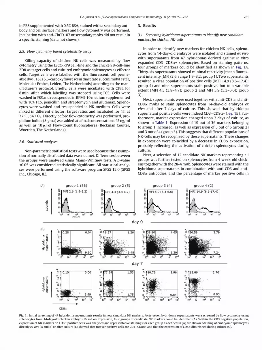
ompa
ibIa
2
c2caMu8wwcm3paW
2
tt<sI
Fsed
C.A. Jansen et al. / Developmental and C
n PBS supplemented with 0.5% BSA, stained with a secondary anti-ody and cell surface markers and flow cytometry was performed.ncubation with anti-ChCD107 or secondary mAbs did not result inspecific staining (data not shown).
.5. Flow cytometry based cytotoxicity assay
Killing capacity of chicken NK-cells was measured by flowytometry using the LSCC-RP9 cell-line and the chicken B-cell-lineD8 as target cells and cultured embryonic splenocytes as effectorells. Target cells were labelled with the fluorescent, cell perme-ble dye CFSE (5,6-carboxyfluorescein diacetate succinimidyl ester,olecular Probes, Leiden, The Netherlands) according to the man-
facturer’s protocol. Briefly, cells were incubated with CFSE formin, after which labelling was stopped using FCS. Cells wereashed in PBS and resuspended in RPMI-10 medium supplementedith 10% FCS, penicillin and streptomycin and glutamax. Spleno-
ytes were washed and resuspended in NK medium. Cells wereixed in different effector: target ratios and incubated for 4 h at
7 ◦C, 5% CO2. Directly before flow cytometry was performed, pro-idium iodide (Sigma) was added at a final concentration of 5 ng/mls well as 10 �l of Flow-Count fluorospheres (Beckman Coulter,oerden, The Netherlands).
.6. Statistical analyses
Non-parametric statistical tests were used because the assump-
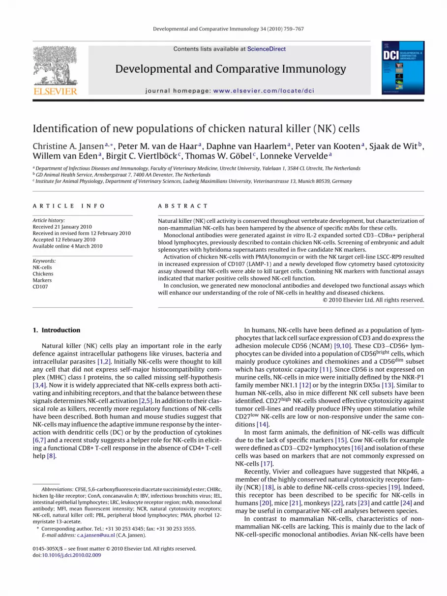
ion of normally distributed data was not met. Differences betweenhe groups were analysed using Mann–Whitney tests. A p-value0.05 was considered statistically significant. All statistical analy-es were performed using the software program SPSS 12.0 (SPSSnc., Chicago, IL).ig. 1. Initial screening of 47 hybridoma supernatants results in new candidate NK markplenocytes from 14-day-old chicken embryos. Based on expression, four groups of canxpression of NK markers on CD8� positive cells was analysed and representative stainiirectly ex vivo (A and B) or after culture (C) showed that marker positive cells are CD3−C
rative Immunology 34 (2010) 759–767 761
3. Results
3.1. Screening hybridoma supernatants to identify new candidatemarkers for chicken NK-cells
In order to identify new markers for chicken NK-cells, spleno-cytes from 14-day-old embryos were isolated and stained ex vivowith supernatants from 47 hybridomas derived against in vitroexpanded CD3−CD8�+ splenocytes. Based on staining patterns,four groups of markers could be identified as shown in Fig. 1A.Thirty-six supernatants showed minimal reactivity (mean fluores-cent intensity (MFI) 2.6, range 1.9–3.2; group 1). Two supernatantsresulted a clear population of positive cells (MFI 14.9 (8.6–17.4);group 4) and nine supernatants stain positive, but to a variableextent (MFI 4.3 (3.8–4.7); group 2 and MFI 5.9 (5.3–6.6); group3).
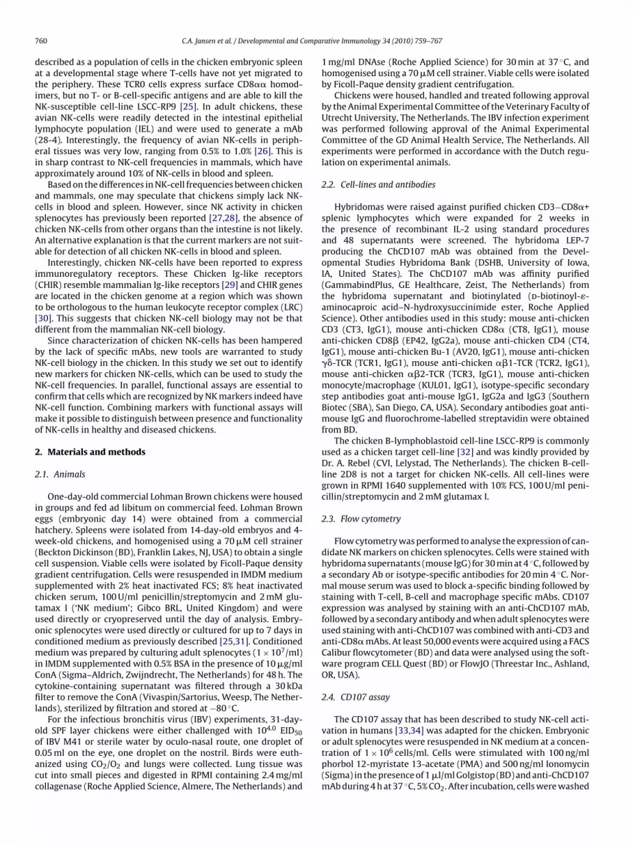
Next, supernatants were used together with anti-CD3 and anti-CD8� mAbs to stain splenocytes from 14-day-old embryos exvivo and after 7 days of culture. This showed that hybridomasupernatant positive cells were indeed CD3−CD8�+ (Fig. 1B). Fur-thermore, marker expression changed upon 7 days of culture, asshown in Table 1. Expression of 19 out of 36 markers belongingto group 1 increased, as well as expression of 3 out of 5 (group 2)and 3 out of 4 (group 3). This suggests that different populations ofNK-cells may be recognized by these supernatants. These changesin expression were coincided by a decrease in CD8� expression,probably reflecting the activation of chicken splenocytes duringculture.
Next, a selection of 12 candidate NK markers representing allgroups was further tested on splenocytes from 4-week-old chick-ens together with the 28-4 mAb. Splenocytes were stained with thehybridoma supernatants in combination with anti-CD3 and anti-CD8� antibodies, and the percentage of marker positive cells in
ers. Forty-seven hybridoma supernatants were screened by flow cytometry usingdidate NK markers could be identified (A). Within the CD3 negative population,
ngs for each group as defined in (A) are shown. Staining of embryonic splenocytesD8�+ and that the expression of CD8� diminished during culture (C).
762 C.A. Jansen et al. / Developmental and Comparative Immunology 34 (2010) 759–767
Table 1Expression of candidate chicken NK-cell markers on ED14 cells on day 0 and after 7 days of culture.
Day 0 Day 7
Group 1 Group 2 Group 3 Group 4
Group 1 14D9 7G6 15E7 2A4 9G6 4H3 6E12 15F85D1 1D11 7A6 20C8 13G3 12H3 15D1 11F710H7 19H6 3C6 14D3 7C1 6E7 12A11 4A515B11 1C4 16B8 9G4 6H7 2G3 7B521D5 4F8 1B1 1D12 17B12
Group 2 21E3 5C715C7 12D7
14H12
Group 3 1G7 14A820E53C4
G
du2(5Cmte
CD3− cells (data not shown). Based on these staining patterns
Fa1aa
Group 4
roups 1, 2, 3 and 4 refer to the grouping that is shown in Fig. 1.
ifferent subsets was analysed. As shown in Fig. 2A and B, the pop-lation of CD3−CD8�+ splenocytes (population 1) is small; median.0% with a range of 0.7–6.9%. The population of CD3−CD8�dimpopulation 2) cells is much larger; median 9.2% with a range of.8–16.8%. When the expression of the candidate NK markers on
D3−CD8�+ cells was analysed (Fig. 2C), differences between thearkers were observed. Staining patterns could be divided intowo groups, based on frequencies of positive cells. Eight mark-rs stained the major fraction of CD3−CD8�+ cells (median 35.3,
ig. 2. Additional screening on adult splenocytes results in a panel of five candidate NKnti-CD8� mAbs. A representative example is shown (A). Frequencies of CD3−CD8�+ ce2 4-week-old chickens (B). Co-staining with 12 candidate NK markers (or 28-4, previounti-CD8� mAbs was performed on splenocytes from 4-week-old chickens. Median exprere indicated by a black dot (C).
12D66B5
range 19.4–46.6), and five markers were expressed on a minor-ity of the CD3−CD8�+ cells (median 4.8, range 1.5–6.2). The 28-4mAb stained 2.1% of the CD3−CD8�+ cells (range 0.8–5.5%). Simi-lar results were observed for the CD3−CD8�dim population and
which suggested that markers from different groups may recog-nize different populations of NK-cells, two markers from group1 and three markers from group 2 were selected for furtheranalyses.
markers. Splenocytes from 4-week-old chickens were stained with anti-CD3 andlls (population 1) and CD3−CD8�dim splenocytes (population 2) were analysed insly described as a marker for intestinal and embryonic NK-cells) and anti-CD3 andssion and interquartile range is shown. Markers that are selected for further study
C.A. Jansen et al. / Developmental and Comparative Immunology 34 (2010) 759–767 763
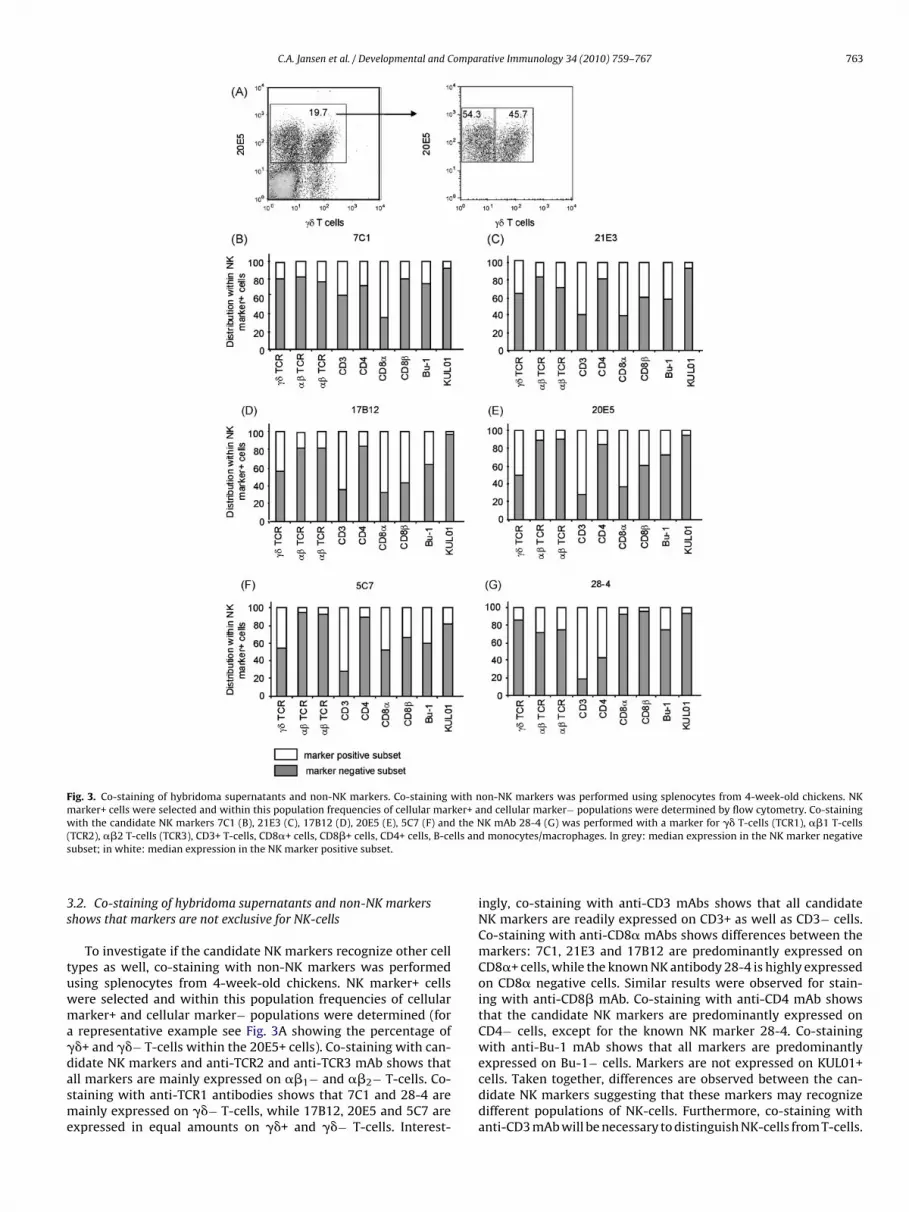
Fig. 3. Co-staining of hybridoma supernatants and non-NK markers. Co-staining with non-NK markers was performed using splenocytes from 4-week-old chickens. NKmarker+ cells were selected and within this population frequencies of cellular marker+ and cellular marker− populations were determined by flow cytometry. Co-stainingw the N( lls ans
3s
tuwma�dasme
ith the candidate NK markers 7C1 (B), 21E3 (C), 17B12 (D), 20E5 (E), 5C7 (F) andTCR2), ��2 T-cells (TCR3), CD3+ T-cells, CD8�+ cells, CD8�+ cells, CD4+ cells, B-ceubset; in white: median expression in the NK marker positive subset.
.2. Co-staining of hybridoma supernatants and non-NK markershows that markers are not exclusive for NK-cells
To investigate if the candidate NK markers recognize other cellypes as well, co-staining with non-NK markers was performedsing splenocytes from 4-week-old chickens. NK marker+ cellsere selected and within this population frequencies of cellulararker+ and cellular marker− populations were determined (forrepresentative example see Fig. 3A showing the percentage of�+ and ��− T-cells within the 20E5+ cells). Co-staining with can-
idate NK markers and anti-TCR2 and anti-TCR3 mAb shows thatll markers are mainly expressed on ��1− and ��2− T-cells. Co-taining with anti-TCR1 antibodies shows that 7C1 and 28-4 areainly expressed on ��− T-cells, while 17B12, 20E5 and 5C7 arexpressed in equal amounts on ��+ and ��− T-cells. Interest-
K mAb 28-4 (G) was performed with a marker for �� T-cells (TCR1), ��1 T-cellsd monocytes/macrophages. In grey: median expression in the NK marker negative
ingly, co-staining with anti-CD3 mAbs shows that all candidateNK markers are readily expressed on CD3+ as well as CD3− cells.Co-staining with anti-CD8� mAbs shows differences between themarkers: 7C1, 21E3 and 17B12 are predominantly expressed onCD8�+ cells, while the known NK antibody 28-4 is highly expressedon CD8� negative cells. Similar results were observed for stain-ing with anti-CD8� mAb. Co-staining with anti-CD4 mAb showsthat the candidate NK markers are predominantly expressed onCD4− cells, except for the known NK marker 28-4. Co-stainingwith anti-Bu-1 mAb shows that all markers are predominantly
expressed on Bu-1− cells. Markers are not expressed on KUL01+cells. Taken together, differences are observed between the can-didate NK markers suggesting that these markers may recognizedifferent populations of NK-cells. Furthermore, co-staining withanti-CD3 mAb will be necessary to distinguish NK-cells from T-cells.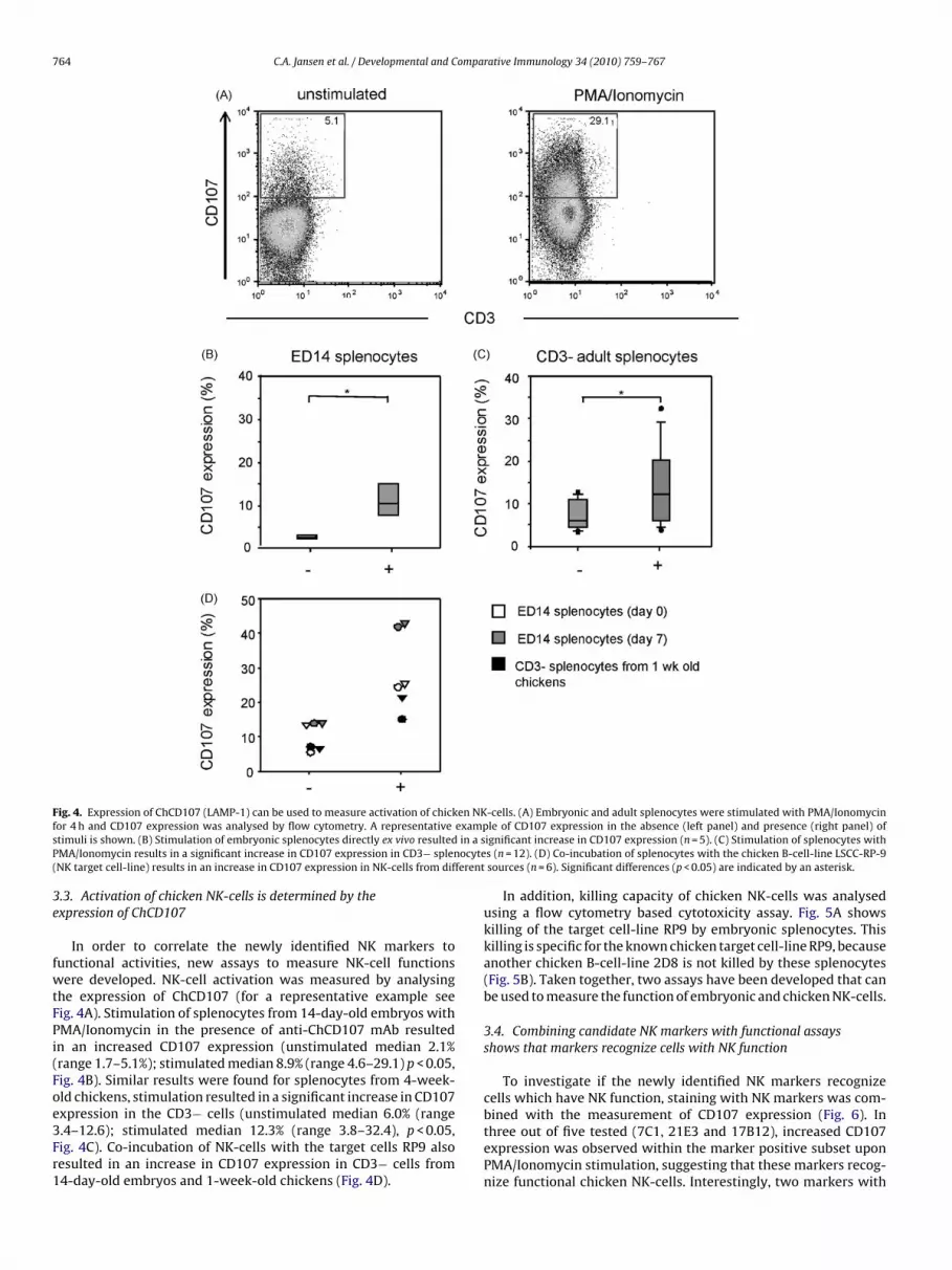
764 C.A. Jansen et al. / Developmental and Comparative Immunology 34 (2010) 759–767
F en NKf examps in a sP ocyte( ferent
3e
fwtFPi(Foe3Fr1
ig. 4. Expression of ChCD107 (LAMP-1) can be used to measure activation of chickor 4 h and CD107 expression was analysed by flow cytometry. A representativetimuli is shown. (B) Stimulation of embryonic splenocytes directly ex vivo resultedMA/Ionomycin results in a significant increase in CD107 expression in CD3− splenNK target cell-line) results in an increase in CD107 expression in NK-cells from dif
.3. Activation of chicken NK-cells is determined by thexpression of ChCD107
In order to correlate the newly identified NK markers tounctional activities, new assays to measure NK-cell functionsere developed. NK-cell activation was measured by analysing
he expression of ChCD107 (for a representative example seeig. 4A). Stimulation of splenocytes from 14-day-old embryos withMA/Ionomycin in the presence of anti-ChCD107 mAb resultedn an increased CD107 expression (unstimulated median 2.1%range 1.7–5.1%); stimulated median 8.9% (range 4.6–29.1) p < 0.05,ig. 4B). Similar results were found for splenocytes from 4-week-ld chickens, stimulation resulted in a significant increase in CD107
xpression in the CD3− cells (unstimulated median 6.0% (range.4–12.6); stimulated median 12.3% (range 3.8–32.4), p < 0.05,ig. 4C). Co-incubation of NK-cells with the target cells RP9 alsoesulted in an increase in CD107 expression in CD3− cells from4-day-old embryos and 1-week-old chickens (Fig. 4D).-cells. (A) Embryonic and adult splenocytes were stimulated with PMA/Ionomycinle of CD107 expression in the absence (left panel) and presence (right panel) of
ignificant increase in CD107 expression (n = 5). (C) Stimulation of splenocytes withs (n = 12). (D) Co-incubation of splenocytes with the chicken B-cell-line LSCC-RP-9sources (n = 6). Significant differences (p < 0.05) are indicated by an asterisk.
In addition, killing capacity of chicken NK-cells was analysedusing a flow cytometry based cytotoxicity assay. Fig. 5A showskilling of the target cell-line RP9 by embryonic splenocytes. Thiskilling is specific for the known chicken target cell-line RP9, becauseanother chicken B-cell-line 2D8 is not killed by these splenocytes(Fig. 5B). Taken together, two assays have been developed that canbe used to measure the function of embryonic and chicken NK-cells.
3.4. Combining candidate NK markers with functional assaysshows that markers recognize cells with NK function
To investigate if the newly identified NK markers recognizecells which have NK function, staining with NK markers was com-
bined with the measurement of CD107 expression (Fig. 6). Inthree out of five tested (7C1, 21E3 and 17B12), increased CD107expression was observed within the marker positive subset uponPMA/Ionomycin stimulation, suggesting that these markers recog-nize functional chicken NK-cells. Interestingly, two markers with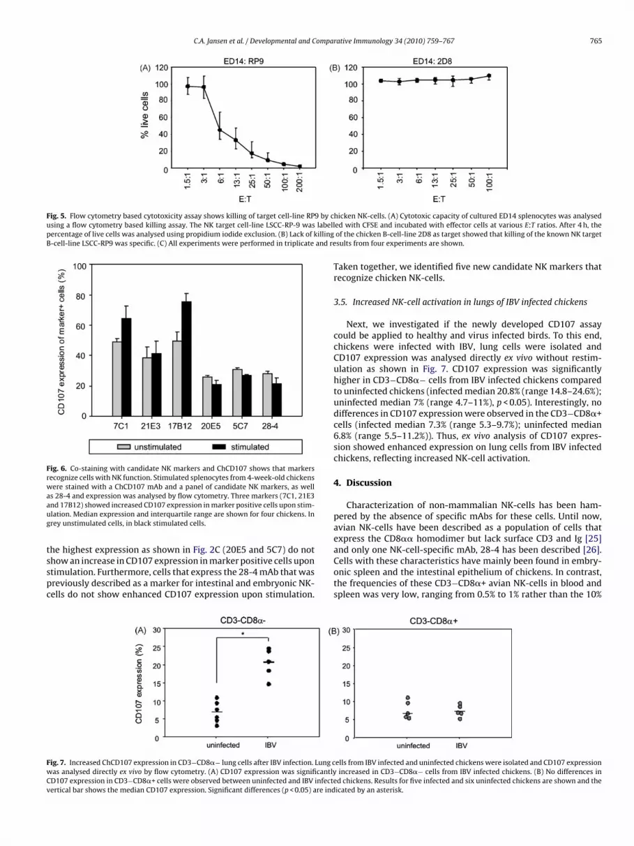
C.A. Jansen et al. / Developmental and Comparative Immunology 34 (2010) 759–767 765
Fig. 5. Flow cytometry based cytotoxicity assay shows killing of target cell-line RP9 by cusing a flow cytometry based killing assay. The NK target cell-line LSCC-RP-9 was labellpercentage of live cells was analysed using propidium iodide exclusion. (B) Lack of killingB-cell-line LSCC-RP9 was specific. (C) All experiments were performed in triplicate and re
Fig. 6. Co-staining with candidate NK markers and ChCD107 shows that markersrecognize cells with NK function. Stimulated splenocytes from 4-week-old chickenswere stained with a ChCD107 mAb and a panel of candidate NK markers, as wellaaug
tsspc
FwCv
s 28-4 and expression was analysed by flow cytometry. Three markers (7C1, 21E3nd 17B12) showed increased CD107 expression in marker positive cells upon stim-lation. Median expression and interquartile range are shown for four chickens. Inrey unstimulated cells, in black stimulated cells.
he highest expression as shown in Fig. 2C (20E5 and 5C7) do nothow an increase in CD107 expression in marker positive cells upontimulation. Furthermore, cells that express the 28-4 mAb that wasreviously described as a marker for intestinal and embryonic NK-ells do not show enhanced CD107 expression upon stimulation.
ig. 7. Increased ChCD107 expression in CD3−CD8�− lung cells after IBV infection. Lung cas analysed directly ex vivo by flow cytometry. (A) CD107 expression was significantlyD107 expression in CD3−CD8�+ cells were observed between uninfected and IBV infecteertical bar shows the median CD107 expression. Significant differences (p < 0.05) are ind
hicken NK-cells. (A) Cytotoxic capacity of cultured ED14 splenocytes was analyseded with CFSE and incubated with effector cells at various E:T ratios. After 4 h, theof the chicken B-cell-line 2D8 as target showed that killing of the known NK targetsults from four experiments are shown.
Taken together, we identified five new candidate NK markers thatrecognize chicken NK-cells.
3.5. Increased NK-cell activation in lungs of IBV infected chickens
Next, we investigated if the newly developed CD107 assaycould be applied to healthy and virus infected birds. To this end,chickens were infected with IBV, lung cells were isolated andCD107 expression was analysed directly ex vivo without restim-ulation as shown in Fig. 7. CD107 expression was significantlyhigher in CD3−CD8�− cells from IBV infected chickens comparedto uninfected chickens (infected median 20.8% (range 14.8–24.6%);uninfected median 7% (range 4.7–11%), p < 0.05). Interestingly, nodifferences in CD107 expression were observed in the CD3−CD8�+cells (infected median 7.3% (range 5.3–9.7%); uninfected median6.8% (range 5.5–11.2%)). Thus, ex vivo analysis of CD107 expres-sion showed enhanced expression on lung cells from IBV infectedchickens, reflecting increased NK-cell activation.
4. Discussion
Characterization of non-mammalian NK-cells has been ham-pered by the absence of specific mAbs for these cells. Until now,avian NK-cells have been described as a population of cells thatexpress the CD8�� homodimer but lack surface CD3 and Ig [25]
and only one NK-cell-specific mAb, 28-4 has been described [26].Cells with these characteristics have mainly been found in embry-onic spleen and the intestinal epithelium of chickens. In contrast,the frequencies of these CD3−CD8�+ avian NK-cells in blood andspleen was very low, ranging from 0.5% to 1% rather than the 10%ells from IBV infected and uninfected chickens were isolated and CD107 expressionincreased in CD3−CD8�− cells from IBV infected chickens. (B) No differences ind chickens. Results for five infected and six uninfected chickens are shown and theicated by an asterisk.
7 ompa
opncNsipsCtomeee
uofNbIct(dwpftii
NoCmwCCoaTItroT�so
tfptostc
ksbw
66 C.A. Jansen et al. / Developmental and C
bserved in many mammals. This implicates that NK-cells are rarelyresent outside the gut or more likely that the current markers areot appropriate for the detection of NK-cells in blood and spleen ofhickens. Therefore, we set out to identify new markers for chickenK-cells. Splenocytes from 14-day-old embryos were isolated and
tained with supernatants from 47 hybridomas generated againstn vitro expanded CD3−CD8�+ splenocytes and based on stainingatterns four different groups of markers could be identified. Next,taining of the supernatants in the presence of anti-CD3 and anti-D8� mAbs on splenocytes from 14-day-old embryos confirmedhat the positive cells were indeed CD3−CD8�+, which fits earlierbservations [34]. Culturing the cells for 7 days with conditionededium showed changes in expression of the different NK mark-
rs and a decrease in CD8� expression. This decrease in CD8�xpression may reflect activation of the cells, similar to loss of CD3xpression upon stimulation.
It is tempting to speculate that the differences in expressionpon culture are due to the existence of different populationsf NK-cells which develop during culture. Staining splenocytesrom healthy 4-week-old chickens with a panel of 12 candidateK markers and the 28-4 mAb resulted in two different groupsased on staining patterns within the CD3−CD8�+ population.
nterestingly, while in the spleen the population of CD3−CD8�+ells is rather small (median 2.0%; range (0.7–6.9%)), the popula-ion of CD3−CD8�dim cells is much bigger (median 9.2%; range5.8–16.8%)). As activation of embryonic splenocytes resulted in aecrease in CD8� expression, these CD3−CD8�dim cells may veryell represent a population of (activated) NK-cells. This is sup-orted by the similar staining patterns of the candidate NK markersor the CD3−CD8�+ and CD3−CD8�dim population. Furthermore,he total frequency of CD3−CD8�dim and CD3−CD8�+ cells is sim-lar to the frequencies of NK-cells observed in mammals and CD107s predominantly expressed in CD8�dim cells (data not shown).
Co-staining with non-NK markers suggests that the candidateK markers recognize different populations. Most markers also rec-gnize CD8� negative cells. As we observed down regulation ofD8� expression upon activation, this suggests that some of thesearkers may recognize activated NK-cells. Furthermore, stainingith the NK markers needs to be combined with staining with anti-D3 mAbs, similar to the human situation in which the NK markerD56 is also expressed on T-cells [35]. Some markers may also rec-gnize �� T-cells (21E3, 17B12, 20E5 and 5C7). In mammals, almostll NK-cell receptors have been found to be expressed by �� or ��-cells [36]. Also bovine �� T-cells that have been stimulated withL-15 express the NK-cell receptor NKp46 [37]. Chickens have upo 50% �� T-cells in blood and spleen [38], and a mAb against a NKeceptor from the C-type lectin family (B-NK) recognizes embry-nic ED14 splenocytes as well as subsets of splenic �� and ��-cells [39]. This implies that markers for NK-cells may recognize� T-cells as well as NK-cells show which is supported by our datahowing that the candidate NK markers are also readily expressedn TCR1+ cells.
The function of chicken NK-cells was determined by analysinghe expression of CD107 (LAMP-1) which is expressed on the sur-ace of NK-cells upon activation. The expression of CD107 hasreviously been shown to correlate cytotoxicity and IFN� produc-ion [34] and is commonly used as an assay to measure activationf mammalian NK-cells [34,40]. CD107 expression was found upontimulation with PMA/Ionomycin and upon stimulation with thearget cell-line LSCC-RP9, showing that activation of chicken NK-ells can be measured in vitro.
In addition, a flow cytometry based cytotoxicity assay showedilling of the target cell-line LSCC-RP9 by cultured ED14 embryonicplenocytes and CD3−CD4-depleted lung cells. Flow cytometryased cytotoxicity assays have previously been shown to correlateell with the standard 51Chromium release assay [41,42]. Further-
rative Immunology 34 (2010) 759–767
more, the advantage of a flow cytometry based assay is that thisassay is not radioactive and multi-color flow cytometry allows theanalysis of cell-specific parameters.
Based on the screening of embryonic and adult splenocytes,staining of five candidate NK markers and the 28-4 mAb was com-bined with the measurement of CD107 expression. Three markers(7C1, 21E3 and 17B12) showed increased CD107 expression withinthe marker positive subset upon PMA/Ionomycin stimulation,suggesting that these markers recognize chicken NK-cells. Inter-estingly, 20E5 and 5C7, two markers with the highest expressionbased on Fig. 2C do not show an increase in CD107 expressionin marker positive cells upon stimulation. This confirms the ear-lier observation that these markers may be expressed to someextend on NK-cells, but are readily expressed by other cells as well.Furthermore, 28-4+ cells in spleen do not show NK-cell function.Interestingly, 28-4+ IEL have been reported to induce lysis of NK-sensitive targets [26]. This suggests that 28-4+ IEL are different from28-4+ splenocytes which are studied here.
Analysis of CD107 expression in lung cells from IBV infectedchickens showed increased CD107 expression compared to lungcells from uninfected chickens. Interestingly, this difference wasonly observed for CD3−CD8�− cells, which again suggests that acti-vation of chicken NK-cells is paralleled by down regulation of CD8�.These data show for the first time that the ChCD107 assay can bereadily used to study chicken NK-cell activation ex vivo. Therefore,this assay is a valuable tool to study NK-cell biology in healthy anddiseased chickens.
In conclusion, we identified five new markers (7C1, 21E3, 17B12,20E5 and 5C7) that recognize chicken NK-cells and developed twoassays to measure NK-cell activation and cytotoxicity. Although theantigens that are recognized by the different markers are not yetknown, the experiments performed in this study suggest that themarkers may recognize different populations of NK-cells. It is pos-sible that the frequencies of NK-cells recognized by the differentmarkers may vary between different organs, similar to the resultswith the 28-4 mAb. These results will lead to a better understand-ing of NK-cell frequencies and distribution in healthy and diseasedchickens.
Acknowledgements
This study was financially supported by the EU sixth frameworkprogram Flupath (grant 044220) and Program “Impulse VeterinaryAvian Influenza Research in The Netherlands”, Dutch Ministry ofAgriculture, Nature and Food Quality. T.W.G. was supported by DFGgrant GO489/5-1.
References
[1] Trinchieri G. Biology of natural killer cells. Adv Immunol 1989;47:187–376.
[2] Lanier LL. NK cell recognition. Annu Rev Immunol 2005;23:225–74.[3] Karre K, Ljunggren HG, Piontek G, Kiessling R. Selective rejection of H-2-
deficient lymphoma variants suggests alternative immune defence strategy.Nature 1986;319:675–8.
[4] Ljunggren HG, Karre K. Host resistance directed selectively against H-2-deficient lymphoma variants. Analysis of the mechanism. J Exp Med1985;162:1745–59.
[5] Moretta L, Bottino C, Pende D, Vitale M, Mingari MC, Moretta A. Different check-points in human NK-cell activation. Trends Immunol 2004;25:670–6.
[6] Cooper MA, Fehniger TA, Fuchs A, Colonna M, Caligiuri MA. NK cell and DCinteractions. Trends Immunol 2004;25:47–52.
[7] Moretta A. Natural killer cells and dendritic cells: rendezvous in abused tissues.Nat Rev Immunol 2002;2:957–64.
[8] Nandakumar S, Woolard SN, Yuan D, Rouse BT, Kumaraguru U. Natural killercells as novel helpers in anti-herpes simplex virus immune response. J Virol2008;82:10820–31.
[9] Lanier LL, Testi R, Bindl J, Phillips JH. Identity of Leu-19 (CD56) leuko-cyte differentiation antigen and neural cell adhesion molecule. J Exp Med1989;169:2233–8.
ompa
[
[
[
[
[
[
[
[
[
[
[
[
[
[
[
[
[
[
[
[
[
[
[
[
[
[
[
[
[
[
[
[41] Radosevic K, Garritsen HS, van Graft M, de Grooth BG, Greve J. A simple and
C.A. Jansen et al. / Developmental and C
10] Ritz J, Schmidt RE, Michon J, Hercend T, Schlossman SF. Characterization offunctional surface structures on human natural killer cells. Adv Immunol1988;42:181–211.
11] Cooper MA, Fehniger TA, Caligiuri MA. The biology of human natural killer-cellsubsets. Trends Immunol 2001;22:633–40.
12] Ryan JC, Turck J, Niemi EC, Yokoyama WM, Seaman WE. Molecular cloning of theNK1.1 antigen, a member of the NKR-P1 family of natural killer cell activationmolecules. J Immunol 1992;149:1631–5.
13] Arase H, Saito T, Phillips JH, Lanier LL. Cutting edge: the mouse NK cell-associated antigen recognized by DX5 monoclonal antibody is CD49b (alpha2 integrin, very late antigen-2). J Immunol 2001;167:1141–4.
14] Hayakawa Y, Smyth MJ. CD27 dissects mature NK cells into two subsets withdistinct responsiveness and migratory capacity. J Immunol 2006;176:1517–24.
15] Evans DL, Jaso-Friedmann L. Natural killer (NK) cells in domestic animals:phenotype, target cell specificity and cytokine regulation. Vet Res Commun1993;17:429–47.
16] Goff WL, Johnson WC, Horn RH, Barrington GM, Knowles DP. The innateimmune response in calves to Boophilus microplus tick transmitted Babesiabovis involves type-1 cytokine induction and NK-like cells in the spleen. Para-site Immunol 2003;25:185–8.
17] Endsley JJ, Endsley MA, Estes DM. Bovine natural killer cells acquire cyto-toxic/effector activity following activation with IL-12/15 and reduce Mycobac-terium bovis BCG in infected macrophages. J Leukoc Biol 2006;79:71–9.
18] Sivori S, Pende D, Bottino C, Marcenaro E, Pessino A, Biassoni R, et al. NKp46is the major triggering receptor involved in the natural cytotoxicity of fresh orcultured human NK cells. Correlation between surface density of NKp46 andnatural cytotoxicity against autologous, allogeneic or xenogeneic target cells.Eur J Immunol 1999;29:1656–66.
19] Walzer T, Blery M, Chaix J, Fuseri N, Chasson L, Robbins SH, et al. Identification,activation, and selective in vivo ablation of mouse NK cells via NKp46. Proc NatlAcad Sci USA 2007;104:3384–9.
20] Pessino A, Sivori S, Bottino C, Malaspina A, Morelli L, Moretta L, et al. Molec-ular cloning of NKp46: a novel member of the immunoglobulin superfamilyinvolved in triggering of natural cytotoxicity. J Exp Med 1998;188:953–60.
21] Biassoni R, Pessino A, Bottino C, Pende D, Moretta L, Moretta A. The murinehomologue of the human NKp46, a triggering receptor involved in the inductionof natural cytotoxicity. Eur J Immunol 1999;29:1014–20.
22] De MA, Biassoni R, Fogli M, Rizzi M, Cantoni C, Costa P, et al. Identification,molecular cloning and functional characterization of NKp46 and NKp30 nat-ural cytotoxicity receptors in Macaca fascicularis NK cells. Eur J Immunol2001;31:3546–56.
23] Falco M, Cantoni C, Bottino C, Moretta A, Biassoni R. Identification of therat homologue of the human NKp46 triggering receptor. Immunol Lett1999;68:411–4.
24] Storset AK, Slettedal IO, Williams JL, Law A, Dissen E. Natural killer cell recep-
tors in cattle: a bovine killer cell immunoglobulin-like receptor multigenefamily contains members with divergent signaling motifs. Eur J Immunol2003;33:980–90.25] Gobel TW, Chen CL, Shrimpf J, Grossi CE, Bernot A, Bucy RP, et al. Characteri-zation of avian natural killer cells and their intracellular CD3 protein complex.Eur J Immunol 1994;25:1685–91.
[
rative Immunology 34 (2010) 759–767 767
26] Gobel TW, Kaspers B, Stangassinger M. NK and T cells constitute two major,functionally distinct intestinal epithelial lymphocyte subsets in the chicken.Int Immunol 2001;13:757–62.
27] Lillehoj HS. Intestinal intraepithelial and splenic natural killer cellresponses to eimerian infections in inbred chickens. Infect Immun 1989;57:1879–84.
28] Myers TJ, Schat KA. Natural killer cell activity of chicken intraepithelial leuko-cytes against rotavirus-infected target cells. Vet Immunol Immunopathol1990;26:157–70.
29] Dennis Jr G, Kubagawa H, Cooper MD. Paired Ig-like receptor homologs in birdsand mammals share a common ancestor with mammalian Fc receptors. ProcNatl Acad Sci USA 2000;97:13245–50.
30] Viertlboeck BC, Habermann FA, Schmitt R, Groenen MA, Du PL, Gobel TW.The chicken leukocyte receptor complex: a highly diverse multigene familyencoding at least six structurally distinct receptor types. J Immunol 2005;175:385–93.
31] Gobel TW. Isolation and analysis of natural killer cells in chickens. MethodsMol Biol 2000;121:337–45.
32] Sharma JM, Okazaki W. Natural killer cell activity in chickens: target cellanalysis and effect of antithymocyte serum on effector cells. Infect Immun1981;31:1078–85.
33] Betts MR, Brenchley JM, Price DA, De Rosa SC, Douek DC, Roederer M, et al.Sensitive and viable identification of antigen-specific CD8+ T cells by a flowcytometric assay for degranulation. J Immunol Methods 2003;281. pp. 281:65–78.
34] Penack O, Gentilini C, Fischer L, Asemissen AM, Scheibenbogen C, Thiel E, et al.CD56dimCD16neg cells are responsible for natural cytotoxicity against tumortargets. Leukemia 2005;19:835–40.
35] Lanier LL, Le AM, Ding A, Evans EL, Krensky AM, et al. Expression of Leu-19(NKH-1) antigen on IL 2-dependent cytotoxic and non-cytotoxic T cell lines. JImmunol 1987;138:2019–23.
36] Lanier LL. Back to the future-defining NK cells and T cells. Eur J Immunol2007;37:1424–6.
37] Johnson WC, Bastos RG, Davis WC, Goff WL. Bovine WC1(−) gammadeltaT cellsincubated with IL-15 express the natural cytotoxicity receptor CD335 (NKp46)and produce IFN-gamma in response to exogenous IL-12 and IL-18. Dev CompImmunol 2008;32:1002–10.
38] Chen CH, Six A, Kubota T, Tsuji S, Kong FK, et al. T cell receptors and T celldevelopment. Curr Top Microbiol Immunol 1996;212:37–53.
39] Viertlboeck BC, Wortmann A, Schmitt R, Plachy J, Gobel TW. Chicken C-typelectin-like receptor B-NK, expressed on NK and T cell subsets, binds to a ligandon activated splenocytes. Mol Immunol 2008;45:1398–404.
40] Alter G, Malenfant JM, Altfeld M. CD107a as a functional marker for the identi-fication of natural killer cell activity. J Immunol Methods 2004;294:15–22.
sensitive flow cytometric assay for the determination of the cytotoxic activityof human natural killer cells. J Immunol Methods 1990;135:81–9.
42] Goldberg JE, Sherwood SW, Clayberger C. A novel method for measuring CTLand NK cell-mediated cytotoxicity using annexin V and two-color flow cytom-etry. J Immunol Methods 1999;224:1–9.