
Deep Sequencing of Chicken microRNAs
9
BioMed Central Page 1 of 9 (page number not for citation purposes) BMC Genomics Open Access Research article Deep Sequencing of Chicken microRNAs Joan Burnside* 1 , Ming Ouyang 3 , Amy Anderson 1 , Erin Bernberg 1 , Cheng Lu 2 , Blake C Meyers 2 , Pamela J Green 2 , Milos Markis 1 , Grace Isaacs 1 , Emily Huang 1 and Robin W Morgan 1 Address: 1 Department of Animal and Food Sciences, Delaware Biotechnology Institute, University of Delaware, Newark, Delaware, 19711, USA, 2 Department of Plant and Soil Sciences, Delaware Biotechnology Institute, University of Delaware, Newark, Delaware, 19711, USA and 3 University of Louisville, Department of Computer Engineering & Computer Science Louisville, Kentucky, 40292, USA Email: Joan Burnside* - [email protected]; Ming Ouyang - [email protected]; Amy Anderson - [email protected]; Erin Bernberg - [email protected]; Cheng Lu - [email protected]; Blake C Meyers - [email protected]; Pamela J Green - [email protected]; Milos Markis - [email protected]; Grace Isaacs - [email protected]; Emily Huang - [email protected]; Robin W Morgan - [email protected] * Corresponding author Abstract Background: The use of new, deep sequencing technologies has greatly accelerated microRNA discovery. We have applied this approach to the identification of chicken microRNAs and to the comparison of microRNAs in chicken embryo fibroblasts (CEF) infected with Marek's disease virus (MDV) to those present in uninfected CEF. Results: We obtained 125,463 high quality reads that showed an exact match to the chicken genome. The majority of the reads corresponded to previously annotated chicken microRNAs; however, the sequences of many potential novel microsRNAs were obtained. A comparison of the reads obtained in MDV-infected and uninfected CEF indicates that infection does not significantly perturb the expression profile of microRNAs. Frequently sequenced microRNAs include miR-221/ 222, which are thought to play a role in growth and proliferation. A number of microRNAs (e.g., let-7, miR-199a-1, 26a) are expressed at lower levels in MDV-induced tumors, highlighting the potential importance of this class of molecules in tumorigenesis. Conclusion: Deep sequencing technology is highly suited for small RNA discovery. This approach is independent of comparative sequence analysis, which has been the primary method used to identify chicken microRNAs. Our results have confirmed the expression of many microRNAs identified by sequence similarity and identified a pool of candidate novel microRNAs. Background MicroRNAs are small (about 22 nt) RNAs that play impor- tant regulatory roles by targeting mRNAs for degradation or translational repression. MicroRNAs were first identi- fied in Caenorhabditis elegans [1] but high evolutionary conservation eventually led to the identification of micro- RNAs in other species. This, coupled with conventional sequencing of small RNA libraries, has greatly expanded the list of known microRNAs. The most recent release of the microRNA database, miRBase 10.0 [2], contains over 5000 microRNA gene loci in a wide variety of animal, plant and viral genomes. Published: 22 April 2008 BMC Genomics 2008, 9:185 doi:10.1186/1471-2164-9-185 Received: 10 November 2007 Accepted: 22 April 2008 This article is available from: http://www.biomedcentral.com/1471-2164/9/185 © 2008 Burnside et al; licensee BioMed Central Ltd. This is an Open Access article distributed under the terms of the Creative Commons Attribution License (http://creativecommons.org/licenses/by/2.0 ), which permits unrestricted use, distribution, and reproduction in any medium, provided the original work is properly cited.
-
Upload
independent -
Category
Documents
-
view
1 -
download
0
Transcript of Deep Sequencing of Chicken microRNAs
BioMed CentralBMC Genomics
ss
Open AcceResearch articleDeep Sequencing of Chicken microRNAsJoan Burnside*1, Ming Ouyang3, Amy Anderson1, Erin Bernberg1, Cheng Lu2, Blake C Meyers2, Pamela J Green2, Milos Markis1, Grace Isaacs1, Emily Huang1 and Robin W Morgan1Address: 1Department of Animal and Food Sciences, Delaware Biotechnology Institute, University of Delaware, Newark, Delaware, 19711, USA, 2Department of Plant and Soil Sciences, Delaware Biotechnology Institute, University of Delaware, Newark, Delaware, 19711, USA and 3University of Louisville, Department of Computer Engineering & Computer Science Louisville, Kentucky, 40292, USA
Email: Joan Burnside* - [email protected]; Ming Ouyang - [email protected]; Amy Anderson - [email protected]; Erin Bernberg - [email protected]; Cheng Lu - [email protected]; Blake C Meyers - [email protected]; Pamela J Green - [email protected]; Milos Markis - [email protected]; Grace Isaacs - [email protected]; Emily Huang - [email protected]; Robin W Morgan - [email protected]
* Corresponding author
AbstractBackground: The use of new, deep sequencing technologies has greatly accelerated microRNAdiscovery. We have applied this approach to the identification of chicken microRNAs and to thecomparison of microRNAs in chicken embryo fibroblasts (CEF) infected with Marek's disease virus(MDV) to those present in uninfected CEF.
Results: We obtained 125,463 high quality reads that showed an exact match to the chickengenome. The majority of the reads corresponded to previously annotated chicken microRNAs;however, the sequences of many potential novel microsRNAs were obtained. A comparison of thereads obtained in MDV-infected and uninfected CEF indicates that infection does not significantlyperturb the expression profile of microRNAs. Frequently sequenced microRNAs include miR-221/222, which are thought to play a role in growth and proliferation. A number of microRNAs (e.g.,let-7, miR-199a-1, 26a) are expressed at lower levels in MDV-induced tumors, highlighting thepotential importance of this class of molecules in tumorigenesis.
Conclusion: Deep sequencing technology is highly suited for small RNA discovery. This approachis independent of comparative sequence analysis, which has been the primary method used toidentify chicken microRNAs. Our results have confirmed the expression of many microRNAsidentified by sequence similarity and identified a pool of candidate novel microRNAs.
BackgroundMicroRNAs are small (about 22 nt) RNAs that play impor-tant regulatory roles by targeting mRNAs for degradationor translational repression. MicroRNAs were first identi-fied in Caenorhabditis elegans [1] but high evolutionaryconservation eventually led to the identification of micro-RNAs in other species. This, coupled with conventional
sequencing of small RNA libraries, has greatly expandedthe list of known microRNAs. The most recent release ofthe microRNA database, miRBase 10.0 [2], contains over5000 microRNA gene loci in a wide variety of animal,plant and viral genomes.
Published: 22 April 2008
BMC Genomics 2008, 9:185 doi:10.1186/1471-2164-9-185
Received: 10 November 2007Accepted: 22 April 2008
This article is available from: http://www.biomedcentral.com/1471-2164/9/185
© 2008 Burnside et al; licensee BioMed Central Ltd. This is an Open Access article distributed under the terms of the Creative Commons Attribution License (http://creativecommons.org/licenses/by/2.0), which permits unrestricted use, distribution, and reproduction in any medium, provided the original work is properly cited.
Page 1 of 9(page number not for citation purposes)

BMC Genomics 2008, 9:185 http://www.biomedcentral.com/1471-2164/9/185
Conventional sequencing favors identification of highlyexpressed species, and comparative genomics will notidentify nonconserved microRNAs. In order to enhancediscovery of small RNA species, massively parallel signa-ture sequencing (MPSS) was used to identify small RNAsin Arabidopsis thaliana [3], and the results showed that thediversity of small RNAs exceeded previous estimates.More recently, newer deep sequencing technologies havebeen used to profile microRNAs in Arabidopsis DICER andRDR2 mutants [4,5], and others have applied this tech-nology to various samples including human and chim-panzee brain [6] and Chlamydomonas reinhardtii [7]. Theseapproaches have the advantage that they not only providesequence of low abundance species, but also providequantitative data since the frequency of sequencing readsreflects the abundance of microRNAs in the population.
We previously reported on the use of deep sequencingtechnologies for identification of microRNAs encoded byMarek's disease virus (MDV), an economically importantpathogenic herpesvirus of chickens [8,9]. In an extensionof the pilot study, we sequenced additional reads fromboth MDV-infected chicken embryo fibroblasts (CEF) anduninfected CEF and now report on the identification ofpotential novel host microRNAs. In addition, thesequence of several new MDV-encoded microRNAs werediscovered by deeper sequencing.
ResultsSmall RNA librariesWe obtained 256,221 reads from two small RNA librariesprepared from uninfected CEF or CEF infected with MDV.As shown in Table 1, a total of 171,783 reads containedboth adapters used in creating the library, and 125,463 ofthese high quality reads showed an exact match to thechicken genome. A total of 1,036 reads from the MDV-infected CEF library matched the MDV genome. The pres-ence of other small RNAs (ribosomal fragments, tRNA,snRNA, mtRNA) was relatively small (less than 3%).
The majority (86%) of the small RNAs match to known orpredicted chicken microRNAs (Additional File 1). Of the149 distinct Gallus gallus (gga) entries in miRbase, wefound 101 distinct species expressed in CEF. There were
93 matches from the MDV-infected CEF library and 87matches from the uninfected CEF library. The infectedcells showed slightly more complexity in microRNA diver-sity, which may be in part due to the larger number ofreads obtained from the infected CEF library whichincreases the chances of revealing low abundance microR-NAs. There were 12 microRNAs in the infected cells thatwere not found in the uninfected CEFs and 9 microRNAsfound in the uninfected CEFs that were not found in theinfected cells. An additional eleven chicken homologs ofknown microRNAs were identified (Additional File 1).The size distribution of reads was not significantly differ-ent in the two libraries, and the majority of the reads hadlengths of 21–25 nt (Figure 1).
microRNA profiling by analysis of read countsThe number of reads obtained should reflect the relativeabundance and expression levels of the microRNAs. Afterscaling for total number of reads obtained for each library,the majority of microRNAs were found at similar levels inthe two libraries. A few microRNAs (listed in Table 2)showed a greater than two-fold difference in the numberof reads between the infected and uninfected CEF librar-ies. We found miR-29b and miR-196 at higher levels inthe MDV-infected cells, and three of the let7 microRNAswere found at lower levels in the MDV-infected CEF com-pared to the uninfected CEF. Northern blot analysis didnot detect these differences, but this could be because ofthe low read numbers or because of cross hybridizationwith microRNAs with similar sequences (miR-29a, let7family).
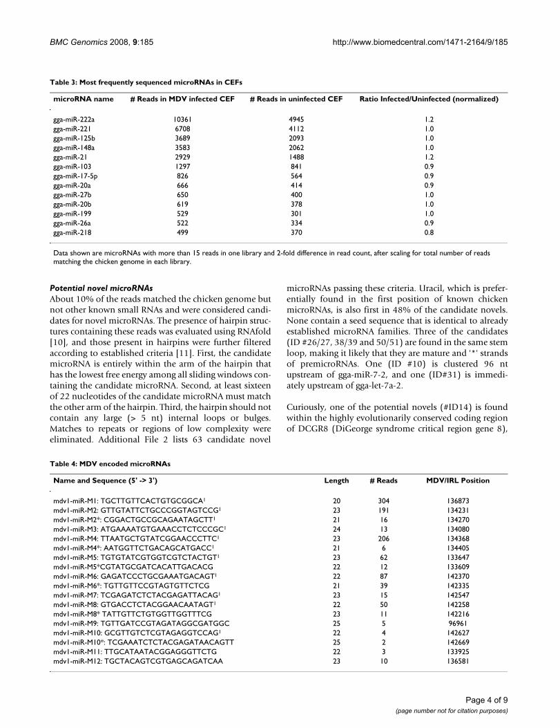
The most frequently sequenced (> 500 reads) microRNAsare found at remarkably similar levels in the two libraries(Table 3). Consistent with findings in our pilot study [9],the highest number of reads was obtained for gga-miR-222 and 221. These are clustered on chromosome 1(114216027–114219024), and in the chicken there aretwo copies of miR-222 in the cluster, which could accountfor the higher number of miR-222 reads. We also see highlevels of gga-miR-125b/148a/21 and 103.
Table 1: Distribution of small RNAs from uninfected CEF and CEF infected with MDV
MDV infected CEF uninfected CEF
High quality/both adapters 107,728 64,055Exact match to chicken genome 79,074 46,389
Match to known miRNAs 67,982 40,173Match to other chicken smalls1 3,249 1,487Match to MDV 1036 -Other potential smalls 7,761 4,666
1tRNA, rRNA, mtRNA, snRNA
Page 2 of 9(page number not for citation purposes)

BMC Genomics 2008, 9:185 http://www.biomedcentral.com/1471-2164/9/185
Viral microRNAsWe previously identified ten MDV-encoded microRNAs ina pilot sequencing project of the MDV-infected CEFlibrary [9]. The deep sequencing revealed an additionalseven microRNAs and '*' strands (Table 4). Four of thenew microRNAs map to the previously identified LAT
cluster (mdv1-miR-6*, 8*, 10, and 10*), two are in thecluster upstream of the meq gene (mdv1-miR-11 and 5*),one is downstream of the meq gene (mdv1-miR-12), andone is antisense to the coding region of the ribonucleotidereductase gene (mdv1-miR-M9). (A preliminary discussionof some of these microRNAs was reviewed in [8]).
Table 2: Relative abundance of differentially expressed microRNAs
microRNA name Length Sequence # Reads in MDV infected CEF
# Reads in uninfected CEF
Ratio Infected/Uninfected (Normalized)
gga-miR-29b 23 TAGCACCATTTGAAATCAGTGTT 64 2 18.8gga-miR-196 21 TAGGTAGTTTCATGTTGTTGG 23 1 13.5gga-miR-133a 22 TTGGTCCCCTTCAACCAGCTGT 15 2 4.4gga-miR-10b 22 TACCCTGTAGAACCGAATTTGT 70 15 2.7gga-miR-30d 22 TGTAAACATCCCCGACTGGAAG 23 6 2.3
gga-let-7f 22 TGAGGTAGTAGATTGTATAGTT 338 454 0.4gga-let-7b 22 TGAGGTAGTAGGTTGTGTGGTT 74 105 0.4gga-miR-130a 22 CAGTGCAATATTAAAAGGGCAT 18 28 0.4gga-let-7a 22 TGAGGTAGTAGGTTGTATAGTT 383 603 0.4gga-miR-1a 21 TGGAATGTAAAGAAGTATGTA 7 12 0.3
Data shown are microRNAs with more than 15 reads in one library and 2-fold difference in read count, after scaling for total number of reads matching the chicken genome in each library.
Size distribution of small RNAsFigure 1Size distribution of small RNAs.
Page 3 of 9(page number not for citation purposes)
BMC Genomics 2008, 9:185 http://www.biomedcentral.com/1471-2164/9/185
Potential novel microRNAsAbout 10% of the reads matched the chicken genome butnot other known small RNAs and were considered candi-dates for novel microRNAs. The presence of hairpin struc-tures containing these reads was evaluated using RNAfold[10], and those present in hairpins were further filteredaccording to established criteria [11]. First, the candidatemicroRNA is entirely within the arm of the hairpin thathas the lowest free energy among all sliding windows con-taining the candidate microRNA. Second, at least sixteenof 22 nucleotides of the candidate microRNA must matchthe other arm of the hairpin. Third, the hairpin should notcontain any large (> 5 nt) internal loops or bulges.Matches to repeats or regions of low complexity wereeliminated. Additional File 2 lists 63 candidate novel
microRNAs passing these criteria. Uracil, which is prefer-entially found in the first position of known chickenmicroRNAs, is also first in 48% of the candidate novels.None contain a seed sequence that is identical to alreadyestablished microRNA families. Three of the candidates(ID #26/27, 38/39 and 50/51) are found in the same stemloop, making it likely that they are mature and '*' strandsof premicroRNAs. One (ID #10) is clustered 96 ntupstream of gga-miR-7-2, and one (ID#31) is immedi-ately upstream of gga-let-7a-2.
Curiously, one of the potential novels (#ID14) is foundwithin the highly evolutionarily conserved coding regionof DCGR8 (DiGeorge syndrome critical region gene 8),
Table 4: MDV encoded microRNAs
Name and Sequence (5' -> 3') Length # Reads MDV/IRL Position
mdv1-miR-M1: TGCTTGTTCACTGTGCGGCA1 20 304 136873mdv1-miR-M2: GTTGTATTCTGCCCGGTAGTCCG1 23 191 134231mdv1-miR-M2*: CGGACTGCCGCAGAATAGCTT1 21 16 134270mdv1-miR-M3: ATGAAAATGTGAAACCTCTCCCGC1 24 13 134080mdv1-miR-M4: TTAATGCTGTATCGGAACCCTTC1 23 206 134368mdv1-miR-M4*: AATGGTTCTGACAGCATGACC1 21 6 134405mdv1-miR-M5: TGTGTATCGTGGTCGTCTACTGT1 23 62 133647mdv1-miR-M5*CGTATGCGATCACATTGACACG 22 12 133609mdv1-miR-M6: GAGATCCCTGCGAAATGACAGT1 22 87 142370mdv1-miR-M6*: TGTTGTTCCGTAGTGTTCTCG 21 39 142335mdv1-miR-M7: TCGAGATCTCTACGAGATTACAG1 23 15 142547mdv1-miR-M8: GTGACCTCTACGGAACAATAGT1 22 50 142258mdv1-miR-M8* TATTGTTCTGTGGTTGGTTTCG 23 11 142216mdv1-miR-M9: TGTTGATCCGTAGATAGGCGATGGC 25 5 96961mdv1-miR-M10: GCGTTGTCTCGTAGAGGTCCAG1 22 4 142627mdv1-miR-M10*: TCGAAATCTCTACGAGATAACAGTT 25 2 142669mdv1-miR-M11: TTGCATAATACGGAGGGTTCTG 22 3 133925mdv1-miR-M12: TGCTACAGTCGTGAGCAGATCAA 23 10 136581
Table 3: Most frequently sequenced microRNAs in CEFs
microRNA name # Reads in MDV infected CEF # Reads in uninfected CEF Ratio Infected/Uninfected (normalized)
gga-miR-222a 10361 4945 1.2gga-miR-221 6708 4112 1.0gga-miR-125b 3689 2093 1.0gga-miR-148a 3583 2062 1.0gga-miR-21 2929 1488 1.2gga-miR-103 1297 841 0.9gga-miR-17-5p 826 564 0.9gga-miR-20a 666 414 0.9gga-miR-27b 650 400 1.0gga-miR-20b 619 378 1.0gga-miR-199 529 301 1.0gga-miR-26a 522 334 0.9gga-miR-218 499 370 0.8
Data shown are microRNAs with more than 15 reads in one library and 2-fold difference in read count, after scaling for total number of reads matching the chicken genome in each library.
Page 4 of 9(page number not for citation purposes)

BMC Genomics 2008, 9:185 http://www.biomedcentral.com/1471-2164/9/185
which interacts with Drosha in the processing of pri-microRNAs [12].
The expression of several of these candidate microRNAswas evaluated by northern blot analysis of different tissues(Figure 2). All hybridize to species the size of maturemicroRNAs. Some of the novel microRNAs are expressedubiquitously (ID#26, 39, 51), while others show moreselective expression (ID#46,61). These microRNAs showno sequence similarity to any known microRNAs, with theexception of #46, which is similar, but not identical todre-miR-730 (21/22) and gga-miR-460 (19/22). ThemicroRNAs analyzed by Northern blots were selectedbased on presence of star strand in sequencing data, pres-ence in a cluster, or some level of sequence conservationwith other species. Other candidate microRNAs in the listhave not been evaluated.
Expression of known host microRNAs in MDV-induced tumorsThere is a large and growing literature on microRNAexpression in tumors, and both up- and down-regulationhave been observed, with microRNA expression patternsreflecting the developmental history and lineage of neo-plasms. We compared expression in MDV-inducedtumors versus normal spleen tissue for selected hostmicroRNAs that were either differentially expressed basedon the deep sequencing or that were interesting based onthe literature. Figure 3 shows that the expression of gga-miR-let 7, 199a-1, 26a, 181a, and -16 were all expressed atlower levels in tumors, compared to normal spleens,using either gga-miR-221 or U6 as a loading control; gga-miR-221 is slightly lower in tumors.
DiscussionOur deep sequencing approach to microRNA discovery inthe chicken confirms the expression of 112 known micro-RNAs and identifies a pool of 63 candidate novel microR-NAs. The majority of the known chicken microRNAs havebeen identified by sequence comparison with microRNAsfrom other species [13], and the expression of many hasbeen confirmed by analysis of EST data and in situ hybrid-ization [14,15]. Cloning from small RNA libraries alsovalidates the expression of microRNAs. A recent study ofchicken microRNA cloning [16] used conventional tech-nology to confirm expression of 25 of the known chickenmicroRNAs and identified one possible novel microRNA.Our study adds to the confirmation of expression of pre-dicted microRNAs, and greatly expands the list of poten-tial novel microRNAs in the chicken.
A large majority (86%) of the reads from chicken CEFsmall RNA libraries matched known microRNAs. Similarnumbers were obtained in deep sequencing of humanbrain [6], where 80% of the reads matched known human
microRNAs. Thus, it could be argued that we areapproaching saturation of microRNA discovery. However,it is possible that only highly and ubiquitously expressedmicroRNAs have been found, and less abundant or tissue-specific microRNAs may still be revealed by deep sequenc-ing of different tissues. This is clearly the case in plants,where a large set of small RNAs have been discovered bydeep sequencing [3]. In an analysis of the human 'color-ectal microRNAome', SAGE was used as a deep sequenc-ing approach, and matches to 200 microRNAs in miRbasewere found, as well as 100 previously unrecognizedmicroRNA* strands and 133 candidate novels [17]. MPSSanalysis of mouse embryo small RNA discovered over 60potential novel microRNAs, some of which are rodentspecific [11]. We have identified a pool of 63 candidatenovel chicken microRNAs and have confirmed expressionof several candidates by northern blot analysis. Some areexpressed in a tissue-specific manner, while others aremore ubiquitously expressed. Further analysis of theseand other candidates with respect to temporal and tissueexpression will be a first step to understanding function.Overall, the deep sequencing approach to microRNA dis-covery suggests that a significant number of novel micro-RNAs remain to be discovered and characterized.
In addition to identifying novel chicken microRNAs, deepsequencing of MDV infected CEF has revealed previouslyuncharacterized MDV-encoded microRNAs, bringing thetotal of MDV microRNAs to 18. Other herpesvirusesencode microRNAs, and these are thought to play a role inimmune evasion, apoptosis and cell cycle control [18,19].MDV causes a well- characterized, virally induced T celllymphoma of chickens and represents an excellent modelsystem for analyzing the function of viral microRNAs inthe pathogenesis of cancer. Many recent studies implicatemicroRNAs as either tumor suppressors or oncogenes[20], and host encoded microRNAs can act in cis (on viraltarget genes) or trans (on host encoded genes) to affectphenotypic changes [21]. Moreover, virally infected cellsare stressed, and it has been proposed that microRNAsplay an important role in the stress response [22]. A com-parison of the reads of MDV-infected CEF versus unin-fected CEF indicates that the majority of the microRNAsare expressed at similar levels. However, CEF are used topropagate virus, and viral infection occurs in a very smallpercentage of cells, thus making it difficult to observechanges when analyzing whole cultures. In addition, CEFare not the in vivo target of the virus, and we might expecta more critical layer of regulation in T cells. A small set ofmicroRNAs appeared to be differentially expressed ininfected vs. uninfected cells, but this was not confirmed bynorthern blot analysis. This lack of concordance betweenthe two techniques is not uncommon when expressionlevels are very low, or when cross-hybridization with sim-ilar species can confound the results. Additionally, in our
Page 5 of 9(page number not for citation purposes)

BMC Genomics 2008, 9:185 http://www.biomedcentral.com/1471-2164/9/185
Page 6 of 9(page number not for citation purposes)
Sequence and expression of novel chicken microRNAsFigure 2Sequence and expression of novel chicken microRNAs. A. Sequence and chromosomal location of selected novel microRNAs (location is based on May 2006 build). B. Northern blot analysis of individual microRNAs shows relative expres-sion in different tissues. Blots were hybridized to gga-miR-221 to verify presence of microRNAs in each lane.
A.
ID# Sequence Chromosome Position
50 TTCGATGCTTGTATGCTACTCC chr7 1330109
25 TCACACCAGAGTAACTGGGATCGATC chr2 141373945
38 CGTAACTCGCTGCTGTGAGAGGC chr3 78710269
45 CACAGCGCATGCAATGTGGACATT chr4 2687452
58 TCCTTAACTCATGCCGCTGTGCT chrZ 34596495
B.
spleen heart muscle brain
#38
#25
#45
liver heart muscle brain
#58
gga-miR-221
gga-miR-221
#50
20-
20-
20-
20-
20-
20 -
20-

BMC Genomics 2008, 9:185 http://www.biomedcentral.com/1471-2164/9/185
system, MDV infects only a small percentage of the cellsand infections vary considerably from culture to culture.This biological noise could also hamper the ability toreproduce differences noted in the sequencing data set.
The two most frequently sequenced microRNAs were gga-miR-222 and -221, which also share sequence identity inthe seed region. These are located within a 3000 nt regionof Chromosome 1, where there are two copies of gga-miR-222 followed by one copy of gga-miR-221. In human,miR-221 and -222 are coordinately expressed from a sin-gle primary transcript [23]. We see about 1.7-fold higherabundance of ggg-miR-222 compared to gga-miR-221,consistent with their sharing a transcript that is highlyexpressed in CEF. Computational analysis has predictedthat p27Kip1 protein, a key inhibitory regulator of the cellcycle [24], is a potential target for this cluster. Down regu-lation of p27Kip1 by miR-221/222 promotes growth andproliferation of cancer cells, and could play a similar rolein dividing CEF [24]. miR-125b and 21 were also abun-dant in our libraries. miR-125b is critical for the prolifer-ation of some human cell lines [25], and mir-21 isthought to function as an oncogene by decreasing apopto-sis [26]. The high levels in rapidly dividing CEF could playa permissive role in the cell cycle in CEF.
Our northern analysis of MDV-induced tumors showsseveral host microRNAs that were noticeably less abun-dant in MDV-induced tumor tissue compared to normalspleen, consistent with previous reports of a generaldown-regulation of microRNAs in tumors [27]. Amongthose down regulated, let7 was particularly interesting.The let7 microRNA is known to down-regulate Hmga2[28], which is a small, non-histone, chromatin-associated
protein that is believed to influence chromatin remode-ling [29]. Hmga2 is expressed robustly in undifferentiatedproliferating cells, and its expression during embryogene-sis and in a variety of benign and malignant tumors hasbeen characterized [30]. Down-regulation of let7 shouldlead to increased expression of Hmga-2, and such a sce-nario would be consistent with the cell proliferation thatcharacterizes tumors. miR-16 is considered a tumor sup-pressor [31], which acts by targeting BCL-2, and repressedexpression is consistent with tumorigenesis. MiR-181swere down-regulated in gliobastoma compared to normalbrain controls [32], and miR-199a was down-regulated inhepatocelluar carcinoma [33]. Thus, it is likely that inMDV-induced tumors, as in other tumors, many microR-NAs act collectively to facilitate cellular transformationand proliferation. More information on the perturbationsof host microRNAs will come from a deep sequencinganalysis of microRNAs in tumors.
ConclusionUnderstanding the biological function of microRNAs firstrequires identification of all microRNAs within a genome.Here we have described the application of deep sequenc-ing technology for the identification of many candidatenovel chicken microRNAs from a single tissue source. Theapplication of this technology to other tissues will nodoubt lead to the identification of other novel microR-NAs, which will improve the annotation of the chickengenome and further our understanding of this importantclass of regulatory molecules.
Expression of chicken microRNAs in MDV-induced splenic tumors and normal spleensFigure 3Expression of chicken microRNAs in MDV-induced splenic tumors and normal spleens. Small RNA from three individual MDV-induced splenic tumors (T) and normal spleen (Sp) were analyzed by Northern blot analysis and hybridization to probes antisense to indicated microRNAs.
gga-miR-199a-1
gga-miR-181a
gga-miR-221
let-7 gga-miR-26a
gga-miR-16
U6
Sp T Sp T Sp T Sp T Sp T Sp T
Page 7 of 9(page number not for citation purposes)
BMC Genomics 2008, 9:185 http://www.biomedcentral.com/1471-2164/9/185
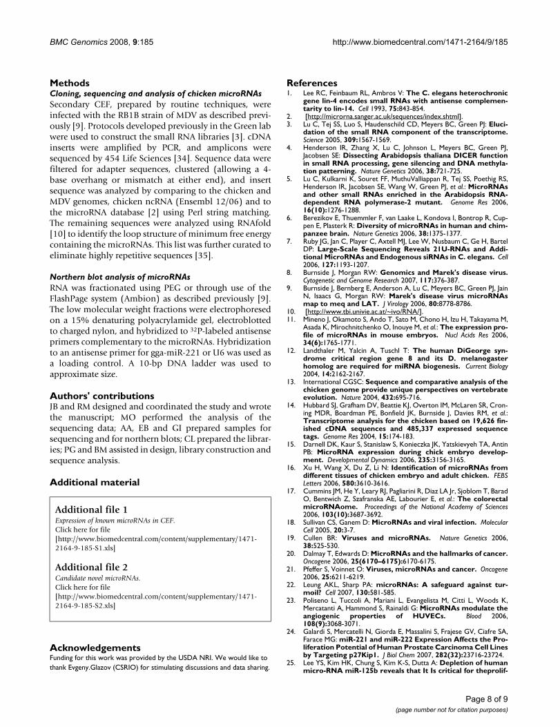
MethodsCloning, sequencing and analysis of chicken microRNAsSecondary CEF, prepared by routine techniques, wereinfected with the RB1B strain of MDV as described previ-ously [9]. Protocols developed previously in the Green labwere used to construct the small RNA libraries [3]. cDNAinserts were amplified by PCR, and amplicons weresequenced by 454 Life Sciences [34]. Sequence data werefiltered for adapter sequences, clustered (allowing a 4-base overhang or mismatch at either end), and insertsequence was analyzed by comparing to the chicken andMDV genomes, chicken ncRNA (Ensembl 12/06) and tothe microRNA database [2] using Perl string matching.The remaining sequences were analyzed using RNAfold[10] to identify the loop structure of minimum free energycontaining the microRNAs. This list was further curated toeliminate highly repetitive sequences [35].
Northern blot analysis of microRNAsRNA was fractionated using PEG or through use of theFlashPage system (Ambion) as described previously [9].The low molecular weight fractions were electrophoresedon a 15% denaturing polyacrylamide gel, electroblottedto charged nylon, and hybridized to 32P-labeled antisenseprimers complementary to the microRNAs. Hybridizationto an antisense primer for gga-miR-221 or U6 was used asa loading control. A 10-bp DNA ladder was used toapproximate size.
Authors' contributionsJB and RM designed and coordinated the study and wrotethe manuscript; MO performed the analysis of thesequencing data; AA, EB and GI prepared samples forsequencing and for northern blots; CL prepared the librar-ies; PG and BM assisted in design, library construction andsequence analysis.
Additional material
AcknowledgementsFunding for this work was provided by the USDA NRI. We would like to thank Evgeny.Glazov (CSRIO) for stimulating discussions and data sharing.
References1. Lee RC, Feinbaum RL, Ambros V: The C. elegans heterochronic
gene lin-4 encodes small RNAs with antisense complemen-tarity to lin-14. Cell 1993, 75:843-854.
2. [http://microrna.sanger.ac.uk/sequences/index.shtml].3. Lu C, Tej SS, Luo S, Haudenschild CD, Meyers BC, Green PJ: Eluci-
dation of the small RNA component of the transcriptome.Science 2005, 309:1567-1569.
4. Henderson IR, Zhang X, Lu C, Johnson L, Meyers BC, Green PJ,Jacobsen SE: Dissecting Arabidopsis thaliana DICER functionin small RNA processing, gene silencing and DNA methyla-tion patterning. Nature Genetics 2006, 38:721-725.
5. Lu C, Kulkarni K, Souret FF, MuthuValliappan R, Tej SS, Poethig RS,Henderson IR, Jacobsen SE, Wang W, Green PJ, et al.: MicroRNAsand other small RNAs enriched in the Arabidopsis RNA-dependent RNA polymerase-2 mutant. Genome Res 2006,16(10):1276-1288.
6. Berezikov E, Thuemmler F, van Laake L, Kondova I, Bontrop R, Cup-pen E, Plasterk R: Diversity of microRNAs in human and chim-panzee brain. Nature Genetics 2006, 38:1375-1377.
7. Ruby JG, Jan C, Player C, Axtell MJ, Lee W, Nusbaum C, Ge H, BartelDP: Large-Scale Sequencing Reveals 21U-RNAs and Addi-tional MicroRNAs and Endogenous siRNAs in C. elegans. Cell2006, 127:1193-1207.
8. Burnside J, Morgan RW: Genomics and Marek's disease virus.Cytogenetic and Genome Research 2007, 117:376-387.
9. Burnside J, Bernberg E, Anderson A, Lu C, Meyers BC, Green PJ, JainN, Isaacs G, Morgan RW: Marek's disease virus microRNAsmap to meq and LAT. J Virology 2006, 80:8778-8786.
10. [http://www.tbi.univie.ac.at/~ivo/RNA/].11. Mineno J, Okamoto S, Ando T, Sato M, Chono H, Izu H, Takayama M,
Asada K, Mirochnitchenko O, Inouye M, et al.: The expression pro-file of microRNAs in mouse embryos. Nucl Acids Res 2006,34(6):1765-1771.
12. Landthaler M, Yalcin A, Tuschl T: The human DiGeorge syn-drome critical region gene 8 and its D. melanogasterhomolog are required for miRNA biogenesis. Current Biology2004, 14:2162-2167.
13. International CGSC: Sequence and comparative analysis of thechicken genome provide unique perspectives on vertebrateevolution. Nature 2004, 432:695-716.
14. Hubbard SJ, Grafham DV, Beattie KJ, Overton IM, McLaren SR, Cron-ing MDR, Boardman PE, Bonfield JK, Burnside J, Davies RM, et al.:Transcriptome analysis for the chicken based on 19,626 fin-ished cDNA sequences and 485,337 expressed sequencetags. Genome Res 2004, 15:174-183.
15. Darnell DK, Kaur S, Stanislaw S, Konieczka JK, Yatskievyeh TA, AntinPB: MicroRNA expression during chick embryo develop-ment. Developmental Dynamics 2006, 235:3156-3165.
16. Xu H, Wang X, Du Z, Li N: Identification of microRNAs fromdifferent tissues of chicken embryo and adult chicken. FEBSLetters 2006, 580:3610-3616.
17. Cummins JM, He Y, Leary RJ, Pagliarini R, Diaz LA Jr, Sjoblom T, BaradO, Bentwich Z, Szafranska AE, Labourier E, et al.: The colorectalmicroRNAome. Proceedings of the National Academy of Sciences2006, 103(10):3687-3692.
18. Sullivan CS, Ganem D: MicroRNAs and viral infection. MolecularCell 2005, 20:3-7.
19. Cullen BR: Viruses and microRNAs. Nature Genetics 2006,38:525-530.
20. Dalmay T, Edwards D: MicroRNAs and the hallmarks of cancer.Oncogene 2006, 25(6170–6175):6170-6175.
21. Pfeffer S, Voinnet O: Viruses, microRNAs and cancer. Oncogene2006, 25:6211-6219.
22. Leung AKL, Sharp PA: microRNAs: A safeguard against tur-moil? Cell 2007, 130:581-585.
23. Poliseno L, Tuccoli A, Mariani L, Evangelista M, Citti L, Woods K,Mercatanti A, Hammond S, Rainaldi G: MicroRNAs modulate theangiogenic properties of HUVECs. Blood 2006,108(9):3068-3071.
24. Galardi S, Mercatelli N, Giorda E, Massalini S, Frajese GV, Ciafre SA,Farace MG: miR-221 and miR-222 Expression Affects the Pro-liferation Potential of Human Prostate Carcinoma Cell Linesby Targeting p27Kip1. J Biol Chem 2007, 282(32):23716-23724.
25. Lee YS, Kim HK, Chung S, Kim K-S, Dutta A: Depletion of humanmicro-RNA miR-125b reveals that It Is critical for theprolif-
Additional file 1Expression of known microRNAs in CEF.Click here for file[http://www.biomedcentral.com/content/supplementary/1471-2164-9-185-S1.xls]
Additional file 2Candidate novel microRNAs.Click here for file[http://www.biomedcentral.com/content/supplementary/1471-2164-9-185-S2.xls]
Page 8 of 9(page number not for citation purposes)
http://www.ncbi.nlm.nih.gov/entrez/query.fcgi?cmd=Retrieve&db=PubMed&dopt=Abstract&list_uids=8252621
http://www.ncbi.nlm.nih.gov/entrez/query.fcgi?cmd=Retrieve&db=PubMed&dopt=Abstract&list_uids=8252621
BMC Genomics 2008, 9:185 http://www.biomedcentral.com/1471-2164/9/185
Publish with BioMed Central and every scientist can read your work free of charge
"BioMed Central will be the most significant development for disseminating the results of biomedical research in our lifetime."
Sir Paul Nurse, Cancer Research UK
Your research papers will be:
available free of charge to the entire biomedical community
peer reviewed and published immediately upon acceptance
cited in PubMed and archived on PubMed Central
yours — you keep the copyright
Submit your manuscript here:http://www.biomedcentral.com/info/publishing_adv.asp
BioMedcentral
eration of differentiated cells but not for the down-regula-tion of putative targets during differentiation. J Biol Chem2005, 280:16635-16641.
26. Si M-L, Zhu S, Wu H, Lu Z, Wu F, Mo Y-Y: miR-21-mediatedtumor growth. Oncogene 2007, 26:2799-2803.
27. Lu J, Getz G, Miska EA, Alvarez-Saavedra E, Lamb J, Peck D, Sweet-Cordero A, Ebert BL, Mak RH, Ferrando AA, et al.: MicroRNAexpression profiles classify human cancers. Nature 2005,435:834-843.
28. Mayr C, Hemann MT, Bartel DP: Disrupting the pairing betweenlet-7 and Hmga2 enhances oncogenic transformation. Science2007, 315(5818):1576-1579.
29. Sgarra R, Rustighi A, Tessari MA, Di Bernardo J, Altamura S, Fusco A,Manfioletti G, Giancotti V: Nuclear phosphoproteins HMGA andtheir relationship with chromatin structure and cancer. FEBSLetters 2004, 574:1-8.
30. Fedele M, Battista S, Manfioletti G, Croce CM, Giancotti V, Fusco A:Role of the high mobility group A proteins in human lipomas.Carcinogenesis 2001, 22(10):1583-1591.
31. Cimmino A, Calin GA, Fabbri M, Iorio MV, Ferracin M, Shimizu M,Wojcik SE, Aqeilan RI, Zupo S, Dono M, et al.: miR-15 and miR-16induce apoptosis by targeting BCL2. Proceedings of the NationalAcademy of Sciences 2005, 102(39):13944-13949.
32. Ciafre SA, Galardi S, Mangiola A, Ferracin M, Liu C-G, Sabatino G,Negrini M, Maira G, Croce CM, Farace MG: Extensive modulationof a set of microRNAs in primary glioblastoma. Biochemicaland Biophysical Research Communications 2005, 334:1351-1358.
33. Murakami Y, Yasuda T, Saigo K, Urashima T, Toyoda H, Okanoue T,Shimotohno K: Comprehensive analysis of microRNA expres-sion patterns in hepatocellular carcinoma and non-tumor-ous tissues. Oncogene 2006, 25:2537-2545.
34. Margulies M, Egholm M, Altman WE, Attiya S, Bader JS, Bemben LA,Berka J, Braverman MS, Chen YJ, Chen Z, et al.: Genome sequenc-ing in microfabricated high-density picolitre reactors. Nature2005, 437(7057):376-380.
35. [http://www.repeatmasker.org/].
Page 9 of 9(page number not for citation purposes)