Circulating microRNAs in Medicine - MDPI
17
Citation: Pozniak, T.; Shcharbin, D.; Bryszewska, M. Circulating microRNAs in Medicine. Int. J. Mol. Sci. 2022, 23, 3996. https://doi.org/ 10.3390/ijms23073996 Academic Editors: Y-h. Taguchi and Hsiuying Wang Received: 14 March 2022 Accepted: 1 April 2022 Published: 3 April 2022 Publisher’s Note: MDPI stays neutral with regard to jurisdictional claims in published maps and institutional affil- iations. Copyright: © 2022 by the authors. Licensee MDPI, Basel, Switzerland. This article is an open access article distributed under the terms and conditions of the Creative Commons Attribution (CC BY) license (https:// creativecommons.org/licenses/by/ 4.0/). International Journal of Molecular Sciences Review Circulating microRNAs in Medicine Tetiana Pozniak 1,2, *, Dzmitry Shcharbin 1, * and Maria Bryszewska 3 1 Institute of Biophysics and Cell Engineering of the National Academy of Sciences of Belarus, 220072 Minsk, Belarus 2 Palladin Institute of Biochemistry, National Academy of Sciences of Ukraine, 02000 Kyiv, Ukraine 3 Department of General Biophysics, Faculty of Biology and Environmental Protection, University of Lodz, 90-236 Lodz, Poland; [email protected] * Correspondence: [email protected] (T.P.); [email protected] (D.S.) Abstract: Circulating microRNAs (c-microRNAs, c-miRNAs), which are present in almost all biologi- cal fluids, are promising sensitive biomarkers for various diseases (oncological and cardiovascular diseases, neurodegenerative pathologies, etc.), and their signatures accurately reflect the state of the body. Studies of the expression of microRNA markers show that they can enable a wide range of diseases to be diagnosed before clinical symptoms are manifested, and they can help to assess a patient’s response to therapy in order to correct and personalize treatments. This review discusses the latest trends in the uses of miRNAs for diagnosing and treating various diseases, viral and non-viral. It is concluded that exogenous microRNAs can be used as high-precision therapeutic agents for these purposes. Keywords: circulating microRNAs (c-microRNAs, c-miRNAs); small interfering RNAs (miRNAs); oncology; cardiovascular diseases; neurodegenerative diseases 1. Introduction More than 20 years ago, a mechanism of negative regulation of gene expression at the level of translation was discovered: mRNA is blocked by small non-coding RNAs, repressing translation or promoting mRNA degradation. This process has been called RNA interference and it leads to “gene silencing”. Several subclasses of small non-coding RNAs (snRNAs) are involved in this powerful post-transcriptional gene silencing process. RNA interference was first described by Andrew Fire and Craig Mello in Nature in 1998. They received the Nobel Prize in Physiology or Medicine (2006) for this discovery [1]. They showed that short double-stranded RNAs can silence homologous genes. There are many groups of snRNAs and the list is growing rapidly. This review details the components of RNA interference, such as microRNAs (miRNAs) and small interfering RNAs (siRNAs). These two species have similar structures and are short (21–23 and 20–24 nucleotides, respectively) single- and double-stranded RNAs that inhibit gene expression. The main differences between them lie in their mechanisms of formation and their degrees of homology with respect to targeting of mRNAs. In 1993, it was determined that the lin-4 gene in Caenorhabditis elegans produced a snRNA [2,3]. In 1998, lin-4 was shown to encode a 61-nucleotide precursor that matured into a 22-nucleotide RNA, later called miRNA. This short RNA contains sequences that are partially complementary to the 3 0 -untranslated region (3 0 -UTR) of mRNA transcribed from the lin-14 nematode gene and represses translation of this mRNA, inhibiting LIN-14 protein synthesis. In 2000, a second miRNA was discovered, a product of let-7 [2], which suppressed the expression of several genes simultaneously and was later identified in a number of organisms, including humans. Although lin-4 and let-7 were identified by standard positional cloning of genetic loci, most miRNA genes are detected by cloning cDNA sequences complementary to the desired RNA fragments. This method involves Int. J. Mol. Sci. 2022, 23, 3996. https://doi.org/10.3390/ijms23073996 https://www.mdpi.com/journal/ijms
-
Upload
khangminh22 -
Category
Documents
-
view
4 -
download
0
Transcript of Circulating microRNAs in Medicine - MDPI
�����������������
Citation: Pozniak, T.; Shcharbin, D.;
Bryszewska, M. Circulating
microRNAs in Medicine. Int. J. Mol.
Sci. 2022, 23, 3996. https://doi.org/
10.3390/ijms23073996
Academic Editors: Y-h. Taguchi and
Hsiuying Wang
Received: 14 March 2022
Accepted: 1 April 2022
Published: 3 April 2022
Publisher’s Note: MDPI stays neutral
with regard to jurisdictional claims in
published maps and institutional affil-
iations.
Copyright: © 2022 by the authors.
Licensee MDPI, Basel, Switzerland.
This article is an open access article
distributed under the terms and
conditions of the Creative Commons
Attribution (CC BY) license (https://
creativecommons.org/licenses/by/
4.0/).
International Journal of
Molecular Sciences
Review
Circulating microRNAs in MedicineTetiana Pozniak 1,2,*, Dzmitry Shcharbin 1,* and Maria Bryszewska 3
1 Institute of Biophysics and Cell Engineering of the National Academy of Sciences of Belarus,220072 Minsk, Belarus
2 Palladin Institute of Biochemistry, National Academy of Sciences of Ukraine, 02000 Kyiv, Ukraine3 Department of General Biophysics, Faculty of Biology and Environmental Protection, University of Lodz,
90-236 Lodz, Poland; [email protected]* Correspondence: [email protected] (T.P.); [email protected] (D.S.)
Abstract: Circulating microRNAs (c-microRNAs, c-miRNAs), which are present in almost all biologi-cal fluids, are promising sensitive biomarkers for various diseases (oncological and cardiovasculardiseases, neurodegenerative pathologies, etc.), and their signatures accurately reflect the state ofthe body. Studies of the expression of microRNA markers show that they can enable a wide rangeof diseases to be diagnosed before clinical symptoms are manifested, and they can help to assess apatient’s response to therapy in order to correct and personalize treatments. This review discusses thelatest trends in the uses of miRNAs for diagnosing and treating various diseases, viral and non-viral.It is concluded that exogenous microRNAs can be used as high-precision therapeutic agents forthese purposes.
Keywords: circulating microRNAs (c-microRNAs, c-miRNAs); small interfering RNAs (miRNAs);oncology; cardiovascular diseases; neurodegenerative diseases
1. Introduction
More than 20 years ago, a mechanism of negative regulation of gene expression atthe level of translation was discovered: mRNA is blocked by small non-coding RNAs,repressing translation or promoting mRNA degradation. This process has been called RNAinterference and it leads to “gene silencing”. Several subclasses of small non-coding RNAs(snRNAs) are involved in this powerful post-transcriptional gene silencing process. RNAinterference was first described by Andrew Fire and Craig Mello in Nature in 1998. Theyreceived the Nobel Prize in Physiology or Medicine (2006) for this discovery [1]. Theyshowed that short double-stranded RNAs can silence homologous genes.
There are many groups of snRNAs and the list is growing rapidly. This review detailsthe components of RNA interference, such as microRNAs (miRNAs) and small interferingRNAs (siRNAs). These two species have similar structures and are short (21–23 and 20–24nucleotides, respectively) single- and double-stranded RNAs that inhibit gene expression.The main differences between them lie in their mechanisms of formation and their degreesof homology with respect to targeting of mRNAs.
In 1993, it was determined that the lin-4 gene in Caenorhabditis elegans produced asnRNA [2,3]. In 1998, lin-4 was shown to encode a 61-nucleotide precursor that maturedinto a 22-nucleotide RNA, later called miRNA. This short RNA contains sequences thatare partially complementary to the 3′-untranslated region (3′-UTR) of mRNA transcribedfrom the lin-14 nematode gene and represses translation of this mRNA, inhibiting LIN-14protein synthesis. In 2000, a second miRNA was discovered, a product of let-7 [2], whichsuppressed the expression of several genes simultaneously and was later identified ina number of organisms, including humans. Although lin-4 and let-7 were identified bystandard positional cloning of genetic loci, most miRNA genes are detected by cloningcDNA sequences complementary to the desired RNA fragments. This method involves
Int. J. Mol. Sci. 2022, 23, 3996. https://doi.org/10.3390/ijms23073996 https://www.mdpi.com/journal/ijms
Int. J. Mol. Sci. 2022, 23, 3996 2 of 17
the isolation of a miRNA that blocks the translation of a specific messenger, followed bycDNA synthesis using reverse transcriptase. One difficulty in finding miRNA genes forfurther cDNA cloning is that not only fragments of the target but also fragments of othernoncoding RNAs (such as rRNA, tRNA, and snRNA), together with mRNAs, are clonedfrom RNA samples of a selected size. However, this difficulty is easily solved by comparingthe candidate miRNA sequence with known miRNA sequences in annotated databases [4].To date, more than 2000 miRNAs have been registered in this database.
There are certain rules for miRNA nomenclature [5,6]: (1) all miRNAs are abbreviatedas miR; (2) newly discovered miRNAs are assigned sequential numerical identifiers (forexample, miR5, miR6, miR7); (3) to designate the species of origin of a miRNA, a prefix of3–4 letters can be added to the name (hsa for Homo sapiens, dme for Drosophila melanogaster,etc.); (4) orthologous miRNAs from different species are given parallel names (hsa-miR-28, ptr-mir-28, crf-miR-28); (5) paralogous miRNAs with one or two different bases aredistinguished by suffixes (e.g., miR-10a and miR-10b); (6) if identical miRNAs originatefrom separate loci in the same organism, they are assigned numerical suffixes (for example,miR-281-1 and miR-281-2 in Drosophila melanogaster).
2. miRNA Biogenesis
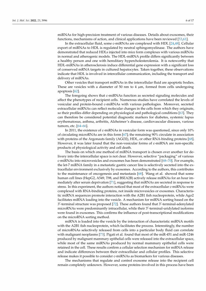
The miRNA precursors, pre-miRNAs, are first transcribed as capped polyadenylatedstrands that form double-stranded stem–loop structures (Figure 1). In the nucleus, thesetranscripts are processed by the Drosha enzyme of the RNase III family to form pre-miRNAsof 70–100 nucleotides with a hairpin structure (two base pairs connected by a loop), a 5′-phosphate group, and a 3′-double nucleotide [7]. The pre-miRNAs are exported to thecytoplasm via exportin-5 [8]; microRNAs of 21–23 nucleotide sequences are formed fromthem by enzymes, including the Dicer ribonuclease complex [9]. This is part of the RISC(RNA-Induced Silencing Complex) that unwinds miRNA chains, cleaving and releasingthe “passenger” chain and the guide chain. Argonaute 2 endonuclease [10,11], whichis also part of the complex, interacts with complementary regions of mRNA, leading todegradation of the latter and/or blocking of translation [12,13].
Int. J. Mol. Sci. 2022, 23, x FOR PEER REVIEW 2 of 18
the isolation of a miRNA that blocks the translation of a specific messenger, followed by cDNA synthesis using reverse transcriptase. One difficulty in finding miRNA genes for further cDNA cloning is that not only fragments of the target but also fragments of other noncoding RNAs (such as rRNA, tRNA, and snRNA), together with mRNAs, are cloned from RNA samples of a selected size. However, this difficulty is easily solved by compar-ing the candidate miRNA sequence with known miRNA sequences in annotated data-bases [4]. To date, more than 2000 miRNAs have been registered in this database.
There are certain rules for miRNA nomenclature [5,6]: (1) all miRNAs are abbreviated as miR; (2) newly discovered miRNAs are assigned sequential numerical identifiers (for example, miR5, miR6, miR7); (3) to designate the species of origin of a miRNA, a prefix of 3-4 letters can be added to the name (hsa for Homo sapiens, dme for Drosophila melanogaster, etc.); (4) orthologous miRNAs from different species are given parallel names (hsa-miR-28, ptr-mir-28, crf-miR-28); (5) paralogous miRNAs with one or two different bases are dis-tinguished by suffixes (e.g., miR-10a and miR-10b); (6) if identical miRNAs originate from separate loci in the same organism, they are assigned numerical suffixes (for example, miR-281-1 and miR-281-2 in Drosophila melanogaster).
2. miRNA Biogenesis The miRNA precursors, pre-miRNAs, are first transcribed as capped polyadenylated
strands that form double-stranded stem–loop structures (Figure 1). In the nucleus, these transcripts are processed by the Drosha enzyme of the RNase III family to form pre-miR-NAs of 70–100 nucleotides with a hairpin structure (two base pairs connected by a loop), a 5′-phosphate group, and a 3′-double nucleotide [7]. The pre-miRNAs are exported to the cytoplasm via exportin-5 [8]; microRNAs of 21–23 nucleotide sequences are formed from them by enzymes, including the Dicer ribonuclease complex [9]. This is part of the RISC (RNA-Induced Silencing Complex) that unwinds miRNA chains, cleaving and releasing the “passenger” chain and the guide chain. Argonaute 2 endonuclease [10,11], which is also part of the complex, interacts with complementary regions of mRNA, leading to deg-radation of the latter and/or blocking of translation [12,13].
Figure 1. Scheme of biogenesis and functions of miRNAs in animals. Reprinted/adapted with per-mission from Ref. [14], 2015, Creative Commons Attribution-NonCommercial 4.0 International License .
Figure 1. Scheme of biogenesis and functions of miRNAs in animals. Reprinted/adapted with permis-sion from Ref. [14], 2015, Creative Commons Attribution-NonCommercial 4.0 International License.
siRNA is formed from its double-stranded RNA (dsRNA) precursor in the sameway as miRNA. However, an important distinguishing feature is that siRNAs are only

Int. J. Mol. Sci. 2022, 23, 3996 3 of 17
partially complementary to the 3′UTR region of their target mRNAs, so they can inactivateseveral different mRNAs simultaneously [15]. They regulate their targets through fourArgonaute proteins, and although they sometimes induce mRNA cleavage and degradationas miRNAs do, they primarily mediate gene silencing through translational repression andmRNA degradation through deadenylation [16]. As a rule, miRNAs pair accurately withtheir targets and promote endonucleotic cleavage of a single specific mRNA [17].
3. Functions of miRNA
The modern scientific approach to the search for therapeutic agents is to develop drugsthat selectively influence processes at the gene level. The main targets of this approachare non-coding double-stranded miRNAs because they cause gene silencing by directinteraction with mRNA, suppressing the production of proteins in various diseases (forexample, anti-apoptotic proteins in malignant neoplasms [18] and anti-miRNA that inhibitsthe activity of miRNA-122 in the liver in hepatitis C patients [19]). snRNAs regulate theexpression of more than 30% of the protein-encoding genes in humans [20]. They inhibitgene expression in several ways [21]: (1) by interacting with mRNA, directly binding to thetarget, leading to blocking of translation (protein synthesis) and to mRNA degradation (ifthere is perfect complementary pairing, which is more characteristic of miRNAs); (2) mRNAdeadenylation; (3) at the level of transcription, when snRNAs within the polyproteincomplex cause epigenetic modifications of the genome—DNA methylation, deacetylation,and histone methylation; (4) by interaction with repressor proteins, blocking translationat the level of elongation (Figure 2) [22]. Additionally, when the cell cycle stops, miRNAscan activate as well as repress translation. This phenomenon was described in 2007 inScience [23]. Through combinations of these mechanisms, snRNAs affect protein synthesisin all cells and are significant in cellular processes, such as differentiation, proliferation,apoptosis, and metabolism. The biological processes can be regulated at several levels andlead to a decrease or increase in the number of miRNAs in a cell. Deregulation of miRNAexpression can be genetic and result from chromosomal loss (deletion), amplification,translocation, or even point mutations of genes. miRNA expression can be affected bycytosine methylation in DNA, with hypermethylation or hypomethylation of CpG regions,post-translational modifications of histones in many types of tumors, and a decrease intranscription factors, such as p53 and c-Myc. Anomalies in proteins involved in differentstages of the maturation process can also disrupt snRNA expression; for example, mutationsaffecting binding to the Drosha protein, miRNA export from the nucleus to the cytoplasmvia exportin-5, or interaction with the Dicer enzyme [24].
In the mammalian genome, a single miRNA can regulate many genes, binding to morethan 60% of the mRNAs involved in a specific signaling cascade or cellular mechanism.This makes them effective biological regulators capable of targeted suppression of “disease-causing” genes.
In 2001, it was demonstrated that the introduction of chemically synthesized miRNAsinto mammalian cells effectively inhibits gene expression [25]. This discovery inspiredmany further studies of the “silencing” of gene expression through RNA interference. It hasbeen established that miRNAs are involved in the regulation of many cellular functions, andaberrant miRNA expression leads to various disorders, cancer in particular, correlating withits various types [26,27]. miRNAs are involved in almost every stage of carcinogenesis: cellgrowth, differentiation, proliferation, angiogenesis, apoptosis, invasion, and metastasis [28].Changes in expression can result from mutation, methylation, deletion, and amplificationof miRNA-coding regions. In this case, miRNAs are either oncogenic and are activated, orfunction by acting on the mRNAs of tumor suppressors or oncogenes. For example, miRNA-suppressors are suppressed in cancer. The study of miRNA expression profiles enablesnormal tissues to be distinguished from cancerous ones, tissues of origin to be identified,and highly accurate information on the subtype of a particular cancer to be provided [29,30].No less important is the fact that miRNA profiles can be used to predetermine the response

Int. J. Mol. Sci. 2022, 23, 3996 4 of 17
to therapy and the further development of the disease. For example, miRNA expressioncan be used to predict a patient’s individual response to a drug [27,31].
Int. J. Mol. Sci. 2022, 23, x FOR PEER REVIEW 4 of 18
Figure 2. Scheme of inhibition of gene expression by miRNA in several ways (description in the article). Ac—acetyl, CH3CO.
In the mammalian genome, a single miRNA can regulate many genes, binding to more than 60% of the mRNAs involved in a specific signaling cascade or cellular mecha-nism. This makes them effective biological regulators capable of targeted suppression of “disease-causing” genes.
In 2001, it was demonstrated that the introduction of chemically synthesized miR-NAs into mammalian cells effectively inhibits gene expression [25]. This discovery in-spired many further studies of the “silencing” of gene expression through RNA interfer-ence. It has been established that miRNAs are involved in the regulation of many cellular functions, and aberrant miRNA expression leads to various disorders, cancer in particu-lar, correlating with its various types [26,27]. miRNAs are involved in almost every stage of carcinogenesis: cell growth, differentiation, proliferation, angiogenesis, apoptosis, in-vasion, and metastasis [28]. Changes in expression can result from mutation, methylation, deletion, and amplification of miRNA-coding regions. In this case, miRNAs are either on-cogenic and are activated, or function by acting on the mRNAs of tumor suppressors or oncogenes. For example, miRNA-suppressors are suppressed in cancer. The study of miRNA expression profiles enables normal tissues to be distinguished from cancerous ones, tissues of origin to be identified, and highly accurate information on the subtype of a particular cancer to be provided [29,30]. No less important is the fact that miRNA pro-files can be used to predetermine the response to therapy and the further development of the disease. For example, miRNA expression can be used to predict a patient’s individual response to a drug [27,31].
Studies of miRNAs that affect the enzymes involved in processing have shown that they also control the differentiation, post-meiotic function, and growth of male germ cells, and the development and maturation of oocytes, through highly regulated gene expres-sion. The expression of miRNA genes at different stages of testicular and ovarian cell de-velopment indicates a potential role in the physiology of the genital organs [32].
Changes in miRNA expression lead not only to cancer [29] but also to many other pathological processes, such as cardiovascular diseases [33,34], autoimmune diseases [35,36], disorders of the central nervous system [37], viral respiratory diseases [38], etc. It
Figure 2. Scheme of inhibition of gene expression by miRNA in several ways (description in thearticle). Ac—acetyl, CH3CO.
Studies of miRNAs that affect the enzymes involved in processing have shown thatthey also control the differentiation, post-meiotic function, and growth of male germcells, and the development and maturation of oocytes, through highly regulated geneexpression. The expression of miRNA genes at different stages of testicular and ovariancell development indicates a potential role in the physiology of the genital organs [32].
Changes in miRNA expression lead not only to cancer [29] but also to many otherpathological processes, such as cardiovascular diseases [33,34], autoimmune diseases [35,36],disorders of the central nervous system [37], viral respiratory diseases [38], etc. It is notsurprising that scientists around the world have focused attention on miRNAs as promisingtools for highly selective post-transcriptional inhibition of gene expression.
4. Circulating miRNAs (c-miRNAs) and Their Differences
All organisms transmit genetic information from parent to offspring through verticalgene transfer. Bacteria also have horizontal (lateral) gene transfer for exchanging geneticinformation, which allows them to diversify their populations and facilitate adaptation tonew conditions. Horizontal information transfer in eukaryotes was discovered relativelyrecently through the intercellular transport of miRNAs [39]. In 2008, circulating miRNA(c-microRNA) was first detected in maternal plasma [40]. Later, c-microRNAs were alsofound in other biological fluids, such as blood (serum, plasma), urine, cerebrospinal fluid,saliva, milk, lacrimal and seminal fluids, and bronchial lavage [41–43]. The appearanceof c-microRNAs in the blood can result from secretion by cells or from cell death duringapoptosis, necrosis, tumors, or trauma.
Interestingly, despite the presence of various nucleases, circulating endogenous miR-NAs remain stable while pure exogenous miRNAs added to plasma degrade rapidly [41].Subsequent studies established that endogenous miRNAs are highly stable and resistantnot only to endogenous ribonucleases but also to extreme temperatures, pH levels, andfreeze–thaw cycles [44]. To ascertain what protects them from enzymatic degradation,

Int. J. Mol. Sci. 2022, 23, 3996 5 of 17
let us consider the mechanisms of miRNA entry into extracellular fluids and the formsin which they are found there. Initially, c-miRNAs can be secreted into the extracellularspace in microvesicles (late endosomal compartments [45]), ectosomes [46], exosomes [47],or microvesicles (liposomes), or they can be associated with high-density lipoproteins(HDL) [48], or released as parts of apoptotic bodies. However, most of them, according toReiner et al., form complexes with the proteins AGO2 or NPM1 (nucleophosmin 1) [49].Regardless of the form in which they enter the extracellular space, miRNAs then pass intoother biological fluids, for example, the general bloodstream.
Microvesicles (microparticles or ectosomes) are plasma membrane-derived particlesreleased into the extracellular space by budding out and detachment from the plasma mem-brane. They range in size from 100 nm to 1 µm and are formed by outward protrusion ofthe plasma membrane, followed by separation [50]. Ectosomes are secreted by various cells,including tumor cells, polymorphonuclear leukocytes, aging erythrocytes, and activatedplatelets. One of their characteristic features is the appearance of phosphatidylserine (PS)on their membrane surfaces. Unlike exosomes, ectosomes bind well to annexin V andcan also bind to prothrombin and blood coagulation factor X to form the prothrombinasecomplex [41].
Exosomes are small, membrane-bound vesicles of endosomal origin, 30–100 nm indiameter, that form intracellularly via endocytic invagination and are released into astructure known as the multivesicular body (MVB). The MVB then fuses with the plasmamembrane, releasing the exosome contents into the extracellular space [51]. Exosomes aresecreted by almost all normal cells (T-cells, B-lymphocytes, dendritic cells, reticulocytes,neurons, intestinal epithelial cells, platelets, etc.) and by pathological cells. It was previouslybelieved that exosomes function only as protein scavengers, but in 2007, Valadi et al. foundthat they can carry nucleic acids (NAs), in particular, RNA [52]. It is now known thatthey can contain various components of their donor cells, including proteins (proteolyticproteins, chaperones (Hsp70 and Hsp90), apoptotic proteins (Alix), translation factors,metabolic enzymes), lipids, mRNA, microRNA, small interfering RNA (siRNA), and DNA.They can also carry viruses and prions from an infected cell [53]. Thus, exosomes functionas horizontal carriers (between cells of the same organism, donor–recipient) of information,mRNA, viruses, and other materials, such as proteins and microRNAs. Some proteins areexosome-specific and do not depend on the donor cell; however, there are also specificones, so it is possible to identify a subpopulation of specific exosomes and infer the celltype of origin [54]. However, how the various components making up exosomes are chosenremains an open question. In 2012, 4563 proteins had been found in exosomes [55], andthe list of exosomal miRNAs numbered about 800. Cells can interact with each otherby transferring exosomes loaded with miRNAs [56,57]. The authors of [57] showed thatmonocyte exosomes deliver miR-150 to endothelial cells and enhance their migration byreducing c-myb 9 expression. The miRNA content of exosomes is critical in this type ofintercellular communication and determines the fate of the recipient cell. Thus, exosomesderived from mesenchymal stromal cells of the bone marrows of myeloma patients promotetumor growth, and this effect depends on their miR-15a content.
Exosomes produced by stressed cells also provide information that induces resis-tance in surrounding cells through a paracrine effect. For example, miRNAs regulate theactivity of pancreatic β-cells by transfer via exosomes [58]. Exosomes are also involvedin transmitting the immune response; miRNAs transferred by exosomes from T-cells toantigen-presenting cells (APCs) can regulate gene expression in the recipient cells [59]. Ex-actly this process accounts for the suppression of antitumor immunity, including inhibitionof T-lymphocyte and natural killer activities, and the suppression of APC differentiation.Tumor exosomes also enhance the activity of immunosuppressive cells and increase theirnumber. There are indications that tumor cells dispose of chemotherapy drugs (in particu-lar, doxorubicin) by secreting them in exosomes. This process underlies the acquisition ofresistance to anticancer therapy by malignant cells [60]. Considering these properties, exo-somes can be considered promising tools for delivering drugs and engineering exogenous
Int. J. Mol. Sci. 2022, 23, 3996 6 of 17
miRNAs for high-precision treatment of various diseases. Details about exosomes, theirfunctions, mechanisms of action, and clinical applications have been reviewed [53,61].
In the extracellular fluid, some c-miRNAs are complexed with HDL [24,48]. Cellularexport of miRNAs to HDL is regulated by neutral sphingomyelinase. The authors havedemonstrated that reduced HDLs injected into mice form complexes with various miRNAsin normal and atherogenic models. The HDL-miRNA profile differs significantly betweena healthy person and one with hereditary hypercholesterolemia. It is noteworthy thatHDL-miRNAs in atherosclerosis induce differential gene expression with a significant lossof conserved mRNA targets in cultured hepatocytes. Taken together, these observationsindicate that HDL is involved in intercellular communication, including the transport anddelivery of miRNAs.
Other vesicles that transport miRNAs in the intercellular fluid are apoptotic bodies.These are vesicles with a diameter of 50 nm to 4 µm, formed from cells undergoingapoptosis [62].
The foregoing shows that c-miRNAs function as secreted signaling molecules andaffect the phenotypes of recipient cells. Numerous studies have correlated the levels ofvesicular and protein-bound c-miRNAs with various pathologies. Moreover, secretedextracellular miRNAs can reflect molecular changes in the cells from which they originate,so their profiles differ depending on physiological and pathological conditions [63]. Theycan therefore be considered potential diagnostic markers for diabetes, systemic lupuserythematosus, asthma, arthritis, Alzheimer’s disease, cardiovascular diseases, varioustumors, etc. [64–66].
In 2011, the existence of c-miRNAs in vesicular form was questioned, since only 10%of circulating microRNAs are in this form [67]; the remaining 90% circulate in associationwith proteins of the Argonauts family (AGO2), HDL, or other RNA-binding proteins [68].However, it was later found that the non-vesicular forms of c-miRNA are non-specificproducts of physiological activity and cell death.
The basis on which one method of miRNA transport is chosen over another for de-livery into the intercellular space is not clear. However, selective “packaging” of variousc-miRNAs into microvesicles and exosomes has been demonstrated [68–70]. For example,the let-7 miRNA family in a metastatic gastric cancer line is selectively secreted into the ex-tracellular environment exclusively by exosomes. According to the authors, this contributesto the maintenance of oncogenesis and metastasis [68]. Wang et al. showed that somehuman cell lines (HepG2, A549, T98, and BSEA2B) actively release miRNAs for an hour im-mediately after serum deprivation [71], suggesting that miRNAs are secreted in response tostress. In this experiment, the authors noticed that most of the extracellular c-miRNAs werecomplexed with RNA-binding proteins, not inside microvesicles or exosomes. Characteris-tic miRNA sequences promote interaction with the A2B1 fish nucleoprotein, while Ago2facilitates miRNA loading into the vesicle. A mechanism for miRNA sorting based on the3′-terminal structure was proposed [72]. These authors found that 3′-terminal-adenylatedmicroRNAs were predominantly intracellular, while their 3′-terminal-uridylated isoformswere found in exosomes. This confirms the influence of post-transcriptional modificationson the microRNA sorting method.
miRNA is loaded into the vesicle by the interaction of characteristic miRNA motifswith the A2B1 fish nucleoprotein, which facilitates the process. Interestingly, the numberof microRNAs selectively released from cells into a particular body fluid can correlatewith malignant neoplasms [73]. Pigati et al. found that most of the miR-451 and miR-1246produced by malignant mammary epithelial cells were released into the extracellular space,while most of the same miRNAs produced by normal mammary epithelial cells wereretained in the cell. These results confirm a cellular selection mechanism for miRNA releaseand indicate differences between their extracellular and cellular profiles. This selectiverelease makes it possible to consider c-miRNAs as biomarkers for various diseases.
The mechanisms that regulate and control exosome release into the recipient cellremain completely unknown. However, some proteins involved in this process have been

Int. J. Mol. Sci. 2022, 23, 3996 7 of 17
identified: TAT-5 and Rab27. TAT-5 is a cell membrane-associated protein that regulatesvesicle detachment [74]. Rab27 is involved in exosomal release and uptake by recipientcells [75]. Exosome release from mammalian cells is also facilitated by the ceramidepathway, which also inhibits the export of miRNAs in combination with HDL.
5. Analysis Methods
Most potential c-miRNA biomarkers are present in both healthy people and cancerpatients, with very slight differences in expression levels. Therefore, for studying miRNAexpression, initial standardization of the stage at which the material is isolated is decisive.In addition, there is still no generally accepted line of markers for all biofluids. However,an advantage of using miRNAs as biomarkers is that their expression levels are seldomaffected by age, gender, body mass index (BMI), smoking status, or other characteristicsunderlying pathogenic potential. The most common body fluids for miRNA testing areplasma and serum, but others such as urine and saliva are also used.
When biofluids are collected to isolate miRNAs, it is important to ensure that thecellular and non-cellular fractions of the sample are properly separated. Failure to dothis can cause large differences in miRNA concentration between samples because cellscan contain many microRNAs. MicroRNAs specific for each biofluid have to be iden-tified so that their levels can be used to assess contamination of samples with cellularmiRNAs. Different amounts of cellular contamination can also introduce variability intothe final concentrations of miRNAs detected. Therefore, highly consistent sample pro-cessing is extremely important. For example, when a sample is isolated from blood, thelevel of hemoglobin must be controlled to avoid sample contamination due to ruptureof red blood cells. Ideally, ethylenediaminetetraacetic acid (EDTA) or citrate is used asan anticoagulant, since heparin can interfere with subsequent experimental steps such asreverse transcription and qPCR. To minimize differences between sample profiles, bloodcollection tubes with the same anticoagulant (no heparin) should be used. When exosomalmiRNAs are examined, ultracentrifugation or a commercial kit such as ExoQuick (SBI,Palo Alto, CA, USA) is used [76]. The RNA is then extracted using commercially availablemethods, including phenol/guanidinium, such as TRIzol (Life Technologies, Carlsbad,CA, USA), and column-based kits, such as mirVana (Life Technologies) and miRNeasy(Qiagen, Hilden, Germany) [77]. The same storage conditions have to be maintained forall samples as temperature and storage time can affect miRNA stability, especially duringlong-term storage.
Further stages in the study depend on whether the task is to examine the expressionof c-miRNAs in a previously installed panel for a specific biomaterial and a number ofdiseases, or to discover new miRNAs.
The most common methods for studying miRNA expression are PCR with real-timereverse transcription (RT-PCR, qRT-PCR). The high specificity, sensitivity and speed of thismethod are advantageous, making it possible to identify a particular microRNA amonghundreds of others using special microplates with specific primers. However, the shortlengths of miRNAs and the lack of a common sequence for all molecules (for example, the3′ poly(A) sequence in mRNAs) make for difficulties, though the the possibility of usingconventional primers is not excluded. To overcome these difficulties, different methodswith different primers and detection methods are available. One of these uses stem–loopprimers to carry out reverse transcription of miRNA; the resulting cDNA is amplifiedwith conventional primers and quantified in real time with labeled fluorophores addedto the reaction medium. This method is used in platforms such as TaqMan Cards (LifeTechnologies). Another method involves polyadenylation of all miRNAs and adding anantisense primer with a poly(T) sequence at both the 5′ and 3′ ends, followed by reversetranscription and amplification together with the sense primer. Amplification productsare detected using the SYBR Green fluorescent dye, the fluorescence intensity of which isincreased about 100-fold when it is incorporated into a double-stranded structure. Thus, aparticular miRNA can be quantified absolutely [24]. The main problem with this method

Int. J. Mol. Sci. 2022, 23, 3996 8 of 17
remains the normalization of microRNA values to the appropriate endogenous control.There are many panels of endogenous miRNA genes, depending on the biomaterial understudy, that maintain constant levels under various conditions and serve as controls formiRNA quantification. For example, reference genes U6, RNU43, RNU44, RNU48, andmiR-16 are used to normalize data on the number of miRNAs in blood serum. RNU43is used in combination with RNU1-4 or miR-16 with miR-30e as a reference in urologicalcancers. Xi Chen and co-author identified a new panel of let-7d, let-7g, and let-7i genes as abenchmark for normalizing serum miRNA, which, according to their study, is statisticallysuperior to those previously used [78]. Lushui Wana et al. [79] identified another panel ofthe most stable reference genes for normalizing the results for serum miRNAs in bladdercancer (BC), which comprised the genes hsa-miR-193a-5p and hsa-miR-16-5p.
When new profiles of c-miRNA markers for various diseases are sought, samples ofthem are obtained from biological material and then sequenced. For this purpose, eithernext-generation sequencing (NGS) or microarray analysis is used. NGS combines methodsfor determining the nucleotide sequence of DNA or RNA, making it possible to identifyseveral regions of the genome simultaneously. NGS is accomplished by repeated cyclesof polymerase-induced chain elongation or multiple ligation of oligonucleotides. Up tohundreds of megabases and gigabases of nucleotide sequences can be generated in oneworking cycle. The advantage of NGS is the ability to identify new miRNAs, but the systemis less cost-effective and less efficient than microarrays.
Microarray analysis allows several thousand microRNAs to be analyzed simultane-ously. The microRNA samples are labeled with fluorescent probes, then further hybridizedwith samples of hundreds or thousands of microRNAs covalently immobilized on a solidsupport (microchip), followed by sample detection. To standardize the melting tempera-tures of the miRNAs, LNA (Locked Nucleic Acid) sequences are used, which are includedin the hybridization probes. This approach helps to reduce differences in melting tempera-ture between probes and also makes it possible to identify microRNAs with similar profiles.However, this method for quantitative analysis can cause technical difficulties, so it is oftencombined with RT-PCR.
One important task is to identify the expression signatures of miRNAs in the biomate-rial studied. These have diagnostic value for the disease under investigation. Xumei Jianget al. [80] examined a panel of six miRNAs (miR-152, miR-148b-3p, miR-3187-3p, miR-15b-5p,miR-27a-3p, and miR-30a-5p) to identify their expression signature in blood serum frombreast cancer patients. The authors showed that the serum miRNA signature can be ofsignificant diagnostic value, and the miR-152 expression level can elucidate the risk ofrecurrence of non-muscle-invasive breast cancer. Lutao Du and colleagues identified apanel of seven miRNAs in urine (miR-7-5p, miR-22-3p, miR-29a-3p, miR-126-5p, miR-200a-3p,miR-375, and miR-423-5p) for diagnosing and predicting bladder cancer recurrence [81].Ghorbanmehr and colleagues identified three other miRNA markers, miR-21-5p, miR-141-3p, and miR-205-5p, which are expressed differently in the urine of patients with bladderand prostate cancers [82]. Research on this theme continues to be productive, and it is obvi-ous that c-miRNAs in bioliquids have great potential as a basis for developing non-invasivetest systems for diagnosing various diseases.
6. Advantages and Disadvantages of miRNA as a Disease Predictor
Although the class of microRNAs was discovered relatively recently [2], they havebeen successfully studied for more than 10 years as biomarkers for various diseases. Theexpression of these molecules changes in cardiovascular diseases, tuberculosis, oncology,Alzheimer’s disease, epilepsy, ischemic stroke, and many other pathologies [83]. Theadvantage of miRNAs over other known markers is their easy accessibility, i.e., they can bedetected in any body fluid, including saliva, urine, and breast milk. Their high stabilityis associated with encapsulation in lipid vacuoles or complexation with proteins, whichprotects them from denaturation.

Int. J. Mol. Sci. 2022, 23, 3996 9 of 17
Another advantage of c-miRNAs is their sensitivity. They can indicate a diseasebefore the clinical picture is manifest (during the latent period), and the profile can differdepending on the degree and severity of the disease, which is especially important fordetermining the stage of an oncological disease [84] and for personalized therapy. Tak Fanet al. showed the predictive power of miRNAs for the effectiveness of therapy for hepatitisvirus [85].
Despite the rapid growth of knowledge about c-miRNAs, lines of these markers forindividual pathologies have not yet been developed as standards; each individual miRNAhas a wide and often non-specific spectrum of action. In addition, c-miRNA signaturesdiffer depending on the biofluid in which they are detected. However, these problemscan be solved if a clear algorithm for selecting markers is created, which is possible whenthe database of detected miRs, which is rapidly being replenished with new samples, hasbeen sufficiently expanded. Furthermore, no standard protocol for c-miRNA detection hasbeen approved so far. A protocol should include methods for isolation and storage and thedetection technology itself. However, all these obstacles to using c-miRNAs as biomarkersfor various diseases can be overcome by further studies.
7. The Use of miRNAs as Targets in the Treatment of Diseases
The therapeutic approach to using miRNAs can be divided into two categories:(1) miRNA inhibition therapy, when they are overexpressed [86]; and (2) miRNA replace-ment therapy, when they are repressed. The former approach uses an anti-miRNA ormiRNA inhibitor consisting of a single-stranded oligonucleotide with a sequence comple-mentary to the mature miRNA. The latter approach uses synthetic miRNA mimics with asequence identical to that of endogenous mature miRNA. The principle is that such miR-NAs are introduced into cells as an exogenous supplementary source. They are deliveredin either plasmids or viruses for further expression, or by modifying the miRNA moleculesthemselves to stabilize them in the cell’s internal environment. The approach to treatingdiseases at the translational level using miRNAs has advantages, since it makes it possibleto “target” a specific gene and thereby inhibit it highly specifically without affecting thegenetic material of the host cell [87]. Already, drugs based on miRNAs are undergoingclinical trials.
8. Targeted miRNA Delivery Strategies In Vivo
To control the silencing of target genes, miRNA molecules need to leave the endosomeand immediately enter the cytoplasm, where they bind to the RISC complex and cleave thecomplementary mRNA pointwise.
MicroRNAs in a complex of nanoparticles or modified molecules are captured bytarget cells through receptor-mediated endocytosis (Figure 3). Endocytic vesicles fuse andform early endosomes, which transfer their contents to late endosomes. Late endosomalvesicles have an internal pH of 5–6 owing to membrane-bound proton pump ATPases.Their contents are then transported to lysosomes, which have an even lower pH of ~4.5.Lysosomes also contain nucleases that promote miRNA degradation. To avoid this, miR-NAs (free or complexed with a carrier) must exit the endosome into the cytosol, where theycan bind to the RISC complex and participate in RNA interference. Endosomal yield isanother obstacle to efficient miRNA delivery [13]. If this stage is overcome, the microRNAguide strand in the RISC complex interacts with complementary mRNA regions, leading tomRNA degradation and/or blocked translation.

Int. J. Mol. Sci. 2022, 23, 3996 10 of 17
Int. J. Mol. Sci. 2022, 23, x FOR PEER REVIEW 10 of 18
To control the silencing of target genes, miRNA molecules need to leave the endo-some and immediately enter the cytoplasm, where they bind to the RISC complex and cleave the complementary mRNA pointwise.
MicroRNAs in a complex of nanoparticles or modified molecules are captured by target cells through receptor-mediated endocytosis (Figure 3). Endocytic vesicles fuse and form early endosomes, which transfer their contents to late endosomes. Late endosomal vesicles have an internal pH of 5–6 owing to membrane-bound proton pump ATPases. Their contents are then transported to lysosomes, which have an even lower pH of ~4.5. Lysosomes also contain nucleases that promote miRNA degradation. To avoid this, miR-NAs (free or complexed with a carrier) must exit the endosome into the cytosol, where they can bind to the RISC complex and participate in RNA interference. Endosomal yield is another obstacle to efficient miRNA delivery [13]. If this stage is overcome, the mi-croRNA guide strand in the RISC complex interacts with complementary mRNA regions, leading to mRNA degradation and/or blocked translation.
Figure 3. Transport of microRNA inside the cell (description in the article).
Thus, a primary and major barrier to miRNA delivery is the plasma membrane. Being hydrophilic and negatively charged, miRNAs cannot penetrate into the cell easily. An-other difficulty in using exogenous miRNAs is their short half-life in the blood owing to the nucleases therein. After the first barrier is overcome, the miRNA must be delivered to the cytoplasm bypassing the lysosome so it can bind to RISC and perform its silencing function. Therefore, the main challenge in using miRNAs as potential therapeutic agents is the development of high-precision platforms that can overcome all the difficulties of delivery and cellular uptake. The aims of these developments are: (1) to increase the resi-dence time of miRNAs in the circulation by reducing the rate of renal clearance; (2) to protect them from serum nucleases; (3) to ensure efficient biodistribution; (4) to facilitate accurate delivery to the cytoplasm and capture by the target cell RISC system [89].
Figure 3. Transport of microRNA inside the cell (description in the article).
Thus, a primary and major barrier to miRNA delivery is the plasma membrane. Beinghydrophilic and negatively charged, miRNAs cannot penetrate into the cell easily. Anotherdifficulty in using exogenous miRNAs is their short half-life in the blood owing to thenucleases therein. After the first barrier is overcome, the miRNA must be delivered tothe cytoplasm bypassing the lysosome so it can bind to RISC and perform its silencingfunction. Therefore, the main challenge in using miRNAs as potential therapeutic agentsis the development of high-precision platforms that can overcome all the difficulties ofdelivery and cellular uptake. The aims of these developments are: (1) to increase theresidence time of miRNAs in the circulation by reducing the rate of renal clearance; (2) toprotect them from serum nucleases; (3) to ensure efficient biodistribution; (4) to facilitateaccurate delivery to the cytoplasm and capture by the target cell RISC system [88].
To date, there have been many approaches to maintaining the stability of miRNAsin vivo and ensuring targeted delivery to cells (Figure 4). The main ones include us-ing: phosphorothioate-containing oligonucleotides [89], 2′-O-methyl-(2′-O-Me), or 2′-O-methoxyethyl oligonucleotides (2′-O-MOE) [90], locked NA (LNA), oligonucleotides [90],peptide NA (PNA) [91], fluorine derivatives (FANA and 2′-F), and others [21,92]) forchemical modifications of miRNAs.
1. Phosphorothioate-containing oligonucleotides [89], 2′-O-methyl-(2′-O-Me), or 2′-O-methoxyethyl oligonucleotides (2′-O-MOE) [90], locked NA (LNA), oligonucleotides [90],peptide NA (PNA) [91], fluorine derivatives (FANA and 2′-F), and others [21,92]) forchemical modifications of miRNAs.

Int. J. Mol. Sci. 2022, 23, 3996 11 of 17
Int. J. Mol. Sci. 2022, 23, x FOR PEER REVIEW 11 of 18
To date, there have been many approaches to maintaining the stability of miRNAs in vivo and ensuring targeted delivery to cells (Figure 4). The main ones include using: phos-phorothioate-containing oligonucleotides [90], 2′-O-methyl-(2′-O-Me), or 2′-O-methoxy-ethyl oligonucleotides (2′-O-MOE) [91], locked NA (LNA), oligonucleotides [91], peptide NA (PNA) [92], fluorine derivatives (FANA and 2′-F), and others [21,93]) for сhemical modifications of miRNAs.
Figure 4. The various approaches to maintaining the stability of miRNAs in vivo and ensuring tar-geted delivery to cells.
1. Phosphorothioate-containing oligonucleotides [90], 2′-O-methyl-(2′-O-Me), or 2′-O-methoxyethyl oligonucleotides (2′-O-MOE) [91], locked NA (LNA), oligonucleotides [91], peptide NA (PNA) [92], fluorine derivatives (FANA and 2′-F), and others [21,93]) for сhemical modifications of miRNAs
2. Cationic nanoparticles, such as liposomes, micelles, vesicles, or dendrimers, which con-dense with negatively charged polynucleic acids through electrostatic interactions, in-creasing permeability and accumulation [94,95]. Since RNAs in liposomes enter the cell by endocytosis, they are usually used in combination with targeted ligands. The lipid na-noparticle–mRNA vaccines are now in clinical use against coronavirus disease 2019 (COVID-19) [96]. Of the many COVID-19 vaccines under development, the two vaccines are composed of mRNA strands encapsulated in lipid nanoparticles. The efficacy of these mRNA vaccines developed by BioNTech/Pfizer and Moderna is about 95%. A drawback of the current mRNA–lipid nanoparticle COVID-19 vaccines is that they have to be stored at low temperatures. Thus, developing new delivery systems for mRNA and miRNA is an important objective in modern pharmacology [97].
3. Fusogenic lipids to improve the delivery of NA into cells. One example is the devel-opment of a multifunctional shell-like structure for efficient delivery [98]. This structure consists of NA molecules enclosed within a liposome enriched with fusogenic lipids. Such structures are called lipoplexes. They can join and merge with anionic membranes, lead-ing to the release of their contents into the cytoplasm. Another approach to gene delivery is to use highly hydrophobic proteins, viroporins, which can form channels and thereby destabilize biological membranes [99]. However, it is difficult to maintain the stability of lipoplexes. One strategy involves modifying them to protect positively charged fusogenic
Figure 4. The various approaches to maintaining the stability of miRNAs in vivo and ensuringtargeted delivery to cells.
2. Cationic nanoparticles, such as liposomes, micelles, vesicles, or dendrimers, which con-dense with negatively charged polynucleic acids through electrostatic interactions, in-creasing permeability and accumulation [93,94]. Since RNAs in liposomes enter the cellby endocytosis, they are usually used in combination with targeted ligands. The lipidnanoparticle–mRNA vaccines are now in clinical use against coronavirus disease 2019(COVID-19) [95]. Of the many COVID-19 vaccines under development, the two vaccinesare composed of mRNA strands encapsulated in lipid nanoparticles. The efficacy of thesemRNA vaccines developed by BioNTech/Pfizer and Moderna is about 95%. A drawbackof the current mRNA–lipid nanoparticle COVID-19 vaccines is that they have to be storedat low temperatures. Thus, developing new delivery systems for mRNA and miRNA is animportant objective in modern pharmacology [96].
3. Fusogenic lipids to improve the delivery of NA into cells. One example is the devel-opment of a multifunctional shell-like structure for efficient delivery [97]. This structureconsists of NA molecules enclosed within a liposome enriched with fusogenic lipids. Suchstructures are called lipoplexes. They can join and merge with anionic membranes, leadingto the release of their contents into the cytoplasm. Another approach to gene deliveryis to use highly hydrophobic proteins, viroporins, which can form channels and therebydestabilize biological membranes [98]. However, it is difficult to maintain the stabilityof lipoplexes. One strategy involves modifying them to protect positively charged fuso-genic liposomes from interaction with serum proteins and macrophages. Kumar et al., forexample, developed pH-sensitive cationic lipoplexes to replace commercially availabletransfection reagents of the Lipofectamine series [99]. Lipoplexes with altered ratios ofmolar masses of cationic pH-sensitive liposomes to pDNA (plasmid) have been developed.Lipoplexes containing pDNA have approximately 1.3-fold higher tumor transfection ratesthan Lipofectamine lipoplexes, indicating a superior ability to deliver genes in vivo. Studiesof pDNA lipoplexes have also shown high cell viability and transfection efficiencies 5.00times higher than those of Lipofectamine lipoplexes. Another example is the work of Guoand colleagues, who also developed biocompatible liposomes sensitive to small changesin pH [100]. They synthesized a hydrophilic, pH-sensitive polymer, polyethylene glycolorthoesther-distearoylglycerol, and used it to modify the outer surfaces of DOPE (dioleoyl-

Int. J. Mol. Sci. 2022, 23, 3996 12 of 17
phosphatidylethanolamine) vesicles. The polymer–liposome conjugate was highly stablein serum, but most of the polymer molecules hydrolyzed rapidly as the pH was lowered,promoting aggregation and fusion of PE-rich lipid vesicles.
4. pH-sensitive polyplexes are used as non-viral vectors for targeted delivery of NA.These consist of positively charged polymers (polycations) and negatively charged NAmolecules, which they condense into complexes, ensuring their stability and protectionfrom nucleases [101]. The best-known example is polyethyleneimine (PEI) [102]. In a physi-ological environment, PEI is positively charged owing to the protonation of amino groupsso it can be used for NA condensation. The polyplexes formed by PEI and NA usuallyretain a net positive charge, facilitating interaction with negatively charged polysaccharideson the cell surface. It is assumed that after interaction with the cell surface, the complexesundergo endocytosis. Examples of this group of miRNA delivery agents used successfullyare PEI [103], PEI short-arm polyurethane [104], and dendrimers [105–107].
5. Positively charged peptides (e.g., polyarginine) or proteins associated with an antibody orligand can be combined with negatively charged miRNAs. The same approach (attachmentof short peptides) is used as an alternative strategy for modifying lipoplexes or polyplexesto improve endosomal miRNA release [108].
6. Targeting molecules such as aptamers, ligands (for example, a cholesterol frag-ment) or natural polymers (chitosan, protamine, atellocatagen) [109–111] and PLGA poly-conjugates (poly(lactic-co-glycolic acid)) for conjugation with miRNAs. The latter arewater-insoluble polymers that protect miRNAs from degradation, have a high loadingcapacity, and allow for many surface modifications to achieve the best pharmacodynamiccharacteristics [112–114].
7. Inorganic materials (gold nanoparticles, silicon dioxide, magnetic particles) [115].8. Photochemical internalization technology is used to optimize the release of endocytosed
macromolecules into the cytosol. Endocytosed photosensitive molecules (photosensitizers)are activated by light to release the contents of endocytic vesicles before they enter lyso-somes [116]. Photosensitizers are natural or synthetic compounds that when exposed tolight stimulate the formation of reactive oxygen species, primarily singlet oxygen, which,having a short lifespan and a limited range of destruction, damage only the endosomalmembrane, promoting the release of contents without affecting other organelles.
9. Conclusions
As can be seen, there is already a huge range of approaches to solve the prob-lems of miRNA delivery to a strictly targeted target. Nevertheless, research in thisdirection continues.
The discovery of miRNAs was a scientific breakthrough, and the study of their func-tional potential opened the possibility of influencing protein synthesis at the gene level.Moreover, miRNAs in this class have proved to be highly sensitive biomarkers for variousdiseases, making it possible not only to detect a disease at early asymptomatic stages, butalso to predict therapeutic efficacy. In the future, this will help in the development ofpersonalized medicine.
Author Contributions: Conceptualization, T.P. and D.S.; writing—review and editing, T.P. and D.S.;supervision, D.S. and M.B.; funding acquisition, D.S. and M.B. All authors have read and agreed tothe published version of the manuscript.
Funding: This work was supported by the Polish National Agency for Academic Exchange, grantEUROPARTNER, No. PPI/APM/2018/1/00007/U/001; by the Belarusian Republican Foundation forFundamental Research and State Committee of Science and Technology of Belarus, grants B20SLKG-002, B21KORG-001, B21TUB-001, B21ARMG-002, B21RM-045, and B21M-001.
Institutional Review Board Statement: Not applicable.
Informed Consent Statement: Not applicable.

Int. J. Mol. Sci. 2022, 23, 3996 13 of 17
Data Availability Statement: The data presented in this study are available on request from thecorresponding author without any restrictions.
Conflicts of Interest: The authors declare no conflict of interest. The funders had no role in the designof the study; in the collection, analyses, or interpretation of data; in the writing of the manuscript, orin the decision to publish the results.
Abbreviations
c-miRNA Circulating microRNAsiRNA Small interfering RNAmnRNA Small non-coding RNAcDNA Complementary DNAPCR Polymerase chain reaction
References1. Fire, A.; Xu, S.; Montgomery, M.K.; Kostas, S.A.; Driver, S.E.; Mello, C.C. Potent and specific genetic interference by double-
stranded RNA in Caenorhabditis elegans. Nature 1998, 391, 806–811. [CrossRef]2. Lee, R.C.; Feinbaum, R.L.; Ambros, V. The C. elegans heterochronic gene lin-4 encodes small RNAs with antisense co mplemen-
tarity to lin-14. Cell 1993, 75, 843–854. [CrossRef]3. Reinhart, B.J.; Slack, F.J.; Basson, M.; Pasquinelli, A.E.; Bettinger, J.C.; Rougvie, A.E.; Horvitz, H.R.; Ruvkun, G. The 21-nucleotide
let-7 RNA regulates developmental timing in Caenorhabditis elegans. Nature 2000, 403, 901–906. [CrossRef] [PubMed]4. Online Database: “miRBase: The microRNA Database”. Manchester University, UK. Available online: www.mirbase.org/
(accessed on 10 March 2022).5. Tian, T.; Wang, J.; Zhou, X. A review: MicroRNA detection methods. Org. Biomol. Chem. 2015, 13, 2226–2238. [CrossRef] [PubMed]6. Griffiths-Jones, S.; Grocock, R.J.; van Dongen, S.; Bateman, A.; Enright, A.J. MiRBase: MicroRNA sequences, targets and gene
nomenclature. Nucleic Acids Res. 2006, 34, D140–D144. [CrossRef] [PubMed]7. Lee, Y.; Ahn, C.; Han, J.; Choi, H.; Kim, J.; Yim, J.; Lee, J.; Provost, P.; Rådmark, O.; Kim, S.; et al. The nuclear RNase III Drosha
initiates microRNA processing. Nature 2003, 425, 415–419. [CrossRef]8. Yi, R.; Qin, Y.; Macara, I.G.; Cullen, B.R. Exportin-5 mediates the nuclear export of pre-microRNAs and short hairpin RNAs. Genes
Dev. 2003, 17, 3011–3016. [CrossRef]9. Macrae, I.; Zhou, K.; Li, F.; Repic, A.; Brooks, A.; Cande, W.; Adams, P.; Doudna, J. Structural basis for double-stranded RNA
processing by Dicer. Science 2006, 5758, 195–198. [CrossRef]10. Liu, J.; Carmell, M.A.; Rivas, F.V.; Marsden, C.G.; Thomson, J.M.; Song, J.J.; Hammond, S.M.; Joshua-Tor, L.; Hannon, G.J.
Argonaute2 is the catalytic engine of mammalian RNAi. Science 2004, 305, 1437–1441. [CrossRef]11. Hutvagner, G.; Simard, M.J. Argonaute proteins: Key players in RNA silencing. Nat. Rev. Mol. Cell Biol. 2008, 9, 22–32. [CrossRef]12. Ambros, V. The functions of animal microRNAs. Nature 2004, 431, 350–355. [CrossRef] [PubMed]13. Whitehead, K.A.; Langer, R.; Anderson, D.G. Knocking down barriers: Advances in siRNA delivery. Nat. Rev. Drug Discov. 2009,
8, 129–138. [CrossRef] [PubMed]14. Hajarnis, S.; Lakhia, R.; Patel, V. MicroRNAs and Polycystic Kidney Disease. In Polycystic Kidney Disease; Li, X., Ed.; Codon
Publications: Brisbane, Australia, 2015; Chapter 13. [CrossRef]15. O’Brien, J.; Hayder, H.; Zayed, Y.; Peng, C. Overview of MicroRNA Biogenesis, Mechanisms of Actions, and Circulation. Front.
Endocrinol. Lausanne 2018, 9, 402. [CrossRef]16. Chen, C.Y.; Shyu, A.B. Mechanisms of deadenylation-dependent decay. Wiley Interdiscip. Rev. RNA 2011, 2, 167–183. [CrossRef]
[PubMed]17. Pillai, R.S.; Bhattacharyya, S.N.; Filipowicz, W. Repression of protein synthesis by miRNAs: How many mechanisms? Trends Cell
Biol. 2007, 17, 118–126. [CrossRef] [PubMed]18. Dzmitruk, V.; Szulc, A.; Shcharbin, D.; Janaszewska, A.; Shcharbina, N.; Lazniewska, J.; Novopashina, D.; Buyanova, M.;
Ionov, M.; Klajnert-Maculewicz, B.; et al. Anticancer siRNA cocktails as a novel tool to treat cancer cells. Part (B). Efficiency ofpharmacological action. Int. J. Pharm. 2015, 485, 288–294. [CrossRef] [PubMed]
19. Janssen, H.L.; Reesink, H.W.; Lawitz, E.J.; Zeuzem, S.; Rodriguez-Torres, M.; Patel, K.; van der Meer, A.J.; Patick, A.K.; Chen, A.;Zhou, Y.; et al. Treatment of HCV infection by tar-geting microRNA. N. Engl. J. Med. 2013, 368, 1685–1694. [CrossRef]
20. Kukreja, R.C.; Yin, C.; Salloum, F.N. MicroRNAs: New Players in Cardiac Injury and Protection. Mol. Pharmacol. 2011, 80, 558–564.[CrossRef]
21. Zhang, Y.; Wang, Z.; Gemeinhart, R.A. Progress in microRNA delivery. J. Control. Release 2013, 172, 962–974. [CrossRef]22. Eiring, A.M.; Harb, J.G.; Neviani, P.; Garton, C.; Oaks, J.J.; Spizzo, R.; Liu, S.; Schwind, S.; Santhanam, R.; Hickey, C.J.; et al. miR-
328 functions as an RNA decoy to modulate hnRNP E2 regulation of mRNA translation in leukemic blasts. Cell 2010, 140, 652–665.[CrossRef]

Int. J. Mol. Sci. 2022, 23, 3996 14 of 17
23. Vasudevan, S.; Tong, Y.; Steitz, J.A. Switching from Repression to Activation: MicroRNAs Can Up-Regulate Translation. Science2007, 318, 1931–19345. [CrossRef] [PubMed]
24. Mahjoob, G.; Ahmadi, Y.; Fatima Rajani, H.; Khanbabaei, N.; Abolhasani, S.J. Circulating microRNAs as predictive biomarkers ofcoronary artery diseases in type 2 diabetes patients. Clin. Lab. Anal. 2022, e24380. [CrossRef] [PubMed]
25. Elbashir, S.M.; Harborth, J.; Lendeckel, W.; Yalcin, A.; Weber, K.; Tuschl, T. Duplexes of 21-nucleotide RNAs mediate RNAinterference in cultured mammalian cells. Nature 2001, 411, 494–498. [CrossRef] [PubMed]
26. Ganju, A.; Khan, S.; Hafeez, B.B.; Behrman, S.W.; Yallapu, M.M.; Chauhan, S.C.; Jaggi, M. miRNA nanotherapeutics for cancer.Drug Discov. Today 2017, 22, 424–432. [CrossRef]
27. De Palma, F.D.E.; Salvatore, F.; Pol, J.G.; Kroemer, G.; Maiuri, M.C. Circular RNAs as Potential Biomarkers in Breast Cancer.Biomedicines 2022, 10, 725. [CrossRef]
28. Negrini, M.; Nicoloso, M.S.; Calin, G.A. MicroRNAs and cancer–new paradigms in molecular oncology. Curr. Opin. Cell Biol.2009, 21, 470–479. [CrossRef]
29. Lee, Y.S.; Dutta, A. MicroRNAs in cancer. Annu. Rev. Pathol. 2009, 4, 199–227. [CrossRef]30. Shao, X.; Huang, P.; Shi, L.; Lei, L.; Cao, W.; Chen, Z.; Wang, X.; Zheng, Y. MicroRNA and LncRNA Expression Profiles in Human
Estrogen Receptor Positive Breast Cancer. Clin. Lab. 2019, 65. [CrossRef]31. Giovannetti, E.; Funel, N.; Peters, G.J.; Del Chiaro, M.; Erozenci, L.A.; Vasile, E.; Leon, L.G.; Pollina, L.E.; Groen, A.; Falcone, A.;
et al. MicroRNA-21 in pancreatic cancer: Correlation with clinical outcome and pharmacologic aspects underlying its role in themodulation of gemcitabine activity. Cancer Res. 2010, 70, 4528–4538. [CrossRef]
32. Hossain, M.M.; Sohel, M.M.H.; Schellander, K.; Tesfaye, D. Characterization and importance of microRNAs in mam-maliangonadal functions. Cell Tissue Res. 2012, 349, 679–690. [CrossRef]
33. Vishnoi, A.; Rani, S. MiRNA Biogenesis and Regulation of Diseases: An Overview. Methods Mol. Biol. 2017, 1509, 1–10. [CrossRef][PubMed]
34. Mellis, D.; Caporali, A. MicroRNA-based therapeutics in cardiovascular disease: Screening and delivery to the target. Biochem.Soc Trans. 2018, 46, 11–21. [CrossRef] [PubMed]
35. Salvi, V.; Gianello, V.; Tiberio, L.; Sozzani, S.; Bosisio, D. Cytokine Targeting by miRNAs in Autoimmune Diseases. Front. Immunol.2019, 10, 15. [CrossRef]
36. Long, H.; Wang, X.; Chen, Y.; Wang, L.; Zhao, M.; Lu, Q. Dysregulation of microRNAs in autoimmune diseases: Patho-genesis,biomarkers and potential therapeutic targets. Cancer Lett. 2018, 428, 90–103. [CrossRef]
37. van den Berg, M.M.J.; Krauskopf, J.; Ramaekers, J.G.; Kleinjans, J.C.S.; Prickaerts, J.; Briedé, J.J. Circulating mi-croRNAs aspotential biomarkers for psychiatric and neurodegenerative disorders. Prog. Neurobiol. 2020, 185, 101732. [CrossRef] [PubMed]
38. DeVincenzo, J.P. RNA interference strategies as therapy for respiratory viral infections. Pediatr. Infect Dis. J. 2008, 27, S118–S122.[CrossRef] [PubMed]
39. Chen, X.; Liang, H.; Zhang, J.; Zen, K.; Zhang, C.Y. Horizontal transfer of microRNAs: Molecular mechanisms and clin-icalapplications. Protein. Cell 2012, 3, 28–37. [CrossRef]
40. Weber, J.A.; Baxter, D.H.; Zhang, S.; Huang, D.Y.; Huang, K.H.; Lee, M.J.; Galas, D.J.; Wang, K. The microRNA spectrum in 12body fluids. Clin. Chem. 2010, 56, 1733–1741. [CrossRef] [PubMed]
41. Mitchell, P.S.; Parkin, R.K.; Kroh, E.M.; Fritz, B.R.; Wyman, S.K.; Pogosova-Agadjanyan, E.L.; Peterson, A.; Noteboom, J.; O’Briant,K.C.; Allen, A.; et al. Circulating microRNAs as stable blood-based markers for cancer detection. Proc. Natl. Acad. Sci. USA 2008,105, 10513–10518. [CrossRef]
42. Gilad, S.; Meiri, E.; Yogev, Y.; Benjamin, S.; Lebanony, D.; Yerushalmi, N.; Benjamin, H.; Kushnir, M.; Cholakh, H.; Melamed, N.;et al. Serum microRNAs are promising novel biomarkers. PLoS ONE 2008, 3, e3148. [CrossRef] [PubMed]
43. Fujimoto, S.; Manabe, S.; Morimoto, C.; Ozeki, M.; Hamano, Y.; Hirai, E.; Kotani, H.; Tamaki, K. Distinct spectrum of microRNAexpression in forensically relevant body fluids and probabilistic discriminant approach. Sci. Rep. 2019, 9, 14332. [CrossRef]
44. Chim, S.S.; Shing, T.K.; Hung, E.C.; Leung, T.C.; Lau, T.K.; Chiu, R.W.; Lo, Y.M. Detection and characterization of pla-centalmicroRNAs in maternal plasma. Clin. Chem. 2008, 54, 482–490. [CrossRef] [PubMed]
45. Gibbings, D.J.; Ciaudo, C.; Erhardt, M.; Voinnet, O. Multivesicular bodies associate with components of miRNA effector complexesand modulate miRNA activity. Nat. Cell Biol. 2009, 11, 1143–1149. [CrossRef] [PubMed]
46. Sadallah, S.; Eken, C.; Schifferli, J.A. Ectosomes as modulators of inflammation and immunity. Clin. Exp. Immunol. 2011, 163,26–32. [CrossRef]
47. Bobrie, A.; Colombo, M.; Raposo, G.; Théry, C. Exosome secretion: Molecular mechanisms and roles in immune re-sponses. Traffic2011, 12, 1659–1668. [CrossRef] [PubMed]
48. Vickers, K.C.; Palmisano, B.T.; Shoucri, B.M.; Shamburek, R.D.; Remaley, A.T. MicroRNAs are transported in plasma and deliveredto recipient cells by high-density lipoproteins. Nat. Cell Biol. 2011, 13, 423–433. [CrossRef] [PubMed]
49. Rayner, K.J.; Hennessy, E.J. Extracellular communication via microRNA: Lipid particles have a new message. J. Lipid Res. 2013, 54,1174–1181. [CrossRef]
50. Cocucci, E.; Racchetti, G.; Meldolesi, J. Shedding microvesicles: Artefacts no more. Trends Cell Biol. 2009, 19, 43–51. [CrossRef]51. Jy, W.; Horstman, L.L.; Ahn, Y.S. Microparticle size and its relation to composition, functional activity, and clinical significance.
Semin. Thromb. Hemost. 2010, 36, 876–880. [CrossRef] [PubMed]

Int. J. Mol. Sci. 2022, 23, 3996 15 of 17
52. Valadi, H.; Ekström, K.; Bossios, A.; Sjöstrand, M.; Lee, J.; Jan Lötvall, O. Exosome-mediated transfer of mRNAs and microRNAsis a novel mechanism of genetic exchange between cells. Nat. Cell Biol. 2007, 9, 654–659. [CrossRef]
53. Kalluri, R.; LeBleu, V.S. The biology, function, and biomedical applications of exosomes. Science 2020, 367, eaau6977. [CrossRef][PubMed]
54. Villarroya-Beltri, C.; Gutiérrez-Vázquez, C.; Sánchez-Cabo, F.; Pérez-Hernández, D.; Martin-Cofreces, J.V.N.; Mar-tinez-Herrera,D.J.; Pascual-Montano, A.; Mittelbrunn, M.; Sánchez-Madrid, F. Sumoylated hnRNPA2B1 controls the sorting of miRNAs intoexosomes through binding to specific motifs. Nat. Commun. 2013, 4, 2980. [CrossRef] [PubMed]
55. Mathivanan, S.; Fahner, C.J.; Reid, G.E.; Simpson, R.J. ExoCarta(2012): Database of exosomal proteins, RNA and lipids. NucleicAcids Res. 2012, 40, D1241–D1244. [CrossRef] [PubMed]
56. Roccaro, A.M.; Sacco, A.; Maiso, P.; Azab, A.K.; Tai, Y.T.; Reagan, M.; Azab, F.; Flores, L.M.; Campigotto, F.; Weller, E.; et al. BMmesenchymal stromal cell-derived exosomes facilitate multiple mye-loma progression. J. Clin. Investig. 2013, 123, 1542–1555.[CrossRef] [PubMed]
57. Zhang, Y.; Liu, D.; Chen, X.; Li, J.; Li, L.; Bian, Z.; Sun, F.; Lu, J.; Yin, Y.; Cai, X.; et al. Secreted monocytic miR-150 enhancestargeted endo-thelial cell migration. Mol. Cell 2010, 39, 133–144. [CrossRef] [PubMed]
58. Guay, C.; Menoud, V.; Rome, S.; Regazzi, R. Horizontal transfer of exosomal microRNAs transduce apoptotic signals be-tweenpancreatic beta-cells. Cell Commun. Signal 2015, 13, 17. [CrossRef] [PubMed]
59. Mittelbrunn, M.; Gutiérrez-Vázquez, C.; Villarroya-Beltri, C.; González, S.; Sánchez-Cabo, F.; González, M.Á.; Bernad, A.;Sánchez-Madrid, F. Unidirectional transfer of microRNA-loaded exosomes from T cells to antigen-presenting cells. Nat. Commun.2011, 2, 282. [CrossRef]
60. Safaei, R.; Larson, B.J.; Cheng, T.C.; Gibson, M.A.; Otani, S.; Naerdemann, W.; Howell, S.B. Abnormal lysosomal traf-ficking andenhanced exosomal export of cisplatin in drug-resistanthuman ovarian carcinoma cells. Mol. Cancer Ther. 2005, 4, 1595–1604.[CrossRef]
61. Zhang, L.; Yu, D. Exosomes in cancer development, metastasis, and immunity. Biochim. Biophys. Acta Rev. Cancer 2019, 1871,455–468. [CrossRef] [PubMed]
62. Zernecke, A.; Bidzhekov, K.; Noels, H.; Shagdarsuren, E.; Gan, L.; Denecke, B.; Hristov, M.; Köppel, T.; Jahantigh, M.N.;Lut-gens, E.; et al. Delivery of icroRNA-126 by apoptotic bodies induces CXCL12-dependent vascular protection. Sci. Signal 2009,2, ra81. [CrossRef]
63. Wittmann, J.; Jäck, H.M. Serum microRNAs as powerful cancer biomarkers. Biochim. Biophys. Acta 2010, 1806, 200–207. [CrossRef][PubMed]
64. Chen, X.; Ba, Y.; Ma, L.; Cai, X.; Yin, Y.; Wang, K.; Guo, J.; Zhang, Y.; Chen, J.; Guo, X.; et al. Characterization of microRNAs inserum: A novel class of biomarkers for diagnosis of cancer and other diseases. Cell Res. 2008, 18, 997–1006. [CrossRef] [PubMed]
65. Hussen, B.M.; Hidayat, H.J.; Salihi, A.; Sabir, D.K.; Taheri, M.; Ghafouri-Fard, S. MicroRNA: A signature for cancer progression.Biomed Pharmacother. 2021, 138, 111528. [CrossRef]
66. Zhu, H.; Fan, G.C. Extracellular/circulating microRNAs and their potential role in cardiovascular disease. Am. J. Cardiovasc. Dis.2011, 1, 138–149. [PubMed]
67. Arroyo, J.D.; Chevillet, J.R.; Kroh, E.M.; Ruf, I.K.; Pritchard, C.C.; Gibson, D.F.; Mitchell, P.S.; Bennett, C.F.; Pogosova-Agadjanyan,E.L.; Stirewalt, D.L.; et al. Argonaute2 complexes carry a population of circulating microRNAs independent of vesicles in humanplasma. Proc. Natl. Acad. Sci. USA 2011, 108, 5003–5008. [CrossRef] [PubMed]
68. Kosaka, N.; Iguchi, H.; Yoshioka, Y.; Takeshita, F.; Matsuki, Y.; Ochiya, T. Secretory mechanisms and intercellular transfer ofmicroRNAs in living cells. J Biol Chem. 2010, 285, 17442–17452. [CrossRef] [PubMed]
69. Colombo, M.; Raposo, G.; Théry, C. Biogenesis, secretion, and intercellular interactions of exosomes and other extracellularvesicles. Annu. Rev. Cell Dev. Biol. 2014, 30, 255–289. [CrossRef] [PubMed]
70. Ohshima, K.; Inoue, K.; Fujiwara, A.; Hatakeyama, K.; Kanto, K.; Watanabe, Y.; Muramatsu, K.; Fukuda, Y.; Ogura, S.; Yama-guchi,K.; et al. Let-7 microRNA family is selectively secreted into the extracellular environment via exo-somes in a metastatic gastriccancer cell line. PLoS ONE 2010, 5, e13247. [CrossRef]
71. Wang, K.; Zhang, S.; Weber, J.; Baxter, D.; Galas, D.J. Export of microRNAs and microRNA-protective protein by mam-maliancells. Nucleic Acids Res. 2010, 38, 7248–7259. [CrossRef] [PubMed]
72. Koppers-Lalic, D.; Hackenberg, M.; Bijnsdorp, I.V.; van Eijndhoven, M.A.J.; Sadek, P.; Sie, D.; Zini, N.; Middeldorp, J.M.; Ylstra,B.; de Menezes, R.X.; et al. Nontemplated nucleotide additions distin-guish the small RNA composition in cells from exosomes.Cell Rep. 2014, 8, 1649–1658. [CrossRef] [PubMed]
73. Pigati, L.; Yaddanapudi, S.C.; Iyengar, R.; Kim, D.J.; Hearn, S.A.; Danforth, D.; Hastings, M.L.; Duelli, D.M. Selective release ofmicroRNA species from normal and malignant mammary epithelial cells. PLoS ONE 2010, 5, e13515. [CrossRef] [PubMed]
74. Naik, J.; Hau, C.M.; Ten Bloemendaal, L.; Mok, K.S.; Hajji, N.; Wehman, A.M.; Meisner, S.; Muncan, V.; Paauw, N.J.; de Vries, H.E.;et al. The P4-ATPase ATP9A is a novel determinant of exosome release. PLoS ONE 2019, 14, e0213069. [CrossRef] [PubMed]
75. Alexander, M.; Ramstead, A.G.; Bauer, K.M.; Lee, S.H.; Runtsch, M.C.; Wallace, J.; Huffaker, T.B.; Larsen, D.K.; Tolmachova,T.; Seabra, M.C.; et al. Rab27-Dependent Exosome Production Inhibits Chronic Inflammation and Enables Acute Responses toInflammatory Stimuli. J. Immunol. 2017, 199, 3559–3570. [CrossRef] [PubMed]

Int. J. Mol. Sci. 2022, 23, 3996 16 of 17
76. Montecalvo, A.; Larregina, A.T.; Shufesky, W.J.; Stolz, D.B.; Sullivan, M.L.; Karlsson, J.M.; Baty, C.J.; Gibson, G.A.; Erdos, G.;Wang, Z.; et al. Mechanism of transfer of functional microRNAs between mouse dendritic cells via exosomes. Blood 2012, 119,756–766. [CrossRef] [PubMed]
77. Nakamura, K.; Sawada, K.; Yoshimura, A.; Kinose, Y.; Nakatsuka, E.; Kimura, T. Clinical relevance of circulating cell-freemicroRNAs in ovarian cancer. Mol. Cancer 2016, 15, 48. [CrossRef] [PubMed]
78. Chen, X.; Liang, H.; Guan, D.; Wang, C.; Hu, X.; Cui, L.; Chen, S.; Zhang, C.; Zhang, J.; Zen, K.; et al. A combina-tion of Let-7d,Let-7g and Let-7i serves as a stable reference for normalization of serum microRNAs. PLoS ONE 2013, 8, e79652. [CrossRef]
79. Wang, L.; Liu, Y.; Du, L.; Li, J.; Jiang, X.; Zheng, G.; Qu, A.; Wang, H.; Wang, L.; Zhang, X.; et al. Identification and validationof reference genes for the detection of serum microRNAs by reverse transcrip-tion-quantitative polymerase chain reaction inpatients with bladder cancer. Mol. Med. 2015, 12, 615–622. [CrossRef]
80. Jiang, X.; Du, L.; Wang, L.; Li, J.; Liu, Y.; Zheng, G.; Qu, A.; Zhang, X.; Pan, H.; Yang, Y.; et al. Serum microRNA ex-pressionsignatures identified from genome-wide microRNA profiling serve as novel noninvasive biomarkers for diagno-sis and recurrenceof bladder cancer. Int. J. Cancer 2015, 136, 854–862. [CrossRef] [PubMed]
81. Du, L.; Jiang, X.; Duan, W.; Wang, R.; Wang, L.; Zheng, G.; Yan, K.; Wang, L.; Li, J.; Zhang, X.; et al. Cell-free microRNA expressionsignatures in urine serve as novel noninvasive biomarkers for diagnosis and recurrence prediction of bladder cancer. Oncotarget2017, 8, 40832–40842. [CrossRef]
82. Ghorbanmehr, N.; Gharbi, S.; Korsching, E.; Tavallaei, M.; Einollahi, B.; Mowla, S.J. miR-21-5p, miR-141-3p, and miR-205-5plevels in urine-promising biomarkers for the identification of prostate and bladder cancer. Prostate 2019, 79, 88–95. [CrossRef]
83. Condrat, C.E.; Thompson, D.C.; Barbu, M.G.; Bugnar, O.L.; Boboc, A.; Cretoiu, D.; Suciu, N.; Cretoiu, S.M.; Voinea, S.C. miRNAsas Biomarkers in Disease: Latest Findings Regarding Their Role in Diagnosis and Prognosis. Cells 2020, 9, 276. [CrossRef][PubMed]
84. Lan, H.; Lu, H.; Wang, X.; Jin, H. MicroRNAs as potential biomarkers in cancer: Opportunities and challenges. Biomed. Res. Int.2015, 125094. [CrossRef] [PubMed]
85. Fan, Z.; Zhang, Q.; Chen, H.; He, P.; Li, Y.; Si, M.; Jiao, X. Circulating microRNAs as a biomarker to predict therapy efficacy inhepatitis C patients with different genotypes. Microb. Pathog. 2017, 112, 320–326. [CrossRef]
86. Montgomery, R.L.; Hullinger, T.G.; Semus, H.M.; Dickinson, B.A.; Seto, A.G.; Lynch, J.M.; Stack, C.; Latimer, P.A.; Olson, E.N.;van Rooij, E. Therapeutic inhibition of miR-208a improves cardiac function and survival during heart failure. Circulation 2011,124, 1537–1547. [CrossRef]
87. Burnett, J.C.; Rossi, J.J.; Tiemann, K. Current progress of siRNA/shRNA therapeutics in clinical trials. Biotechnol. J. 2011, 6,1130–1146. [CrossRef] [PubMed]
88. Dominska, M.; Dykxhoorn, D.M. Breaking down the barriers: siRNA delivery and endosome escape. J. Cell Sci. 2010, 123 Pt 8,1183–1189. [CrossRef] [PubMed]
89. Crooke, S.T.; Graham, M.J.; Zuckerman, J.E.; Brooks, D.; Conklin, B.S.; Cummins, L.L.; Greig, M.J.; Guinosso, C.J.; Kornbrust, D.;Manoharan, M.; et al. Pharmacokinetic properties of several novel oligonucleotide analogs in mice. J. Pharmacol. Exp. Ther. 1996,277, 923–937. [PubMed]
90. Yoo, B.H.; Bochkareva, E.; Bochkarev, A.; Mou, T.C.; Gray, D.M. 2’-O-methyl-modified phosphorothioate antisense oligonu-cleotides have reduced non-specific effects in vitro. Nucleic Acids Res. 2004, 32, 2008–2016. [CrossRef] [PubMed]
91. Hyrup, B.; Nielsen, P.E. Peptide nucleic acids (PNA): Synthesis, properties and potential applications. Bioorg. Med. Chem. 1996, 4,5–23. [CrossRef]
92. Wahlestedt, C.; Salmi, P.; Good, L.; Kela, J.; Johnsson, T.; Hökfelt, T.; Broberger, C.; Porreca, F.; Lai, J.; Ren, K.; et al. Potent andnontoxic antisense oligonucle-otides containing locked nucleic acids. Proc. Natl. Acad. Sci. USA 2000, 97, 5633–5638. [CrossRef][PubMed]
93. Love, K.T.; Mahon, K.P.; Levins, C.G.; Whitehead, K.A.; Querbes, W.; Dorkin, J.R.; Qin, J.; Cantley, W.; Qin, L.L.; Racie, T.; et al.Lipid-like materials for low-dose, in vivo gene silencing. Proc. Natl. Acad. Sci. USA 2010, 107, 1864–1869. [CrossRef] [PubMed]
94. Haque, M.E.; McIntosh, T.J.; Lentz, B.R. Influence of lipid composition on physical properties and peg-mediated fusion of curvedand uncurved model membrane vesicles: “nature’s own” fusogenic lipid bilayer. Biochemistry 2001, 40, 4340–4348. [CrossRef][PubMed]
95. Hou, X.; Zaks, T.; Langer, R.; Dong, Y. Lipid nanoparticles for mRNA delivery. Nat. Rev. Mater. 2021, 6, 1078–1094. [CrossRef]96. Schoenmaker, L.; Witzigmann, D.; Kulkarni, J.A.; Verbeke, R.; Kersten, G.; Jiskoot, W.; Crommelin, D.J. mRNA-lipid nanoparticle
COVID-19 vaccines: Structure and stability. Int. J. Pharm. 2021, 601, 120586. [CrossRef] [PubMed]97. Wiedenhoeft, T.; Tarantini, S.; Nyúl-Tóth, Á.; Yabluchanskiy, A.; Csipo, T.; Balasubramanian, P.; Lipecz, A.; Kiss, T.; Csiszar, A.;
Csiszar, A.; et al. Fusogenic liposomes effectively deliver resveratrol to the cerebral microcirculation and improve endothe-lium-dependent neurovascular coupling responses in aged mice. Geroscience 2019, 41, 711–725. [CrossRef] [PubMed]
98. Wang, K.; Xie, S.; Sun, B. Viral proteins function as ion channels. Biochim. Biophys. Acta 2011, 1808, 510–515. [CrossRef] [PubMed]99. Kumar, Y.; Kuche, K.; Swami, R.; Katiyar, S.S.; Chaudhari, D.; Katare, P.B.; Banerjee, S.K.; Jain, S. Exploring the poten-tial of novel
pH sensitive lipoplexes for tumor targeted gene delivery with reduced toxicity. Int. J. Pharm. 2020, 573, 118889. [CrossRef]100. Guo, X.; MacKay, J.A.; Szoka, F.C., Jr. Mechanism of pH-triggered collapse of phosphatidylethanolamine liposomes stabilized by
an ortho ester polyethyleneglycol lipid. Biophys. J. 2003, 84, 1784–1795. [CrossRef]

Int. J. Mol. Sci. 2022, 23, 3996 17 of 17
101. Pilipenko, I.; Korzhikov-Vlakh, V.; Sharoyko, V.; Zhang, N.; Schäfer-Korting, M.; Rühl, E.; Zoschke, C.; Tennikova, T. pH-SensitiveChitosan-Heparin Nanoparticles for Effective Delivery of Genetic Drugs into Epithelial Cells. Pharmaceutics 2019, 11, 317.[CrossRef] [PubMed]
102. Knorr, V.; Allmendinger, L.; Walker, G.F.; Paintner, F.F.; Wagner, E. An acetal-based PEGylation reagent for pH-sensitive shieldingof DNA polyplexes. Bioconjug Chem. 2007, 18, 1218–1225. [CrossRef] [PubMed]
103. Ibrahim, A.F.; Weirauch, U.; Thomas, M.; Grünweller, A.; Hartmann, R.K.; Aigner, A. MicroRNA replacement therapy for miR-145and miR-33a is efficacious in a model of colon carcinoma. Cancer Res. 2011, 71, 5214–5224. [CrossRef] [PubMed]
104. Chiou, G.Y.; Cherng, J.Y.; Hsu, H.S.; Wang, M.L.; Tsai, C.M.; Lu, K.H.; Chien, Y.; Hung, S.C.; Chen, Y.W.; Wong, C.I.; et al. Cationicpolyurethanes-short branch PEI-mediated de-livery of Mir145 inhibited epithelial-mesenchymal transdifferentiation and cancerstem-like properties and in lung adeno-carcinoma. J. Control. Release 2012, 159, 240–250. [CrossRef]
105. Ren, Y.; Zhou, X.; Mei, M.; Yuan, X.B.; Han, L.; Wang, G.X.; Jia, Z.F.; Xu, P.; Pu, P.Y.; Kang, C.S. MicroRNA-21 inhibi-tor sensitizeshuman glioblastoma cells U251 (PTEN-mutant) and LN229 (PTEN-wild type) to taxol. BMC Cancer 2010, 10, 27. [CrossRef][PubMed]
106. Dutta, T.; Jain, N.K.; McMillan, N.A.; Parekh, H.S. Dendrimer nanocarriers as versatile vectors in gene delivery. Nanomedicine2010, 6, 25–34. [CrossRef] [PubMed]
107. Shcharbin, D.; Shcharbina, N.; Dzmitruk, V.; Pedziwiatr-Werbicka, E.; Ionov, M.; Mignani, S.; de la Mata, F.J.; Gómez, R.;Muñoz-Fernández, M.A.; Majoral, J.P.; et al. Dendrimer-protein interactions versus dendrimer-based na-nomedicine. ColloidsSurf. B Biointerfaces 2017, 152, 414–422. [CrossRef] [PubMed]
108. Endoh, T.; Ohtsuki, T. Cellular siRNA delivery using cell-penetrating peptides modified for endosomal escape. Adv. Drug Deliv.Rev. 2009, 61, 704–709. [CrossRef] [PubMed]
109. Lallana, E.; Rios de la Rosa, J.M.; Tirella, A.; Pelliccia, M.; Gennari, A.; Stratford, I.J.; Puri, S.; Ashford, M.; Tirelli, N. Chi-tosan/Hyaluronic Acid Nanoparticles: Rational Design Revisited for RNA Delivery. Mol. Pharm. 2017, 14, 2422–2436. [CrossRef]
110. Wysocki, T.; Sacewicz, I.; Wiktorska, M.; Niewiarowska, J. Atelokolagen jako potencjalny nosnik terapeutyków [Atelo-collagen asa potential carrier of therapeutics]. Postepy Hig. Med. Dosw 2007, 61, 646–654.
111. Sano, A.; Maeda, M.; Nagahara, S.; Ochiya, T.; Honma, K.; Itoh, H.; Miyata, T.; Fujioka, K. Atelocollagen for protein and genedelivery. Adv. Drug Deliv. Rev. 2003, 55, 1651–1677. [CrossRef] [PubMed]
112. Blum, J.S.; Saltzman, W.M. High loading efficiency and tunable release of plasmid DNA encapsulated in submicron particlesfabricated from PLGA conjugated with poly-L-lysine. J. Control. Release 2008, 129, 66–72. [CrossRef] [PubMed]
113. Kapoor, D.N.; Bhatia, A.; Kaur, R.; Sharma, R.; Kaur, G.; Dhawan, S. PLGA: A unique polymer for drug delivery. Ther. Deliv. 2015,6, 41–58. [CrossRef] [PubMed]
114. Mir, M.; Ahmed, N.; Rehman, A.U. Recent applications of PLGA based nanostructures in drug delivery. Colloids Surf. BBiointerfaces 2017, 159, 217–231. [CrossRef] [PubMed]
115. Graczyk, A.; Pawlowska, R.; Jedrzejczyk, D.; Chworos, A. Gold Nanoparticles in Conjunction with Nucleic Acids as a ModernMolecular System for Cellular Delivery. Molecules 2020, 25, 204. [CrossRef] [PubMed]
116. Oliveira, S.; Høgset, A.; Storm, G.; Schiffelers, R.M. Delivery of siRNA to the target cell cytoplasm: Photochemical in-ternalizationfacilitates endosomal escape and improves silencing efficiency, in vitro and in vivo. Curr. Pharm. Des. 2008, 14, 3686–3697.[CrossRef] [PubMed]