microRNAs exhibit high frequency genomic alterations in human cancer
Upload
khangminh22Category
view
0download
0
Oncogenehttps://doi.org/10.1038/s41388-019-0823-5
ARTICLE
Androgen receptor-modulatory microRNAs provide insight intotherapy resistance and therapeutic targets in advanced prostatecancer
Claire E. Fletcher 1● Eric Sulpice2 ● Stephanie Combe2 ● Akifumi Shibakawa1 ● Damien A. Leach1
●
Mark P. Hamilton3● Stelios L. Chrysostomou1
● Adam Sharp4● Jon Welti4 ● Wei Yuan4
● Dafydd. A. Dart1 ●
Eleanor Knight5 ● Jian Ning5● Jeffrey C. Francis5 ● Evangelia E. Kounatidou6
● Luke Gaughan6● Amanda Swain5
●
Shawn E. Lupold 7● Johann S. de Bono4
● Sean E. McGuire8,9,10 ● Xavier Gidrol2 ● Charlotte L. Bevan1
Received: 3 May 2018 / Revised: 5 February 2019 / Accepted: 5 February 2019© The Author(s) 2019. This article is published with open access
AbstractAndrogen receptor (AR) signalling is a key prostate cancer (PC) driver, even in advanced ‘castrate-resistant’ disease(CRPC). To systematically identify microRNAs (miRs) modulating AR activity in lethal disease, hormone-responsive and-resistant PC cells expressing a luciferase-based AR reporter were transfected with a miR inhibitor library; 78 inhibitorssignificantly altered AR activity. Upon validation, miR-346, miR-361-3p and miR-197 inhibitors markedly reduced ARtranscriptional activity, mRNA and protein levels, increased apoptosis, reduced proliferation, repressed EMT, and inhibitedPC migration and invasion, demonstrating additive effects with AR inhibition. Corresponding miRs increased AR activitythrough a novel and anti-dogmatic mechanism of direct association with AR 6.9 kb 3′UTR and transcript stabilisation. Inaddition, miR-346 and miR-361-3p modulation altered levels of constitutively active AR variants, and inhibited variant-driven PC cell proliferation, so may contribute to persistent AR signalling in CRPC in the absence of circulating androgens.Pathway analysis of AGO-PAR-CLIP-identified miR targets revealed roles in DNA replication and repair, cell cycle, signaltransduction and immune function. Silencing these targets, including tumour suppressors ARHGDIA and TAGLN2,phenocopied miR effects, demonstrating physiological relevance. MiR-346 additionally upregulated the oncogene,YWHAZ, which correlated with grade, biochemical relapse and metastasis in patients. These AR-modulatory miRs andtargets correlated with AR activity in patient biopsies, and were elevated in response to long-term enzalutamide treatment ofpatient-derived CRPC xenografts. In summary, we identified miRs that modulate AR activity in PC and CRPC, via novelmechanisms, and may represent novel PC therapeutic targets.
* Charlotte L. [email protected]
1 Imperial Centre for Translational and Experimental Medicine,Department of Surgery & Cancer, Imperial College London,Hammersmith Hospital, Du Cane Road, London W12 0NN, UK
2 Université Grenoble Alpes, CEA, INSERM, BIG, BGE, 17Avenue des Martyrs, 38054 Grenoble, France
3 Department of Molecular and Cellular Biology, Baylor College ofMedicine, 1 Baylor Plaza Houston M822, Houston, TX 77030,USA
4 Prostate Cancer Target Therapy Group, Institute of CancerResearch and The Royal Marsden NHS Foundation Trust,Sutton, UK
5 Tumour Profiling Unit, Institute of Cancer Research, London SW36JB, UK
6 Northern Institute for Cancer Research, Newcastle University,Newcastle upon Tyne NE2 4HH, UK
7 The James Buchanan Brady Urological Institute, Johns HopkinsUniversity School of Medicine, Baltimore, MD, USA
8 Department of Molecular and Cell Biology, Baylor College ofMedicine Hospital, Houston, TX, USA
9 Dan L. Duncan Cancer Center, Baylor College of Medicine,Houston, TX, USA
10 Department of Radiation Oncology, The University of Texas MDAnderson Cancer Center, Houston, TX, USA
Supplementary information The online version of this article (https://doi.org/10.1038/s41388-019-0823-5) contains supplementarymaterial, which is available to authorized users.
1234
5678
90();,:
1234567890();,:

Introduction
Prostate cancer (PC) is the most prevalent malignancy inWestern males [1]. Initially, growth of prostate tumours isdependent on circulating androgens activating the androgenreceptor (AR) [2]. Targets of this ligand-activated tran-scription factor include effectors of cell cycle progressionand proliferation, hence androgen deprivation remains afirst-line approach for treatment of metastatic PC. However,relapse almost invariably occurs as tumours progress torefractory and largely lethal ‘castrate-resistant’ status(CRPC). Despite the lack of requirement of CRPC tumoursfor circulating androgens, it is widely accepted that ARsignalling is still driving their growth. Mechanisms of per-sistent AR activity include increased sensitivity to remain-ing androgens (e.g. adrenal androgens), ligand promiscuity,AR gene amplification or mutation, altered AR mRNAsplicing leading to constitutively active forms, aberrantexpression of AR coregulators and intra-tumoural androgensynthesis [3]. Thus compounds reducing AR levels and/oractivity are likely to be effective in CRPC, for whichtreatment options are severely limited. Hence, Abiraterone(Abi – a CYP17A inhibitor with AR-inhibitory activity) andEnzalutamide (Enza – an anti-androgen) are recommendedfor CRPC treatment [4]. Unfortunately, acquired resistanceto drugs targeting the AR ligand binding domain (LBD) isan increasing clinical problem, so novel therapeutics thatrepress AR through non-LBD-mediated mechanisms will beimportant additions to the CRPC treatment repertoire.
MiRs are 20–25 bp non-coding RNAs that regulate geneexpression through interaction with complementary bindingsites in target mRNAs, most commonly in 3′ untranslatedregions (3′UTRs). Mammalian miRs are transcribed byRNA polymerase II, generating a primary miR (pri-miR)transcript that undergoes several processing steps to themature miR by way of a precursor stage (pre-miR), usuallyrequiring the nuclear Drosha- and DiGeorge Critical Region8 (DGCR8)-containing Microprocessor (MP) complex, theRNase III enzyme, Dicer, and the RNA-induced silencingcomplex (RISC) [5]. Then, mature miR associates withArgonaute 2 (AGO2) of the RISC and guides the complexto complementary sequences within the 3′-UTR of targetmRNAs, usually resulting in translational repression and/ortranscript degradation. There is a wealth of evidence for theimportance of miRs, several of which can function astumour-promoting oncogenes (or “oncomiRs”) and alsotumour suppressors in hormone-regulated cancers [6–9], inPC. Androgen regulation of numerous miRs has beendemonstrated, also miR expression profiles are alteredduring PC progression, with dysregulation in CRPC com-pared to androgen-dependent tumours [7, 10–14]. Theirroles in PC likely include altering androgen signalling viatargeting of AR itself, AR target genes or AR coregulatory
proteins, opening up multiple avenues for future therapeuticdevelopment. However, surprisingly little is known con-cerning the repertoire of miRs that modulate AR activity.
Previously, Östling et al. [15] used a pre-miR library toperform gain-of-function screening for AR-modulatorymiRs in PC cell lines. Thirteen miRs were validated assignificantly reducing AR 3′-UTR activity: miR-135b, miR-185, miR-297, miR-299-3p, miR-34a, miR-34c, miR-371-3p, miR-421, miR-449a, miR-449b, miR-634, miR-654-5pand miR-9 [15]. AR targeting by miR-185 was also shownby Qu et al. [16], who demonstrated that miR-185 sig-nificantly reduces AR protein (but not mRNA) in LNCaPcells, and decreases LNCaP proliferation and xenografttumourigenicity. AR targeting by miR-34a was previouslyshown by Kashat et al. [17], in C4-2B and CWR22Rv-1cells. Other miRs also target AR directly: Hagman et al.[18] demonstrated ectopic miR-205 overexpression to sig-nificantly decrease AR protein levels and 3′-UTR activity inVCaP cells, although this was not recapitulated in LNCaPcells. In addition, miR-205 levels were low in all but one PCcell line, so the physiological relevance in PC is in question.Similarly, AR mRNA and protein levels, AR transcriptionalactivity and AR 3′UTR reporter activity were all decreasedfollowing ectopic expression of miR-488-5p in androgen-dependent and -independent PC cells [19]. In 2016, com-prehensive, complementary functional screening for ARactivity-regulating miRs, using a library of miR mimics [13]highlighted miR-30 family members as directly inhibitingAR activity through 3′UTR association; it also showedthem to reduce PCa cell proliferation and inversely correlatewith mRNA levels of the AR target gene PSA in clinicalsamples. This work also corroborated miR-488-5p as anAR-targeting miR. Our work complements the above miRmimic screening approach through use of a miR inhibitorlibrary in androgen-dependent and -independent PC celllines. A miR inhibitor library was selected since, unlikemiR mimics, inhibitors do not deplete cells of endogenousmiR processing or effector complexes (such as RISC),which could lead to off-target or non-specific effects.
Indirect regulation of AR by miRs is also evident in PCcells. For example, the tumour-suppressive miR, let-7c,reduces AR activity and PC cell growth by repressing theoncogenes Ras and c-Myc [20, 21], as Myc is required forAR transcriptional activity [22]. Let-7c expression is fre-quently reduced, or its gene deleted, in cancer [12, 23].Similarly, miR-331-3p reduces AR activity in several PCcell lines by targeting the oncogene ERBB2 [24], which canactivate transcription of AR target genes in the absence ofandrogens [25]. Tumour-suppressive, AR-inhibitory miRssuch as these could be exploited as therapeutics for PC,alone or in combination with anti-androgens.
The above findings demonstrate the importance of miRsin regulation of AR activity, both directly and indirectly.
C. E. Fletcher et al.

However, much remains to be learnt surrounding mutualregulation of miRs and AR. Thus we sought a miRnome-wide approach to identify AR-modulatory miRs that mayrepresent novel therapeutics, drug targets or biomarkers inPC. We used LNCaP and C42 cell lines to model androgen-dependent and CRPC stages of the disease respectively,with expression of an integrated luciferase-based ARactivity reporter as an easily-assayable end-point [26]. Toperform a non-biased screen with high coverage of thepotential miR repertoire, we transfected these with a librarycomprising LNA-modified antisense inhibitors against 983human miRs. Importantly, this strategy identified not onlymiRs that target AR directly, but also those that modulateAR activity indirectly by targeting AR cofactors, chaper-ones, pioneer factors and effector proteins.
Results
Identification and validation of AR-modulatory MiRs
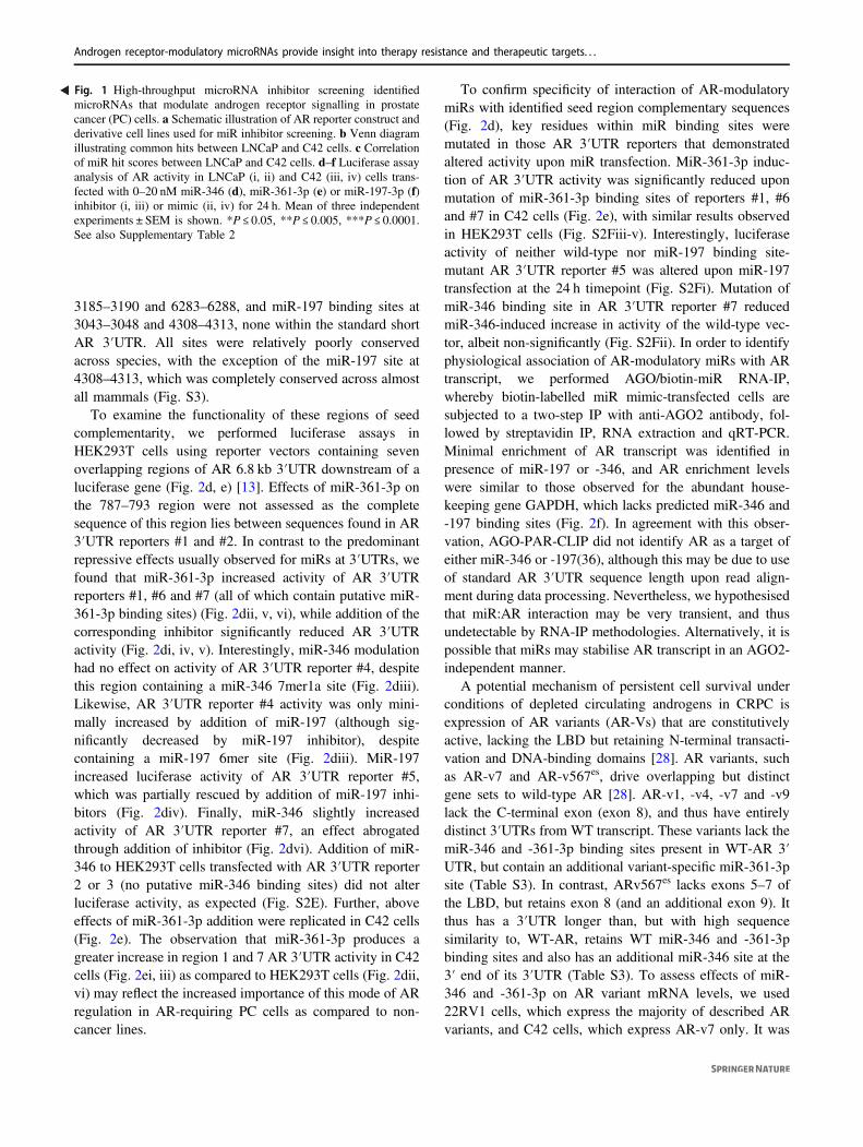
In order to identify miRs that modulate AR activity inandrogen-dependent and -independent PC, LNCaP and C42cells stably expressing a luciferase-based AR activityreporter [26] (Fig. 1a) were transfected with a library of 983miR inhibitors. Luciferase assays were performed and BScore calculated. Using a stringent B score of ±6, 78 miRinhibitors were found to significantly modulate AR reporteractivity (B Score ≥ 6 in at least one PC cell line), 63%negatively and 37% positively. B score correlation betweenLNCaP and C42 cells, and top 30 hits are shown (Fig. 1c,Table S2, respectively). Eight miR inhibitors significantlyaltered AR activity in the same direction in both LNCaP andC42 cells (B Score ≥ 6, Fig. 1b, Table S2), while no inhi-bitors significantly modulated AR activity in oppositedirections in the two cell lines at B Score ≥6. MiR-346,-361-3p and -197-3p were selected from top ten ‘hits’ due totheir high B scores and confirmed expression in cell lines(Ct < 35 in LNCaP cells). To confirm the ability of thesemiRs to regulate AR activity in androgen-dependent and-independent settings, inhibitors or mimics were transfectedinto LNCaP/ARE and C42/ARE and luciferase assays per-formed. Figure S1a confirms significantly increased miRlevels in C42 cells transfected with miR mimics. MiR-346inhibitor significantly reduced AR activity by up to 90% ina dose-dependent manner in LNCaP/ARE and C42/AREcells (Fig. 1di, ii). AR activity was not altered followingtransfection of a non-targeting inhibitor (Fig. S1b). Con-versely, miR-346 mimic increased AR activity by 70% inLNCaP/ARE cells, an effect not significant in C42/AREcells (Fig. 1diii, iv). Similarly, miR-361-3p inhibitor sig-nificantly repressed AR activity by up to 80% in both celllines (Fig. 1ei, ii), while miR-361-3p mimic significantly
increased AR activity by up to fivefold (Fig. 1eiii, iv).Comparable data were obtained for miR-197 inhibitor,which decreased AR activity by up to 50% (Fig. 1fi, ii),although miR-197 mimic did not significantly increase ARactivity in either cell line (Fig. 1fiii, iv). Further, miRmimics are able to rescue effects of miR inhibitors, ver-ifying that these are not off-target effects (Fig. 2a, c).
Modulation of MiR-346, -361-3p and -197 alters ARwild-type and variant mRNA, protein and 3′UTRactivity
Given that modulating miR-346, -361-3p and -197 sig-nificantly altered AR activity, we hypothesised that this maybe due to changes in AR mRNA or protein levels. Thus,C42 and LNCaP cells were transfected with inhibitor and/ormimic and AR mRNA and protein levels assessed 24 hpost-transfection. Non-targeting miR inhibitors and mimicsdid not significantly modulate AR activity in either cell line(Fig S1b). It was shown that inhibition of each miR underinvestigation significantly reduced AR mRNA levels by upto 70% (Fig. 2a, Fig. S1c), and that addition of mimic in thepresence of inhibitor could rescue AR mRNA loss in adose-dependent manner, an effect significant for miR-346(Fig. 2ai). Further, inhibition of miR-346, -361-3p and-197 significantly reduced AR protein levels in a dose-dependent manner, also rescued through addition of miRmimic (Fig. 2b, Fig. S1d–h). Similar effects were observedin LNCaP cells (Fig. S1f, g, h).
To examine effects of miR manipulation on AR tran-scriptional output, we assessed levels of the endogenous ARtarget genes, PSA, TMPRSS2 and DRG1 in both LNCaP andC42 cells. Inhibition of miR-346, -361-3p or -197 wasfound to significantly reduce PSA mRNA levels by up to75% in LNCaP cells (Fig. 2c). Loss of PSA mRNA wasrescued through addition of miR-346 mimic, and miR-346mimic alone was found to significantly increase PSAmRNA levels compared to mock-transfected cells (Fig. 2ci).Similar results were obtained for other AR target genes inboth LNCaP and C42 (Fig. S2a–d). To assess whetherupregulation of AR activity and protein levels occursthrough direct miR activity at the AR 3′UTR, we analysedAR 3′UTR for miR-346, -361-3p and -197 seed regioncomplementarity. Although algorithm-based miR bindingprediction tools such as microrna.org and DIANAmicroTpredict miR associations with an AR 3′UTR of 436 nt andc. 3 kb, respectively, a number of studies have identified AR3′UTR lengths of between 6.6 and 6.9 kb in PC cells [27]resulting from alternative polyadenylation [15], meaningthat large numbers of biologically important potential miR:AR 3′UTR interactions may be missed during bioinformaticanalysis. Thus, 6.8 Kb AR 3′UTR sequence (fromNM_000044 was examined for miR binding sites). Two
Androgen receptor-modulatory microRNAs provide insight into therapy resistance and therapeutic targets. . .

miR-361-3p binding sites (complete seed complementarity)were identified within the proximal region of AR 6.8 kb 3′UTR at nucleotides 407–412 and 787–793 (although only
the first is within the 436 nt short AR 3′UTR), with a furthertwo sites identified distally at 5772–5777 and 6070–6075(Fig. 2di). MiR-346 binding sites were identified at
29 418
C42/ARELNCaP/ARE
miRs with roles in
castration-resistance?
miRs that alter AR activity in androgen-
dependent and –independent setting?
B
C42• AR-positive• LNCaP derivative• Model of CRPC/ metastasis
C42-ARE
LNCaP• AR-positive• Epithelial• Anti-androgen responsive
LNCaP-ARE
AARE Tk prom Luc
020406080
100120140160180200
0 2 20
AR
Rep
orte
r Act
ivity
/Pro
tein
C
onte
nt (%
Moc
k)
AR Reporter Activity:
LNCaP/MAR4
miR-346 mimic (nM)
**
0
20
40
60
80
100
120
0 2 20
AR
Rep
orte
r Act
ivity
/Pro
tein
C
onte
nt (%
Moc
k)
AR Reporter Activity:
LNCaP/MAR4
miR-346 inhib (nM)
******
0
20
40
60
80
100
120
0 2 20
AR
Rep
orte
r Act
ivity
/Pro
tein
C
onte
nt (%
Moc
k)
miR-346 inhib (nM)
AR Reporter Activity: C42/MAR4
***
0
20
40
60
80
100
120
140
0 2 20
AR
Rep
orte
r Act
ivity
/Pro
tein
C
onte
nt (%
Moc
k)
miR-346 mimic (nM)
AR Reporter Activity: C42/MAR4
Di ii
iii iv
0
20
40
60
80
100
120
0 2 20
AR
Rep
orte
r Act
ivity
/Pro
tein
C
onte
nt (%
Moc
k)
AR Reporter Activity:
LNCaP/MAR4**
**
miR-361-3p inhib (nM)
0
100
200
300
400
500
600
0 2 20
AR
Rep
orte
r Act
ivity
/Pro
tein
C
onte
nt (%
Moc
k)
AR Reporter Activity:
LNCaP/MAR4
miR-361-3p mimic (nM)
***
0
20
40
60
80
100
120
0 2 20
AR
Rep
orte
r Act
ivity
/Pro
tein
C
onte
nt (%
Moc
k)
miR-361-3p inhib(nM)
AR Reporter Activity: C42/MAR4
***
0
50
100
150
200
250
300
0 2 20
AR
Rep
orte
r Act
ivity
/Pro
tein
C
onte
nt (%
Moc
k)
miR-361-3p mimic (nM)
AR Reporter Activity: C42/MAR4
***
0
20
40
60
80
100
120
140
0 2 20
AR
Rep
orte
r Act
ivity
/Pro
tein
C
onte
nt (%
Moc
k)
miR-197-3p mimic (nM)
AR Reporter Activity: C42/MAR4
0
20
40
60
80
100
120
0 2 20
AR
Rep
orte
r Act
ivity
/Pro
tein
C
onte
nt (%
Moc
k)
miR-197-3p mimic (nM)
AR Reporter Activity:
LNCaP/MAR4
0
20
40
60
80
100
120
0 2 20
AR
Rep
orte
r Act
ivity
/Pro
tein
C
onte
nt (%
Moc
k)
miR-197-3p inhib (nM)
AR Reporter Activity:
LNCaP/MAR4
* *
0
20
40
60
80
100
120
0 2 20AR
Rep
orte
r Act
ivity
/Pro
tein
C
onte
nt (%
Moc
k)
miR-197-3p inhib (nM)
AR Reporter Activity: C42/MAR4
***
E Fi ii
iii iv
i ii
iii iv
C
miR-361-3pmiR-346
miR-197
miR-378c
miR-26b*
miR-1260
let-7b*
miR-1271
miR-143*
miR-203
miR-96*miR-2110
miR-548qmiR-635miR-512-5pmiR-943miR-3152
miR-16
miR-195
miR-651
miR-135b*miR-516b
miR-518f* miR-548b-5p
C. E. Fletcher et al.
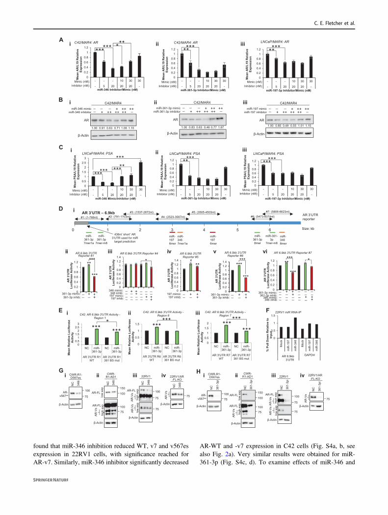
3185–3190 and 6283–6288, and miR-197 binding sites at3043–3048 and 4308–4313, none within the standard shortAR 3′UTR. All sites were relatively poorly conservedacross species, with the exception of the miR-197 site at4308–4313, which was completely conserved across almostall mammals (Fig. S3).
To examine the functionality of these regions of seedcomplementarity, we performed luciferase assays inHEK293T cells using reporter vectors containing sevenoverlapping regions of AR 6.8 kb 3′UTR downstream of aluciferase gene (Fig. 2d, e) [13]. Effects of miR-361-3p onthe 787–793 region were not assessed as the completesequence of this region lies between sequences found in AR3′UTR reporters #1 and #2. In contrast to the predominantrepressive effects usually observed for miRs at 3′UTRs, wefound that miR-361-3p increased activity of AR 3′UTRreporters #1, #6 and #7 (all of which contain putative miR-361-3p binding sites) (Fig. 2dii, v, vi), while addition of thecorresponding inhibitor significantly reduced AR 3′UTRactivity (Fig. 2di, iv, v). Interestingly, miR-346 modulationhad no effect on activity of AR 3′UTR reporter #4, despitethis region containing a miR-346 7mer1a site (Fig. 2diii).Likewise, AR 3′UTR reporter #4 activity was only mini-mally increased by addition of miR-197 (although sig-nificantly decreased by miR-197 inhibitor), despitecontaining a miR-197 6mer site (Fig. 2diii). MiR-197increased luciferase activity of AR 3′UTR reporter #5,which was partially rescued by addition of miR-197 inhi-bitors (Fig. 2div). Finally, miR-346 slightly increasedactivity of AR 3′UTR reporter #7, an effect abrogatedthrough addition of inhibitor (Fig. 2dvi). Addition of miR-346 to HEK293T cells transfected with AR 3′UTR reporter2 or 3 (no putative miR-346 binding sites) did not alterluciferase activity, as expected (Fig. S2E). Further, aboveeffects of miR-361-3p addition were replicated in C42 cells(Fig. 2e). The observation that miR-361-3p produces agreater increase in region 1 and 7 AR 3′UTR activity in C42cells (Fig. 2ei, iii) as compared to HEK293T cells (Fig. 2dii,vi) may reflect the increased importance of this mode of ARregulation in AR-requiring PC cells as compared to non-cancer lines.
To confirm specificity of interaction of AR-modulatorymiRs with identified seed region complementary sequences(Fig. 2d), key residues within miR binding sites weremutated in those AR 3′UTR reporters that demonstratedaltered activity upon miR transfection. MiR-361-3p induc-tion of AR 3′UTR activity was significantly reduced uponmutation of miR-361-3p binding sites of reporters #1, #6and #7 in C42 cells (Fig. 2e), with similar results observedin HEK293T cells (Fig. S2Fiii-v). Interestingly, luciferaseactivity of neither wild-type nor miR-197 binding site-mutant AR 3′UTR reporter #5 was altered upon miR-197transfection at the 24 h timepoint (Fig. S2Fi). Mutation ofmiR-346 binding site in AR 3′UTR reporter #7 reducedmiR-346-induced increase in activity of the wild-type vec-tor, albeit non-significantly (Fig. S2Fii). In order to identifyphysiological association of AR-modulatory miRs with ARtranscript, we performed AGO/biotin-miR RNA-IP,whereby biotin-labelled miR mimic-transfected cells aresubjected to a two-step IP with anti-AGO2 antibody, fol-lowed by streptavidin IP, RNA extraction and qRT-PCR.Minimal enrichment of AR transcript was identified inpresence of miR-197 or -346, and AR enrichment levelswere similar to those observed for the abundant house-keeping gene GAPDH, which lacks predicted miR-346 and-197 binding sites (Fig. 2f). In agreement with this obser-vation, AGO-PAR-CLIP did not identify AR as a target ofeither miR-346 or -197(36), although this may be due to useof standard AR 3′UTR sequence length upon read align-ment during data processing. Nevertheless, we hypothesisedthat miR:AR interaction may be very transient, and thusundetectable by RNA-IP methodologies. Alternatively, it ispossible that miRs may stabilise AR transcript in an AGO2-independent manner.
A potential mechanism of persistent cell survival underconditions of depleted circulating androgens in CRPC isexpression of AR variants (AR-Vs) that are constitutivelyactive, lacking the LBD but retaining N-terminal transacti-vation and DNA-binding domains [28]. AR variants, suchas AR-v7 and AR-v567es, drive overlapping but distinctgene sets to wild-type AR [28]. AR-v1, -v4, -v7 and -v9lack the C-terminal exon (exon 8), and thus have entirelydistinct 3′UTRs from WT transcript. These variants lack themiR-346 and -361-3p binding sites present in WT-AR 3′UTR, but contain an additional variant-specific miR-361-3psite (Table S3). In contrast, ARv567es lacks exons 5–7 ofthe LBD, but retains exon 8 (and an additional exon 9). Itthus has a 3′UTR longer than, but with high sequencesimilarity to, WT-AR, retains WT miR-346 and -361-3pbinding sites and also has an additional miR-346 site at the3′ end of its 3′UTR (Table S3). To assess effects of miR-346 and -361-3p on AR variant mRNA levels, we used22RV1 cells, which express the majority of described ARvariants, and C42 cells, which express AR-v7 only. It was
Fig. 1 High-throughput microRNA inhibitor screening identifiedmicroRNAs that modulate androgen receptor signalling in prostatecancer (PC) cells. a Schematic illustration of AR reporter construct andderivative cell lines used for miR inhibitor screening. b Venn diagramillustrating common hits between LNCaP and C42 cells. c Correlationof miR hit scores between LNCaP and C42 cells. d–f Luciferase assayanalysis of AR activity in LNCaP (i, ii) and C42 (iii, iv) cells trans-fected with 0–20 nM miR-346 (d), miR-361-3p (e) or miR-197-3p (f)inhibitor (i, iii) or mimic (ii, iv) for 24 h. Mean of three independentexperiments ± SEM is shown. *P ≤ 0.05, **P ≤ 0.005, ***P ≤ 0.0001.See also Supplementary Table 2
Androgen receptor-modulatory microRNAs provide insight into therapy resistance and therapeutic targets. . .
found that miR-346 inhibition reduced WT, v7 and v567esexpression in 22RV1 cells, with significance reached forAR-v7. Similarly, miR-346 inhibitor significantly decreased
AR-WT and -v7 expression in C42 cells (Fig. S4a, b, seealso Fig. 2a). Very similar results were obtained for miR-361-3p (Fig. S4c, d). To examine effects of miR-346 and
0
0.2
0.4
0.6
0.8
1
1.2
- - - 10 30 30
- 5 20 20 20 -
Mea
n A
R/L
19 R
elat
ive
Expr
essi
on
miR-346 Inhibitor/Mimic (nM)
C42/MAR4: AR
*********
Mimic (nM):Inhibitor (nM):
00.20.40.60.8
11.2
- - - 10 30 30
- 5 20 20 20 -
Mea
n A
R/L
19 R
elat
ive
Expr
essi
on
miR-361-3p Inhibitor/Mimic (nM)
C42/MAR4: AR
*****
Mimic (nM):Inhibitor (nM):
00.20.40.60.8
11.2
- - - 10 30 30
- 5 20 20 20 -
Mea
n A
R/L
19 R
elat
ive
Expr
essi
on
miR-197-3p Inhibitor/Mimic (nM)
LNCaP/MAR4: AR
*****
Mimic (nM):Inhibitor (nM):
Aiiiiii
iiiii
00.5
11.5
22.5
3
- - - 10 30 30
- 5 20 20 20 -
Mea
n PS
A/L
19 R
elat
ive
Expr
essi
on
miR-346 Mimic/Inhibitor (nM)
LNCaP/MAR4: PSA
**************
Mimic (nM):Inhibitor (nM):
00.20.40.60.8
11.2
- - - 10 30 30
- 5 20 20 20 -
Mea
n PS
A/L
19 R
elat
ive
Expr
essi
on
miR-361-3p Inhibitor/Mimic (nM)
LNCaP/MAR4: PSA
*****
Mimic (nM):Inhibitor (nM):
00.20.40.60.8
11.2
- - - 10 30 30
- 5 20 20 20 -
Mea
n PS
A/L
19 R
elat
ive
Expr
essi
on
miR-197-3p Inhibitor/Mimic (nM)
LNCaP/MAR4: PSA
Mimic (nM):Inhibitor (nM):
******
C i ii iii
D i
B i
AR
β-Actin
C42/MAR4
_ + ++ ++ ++ __ _ _ +++ ++
miR-346 inhibitormiR-346 mimic
1.00 0.91 0.63 0.71 1.06 1.10
AR 3’UTR – 6.9kb
0 1 2 3 4 5 6miR-3467mer1a
miR-3466mer
miR-361-3p
7mer-m8
miR-361-3p7mer1a
miR-361-3p8mer
miR-361-3p
7mer-m8
miR-1976mer
miR-1976mer
Size: kb
AR 3’UTR reporter#1: (1-769nt) #2: (791-1703nt)
#3: (1537-2672nt)#4: (2523-3567nt)
#5: (3505-4503nt)#6: (5473-6074nt)
#7: (5806-6623nt)
436nt ‘short’ AR 3’UTR used for miR
target prediction
AR
β-Actin
C42/MAR4
_ + ++ ++ ++ __ _ _ +++ ++
miR-361-3p inhibitormiR-361-3p mimic
1.00 0.83 0.63 0.46 0.77 1.67
AR
β-Actin
C42/MAR4
_ + ++ ++ ++ __ _ _ +++ ++
miR-197 inhibitormiR-197 mimic
1.00 0.85 0.49 0.55 1.01 1.15
0
0.2
0.4
0.6
0.8
1
1.2
NC
361…
361…
AR 6.9kb 3'UTR Reporter #1
**
***
***
361-3p mimic:361-3p inhib:
_ + +_ _ +
AR
3’U
TR
Luci
fera
se A
ctiv
ity
00.20.40.60.8
11.21.4
NC
346…
346…
NC
197…
197…
AR 6.9kb 3'UTR Reporter #4
*
346 mimic:346 inhib:
197 mimic:197 inhib:
_ + +_ _ +_ _ __ _ _
_ _ __ _ __ + +_ _ +
*
AR
3’U
TR
Luci
fera
se A
ctiv
ity
00.20.40.60.8
11.21.4
AR 6.9kb 3'UTR Reporter #5
***
_ + +_ _ +197 mimic:197 inhib:
AR
3’U
TR
Luci
fera
se A
ctiv
ity
00.20.40.60.8
11.21.4
NC361 mim361 mim
***
***
***
_ + +_ _ +361-3p mimic:361-3p inhib:
AR 6.9kb 3’UTR Reporter #6
AR
3’U
TR
Luci
fera
se A
ctiv
ity
00.20.40.60.8
11.2
AR 6.9kb 3'UTR Reporter #7
**
***
*** *
361-3p mimic:361-3p inhib:
346 mimic:346 inhib:
AR
3’U
TR
Luci
fera
se A
ctiv
ity
_ + + _ _ __ _ + _ _ __ _ _ _ + +_ _ _ _ _ +
ii iii iv v vi
00.5
11.5
22.5
3
NC miR-361-3p
NC miR-361-3p
AR 3'UTR R1WT
AR 3'UTR R1361 BS mut
Mea
n R
elat
ive
Luci
fera
se
Act
ivity
C42: AR 6.9kb 3'UTR Activity -Region 1
*** ****
E
0
0.5
1
1.5
Moc
k
miR
-197
miR
-346
Moc
k
miR
-197
miR
-346
AR long 3'UTRreg7
GAPDH
% P
ull D
own
Rel
ativ
e to
In
put
22RV1 miR RNA-IP
AR 6.9kb 3’UTR
F
00.5
11.5
2
NC miR-361-3p
NC miR-361-3p
AR 3'UTR R7WT
AR 3'UTR R7361 BS mut
Mea
n R
elat
ive
Luci
fera
se
Act
ivity
C42: AR 6.9kb 3'UTR Activity -Region 7
******
***
0
0.5
1
1.5
NC miR-361-3p
NC miR-361-3p
AR 3'UTR R6WT
AR 3'UTR R6361 BS mut
Mea
n R
elat
ive
Luci
fera
se
Act
ivity
C42: AR 6.9kb 3'UTR Activity -Region 6
******
ii
β-Actin
NC
346i
CWR-R1-D567es
AR-v567
100
75
NC
346i
CWR-R1-AD1
low
exp
high
exp
100
150
100
75
AR-FL
AR-FL
AR
-Vs
β-Actin
β-Actin
22RV1
NC
346i
100
75
75
AR-FL
high
exp
AR
-Vs
low
exp
AR
-Vs
NC
346i
22RV1/AR-FL-KO
AR
-Vs
β-Actin
75
NC
361-
3pi
CWR-R1-D567es
AR-v567
100
75
β-Actin
NC
361-
3pi
CWR-R1-AD1
low
exp
high
exp
100
150
100
75
AR-FL
AR-FL
AR
-Vs
β-Actinβ-Actin
22RV1
NC
361-
3pi
100
75
75
AR-FL
high
exp
AR
-Vs
low
exp
AR
-Vs
NC
361-
3pi
22RV1/AR-FL-KO
AR
-Vs
75
β-Actin
iiii
Gi H iii iii iv ii iii iv
C. E. Fletcher et al.

-361-3p inhibition on WT and variant-AR protein levels,Western blotting was performed in 22RV1 cells, WT-knockout derivatives (22RV1-AR-FL-KO—all variants expressedbut not WT), parental CWR-R1-AD1 cells (lacking AR-v567es) and the CWR-R1-D567es derivative lacking WT-AR but overexpressing v567es. An N-terminal AR antibodywas used in order to detect both WT and variant-AR. Thisantibody is unable to identify individual variants, althoughAR-v567es is distinguishable from others due to its size(80.7 kDa vs. 50.6–74.9 kDa for variants 1, 4, 7 and 9 and110 kDa for WT-AR). Of note: the 22rv1 line carries a35 kb tandem duplication encompassing AR exon 3 andcryptic exons, which are spliced as alternative 3′ exons ofAR-variants, including v7 [29]. This may result in increasedlevels of cryptic exon-derived variants in this cell line,potentially in different ratios compared to cells lacking theduplication. These, as above, are indistinguishable usingavailable antibodies. It was shown that miR-346 inhibitorreduces protein levels of full-length and multiple variants ofAR (including v567es) across the above panel of cell linesafter 24 h (Fig. 2g). MiR-361-3p inhibitor produced similar
results, demonstrating a greater extent of repression (Fig.2h). Biological replicates are shown in Fig. S5. Thus weconclude that miR-346 and miR-361-3p modulation alterslevels of constitutively active AR variants in PC, and maycontribute to persistent AR signalling in CRPC in theabsence of circulating androgens.
AR-modulatory MiRs stabilise AR transcript andtheir inhibition demonstrates additive effects withAR silencing
In light of their effects on AR protein levels (Fig. 2b), weinvestigated the ability of AR-modulatory miRs to enhanceAR transcript stability. C42 cells were transfected with miRmimics for 24 h prior to 4 h Actinomycin D (ActD) treat-ment. In agreement with Fig. 2a, all three miRs increasedAR mRNA levels in vehicle-treated conditions compared tonegative control (Fig. S2g). It was demonstrated that addi-tion of either miR-346 or miR-197 rescued ActD-mediatedloss of AR transcript compared to negative control-transfected cells in a significant manner, while miR-361-3p transfection was not able to rescue ActD-mediated ARmRNA loss (Fig. S2g). Given that miR-346, -361-3p and-197 have profound effects on AR protein levels andactivity in PC cells, we hypothesised that modulation ofthese miRs may alter PC cell proliferation, and that miRinhibition may demonstrate combinatorial effects with ARsilencing, potentially revealing a novel therapeutic avenuefor PC treatment. To this end, LNCaP cells expressing adoxycycline (Dox)-inducible AR siRNA [30] were treated± 1 µM Dox and transfected with miR-346 or -361-3pinhibitors or mimics prior to SRB cell proliferation analysis.It was shown that miR-346 inhibitor alone significantlyreduced cell number to 40% of day 0 numbers (Fig. 3ai),suggesting that miR-346 inhibition has cytotoxic, ratherthan cytostatic effects. AR siRNA alone reduced growth ofLNCaP cells by ~50% over 6 days (Fig. 3a). Induction ofAR siRNA in the presence of miR-346 inhibitor did notenhance effects of miR-346 inhibitor alone, presumably dueto the very marked effects of miR-346 inhibitor at con-centrations used in this experiment (20 nM) (Fig. 3ai).Converse experiments showed that miR-346 mimic wasable to significantly enhance proliferation of LNCaP cellsby 80%, and partially rescue effects of AR silencing in asignificant manner (Fig. 3aii). Inhibition of miR-361-3psignificantly repressed growth of LNCaP cells by 70%, andfurther addition of AR siRNA in combination repressed cellgrowth at day 6 to day 0 levels (Fig. 3bi). MiR-361-3pmimic significantly increased proliferation of LNCaP cellsand partially rescued AR siRNA-induced reduction in cellgrowth in a significant manner (Fig. 3bii). It was addition-ally demonstrated that while miR-346 inhibitor repressesC42 and LNCaP proliferation at both 5 nM and 20 nM
Fig. 2 MiR-346, -361-3p and -197-3p alter wild-type and variantandrogen receptor activity in prostate cancer (PC) cells partiallythrough association with AR 3′UTR. a qRT-PCR analysis of ARmRNA levels in C42/MAR4 cells transfected with (i) miR-346, (ii)miR-361-3p or (iii) miR-197-3p mimic and/or inhibitor for 24 h.bWestern blot analysis of AR protein levels in C42/MAR4 transfectedwith (i) miR-346, (ii) miR-361-3p or (iii) miR-197-3p mimic and/orinhibitor for 24 h. β-actin was used as a control for loading. Repre-sentative blots of three independent experiments are shown. Additionalbiological replicates and densitometry for three independent experi-ments are shown (Fig. S1c–g). c qRT-PCR analysis of PSA mRNAlevels in LNCaP/MAR4 cells transfected with (i) miR-346, (ii) miR-361-3p or (iii) miR-197-3p mimic and/or inhibitor for 24 h. L19 wasused as a normalisation gene. d, e Luciferase assay analysis of 6.9 kbAR 3′UTR activity in HEK293T (d) or C42 (e) cells transfected withpMiR-Report vector containing WT (d) or miR binding site-mutant (e)regions of the AR 6.9 kb 3′UTR as depicted (Fig. S2Di) ± miR mimicsor inhibitors (20 nM) for 48 h (d) or 72 h (e) Luciferase activity wasnormalised to β-galactosidase activity to correct for transfection effi-ciency. Columns: mean normalised AR reporter luciferase activityfrom three independent experiments performed in duplicate ± SEM.f AGO2/biotin-miR RNA-IP analysis of miR-197 and miR-346 asso-ciation with AR 6.9 kb 3′UTR. 22RV1 cells transfected with biotin-labelled miR (200 pmol) for 24 h, followed by two-step immunopre-cipitation with AGO2 antibody- and streptavidin-coated beads. RNAwas extracted from input and IP samples and qRT-PCR performed for6.9 kb AR 3′UTR. Data are presented relative to input values. g, hWestern blot analysis of wild-type AR (110 kDa) and variant-AR(65–90 kDa) in (i) CWR-R1-D567es (lacking WT-AR but over-expressing v567es), (ii) CWR-R1-AD1 (parental cells expressing WT-and variant-AR), (iii) 22RV1 (expressing WT- and variant-AR) and(iv) 22RV1/AR-FL-KO cells (expressing AR-variants but with WT-AR knocked out) cells transfected with (a) miR-346 or (b) miR-361-3p inhibitor (both 20 nM) for 24 h. β-actin was used as a control forloading. Representative western blot images are shown. See Fig. S5 forindependent biological replicates. *P ≤ 0.05, **P ≤ 0.005, ***P ≤0.0001. See also Figs. S1, S2, S3, S4 and S5
Androgen receptor-modulatory microRNAs provide insight into therapy resistance and therapeutic targets. . .

(Fig. S6a), miR-361-3p inhibitor reduced cell number in adose-dependent manner in both lines (Fig. S6c). In contrastto the observed increase in LNCaP cell proliferation
following transfection of 7.5 nM miR-346 mimic, con-centrations of 10 and 50 nM miR-346 mimic significantlyreduced proliferation of both C42 and LNCaP cells (Fig.
0
200
400
600
0 2 4 6Abs
orba
nce
(% d
ay 0
moc
k)
Time (d)
LNCaP/ARsiRNANCNC346 mimic346 mimic
*
***
0
100
200
300
400
500
0 2 4 6
Abs
orba
nce
(% d
ay 0
moc
k)
Time (d)
LNCaP/AR siRNANCARKO346 inhib346 inhib + ARKO
0
100
200
300
400
500
0 2 4 6Abs
orba
nce
(% d
ay 0
moc
k)
Time (d)
LNCaP/AR siRNANCARKO361-3p inhib361-3p inhib + ARKO
*
*
*
0
100
200
300
400
500
600
0 2 4 6Abs
orba
nce
(% d
ay 0
moc
k)
Time (d)
LNCaP/AR siRNANCARKO361-3p mimic361-3p mimic + ARKO
Biii
0
200
400
600
800
1000
0 2 4 6Abs
orba
nce
(% d
ay 0
moc
k)
Time (d)
DU145/MAR4 mock346 5nM346 20nM
***
0
200
400
600
800
1000
0 2 4 6Abs
orba
nce
(% d
ay 0
moc
k)
Time (d)
DU145/MAR4mock361-3p 5nM361-3p 20nM
*
Aiii
Ciii
inhib (5nM)inhib (20nM)
inhib (5nM)inhib (20nM)
0
20
40
60
80
100
- + - + - +
Cas
pase
3/7
Glo
Luc
A
ctiv
ity/C
ell N
umbe
r
Caspase 3/7 Glo Luc Activity: LNCaP/ARsiRNA
+_ +_ +_AR siRNA:miR-346 inhib:
miR-346 mimic:_ _ _ _+ +_ _ _ _ + +
***
***
*****
0
2
4
6
8
10
12
- + - + - +
Cas
pase
3/7
Glo
Luc
A
ctiv
ity/C
ell N
umbe
r
Caspase 3/7 Glo Luc Activity: LNCaP/ARsiRNA
+_ +_ +_AR siRNA:miR-361-3p inhib:
miR-361-3p mimic:_ _ _ _+ +_ _ _ _ + +
***
**
*** **
0
5
10
15
- + - + - +
Cas
pase
3/7
Glo
Luc
A
ctiv
ity/ C
ell N
umbe
r
Caspase 3/7 Glo Luc Activity: LNCaP/ARsiRNA P=0.089
***
*** ******
*
+_ +_ +_AR siRNA:miR-197-3p inhib:
miR-197-3p mimic:_ _ _ _+ +_ _ _ _ + +
iiiiiiK
0
0.5
1
1.5
2
- + + +
NC 361-3p
inhib
361-3p
mimic
AR
/L19
Rel
ativ
e Ex
pres
sion
AR* *
***
AR siRNA:0
0.2
0.4
0.6
0.8
1
1.2
- + + +
NC 346inhib
346mimic
PSA
/L19
Rel
ativ
e Ex
pres
sion
PSA*
**
AR siRNA:
00.20.40.60.8
11.2
- + + +
NC 361-3p
inhib
361-3p
mimic
PSA
/L19
Rel
ativ
e Ex
pres
sion
PSA* *
AR siRNA:
*
0
0.5
1
1.5
2
- + + +
NC 346inhib
346mimic
AR
/L19
Rel
ativ
e Ex
pres
sion
AR
AR siRNA:
*
**
0
5
10
15
20
25
0 3 6
Abs
orba
nce
(% D
ay 0
NC
5n
M)
Time (d)
22Rv1: MiR-346 InhibitorNC 5nMNC 20nM346 5nM346 20nM
0
5
10
15
0 3 6
Abs
orba
nce
(% d
ay 0
NC
5n
M)
Time (d)
22Rv1 AR-FL KO: MiR-346 Inhibitor
NC 5nMNC 20nM346 5nM346 20nM
*
0
3
6
9
12
0 3 6
Abs
orba
nce
(% d
ay 0
NC
5n
M)
Time (d)
CWR-R1-AD1: MiR-346 inhibitorNC 5nMNC 20nM346 5nM346 20nM
0
3
6
9
12
15
0 3 6
Abs
orba
nce
(% d
ay 0
NC
5n
M)
Time (d)
CWR-R1-D567es: MiR-346 Inhibitor
NC 5nMNC 20nM346 5nM346 20nM
D iiiiii E
0
5
10
15
20
25
0 3 6
Abs
orba
nce
(% D
ay 0
NC
5n
M)
Time (d)
22Rv1: MiR-361-3p inhibitorNC 5nMNC 20nM361-3p 5nM361-3p 20nM
**
0
5
10
15
0 3 6Abs
orba
nce
(% d
ay 0
NC
5n
M)
Time (d)
22Rv1 AR-FL KO: MiR-361 InhibitorNC 5nMNC 20nM361-3p 5nM361-3p 20nM
*
**
0
3
6
9
12
0 3 6
Abs
orba
nce
(% d
ay 0
NC
5n
M)
Time (d)
CWR-R1-AD1: MiR-361-3p Inhibitor
NC 5nMNC 20nM361-3p 5nM361-3p 20nM
0
3
6
9
12
15
0 3 6
Abs
orba
nce
(% d
ay 0
NC
5n
M)
Time (d)
CWR-R1-D567es: MiR-361-3p Inhibitor
NC 5nMNC 20nM361-3p 5nM361-3p 20nM
*
F iiiiiiG
iiiiii JH
C. E. Fletcher et al.

S6b). We hypothesise that this is due to threshold effects ofmiR-346 manipulation, with lower doses activating pro-liferative responses, while higher concentrations inducetoxicity, leading to apoptotic induction as illustrated in Fig.4a. Such a threshold is likely to be different in different celllines. MiR-361-3p mimic significantly increased C42 andLNCaP cell growth at both 10 and 50 nM (Fig. S6d).Interestingly, both miR-346 and -361-3p inhibitors sig-nificantly repressed proliferation of AR-negative DU145cells in a dose-dependent manner (Fig. 3c), suggesting thatat least some of miR-346 and -361-3p effects are mediatedvia non-AR pathways.
We further investigated the ability of miR-346, 1361-3pand -197 to regulate proliferation of AR variant-driven PCcell lines, by comparing proliferation between parental22RV1 cells (all variants and WT expressed) and WT-knock out derivatives (22RV1-AR-FL-KO), and alsobetween parental CWR-R1-AD1 cells (lacking AR-v567es)and the CWR-R1-D567es derivative lacking WT-AR butoverexpressing v567es. Interestingly, miR-346 inhibitionsignificantly reduced proliferation of 22RV1 cells expres-sing variants only, but had less effect on the parental cellswith FL AR (Fig. 3d). Although the enhanced effect ismodest, it may reflect the additional miR-346 seed com-plementarity site present in AR-v567es 3′UTR (nts 8353-8358)—see Table S3. This is supported by the observationthat miR-346 inhibition non-significantly represses AR-v567es-driven growth of CWR-R1-D567es cells, with noeffect observed in parental cells (Fig. 3e). MiR-361-3pinhibition completely blocked proliferation of 22RV1 and
the variant-driven derivative, with similar effects in v567es-driven CWR-R1 (Fig. 3f, g). This is consistent with effectsin LNCaP, C42 and DU145 cells (Fig. 3a–c, Fig. S6c, d),and presence of similar numbers of miR-361-3p seedcomplementarity sites in 3′UTRs of AR-variants and -WT(Table S3).
We additionally investigated the ability of AR-modulatory miRs to modulate apoptosis and apoptoticresponse to AR silencing. It was demonstrated that miR-346, -361-3p and 197 inhibitors significantly enhanced ARsiRNA-induced AR loss at transcript level, while ARmRNA was significantly rescued by addition of miRmimics in the presence of AR siRNA (Fig. 3h and Fig. S6e).Accordingly, levels of AR targets PSA, TMPRSS2 andKLK2 were significantly reduced following miR-346, -361-3p of 197 inhibition in the presence of AR siRNA comparedto AR siRNA alone, while miR mimics rescued this (Fig. 3j,S6f–h). MiR-346 inhibition significantly increased ARsiRNA-induced apoptosis by 85-fold, while miR-346 mimicsignificantly reduced such apoptosis (Fig. 3ki). Likewise,miR-361-3p significantly increased apoptosis of LNCaPcells by fourfold compared to untreated cells, and sig-nificantly enhanced apoptosis induced by AR siRNA bysixfold, with a 50% reduction in apoptosis observed formiR-361-3p mimic compared to AR siRNA alone (Fig.3kii). In addition, miR-197 inhibitor significantly increasedapoptosis tenfold compared to untreated cells, and sig-nificantly enhanced AR siRNA-induced cell death by 8.5-fold, whereas miR-197 mimic reduced AR silencing-induced apoptosis by 50% (Fig. 3kiii). Taken together,these data indicate that miR-346 and -197 stabilise ARtranscript, and that miR inhibition shows additive effectswith AR silencing on proliferation and apoptosis, identify-ing a rationale for potential combined use of AR-inhibitorytherapies and miR-346, -361-3p or -197 inhibitors in PC.
AR-modulatory MiR inhibition demonstratesadditive effects with enzalutamide
To provide further evidence to support the rationale ofcombined miR-346, -361-3p or -197 inhibitor and anti-androgen treatment, we investigated the effects of AR-modulatory miR manipulation on apoptotic and pro-liferative response to the anti-androgen, Enzalutamide(Enza), in PC cells. It was demonstrated that miR-346inhibitor significantly enhances apoptosis induced by Enzaby 52%, while miR-346 mimic (20 nM) also increasedEnza-induced apoptosis (Fig. 4ai), consistent with miR-346mimic effects on proliferation in LNCaP cells at 10 and50 nM (Fig. S6Bii). MiR-361-3p inhibitor significantlyincreased Enza-induced apoptosis by 4.3-fold, while miR-361-3p mimic significantly reduced Enza-induced apoptosisby 40% compared to negative control-transfected, Enza-
Fig. 3 AR-modulatory miRs stabilise AR transcript and regulateapoptosis and WT- and variant-AR-driven proliferation, and MiR-346,-361-3p and -197-3p inhibitors show additive effects with AR silen-cing. a, b SRB proliferation assay analysis of LNCaP/ARsiRNA cellstransfected with miR-346 (a) or miR-361-3p (b) mimic (7.5 nM –
miR-346, 20 nM – miR-361-3p) or inhibitor (10 nM – miR-346,20 nM – miR-361-3p) ± 1 µM Dox to induce AR siRNA (ARKO) for6 days. c SRB proliferation assay analysis of DU145 cells transfectedwith miR-346 (i) or miR-361-3p (ii) inhibitor (5 and 20 nM) for6 days. d–g SRB proliferation assay analysis of 22RV1 (di, fi),22RV1-AR-FL-KO (AR variant-driven) (dii, fii), CWR-R1-AD1 (ei,gi) and CWR-R1-D567es (AR-v567es-driven) cells (eii, gii) transfectedwith: d, e miR-346 inhibitor (5 and 20 nM) or f, g miR-361-3p inhi-bitor (5 and 20 nM) for 6 days. a–g Data are presented relative toabsorbance at day 0. Points: mean absorbance at 492 nm for threeindependent experiments performed in quadruplicate ± SEM. h, j qRT-PCR analysis of (h) AR and (j) PSA mRNA levels in LNCaP/ARsiRNA cells transfected with miR-346 (i) or miR-361-3p (ii)inhibitor or mimic (20 nM) ± Doxycycline (1 µM) for 24 h. L19 wasused as a normalisation gene. Columns: mean ± SEM for three inde-pendent experiments performed in triplicate. k Caspase 3/7 Glo assayanalysis of apoptosis in LNCaP/AR siRNA cells ± miR mimic orinhibitor (20 nM) ± Doxycycline (1 µM) for 72 h. Columns: meanrelative luminescence for three independent experiments performed intriplicate ± SEM. *P ≤ 0.05, **P ≤ 0.005, ***P ≤ 0.0001. See also Fig.S6
Androgen receptor-modulatory microRNAs provide insight into therapy resistance and therapeutic targets. . .

0.8
1.3
1.8
2.3
2.8
3.3
3.8
4.3
4.8
5.3
- 0.8 - 0.8 - 2 - 2
NC inhib 361-3pinhib
NCmimic
361-3pmimic
Mea
n C
aspa
se 3
/7 G
lo A
ctiv
ity/C
ell N
umbe
r Caspase Activity + Enza: miR-361-3p
***
*
*****
** n.s.
*
Enza:
0
50
100
150
200
250
300
350
400
450
0 3 6
Abs
orba
nce
(% d
ay 0
moc
k)
Time (d)
LNCaP/MAR4: miR-346 mimic ±Enza
NCNC + EnzamiR-346 mimicmiR-346 mimic + Enza
0
100
200
300
400
500
600
700
800
0 3 6
Abs
orba
nce
(% d
ay 0
moc
k)
Time (d)
LNCaP/MAR4: miR-361-3p mimic ±Enza
NCNC + EnzamiR-361-3p mimicmiR-361-3p mimic + Enza
0
100
200
300
400
500
600
700
0 3 6
Abs
orba
nce
(% d
ay 0
moc
k)
Time (d)
LNCaP/MAR4: miR-346 inhibitor ±Enza
NCNC + EnzamiR-346 inhibmiR-346 inhib + Enza
0
100
200
300
400
500
600
700
0 3 6
Abs
orba
nce
(% d
ay 0
moc
k)
Time (d)
LNCaP/MAR4: miR-361-3p inhibitor ± Enza
NCNC + EnzamiR-361-3p inhibmiR-361-3p inhib + Enza
0
100
200
300
400
500
600
700
0 3 6
Abs
orba
nce
(% d
ay 0
moc
k)
Time (d)
LNCaP/MAR4: miR-197-3p inhibitor ± Enza
NCNC + EnzamiR-197-3p inhibmiR-197-3p inhib + Enza
B
A iiiii
iiiiii
vvi
50
100
150
200
250
0 3 6
Abs
orba
nce
(% d
ay 0
moc
k)
Time (d)
LNCaP/MAR4: MiR-197-3p mimic ±Enza
NCNC + EnzamiR-197-3p mimicmiR-197-3p mimic + Enza
vi
0.8
1.3
1.8
2.3
2.8
3.3
3.8
4.3
4.8
5.3
- 0.8 - 0.8 - 2 - 2
NC inhib 346inhib
NCmimic
346mimic
Mea
n C
aspa
se 3
/7 G
lo A
ctiv
ity/C
ell N
umbe
r
Caspase Activity + Enza: miR-346
****
***** **
*** **n.s.
Enza:0.8
1.3
1.8
2.3
2.8
3.3
3.8
4.3
4.8
5.3
- 0.8 - 0.8 - 2 - 2
NC inhib 197inhib
NCmimic
197mimic
Mea
n C
aspa
se 3
/7 G
lo A
ctiv
ity/C
ell N
umbe
r
Caspase Activity + Enza: miR-197
**
*
***
** ***
Enza:
i
Fig. 4 AR-modulatory miRs inhibitors enhance enzalutamide efficacy.a Caspase 3/7 Glo assay analysis of apoptosis in LNCaP/MAR4 cells± miR-346 (i), miR-361-3p (ii) or miR-197-3p (iii) mimic or inhibitor(20 nM) ± Enzalutamide (0.8 µM – in presence of inhibitors, 2 µM inpresence of mimics) for 72 h. Columns: mean relative luminescencefor three independent experiments performed in triplicate ± SEM. *P ≤0.05, **P ≤ 0.005, ***P ≤ 0.0001. b SRB proliferation assay analysis
of LNCaP/MAR4 cells transfected with inhibitors of miR-346 (i –2.5 nM), miR-361-3p (ii – 5 nM) or miR-197-3p (iii - 10 nM) ormimics of miR-346 (iv – 10 nM), miR-361-3p (v – 30 nM) or miR-197-3p (vi – 20 nM) ± Enzalutamide (0.8 µM) for 6 days. Data arepresented relative to absorbance at day 0 and one representativeexperiment is shown. Additional biological replicates are shown inFig. S7
C. E. Fletcher et al.

treated cells (Fig. 4aii). Similar Enza-induced apoptosis-modulatory effects were observed for miR-197 inhibitor andmimic (Fig. 4aiii). We additionally investigated effects ofmiR-346, -361-3p and -197 modulation on PC growthinhibition by Enza. Inhibition of miR-346, -361-3p or -197considerably enhanced Enza-induced reduction in LNCaPproliferation (Fig. 4bi–iii). Conversely, miR-346 and -361-3p mimics were able to completely rescue Enza-inducedrepression of LNCaP proliferation (Fig. 4biv, v). MiR-197mimic increased LNCaP/MAR4 proliferation, but did notsignificantly alter cell growth in presence of Enza (Fig.4bvi). Representative data is shown and results of replicateexperiments are shown in Fig. S7.
AR-modulatory MiR-361-3p increases migration andinvasion of PC cells and induces Snail-independentEMT
Having demonstrated that miR-346, -361-3p and -197 mod-ulate AR activity, proliferation and apoptosis in PC, wehypothesised that these miRs may alter other cancerprogression-associated processes. To this end, transwellmigration assays were performed in C42 cells transfected withmiR-361-3p mimic or negative control mimic. It wasdemonstrated that miR-361-3p significantly increases migra-tion of PC cells by fourfold (Fig. 5a). In agreement with this,miR-361-3p mimic also significantly enhanced C42 cellmigration when assessed by wound healing assay (Fig. 5b).Further, miR-361-3p mimic was shown to increase invasionof C42 cells through matrigel matrix (Fig. 5c), suggesting thatmiR-361-3p overexpressing cells show increased capacity toactively remodel extracellular matrix, potentially illustrativeof increased metastatic potential. Overexpression of neithermiR-197 nor miR-346 significantly increased invasion. Toinvestigate whether increased invasive potential is attributableto adoption of a more mesenchymal PC cell phenotype (aninitiating event in metastasis), mRNA levels of the establishedmesenchymal markers, Snail and ZEB2, were assessed in C42cells post-miR-346, -361-3p or -197 transfection. mRNAlevels of both markers were significantly increased by aminimum of 1.5-fold following miR overexpression (Fig. 5di,ii). MiR overexpression also increased expression of addi-tional mesenchymal markers, Slug and Twist-1 at mRNAlevel (Fig. S8a), and concomitant loss of the epithelial mar-kers, TSPAN-13 and E-Cadherin was observed followingmiR-346 or -197 overexpression (TSPAN-13) and miR-346or 361-3p overexpression (E-Cadherin) (Fig. 5diii, iv).Similarly, while miR-346 and -361-1p mimics significantlyincreased Snail and ZEB2 mRNA levels in LNCaP cells, theirinhibition led to reduction of these mesenchymal markers(Fig. S8b, c).
To establish whether such gene expression changestranslated to modulation of EMT-associated protein levels,
Western blotting was performed to assess epithelial proteinlevels in epithelial C42 cells and mesenchymal proteinlevels in more mesenchymal-like PC3 and DU145 cellsfollowing transfection of miR mimics and/or inhibitors. Itwas demonstrated that overexpression of miR-346 and miR-197 significantly reduced β-catenin and E-Cadherin proteinlevels in C42 cells (Fig. 5e and S8E-iii). In support ofenhanced EMT by miR-346 and -361-3p, addition of miRinhibitors in presence of miR mimics reduced protein levelsof the mesenchymal markers, N-Cadherin and Vimentin inPC3 cells (Fig. 5f, Fig. S8d). Unexpectedly given increasedSnail mRNA levels in presence of miR mimics (Fig. 5di,Fig. S8B), Snail protein levels were markedly induced, by50-fold and 220-fold, respectively, following miR-346 or-361-3p inhibitor transfection of PC3 cells in presence ofthe corresponding miR mimic (Fig. 5f, Fig. S8d). Similarsignificant effects were observed in the presence of miRinhibitors alone (Fig. S8f): while most mesenchymal mar-kers (N-Cadherin, Vimentin and Slug) are lost followingmiR-346 or -361-3p inhibition, Snail levels are significantlyincreased. To demonstrate that this was not a cell line-specific effect, the more mesenchymal-like DU145 cellswere transfected with miR-346 or -361-3p inhibitor and/ormimic. Again, Snail protein levels were significantly andmarkedly increased in presence of miR inhibitors, an effectrescued by addition of miR mimic (Fig. 5g, Fig. S9). Incontrast, protein levels of closely-related Slug are sig-nificantly reduced in the presence of miR inhibitor (Fig. 5g,Fig. S9), consistent with enhanced EMT in presence of miRmimics and reversal of EMT when miRs are inhibited. Wehypothesised that this unique EMT protein signature mayresult from direct regulation of Snail-regulatory proteins byAR-modulatory miRs. Thus we mined AGO-PAR-CLIPdata from PC cells [31] for association of miR-346, -361-3por -197 with 58 unique Snail interactors identified frombiogrid.org. 20 of these (although not Snail itself) werefound to contain binding sites for at least one of AR-modulatory miR-346, -361-3p or -197 in at least one PC cellline. These include LATS, PARP1, MDM2, GSK3β andPKCµ (Fig. S10a, Fig. 5h). We hypothesised that AR-modulatory miRs may repress positive regulators (greenarrows, Fig. 5h) and upregulate negative regulators (redarrows, Fig. 5h) of Snail protein levels in PC cells, withmiR inhibitors showing opposing effects. To confirm thishypothesis, Western blotting was performed followingtransfection of C42 cells with miR-346, -361-3p or 197mimics. It was demonstrated that, as hypothesised, levels ofpositive Snail regulators PARP1 and MDM2 were reducedfollowing transfection of miRs for which AGO-PAR-CLIP-identified association in PC cells (Fig. 5i, Fig. S9b, c, d). Inaddition, increased levels of negative Snail regulators,GSK3β and PKCµ were identified following transfection ofAGO-PAR-CLIP-implicated miRs (Fig. 5i, S9b, c, d).
Androgen receptor-modulatory microRNAs provide insight into therapy resistance and therapeutic targets. . .

0
1
2
3
4
Mea
n Sn
ail/L
19 R
elat
ive
Expr
essi
on
C42: Snail
***
***
Neg Ctrl A
MiR-361-3p Mimic
miR
-361
-3p
mim
icN
egct
rl
0h 24hA i B i
β-Catenin
E-Cadherin
β-Actin
Neg
Ctrl
MiR
-346
mim
C42
Neg
Ctrl
MiR
-361
-3p
mim
C42
Neg
Ctrl
MiR
-197
-3p
mim
C42
05
10152025303540
Mea
n N
umbe
r Inv
adin
g C
ells
/Cel
l P
rolif
erat
ion
C42: Matrigel Invasion Assay
***
D
F
0
20
40
60
80
100
Mig
rate
d C
ell N
umbe
r/Tot
al C
ell
Num
ber a
t 48h
Transwell Migration Assay: C42/MAR4
ii
0
2
4
6
8
Mea
n ZE
B2/
L19
Rel
ativ
e Ex
pres
sion
C42: ZEB2
*
*
**
0
0.5
1
1.5
Mea
n E-
Cad
herin
/L19
R
elat
ive
Expr
essi
on
C42: E- Cadherin
* ***
0
0.5
1
1.5
2
Mea
n TS
PAN
-13/
L19
Rel
ativ
e Ex
pres
sion
C42: TSPAN-13
*
*
***
E
ii
0
20
40
60
80
100
120
0 20 40 60 80
Mea
n %
Wou
nd R
emai
ning
Time (h)
Wound Healing Assay: C42/MAR4
Neg CtrlMiR-361-3p Mimic
*
C i ii iii iv
i
SnailLATS2miR-197
miR-197inhib
PARP1
3’UTR
3’UTR
CSmiR-346
miR-361-3p8
2
3miR-346inhib
miR-361-3pinhib
MDM2
miR-346
3
miR-346inhib
GSK3B
3’UTR
miR-361-3p
miR-361-3pinhib
PKCμ
3’UTR
CS
miR-197
miR-197inhib
7miR-346
miR-346inhib
1
miR-361-3p
5
miR-361-3pinhib
CS
miR-197
miR-197inhib
13
1
NC
miR
-346
m
im
C42
MDM2
PARP1
Cleaved PARP
PKCμ
β-Actin
*
*
***
β-Actin
GSK3β
NC
miR
-361
-3p
mim
C42
PARP1
PKCμ
β-Actin
*
**
β-Actin
LATS2
NC
miR
-197
m
imC42
GSK3β
PKCμ
β-Actin
*
HI
Snail
β-Actin
NC
Mim
ic
Inhi
b
Mim
ic +
inhi
b
DU145: miR-346
Slug
NC
Mim
ic
Inhi
b
Mim
ic +
inhi
b
DU145: miR-361-3p
G
***
miR-346 mim miR-361-3p mim
miR-197 mim
β-Catenin 0.833±0.0097 ** 1.174±0.264 0.731±0.0664E-Cadherin 0.777±0.034 * 0.902±0.201 0.393±0.089
ii
Neg
Ctrl
MiR
-346
MiR
-346
+ in
h
MiR
-361
-3p
MiR
-361
-3p
+ in
h
PC3
Snail
N-Cadherin
Vimentin
β-Actin
1.00 1.91 56.49 220.344.44
1.00 0.85 0.68 0.210.69
1.00 1.50 1.09 0.410.98
Mimic:
Mimic: Mimic: Mimic: Mimic:
C. E. Fletcher et al.

These data confirm that AR-modulatory miR-346, -361-3pand -197 can target Snail regulators, potentially contributingto Snail-independent EMT in PC.
MiR-346, -361-3p and -197 downregulate the ARcorepressors, ARHGDIA and TAGLN2, andupregulate the oncogene, YWHAZ, in PC
We have observed non-AR-mediated effects of miR-346,-361-3p and -197 in PC. Given that pathway analysis
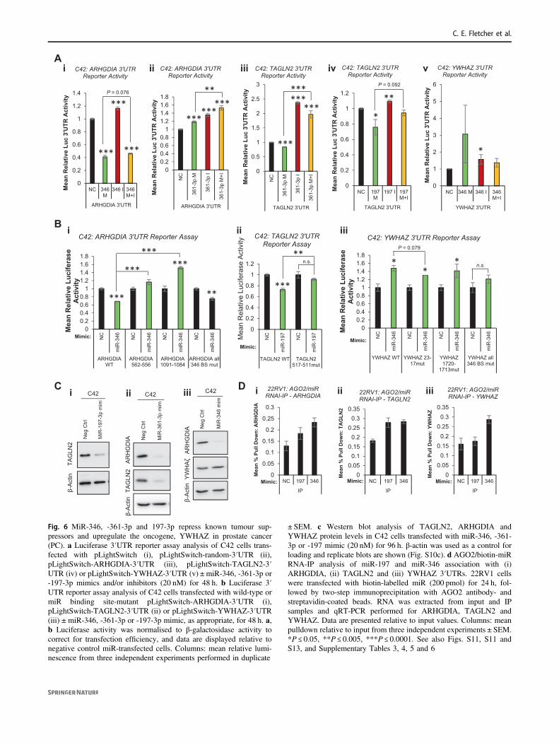
revealed additional roles for these miRs in such processes assignal transduction, cell cycle and DNA replication (Fig.S12), we sought to identify additional relevant targets ofmiR-346, -361-3p and -197. Top AGO-PAR-CLIP targetsidentified in at least three of five PC cell lines are shown(Tables S4, 5 and 6). ARHGDIA, TAGLN2 and YHWAZwere selected for further analysis based on their interactionwith at least one of miR-346, -361-3p or -197 in multiplePC cell lines (Table S7), and also their links to PC pro-gression or AR signalling. The majority of miR bindingsites identified were located within the 3′UTR of thesegenes. ARHGDIA was identified as a target of miR-346 inthree PC cell lines, and as a miR-361-3p target in four lines(Table S7). TAGLN2 3′UTR was identified as a miR-361-3p and -197 target in four cell lines. Interestingly, TAGLN2pseudogene, TAGLN2P1, was also identified as a miR-197target in two PC lines (Table S7). It is tempting to speculatethat TAGLN2 pseudogene could act as a miR sponge toregulate activity of TAGLN2. Finally, miR-346 3′UTRbinding sites were identified in YWHAZ 3′UTR in four PCcell lines, while promoters of YWHAZ pseudogenes,YWHAZP2 and YWHAZP7, were found to contain miR-346 binding sites (Table S7). To validate functional asso-ciation of miRs with identified regions of seed com-plementarity, C42 cells were transfected with reportervectors containing ARHGDIA, TAGLN2 or YWHAZ 3′UTRs downstream of Renilla luciferase, ±miR mimics orinhibitors. MiR-346 significantly reduced ARHGDIA 3′UTR activity, while inhibitor alone significantly increased3′UTR activity by 15% (Fig. 6ai). MiR-346 increasedYWHAZ 3′UTR activity, which was partially rescued byaddition of miR-346 inhibitor (Fig. 6av), but did not sig-nificantly alter activity of empty pLightSwitch, or vectorcontaining random 3′UTR in C42 (Fig. S11Aii) orHEK293T cells (Fig. S11Bi,ii). MiR-361-3p significantlyreduced TAGLN2 3′UTR activity in C42 cells, with anopposing increase in 3′UTR activity observed upon inhi-bitor transfection (Fig. 6aiii), while it enhanced ARHGDIA3′UTR activity (Fig. 6aii). This may be due to off-targeteffects on the vector, as miR-361-3p increased luciferaseactivity both of empty pLightSwitch and vector containing arandom 3′UTR sequence (Fig. S11A). Finally, miR-197mimic significantly reduced 3′UTR activity of TAGLN2,which was significantly opposed through addition ofmiR-197 inhibitor (Fig. 6aiv). Similar results were obtainedupon repetition of experiments in HEK293T cells (Fig.S11biii-iv).
To confirm miR targeting of 3′UTRs through associationwith AGO-PAR-CLIP-identified seed region binding sites,key residues within such seed binding sites were mutated(minimum of three residues within seed-complementaryregion) and 3′UTR luciferase reporter assays performed ±miR mimics. AGO-PAR-CLIP-identified miR-346 seed
Fig. 5 AR-modulatory miRs modulate migration and invasion andinduce a unique EMT progression phenotype through modulation ofsnail-regulatory proteins. a Transwell migration assay analysis of C42cells transfected with miR-361-3p mimic (20 nM) for 48 h prior toseeding onto rat tail collagen-coated transwell membranes for 48 h.Representative image shown in (i), (ii) shows Columns: meanmigrated cell number for three independent experiments performed induplicate normalised to cell number by SRB proliferation assay ±SEM. b Wound healing assay analysis of C42 cells transfected withnegative control or miR-361-3p mimic (20 nM) for 0–72 h. Repre-sentative image shown in (i), in (ii) data are presented as % of day 0wound remaining and represent mean ± SEM for three independentexperiments performed in triplicate (ii). c Matrigel invasion assayanalysis of C42 cells transfected with miR-346, -361-3p or -197-3pmimic (20 nM) for 48 h prior to seeding onto rat tail collagen- and 5 µgmatrigel-coated transwell membranes for 48 h. Columns: meanmigrated cell number for three independent experiments performed induplicate normalised to cell number by SRB proliferation assay ±SEM. d qRT-PCR analysis of Snail (i), ZEB2 (ii) TSPAN-13 (iii) andE-Cadherin (iv) mRNA levels in C42 cells transfected with miR-346,-361-3p or -197-3p mimics (20 nM) for 96 h. L19 was used as anormalisation gene. Columns: mean ± SEM for three independentexperiments performed in triplicate. e Western blot analysis of theepithelial markers β-catenin and E-Cadherin in C42 cells transfectedwith miR-346, -361-3p or -197-3p mimic (20 nM) for 72 h. β-actin wasused as a control for loading. Representative blots of three independentexperiments are shown in (i). Images from replicate experiments maybe found in Fig. S8E. Densitometry was performed using Image Jsoftware and relative protein levels are displayed (ii and Fig. SEii). fWestern blot analysis of mesenchymal marker proteins Snail, N-Cadherin and Vimentin in PC3 cells transfected with miR-346 or -361-3p mimic (20 nM) ± inhibitor (20 nM) for 96 h. β-actin was used as acontrol for loading. Densitometry was performed using Image J soft-ware and relative protein levels are displayed below bands and in Fig.S8F. g Western blot analysis of mesenchymal marker proteins, Snailand Slug, in DU145 cells transfected with miR-346 or -361-3p inhi-bitor (20 nM) ± mimic (20 nM) for 48 h. β-actin was used as a controlfor loading. Representative blots of three independent experiments areshown. Images from replicate experiments may be found in Fig. S9.Densitometry was performed using Image J software and relativeprotein levels are displayed (Fig. S9). h MiR-346, -361-3p and -197-3p prostate cancer (PC) AGO-PAR-CLIP targets were enriched forSnail-regulatory proteins. Interactions and direction of modulation bymiRs is shown. MiR binding sites from AGO-PAR-CLIP are shown inFig. S10A. i Western blot analysis of Snail-regulatory proteins in C42cells transfected with miR-346, -361-3p or -197-3p mimic (20 nM) for96 h. β-actin was used as a control for loading. Representative blots ofthree independent experiments are shown and P-values are indicatedby asterix. Images from replicate experiments may be found in Fig.S10b–d. Densitometry was performed using Image J software andrelative protein levels are displayed (Fig. S10b–d). *P ≤ 0.05, **P ≤0.005, ***P ≤ 0.0001. See also Figs. S8, S9 and S10
Androgen receptor-modulatory microRNAs provide insight into therapy resistance and therapeutic targets. . .
Neg
Ctrl
MiR
-346
mim
AR
HG
DIA
YW
HA
ζ
C42
β-A
ctin
Neg
Ctrl
MiR
-361
-3p
mim
AR
HG
DIA
TAG
LN2
β -A
ctin
C42
Neg
Ctrl
MiR
-197
-3p
mim
TAG
LN2
β-A
ctin
C42C i ii iii
00.20.40.60.8
11.21.41.61.8
NC
miR
-346 NC
miR
-346 NC
miR
-346 NC
miR
-346
ARHGDIAWT
ARHGDIA562-556
ARHGDIA1091-1084
ARHGDIA all346 BS mut
Mea
n R
elat
ive
Luci
fera
se
Act
ivity
C42: ARHGDIA 3'UTR Reporter Assay
***
***
**
***
***
00.20.40.60.8
11.21.41.61.8
NC
miR
-346 NC
miR
-346 NC
miR
-346 NC
miR
-346
YWHAZ WT YWHAZ 23-17mut
YWHAZ1720-
1713mut
YWHAZ all346 BS mut
Mea
n R
elat
ive
Luci
fera
se
Act
ivity
C42: YWHAZ 3'UTR Reporter Assay
**
* n.s.
0
0.05
0.1
0.15
0.2
0.25
0.3
NC 197 346
IP
Mea
n %
Pul
l Dow
n: A
RH
GD
IA
22RV1: AGO2/miR RNAI-IP - ARHGDIA
00.05
0.10.15
0.20.25
0.30.35
NC 197 346
IP
Mea
n %
Pul
l Dow
n: Y
WH
AZ
22RV1: AGO2/miR RNAI-IP - YWHAZ
00.05
0.10.15
0.20.25
0.30.35
NC 197 346
IP
Mea
n %
Pul
l Dow
n: T
AG
LN2
22RV1: AGO2/miR RNAI-IP - TAGLN2
iiiiii
D i ii iii
0
0.2
0.4
0.6
0.8
1
1.2
1.4
NC 346M
346 I 346M+I
ARHGDIA 3'UTR
Mea
n R
elat
ive
Luc
3'U
TR A
ctiv
ityC42: ARHGDIA 3'UTR
Reporter Activity
***
***
***
P = 0.076
00.20.40.60.8
11.21.41.61.8
NC
361-
3p M
361-
3p I
361-
3p M
+I
ARHGDIA 3'UTR
Mea
n R
elat
ive
Luc
3'U
TR A
ctiv
ity
C42: ARHGDIA 3'UTR Reporter Activity
******
*****
0
0.5
1
1.5
2
2.5
3
NC
361-
3p M
361-
3p I
361-
3p M
+I
TAGLN2 3'UTR
Mea
n R
elat
ive
Luc
3'U
TR A
ctiv
ity
C42: TAGLN2 3'UTR Reporter Activity
***
******
***
0
1
2
3
4
5
6
NC 346 M 346 I 346M+I
YWHAZ 3'UTR
Mea
n R
elat
ive
Luc
3'U
TR A
ctiv
ity
C42: YWHAZ 3'UTR Reporter Activity
*
vA
B
i
0
0.2
0.4
0.6
0.8
1
1.2
NC 197M
197 I 197M+I
TAGLN2 3'UTR
Mea
n R
elat
ive
Luc
3'U
TR A
ctiv
ity
C42: TAGLN2 3'UTR Reporter Activity
**
*
P = 0.092
ii iii iv
P = 0.079
Mimic:0
0.2
0.4
0.6
0.8
1
1.2N
C
miR
-197 NC
miR
-197
TAGLN2 WT TAGLN2517-511mut
Mea
n R
elat
ive
Luci
fera
se A
ctiv
ityC42: TAGLN2 3'UTR
Reporter Assay
***
**n.s.
Mimic:Mimic:
Mimic: Mimic: Mimic:
Fig. 6 MiR-346, -361-3p and 197-3p repress known tumour sup-pressors and upregulate the oncogene, YWHAZ in prostate cancer(PC). a Luciferase 3′UTR reporter assay analysis of C42 cells trans-fected with pLightSwitch (i), pLightSwitch-random-3′UTR (ii),pLightSwitch-ARHGDIA-3′UTR (iii), pLightSwitch-TAGLN2-3′UTR (iv) or pLightSwitch-YWHAZ-3′UTR (v) ± miR-346, -361-3p or-197-3p mimics and/or inhibitors (20 nM) for 48 h. b Luciferase 3′UTR reporter assay analysis of C42 cells transfected with wild-type ormiR binding site-mutant pLightSwitch-ARHGDIA-3′UTR (i),pLightSwitch-TAGLN2-3′UTR (ii) or pLightSwitch-YWHAZ-3′UTR(iii)±miR-346, -361-3p or -197-3p mimic, as appropriate, for 48 h. a,b Luciferase activity was normalised to β-galactosidase activity tocorrect for transfection efficiency, and data are displayed relative tonegative control miR-transfected cells. Columns: mean relative lumi-nescence from three independent experiments performed in duplicate
± SEM. c Western blot analysis of TAGLN2, ARHGDIA andYWHAZ protein levels in C42 cells transfected with miR-346, -361-3p or -197 mimic (20 nM) for 96 h. β-actin was used as a control forloading and replicate blots are shown (Fig. S10c). d AGO2/biotin-miRRNA-IP analysis of miR-197 and miR-346 association with (i)ARHGDIA, (ii) TAGLN2 and (iii) YWHAZ 3′UTRs. 22RV1 cellswere transfected with biotin-labelled miR (200 pmol) for 24 h, fol-lowed by two-step immunoprecipitation with AGO2 antibody- andstreptavidin-coated beads. RNA was extracted from input and IPsamples and qRT-PCR performed for ARHGDIA, TAGLN2 andYWHAZ. Data are presented relative to input values. Columns: meanpulldown relative to input from three independent experiments ± SEM.*P ≤ 0.05, **P ≤ 0.005, ***P ≤ 0.0001. See also Figs. S11, S11 andS13, and Supplementary Tables 3, 4, 5 and 6
C. E. Fletcher et al.

binding sites at nucleotides 556–562 and 1084–1091 of theARHGDIA 3′UTR. The more 5’ site shows greater con-servation across mammals than the 3′ site. Mutation of eachsite individually, and of both sites in the same vector, wasperformed. Mutation of either region was able to completelyabolish miR-346 induced loss of ARHGDIA 3′UTR activ-ity. Interestingly, although the extent of miR-346-inducedloss of 3′UTR activity was reduced following mutation ofboth sites, complete rescue of repression was not observed(as seen for individual sites) (Fig. 6bi). One poorly con-served miR-197 binding site was identified by AGO-PAR-CLIP at nucleotides 511–517 of TAGLN2 3′UTR. Mutationof this seed-complementary region significantly abrogatedmiR-197-induced loss of TAGLN2 3′UTR activity (Fig.6bii). Finally, two miR-346 binding sites were identified byAGO-PAR-CLIP at nucleotides 17–23 and 1713–1720 ofYWHAZ 3′UTR, with the proximal site very highly con-served across mammals, and the more distal site relativelypoorly conserved. Mutation of these seed-complementaryregions individually only minimally reduced miR-346-induced increase in YWHAZ 3′UTR activity, although thiseffect was greater for the proximal site (Fig. 6biii). How-ever, mutation of both sites significantly abrogated miR-346-mediated increase in YWHAZ 3′UTR activity(Fig. 6biii).
To investigate whether altered miR target 3′UTRactivity upon miR transfection is translated into alteredprotein levels, Western blots were performed in C42 cellsfollowing transfection of miR-197, -361-3p or -346. Itwas shown that miR-197 significantly reduced levels of itsputative target, TAGLN2, by 56% (Fig. 6ci, Fig. S11Ciii,iv). Similarly, miR-361-3p significantly decreased proteinlevels of TAGLN2 by 70%, and ARHGDIA by 59% (Fig.6cii, Fig. S11Cii, iv). In addition, miR-346 significantlyreduced ARHGDIA protein levels by 74% but increasedYHWAZ protein by 1.2-fold (Fig. 6ciii, Fig. S11Ci, iv).Finally, to confirm physiological association of miRs withtarget transcripts, we performed AGO/biotin-miR RNA-IP. It was found that levels of ARHGDIA transcript aresignificantly enriched in 22RV1 cells following IP withmiR-346 as compared to miR-197 or non-targeting con-trol (Fig. 6di). Although pulldown of TAGLN2 wasgreater in presence of miR-197 compared to non-targetingcontrol, there was no significant enrichment when com-pared to miR-346, which was not identified as targetingTAGNL2 by AGO-PAR-CLIP in 22RV1 cells (Fig. 6dii).In contrast, enrichment of YWHAZ was increased twofoldupon miR-346 transfection, compared to non-targetingcontrol or miR-197 (Fig. 6diii). Taken together, these dataconfirm functional repressive targeting of ARHGDIA bymiR-346 and -361-3p and TAGLN2 by miR-361-3p and-197, and upregulatory targeting of the PC oncogene,YWHAZ, by miR-346.
Additional putative miR-346 targets (IL18, LIF, BTKand TAP1), miR-361-3p targets (ATXN7L3, MAPT andPRMT2) and -197 targets (ACVR1, TSPAN3, TUSC2), asidentified from miRTarBase, were validated in PC cellsthrough a combination of AGO/biotin-miR RNA-IP, qPCRand western blotting (Fig. S13).
siRNA-mediated silencing of ARHGDIA, TAGLN2 andYWHAZ phenocopies effects of MiRs (or MiRinhibition) in PC
If ARHGDIA, TAGLN2 and YWHAZ represent physiolo-gically relevant miR-346, -361-3p and -197 targets in PC,we reasoned that siRNA-mediated silencing of these genesshould phenocopy effects of miR overexpression (or miRinhibition in the case of YHWAZ, since miR-346 increasesYHWAZ 3′UTR activity and protein levels). To addressthis, we first examined AR protein levels in LNCaP cellstransfected with several single siRNAs targeting ARHG-DIA, TAGLN2 or YWHAZ. It was found that bothARHGDIA siRNAs tested reduced ARHGDIA proteinlevels by up to 80%, with AR protein levels concomitantlyincreased by up to 4.5-fold (Fig. 7ai, Fig. S14a). Similarly,TAGLN2 siRNAs reduced TAGLN2 protein levels by up to90% and increased AR protein levels up to 10.5-fold (Fig.7aii, Fig. S14b). Interestingly, YWHAZ siRNAs reducedYWHAZ protein levels by up to 70%, but contrasting effectswere observed on AR protein: where siRNAs efficientlyreduced YWHAZ protein levels (for example, siRNA #8 andpooled siRNAs), AR protein levels were reduced by up to60% (Fig. 7aiii, Fig. S14c), while less efficient silencing ofYWHAZ (for example, by siRNAs #3 and #6) led to a smallincrease in AR protein levels of up to 20%, suggestive ofthreshold effects of YWHAZ on AR protein levels. Thesedata demonstrate that siRNA-mediated silencing ofARHGDIA and TAGLN2 phenocopies miR-346, -361-3pand -197-induced increase in AR protein levels.
In support of the above data, siRNA silencing ofTAGLN2 significantly increased AR mRNA levels andexpression of the AR target gene, PSA, similar to effectsobserved for the TAGLN2-targeting miRs 361-3p and 197(Fig. 7b). As anticipated, siYWHAZ phencopied effects ofmiR-346 inhibition, resulting in significant reduction ofboth AR and PSA mRNA levels (Fig. 7b). siRNA silencingof ARHGDIA did not significantly modulate AR mRNAlevels, but unexpectedly and significantly reduced PSAlevels in LNCaP cells (Fig. 7b). To ascertain whethersiRNA silencing of miR targets can replicate phenotypiceffects of miR manipulation, SRB assays were performed inLNCaP cells following transfection of ARHGDIA,TAGLN2 or YWHAZ siRNAs. It was demonstrated thatknockdown of ARHGDIA increased cell number by 1.3-fold (Fig. 7ci) and that TAGLN2 siRNA enhanced
Androgen receptor-modulatory microRNAs provide insight into therapy resistance and therapeutic targets. . .

proliferation by 1.25-fold (Fig. 7cii). In contrast, knock-down of YWHAZ significantly reduced LNCaP cell pro-liferation by 20% (Fig. 7ciii). Taken together, these datademonstrate that targeting of ARHGDIA, TAGLN2,
YWHAZ are likely to significantly contribute to oncogenicmiR-346, -361-3p and -197-induced phenotypes.
To examine the relevance of AR-modulatory miRexpression and levels of their targets genes to clinical PC
50
100
150
200
250
300
0 2 4 6
Mea
n A
bsor
banc
e (%
Day
0 M
ock)
Time (Days)
LNCaPNCsiARHGDIA
P =
0.0
51
50
100
150
200
250
300
350
0 2 4 6Mea
n A
bsor
banc
e (%
Day
0 M
ock)
Time (Days)
LNCaP
NCsiTAGLN2
P =
0.0
63
50
70
90
110
130
150
170
190
210
0 2 4 6Mea
n A
bsor
banc
e (%
Day
0 M
ock)
Time (Days)
LNCaPNCsiYWHAZ
*
Cii iiii
00.20.40.60.8
11.21.41.6
Mea
n A
R/L
19 R
elat
ive
Expr
essi
on
LNCaP: AR
***
00.20.40.60.8
11.21.41.6
Mea
n PS
A/L
19 R
elat
ive
Expr
essi
onLNCaP: PSA
**
*** *
Bi ii
TAGLN2
β-Actin
1 10 1 10
NTC #8 #9
LNCaP: TAGLN2 siRNA
1 10
AR
iiA
i1 10 1 10 1 10
NTC #2 #3
LNCaP: ARHGDIA siRNA
ARHGDIA
β-Actin
AR
1 10 1 10 1 10 1 10 1 10
NTC #3 #6 #7 #8 Pool
LNCaP: YWHAZ siRNA
YWHAZ
β-actin
1 10
AR
iii
0
1
2
3
4
5
Exp
ress
ion
(log2
)
MiR-361-3p
0
1
2
3
4
5
Exp
ress
ion
(log2
)
MiR-346*
0123456
Exp
ress
ion
(log2
)
MiR-197
*
02468
101214
Normal Cancer
Expr
essio
n (log
2)
TAGLN2p=0.037
02468
101214
Normal Cancer
Expr
essio
n (log
2)
YWHAZp=0.082
0
2
4
6
8
Normal Cancer
Expr
essio
n (log
2)
ARHGDIAp=0.014
P = 1.118E-17
16.0
15.0
14.0
13.0
12.0
11.0
10.0
YW
HA
Z m
RN
A
D i ii iiiE i ii iii
F iii
No LN Mets
LN Mets
1.0
0
-1.0
-2.0
-3.0
-4.0
-5.0
miR
-346
H
Veh Enza Veh Enza Veh Enza
10d treatment:
P = 0.023 P = 0.0049
Veh Enza Veh Enza Veh Enza
Treatment to 1200 mm , 22-46d:
i
ii
6 7 8 9 10Gleason Grade
G i ii iiiP = 0.03 P = 0.003P = 5x10
C. E. Fletcher et al.

development and progression, bioinformatic analyses ofpublically available expression data sets were performed.Upon examination of AR-modulatory miR levels in fresh-frozen tissue from five PCs and six normal prostates(GSE34932), it was found that miR-197 was significantlydecreased in cancer vs normal, while miR-361-3p isunchanged and miR-346 significantly increased (Fig. 7d).TAGLN2 mRNA levels were significantly lower in cancervs normal and ARHGDIA and YWHAZ levels elevatedsignificantly and non-significantly, respectively, in the samedata set (Fig. 7e). In analysis of TCGA-PRAD data from333 primary PCs, increased YWHAZ transcript correlatedwith Gleason grade in a highly-significant manner (Fig. 7fi),and was higher in patients with lymph node metastases,biochemical recurrence and in non-organ-confined disease(Fig. S15). No such relationships were identified forTAGLN2 or ARHGDIA. In the same data set, miR-346, butnot -361-3p nor -197, was significantly elevated in lymphnode-positive disease (Fig. 7fii). As anticipated, ARHGDIAexpression inversely correlated with its targeting miRs,miR-346 and miR-361-3p, in the TCGA-PRAD data set(Fig. S16a, b). However, miR-361-3p was found to posi-tively correlate with its target, TAGLN2 (Fig. S16c), andmiR-346 negatively correlated with its positively regulated
target, YWHAZ (Fig. S16d). When linear regression ana-lysis was conducted to assess correlation between ARactivity score and miR targets in CRPC patient tumours(n= 122) from SU2C [32], a significant positive correlationof ARHGDIA expression with AR activity was found (P=5 × 10⁻⁸) (Fig. 7gi), while TAGLN2 is significantly nega-tively correlated (P= 0.03) (Fig. 7gii). Similarly, YHWAZis negatively associated with AR activity, although samplesseparate into two populations according to YWHAZexpression, and both correlate positively with AR activitywhen assessed individually (Fig. 7giii). In the same data set,patients with high levels of miR-197 showed a near-significant (P= 0.0501) decreased survival compared tomiR-197 low patients (Fig. S16e). Neither miR-346 (Fig.S16f) nor miR-361-3p (alone, Fig. S16g, or combined withmiR-197, not shown) correlated with survival in TCGA-PRAD. Of note, MiR-361-3p significantly correlated withAR activity in this cohort (Fig. S16h), albeit with an r2
value of 0.13.We next investigated the effect of treatment on AR-
modulatory miRs in patient-derived models. CP50 PDXwas derived from a CRPC patient biopsy with chromosome8 gain and AR amplification who had progressed throughall standard of care therapies (Welti et al; submitted toClinical Cancer Research). Short-term (10 days) treatmentwith enzalutamide (10 mg/kg; n= 3) did not significantlyalter miR levels compared to vehicle treatment (KeptoseHPB; n= 3) (Fig. 7Hi). However, long-term treatment(mean 35.5 days) with enzalutamide (n= 6) resulted insignificant increases in miR-197 and -361-3p levels ascompared to vehicle (n= 6) (Fig. 7hii). MIR-346 levelswere not altered (Fig. 7hii).
Taken together, these data confirm that AR-modulatorymiRs and their biologically relevant PC targets are altered incancer vs normal tissue, correlate with disease progressionand AR activity, and are altered in response to hormonetherapy in a CRPC patient tumour.
Discussion
In PC that is refractory to conventional LBD-targeting anti-androgens, AR-targeting miRs may represent promisingtherapeutics or drug targets. A handful of studies havedemonstrated direct AR targeting by miRs, however withlittle consensus. Much remains to be learnt regarding theroles of miRs and the AR 3′UTR in disease progression, theability of miRs to integrate AR signalling with other path-ways, and relationships between AR and other targets ofAR 3′UTR-associating miRs. In an effort to increase con-sensus on AR-modulatory miRs, we performed a miRinhibitor library screen in LNCaP and C42 PCa cells stablyexpressing a luciferase-based AR activity reporter.
Fig. 7 AR-modulatory miR target siRNA silencing phenocopies miRs.a Western blot analysis of ARHGDIA, TAGLN2, YWHAZ and ARprotein levels in LNCaP cells transfected with siRNAs (1 or 10 nM)targeting (i) YWHAZ, (ii) TAGLN2 or (iii) ARHGDIA for 72 h. Aminimum of two different siRNAs were used for each gene. β-actinwas used as a control for loading. b qRT-PCR analysis of (i) AR or (ii)PSA mRNA levels in LNCaP cells transfected with ARHGDIA,TAGLN2 or YWHAZ siRNAs (10 nM) for 48 h. L19 was used as anormalisation gene. Columns: mean ± SEM for three independentexperiments performed in triplicate. c SRB proliferation assay analysisof LNCaP cells transfected with siRNA targeting ARHGDIA,TAGLN2 or YWHAZ (10 nM) for 6 days. Data are presented relativeto absorbance at day 0. Points: mean absorbance at 492 nm for threeindependent experiments performed in quadruplicate ± SEM. d, eMiR-197 (di), miR-346 (dii), miR-361-3p levels (diii), TAGLN2 (ei),ARHGDIA (eii) and YWHAZ (eiii) RNA levels in fresh-frozen tissuefrom five PCs and six normal prostates (GSE34932). f YWHAZmRNA levels in PC tumours of Gleason Grades 6–10 (i) and miR-346levels in PC tumours of lymph node-positive and –negative patientsfrom the TCGA-PRAD data set of 333 primary prostate cancers (PCs).g Linear regression analysis of correlation between AR activity andARHGDIA (i), TAGLN2 (ii) and YWHAZ (iii) levels in CRPC patienttumours (n= 122, transcriptome data from SU2C-PCF). AR activityscore is an accumulation measurement of AR pathway activity basedon 43 genes whose expression level is regulated by AR in PC cell lines[56] and metastatic PC [57]. h qRT-PCR analysis of miR-197, miR-346 and -361-3p levels in patient-derived xenografts implanted intonude mice and derived from a single biopsy from an AR-amplified, C-MYC-gain CRPC patient. Enzalutamide (10 mg/kg) or vehicle treat-ment was initiated when xenografts reached 300 mm3, and mice killedat day 10 (i) or when tumours reached 100 mm3 (ii) (mean treatmentduration= 35.5 days). (i) n= 3 per treatment group, (ii) n= 6 pertreatment group. Data were normalised to U6 and SNORD48 levels.*P ≤ 0.05, **P ≤ 0.005, ***P ≤ 0.0001. See also Figs. S14, S15, S16
Androgen receptor-modulatory microRNAs provide insight into therapy resistance and therapeutic targets. . .

Although little agreement was observed between top hitsfrom our screen and that of Kumar et al. [13], which used amiR mimic library, 22 miR inhibitors that increased ARactivity above our stringent threshold in LNCaP and/or C42cells agreed with their screen in that the corresponding miRmimic significantly reduced LNCaP cell viability, anddecreased PSA and/or AR protein levels/ activity in at leasttwo of three PC cell lines tested (LNCaP, VCaP, LAPC4).Four additional miRs, inhibitors of which decreased ARactivity in our screen, were likewise identified by thesecriteria in the Kumar study. Of thirteen miRs found byÖstling et al. to significantly reduce AR 3′-UTR activity,although corresponding inhibitors did not found to mod-ulate AR activity above our threshold, there was someagreement for seven, as we found that miR-421 and -449aincreased AR activity in C42 cells; miR-135b, -634 and-654-5p increased AR activity in LNCaP; while miR-34cand miR-9 increased AR activity in both lines. We wereunable to validate previously reported reduction of ARlevels, in various cell lines, by miR-185 [16], miR-34a [33],or miR-488-5p [13, 19]. We identified agreement with ourscreening data (LNCaP and/or C42) for 26 of 64 miRsidentified to modulate PSA protein levels in LNCaP andLAPC4 cells in a miR mimic screen by Larne et al. [34],although only three had B Score > 6 in our screen. There aremany likely reasons for lack of agreement between studies:differences in library types (mimic or inhibitor), inhibitor/mimic concentrations, cell lines with different transfectionefficiencies and endogenous miR levels, end-point assays,and statistical scores and thresholds for analysis.
Upon validation of screen hits, we demonstrated thatmiR-346, -361-3p and -197 inhibitors markedly reducedAR activity in both LNCaP and C42 cells, with oppositeeffects observed for corresponding miR mimics. In corro-boration we also showed that the inhibitors significantlyreduced AR mRNA and protein levels, and target geneexpression, effects rescued by addition of miR mimics.Since these effects were observed after only 24 h, it is likelythat these miRs directly regulate AR through activatoryassociation with the 3′UTR, in contrast to the classicalmodel of target repression by miRs. In support of this, weidentified binding sites for all three miRs in the extended6.9 kb AR 3′UTR, which has been described in PC cells bya number of studies [15, 27], but is not used by miR bindingprediction tools, which use the UCSC genome browser andGenbank-defined 436 nt AR 3′UTR sequence(NM_000044). This could potentially result in exclusion ofcritical but uncharacterised regulators of AR activity fromprevious investigations. Direct miR-346, -361-3p and -197upregulation of AR 3′UTR activity was confirmed throughuse of AR 3′UTR reporters in combination with miRmimics, which significantly increased AR 3′UTR activity,with specificity demonstrated through additional
transfection of miR inhibitors and miR seed binding sitemutagenesis. Such miR-mediated AR upregulation couldprovide a mechanism for maintenance of AR activity incastrate conditions. Further, miR-346 and -197 preventedActD-induced loss of AR transcript, so may additionallyrepress AR mRNA turnover. This study is not the first toreport miR-346 upregulation of target genes: miR-346increases AGO2 mRNA and protein levels in cervical,gastric and colorectal cancer cell lines through formation ofa miR-346 mid-region ‘bulge’ upon association with AGO23′UTR, which in turn increases recruitment of GRSF1 [35].The same authors showed that a similar mechanismincreases recruitment of miR-346-bound hTERT mRNA toribosomes to enhance translation in a GRSF1-dependentmanner [36]. It will be interesting to assess whether miR-346:AR 3′UTR association results in similar miR loopingand recruitment of RNA binding proteins.
The identification of an extended AR 3′-UTR in PCa celllines is interesting in the context of a proposed novelmechanism for altered gene regulation, whereby dynamic 3′UTR shortening prevents miR targeting. In cancer cells,mRNAs with shortened 3′-UTRs produced tenfold moreprotein, in part due to loss of miR-mediated repression [37].While it has been proposed that active AR 3′-UTR short-ening represents a further mechanism of PCa progression[38], permitting AR to evade regulation by tumour-suppressive miRs, such miR-34a and miR-34c [15], thereis currently no evidence for reduced miR binding andconsequently higher AR protein levels following AR 3′-UTR shortening. Further studies are required to assess itspotential role in cancer progression, especially given ourdata showing AR 3′UTR as a region positively regulated byoncogenic miRs.
Expression of constitutively active AR variants lackingthe LBD represent a potential mechanism of cell survival inCRPC. AR-v7 has a distinct 3′UTR sequence from WT-AR, while ARv567es 3′UTR has considerable overlap withWT-AR 3′UTR but is extended at the 3′ end [39]. Wetherefore hypothesised that ARv567es would be subject toregulation through the same miR-346 and -361-3p bindingsites as WT-AR, while AR-v7 would not. Indeed, weobserved decreased ARv567es mRNA and protein levelsfollowing transfection of miR-346 and miR-361-3p inhibi-tors in 22RV1 and CWR-R1-D567es cells. AR-v7 mRNAwas significantly decreased following miR-361-3p inhibitortransfection in 22RV1 and C42 cells, possibly due to anAR-v7/9/4-unique miR-361-3p 7mer site between 981-987nt of AR-v7 3′UTR (Table S3). AR-v7 does not containany predicted miR-346 binding sites however, and theunexpected decrease in its expression in both 22RV1 andC42 cells following miR-346 inhibitor transfection (Fig.S4c) may be attributable to feedback regulation from loss ofWT-AR. Protein levels of AR variants of size consistent
C. E. Fletcher et al.

with AR-v1, -v4 and -v7 were markedly downregulatedupon miR-361-3p inhibitor treatment in 22RV1 and CWR-R1-AD1 cells. Together, these data indicate that modulationof miR-346 and miR-361-3p can alter levels of con-stitutively active AR variants as well as WT/full-length AR.
Given their effects on AR activity, we examined phe-notypic consequences using proliferation assays and foundthat miR-346 and -361-3p inhibitors reduce proliferation,supporting a tumour-promoting/oncogenic effect of thecorresponding miRs. In AR-positive cells the inhibitorsshowed additive effects with AR silencing and Enzaluta-mide treatment, conversely, mimics increased PC cell pro-liferation and abrogated AR siRNA- or Enza-induced lossof cell proliferation. This extended to AR-variant-drivenlines, and the enhanced effects of miR-346 inhibition onAR-variant-driven vs parental PC cell growth likely reflectthe presence of an additional variant-specific miR-346 seedcomplementarity site in v567es 3′UTR. However, miRshave potentially dozens of target mRNAs, and the pheno-typic consequences of miR inhibition are likely to be bothAR-mediated and non-AR-mediated Indeed, miR-346,-361-3p and -197 inhibitors were able significantly reducegrowth of both AR-positive and AR-negative cells in adose-dependent manner. Looking into potential modes ofaction by which miR-346, -361-3p and -197 inhibitorsreduce proliferation, we found that all three significantlyincreased apoptosis in LNCaP cells, and enhanced celldeath induced by AR inhibition//ablation, while the mimicsmostly abrogated this. The observation that, contrary toexpectation, miR-346 mimic showed similar effects toinhibitor in increasing Enza-induced apoptosis may beattributable to very large (280,000-fold) upregulation ofmiR-346 upon transfection of mimic at 20 nM final con-centration (Fig. S1Ai), which may generate toxicity. Indeed,10 nM miR-346 mimic was shown to repress C42 andLNCaP proliferation, and cells showed signs of toxicity atthis concentration (Fig. S6b). All three inhibitors showedadditive effects with AR siRNA on AR target geneexpression, while corresponding mimics rescued ARsiRNA-mediated loss of AR, PSA and KLK2 expression.However, the observation that they could not completelyrescue effects of AR siRNA on proliferation, apoptosis andAR target gene expression supports that these miRs alsofunction, in part, through non-AR pathways. This is rein-forced by the observation that miR-346 and -361-3p inhi-bition significantly reduces proliferation of AR-negativeDU145 cells. Together, these data suggest that miR-346,miR-361-3p and miR-197 can act as oncomiRs to increasePC proliferation and inhibit apoptosis, partly, but notwholly, through regulation of AR signalling.
As further evidence of its oncomiR function, miR-361-3p significantly increases migration and invasion of C42 PCcells. It is thus interesting that significant downregulation of
miR-361-3p was observed in prostatic secretions of 23 (lowgrade) PC patients compared to 25 BPH controls [40]. Itwould be interesting to know whether observed miR dys-regulation was also identified in tissue, urine or plasma, andwhether the correlation is retained in men with higher-gradetumours. Conceivably, these data may suggest activeretention of oncomiR-361-3p within PC cells, to drive EMTand migration/invasion. Indeed, miR-361-3p, -346 and -197all reduced levels of epithelial markers in C42 cells (whichhave epithelial morphology), while miR-346 and -361-3pinhibitors decreased protein levels of mesenchymal markersin the more aggressive PC3 and DU145 cells. The exceptionwas Snail, a mesenchymal marker, which was markedlyincreased. We hypothesised that miR-346, -361-3p and -197modify levels of Snail-regulatory proteins, and analysis ofAGO-PAR-CLIP data confirmed these to be enriched intheir targets: Snail repressors GSK3β and PKCµ appear tocontain activatory miR binding sites (levels are increased inpresence of targeting miRs), while Snail activators MDM2,PARP1 and LATS2 contain repressive miR binding sites.An alternative, possibly complementary, explanation is thatthe miRs induce intermediate EMT ‘plasticity’ that pro-motes migration and invasion in a Snail-independent man-ner. Evidence for such Snail-independent EMT has beensuggested by a number of studies: p38 MAP kinase and p38interacting protein (p38IP) are required for E-Cadherindownregulation during EMT in mouse embryo gastrulation,in a Snail-independent manner [41]; Snail and Slug inducedistinct EMT programmes in mammary stem cells ascompared to tumour initiating cells [42]; and high levels ofthe E-Cadherin repressors, ZEB1 and ZEB2, which couldinitiate EMT independently of Snail, were found in recur-rent oral squamous cell cancer as compared to primarytumours, suggestive of non-canonical EMT progression inadvanced cancer [43].
To identify additional PC-relevant targets of these miRsthat mediate the non-AR-mediated effects, we exploitedAGO-PAR-CLIP to identify miR:target association in a panelof PC cell lines. We focussed on genes previously implicatedin PC and with multiple 3′UTR binding sites for more thanone of miR-346, 361-3p and -197, selecting YWHAZ,ARHGDIA and TAGLN2 for further analysis. Our dataconfirm ARHGDIA and TAGLN2 as bona fide AR-modulatory miR targets, downregulated by miR-346 and-361-3p, and miR-361-3p and -197, respectively. ARHGDIAand TAGLN2 have both previously been shown to repressAR transcriptional activity, and to have tumour-suppressiveroles [44–47]; their downregulation has clear implications forPC development, and further supports oncogenic activity ofmiR-346, 361-3p and -197. Conversely, the androgen-regulated PC oncogene YWHAZ was upregulated by miR-346, likely through association between the miR andnucleotides 17–23 of YWHAZ 3′UTR. YWHAZ has been
Androgen receptor-modulatory microRNAs provide insight into therapy resistance and therapeutic targets. . .
shown to directly associate with AR, to promote proliferation,migration, survival and resistance to apoptosis in PC cells[48], to increase in PC patient tumours as compared to benigntissue, and to positively correlate with presence of metastasesin clinical tumour samples [49]. Thus, miR-346-mediatedYWHAZ upregulation could enhance PC progression.
To confirm biological relevance of miR targeting ofARHGDIA, TAGLN2 and YWHAZ, we examined the abilityof siRNAs against these genes to phenocopy effects of miR-346, -361-3p and -197 in PC cells. Silencing ARHGDIA orTAGLN2 resulted in increased AR protein levels, target geneexpression and PC cell growth, similarly to miR-346, -361-3por -197 mimics. Conversely, and as anticipated, siYWHAZphenocopied miR-346 inhibition reducing AR protein levelsand represseing PC cell growth. We hypothesise that AR-modulatory miRs may have important functions at differentstages of PC: miR-346 levels are higher in cancerous vsnormal prostate tissue and elevated in tumours of patientswith lymph node metastases compared to those without, whilenot altered in Enza-treated CRPC patient xenografts. In con-trast, miR-197 is reduced and -361-3p unchanged in cancer-ous vs normal prostate, but both were significantly elevated inEnza-treated CRPC xenografts, although only after long-termtreatment. This suggests that while miR-346 may contributeto progression of early-stage PC (through upregulation ofYWHAZ and AR activity), miR-197 and -361-3p may act atthe castrate-resistant stage of the disease to promote ormaintain resistance to AR-targetting therapies, throughdownregulation of tumour-suppressive targets and stabilisa-tion of AR transcript.
Taken together, these data identify miR-346, miR-361-3p and miR-197 as AR-modulatory miRs in PC thatassociate with an extended form of the AR 3′UTR toincrease transcript stability and AR activity. AR-modulatorymiRs modulate apoptosis, proliferation, EMT, migrationand invasion of PC cells, and their inhibition shows additiveeffects with anti-androgens, highlighting the potential forcombination approaches for the treatment of PC. SilencingPC-relevant miR targets phenocopies effects of these miRs,supporting their physiological relevance.
Materials and methods
Mammalian cell culture
Cells were maintained at 37 °C in 5% CO2. PC3, 22RV1,LNCaP, C42, DU145 and HEK293T cells were maintainedand passaged in RPMI-1640 or Dulbecco’s ModifiedEagle’s Medium (DMEM – HEK293T only) (Sigma) sup-plemented with 10% fetal bovine serum (FBS), 100 U/mlpenicillin, 100 µg/ml streptomycin and 2mM L-glutamine(Sigma). LNCaP/ARE, C42/ARE and DU145/ARE cells
were maintained and passaged as above but with 10%tetracycline-free FBS, 12 µg/ml blasticidin and 500 µg/mlG418. LNCaP/ARsiRNA cells (a kind gift of Paul Rennie,Vancouver Prostate Centre, Canada) were maintained asabove, but with 2.4 µg/ml blasticidin and 1 µg/ml pur-omycin. Parental 22RV1 cells and AR WT-knock outderivatives, and parental CWR-R1-AD1 cells and CWR-R1-D567es derivative lacking WT-AR but expressing AR-v567es were kind gifts from Luke Gaughan, NewcastleUniversity, UK, and Scott Dehm, University of Minnesota,USA, respectively [50]. Cell lines were routinely tested forMycoplasma contamination on a monthly basis
Plasmid stocks
pMiR-Report-AR 6.9 kb 3′UTR reporter plasmids 1–7,representing seven different regions of the 6.9 kb AR 3′UTR (Fig. 2d) were a kind gift from Dr Shawn Lupold(36).pLightSwitch-Empty, pLightSwitch-Random#2,pLightSwitch-ARHGDIA 3′UTR, pLightSwitch-TAGLN23′UTR and pLightSwitch-YWHAZ 3′UTR were purchasedfrom OriGene. Putative miR-346, -361-3p and -197-3pbinding sites were mutated in the above vectors by site-directed mutagenesis using the QuikChange Lightning Site-Directed Mutagenesis Kit (Stratagene).
High-throughput LNA microRNA inhibitor screening
C42/ARE and LNCaP/ARE cells were seeded into thirteen96-well, white-walled, flat-bottomed cell culture plates percell line in hormone-depleted, antibiotic-free RPMI medium48 h prior to transfection. Seeding densities were 3000 cellsper well (C42/ARE) and 4000 cells per well (LNCaP/ARE).8 h prior to transfection, cells were supplemented with0.1 nM Mibolerone. Cells were transfected with LNAMicroRNA Inhibitor Library (Exiqon) to a final con-centration of 20 nM using Lipofectamine RNAiMax for48 h. Controls were AllStars Hs Cell Death Control siRNA(Qiagen), AllStars Negative Control siRNA (Qiagen) andLNA Negative Control A (Exiqon).
MicroRNA inhibitor/mimic, siRNA transfection
MiRCURY LNA microRNA inhibitors and mimics (Exi-qon) and/or miRidian microRNA mimics (GE healthcare)were transfected into cells at final concentration of 0–30 nMin antibiotic-free conditions using Lipofectamine RNAiMaxas per the manufacturer’s recommendations. siRNAs (Flex-itube, Qiagen) were transfected as above, final concentration10 nM. Co-transfection of plasmids and siRNAs or micro-RNA inhbitors/mimics was performed using JetPrimetransfection reagent (Polyplus) according to the manu-facturer’s protocol.
C. E. Fletcher et al.

RNA extraction
RNA was extracted using Trizol according to the manu-facturer’s specifications. For RNA extraction from PDXtissue, samples were subjected to homogenisation usingPrecellys24 Homogeniser in the presence of Trizol.
MiR and mRNA quantitative real-time PCR
Mature miR expression was quantified by quantitative real-time RT-PCR using TaqMan microRNA assays and Taq-Man Universal PCR Master Mix (Applied Biosystems)according to the manufacturer’s protocol. For detection ofmRNAs, cDNA was prepared from 500 ng total RNA usingPrecision qScript Reverse Transcription kit (PrimerDesign)and oligo d(T) primers. cDNAs were amplified using 2×Fast SYBR Green Master Mix (Applied Biosystems) and250 nM forward and reverse primers (Table S1). All datawere analysed using the ΔΔCt method, with U18 and L19as endogenous references for miR and mRNA levelsrespectively, using DMSO/ethanol-treated samples as cali-brators where appropriate.
3′UTR reporter assays
HEK293T or C42 cells seeded in 24-well plates were co-transfected with 400 ng pMiR-Report AR 6.9 kb 3′UTR#1–7, pLightSwitch-Empty, pLightSwitch-Random#2,pLightSwitch-ARHGDIA 3′UTR, pLightSwitch-TAGLN23′UTR, or pLightSwitch-YWHAZ 3′UTR (Origene)alongside 50 ng pdm-LacZ (β-galactosidase expressionvector) and 0–20 nM final concentration non-targeting,miR-346, miR-361-3p or miR-197 mimic and/or inhibitors(see above) using JetPrime Transfection reagent for 48 h.For AR 3′UTR reporter assays, luciferase assays wereperformed using Luclite assay as below. For pLightSwitchplasmids, LightSwitch luciferase assay (Active Motif) wasused according to the manufacturer’s protocol. For bothassays, luciferase activity was normalised for transfectionefficiency using the Galacton kit (Tropix) as previouslydescribed [51].
Luciferase assays
Luciferase assays were performed using the Luclite assay(Packard, USA). Cell medium was aspirated from wellsand 30 µl (96-well plates) or 60 µl (24-well plates) ofReporter Lysis Buffer (Promega) added per well. Plateswere stored at −80 °C for a minimum of 15 min. Plateswere thawed and 30 µl luciferin substrate added per well(96-well plates) or 20ul of cell lysate dispensed in duplicateinto white-walled 96-well plates and 20ul luciferin sub-strate added per well (24-well plates). Plates were
incubated for 15 min in the dark prior to luminescencereading using the GloMax system (Promega) or Victorluminescence counter. Luciferase activity was normalisedfor cell number by Bradford assay for protein content,where appropriate.
Actinomycin D transcriptional inhibitionexperiments
C42 cells in 6-well plates previously transfected with miRmimics for 24 h were treated with Actinomycin D (1 µg/ml)for 4 h prior to harvesting and RNA extraction.
Cell Lysis, western blotting and antibodies
Cells were lysed and protein extracted as described [26].Proteins were resolved by 8–12% SDS-polyacrylamidegel electrophoresis and electroblotted to nitrocellulosemembrane (Bio-Rad). After blocking (5% non-fat driedmilk powder in 0.05% Tween-20 in 1x TBS) for 40 min,membrane was incubated with mouse anti-androgenreceptor mAb (ThermoFisher, MA513426), rabbit anti-beta-catenin pAb (Cell Signalling Technology, 8480),rabbit anti-E-Cadherin pAb (Cell Signalling Technology,3195), rabbit anti-Snail pAb (Cell Signalling Technology,3879), rabbit anti-N-Cadherin pAb (Cell SignallingTechnology, 13116), rabbit anti-Vimentin pAb (CellSignalling Technology, 5741), rabbit anti-Slug pAb (CellSignalling Technology, 9585), rabbit anti-MDM2 mAb(Abcam, ab178938), mouse anti-PARP1 mAb (Santa CruzBiotechnology, sc-8007), rabbit anti-PKCµ mAb (Abcam,ab108963), mouse anti-GSK3β mAb (Abcam, ab93926),rabbit anti-LATS2 pAb (Abcam, ab70565), rabbit anti-ARHGDIA pAb (Santa Cruz Biotechnology, sc-360),mouse anti-TAGLN2 mAb (Santa Cruz Biotechnology,sc-373928), rabbit anti-YWHAζ pAb (Abcam, ab51129),mouse anti-BTK mAb (R+D Systems, MAB5807),mouse anti-LIF mAb (Abcam, ab34427), mouse anti-β-tubulin mAb (Sigma, T4126), or mouse anti-β-actinmAb (Abcam, ab6276) for 1 h and visualised using goatanti-mouse, goat anti-rabbit, goat anti-rat or rat anti-goatIgG-HRP as appropriate. Detection was by LuminataForte HRP substrate (Millipore).
Sulphorhodamine B (SRB) Assay
Cell number was assayed using the SRB assay as previouslydescribed [52].
Luminescent caspase 3/7 activation assay
Caspase assays were performed using the Caspase-Glo 3/7 kit(Promega) according to the manufacturer’s instructions, 48 h
Androgen receptor-modulatory microRNAs provide insight into therapy resistance and therapeutic targets. . .
post-transfection with miR mimics or inhibitors. Luminescentsignal was detected using a Victor luminometer.
Wound healing assay
Cells seeded in 6-well plates were transfected with miRmimics as described. 48 h post-transfection, an artificial‘wound’ was created in the cell monolayer using a 200 µlpipette. Medium was replaced with antibiotic-free, phenolred-free RPMI supplemented with 5% sFCS to minimiseproliferation. Wounds were imaged at 0 and 24 h andwound area quantified using Image J.
Transwell migration/matrigel invasion assays
Cells seeded in 6-well plates were transfected with miRmimics as described. 48 h post-transfection, outer surfaces of8 µm pore transwell inserts were pre-coated with 2.5 μg rattail collagen type I in cold, non-supplemented, phenol red-free, RPMI and air-dried for 2 h and placed in 24-wellcompanion plates. Transfected cells were harvested andseeded (3 × 10⁴ cells per condition) onto collagen-coatedtranswell membranes in 100 μl serum-free RPMI in dupli-cate. Lower chamber of companion plate was filled with600 μl antibiotic-free complete growth medium as a che-moattractant. Cells were incubated for a further 48 h at37 °C. Inner surfaces of transwells were scrapped with aPBS-soaked cotton bud to remove non-migrated cells.Migrated cells were fixed with 4% paraformaldehyde in PBSfor 10 min at RT, washed with PBS and stained with 0.4%crystal violet in 20% methanol for 45 min at RT. Membraneswere washed in ddH2O and stored in PBS at 4 °C. Aminimum of five fields per transwell membrane were imagedat ×10 magnification using the digital inverted microscopeEVOS Cell Imaging System (Life Technologies, CA, USA).Total number of migrated cells was manually counted foreach imaged field, the mean was calculated, and normalisedto total protein content by SRB assay. Invasion assays wereperformed as above but with additional coating of innertranswell surfaces with 5 µg Matrigel.
Biotinylated miR/AGO2 pulldown for miR targetidentification
Cells seeded in 2 × 10 cm dishes per condition were trans-fected with 200 pmol biotinylated miR mimic (Exiqon) perplate for 24 h prior to harvesting, pooling and lysis in 500 µlper pellet of ice-cold hypotonic lysis buffer (10 mM KCl,1.5 mM MgCl2, 10 mM Tris-HCl pH7.5, 5 mM DTT, 0.5%NP40, 60 U/ml SUPERaseIn (Ambion), 5 µl/ml proteaseinhibitor cocktail). Lysates were passed through needles(2 × 23 G, 2 × 27 G) to ensure efficient lysis. Lysates werecleared by centrifugation at 5000×g for 5 min at 4 °C. 50 µl
of cleared lysate was retained for input RNA extraction.10 µg AGO2 antibody (Abcam ab57113) diluted in 200 µlhypotonic lysis buffer was conjugated to 50 µl DynabeadsProtein G (Invitrogen) per condition by incubation at roomtemperature for 20 min with rotation. AGO2-conjugatedbeads were washed 3× in hypotonic lysis buffer prior toincubation with lysate (500 µl) at room temperature for30 min with rotation. Beads were then washed 3× withhypotonic lysis buffer and eluted in 50 µl elution buffer(Dynabeads Protein G IP kit, Invitrogen). NaCl was addedto eluted immunoprecipitated AGO2 complex to a finalconcentration of 1M. Reconstituted MyOne C1 streptavidinbeads (LifeTech) were washed 2× in 0.1M NaOH, 0.05MNaCl, 1× in 0.1 M NaCl and 1× in hypotonic lysis bufferand blocked with 1 µg/µl BSA and 1 µg/µl yeast tRNA inhypotonic lysis buffer for 3 h at 4 °C with rotation. Blockedbeads were washed 3× with hypotonic lysis buffer prior toincubation with immunoprecipitated AGO2 complex for16 h at 4 °C. Beads were washed 3× with hypotonic lysisbuffer and RNA extracted from both streptavidin beads andinput lysates using 300 µl Trizol LS according to the man-ufacturer’s protocol. Isolated RNA was reverse transcribedand qPCR performed as described. RNase-free conditionswere maintained throughout.
AGO-PAR-CLIP-seq
AGO-PAR-CLIP-seq data represented is extracted fromprior published data [31]. Methods are as previously pub-lished [31].
Patient-derived xenograft (PDX) development CP50
The development of CP50 PDX has been described recently(Welti et al; submitted to Clinical Cancer Research).Briefly, a tissue biopsy from a patient with CRPC who hadprogressed through all standard of care treatments (doc-etaxel, cabazitaxel, enzalutamide and abiraterone) wasimplanted into 2 NOD scid gamma (NSG) (JAX Mice) malemice. Next generation sequencing of the parental lymphnode biopsy and generated CP50 PDX demonstrated com-parable copy number analysis, with chromosome 8 gain andandrogen receptor amplification (Welti et al; submitted toClinical Cancer Research). To develop a castrate line,CP50 PDX was castrated when tumours reached 300 to400 mm3, and passaged again at a size of 1200 mm3 intocastrate mice, this process was repeated for ongoing main-tenance. The effect of enzalutamide on castrate CP50 PDXmiR levels was determined at two time points. For bothexperiments, animals were treated daily by intraperitonealinjection with enzalutamide (10 mg/kg) or vehicle (KeptoseHPB) when implanted tumours reached a size of 300 to400 mm3. Six (n= 3 enzalutamide and n= 3 vehicle) were
C. E. Fletcher et al.

treated for 10 days (short term) and 12 (n= 6 enzalutamideand n= 6 vehicle) were treated until tumours reached a size1200 mm3 (long term: 22–48 days).
Statistical analyses
Normally distributed continuous variables were assessed byStudent’s t-test. P ≤ 0.05 was interpreted to denote statisticalsignificance. Data from miR inhibitor library screening wereanalysed using the B score method [53]. The simplest andmost widely used statistical scoring method in high-contentscreening is the Z score, however, B score was used here asit additionally adjusts for positional effects within each 96-well plate. The expression of mRNA and miRNA in PCsamples was analysed from publically available data setsGSE34932 and TCGA (mRNA and miRNA-seq). TCGAdata (processed, and Log2 transformed) was accessed fromhttps://tcga-data.nci.nih.gov/docs/publications/prad_2015/.IBM-SPSS statistics software was used for analysis ofrelationships between miR levels and target gene mRNAlevels, and with clinical attributes. CRPC (n= 122) tran-scriptome data from SU2C-PCF [32] was re-analysed.Paired-end transcriptome sequencing reads were aligned tothe human reference genome (GRCh37/hg19) using a RNA-seq spliced read mapper Tophat2 [54] (Tophat2.0.7), withdefault settings. Gene expression, fragments per kilobase oftranscript per million mapped reads (FPKM), was calculatedusing Cufflinks [55]. AR activity score is an accumulationmeasurement of AR pathway activity based on 43 geneswhose expression level is regulated by AR in PC cell lines[56] and metastatic PC [57].
Acknowledgements We are grateful for the assistance of LaurentGuyon and Kristina Skare with statistical analysis of screening data.This work was supported by Prostate Cancer UK and the MovemberFoundation through the London Movember Centre of Excellence(CEO13_2-002). We also gratefully acknowledge funding fromMovember, Prostate Cancer UK and also the Prostate Cancer Foun-dation, and infrastructure support from the Cancer Research UKImperial Centre, the Imperial Experimental Cancer Medicine Centreand the National Institute for Health Research Imperial BiomedicalResearch Centre.
Author contributions CEF, XG and CLB devised the study. CEF, ES,AS, DAL, SLC, EEK, EK, JN and JF performed experiments. CEF,ES, SC, DAL, MPH, WY, AS, SEL, JSDB, AS, JW, SEM, LG, XG,CLB analysed data. AS, EEK, LG, EK, JN, JF, AS and JW providedmaterial. CEF and CLB wrote and revised the manuscript. All authorsrevised and approved the manuscript.
Compliance with ethical standards
Conflict of interest The authors declare that they have no conflict ofinterest.
Publisher’s note: Springer Nature remains neutral with regard tojurisdictional claims in published maps and institutional affiliations.
Open Access This article is licensed under a Creative CommonsAttribution 4.0 International License, which permits use, sharing,adaptation, distribution and reproduction in any medium or format, aslong as you give appropriate credit to the original author(s) and thesource, provide a link to the Creative Commons license, and indicate ifchanges were made. The images or other third party material in thisarticle are included in the article’s Creative Commons license, unlessindicated otherwise in a credit line to the material. If material is notincluded in the article’s Creative Commons license and your intendeduse is not permitted by statutory regulation or exceeds the permitteduse, you will need to obtain permission directly from the copyrightholder. To view a copy of this license, visit http://creativecommons.org/licenses/by/4.0/.
References
1. Siegel RL, Miller KD, Jemal A. Cancer statistics, 2016. CA ACancer J Clin. 2016;66:7–30.
2. Koivisto P, Kolmer M, Visakorpi T, Kallioniemi O. Androgenreceptor gene and hormonal therapy failure of prostate caner. AmJ Pathol. 1998;152:1–9.
3. Wadosky KM, Koochekpour S. Molecular mechanisms under-lying resistance to androgen deprivation therapy in prostate can-cer. Oncotarget. 2016;7:64447–70.
4. James ND, de Bono JS, Spears MR, Clarke NW, Mason MD,Dearnaley DP, et al. Abiraterone for prostate cancer not pre-viously treated with hormone therapy. N Engl J Med.2017;377:338–51.
5. Krol J, Loedige I, Filipowicz W. The widespread regulation ofmicroRNA biogenesis, function and decay. Nat Rev Genet.2010;11:597.
6. Pang Y, Young CYF, Yuan H. MicroRNAs and prostate cancer.Acta Biochim et Biophys Sin. 2010;42:363–9.
7. Porkka KP, Pfeiffer MJ, Waltering KK, Vessella RL, TammelaTLJ, Visakorpi T. MicroRNA expression profiling in prostatecancer. Cancer Res. 2007;67:6130–5.
8. Shenouda S, Alahari S. MicroRNA function in cancer: oncogeneor a tumor suppressor? Cancer Metastas Rev. 2009;28:369–78.
9. Volinia S, Calin GA, Liu C-G, Ambs S, Cimmino A, Petrocca F,et al. A microRNA expression signature of human solid tumorsdefines cancer gene targets. Proc Natl Acad Sci USA.2006;103:2257–61.
10. Ambs S, Prueitt RL, Yi M, Hudson RS, Howe TM, Petrocca F,et al. Genomic profiling of microRNA and messenger RNAreveals deregulated microRNA expression in prostate cancer.Cancer Res. 2008;68:6162–70.
11. Gandellini P, Folini M, Zaffaroni N. Emerging role of microRNAsin prostate cancer: implications for personalized medicine. DiscovMed. 2010;9:212–8.
12. Ozen M, Creighton CJ, Ozdemir M, Ittmann M. Widespreadderegulation of microRNA expression in human prostate cancer.Oncogene. 2008;27:1788–93.
13. Kumar B, Khaleghzadegan S, Mears B, Hatano K, Kudrolli TA,Chowdhury WH, et al. Identification of miR-30b-3p and miR-30d-5p as direct regulators of androgen receptor signaling inprostate cancer by complementary functional microRNA libraryscreening. Oncotarget. 2016;7:72593–607.
14. Ribas J, Ni X, Haffner M, Wentzel EA, Salmasi AH, ChowdhuryWH, et al. miR-21: an androgen receptor-regulated microRNAthat promotes hormone-dependent and hormone-independentprostate cancer growth. Cancer Res. 2009;69:7165–9.
15. Östling P, Leivonen S-K, Aakula A, Kohonen P, Mäkelä R,Hagman Z, et al. Systematic analysis of microRNAs targeting theandrogen receptor in prostate cancer cells. Cancer Res.2011;71:1956–67.
Androgen receptor-modulatory microRNAs provide insight into therapy resistance and therapeutic targets. . .

16. Qu F, Cui X, Hong Y, Wang J, Li Y, Chen L, et al. MicroRNA-185 suppresses proliferation, invasion, migration, and tumor-igenicity of human prostate cancer cells through targeting andro-gen receptor. Mol Cell Biochem. 2013;377:121–30.
17. Kashat M, Azzouz L, Sarkar S, Kong D, Li Y, Sarkar F. Inacti-vation of AR and Notch-1 signaling by miR-34a attenuatesprostate cancer aggressiveness. Am J Transl Res. 2013;4:432–42.
18. Hagman Z, Haflidadottir BS, Ceder JA, Larne O, Bjartell A, LiljaH, et al. miR-205 negatively regulates the androgen receptor andis associated with adverse outcome of prostate cancer patients. BrJ Cancer. 2013;108:1668–76.
19. Sikand K, Slaibi JE, Singh R, Slane SD, Shukla GC. miR 488*inhibits androgen receptor expression in prostate carcinoma cells.Int J Cancer. 2011;129:810–9.
20. Johnson SM, Grosshans H, Shingara J, Byrom M, Jarvis R, ChengA, et al. RAS is regulated by the let-7 microRNA family. Cell.2005;120:635–47.
21. Kumar MS, Lu J, Mercer KL, Golub TR, Jacks T. ImpairedmicroRNA processing enhances cellular transformation andtumorigenesis. Nat Genet. 2007;39:673–7.
22. Nadiminty N, Tummala R, Lou W, Zhu Y, Zhang J, Chen X, et al.MicroRNA let-7c suppresses androgen receptor expression andactivity via regulation of Myc expression in prostate cancer cells. JBiol Chem. 2012;287:1527–37.
23. Calin GA, Sevignani C, Dumitru CD, Hyslop T, Noch E, Yen-damuri S, et al. Human microRNA genes are frequently located atfragile sites and genomic regions involved in cancers. Proc NatlAcad Sci USA. 2004;101:2999–3004.
24. Epis MR, Giles KM, Barker A, Kendrick TS, Leedman PJ. miR-331-3p regulates ERBB-2 expression and androgen receptor sig-naling in prostate cancer. J Biol Chem. 2009;284:24696–704.
25. Mellinghoff IK, Vivanco I, Kwon A, Tran C, Wongvipat J,Sawyers CL. HER2/neu kinase-dependent modulation of andro-gen receptor function through effects on DNA binding and sta-bility. Cancer Cell. 2004;6:517–27.
26. Dart DA, Spencer-Dene B, Gamble SC, Waxman J, Bevan CL.Manipulating prohibitin levels provides evidence for an in vivorole in androgen regulation of prostate tumours. Endocr RelatCancer. 2009;16:1157–69.
27. Waltering KK, Wallen MJ, Tammela TLJ, Vessella RL, VisakorpiT. Mutation screening of the androgen receptor promoter anduntranslated regions in prostate cancer. Prostate.2006;66:1585–91.
28. Ware KE, Garcia-Blanco MA, Armstrong AJ, Dehm SM. Biolo-gic and clinical significance of androgen receptor variants incastration resistant prostate cancer. Endocr Relat Cancer. 2014;21:T87–T103.
29. Van Etten JL, Nyquist M, Li Y, Yang R, Ho Y, Johnson R, et al.Targeting a single alternative polyadenylation site coordinatelyblocks expression of androgen receptor mRNA splice variants inprostate cancer. Cancer Res. 2017;77:5228–35.
30. Cheng H, Leblanc E, Rennie PS. In vitro and in vivo silencing ofthe androgen receptor. In: Saatcioglu F, editor. Androgen action:methods and protocols. Totowa, NJ: Humana Press; 2011. p.95–112.
31. Hamilton MP, Rajapakshe KI, Bader DA, Cerne JZ, Smith EA,Coarfa C, et al. The landscape of microRNA targeting in prostatecancer defined by AGO-PAR-CLIP. Neoplasia. 2016;18:356–70.
32. Robinson D, Van Allen EM, Wu Y-M, Schultz N, Lonigro RJ,Mosquera J-M, et al. Integrative clinical genomics of advancedprostate. Cancer Cell. 2015;162:454.
33. Kashat M, Azzouz L, Sarkar SH, Kong D, Li Y, Sarkar FH.Inactivation of AR and Notch-1 signaling by miR-34aattenuates prostate cancer aggressiveness. Am J Transl Res.2012;4:432–42.
34. Larne O, Östling P, Haflidadóttir BS, Hagman Z, Aakula A,Kohonen P, et al. miR-183 in prostate cancer cells positivelyregulates synthesis and serum levels of prostate-specific antigen.Eur Urol. 2015;68:581–8.
35. Guo J, Lv J, Liu M, Tang H. miR-346 up-regulates argonaute 2(AGO2) protein expression to augment the activity of othermicroRNAs (miRNAs) and contributes to cervical cancer cellmalignancy. J Biol Chem. 2015;290:30342–50.
36. Song G, Wang R, Guo J, Liu X, Wang F, Qi Y, et al. miR-346 andmiR-138 competitively regulate hTERT in GRSF1- and AGO2-dependent manners, respectively. Sci Rep. 2015;5:15793.
37. Mayr C, Bartel DP. Widespread shortening of 3′UTRs by alter-native cleavage and polyadenylation activates oncogenes in cancercells. Cell. 2009;138:673–84.
38. Sikand K, Barik S, Shukla G. MicroRNAs and androgen receptor3’ untranslated region: a missing link in castration-resistantprostate cancer? Mol Cell Pharmacol. 2011;3:107–13.
39. Hu R, Isaacs WB, Luo J. A snapshot of the expression signature ofandrogen receptor splicing variants and their distinctive tran-scriptional activities. Prostate. 2011;71:1656–67.
40. Guzel E, Karatas OF, Semercioz A, Ekici S, Aykan S, Yentur S,et al. Identification of microRNAs differentially expressed inprostatic secretions of patients with prostate cancer. Int J Cancer.2015;136:875–9.
41. Zohn IE, Li Y, Skolnik EY, Anderson KV, Han J, Niswander L.p38 and a p38-interacting protein are critical for downregulationof E-cadherin during mouse gastrulation. Cell. 2006;125:957–69.
42. Ye X, Tam WL, Shibue T, Kaygusuz Y, Reinhardt F, Ng Eaton E,et al. Distinct EMT programs control normal mammary stem cellsand tumour-initiating cells. Nature. 2015;525:256–60.
43. Takkunen M, Grenman R, Hukkanen M, Korhonen M, HerrerosAGd, Virtanen I. Snail-dependent and -independent epithelial-mesenchymal transition in oral squamous carcinoma cells. JHistochem Cytochem. 2006;54:1263–75.
44. Yang Z, Chang Y-J, Miyamoto H, Ni J, Niu Y, Chen Z, et al.Transgelin functions as a suppressor via inhibition of ARA54-enhanced androgen receptor transactivation and prostate cancercell growth. Mol Endocrinol. 2007;21:343–58.
45. Zhang B, Zhang Y, Dagher M-C, Shacter E. Rho GDP dissocia-tion inhibitor protects cancer cells against drug-induced apoptosis.Cancer Res. 2005;65:6054–62.
46. Zhang Z-W, Yang Z-M, Zheng Y-C, Chen Z-D. Transgelininduces apoptosis of human prostate LNCaP cells through itsinteraction with p53. Asian J Androl. 2010;12:186–95.
47. Zhu Y, Liu C, Tummala R, Nadiminty N, Lou W, Gao AC.RhoGDIα downregulates androgen receptor signaling in prostatecancer cells. Prostate. 2013;73:1614–22.
48. Goc A, Abdalla M, Al-Azayzih A, Somanath PR. Rac1 activationdriven by 14-3-3ζ dimerization promotes prostate cancer cell-matrix interactions, motility and transendothelial migration. PLoSONE. 2012;7:e40594.
49. Murata T, Takayama K-i, Urano T, Fujimura T, Ashikari D,Obinata D, et al. 14-3-3ζ, a novel androgen-responsive gene, isupregulated in prostate cancer and promotes prostate cancer cellproliferation and survival. Clin Cancer Res. 2012;18:5617–27.
50. Nyquist MD, Li Y, Hwang TH, Manlove LS, Vessella RL, Sil-verstein KAT, et al. TALEN-engineered AR gene rearrangementsreveal endocrine uncoupling of androgen receptor in prostatecancer. Proc Natl Acad Sci USA. 2013;110:17492–7.
51. Gamble SC, Chotai D, Odontiadis M, Dart DA, Brooke GN, PowellSM, et al. Prohibitin, a protein downregulated by androgens,represses androgen receptor activity. Oncogene. 2006;26:1757–68.
52. Gamble SC, Odontiadis M, Waxman J, Westbrook JA, Dunn MJ,Wait R, et al. Androgens target prohibitin to regulate proliferationof prostate cancer cells. Oncogene. 2004;23:2996–3004.
C. E. Fletcher et al.

53. Brideau C, Gunter B, Pikounis B, Liaw A. Improved statisticalmethods for hit selection in high-throughput screening. J BiomolScreen. 2003;8:634–47.
54. Kim D, Salzberg SL. TopHat-Fusion: an algorithm for discoveryof novel fusion transcripts. Genome Biol. 2011;12:R72.
55. Trapnell C, Roberts A, Goff L, Pertea G, Kim D, Kelley DR, et al.Differential gene and transcript expression analysis of RNA-seqexperiments with TopHat and Cufflinks. Nat Protoc. 2012;7:562–78.
56. Wang G, Jones SJM, Marra MA, Sadar MD. Identification ofgenes targeted by the androgen and PKA signaling pathways inprostate cancer cells. Oncogene. 2006;25:7311–23.
57. Kumar A, Coleman I, Morrissey C, Zhang X, True LD, Gulati R,et al. Substantial interindividual and limited intraindividualgenomic diversity among tumors from men with metastaticprostate cancer. Nat Med. 2016;22:369–78.
Androgen receptor-modulatory microRNAs provide insight into therapy resistance and therapeutic targets. . .
Copyright © 2022 FDOKUMEN