Androgen metabolism and reproductive outcome - Med Uni Graz
98
1 I. Dissertation Androgen metabolism and reproductive outcome submitted by Dr.med.univ., MSc Martina Brigitte Kollmann for the Academic Degree of Doctor of Medical Science (Dr. scient. med.) at the Medical University of Graz Department of Obstetrics and Gynecology Division of Endocrinology and Diabetology, Department of Internal Medicine under the Supervision of Univ.-Prof. in Dr. in med.univ. Barbara Obermayer-Pietsch Assoz. Prof. Priv.-Doz. Dr.med.univ. Philipp Klaritsch Priv.-Doz. in Dr. in med.univ. et scient.med. Elisabeth Lerchbaum Univ.-Prof. Dr.med. Uwe Lang 2018
-
Upload
khangminh22 -
Category
Documents
-
view
4 -
download
0
Transcript of Androgen metabolism and reproductive outcome - Med Uni Graz

1
I. Dissertation
Androgen metabolism and reproductive outcome
submitted by
Dr.med.univ., MSc
Martina Brigitte Kollmann
for the Academic Degree of
Doctor of Medical Science
(Dr. scient. med.)
at the
Medical University of Graz
Department of Obstetrics and Gynecology
Division of Endocrinology and Diabetology,
Department of Internal Medicine
under the Supervision of
Univ.-Prof. in
Dr. in
med.univ. Barbara Obermayer-Pietsch
Assoz. Prof. Priv.-Doz. Dr.med.univ. Philipp Klaritsch
Priv.-Doz.in
Dr.in
med.univ. et scient.med. Elisabeth Lerchbaum
Univ.-Prof. Dr.med. Uwe Lang
2018

2
Declaration
I hereby declare that this thesis is my own original work and that I have fully acknowledged
by name all of those individuals and organisations that have contributed to the research for
this thesis. Due acknowledgement has been made in the text to all other material used.
Throughout this thesis and in all related publications I followed the guidelines of “Standards
of Good Scientific Practice and Ombuds Committee at the Medical University of Graz”.
Disclosures
Part of this thesis has been published in M Kollmann1, P Klaritsch
1, WP Martins
2, F
Guenther1, V Schneider
1, SA Herzog
3, L Craciunas
4, U Lang
1, B Obermayer-Pietsch
5, E
Lerchbaum5, N Raine-Fenning
4: Maternal and neonatal outcomes in pregnant women with
PCOS: Comparison of different diagnostic definitions. Human Reproduction 07/2015; 30(10).
1Division of Obstetrics and Maternal Fetal Medicine, Department of Obstetrics and
Gynecology, Medical University of Graz, 8036 Graz, Austria.
2Department of Obstetrics and Gynecology, Medical School of Ribeirao Preto, University of
Sao Paulo, 14048-900 Ribeirao Preto, Brazil.
3Institute for Medical Informatics, Statistics and Documentation (IMI), Medical University of
Graz, 8036 Graz, Austria.
4Division of Child Health, Obstetrics & Gynaecology, School of Medicine, University of
Nottingham, NG7 2UH Nottingham, UK.
5Division of Endocrinology and Diabetology, Department of Internal Medicine, Medical
University of Graz, 8036 Graz, Austria.
I confirm that all co-authors have explicitly agreed to the use of their data in the thesis and
that I have obtained permission to reproduce figures and tables published in Human
Reproduction (Maternal and neonatal outcomes in pregnant women with PCOS:
comparison of different diagnostic definitions), Fertility and Sterility (Genetic determinants
of polycystic ovary syndrome: progress and future directions), and Human Reproduction
Update (Pregnancy complications in women with polycystic ovary syndrome and Androgens
in pregnancy: roles in parturition).
Acknowledgements
Sustainable Health Research: Doctoral student Martina Kollmann received funding from the
Austrian Agency for International Cooperation in Education and Research (OeAD-GmbH)
and the Medical University of Graz through the Doctoral School “Sustainable Health
Research”.
Graz, 13.02.2018

3
II. Table of Contents
I. Dissertation ...................................................................................................................................... 1
III. Abbreviations and Definitions ......................................................................................................... 5
V. List of Figures ................................................................................................................................. 7
VI. List of Tables ................................................................................................................................... 8
VII. Abstract (Deutsch) ........................................................................................................................... 9
VIII. Abstract (English) .......................................................................................................................... 10
IX. Introduction ................................................................................................................................... 12
A. History of polycystic ovary syndrome (PCOS) ......................................................................... 12
B. Current definitions/criteria ........................................................................................................ 12
C. Phenotypes in polycystic ovary syndrome ................................................................................ 13
1. ESHRE/ASRM criteria .......................................................................................................... 14
D. Recent studies ............................................................................................................................ 15
E. Etiology ..................................................................................................................................... 17
F. Pathophysiology ........................................................................................................................ 19
G. Short-and long-term consequences of polycystic ovary syndrome ........................................... 20
H. Polycystic ovary syndrome and reproductive consequences ..................................................... 21
1. Therapeutics .......................................................................................................................... 22
I. Subfertility ................................................................................................................................. 23
1. Therapeutics .......................................................................................................................... 24
J. Hormone levels during pregnancy ............................................................................................. 25
1. Androgens in pregnancy ........................................................................................................ 25
2. AMH in pregnancy ................................................................................................................ 28
K. Aims .......................................................................................................................................... 29
1. 1st study - Maternal and neonatal outcomes in pregnant women with PCOS: comparison of
different diagnostic definitions (Kollmann et al., 2015) ............................................................... 29
2. 2nd
study – Longitudinal study of pregnant PCOS women .................................................... 30
3. 3rd
study – Cross-sectional study of pregnant PCOS women and their offspring compared to
non-PCOS women and their offspring .......................................................................................... 30
X. 1st study - Maternal and neonatal outcomes in pregnant women with PCOS: comparison of
different diagnostic definitions (Kollmann et al., 2015) ....................................................................... 31
A. Material and Methods ................................................................................................................ 31
B. Results ....................................................................................................................................... 35
C. Discussion ................................................................................................................................. 39
XI. 2nd study – Longitudinal study of pregnant PCOS women .......................................................... 43

4
A. Material and Methods ................................................................................................................ 43
B. Results ....................................................................................................................................... 47
C. Discussion ................................................................................................................................. 53
XII. 3rd study – Cross-sectional study of pregnant PCOS women and their offspring compared to non-
PCOS women and their offspring.......................................................................................................... 55
A. Material and Methods ................................................................................................................ 55
B. Results ....................................................................................................................................... 58
C. Discussion ................................................................................................................................. 66
XIII. Conclusion ..................................................................................................................................... 72
XIV. References ............................................................................................................................. 73
XV. Appendix ....................................................................................................................................... 92
A. Curriculum Vitae ....................................................................................................................... 92

5
III. Abbreviations and Definitions
AE-PCOS Androgen Excess and PCOS Society
AMH Anti-Müllerian Hormone
ANDR Androstenedione
AR Androgen Receptor
ART Assisted Reproductive Techniques
ASA Acetylsalicylic Acid
BMI Body Mass Index
CL Corpus Luteum
CVD Cardiovascular Disease
DHEAS Dehydroepiandrosterone Sulphate
DHT Dihydrotestosterone
E1 Estrone
E2 Estradiol
E3 Estriol
ESHRE European Society of Human Reproduction and
Endocrinology
FAI Free Androgen Index
FNPO Follicle Number per Ovary
FSH Follicle-Stimulating Hormone
FT Free Testosterone
G Grams
GDM Gestational Diabetes Mellitus
GnRH Gonadotropin Releasing Hormon
GWAS Genome-wide Association Studies
HA Hyperandrogenism
hCG Human Chorionic Gonadotrophin
ICU Intensive Care Unit
ICSI Intracytoplasmic Sperm Injection

6
IGT Impaired Glucose Tolerance
IUFD Intrauterine Fetal Death
IUGR Intrauterine Growth Restriction
IVF In-vitro Fertilization
IVM In-vitro Maturation
LH Luteinizing Hormone
LGA Large for Gestational Age
LMWH Low-Molecular-Weight Heparin
NIH National Institutes of Health
OA Oligoanovulation
OD Ovulatory Dysfunction
oGTT Oral Glucose Tolerance Test
OHSS Ovarian Hyperstimulation Syndrome
OR Odds ratio
P Progesterone
PCO Polycystic Ovary
PCOM Polycystic Ovarian Morphology
PCOS Polyzystisches Ovar Syndrom/Polycystic Ovary
Syndrome
RIA Radioimmunoassay
RCT Randomized Controlled Trials
SGA Small for Gestational Age
SHGB Sex Hormone Binding Globulin
SNP Single-Nucleotide Polymorphism
T Testosterone
TT Total Testosterone
T2DM Type 2 Diabetes mellitus

7
V. List of Figures
Figure 1 Polycystic ovary ...................................................................................................................... 16
Figure 2 Risk of pregnancy complications in PCOS women (Palomba et al., 2015) ............................ 23
Figure 3 Possible sources of androgens during pregnancy (Makieva et al., 2014) ............................... 28
Figure 4 Flow chart shows the study population and distribution (Kollmann et al., 2015) .................. 34
Figure 5 Flow chart shows the control group (Kollmann et al., 2015) .................................................. 34
Figure 6 Testosterone levels throughout pregnancy .............................................................................. 49
Figure 7 Free Testosterone levels throughout pregnancy ...................................................................... 49
Figure 8 DHEAS levels throughout pregnancy ..................................................................................... 50
Figure 9 Androstenedione levels throughout pregnancy ....................................................................... 51
Figure 10 AMH levels throughout pregnancy ....................................................................................... 51
Figure 11 SHBG levels throughout pregnancy ..................................................................................... 52
Figure 12 Testosterone levels in mothers, girls and boys...................................................................... 60
Figure 13 Free Testosterone levels in mothers, girls and boys ............................................................. 61
Figure 14 DHEAS levels in mothers, girls and boys ............................................................................. 61
Figure 15 Androstenedione levels in mothers, girls and boys ............................................................... 62
Figure 16 AMH levels in mothers, girls and boys ................................................................................. 63
Figure 17 SHBG levels in girls and boys .............................................................................................. 63

8
VI. List of Tables
Table 1 Current possible phenotypes (Kollmann et al., 2015) .............................................................. 13
Table 2 Genome-wide association studies (GWAS). (Jones and Goodarzi, 2016) ............................... 18
Table 3 Comparison of demographics, clinical history, and PCOS features (Kollmann et al., 2015) .. 36
Table 4 Maternal and neonatal complications in PCOS pregnancies (Kollmann et al., 2015) ............. 38
Table 5 Maternal and neonatal complications: PCOS versus control (Kollmann et al., 2015) ............. 39
Table 6 Cut-off values for the investigated parameters ......................................................................... 45
Table 7 Data regarding available samples and values ........................................................................... 48
Table 8 Comparison of demographics ................................................................................................... 59
Table 9 Comparison PCOS women and Non-PCOS women ................................................................ 64
Table 10 Comparison female offspring of PCOS women and Non-PCOS women. ............................. 64
Table 11 Comparison male offspring of PCOS women and Non-PCOS women ................................. 64
Table 12 Maternal and neonatal complications in PCOS pregnancies .................................................. 65
9
VII. Abstract (Deutsch)
Einleitung: Das Polyzystische Ovar Syndrom (PCOS) ist eine heterogene Erkrankung, welche
verschiedene Körpersysteme beeinflusst und rund 10% der Frauen im reproduktiven Alter
betrifft. Frauen mit einem PCOS leiden häufig an unerfülltem Kinderwunsch und entwickeln
während Kinderwunschbehandlungen wie auch während Schwangerschaften häufiger
Komplikationen. Das übergeordnete Ziel dieser Arbeit war die Untersuchung der
Perinatalperiode von schwangeren Frauen mit PCOS.
Material und Methoden: Drei Studien wurden geplant und an der Medizinischen Universität
Graz durchgeführt. In der ersten Studie (1), einer retrospektiven Kohortenstudie, wurde die
Prävalenz von mütterlichen und kindlichen Komplikationen bei Frauen mit einem PCOS in
Abhängigkeit der derzeit gültigen diagnostischen Kriterien erhoben. Im Rahmen der zweiten
Studie (2), einer prospektive Kohortenstudie, wurden bei Frauen mit PCOS die Serumspiegel
von Androgenen und Anti-Müller Hormon (AMH) vor, während und nach der
Schwangerschaft evaluiert. In der dritten Studie (3) wurde im Rahmen einer prospektiven
Kohortenstudie untersucht, ob Kinder von Frauen mit PCOS und Kinder von Frauen ohne
PCOS vergleichbare Androgenspiegel haben.
Ergebnis und Zusammenfassung: (1) Unabhängig von den diagnostischen Kriterien weisen
rund 60% der Frauen mit einem PCOS und 30% ihrer Kinder perinatale und neonatale
Komplikationen auf. Im Vergleich zu Frauen ohne PCOS weisen Frauen mit einem PCOS eine
signifikant höhere Rate an mütterlichen Komplikationen auf. (2) Die Serumspiegel von
Testosteron, freiem Testosteron, Androstendion (ANDR) und Sexualhormon-Binding-
Globulin nehmen mit zunehmendem Gestationsalter zu, während jene von
Dehydroepiandrosteron-Sulphat und AMH abnehmen. (3) Die postnatalen Androgenspiegel
der Kinder von Frauen mit PCOS und Frauen ohne PCOS unterscheiden sich unabhängig vom
Geschlecht nicht signifikant; nur die Serumspiegel von ANDR sind bei Buben von Frauen mit
PCOS signifikant höher.

10
VIII. Abstract (English)
Introduction: Polycystic ovary syndrome (PCOS) is a heterogeneous endocrine disorder which
occurs in about 10% of women in reproductive age, affects several body systems and leads to
reproductive and metabolic complications. Women with PCOS suffer from impaired fertility
and higher complication rates during infertility treatment, pregnancy and the perinatal period.
The superordinate aim of the project was the investigation of the perinatal period of women
with PCOS.
Material and Methods: Three studies were designed and performed at the Medical University
of Graz. The first study (1) was a retrospective matched cohort study designed to compare the
prevalence of adverse maternal and neonatal outcomes in pregnant women classified with
PCOS according to different definitions. The second study (2) was a prospective cohort study
investigating serum levels of androgens and anti-Müllerian hormone (AMH) before, during
and after pregnancy, and the perinatal outcome of pregnant women with PCOS. The third
study (3) was a prospective cohort study evaluating whether androgen and AMH levels in the
offspring of women with PCOS are different from those of women without PCOS and whether
the perinatal outcome of PCOS and non-PCOS women is different.
Results and Conclusion: (1) Regardless of the PCOS definition, about 60% of women with
PCOS and about 30% of their infants were subject to perinatal and neonatal complications. In
comparison to healthy controls, the risk for maternal complications was significantly increased
in PCOS women, while there was no difference in neonatal complications. (2) Androgen
(testosterone, free testosterone, and androstenedione) and sexual hormone binding globulin
(SHBG) levels increased throughout pregnancy, while those of dehydroepiandrosterone
sulphate (DHEAS) and AMH decreased. (3) Androgen levels in female offspring of PCOS and
non-PCOS women did not differ, although maternal hormone levels differed significantly.
Postnatal hormone levels were not different in girls and boys of women with PCOS as

11
compared to those without PCOS women; only androstenedione levels were higher in boys of
PCOS women.
12
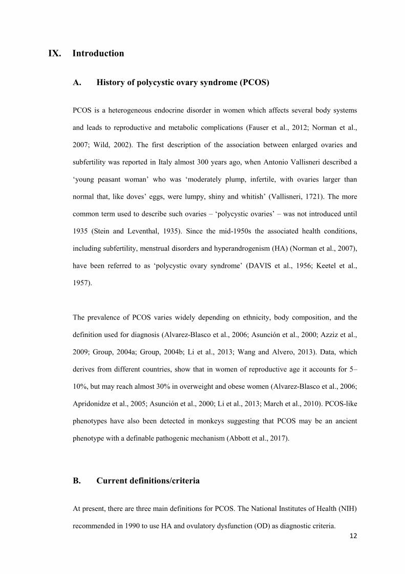
IX. Introduction
A. History of polycystic ovary syndrome (PCOS)
PCOS is a heterogeneous endocrine disorder in women which affects several body systems
and leads to reproductive and metabolic complications (Fauser et al., 2012; Norman et al.,
2007; Wild, 2002). The first description of the association between enlarged ovaries and
subfertility was reported in Italy almost 300 years ago, when Antonio Vallisneri described a
‘young peasant woman’ who was ‘moderately plump, infertile, with ovaries larger than
normal that, like doves’ eggs, were lumpy, shiny and whitish’ (Vallisneri, 1721). The more
common term used to describe such ovaries – ‘polycystic ovaries’ – was not introduced until
1935 (Stein and Leventhal, 1935). Since the mid-1950s the associated health conditions,
including subfertility, menstrual disorders and hyperandrogenism (HA) (Norman et al., 2007),
have been referred to as ‘polycystic ovary syndrome’ (DAVIS et al., 1956; Keetel et al.,
1957).
The prevalence of PCOS varies widely depending on ethnicity, body composition, and the
definition used for diagnosis (Alvarez-Blasco et al., 2006; Asunción et al., 2000; Azziz et al.,
2009; Group, 2004a; Group, 2004b; Li et al., 2013; Wang and Alvero, 2013). Data, which
derives from different countries, show that in women of reproductive age it accounts for 5–
10%, but may reach almost 30% in overweight and obese women (Alvarez-Blasco et al., 2006;
Apridonidze et al., 2005; Asunción et al., 2000; Li et al., 2013; March et al., 2010). PCOS-like
phenotypes have also been detected in monkeys suggesting that PCOS may be an ancient
phenotype with a definable pathogenic mechanism (Abbott et al., 2017).
B. Current definitions/criteria
At present, there are three main definitions for PCOS. The National Institutes of Health (NIH)
recommended in 1990 to use HA and ovulatory dysfunction (OD) as diagnostic criteria.
13
The ESHRE/ASRM definition was published in 2004 (Group REA-SPCW, 2004a,b).
Accordingly, PCOS is diagnosed when two of the following criteria are present: oligo-
/amenorrhea, clinical and/or biochemical signs of HA and polycystic ovarian morphology
(PCOM) on ultrasound.
The Androgen Excess and PCOS Society (AE-PCOS) published the most recent definition in
2006 suggesting to involve the presence of HA (clinical and/or biochemical) and ovarian
dysfunction [oligoanovulation (OA) and/or polycystic ovaries] (Azziz et al., 2009).
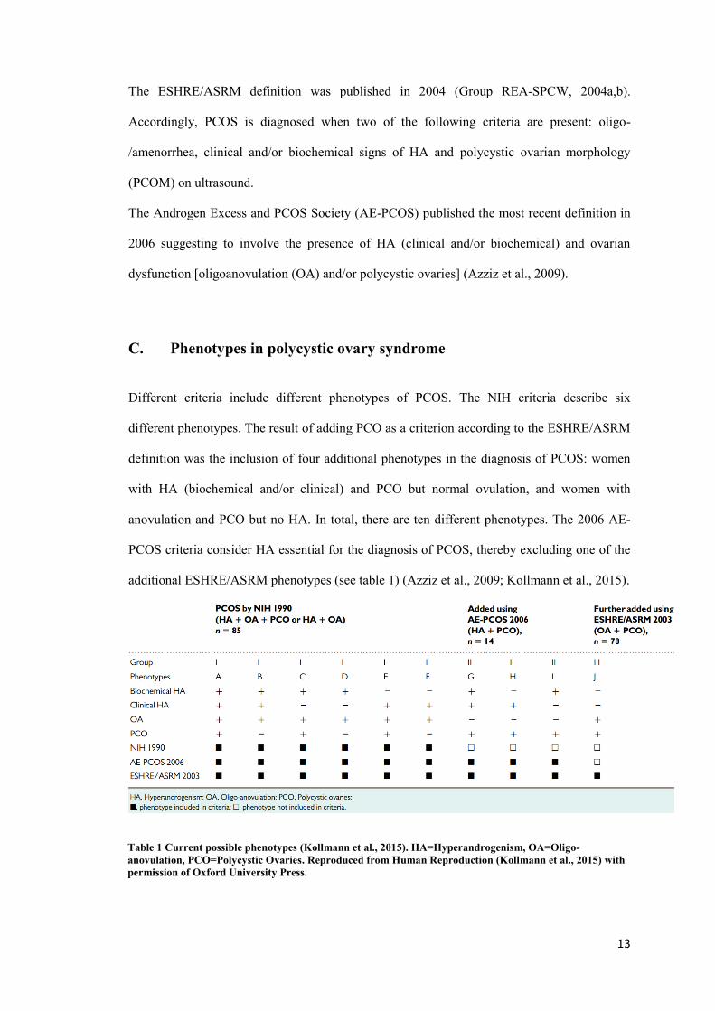
C. Phenotypes in polycystic ovary syndrome
Different criteria include different phenotypes of PCOS. The NIH criteria describe six
different phenotypes. The result of adding PCO as a criterion according to the ESHRE/ASRM
definition was the inclusion of four additional phenotypes in the diagnosis of PCOS: women
with HA (biochemical and/or clinical) and PCO but normal ovulation, and women with
anovulation and PCO but no HA. In total, there are ten different phenotypes. The 2006 AE-
PCOS criteria consider HA essential for the diagnosis of PCOS, thereby excluding one of the
additional ESHRE/ASRM phenotypes (see table 1) (Azziz et al., 2009; Kollmann et al., 2015).
Table 1 Current possible phenotypes (Kollmann et al., 2015). HA=Hyperandrogenism, OA=Oligo-
anovulation, PCO=Polycystic Ovaries. Reproduced from Human Reproduction (Kollmann et al., 2015) with
permission of Oxford University Press.

14
1. ESHRE/ASRM criteria
The ESHRE/ASRM criteria, which were published in 2004, are most commonly used in
Europe and will therefore be described in a more detailed way. PCOS is diagnosed when two
of the following criteria are present: oligo-/amenorrhea, clinical and/or biochemical signs of
HA and polycystic ovarian morphology (PCOM) on ultrasound. Disorders with a similar
clinical presentation, such as congenital adrenal hyperplasia, Cushing‘s syndrome, and
androgen-secreting tumours, must be excluded (Group, 2004b; group, 2004).
a. Hyperandrogenism and metabolic markers
The consensus statement suggests that patients can either have a clinical hyperandrogenism, a
biochemical hyperandrogenism or both. The clinical hyperandrogenism can be expressed by
hirsutism, acne and androgenic alopecia. However, normative data in large populations are
still lacking and the assessment of clinical features is relatively subjective (Group, 2004b;
group, 2004). The statement suggests to measure biochemical hyperandrogenism, although
there are limitations mostly due to the inaccuracy and variability of the laboratory methods of
measurement that are often used. They comment on total testosterone (T), free testosterone
(FT), free testosterone (free androgen) index (FAI), dehydroepiandrosterone sulphate
(DHEAS), and androstenedione (ANDR). Measuring FT or FAI was the more sensitive
methods of assessing hyperandrogenemia (Cibula et al., 2000; Imani et al., 2000; Vermeulen
et al., 1999). The measurement of T only may not be a very sensitive marker of androgen
excess and some PCOS patients may have isolated elevations in DHEAS levels (ESHRE
2004HR/FS). Finally, they state that little data are available on the value of routinely
measuring ANDR in hyperandrogenic patients (Laven et al., 2002).

15
b. Oligo- and/or anovulation
Oligoovulation means infrequent or irregular ovulation and anovulation means absence of
ovulation. PCOS patients often present with oligomenorrhea, which means infrequent, often
light menstrual periods (intervals exceeding 35 days) or amenorrhea which describes the
absence of a menstrual period in a woman of reproductive age. Those symptoms are associated
with oligo- and/or anovulation.
c. Polycystic ovary
As previously described it was already 300 years ago that Vallisneri wrote about a ‘young
peasant woman’ who was ‘moderately plump, infertile, with ovaries larger than normal that,
like doves’ eggs, were lumpy, shiny and whitish’ (Vallisneri, 1721). However, the more
common term used to describe such ovaries – ‘polycystic ovaries’ – was not introduced until
1935 (Stein and Leventhal, 1935). And it was not until 2003 that PCO became part of
diagnostic criteria (Group, 2004b; group, 2004). The 2003 ESHRE/ASRM definition
emphasizes the importance of ultrasound as a diagnostic tool (Group REA-SPCW, 2004a,b).
Based on one publication by Jonard et al., the statement suggests that the presence of 12 or
more follicles in each ovary measuring 2–9 mm in diameter, and/or increased ovarian volume
(>10 mL) should be taken to define a ‘polycystic ovary’ (Group, 2004b; Jonard et al., 2003).
Only one ovary fitting this definition is sufficient to define PCO. This definition does not
apply to women taking an oral contraceptive pill and the scan should be repeated during the
next cycle if the ovaries contain a dominant follicle (>10 mm) or a corpus luteum (Group,
2004b).
D. Recent studies
Since the publication of the consensus statement in 2004, many studies have been performed
looking at PCO diagnosis by ultrasound and further biomarkers.

16
Regarding the diagnosis of PCOM: The threshold of 12 follicles was based on a study
published by Jonard et al. in 2003, which reported that a FNPO of 12 or more offered the best
compromise between specificity (99%) and sensitivity (75%) in the detection of
hyperandrogenic anovulation (Jonard et al., 2003). However, recent studies, using new
ultrasound machines with improved resolution, have shown a high prevalence of ovaries with
more than 12 follicles in healthy young women (Duijkers and Klipping, 2010; Johnstone et al.,
2010; Jokubkiene et al., 2012). A task force from the Androgen Excess and Polycystic Ovary
Syndrome Society (AE-PCOS) reviewed results from recent studies (Dewailly et al., 2011;
Duijkers and Klipping, 2010; Johnstone et al., 2010; Kristensen et al., 2012; Lujan et al.,
2013) and reported an urgent need to update the diagnostic criteria (Dewailly et al., 2013).
They recommend that the threshold should be increased to≥25 follicles when using new
ultrasound machines and that ovarian volume (≥10 mL) should be used in the detection of HA
(in the absence of a dominant follicle or a corpus luteum) when a new ultrasound machine is
not available (Dewailly et al., 2013). We welcome the call to update the current criteria and
hope that these findings will lead to a revision of the present criteria and some long overdue
changes (Kollmann et al., 2014a, b; Martins et al., 2014).
Figure 1 Polycystic ovary (a). Automatic volume calculation (SonoAVC) was used to automatically calculate
the volume of the follicle (b)
Anti-Müllerian hormone (AMH), a peptide produced by the granulosa cells (GC) of ovarian
follicles is discussed as an additional biomarker in diagnosis and management of PCOS
patients (Dewailly et al., 2011; Dewailly et al., 2013; La Marca and Sunkara, 2013). There
seems to be a consistent relationship between AMH serum levels and the FNPO seen on
b a

17
ultrasound (Dewailly et al., 2011; Pigny et al., 2006). Assays have changed over the last years
and it seems that the modification of the Beckman-Coulter second-generation enzyme-linked
immunosorbent assay protocol improves the reliability of serum AMH measurement
(Craciunas et al., 2015). However, only a few studies have assessed specifically whether AMH
serum concentrations might be an effective surrogate marker of PCOM (Dewailly et al., 2011;
Dewailly et al., 2013; Pigny et al., 2006).
E. Etiology
The etiology of PCOS is not particularly mapped, but a complex interaction of various
factors can be assumed. It seems that the disorder arises from interactions between genetic,
environmental and intrauterine factors (de Melo et al., 2015).
The first evidence for a genetic basis of PCOS was already reported in 1968 (Cooper et al.,
1968). With the performance of genome-wide association studies (GWAS) new insights were
brought into the heritability of PCOS (Jones and Goodarzi, 2016; Liu et al., 2016). Currently,
six different GWAS studies of PCOS have been published and 16 genes/loci have been
identified (Jones and Goodarzi, 2016; Liu et al., 2016) (table 2 (Jones and Goodarzi, 2016)).

18
Table 2 PCOS risk loci reported in genome-wide association studies (GWAS). (Jones and Goodarzi, 2016).
Reproduced from Fertility and Sterility (Jones and Goodarzi, 2016) with permission of Elsevier.
More and more studies also discover that epigenetic variations, including gene methylation,
histone modification, microRNAs, and RNA binding proteins play a role in determining the
PCOS phenotype (Ilie and Georgescu, 2015; Xu et al., 2010; Xu et al., 2011; Yu et al., 2015).
However, the heterogeneity of PCOS and the presence of different phenotypes may also
suggest further etiological factors.
Environmental determinants of PCOS were summarized in a review by Merkin et al. (Merkin
et al., 2016). They main factors they considered included environmental toxins, diet and
nutrition, socioeconomic status, and geography. The authors found some evidence that
environmental toxins play a role in disrupting reproductive health, but there is limited research
as to how these toxins may affect the development of PCOS (Merkin et al., 2016). Studies
showed that PCOS symptoms are reduced with certain dietary supplements and with weight
loss among obese women. However, additional research is needed to compare various
approaches to weight loss (Merkin et al., 2016). Furthermore, an association of low
socioeconomic status with certain PCOS phenotypes has been indicated (Merkin et al., 2016).
19
The third factor which seems to play a crucial role in the development of PCOS is the
intrauterine milieu during pregnancy and the early childhood (Barker, 1995; de Melo et al.,
2015; Dumesic et al., 2007; Dumesic et al., 2014). Hales and Barker published already in 1992
that a poor fetal and early post-natal nutrition imposes mechanisms of nutritional thrift upon
the growing individual which subsequently can lead to an impaired development of the
endocrine pancreas (Barker, 1992; Hales and Barker, 1992, 2013). And it has been suggested
that prenatal growth restraint followed by catch-up of weight during infancy can lead to PCOS
(de Zegher and Ibáñez, 2006). Experimental studies in non-humans and clinical observations
in humans support the hypothesis that developmental programming by steroid excess plays a
role in the development of PCOS (Abbott et al., 2005; Abbott et al., 2002; Abbott et al., 2013;
Melo et al., 2010; Palomba et al., 2012). Sex differences in prenatal androgen levels have been
observed and testosterone levels in umbilical cord blood and in amniotic fluid are higher in
healthy male babies than in healthy female babies (Maccoby et al., 1979; van de Beek et al.,
2004). There are just a few studies reporting on the relation between maternal androgen levels
during pregnancy and the respective offspring in PCOS women (Abbott et al., 2005; Barry et
al., 2011; Barry et al., 2010; Caanen et al., 2016; Dahlgren et al., 1992; Dunaif et al., 1989;
Helseth et al., 2014). Barry showed that umbilical vein (UV) T in PCOS girls was
significantly raised, compared with control girls (Barry et al., 2010). Another study did not
find any significant differences between girls of PCOS mothers and girls of mothers without
PCOS (Caanen et al., 2016).
F. Pathophysiology
As ovarian steroidogenesis requires gonadotropin stimulation, luteinizing hormone (LH) has a
key role in hyperandrogenemia of PCOS (Burt Solorzano et al., 2012). The pulsatile release of
gonadotropin-releasing hormone (GnRH) from the hypothalamus is often disturbed in PCOS
patients, which subsequently leads to a LH hypersecretion. PCOS patients often show a
20
resistance of their GnRH pulse generator to progesterone, which regulates GnRH pulse
frequency (Pastor et al., 1998). The resistance to progesterone seems to be mediated by
androgen excess (Eagleson et al., 2000). Serum follicle-stimulating hormone (FSH) levels are
usually normal. However, ovarian follicles seem to be more resistant to FSH in women with
PCOS than in controls, an effect which might be due to increased levels of intra-ovarian AMH
(Burt Solorzano et al., 2012; Burt Solorzano et al., 2010; McCartney and Marshall, 2016).
Ovaries from PCOS women have an exaggerated steroidogenic response to gonadotropins
(Ehrmann, 2005). PCOS is often associated with insulin resistance which leads to
hyperinsulinemia, a status which contributes to hyperandrogenemia as well (McCartney and
Marshall, 2016). Hyperinsulinemia increases LH-stimulated androgen synthesis by ovarian
theca cells, it potentiates corticotropin-mediated adrenal androgen production, and it reduces
hepatic production of sex hormone–binding globulin (SHBG), which augments free
testosterone levels (McCartney and Marshall, 2016).
G. Short-and long-term consequences of polycystic ovary syndrome
PCOS is associated with different short- and long-term comorbidities. Insulin resistance can
be seen in approximately 60–80% of women with PCOS and in 95% of obese women with
PCOS (Wild et al., 2010). Studies show that the incidence of metabolic syndrome, gestational
diabetes mellitus (GDM), impaired glucose tolerance (IGT) and type-2 diabetes mellitus
(T2DM) is increased in premenopausal women with PCOS compared with age-matched and
BMI-matched controls (Moran et al., 2010). Hirsutism, acne and androgenic alopecia are
clinical signs of hyperandrogenism and more often seen in PCOS patients (Azziz et al., 2009).
Even though PCOS occurs in obese and lean women, a recent meta-analysis found that obesity
was more prevalent in women with PCOS than in women without PCOS (Lim et al., 2012).
Cardiovascular disease (CVD) markers indicate a higher risk of CVD in women with PCOS
than in controls (Carmina, 2014): coronary calcification is more prevalent in women with
PCOS (Christian et al., 2003; Talbott et al., 2000; Talbott et al., 2004), the intimal layer

21
thickness of the carotid wall has been reported to be greater in women with PCOS (Lass et al.,
2011; Luque-Ramírez et al., 2007), and PCOS patients show an elevated incidence of aortic
calcification (Talbott et al., 2004). Mood disturbances, mostly severe depression, are more
frequent in PCOS women. A meta-analysis found that abnormal depression scores are
higher in women with PCOS compared with those in the control groups (Dokras et al.,
2011). Due to the concurrence of hyperestrogenic anovulation and hyperinsulinemia, a
higher risk for endometrial cancer was found in women with PCOS (odds ratio [OR] 2.7)
(Dumesic and Lobo, 2013). PCOS women may also have a higher risk for ovarian cancer,
though the OR for this malignancy is not clear (Dumesic and Lobo, 2013).
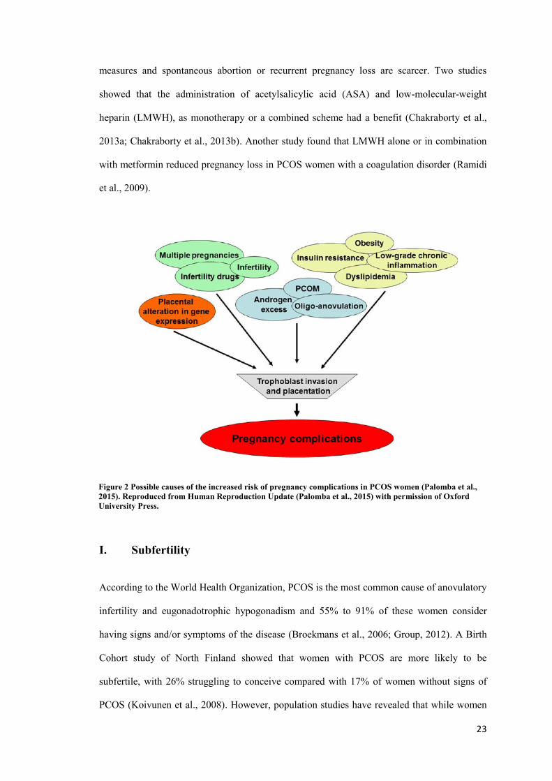
H. Polycystic ovary syndrome and reproductive consequences
Women with PCOS have an impaired fertility and significant higher complication rates during
infertility treatment, pregnancy and the perinatal period (Azziz et al., 2016; Boomsma et al.,
2006; Palomba et al., 2015; Qin et al., 2013). Complications include the occurrence of
multiple gestations, ovarian hyperstimulation syndrome (OHSS), and early pregnancy loss
(Azziz et al., 2016; McCartney and Marshall, 2016; Norman et al., 2007). When pregnant,
these women are susceptible to perinatal complications, including elevated risk of GDM,
pregnancy-induced hypertension (PIH), pre-eclampsia, preterm delivery and caesarean section
(Boomsma et al., 2006; Kollmann et al., 2015; Palomba et al., 2015; Qin et al., 2013). The
offspring of PCOS women is affected by higher intensive care unit (ICU) admission rates and
lower birthweight (Qin et al., 2013). The exact aetiology remains unclear, but it seems that the
complications are the result of several coinciding factors which have an effect on trophoblast
invasion and placentation (Figure 1) (Boomsma et al., 2006; Kollmann et al., 2015; Palomba
et al., 2015; Qin et al., 2013). Maternal hyperandrogenism and insulin resistance play a key
role (Makieva et al., 2014). The clinical relevance of ovarian dysfunction, defined by
polycystic ovarian morphology (PCOM) or anovulation, was discussed controversial
(Palomba et al., 2010; Shroff et al., 2007). Therefore, the aim of our first study was to

22
compare the prevalence of adverse maternal and neonatal outcomes in pregnant women
classified with PCOS according to different definitions (Kollmann et al., 2015). Some studies
have investigated the prevention and management of pregnancy complications in women with
PCOS (Agha et al., 2014; Balsells et al., 2015; Chakraborty et al., 2013a; Chakraborty et al.,
2013b; Løvvik et al., 2015; Magnussen et al., 2011; Palomba et al., 2015; Palomba et al.,
2014a; Palomba et al., 2014b; Peterson et al., 2015; Ramidi et al., 2009). Many observational
findings showed a lower risk of obstetric and neonatal adverse outcomes in normal-weight
women compared to overweight/obese women and therefore losing weight before conception
up to an optimal body weight is suggested. Pregnant women, who are obese, irrespective of
having PCOS, should be informed about the beneficial effects of dietary and/or physical
activity during pregnancy on the gestational weight gain (Agha et al., 2014). The American
Diabetes Association suggests testing PCOS women for GDM at the first prenatal visit
(Association, 2016a, b). In infertile women with PCOS maximum effort should be made to
avoid multiple pregnancies (Løvvik et al., 2015).
1. Therapeutics
Different pharmacological measures have been discussed in women with PCOS during
pregnancy in order to reduce the obstetric and neonatal risks. The best tested drug in PCOS
women is metformin. It seems that metformin is effective and safe for the treatment of GDM,
especially in overweight and obese women (Balsells et al., 2015). There might further be
beneficial effects of using metformin compared to insulin in GDM. Those are related to
maternal weight gain during pregnancy, neonatal outcomes and patient compliance (Lautatzis
et al., 2013; Rowan et al., 2008; Sivalingam et al., 2014). However, the beneficial effects of
metformin were all seen in non-randomized controlled trials (RCTs). The only RCT on
metformin administration during pregnancy in PCOS women did not show a beneficial effect
on the development of GDM (Vanky et al., 2010). For the prevention of PIH and PE
metformin seems not beneficial (Palomba et al., 2009). Studies regarding pharmacological
23
measures and spontaneous abortion or recurrent pregnancy loss are scarcer. Two studies
showed that the administration of acetylsalicylic acid (ASA) and low-molecular-weight
heparin (LMWH), as monotherapy or a combined scheme had a benefit (Chakraborty et al.,
2013a; Chakraborty et al., 2013b). Another study found that LMWH alone or in combination
with metformin reduced pregnancy loss in PCOS women with a coagulation disorder (Ramidi
et al., 2009).
Figure 2 Possible causes of the increased risk of pregnancy complications in PCOS women (Palomba et al.,
2015). Reproduced from Human Reproduction Update (Palomba et al., 2015) with permission of Oxford
University Press.
I. Subfertility
According to the World Health Organization, PCOS is the most common cause of anovulatory
infertility and eugonadotrophic hypogonadism and 55% to 91% of these women consider
having signs and/or symptoms of the disease (Broekmans et al., 2006; Group, 2012). A Birth
Cohort study of North Finland showed that women with PCOS are more likely to be
subfertile, with 26% struggling to conceive compared with 17% of women without signs of
PCOS (Koivunen et al., 2008). However, population studies have revealed that while women
24
with PCOS may take longer than expected to conceive, their ‘lifetime fertility’ does not appear
to be significantly impaired (Group, 2012; Koivunen et al., 2008).
1. Therapeutics
The first line treatment of PCOS-related infertility includes lifestyle modification (weight loss
in overweight/obese) and the use of drugs to induce monofollicular ovulation. Drug treatments
normally begin with the use of clomiphene citrate followed by the administration of
exogenous gonadotropins, with timed intercourse or intrauterine insemination. Assisted
reproductive techniques (ART), particularly in-vitro fertilization (IVF) or intracytoplasmic
sperm injection (ICSI), represent the third line of treatment (Broekmans et al., 2006). Ovarian
hyperstimulation syndrome (OHSS) and multiple pregnancies remain the major complications
of ovulation induction and occur despite ultrasound monitoring (Brown et al., 2009; Nastri et
al., 2010). PCOS women are typically more difficult to stimulate in a controlled manner,
whether the intention is to induce a monofollicular or multifollicular response, are more likely
to demonstrate resistance to stimulation and/or an exaggerated response and experience a
higher cycle cancellation rate than women without PCOS. Whereas a high number of oocytes
may be obtained during ART, there are concerns that the quality and maturity of these oocytes
may be impaired (Baumgarten et al., 2013; Coffler et al., 2003a; Coffler et al., 2003b;
Doronzo et al., 2004; Jayaprakasan et al., 2012; Kumar et al., 2013; Ocal et al., 2011;
Siristatidis et al., 2013). Recent developments have seen the introduction of various actions to
reduce the risks of OHSS and cycle cancellation and to improve oocyte quality. These
measures include priming with metformin, the use of GnRH antagonist cycles as opposed to
the conventional long GnRH agonist protocol, the administration of dopamine agonists and
oocyte retrieval without controlled ovarian stimulation through in-vitro maturation (IVM) of
oocytes (Al-Inany et al., 2011; Baumgarten et al., 2013; Nardo et al., 2013; Nastri et al., 2010;
Ortega-Hrepich et al., 2013; Tang et al., 2012; Tso et al., 2014). To examine the efficacy of all

25
strategies we performed a systematic review and meta-analysis of randomized controlled trials
(RCTs) aiming to improving ART outcomes in women with PCOS (Kollmann et al., 2016) .
There is low- to moderate-quality evidence suggesting that antagonist protocols are preferable
to agonist ones, because they reduce the incidence of OHSS without interfering with clinical
pregnancy and live birth for POCS women. Moreover there is low-quality evidence pointing
to a benefit of metformin supplementation on clinical pregnancy and live birth; and that
ovulation induction and administration of estradiol seem to be equally effective for
endometrial preparation before frozen embryo transfer for women with PCOS (Kollmann et
al., 2016).
J. Hormone levels during pregnancy
1. Androgens in pregnancy
Androgens are especially important for the development of the male reproductive tract during
fetal life and they do act as pro-hormones for biosynthesis of estrogens in both sexes (Macleod
et al., 2010; Purohit and Foster, 2012; Rivas et al., 2002; Scott et al., 2007; Traish et al., 2011;
Welsh et al., 2008). In women, androgens are normally synthesized by ovaries, the adrenal
glands, and also in adipose tissue. Studies show that some androgen levels increase during
normal pregnancy and it has been hypothesized that androgens act as substrates for estrogen
formation in the placenta (Edman et al., 1981; Makieva et al., 2014; PION et al., 1965; Siiteri
and MacDonald, 1966; Smith, 2007). DHEAS, which is produced by the fetal and maternal
adrenals, enters the placenta, where it is metabolized to ANDR and T. This gets further
metabolized to estrone (E1) and estradiol (E2) (Strauss et al., 1996). E2 can enter the fetal
circulation, taken up by the liver and transformed into estriol (E3), which subsequently can
pass to maternal circulation (HIRANO, 1961; Schwarzel et al., 1973; Willows, 1966). In
women who are not pregnant, 50% of all DHEA is produced by the adrenal glands, 20% by
the ovaries, and 30% by peripheral tissue (Abraham, 1974). Regarding ANDR, 50% is
synthesized in the adrenal glands and 50% in the ovaries (Longcope, 1986). One half of
26
testosterone is synthesized in the peripheral tissue, the other half is produced by the ovaries
and adrenal glands (25% and 25% respectively) (Piltonen et al., 2002). In pregnant women,
the fetus and the placenta are additional sources of androgens (Cantineau et al., 1985; Makieva
et al., 2014). As mentioned above, numeral studies have found elevated levels of some
circulating androgens during normal pregnancy (Bammann et al., 1980; Buster et al., 1979;
Dawood and Saxena, 1977; Mizuno et al., 1968; Rivarola et al., 1968; Saez et al., 1972). Total
testosterone (tT) increases from the first trimester of pregnancy and towards term (Bammann
et al., 1980; Berger et al., 1984; Saez et al., 1972), free testosterone (fT) levels only at the third
trimester (Dawood and Saxena, 1977). ANDR seems to be increased between 37-42 weeks of
gestation (Mizuno et al., 1968). On the contrary, DHEAS levels are up to 50% lower in
pregnant women that in non-pregnant women (Milewich et al., 1978). Sexual hormone
binding globulin (SHBG) levels rise dramatically from the first trimester until term (Wilke and
Utley, 1987).
Fetal hormone levels depend on fetal sex and gestation. T levels are higher in male fetuses and
increase until the end of first trimester (Rodeck et al., 1985). At 12 weeks of gestation, T
levels reach a peak of around 150 ng/dL and fall by around 70% afterward (Rodeck et al.,
1985). In female fetuses, T levels are generally lower until the second trimester and they fall
further at term (Diez d'Aux and Pearson Murphy, 1974). DHEA concentrations are also higher
in male fetus compared to female fetuses (Keelan et al., 2012). On the contrary, ANDR levels
are similar in both sexes (Keelan et al., 2012). Interestingly, one study shows that labour is
associated with an increase in concentrations of ANDR, DHEA and SHBG, and decrease of tT
and fT (Keelan et al., 2012). Rivarola et al. investigated the association between fetal sex and
maternal serum levels of any androgen and found none (Rivarola et al., 1968).
As mentioned above, it has been shown that some androgen levels increase during normal
pregnancy (Edman et al., 1981; Makieva et al., 2014; PION et al., 1965; Siiteri and
MacDonald, 1966; Smith, 2007). Although the origin and cause of the higher levels are not
known exactly, it is probable that the production involves the ovary and the placenta (Makieva

27
et al., 2014). After ovulation, androgens are produced by small luteal cells of the corpus
luteum (CL) (Sanders et al., 1996). Within the first trimester, human chorionic gonadotrophin
(hCG) stimulates the CL and increases until the end. The increase of hCG could therefore
cause the augmentation of T levels (Braunstein et al., 1976; Liu and Hsueh, 1986). On the
other hand, we see a steady rise of T after the known peak and further decrease of hCG levels
at the end of the first trimester. This suggests either that the androgen production is regulated
alternatively or that there is a further source of androgens after the end of first trimester
(Braunstein et al., 1976). Maternal adrenal glands are a further important source of androgen
production during pregnancy (Makieva et al., 2014). Studies show that the production of
DHEA during pregnancy is suppressed by E2 (Albrecht and Pepe, 1995; Tagawa et al., 2004;
Umezaki et al., 2001).
Former studies found that the placenta does not have the capacity to synthesize androgens de
novo (PION et al., 1965; Siiteri and MacDonald, 1966). However, a recent study found that
the placental syncytiotrophoblast has this ability (Escobar et al., 2011). Precisely, the study
found that the syncytiotrophoblast expresses the enzyme CYP17, which converts C21 steroids
to C19 steroids (Escobar et al., 2011).
A possible role of the myometrium in androgen production has been shown in an animal
study. The study found that uteri from non-pregnant and early pregnant pigs can synthesize
ANDR and T in vitro (Franczak, 2008). Possible origins of androgens are shown in figure 2.

28
Figure 3 Possible sources of androgens during pregnancy; dehydroepiandrosterone (DHEA), testosterone
(T), dihydrotestosterone (DHT), dehydroepiandrosterone sulphates (DHEAS), androstenedione (A4)
(Makieva et al., 2014). Reproduced from Human Reproduction Update (Makieva et al., 2014) with
permission of Oxford University Press.
As mentioned above, androgen excess is suspected to play a role in the development of
pregnancy complications (Palomba et al., 2015). Likewise, we know that there are pregnancy-
specific mechanisms to protect both the mother and fetus from pregnancy-induced androgen
excess (Crisosto et al., 2012; Hensleigh et al., 1975; Phelan and Conway, 2011). One is the
physiological increase of maternal SHBG, which binds elevated androgens (Hammond, 2011).
Another mechanism is the increase of progesterone (P), which competes for androgen receptor
(AR) binding (Birrell et al., 2007; Slayden et al., 2001). A third mechanism might be that P
has an affinity for 5a-reductase which results in an inhibition of the conversion of T to the
more potent dihydrotestosterone (DHT) (Cabeza et al., 1999; Hodgins, 1982).
2. AMH in pregnancy
There are some reports on AMH levels during pregnancy (Kuijper et al., 2013; Köninger et al.,
2013; Köninger et al., 2015; La Marca et al., 2005; Massé et al., 2011; Nelson et al., 2010;

29
Plante et al., 2010; Shand et al., 2014; Vanky and Carlsen, 2012). As previously described, we
know that AMH is produced in women by the granulosa cells of the ovarian (pre-)antral
follicles. Until puberty AMH is very low in females (Kuijper et al., 2013). In male fetuses,
AMH is produced by Sertoli cells soon after testicular differentiation and it is essential for
regression of the Müllerian ducts (La Marca et al., 2005). The hormone is measureable in the
serum of males during their whole life. However, after puberty T suppresses AMH secretion
(Bergadá et al., 2006; Lee et al., 1996; Pierik et al., 2009). One study investigated the effect of
fetal gender on maternal AMH levels and found no connection (La Marca et al., 2005). The
more recent and bigger studies suggest a decline of AMH in maternal serum during gestation
(Köninger et al., 2013; Köninger et al., 2015; La Marca et al., 2005; Li et al., 2010; Nelson et
al., 2010). A possible explanation for the decline could be the inactivated menstrual cycle
during pregnancy and absence of follicular development (Kuijper et al., 2013).
K. Aims
The superordinate aim of the project was the investigation of the perinatal period of women
with PCOS. Therefore, we designed three major studies:
1. 1st study - Maternal and neonatal outcomes in pregnant women with
PCOS: comparison of different diagnostic definitions (Kollmann et al., 2015)
Studies show that PCOS women are susceptible to perinatal complications including elevated
risk of gestational diabetes, pregnancy-induced hypertension, pre-eclampsia, preterm delivery
and caesarean section (Boomsma et al., 2006; Palomba et al., 2015; Qin et al., 2013) and that
their offspring is affected by higher intensive care unit (ICU) admission rates and lower birth
weight (Qin et al., 2013).

30
At present, there are three main definitions for PCOS: the NIH criteria, the ESHRE/ASRM
criteria, and the AE-PCOS criteria (Azziz et al., 2009; Group, 2004b). Thus, there are several
different phenotypes of PCOS, which are subject of intense debate (Carmina et al., 2005;
Chang et al., 2005; Palomba et al., 2013; Palomba et al., 2010; Shroff et al., 2007).
The aim of this study was to compare the prevalence of adverse maternal and neonatal
outcomes in pregnant women classified with PCOS according to different definitions.
2. 2nd
study – Longitudinal study of pregnant PCOS women
The aim of the longitudinal study of pregnant PCOS women was to evaluate the androgen and
AMH levels before, during and after pregnancy and to evaluate the perinatal outcome.
3. 3rd
study – Cross-sectional study of pregnant PCOS women and their
offspring compared to non-PCOS women and their offspring
The first aim of the cross-sectional study of pregnant PCOS and non-PCOS women and their
offspring was to investigate whether the offspring from PCOS women already show higher
androgen and AMH levels compared to the offspring from non-PCOS women. The second aim
was to evaluate the perinatal outcome of PCOS and non-PCOS women.

31
X. 1st study - Maternal and neonatal outcomes in pregnant women with
PCOS: comparison of different diagnostic definitions (Kollmann et al.,
2015)
A. Material and Methods
Study design
Retrospective matched cohort study
Setting
Data of primiparous women with PCOS according to ESHRE/ASRM 2003 criteria and healthy
controls giving birth to neonates ≥ 500g at the Medical University of Graz, Austria, between
January 2004 and March 2012 were retrospectively retrieved. Data were extracted from the
local perinatal database (PIA, ViewPoint, GE Healthcare, Zipf, Austria) and the medical
documentation system or patient files (Kollmann et al., 2015).
Ethical Approval
The study was approved by the institutional review board (No: 24-282ex11/12).
Participants
PCOS was diagnosed following clinical and sonographic evaluation and a hormonal analysis.
Ultrasound examinations are routinely performed by medical doctors specialized in obstetrics
and gynaecology. Clinical investigations are performed in cooperation with the Division of
Endocrinology and Metabolism at the Department of Internal Medicine at Medical University
of Graz, Austria. We report on a population of women that were treated in our specialized unit
and were delivered at our institution (Kollmann et al., 2015).
Women were assigned to three groups according to the different definitions for PCOS:
Group 1: PCOS by NIH criteria = HA + OA + PCO or HA + OA.
Group 2: Added using AE-PCOS criteria = HA + PCO.
Group 3: Further added using ESHRE/ASRM criteria = OA + PCO.

32
The healthy control group comprised women without PCOS, pre-gestational diabetes, or pre-
gestational hypertension. For all PCOS case per year four control cases with information on
BMI were randomly selected (Kollmann et al., 2015).
Variables
Primary outcome parameter was the composite complication rate per women and newborns.
Secondary outcome parameters were specific maternal and neonatal complications including
gestational diabetes, pregnancy-induced hypertension, pre-eclampsia, operative delivery
(elective and non-elective caesarean section and operative vaginal delivery), large or small for
gestational age infants, preterm birth (< 37 and < 34 weeks of gestation, respectively),
acidosis, ICU admission, and pre- and perinatal mortality (Kollmann et al., 2015).
Data sources/ Measurement/ Quantitative variables
The dataset was investigated on the occurrence of maternal or neonatal complications and
categorized as either ‘normal’ or ‘complicated’(Kollmann et al., 2015). The total complication
rate per women and newborns is a composite outcome consisting of the cumulative incidence
of the following parameters: gestational diabetes, pregnancy-induced hypertension, pre-
eclampsia, operative delivery (elective and non-elective caesarean section and operative
vaginal delivery), large or small for gestational age infants, preterm birth (< 37 and < 34
weeks of gestation, respectively), acidosis, ICU admission, and pre- and perinatal mortality. At
our unit maternal gestational diabetes is diagnosed based on the oral glucose tolerance test
(oGTT, HemoCue, Ängelholm, Sweden) which is implemented in routine pregnancy care in
Austria as well as on cord blood insulin testing which is routinely performed in all infants born
with a weight ≥ 4000 g (Kollmann et al., 2015). The normal range of cord blood insulin is 3 -
25 mU/l (ADVIA Centaur, Siemens, Germany) (Chevenne et al., 1999). In the observed
period oGTT was performed between 24 – 28 weeks of gestation by capillary blood analysis
after 12h fasting and one and two hours following administration of 75g glucose (Kollmann et
al., 2015). Glucose cut-off values to diagnose maternal gestational diabetes were 90 / 160 /
140 mg/dl or 5 / 8.9 / 7.8 mmol/l (Metzger et al., 2010). Measurement of blood pressure

33
(systolic and diastolic) and analysis of a urine sample were performed at admission.
Pregnancy-induced hypertension (PIH) was defined as systolic blood pressure ≥ 140 mmHg or
diastolic blood pressure ≥ 90 mmHg in 5 out of 21 measurements arising after 20 weeks of
gestation. Pre-eclampsia was defined as PIH with proteinuria (≥ 300 mg in 24 hours) (Brown
et al., 2001). Operative delivery was defined as birth by forceps, ventouse or caesarean section
(Kollmann et al., 2015). Foetal umbilical pH value and neonatal birth weight are determined
after delivery. Small for gestational age and large for gestational age was diagnosed, when
birth weight was below the 10th percentile or above the 90
th percentile (Voigt et al., 2010).
Intrauterine growth restriction was defined as SGA and the occurrence of distinct signs of
placental insufficiency such as pathological Doppler waveforms in the umbilical (elevated
pulsatility index, absent or reversed end-diastolic flow) or middle cerebral artery (decreased
pulsatility index) as well as a cerebroplacental Doppler ratio (middle cerebral artery pulsatility
index/umbilical artery pulsatility index) below 1 (Bahado-Singh et al., 1999; Baschat and
Gembruch, 2003; Odibo et al., 2005; Oros et al., 2011). For evaluation of foetal acidosis
umbilical arterial blood was examined (ABL 800 FLEX analyser, Akandevej, Denmark). Fetal
acidosis was defined as an umbilical artery pH < 7.10 at birth (Reif et al., 2014; Reif et al.,
2015).
Study size
Data of primiparous women with PCOS according to ESHRE/ASRM 2003 criteria and healthy
controls giving birth to neonates ≥ 500g between January 2004 and March 2012 was included.
The study population and control group are shown in Figure 3 and Figure 4. Power calculation
was not feasible since data on the rate of the composite maternal and neonatal complications
were not available before the study was designed (Kollmann et al., 2015).
34
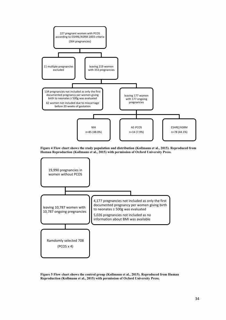
Figure 4 Flow chart shows the study population and distribution (Kollmann et al., 2015). Reproduced from
Human Reproduction (Kollmann et al., 2015) with permission of Oxford University Press.
Figure 5 Flow chart shows the control group (Kollmann et al., 2015). Reproduced from Human
Reproduction (Kollmann et al., 2015) with permission of Oxford University Press.
227 pregnant women with PCOS according to ESHRE/ASRM 2003 criteria
(364 pregnancies)
11 multiple pregnancies excluded
leaving 219 women with 353 pregnancies
134 pregnancies not included as only the first documented pregnancy per women giving
birth to neonates ≥ 500g was evaluated
42 women not included due to miscarriage before 20 weeks of gestation
leaving 177 women with 177 ongoing
pregnancies
NIH
n=85 (48.0%)
AE-PCOS
n=14 (7.9%)
ESHRE/ASRM
n=78 (44.1%)
19,990 pregnancies in women without PCOS
4,177 pregnancies not included as only the first documented pregnancy per women giving birth to neonates ≥ 500g was evaluated
5,026 pregnancies not included as no information about BMI was available
leaving 10,787 women with 10,787 ongoing pregnancies
Ramdomly selected 708
(PCOS x 4)

35
Statistical methods
For categorical variables relative and absolute proportions are indicated, continuous variables
are expressed as mean ± standard deviation or median and range, respectively. We had no
missing data for the primary outcomes and no imputation was done for secondary outcomes.
For all outcomes, the three PCOS groups were compared with each other. Categorical
variables were analysed by using Fisher’s exact test or chi-squared test with p-value computed
by Monte Carlo simulation, while for continuous outcomes one-way analysis of variance
(ANOVA) or Kruskal-Wallis rank sum test followed by Bonferroni correction was applied. No
modelling for confounding or interactions was conducted in the comparison between the
PCOS groups due to small sample size in one group. The PCOS group was compared to the
control group using logistic regression to report odds ratios (ORs) adjusted for BMI and age.
All analyses were performed using the statistic software R (version 3.1.1, Vienna, Austria). A
p-value <0.05 was considered to be statistically significant. Taking the number of patients into
account this is an explorative analysis. (Kollmann et al., 2015)
B. Results
Participants
A total of 227 pregnant women (364 pregnancies) with PCOS according to ESHRE/ASRM
2003 definition were identified during the study period. Thereafter, 11 cases with multiple
pregnancies, another 134 cases with secondary pregnancies and 42 cases with miscarriage
before 20 weeks of gestation were excluded. The final study population included 177
primiparous women with singleton pregnancies. Eighty-five women (48.0%) met the NIH
1990 criteria, another 14 (total of 99 = 55.9%) represented the additional phenotypes defined
by the AE-PCOS 2006 criteria and 78 (44.1%) were classified as PCOS exclusively by the
ESHRE/ASRM 2003 definition (Figure 3).

36
For the healthy control group, we identified a total of 15,813 women with 19,990 pregnancies.
4,177 pregnancies were not included as only the first documented pregnancy per women ≥
500g was evaluated and 5,026 pregnancies were not included as no information about BMI
was present. For all PCOS case per year four control cases were randomly selected from the
above population (n = 10.787). The final control group consisted of 708 women (Figure 4)
(Kollmann et al., 2015).
Descriptive data
Maternal age (p=0.50) was comparable between all groups. BMI (p<0.001) and the proportion
of overweight women (BMI ≥ 25; p<0.001) were different between PCOS groups and the
control group; however there was no difference within the PCOS groups (Kollmann et al.,
2015). Nonetheless, the percentage of women with pre-gestational diabetes, impaired glucose
tolerance, and hyperinsulinemia did differ statistically between the three groups (p=0.007).
Regarding those three parameters, women from the ESHRE/ASRM group showed lower rates
than women from the NIH group (p=0.01). No difference was observed between the
ESHRE/ASRM and the AE-PCOS group. Pre-gestational hypertension did not differ (p=0.10).
Table 3 indicates baseline characteristics and the distribution of PCOS features within the
groups.
Table 3 Comparison of demographics, clinical history, and PCOS features (Kollmann et al., 2015).
Reproduced from Human Reproduction (Kollmann et al., 2015) with permission of Oxford University Press.

37
Main results
The composite maternal complication rates were similar in all three groups: 42/85 (49.4%) vs.
9/14 (64.3%) vs. 47/78 (60.3%) (p=0.31) (Kollmann et al., 2015).
The composite neonatal complication rates did not differ significantly: 23/85 (27.1%) vs. 5/14
(35.7%) vs. 18/78 (23.1%) (p=0.62). In comparison to healthy controls, the odds for maternal
complications was increased in PCOS women (OR 2.57 95% confidence interval [CI] 1.82-
3.64; p<0.001), while there was no significant difference in neonatal complications (OR 0.83
95% CI 0.56-1.21; p=0.343) (Kollmann et al., 2015).
Secondary results
Operative deliveries were more common in PCOS patients than in controls (OR=1.7 [95% CI
1.21-2.42]). Within the PCOS groups the prevalence was different between the NIH and
ESHRE group (p=0.007). Elective caesarean section, non-elective caesarean section and
operative vaginal delivery occurred in 7.2%, 20.5%, and 13.3% (NIH), 21.4%, 21.4%, and
21.4% (AE-PCOS), 22.1%, 32.5%, and 10.4% (ESHRE/ASRM), and 10.9%, 16.7%, and
12.3% (Controls) respectively (Kollmann et al., 2015). The share of women with gestational
diabetes (p=0.36), pregnancy-induced hypertension (p=0.81), and pre-eclampsia (p=0.32)
were similar within the PCOS groups (Table 4) (Kollmann et al., 2015). In comparison to
healthy controls women with PCOS had a higher odds of developing gestational diabetes
(OR=10.97 [95% CI 6.02-20.72]) and pregnancy-induced hypertension (OR=8.25 [95% CI
3.60-20.23]); while pre-eclampsia was not significantly different (OR=1.91 [95% CI 0.63-
5.20]) (Table 5) (Kollmann et al., 2015).
Preterm birth < 37 weeks of gestation (p=0.72), preterm birth < 34 weeks of gestation
(p=0.32), acidosis (p=0.72), and large or small for gestational age newborns (p=0.97) emerged
with a comparable frequency within the PCOS groups. The ICU admission rate was different
between the groups (p=0.02). However, when using Bonferroni correction no statistical
difference between the individual groups was observed (Kollmann et al., 2015). In comparison
38
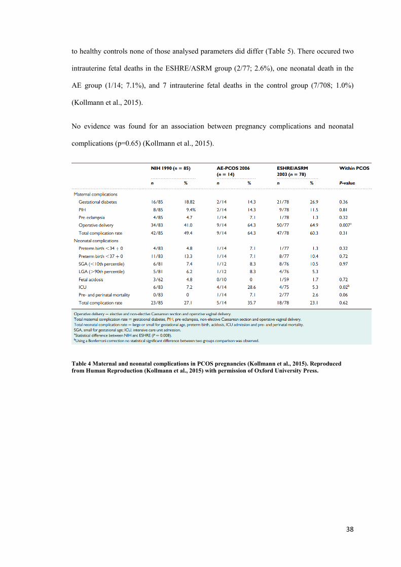
to healthy controls none of those analysed parameters did differ (Table 5). There occured two
intrauterine fetal deaths in the ESHRE/ASRM group (2/77; 2.6%), one neonatal death in the
AE group (1/14; 7.1%), and 7 intrauterine fetal deaths in the control group (7/708; 1.0%)
(Kollmann et al., 2015).
No evidence was found for an association between pregnancy complications and neonatal
complications (p=0.65) (Kollmann et al., 2015).
Table 4 Maternal and neonatal complications in PCOS pregnancies (Kollmann et al., 2015). Reproduced
from Human Reproduction (Kollmann et al., 2015) with permission of Oxford University Press.

39
Table 5 Maternal and neonatal complications: PCOS versus control (Kollmann et al., 2015). Reproduced
from Human Reproduction (Kollmann et al., 2015) with permission of Oxford University Press.
C. Discussion
Regardless of the PCOS definition, about 60% of women with PCOS and about 30% of their
infants are subject to perinatal and neonatal complications. In comparison to healthy controls
the risk for maternal complications was increased in PCOS women (OR 2.57 [1.82-3.64])
while there was no difference in neonatal complications (OR 0.83 [0.56-1.21]) (Kollmann et
al., 2015).
A limitation or the present study is its retrospective design that increases the likelihood of bias
and omits several parameters. We took care to guarantee correct group allocation, however,
even in the most careful manner this remains to be a subjective discrimination that is
ultimately a simplification of the underlying pathophysiology (Kollmann et al., 2015). To
prevent potential confounders, multiple gestations and additional pregnancies of women were
excluded and only primiparous women giving birth to neonates ≥ 500g were included. On the
one hand this may be advantageous regarding confounders; on the other hand it could induce
selection bias. Primiparous women might be different from both nulliparous and multiparous

40
women with PCOS (Kollmann et al., 2015). A further limitation was the limited number of
participants, particularly in the AE-PCOS subgroup (Kollmann et al., 2015).
Our findings confirm previous observations that PCOS women have an increased risk of
perinatal complications (Boomsma et al., 2006; Qin et al., 2013). However, on the contrary to
the study by Palomba et al., who found that the increased risk varies widely according to the
different phenotypes and features of PCOS, we found that all definitions used to define PCOS
are associated with a substantial risk for perinatal complications (Palomba et al., 2010).
A study from 2007 proposed that the non-hyperandrogenic phenotype (ESHRE group) may
represent a subgroup of PCOS associated with a milder metabolic profile (Shroff et al., 2007).
The scientist concluded that women presenting with oligo-/anovulation and polycystic ovaries
only, may feature a significantly lower prevalence of metabolic syndrome. As insulin
resistance as well as the compensatory hyperinsulinemia look to play a major role in the
pathophysiology of metabolic syndrome, it is interesting that there was no decrease in insulin
resistance in the respective ‘low risk’ group (Shroff et al., 2007). When analysing our data we
found that the proportion of women with pre-gestational diabetes, impaired glucose tolerance,
and hyperinsulinemia did differ statistically between the three groups (p=0.007) (Kollmann et
al., 2015). Regarding those parameters, women from the ESHRE/ASRM had a lower risk than
women from the NIH group (p=0.01 (Kollmann et al., 2015)). Interestingly, the proportion of
women with gestational diabetes did not differ within the groups (p=0.36) suggesting that the
ESHRE group is as representative of risk as the other two groups (Kollmann et al., 2015).
Those results highlight the importance of screening in women with PCOS although the timing
and nature of this remains controversial.
The proportionately higher number of women with metabolic problems in the ESHRE group
before and during pregnancy, may explain the discrepancy in mode of delivery between NIH
and ESHRE (p=0.008) (Hartling et al., 2014; Kollmann et al., 2015).

41
The total rate of neonatal complications was, as reported in previous studies, high (27.1%,
35.7% and 23.1% respectively) (Boomsma et al., 2006; Kollmann et al., 2015; Qin et al.,
2013). Maternal androgen excess in utero has been proposed to be an important risk factor for
impaired fetal growth and might have an effect on fetal outcome (Anderson et al., 2010; Barry
et al., 2010). However, in our study we could not found a difference when compared to
controls (Kollmann et al., 2015).
Maternal hyperandrogenism and insulin resistance may indeed play an important role in
maternal and neonatal outcome. The clinical relevance of ovarian dysfunction, in particular
PCOM, stays a matter of debate (Palomba et al., 2010; Shroff et al., 2007). Our data shows
that the additional phenotype, introduced as part of the ESHRE/ASRM 2003 criteria, does
have clinical significance (Kollmann et al., 2015). However, further studies are needed to
investigate the long term outcome of the offspring of PCOS women in general and with
ovarian dysfunction only.
The conflicting findings regarding metabolic problems and perinatal outcome of different
phenotypes may be caused partly by the impreciseness of defining PCOM. The follicle
number per ovary (FNPO ≥ 12) was introduced as a diagnostic criterion more than a decade
ago (group, 2004, Jonard et al., 2003). The prevalence of ovaries with more than 12 follicles in
healthy young women is truly very high when assessed with advanced ultrasound machines,
demanding the need to update the diagnostic criteria (Azziz et al., 2009; Duijkers and
Klipping, 2010; Johnstone et al., 2010; Jokubkiene et al., 2012; Kollmann et al., 2014a, b;
Martins et al., 2014). If the diagnostic criterion for PCO on ultrasound is not altered to take
into consideration technological developments, studies are needed to investigate how the
prevalence of adverse maternal and neonatal outcome is affected by the use of new ultrasound
machines.
The fact that the prevalence of adverse maternal and neonatal outcomes did not differ within
the three PCOS groups may be biased by small sample size. However, PCOS women are at
increased risk for obstetric complications. Pregnant women with PCOS should be informed

42
and advised to follow regular checks in units where troubles can be identified early to allow
specialized care (Kollmann et al., 2015).
43
XI. 2nd study – Longitudinal study of pregnant PCOS women
A. Material and Methods
Study design
Prospective cohort study
Setting
Women with PCOS according to ESHRE/ASRM 2003 criteria, who were treated at the
Medical University of Graz, Austria, between March 2013 and December 2015, were
prospectively included in the study.
Ethical Approval
The study was approved by the institutional review board (No: 24-179ex11/12).
Participants
Women, over 18 years with PCOS according to ESHRE/ASRM 2003 criteria, were included.
PCOS was diagnosed following clinical and sonographic evaluation and a hormonal analysis.
Study examinations were performed in the outpatients department at following gestational
weeks: 12-14, 20-22. 24-28, and 34. Ultrasound examinations were performed by medical
doctors specialized in obstetrics and gynaecology. Clinical investigations were performed at
the Division of Endocrinology and Metabolism at the Department of Internal Medicine at
Medical University of Graz, Austria. Patient data were extracted from the local perinatal
database (PIA, ViewPoint, GE Healthcare, Zipf, Austria) and the medical documentation
system or patient files. We report on a population of women that were treated in our
specialized unit and were delivered at our institution.
Variables
Following examinations were performed:
At 12-14 weeks of gestation:

44
Determination of maternal serum hormone levels (T, fT, SHBG, DHEAS, ANDR,
AMH)
Biometry and ultrasound evaluation of the fetus
Early oral glucose tolerance test (oGTT, HemoCue, Ängelholm, Sweden)
Evaluation of maternal blood pressure, urine probe (proteins), body weight
At 20-22 weeks of gestation:
Determination of maternal serum hormone levels (T, fT, SHBG, DHEAS, ANDR,
AMH)
Biometry and ultrasound evaluation of the fetus
Evaluation of maternal blood pressure, urine probe (proteins), body weight
At 24-28 weeks of gestation:
Determination of maternal serum hormone levels (T, fT, SHBG, DHEAS, ANDR,
AMH)
Biometry and ultrasound evaluation of the fetus
Late oral glucose tolerance test (oGTT, HemoCue, Ängelholm, Sweden)
Evaluation of maternal blood pressure, urine probe (proteins), body weight
At 34 weeks of gestation:
Determination of maternal serum hormone levels (T, fT, SHBG, DHEAS, ANDR,
AMH)
Biometry and ultrasound evaluation of the fetus
Evaluation of maternal blood pressure, urine probe, body weight
At delivery:
Determination of maternal serum hormone levels (T, fT, SHBG, DHEAS, ANDR,
AMH)
Biometry and ultrasound evaluation of the fetus
45
Evaluation of maternal blood pressure, urine probe, body weight
After ablactation:
Determination of maternal serum hormone levels (T, fT, SHBG, DHEAS, ANDR,
AMH)
Primary outcome parameter was the course of maternal serum hormone levels (T, fT, SHBG,
DHEAS, ANDR, and AMH) before, during and after pregnancy. Secondary outcome
parameters were specific perinatal complications including gestational diabetes, pregnancy-
induced hypertension, pre-eclampsia, operative delivery, preterm delivery, intrauterine growth
restriction, intrauterine fetal death, small for gestational age, large for gestational age,
polyhydramnion, and oligohydramnion.
Data sources/ Measurement/ Quantitative variables
The evaluation of maternal serum hormone levels was performed at the Division of
Endocrinology and Metabolism at the Department of Internal Medicine at Medical University
of Graz, Austria. Cut-off values for the investigated parameters are listed in table 6.
Hormone Range
Women
Range
Men
Unit Additional
information
Testosterone 0,14 - 0,77 2,41 - 8,3 ng/ml -
Free Testosterone 0,29 - 3,18 8,69 - 54,69 pg/ml -
Sexual hormone binding globulin 19 - 117 16 - 76 nmol/L -
Dehydroepiandrosterone Sulphate
0,46 - 2,75
0,39 - 4,63
μg/ml
-
Androstenedione 0,94 - 3,2 0,6 - 2,7 ng/ml -
Anti-Müllerian hormone 0,19 - 9,13 0,6 - 13,7 ng/ml
aged 25 -40
Table 6 Cut-off values for the investigated parameters

46
Maternal gestational diabetes was diagnosed based on the oral glucose tolerance test (oGTT,
HemoCue, Ängelholm, Sweden). oGTT was performed between 12 – 14 weeks of gestation
and 24 – 28 weeks of gestation by venous blood analysis after 12h fasting and one and two
hours following administration of 75g glucose. Glucose cut-off values to diagnose maternal
gestational diabetes were 92 / 180 / 153 mg/dl (Metzger et al., 2010). Measurement of blood
pressure (systolic and diastolic), maternal body weight and analysis of a urine sample were
performed at admission. If blood pressure was high patients were advised to perform self-
measurements at home. Pregnancy-induced hypertension (PIH) was defined as systolic blood
pressure ≥ 140 mmHg or diastolic blood pressure ≥ 90 mmHg in 5 out of 21 measurements
arising after 20 weeks of gestation. Pre-eclampsia was defined as PIH with proteinuria (≥ 300
mg in 24 hours) (Brown et al., 2001). Small for gestational age and large for gestational age
was diagnosed, when birth weight was below the 10th percentile or above the 90
th percentile
(Voigt et al., 2010). Intrauterine growth restriction was defined as SGA and the occurrence of
distinct signs of placental insufficiency such as pathological Doppler waveforms in the
umbilical (elevated pulsatility index, absent or reversed end-diastolic flow) or middle cerebral
artery (decreased pulsatility index) as well as a cerebroplacental Doppler ratio (middle
cerebral artery pulsatility index/umbilical artery pulsatility index) below 1 (Bahado-Singh et
al., 1999; Baschat and Gembruch, 2003; Odibo et al., 2005; Oros et al., 2011).
Study size
For this study we planned to recruit 30 pregnant PCOS women. As it was not clear how long it
would take to recruit that number and if it would be possible at all, we planned a feasibility
study.
Statistical methods
For categorical variables relative and absolute proportions are reported, continuous variables
are expressed as mean ± standard deviation or median and range, respectively

47
B. Results
Demographics
A total of 23 pregnant women were recruited and included for analysis. Mean age was
30.8±4.7 years and body mass index was 27.0±6.6. 4.3% of women did smoke during
pregnancy. Eighteen (78.3%) women presented with hyperandrogenemia, 16 (69.6%) with
clinical hyperandrogenism, 20 (87.9%) had polycystic ovaries, and 20 (87.9%) with oligo-,
amenorrhoea.
Seven women (30.4%) experienced a previous miscarriage. Seventeen (73.9%) women
reported that they did not conceive within one year. Regarding the actual pregnancy, 8
(34.8%) women conceived spontaneously, 12 (52%) got pregnant using metformin, 2 (8.7%)
after stimulation and ovulation induction, and one (4.3%) woman was treated with ICSI.
Three (13%) women reported pregestational hypertension and pregestational diabetes,
respectively. Thirteen (56.5%) women had a chronic autoimmune thyroiditis with thyroxin
supplementation before pregnancy.

48
Primary outcome
The course of hormonal levels before, during and after pregnancy is shown in figures 5-10.
Data regarding available samples and values are presented in table 7.
Table 7 Data regarding available samples (n) and values
Testosterone
Testosterone was lowest after pregnancy (0.1 ng/ml [0.1-0.3]) and highest at birth (1.2 ng/ml
[0.1-0.3]. Levels increased throughout pregnancy irrespective of fetal gender (see table 7 and
figure 5).
Before
pregnancy
(n) 12-14(n) 20-22(n)
24-28(n)
34(n)
Birth(n)
postpartal(n)
Testosterone
(ng/ml)**
0.5 [0.4-
0.6] (20)
0.7 [0.5-0.8]
(18)
0.6 [0.5-0.8]
(23)
0.7 [0.5-1.1]
(22)
0.7 [0.5-1.3]
(22)
1.2 [0.8-1.9]
(16)
0.1 [0.1-0.3]
(18)
Free
Testosterone
(pg/ml) **
2.44 [1.46-
3.13] (20)
1.63 [1.33-
2.23] (18)
2.41 [2.14-
3.33] ()23
3.02 [2.01-
3.5] (22)
4.1 [2.88-
5.51] (22)
-
-
DHEAS
(μg/ml)
1.55 [1.06-
2.32] (20)
1.19 [0.93-
1.77] (18)
1.18 [0.8-
1.51] (23)
1.0 [0.7-
1.58] (22)
0.97 [0.59-
1.04] (22)
1.1 [0.76-
1.35] (17)
0.94 [0.82-
1.56] (18)
Androstenedi
one (ng/ml)
**
3.23 [1.99-
4.02] (21)
2.62 [1.99-
3.82] (18)
2.44 [2.0-
3.13] (23)
2.53 [1.91-
3.52] (22)
2.76 [1.96-
3.52] (22)
3.4 [2.29-
5.58] (17)
1.92 [1.38-
2.23] (18)
AMH
(ng/ml)
6.1 [3.8-
8.0] (16)
4.2 [2.7-7.0]
(18)
2.6 [1.7-4.7]
(23)
2.2 [1.8-4.1]
(22)
1.9 [1.2-2.2]
(22)
1.4 [0.9-2.0]
(17)
3.2 [1.7-5.9]
(18)
SHBG
(nmol/L) -
252.98
[208.9-
297.8] (10)
280.32
[240.0-
291.48] (9)
296.0
[244.0-
351.4] (9)
289.62
[2.77.88-
303.88] (6)
297.06
[241.0-
326.9] (5)
76.31 [57.31-
100.01]

49
Figure 6 Testosterone levels throughout pregnancy
Free Testosterone
Free Testosterone increased throughout pregnancy irrespective of fetal gender. However,
levels decreased at the beginning of pregnancy (see table 7 and figure 6).
Figure 7 Free Testosterone levels throughout pregnancy
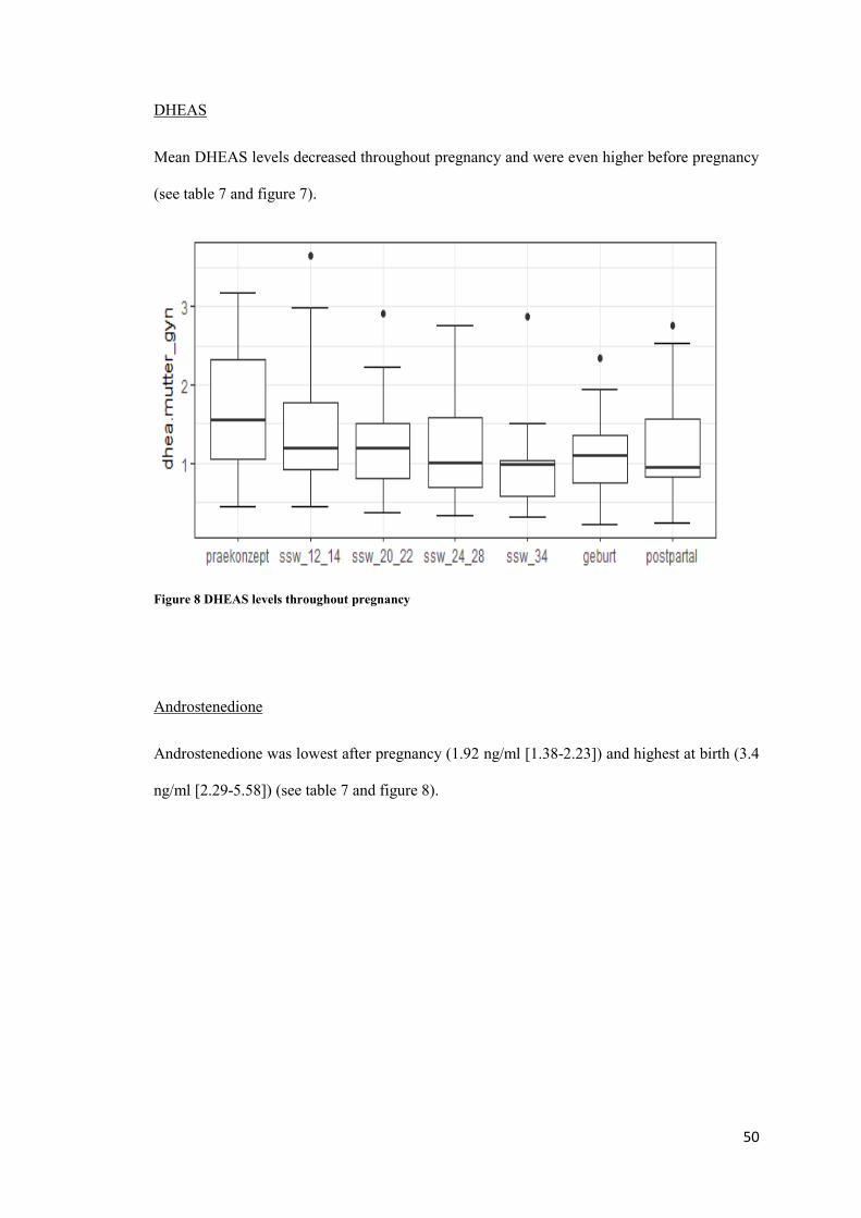
50
DHEAS
Mean DHEAS levels decreased throughout pregnancy and were even higher before pregnancy
(see table 7 and figure 7).
Figure 8 DHEAS levels throughout pregnancy
Androstenedione
Androstenedione was lowest after pregnancy (1.92 ng/ml [1.38-2.23]) and highest at birth (3.4
ng/ml [2.29-5.58]) (see table 7 and figure 8).

51
Figure 9 Androstenedione levels throughout pregnancy
AMH
AMH levels decreased throughout pregnancy. Highest levels were measured before pregnancy
(6.1 ng/ml [3.8-8.0]). After delivery levels went up again (see table 7 and figure 9).
Figure 10 AMH levels throughout pregnancy

52
SHBG
SHBG levels were high throughout pregnancy and decreased after delivery (see table 7 and
figure 10).
Figure 11 SHBG levels throughout pregnancy
Secondary outcomes
Mean gestational age at delivery was 269.7±30.9 days and mean birth weight was
3131.4±866.4 grams. One (4.3%) fetus died in utero (gestational age 152 days). An
intrauterine growth restriction was found in 2 (8.7%) fetuses. One (4.3%) newborn was large
for gestational age and 3 (13%) were small for gestational age. Three (13%) women presented
with a polyhydramnion and 2 (8.7%) women developed gestational diabetes – additionally to
the 3 women with pregestational diabetes. Pregnancy-induced hypertension was present in 5
(21.7%) women and 2 (8.7%) women developed pre-eclampsia. Operative delivery, such as
elective caesarean section, non-elective caesarean section and operative vaginal delivery was
necessary in 14 (60.9%) women.

53
C. Discussion
This study found that androgens (testosterone, free testosterone, and androstenedione) and
SHBG levels increase throughout pregnancy, while DHEAS and AMH levels decrease.
Perinatal complications were frequent in the investigated cohort.
As suspected, only a limited number of PCOS patients could be recruited. However, data
dealing with this subject are scarce.
Our results regarding hormone levels throughout pregnancy in PCOS women are in
accordance with findings in women without PCOS. As described in the introduction section,
numerous studies have found elevated levels of circulating androgens during normal
pregnancy (Bammann et al., 1980; Buster et al., 1979; Dawood and Saxena, 1977; Mizuno et
al., 1968; Rivarola et al., 1968; Saez et al., 1972). Also in agreement with the findings of a
previous study, we found that DHEAS levels are lower throughout pregnancy in PCOS
women (Milewich et al., 1978). SHBG levels do rise dramatically in PCOS women from the
first trimester until term as described in women without PCOS (Wilke and Utley, 1987).
Finally, the decline of AMH levels throughout gestation which is known from controls was
also seen in our cohort (Köninger et al., 2013; Köninger et al., 2015; La Marca et al., 2005; Li
et al., 2010; Nelson et al., 2010).
Further studies should compare the hormone levels throughout pregnancy in PCOS women
and controls. Although the dynamic is similar, subtle changes in increase and decrease of
levels cannot be answered with this study design. Maliqueo et al. aimed to evaluate the
placental activities of steroid sulfatase, 3β-hydroxysteroid dehydrogenase type 1 and P450
aromatase in pregnant PCOS women compared to normal pregnant women. Two important
enzymes for steroid synthesis, 3β-hydroxysteroid dehydrogenase type 1 and P450 aromatase,
showed an altered activity in placental tissue of women with PCOS. There was a higher
activity of 3β-hydroxysteroid dehydrogenase type 1 and a lower activity of P450 aromatase.
54
This finding could explain the increased androgen production during pregnancy (Maliqueo et
al., 2013).
As mentioned above, perinatal complications were frequent in the investigated cohort.
Gestational diabetes and pregnancy-induced hypertension were present in 21.7% of women,
respectively and 8.7% of women developed pre-eclampsia. In comparison to our other two
bigger cohorts the rates are higher for pregnancy-induced hypertension and pre-eclampsia
(Kollmann et al., 2015). Rates for gestational diabetes are comparable (Kollmann et al., 2015).
An explanation for the variable findings might be the relatively small sample size in this
cohort. As we know from the other two cohorts, PCOS women develop more perinatal
complications and should therefore be informed and advised to follow regular checks in units
where troubles can be identified early to allow specialized care (Kollmann et al., 2015).

55
XII. 3rd study – Cross-sectional study of pregnant PCOS women and their
offspring compared to non-PCOS women and their offspring
A. Material and Methods
Study design
Prospective cohort study
Setting
Women with PCOS according to ESHRE/ASRM 2003 criteria and non-PCOS women, who
were treated at the Medical University of Graz, Austria, between March 2013 and December
2015, were prospectively included in the study.
Ethical Approval
The study was approved by the institutional review board (No: 24-179ex11/12).
Participants
Women, over 18 years, with PCOS according to ESHRE/ASRM 2003 criteria and non PCOS-
women were included. PCOS was diagnosed following clinical and sonographic evaluation
and a hormonal analysis. The (healthy) control group comprised women without PCOS. Study
examinations were performed at birth. Ultrasound examinations were performed by medical
doctors specialized in obstetrics and gynaecology. Clinical investigations were performed at
the Division of Endocrinology and Metabolism at the Department of Internal Medicine at
Medical University of Graz, Austria. Patient data were extracted from the local perinatal
database (PIA, ViewPoint, GE Healthcare, Zipf, Austria) and the medical documentation
system or patient files. We report on a population of women that were treated in our
specialized unit and were delivered at our institution.
Variables
Following examinations were performed:

56
At birth:
• Determination of maternal serum hormone levels (T, fT, SHBG, DHEAS, ANDR,
AMH)
• Determination of newborn cord blood hormone levels (T, fT, SHBG, DHEAS, ANDR,
AMH)
• Documentation and evaluation of perinatal outcome parameters:
Maternal pregnancy complications: gestational diabetes, pregnancy-induced
hypertension, pre-eclampsia, eclampsia, polyhydramnios, oligohydramnios, operative
delivery (elective and non-elective caesarean section and operative vaginal delivery)
Neonatal complications: preterm birth (< 37 and < 34 weeks of gestation,
respectively), intrauterine growth restriction (IUGR), small for gestational age (SGA),
large for gestational age (LGA), fetal acidosis, pre- and perinatal mortality, ICU
admission rate, intrauterine fetal death (IUFT)
Primary outcome parameters were maternal and newborn hormone levels (T, fT, SHBG,
DHEAS, ANDR, and AMH). Secondary outcome parameters were specific perinatal
complications.
Data sources/ Measurement/ Quantitative variables
The evaluation of maternal serum hormone levels was performed at the Division of
Endocrinology and Metabolism at the Department of Internal Medicine at Medical University
of Graz, Austria. Cut-off values are listed in table 6.
Maternal gestational diabetes was diagnosed based on the oral glucose tolerance test (oGTT,
HemoCue, Ängelholm, Sweden). At our unit maternal gestational diabetes is diagnosed based
on the oral glucose tolerance test (oGTT, HemoCue, Ängelholm, Sweden) which is
implemented in routine pregnancy care in Austria as well as on cord blood insulin testing
which is routinely performed in all infants born with a weight ≥ 4000 g. The normal range of

57
cord blood insulin is 3 - 25 mU/l (ADVIA Centaur, Siemens, Germany) (Chevenne et al.,
1999). In the observed period oGTT was performed between 24 – 28 weeks of gestation by
venous blood analysis after 12h fasting and one and two hours following administration of 75g
glucose. Glucose cut-off values to diagnose maternal gestational diabetes were 92 / 180 / 153
mg/dl (Metzger et al., 2010). Measurement of blood pressure (systolic and diastolic), maternal
body weight and analysis of a urine sample were performed at admission. If blood pressure
was high patients were advised to perform self-measurements at home. Pregnancy-induced
hypertension (PIH) was defined as systolic blood pressure ≥ 140 mmHg or diastolic blood
pressure ≥ 90 mmHg in 5 out of 21 measurements arising after 20 weeks of gestation. Pre-
eclampsia was defined as PIH with proteinuria (≥ 300 mg in 24 hours) (Brown et al., 2001).
Operative delivery was defined as birth by forceps, ventouse or caesarean section. Neonatal
birth weight and foetal umbilical pH value are determined straight after delivery. Small for
gestational age and large for gestational age was assumed, when birth weight was below the
10th percentile or above the 90
th percentile (Voigt et al., 2010). Intrauterine growth restriction
was defined as SGA and the presence of distinct signs of placental insufficiency such as
pathological Doppler waveforms in the umbilical (elevated pulsatility index, absent or
reversed end-diastolic flow) or middle cerebral artery (decreased pulsatility index) as well as a
cerebroplacental Doppler ratio (middle cerebral artery pulsatility index/umbilical artery
pulsatility index) below 1 (Bahado-Singh et al., 1999; Baschat and Gembruch, 2003; Odibo et
al., 2005; Oros et al., 2011). For evaluation of fetal acidosis umbilical arterial blood was
analysed (ABL 800 FLEX analyser, Akandevej, Denmark). Fetal acidosis was defined as an
umbilical artery pH < 7.10 at birth (Reif et al., 2014; Reif et al., 2015).
Study size
To detect significant differences with an alpha of 0.05 and a power of 80%, we planned to
recruit at least 35 patients with PCOS and 350 non-PCOS patients. As we expected some drop
outs and difficulties with cord blood analysis, we aimed to include at least 400 patients. An
interims analysis revealed that more patients than expected had to be excluded due to

58
comorbidities in the non-PCOS group and that cord blood analysis was not feasible in some
cases due to insufficient material. We therefore aimed to recruit at least 80 PCOS patients and
420 non-PCOS patients.
Statistical methods
For categorical variables relative and absolute proportions are indicated, continuous variables
are expressed as mean ± standard deviation or median and range, respectively. Categorical
variables were analysed by using Fisher’s exact test or chi-squared test with p-value computed
by Monte Carlo simulation, while for continuous outcomes one-way analysis of variance
(ANOVA) or Kruskal-Wallis rank sum test followed by Bonferroni correction was applied.
All analyses were performed using the statistic software R (version 3.1.1, Vienna, Austria). A
p-value <0.05 was considered to be statistically significant.
B. Results
Participants
A total of 499 pregnant women were assessed for eligibility and 433 were finally included for
analysis (79 PCOS and 354 Non-PCOS women). Four women did not want to participate, 7
women were not included due to twin pregnancies (4 PCOS, 3 Non-PCOS) [488], and 55
patients (11 PCOS, 44 Non-PCOS) were not included due to severe maternal or fetal
comorbidities.
PCOS women gave birth to 36 (45.6%) girls and 43 (54.4%) boys. In the Non-PCOS group
178 (50.3%) girls and 176 (49.7%) boys were born.
Baseline data
Mean age in PCOS women was 30.6±4.6 years and 30.3±5.1 years in Non-PCOS women.
Body mass index was 29.8±6.1 kg/m² in the PCOS group and 28.9±5.0 kg/m² in the Non-

59
PCOS group, respectively. 3.8% of PCOS women and 8.8 % of Non-PCOS women did smoke
during pregnancy. Mean gestational age at delivery was 279.0±9.6 days in the PCOS and
281.2±6.9 in the control group (see table 8).
PCOS patients showed the following symptoms: 46 (58.2%) women presented with
hyperandrogenemia, 67 (84.8%) with clinical hyperandrogenism, 53 (67.1%) had polycystic
ovaries, and 68 (86.1%) with oligo-, amenorrhoea. Forty one (51.9%) women in the PCOS
group and 27 (7.6%) in the control group reported that they did not conceive within one year.
Regarding the actual pregnancy, 67 (84.8%) in the PCOS group versus 346 (97.7%) in the
control group conceived spontaneously; 7 (8.9%) versus 1 (0.3%) got pregnant after
stimulation and ovulation induction, 5 (6.3%) versus 7 (1.9%) were treated with IVF/ICSI.
Table 8 Comparison of demographics
PCOS women
n=79
Non-PCOS women
n=354
Mean
n
± SD
%
Mean
n
± SD
%
p-
value
Maternal characteristics
Maternal Age (years) 30.6 ±4.6 30.3 ±5.1 0.660
Body mass index (kg/m2)*
29.8 ±6.1 28.9 ±5.0 0.241
Smoking
3/79 3.8% 31/354 8.8% 0.169
Neonatal characteristics
Gestational age (days) at delivery 279 ±9.6 281.2 ±6.9 0.058

60
Hormone levels
Testosterone
Testosterone was measureable in 84.81% (67/79) of women with PCOS and in 89.26%
(316/354) of Non-PCOS women. Neonatal samples were available in 27 PCOS girls (75%),
151 Non-PCOS girls (84%). 29 PCOS boys (67.4%), and 149 Non-PCOS boys (84.7%).
Mean maternal testosterone was 1.17 ng/ml [range 0.44-4.23] in PCOS women and 0.97 ng/ml
[0.18-5.56] in Non-PCOS women. Levels were significantly higher in PCOS women
(p<0.001).
Mean testosterone levels from PCOS girls were 1.54 ng/ml [0.84-5.82] and 1.82 ng/ml [0.88-
13.05] from Non-PCOS girls. Those levels did not differ significantly (p=0.230).
Mean testosterone levels from PCOS boys (2.17 ng/ml [1.16-10-10]) and Non-PCOS boys
(1.76 ng/ml [0.95-9.52]) did not differ either (p=0.120) (figure 11).
Figure 12 Testosterone levels in mothers, girls and boys
Free Testosterone
Free Testosterone was measureable in 84.81% (67/79) of women with PCOS and in 89.26%
(316/354) of Non-PCOS women. Neonatal samples were available in 26 PCOS girls (72.2%),
150 Non-PCOS girls (84.27%). 29 PCOS boys (67.4%), and 149 Non-PCOS boys (84.7%).
Mean maternal free testosterone was 6.79 pg/ml [1.37-26.80] in PCOS women and 6.72 pg/ml
[0.48-27.91] in Non-PCOS women. Levels did not differ significantly (p=0.563).
Mean testosterone levels from PCOS girls were 22.66 pg/ml [10.52-47.0] and 23.89 pg/ml
[6.12-72.01] from Non-PCOS girls. Those levels did not differ significantly (p=0.196).
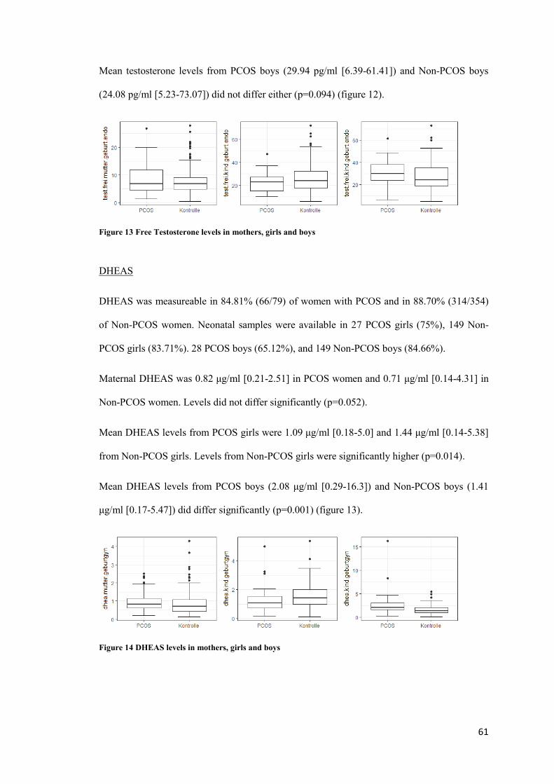
61
Mean testosterone levels from PCOS boys (29.94 pg/ml [6.39-61.41]) and Non-PCOS boys
(24.08 pg/ml [5.23-73.07]) did not differ either (p=0.094) (figure 12).
Figure 13 Free Testosterone levels in mothers, girls and boys
DHEAS
DHEAS was measureable in 84.81% (66/79) of women with PCOS and in 88.70% (314/354)
of Non-PCOS women. Neonatal samples were available in 27 PCOS girls (75%), 149 Non-
PCOS girls (83.71%). 28 PCOS boys (65.12%), and 149 Non-PCOS boys (84.66%).
Maternal DHEAS was 0.82 μg/ml [0.21-2.51] in PCOS women and 0.71 μg/ml [0.14-4.31] in
Non-PCOS women. Levels did not differ significantly (p=0.052).
Mean DHEAS levels from PCOS girls were 1.09 μg/ml [0.18-5.0] and 1.44 μg/ml [0.14-5.38]
from Non-PCOS girls. Levels from Non-PCOS girls were significantly higher (p=0.014).
Mean DHEAS levels from PCOS boys (2.08 μg/ml [0.29-16.3]) and Non-PCOS boys (1.41
μg/ml [0.17-5.47]) did differ significantly (p=0.001) (figure 13).
Figure 14 DHEAS levels in mothers, girls and boys

62
Androstenedione
Androstenedione was measureable in 83.54% (66/79) of women with PCOS and in 88.14%
(312/354) of Non-PCOS women. Neonatal samples were available in 26 PCOS girls (72.22%),
147 Non-PCOS girls (82.58%). 27 PCOS boys (62.79%), and 148 Non-PCOS boys (84.09%).
Maternal androstenedione was 3.44 ng/ml [1.06-10] in PCOS women and 2.74 ng/ml [0.49-
10] in Non-PCOS women. Levels did differ significantly (p=0.002).
Mean androstenedione levels from PCOS girls were 2.19 ng/ml [1.08-7.77] and 2.78 ng/ml
[0.83-8.06] from Non-PCOS girls. Those levels did not differ significantly (p=0.113).
Mean androstenedione levels from PCOS boys (3.47 ng/ml [1.26-7.93]) and Non-PCOS boys
(2.92 ng/ml [0.83-9.24]) did differ significantly (p=0.039) (figure 14).
Figure 15 Androstenedione levels in mothers, girls and boys
AMH
AMH was measureable in 84.81% (66/79) of women with PCOS and in 88.42% (313/354) of
Non-PCOS women. Neonatal samples were available in 27 PCOS girls (75%), 147 Non-PCOS
girls (82.58%). 27 PCOS boys (62.79%), and 148 Non-PCOS boys (84.1%).
Maternal AMH was 1.10 ng/ml [0.10-25.0] in PCOS women and 0.72 ng/ml [0.02-49.0] in
Non-PCOS women. Levels did differ significantly (p=0.001).
Mean AMH levels from PCOS girls were 0.2 ng/ml [0.0-9.2] and 0.2 ng/ml [0.0-25.0] from
Non-PCOS girls. Levels did not differ significantly (p=0.975).

63
Mean AMH levels from PCOS boys (22.0 ng/ml [14.4-45.6]) and Non-PCOS boys (20.01
ng/ml [1.6-124.2]) did not differ significantly (p=0.395) (figure 15).
Figure 16 AMH levels in mothers, girls and boys
SHBG
SHBG was measureable in 84.81% (67/79) of women with PCOS and in 89.27% (316/354) of
Non-PCOS women. Neonatal samples were available in 27 PCOS girls (75%), 150 Non-PCOS
girls (84.27%). 29 PCOS boys (67.44%), and 148 Non-PCOS boys (84.1%).
Maternal SHBG levels were >200 nmol/L in all patients in both groups.
Mean SHBG levels from PCOS girls were 32.0 nmol/L [15.0-35.0] and 35.0 nmol/L [1.20-
81.6] from Non-PCOS girls. Levels did not differ significantly (p=0.292).
Mean SHBG levels from PCOS boys (38.5 nmol/L [20.1-62.2]) and Non-PCOS boys (37.85
nmol/L [18.3-105.9]) did not differ significantly (p=0.919) (figure 16).
Figure 17 SHBG levels in girls and boys
Comparisons between PCOS women and Non-PCOS women, female offspring of PCOS
women and Non-PCOS women, and male offspring of PCOS women and Non-PCOS women
are summarized in tables 9-11.

64
PCOS women Non-PCOS women p
Testosterone (ng/ml)** 1.17 [0.44-4.23] 0.97 [0.18-5.56] <0.001
Free Testosterone
(pg/ml) ** 6.79 [1.37-26.8] 6.72 [0.48-27.91] 0.563
Androstenedione
(ng/ml) ** 3.44 [1.06-10.0] 2.74 [0.49-10.0] 0.002
DHEAS (μg/ml) 0.82 [0.21-2.51] 0.71 [0.14-4.31] 0.052
AMH (ng/ml) 1.10 [0.10-25.0] 0.72 [0.02-49.0] 0.001
SHBG (nmol/L) - - - Table 9 Comparison PCOS women and Non-PCOS women; Data presented as median [range] and comparison by U-
test.
Female offspring of PCOS
women
Female offspring
of Non-PCOS
women p
Testosterone (ng/ml)** 1.54 [0.84-5.82] 1.82 [0.88-13.05] 0.230
Free Testosterone
(pg/ml) ** 22.66 [10.52-47.0] 23.89 [6.12-72.01] 0.196
Androstenedione
(ng/ml) ** 2.19 [1.08-7.77] 2.78 [0.83-8.06] 0.113
DHEAS (μg/ml) 1.09 [0.18-18.5] 1.44 [0.14-5.38] 0.014
AMH (ng/ml) 0.20 [0.00-9.2] 0.20 [0.00-25.0] 0.975
SHBG (nmol/L) 32.0 [20.17-39.0] 35.0 [1.2-81.6]] 0.292 Table 10 Comparison female offspring of PCOS women and Non-PCOS women; Data presented as median [range]
and comparison by U-test.
Male offspring of PCOS
women
Male offspring of Non-PCOS
women p
Testosterone (ng/ml)** 2.17 [1.16-10.10] 1.76 [0.95-9.52] 0.120
Free Testosterone
(pg/ml) ** 29.94 [6.39-61.41] 24.08 [5.23-73.07] 0.094
Androstenedione
(ng/ml) ** 3.47 [1.26-7.93] 2.92 [0.83-9.24] 0.039
DHEAS (μg/ml) 2.08 [0.29-16.3] 1.41 [0.17-5.47] 0.001
AMH (ng/ml) 22.0 [14.4-45.6] 20.01 [1.6-124.2] 0.395
SHBG (nmol/L) 38.5 [20.1-62.0] 37.85 [18.3-105.9] 0.919 Table 11 Comparison male offspring of PCOS women and Non-PCOS women; Data presented as median [range] and
comparison by U-test.

65
Pregnancy outcome
Compared to healthy controls, the risk for maternal complications was increased in PCOS
women (51/79 [64.56%] versus 171/354 [48.31%]; p=0.009), while there was no significant
difference in neonatal complications (17/79 [21.52%] versus 79/354 [22.32%]; p=1.0) (table
12).
PCOS women
n=79
Non-PCOS women
n=354
n % n % p-value
Maternal complications
Gestational diabetes 12/79 15.19 21/354 5.93 0.009
Pregnancy-induced hypertension 8/79 10.13 17/354 4.8 0.103
Pre-eclampsia 3/79 3.8 5/354 1.41 0.164
Operative delivery 41/79 51.89 148/354 41.8 0.259
Total complication rate 51/79 64.56 171/354 48.31 0.009
Neonatal complications
SGA (< 10th
percentile) 9/79 11.39 44/354 12.43 1.0
LGA (> 90th
percentile) 3/79 3.8 19/354 5.37 0.779
IUGR 1/79 1.27 3/354 0.85 0.555
Fetal acidosis 2/79 2.53 10/354 2.82 1.0
ICU 1/79 1.27 5/354 1.41 1.0
Total complication rate 17/79 21.52 79/354 22.32 1.0
Table 12 Maternal and neonatal complications in PCOS pregnancies
Operative delivery = elective and non-elective caesarean section and operative vaginal delivery
Total maternal complication rate = gestational diabetes, pregnancy-induced hypertension, pre-eclampsia, non-elective
caesarean section, and operative vaginal delivery
SGA = small for gestational age; ICU = intensive care unit admission, IUGR=intrauterine growth restriction
Total neonatal complication rate = large or small for gestational age, IUGR, acidosis, ICU admission, and pre- and
perinatal mortality
66
Women with PCOS had a higher risk of developing gestational diabetes (12/79 [15.19%]
versus 21/354 [5.93%]; p=0.009) when compared to healthy controls. No significant
difference was found for pregnancy-induced hypertension (8/79 [10.13%] versus 17/354
[4.8%]; p=0.103), pre-eclampsia (3/79 [3.8%] versus 5/354 [1.41%]; p=0.164), and operative
delivery (41/79 [51.89%] versus 148/354 [41.8%]; p=0.259).
Large (p=0.779) or small (p=1.0) for gestational age newborns, intrauterine growth restriction
(p=0.555), fetal acidosis (p=1.0), and ICU admission rate (p=1.0) occurred with a comparable
frequency in both groups (table 12).
C. Discussion
Our main findings are that androgen levels in female offspring of PCOS and non-PCOS
women do not differ although maternal hormone levels differ significantly. Boys of PCOS
women show similar hormone levels compared to boys of non-PCOS women; only
androstenedione levels were higher in boys of PCOS women. Compared to healthy controls,
the risk for maternal complications was increased in PCOS women, while there was no
significant difference in neonatal complications.
Our findings do not support the hypothesis that elevated androgen concentrations in PCOS
mothers lead to elevated levels in the offspring. This hypothesis has been strongly encouraged
by Abbott et al., who performed studies in rhesus monkeys, which were prenatally exposed to
androgens (Abbott and Bacha, 2013; Abbott et al., 2005; Abbott et al., 2008a; Abbott et al.,
2002; Abbott et al., 2013; Abbott et al., 2009; Abbott et al., 2008b; Dumesic et al., 1997).
However, there are not many available human studies dealing with this subject. The most
recent study was published in 2016 (Caanen et al., 2016). In the latter study researchers
investigated androgen levels in 20 PCOS women and 83 controls at 20 weeks of gestation and
at delivery. They also looked at the umbilical cord blood of the respective offspring. Analysed
parameters were testosterone, androstenedione, dehydroepiandrostenedione, estrone, estradiol,

67
and estriol. Hormones were measured using the liquid chromatography tandem mass
spectrometry methods. The results were similar to ours. In spite of elevated maternal androgen
concentrations during pregnancy in PCOS women, offspring showed no signs of elevated
androgen concentrations in cord blood at birth (Caanen et al., 2016). Other available studies
show assimilable results (Anderson et al., 2010; Boutzios et al., 2013; Hickey et al., 2009;
Maliqueo et al., 2013). Anderson et al included 39 PCOS patients and 31 controls. They
investigated mixed cord blood testosterone, androstenedione, dehydroepiandrosterone, 17-
hydroxyprogesterone, estradiol, and dihydrotestosterone levels. Testosterone and
dehydroepiandrosterone did not differ significantly in the offspring (Anderson et al., 2010).
Interestingly, the study found, equal to further two studies, lower levels of androstenedione in
cord blood of female offspring of PCOS women in comparison to female offspring of non-
PCOS women (Anderson et al., 2010; Caanen et al., 2016; Maliqueo et al., 2013). Cord blood
estradiol levels were also significantly lower in PCOS women, without any difference in the
testosterone to estradiol ratio. Authors suggest a decreased fetal or placental production of
steroids(Anderson et al., 2010). Boutzios et al. performed a study on PCOS patients (n=41),
patients with a gestational diabetes (n=54), controls (n=56) and the respective offspring
(Boutzios et al., 2013). Testosterone levels did differ in mothers, but did not show any
difference in the offspring (Boutzios et al., 2013). Maliqueo et al. examined 20 pregnant
PCOS women and 30 controls and determined progesterone, DHEAS, DHEA,
androstenedione, testosterone, estrone, estradiol and total estriol (Maliqueo et al., 2013). They
further investigated the activities of steroid sulfatase, 3β-hydroxysteroid dehydrogenase type
1, and P450 aromatase in placental tissue (Maliqueo et al., 2013). Findings regarding
hormonal levels were similar to ours. Despite of elevated maternal androgen concentrations in
PCOS women they did not find a difference in the offspring (Maliqueo et al., 2013).But as
mentioned above this group found lower levels of androstenedione and higher estriol
concentrations in female newborns of women with PCOS compared to daughters of control
women (Maliqueo et al., 2013). In comparison to control women, placental tissue from women
with PCOS showed higher 3β-hydroxysteroid dehydrogenase type 1 and lower P450

68
aromatase activities which could increase androgen production during pregnancy (Maliqueo et
al., 2013). There is only one study which showed elevated testosterone levels in cord blood in
female offspring of PCOS mothers compared to controls (Barry et al., 2010). Overall, it seems
that the hypothesis that maternal androgen excess contributes to elevated androgen
concentrations in the offspring and therefore lead to the development of PCOS cannot be
supported. A large prospective cohort study, which evaluated the relationship between prenatal
androgen exposure and PCOS in adolescence in normal pregnancy, did not find a statistically
significant relationship too (Hickey et al., 2009).
Reasons for this finding may be protective mechanisms which work throughout pregnancy. As
mentioned in the introduction section we are aware of the fact that there are pregnancy-
specific mechanisms to protect both the mother and fetus from pregnancy-induced androgen
excess (Crisosto et al., 2012; Hensleigh et al., 1975; Phelan and Conway, 2011). First of all the
placenta forms an effective barrier by producing placental aromatase (Kragie, 2002). The
aromatase quickly catalyses the conversion of androstenedione to estrone, 16-
hydroxytestosterone to estriol and testosterone to estradiol (Kragie, 2002). Another
mechanism is the increase of progesterone, which competes for androgen receptor binding
(Birrell et al., 2007; Slayden et al., 2001) and has an affinity for 5a-reductase which further
results in an inhibition of the conversion of T to the more potent dihydrotestosterone (DHT)
(Cabeza et al., 1999; Hodgins, 1982). A further protective factor is the physiological increase
of maternal SHBG which was found in both groups and which leads to higher share of
bounded and biologically inactive sex steroids.
Although we did not find higher androgen levels in the offspring, a negative effect of the
higher androgen concentrations in PCOS mothers during pregnancy cannot be ruled out.
Altered hormone levels might have their effect on PCOS offspring in a more indirect way such
as fetal programming. A study on zebrafish embryos which were exposed to androgens
(testosterone and dihydrotestosterone) showed altered global methylation levels in the ovary
and elevated postprandial glucose levels (Xu et al., 2014). Similar results were found by

69
Zhang et al and Xu et al who examined the effect of a hyperandrogenic milieu in utero on rats
(Zhang et al., 2014) and on rhesus monkeys (Xu et al., 2011). Data on humans are scarce. The
first pilot study on PCOS patients and controls did not find a significant difference in global
methylation (6.7% for PCOS women and 7.1% for controls) (Xu et al., 2010), whereas a more
recent study report a significant difference (Yu et al., 2015). The latter study also looked at
seven potentially interesting gene loci and found a hypermethylation in the promotor region of
SLC2A8, NRIP1, IGF2BP2, AMHR2 and a hypomethylation of INSR und AMH (Yu et al.,
2015).
The effect of different PCOS phenotypes on perinatal outcome has been studied in our first
cohort (Kollmann et al., 2015). While a study by Shroff et al. suggests that the non-
hyperandrogenic phenotype may represent a subgroup of PCOS associated with a milder
metabolic profile we did not find a difference regarding perinatal outcome (Kollmann et al.,
2015; Shroff et al., 2007). Regarding this cohort we have to bear in mind that not all mothers
had biochemically proven hyperandrogenism. However, 58.2% of women presented with
hyperandrogenemia and 84.8% with clinical hyperandrogenism. Moreover, PCOS women had
significant higher androgen levels when compared to non-PCOS women. Nevertheless, further
studies should investigate how different maternal PCOS phenotypes influence the hormonal
levels of the offspring.
The ideal way to examine hormone exposure during prenatal life would be the measurement of
circulating fetal hormone levels at repeated time points during pregnancy. However, this is not
without significant risk to the fetus and hence surrogate markers of fetal hormone levels must
be applied. A relatively simple way is the collection of umbilical cord blood at birth (Hollier et
al., 2014). Alternatively, amniotic fluid samples could be investigated. However, this approach
is also associated with a higher complication rate and can therefore not be used in study
settings. Furthermore, the exact correlation between hormone levels in fetal blood and
amniotic fluid is not exactly known (Hollier et al., 2014).

70
Fetal blood, which is loaded with placental steroid metabolites and some maternal steroids
leaves the placenta via the umbilical vein and returns from the fetus to the placenta via the
umbilical artery (Ishimoto and Jaffe, 2011). A review published in 2014 examined the
accuracy and biological interpretation of the measurement of androgens and estrogens in cord
blood (Hollier et al., 2014). Usually venous and arterial blood samples are taken at once and
the exact proportion of each component is not known precisely. None the less, in spite of
different steroid concentrations in umbilical artery and vein, Pašková et al. showed that
concentrations strongly correlate (Hollier et al., 2014; Pašková et al., 2014). However, one has
to be cautious when interpreting hormone levels which were measured in umbilical cord
blood. Various factors, such as obstetric and maternal ones can influence the concentrations,
although the amount of this influence is not exactly known (Hollier et al., 2014). On the one
hand it is known that gestational age and labour have an effect on fetal adrenal steroid
production and on the other hand we know that levels of steroid metabolizing enzymes in the
placenta are modulated by factors connected to labour and delivery such as glucocorticoids,
pro-inflammatory cytokines, and exposure to reactive oxygen species (Albrecht and Pepe,
1990; Hollier et al., 2014). Recently published studies showed that gestational age and
delivery significantly influence androgen and estrogen concentrations in cord blood (Hickey et
al., 2014; Keelan et al., 2012). Labour was associated with significant lower median cord
blood testosterone and free testosterone levels and higher SHBG, androstenedione and
dehydroepiandrosterone levels. Gestational age at delivery was significantly negatively
correlated with testosterone and free testosterone and significantly positive correlated with
SHBG, androstenedione, and dehydroepiandrosterone. They further investigated the effect of
an antenatal glucocorticoid administration and also found a significant effect (Keelan et al.,
2012). Looking at our cohort we report no significant difference for those factors when we
compared PCOS women and non-PCOS women. However, we found a significant higher rate
of gestational diabetes and total maternal complication rate in PCOS women and a possible
association should be investigated in a further step.

71
Radioimmunoassay (RIA) was used as the detection methodology for hormonal levels in our
study and we are aware of issues related to this method. A main limitation of RIA is the
measurement of low concentrations of sex steroids (Demers, 2010; Hollier et al., 2014; Rosner
et al., 2007; Stanczyk et al., 2003). Particularly for testosterone we know that levels derived
from mass spectrometry are consistently lower than those derived by RIA (Demers, 2010;
Krogh et al., 2011; Soldin et al., 2005; Stanczyk and Clarke, 2010; Vicente et al., 2006). Our
levels did not show a significant difference and we do not expect testosterone levels to be
significantly different in the investigated groups when using mass spectrometry.
Regarding our secondary outcome we found that the risk for maternal complications was
increased in PCOS women when compared to healthy controls, while there was no significant
difference in neonatal complications. This finding is identical to the results from our first
study and in accordance to published data (Boomsma et al., 2006; Kollmann et al., 2015;
Palomba et al., 2015; Qin et al., 2013).
72
XIII. Conclusion
Regardless of the PCOS definition, about 60% of women with PCOS and about 30% of their
infants were subject to perinatal and neonatal complications. In comparison to healthy controls
the risk for maternal complications was increased in PCOS women (OR 2.57 [1.82-3.64])
while there was no difference in neonatal complications (OR 0.83 [0.56-1.21]).
Serum levels of androgens (testosterone, free testosterone, and androstenedione) and SHBG
increased throughout pregnancy, while those of DHEAS and AMH decreased. Postnatal
Androgen levels in female offspring of PCOS and non-PCOS women did not differ, although
maternal hormone levels differed significantly. Boys of PCOS women showed similar
hormone levels postpartum compared to boys of non-PCOS women; only androstenedione
levels were higher in boys of PCOS women.

73
XIV. References
Abbott, D. H., and F. Bacha, 2013, Ontogeny of polycystic ovary syndrome and insulin resistance in utero and early childhood: Fertil Steril, v. 100, p. 2-11.
Abbott, D. H., D. K. Barnett, C. M. Bruns, and D. A. Dumesic, 2005, Androgen excess fetal programming of female reproduction: a developmental aetiology for polycystic ovary syndrome?: Hum Reprod Update, v. 11, p. 357-74.
Abbott, D. H., D. K. Barnett, J. E. Levine, V. Padmanabhan, D. A. Dumesic, S. Jacoris, and A. F. Tarantal, 2008a, Endocrine antecedents of polycystic ovary syndrome in fetal and infant prenatally androgenized female rhesus monkeys: Biol Reprod, v. 79, p. 154-63.
Abbott, D. H., D. A. Dumesic, and S. Franks, 2002, Developmental origin of polycystic ovary syndrome - a hypothesis: J Endocrinol, v. 174, p. 1-5.
Abbott, D. H., L. E. Nicol, J. E. Levine, N. Xu, M. O. Goodarzi, and D. A. Dumesic, 2013, Nonhuman primate models of polycystic ovary syndrome: Mol Cell Endocrinol, v. 373, p. 21-8.
Abbott, D. H., B. H. Rayome, D. A. Dumesic, K. C. Lewis, A. K. Edwards, K. Wallen, M. E. Wilson, S. E. Appt, and J. E. Levine, 2017, Clustering of PCOS-like traits in naturally hyperandrogenic female rhesus monkeys: Hum Reprod, v. 32, p. 923-936.
Abbott, D. H., A. F. Tarantal, and D. A. Dumesic, 2009, Fetal, infant, adolescent and adult phenotypes of polycystic ovary syndrome in prenatally androgenized female rhesus monkeys: Am J Primatol, v. 71, p. 776-84.
Abbott, D. H., R. Zhou, I. M. Bird, D. A. Dumesic, and A. J. Conley, 2008b, Fetal programming of adrenal androgen excess: lessons from a nonhuman primate model of polycystic ovary syndrome: Endocr Dev, v. 13, p. 145-58.
Abraham, G. E., 1974, Ovarian and adrenal contribution to peripheral androgens during the menstrual cycle: J Clin Endocrinol Metab, v. 39, p. 340-6.
Agha, M., R. A. Agha, J. Sandall, and J. Sandell, 2014, Interventions to reduce and prevent obesity in pre-conceptual and pregnant women: a systematic review and meta-analysis: PLoS One, v. 9, p. e95132.
Al-Inany, H. G., M. A. F. M. Youssef, M. Aboulghar, F. Broekmans, M. Sterrenburg, J. Smit, and A. M. Abou-Setta, 2011, GnRH antagonists are safer than agonists: An update of a Cochrane review: Hum Reprod Update, v. 17, p. 435.

74
Albrecht, E. D., and G. J. Pepe, 1990, Placental steroid hormone biosynthesis in primate pregnancy: Endocr Rev, v. 11, p. 124-50.
Albrecht, E. D., and G. J. Pepe, 1995, Suppression of maternal adrenal dehydroepiandrosterone and dehydroepiandrosterone sulfate production by estrogen during baboon pregnancy: J Clin Endocrinol Metab, v. 80, p. 3201-8.
Alvarez-Blasco, F., J. I. Botella-Carretero, J. L. San Millán, and H. F. Escobar-Morreale, 2006, Prevalence and characteristics of the polycystic ovary syndrome in overweight and obese women: Arch Intern Med, v. 166, p. 2081-6.
Anderson, H., N. Fogel, S. K. Grebe, R. J. Singh, R. L. Taylor, and A. Dunaif, 2010, Infants of women with polycystic ovary syndrome have lower cord blood androstenedione and estradiol levels: J Clin Endocrinol Metab, v. 95, p. 2180-6.
Apridonidze, T., P. A. Essah, M. J. Iuorno, and J. E. Nestler, 2005, Prevalence and characteristics of the metabolic syndrome in women with polycystic ovary syndrome: J Clin Endocrinol Metab, v. 90, p. 1929-35.
Association, A. D., 2016a, Erratum. Classification and diagnosis of diabetes. Sec. 2. In Standards of Medical Care in Diabetes-2016. Diabetes Care 2016;39(Suppl. 1):S13-S22: Diabetes Care, v. 39, p. 1653.
Association, A. D., 2016b, Standards of Medical Care in Diabetes-2016 Abridged for Primary Care Providers: Clin Diabetes, v. 34, p. 3-21.
Asunción, M., R. M. Calvo, J. L. San Millán, J. Sancho, S. Avila, and H. F. Escobar-Morreale, 2000, A prospective study of the prevalence of the polycystic ovary syndrome in unselected Caucasian women from Spain: J Clin Endocrinol Metab, v. 85, p. 2434-8.
Azziz, R., E. Carmina, Z. Chen, A. Dunaif, J. S. Laven, R. S. Legro, D. Lizneva, B. Natterson-Horowtiz, H. J. Teede, and B. O. Yildiz, 2016, Polycystic ovary syndrome: Nat Rev Dis Primers, v. 2, p. 16057.
Azziz, R., E. Carmina, D. Dewailly, E. Diamanti-Kandarakis, H. F. Escobar-Morreale, W. Futterweit, O. E. Janssen, R. S. Legro, R. J. Norman, A. E. Taylor, S. F. Witchel, and T. F. o. t. P. o. t. P. O. S. o. T. A. E. a. P. Society, 2009, The Androgen Excess and PCOS Society criteria for the polycystic ovary syndrome: the complete task force report: Fertil Steril, v. 91, p. 456-88.
Bahado-Singh, R. O., E. Kovanci, A. Jeffres, U. Oz, O. Deren, J. Copel, and G. Mari, 1999, The Doppler cerebroplacental ratio and perinatal outcome in intrauterine growth restriction: Am J Obstet Gynecol, v. 180, p. 750-6.
Balsells, M., A. García-Patterson, I. Solà, M. Roqué, I. Gich, and R. Corcoy, 2015, Glibenclamide, metformin, and insulin for the treatment of gestational diabetes: a systematic review and meta-analysis: BMJ, v. 350, p. h102.

75
Bammann, B. L., C. B. Coulam, and N. S. Jiang, 1980, Total and free testosterone during pregnancy: Am J Obstet Gynecol, v. 137, p. 293-8.
Barker, D. J., 1992, The fetal origins of adult hypertension: J Hypertens Suppl, v. 10, p. S39-44.
Barker, D. J., 1995, Intrauterine programming of adult disease: Mol Med Today, v. 1, p. 418-23.
Barry, J. A., P. J. Hardiman, M. R. Siddiqui, and M. Thomas, 2011, Meta-analysis of sex difference in testosterone levels in umbilical cord blood: J Obstet Gynaecol, v. 31, p. 697-702.
Barry, J. A., A. R. Kay, R. Navaratnarajah, S. Iqbal, J. E. Bamfo, A. L. David, M. Hines, and P. J. Hardiman, 2010, Umbilical vein testosterone in female infants born to mothers with polycystic ovary syndrome is elevated to male levels: J Obstet Gynaecol, v. 30, p. 444-6.
Baschat, A. A., and U. Gembruch, 2003, The cerebroplacental Doppler ratio revisited: Ultrasound Obstet Gynecol, v. 21, p. 124-7.
Baumgarten, M., L. Polanski, B. Campbell, and N. Raine-Fenning, 2013, Do dopamine agonists prevent or reduce the severity of ovarian hyperstimulation syndrome in women undergoing assisted reproduction? A systematic review and meta-analysis: Hum Fertil (Camb), v. 16, p. 168-74.
Bergadá, I., C. Milani, P. Bedecarrás, L. Andreone, M. G. Ropelato, S. Gottlieb, C. Bergadá, S. Campo, and R. A. Rey, 2006, Time course of the serum gonadotropin surge, inhibins, and anti-Müllerian hormone in normal newborn males during the first month of life: J Clin Endocrinol Metab, v. 91, p. 4092-8.
Berger, N. G., J. T. Repke, and J. D. Woodruff, 1984, Markedly elevated serum testosterone in pregnancy without fetal virilization: Obstet Gynecol, v. 63, p. 260-2.
Birrell, S. N., L. M. Butler, J. M. Harris, G. Buchanan, and W. D. Tilley, 2007, Disruption of androgen receptor signaling by synthetic progestins may increase risk of developing breast cancer: FASEB J, v. 21, p. 2285-93.
Boomsma, C. M., M. J. Eijkemans, E. G. Hughes, G. H. Visser, B. C. Fauser, and N. S. Macklon, 2006, A meta-analysis of pregnancy outcomes in women with polycystic ovary syndrome: Hum Reprod Update, v. 12, p. 673-83.
Boutzios, G., S. Livadas, C. Piperi, N. Vitoratos, C. Adamopoulos, D. Hassiakos, C. Iavazzo, and E. Diamanti-Kandarakis, 2013, Polycystic ovary syndrome offspring display increased oxidative stress markers comparable to gestational diabetes offspring: Fertil Steril, v. 99, p. 943-50.

76
Braunstein, G. D., J. Rasor, H. Danzer, D. Adler, and M. E. Wade, 1976, Serum human chorionic gonadotropin levels throughout normal pregnancy: Am J Obstet Gynecol, v. 126, p. 678-81.
Broekmans, F. J., E. A. Knauff, O. Valkenburg, J. S. Laven, M. J. Eijkemans, and B. C. Fauser, 2006, PCOS according to the Rotterdam consensus criteria: Change in prevalence among WHO-II anovulation and association with metabolic factors: BJOG, v. 113, p. 1210-7.
Brown, J., C. Farquhar, J. Beck, C. Boothroyd, and E. Hughes, 2009, Clomiphene and anti-oestrogens for ovulation induction in PCOS: Cochrane Database Syst Rev, p. CD002249.
Brown, M. A., M. D. Lindheimer, M. de Swiet, A. Van Assche, and J. M. Moutquin, 2001, The classification and diagnosis of the hypertensive disorders of pregnancy: statement from the International Society for the Study of Hypertension in Pregnancy (ISSHP): Hypertens Pregnancy, v. 20, p. IX-XIV.
Burt Solorzano, C. M., J. P. Beller, M. Y. Abshire, J. S. Collins, C. R. McCartney, and J. C. Marshall, 2012, Neuroendocrine dysfunction in polycystic ovary syndrome: Steroids, v. 77, p. 332-7.
Burt Solorzano, C. M., C. R. McCartney, S. K. Blank, K. L. Knudsen, and J. C. Marshall, 2010, Hyperandrogenaemia in adolescent girls: origins of abnormal gonadotropin-releasing hormone secretion: BJOG, v. 117, p. 143-9.
Buster, J. E., R. J. Chang, D. L. Preston, R. M. Elashoff, L. M. Cousins, G. E. Abraham, C. J. Hobel, and J. R. Marshall, 1979, Interrelationships of circulating maternal steroid concentrations in third trimester pregnancies. I. C21 steroids: progesterone, 16 alpha-hydroxyprogesterone, 17 alpha-hydroxyprogesterone, 20 alpha-dihydroprogesterone, delta 5-pregnenolone, delta 5-pregnenolone sulfate, and 17-hydroxy delta 5-pregnenolone: J Clin Endocrinol Metab, v. 48, p. 133-8.
Caanen, M. R., E. A. Kuijper, P. G. Hompes, M. M. Kushnir, A. L. Rockwood, W. A. Meikle, R. Homburg, and C. B. Lambalk, 2016, Mass spectrometry methods measured androgen and estrogen concentrations during pregnancy and in newborns of mothers with polycystic ovary syndrome: Eur J Endocrinol, v. 174, p. 25-32.
Cabeza, M., E. Gutiérrez, R. Miranda, I. Heuze, E. Bratoeff, G. Flores, and E. Ramírez, 1999, Androgenic and anti-androgenic effects of progesterone derivatives with different halogens as substituents at the C-6 position: Steroids, v. 64, p. 413-21.
Cantineau, R., P. Kremers, J. De Graeve, J. E. Gielen, and R. Lambotte, 1985, 15- and 16-hydroxylations of androgens and estrogens in the human fetal liver: a critical step in estetrol biosynthesis: J Steroid Biochem, v. 22, p. 195-201.

77
Carmina, E., 2014, Polycystic ovary syndrome: metabolic consequences and long-term management: Scand J Clin Lab Invest Suppl, v. 244, p. 23-6; discussion 26.
Carmina, E., M. C. Chu, R. A. Longo, G. B. Rini, and R. A. Lobo, 2005, Phenotypic variation in hyperandrogenic women influences the findings of abnormal metabolic and cardiovascular risk parameters: J Clin Endocrinol Metab, v. 90, p. 2545-9.
Chakraborty, P., S. Banerjee, P. Saha, S. S. Nandi, S. Sharma, S. K. Goswami, B. Chakravarty, and S. N. Kabir, 2013a, Aspirin and low-molecular weight heparin combination therapy effectively prevents recurrent miscarriage in hyperhomocysteinemic women: PLoS One, v. 8, p. e74155.
Chakraborty, P., S. K. Goswami, S. Rajani, S. Sharma, S. N. Kabir, B. Chakravarty, and K. Jana, 2013b, Recurrent pregnancy loss in polycystic ovary syndrome: role of hyperhomocysteinemia and insulin resistance: PLoS One, v. 8, p. e64446.
Chang, W. Y., E. S. Knochenhauer, A. A. Bartolucci, and R. Azziz, 2005, Phenotypic spectrum of polycystic ovary syndrome: clinical and biochemical characterization of the three major clinical subgroups: Fertil Steril, v. 83, p. 1717-23.
Chevenne, D., F. Trivin, and D. Porquet, 1999, Insulin assays and reference values: Diabetes Metab, v. 25, p. 459-76.
Christian, R. C., D. A. Dumesic, T. Behrenbeck, A. L. Oberg, P. F. Sheedy, and L. A. Fitzpatrick, 2003, Prevalence and predictors of coronary artery calcification in women with polycystic ovary syndrome: J Clin Endocrinol Metab, v. 88, p. 2562-8.
Cibula, D., M. Hill, and L. Starka, 2000, The best correlation of the new index of hyperandrogenism with the grade of increased body hair: Eur J Endocrinol, v. 143, p. 405-8.
Coffler, M. S., K. Patel, M. H. Dahan, P. J. Malcom, T. Kawashima, R. Deutsch, and R. J. Chang, 2003a, Evidence for abnormal granulosa cell responsiveness to follicle-stimulating hormone in women with polycystic ovary syndrome: J Clin Endocrinol Metab, v. 88, p. 1742-7.
Coffler, M. S., K. Patel, M. H. Dahan, R. Y. Yoo, P. J. Malcom, and R. J. Chang, 2003b, Enhanced granulosa cell responsiveness to follicle-stimulating hormone during insulin infusion in women with polycystic ovary syndrome treated with pioglitazone: J Clin Endocrinol Metab, v. 88, p. 5624-31.
Cooper, H. E., W. N. Spellacy, K. A. Prem, and W. D. Cohen, 1968, Hereditary factors in the Stein-Leventhal syndrome: Am J Obstet Gynecol, v. 100, p. 371-87.
Craciunas, L., S. A. Roberts, A. P. Yates, A. Smith, C. Fitzgerald, and P. W. Pemberton, 2015, Modification of the Beckman-Coulter second-

78
generation enzyme-linked immunosorbent assay protocol improves the reliability of serum antimüllerian hormone measurement: Fertil Steril, v. 103, p. 554-59.e1.
Crisosto, N., B. Echiburú, M. Maliqueo, V. Pérez, A. Ladrón de Guevara, J. Preisler, F. Sánchez, and T. Sir-Petermann, 2012, Improvement of hyperandrogenism and hyperinsulinemia during pregnancy in women with polycystic ovary syndrome: possible effect in the ovarian follicular mass of their daughters: Fertil Steril, v. 97, p. 218-24.
Dahlgren, E., S. Johansson, G. Lindstedt, F. Knutsson, A. Odén, P. O. Janson, L. A. Mattson, N. Crona, and P. A. Lundberg, 1992, Women with polycystic ovary syndrome wedge resected in 1956 to 1965: a long-term follow-up focusing on natural history and circulating hormones: Fertil Steril, v. 57, p. 505-13.
DAVIS, C. D., J. R. ASHE, and J. AUSTIN, 1956, Sclerotic polycystic ovary syndrome: South Med J, v. 49, p. 856-61.
Dawood, M. Y., and B. B. Saxena, 1977, Testosterone and dihydrotestosterone in maternal and cord blood and in amniotic fluid: Am J Obstet Gynecol, v. 129, p. 37-42.
de Melo, A. S., S. V. Dias, R. e. C. Cavalli, V. C. Cardoso, H. Bettiol, M. A. Barbieri, R. A. Ferriani, and C. S. Vieira, 2015, Pathogenesis of polycystic ovary syndrome: multifactorial assessment from the foetal stage to menopause: Reproduction, v. 150, p. R11-24.
de Zegher, F., and L. Ibáñez, 2006, Prenatal growth restraint followed by catch-up of weight: a hyperinsulinemic pathway to polycystic ovary syndrome: Fertil Steril, v. 86 Suppl 1, p. S4-5.
Demers, L. M., 2010, Androgen deficiency in women; role of accurate testosterone measurements: Maturitas, v. 67, p. 39-45.
Dewailly, D., H. Gronier, E. Poncelet, G. Robin, M. Leroy, P. Pigny, A. Duhamel, and S. Catteau-Jonard, 2011, Diagnosis of polycystic ovary syndrome (PCOS): revisiting the threshold values of follicle count on ultrasound and of the serum AMH level for the definition of polycystic ovaries: Hum Reprod, v. 26, p. 3123-9.
Dewailly, D., M. E. Lujan, E. Carmina, M. I. Cedars, J. Laven, R. J. Norman, and H. F. Escobar Morreale, 2013, Definition and significance of polycystic ovarian morphology: a task force report from the Androgen Excess and Polycystic Ovary Syndrome Society: Hum Reprod Update.
Diez d'Aux, R. C., and B. E. Pearson Murphy, 1974, Androgens in the human fetus: J Steroid Biochem, v. 5, p. 207-10.
Dokras, A., S. Clifton, W. Futterweit, and R. Wild, 2011, Increased risk for abnormal depression scores in women with polycystic ovary syndrome: a systematic review and meta-analysis: Obstet Gynecol, v. 117, p. 145-52.
79
Doronzo, G., I. Russo, L. Mattiello, G. Anfossi, A. Bosia, and M. Trovati, 2004, Insulin activates vascular endothelial growth factor in vascular smooth muscle cells: influence of nitric oxide and of insulin resistance: Eur J Clin Invest, v. 34, p. 664-73.
Duijkers, I. J., and C. Klipping, 2010, Polycystic ovaries, as defined by the 2003 Rotterdam consensus criteria, are found to be very common in young healthy women: Gynecol Endocrinol, v. 26, p. 152-60.
Dumesic, D. A., D. H. Abbott, J. R. Eisner, and R. W. Goy, 1997, Prenatal exposure of female rhesus monkeys to testosterone propionate increases serum luteinizing hormone levels in adulthood: Fertil Steril, v. 67, p. 155-63.
Dumesic, D. A., D. H. Abbott, and V. Padmanabhan, 2007, Polycystic ovary syndrome and its developmental origins: Rev Endocr Metab Disord, v. 8, p. 127-41.
Dumesic, D. A., M. O. Goodarzi, G. D. Chazenbalk, and D. H. Abbott, 2014, Intrauterine environment and polycystic ovary syndrome: Semin Reprod Med, v. 32, p. 159-65.
Dumesic, D. A., and R. A. Lobo, 2013, Cancer risk and PCOS: Steroids, v. 78, p. 782-5.
Dunaif, A., K. R. Segal, W. Futterweit, and A. Dobrjansky, 1989, Profound peripheral insulin resistance, independent of obesity, in polycystic ovary syndrome: Diabetes, v. 38, p. 1165-74.
Eagleson, C. A., M. B. Gingrich, C. L. Pastor, T. K. Arora, C. M. Burt, W. S. Evans, and J. C. Marshall, 2000, Polycystic ovarian syndrome: evidence that flutamide restores sensitivity of the gonadotropin-releasing hormone pulse generator to inhibition by estradiol and progesterone: J Clin Endocrinol Metab, v. 85, p. 4047-52.
Edman, C. D., A. Toofanian, P. C. MacDonald, and N. F. Gant, 1981, Placental clearance rate of maternal plasma androstenedione through placental estradiol formation: an indirect method of assessing uteroplacental blood flow: Am J Obstet Gynecol, v. 141, p. 1029-37.
Ehrmann, D. A., 2005, Polycystic ovary syndrome: N Engl J Med, v. 352, p. 1223-36.
Escobar, J. C., S. S. Patel, V. E. Beshay, T. Suzuki, and B. R. Carr, 2011, The human placenta expresses CYP17 and generates androgens de novo: J Clin Endocrinol Metab, v. 96, p. 1385-92.
Fauser, B. C., B. C. Tarlatzis, R. W. Rebar, R. S. Legro, A. H. Balen, R. Lobo, E. Carmina, J. Chang, B. O. Yildiz, J. S. Laven, J. Boivin, F. Petraglia, C. N. Wijeyeratne, R. J. Norman, A. Dunaif, S. Franks, R. A. Wild, D. Dumesic, and K. Barnhart, 2012, Consensus on women's health aspects of polycystic ovary syndrome (PCOS): the Amsterdam ESHRE/ASRM-Sponsored 3rd PCOS Consensus Workshop Group: Fertil Steril, v. 97, p. 28-38.e25.

80
Franczak, A., 2008, Endometrial and myometrial secretion of androgens and estrone during early pregnancy and luteolysis in pigs: Reprod Biol, v. 8, p. 213-28.
Group, E. C. W., 2004a, Diagnosis and management of the infertile couple: missing information: Hum Reprod Update, v. 10, p. 295-307.
Group, E. C. W., 2012, Health and fertility in World Health Organization group 2 anovulatory women: Hum Reprod Update, v. 18, p. 586-99.
Group, R. E. A.-S. P. C. W., 2004b, Revised 2003 consensus on diagnostic criteria and long-term health risks related to polycystic ovary syndrome: Fertil Steril, v. 81, p. 19-25.
group, R. E. A.-S. P. c. w., 2004, Revised 2003 consensus on diagnostic criteria and long-term health risks related to polycystic ovary syndrome (PCOS): Hum Reprod, v. 19, p. 41-7.
Hales, C. N., and D. J. Barker, 1992, Type 2 (non-insulin-dependent) diabetes mellitus: the thrifty phenotype hypothesis: Diabetologia, v. 35, p. 595-601.
Hales, C. N., and D. J. Barker, 2013, Type 2 (non-insulin-dependent) diabetes mellitus: the thrifty phenotype hypothesis. 1992: Int J Epidemiol, v. 42, p. 1215-22.
Hammond, G. L., 2011, Diverse roles for sex hormone-binding globulin in reproduction: Biol Reprod, v. 85, p. 431-41.
Hartling, L., D. M. Dryden, A. Guthrie, M. Muise, B. Vandermeer, and L. Donovan, 2014, Diagnostic thresholds for gestational diabetes and their impact on pregnancy outcomes: a systematic review: Diabet Med, v. 31, p. 319-31.
Helseth, R., E. Vanky, S. Stridsklev, C. Vogt, and S. M. Carlsen, 2014, Maternal and fetal insulin levels at birth in women with polycystic ovary syndrome: data from a randomized controlled study on metformin: Eur J Endocrinol, v. 170, p. 769-75.
Hensleigh, P. A., R. P. Carter, and H. E. Grotjan, 1975, Fetal protection against masculinization with hyperreactio luteinalis and virilization: J Clin Endocrinol Metab, v. 40, p. 816-23.
Hickey, M., R. Hart, and J. A. Keelan, 2014, The relationship between umbilical cord estrogens and perinatal characteristics: Cancer Epidemiol Biomarkers Prev, v. 23, p. 946-52.
Hickey, M., D. M. Sloboda, H. C. Atkinson, D. A. Doherty, S. Franks, R. J. Norman, J. P. Newnham, and R. Hart, 2009, The relationship between maternal and umbilical cord androgen levels and polycystic ovary syndrome in adolescence: a prospective cohort study: J Clin Endocrinol Metab, v. 94, p. 3714-20.
HIRANO, M., 1961, Formation and biological activity of estriol in placenta: Tohoku J Exp Med, v. 75, p. 89-102.

81
Hodgins, M. B., 1982, Binding of androgens in 5 alpha-reductase-deficient human genital skin fibroblasts: inhibition by progesterone and its metabolites: J Endocrinol, v. 94, p. 415-27.
Hollier, L. P., J. A. Keelan, M. Hickey, M. T. Maybery, and A. J. Whitehouse, 2014, Measurement of androgen and estrogen concentrations in cord blood: accuracy, biological interpretation, and applications to understanding human behavioral development: Front Endocrinol (Lausanne), v. 5, p. 64.
Ilie, I. R., and C. E. Georgescu, 2015, Polycystic Ovary Syndrome-Epigenetic Mechanisms and Aberrant MicroRNA: Adv Clin Chem, v. 71, p. 25-45.
Imani, B., M. J. Eijkemans, F. H. de Jong, N. N. Payne, P. Bouchard, L. C. Giudice, and B. C. Fauser, 2000, Free androgen index and leptin are the most prominent endocrine predictors of ovarian response during clomiphene citrate induction of ovulation in normogonadotropic oligoamenorrheic infertility: J Clin Endocrinol Metab, v. 85, p. 676-82.
Ishimoto, H., and R. B. Jaffe, 2011, Development and function of the human fetal adrenal cortex: a key component in the feto-placental unit: Endocr Rev, v. 32, p. 317-55.
Jayaprakasan, K., Y. Chan, R. Islam, Z. Haoula, J. Hopkisson, A. Coomarasamy, and N. Raine-Fenning, 2012, Prediction of in vitro fertilization outcome at different antral follicle count thresholds in a prospective cohort of 1,012 women: Fertil Steril, v. 98, p. 657-63.
Johnstone, E. B., M. P. Rosen, R. Neril, D. Trevithick, B. Sternfeld, R. Murphy, C. Addauan-Andersen, D. McConnell, R. R. Pera, and M. I. Cedars, 2010, The polycystic ovary post-rotterdam: a common, age-dependent finding in ovulatory women without metabolic significance: J Clin Endocrinol Metab, v. 95, p. 4965-72.
Jokubkiene, L., P. Sladkevicius, and L. Valentin, 2012, Number of antral follicles, ovarian volume, and vascular indices in asymptomatic women 20 to 39 years old as assessed by 3-dimensional sonography: a prospective cross-sectional study: J Ultrasound Med, v. 31, p. 1635-49.
Jonard, S., Y. Robert, C. Cortet-Rudelli, P. Pigny, C. Decanter, and D. Dewailly, 2003, Ultrasound examination of polycystic ovaries: is it worth counting the follicles?: Hum Reprod, v. 18, p. 598-603.
Jones, M. R., and M. O. Goodarzi, 2016, Genetic determinants of polycystic ovary syndrome: progress and future directions: Fertil Steril, v. 106, p. 25-32.
Keelan, J. A., E. Mattes, H. Tan, A. Dinan, J. P. Newnham, A. J. Whitehouse, P. Jacoby, and M. Hickey, 2012, Androgen concentrations in umbilical cord blood and their association with maternal, fetal and obstetric factors: PLoS One, v. 7, p. e42827.

82
Keetel, W., J. Bradbury, and F. Stoddard, 1957, Observations on the polycystic ovary syndrome, Am J Obstet Gynecol, p. 954–962.
Koivunen, R., A. Pouta, S. Franks, H. Martikainen, U. Sovio, A. L. Hartikainen, M. I. McCarthy, A. Ruokonen, A. Bloigu, M. R. Järvelin, L. Morin-Papunen, and N. F. B. C. Study, 2008, Fecundability and spontaneous abortions in women with self-reported oligo-amenorrhea and/or hirsutism: Northern Finland Birth Cohort 1966 Study: Hum Reprod, v. 23, p. 2134-9.
Kollmann, M., P. Klaritsch, W. P. Martins, F. Guenther, V. Schneider, S. A. Herzog, L. Craciunas, U. Lang, B. Obermayer-Pietsch, E. Lerchbaum, and N. Raine-Fenning, 2015, Maternal and neonatal outcomes in pregnant women with PCOS: comparison of different diagnostic definitions: Hum Reprod, v. 30, p. 2396-403.
Kollmann, M., W. P. Martins, M. L. Lima, L. Craciunas, C. O. Nastri, A. Richardson, and N. Raine-Fenning, 2016, Strategies for improving outcome of assisted reproduction in women with polycystic ovary syndrome: systematic review and meta-analysis: Ultrasound Obstet Gynecol.
Kollmann, M., W. P. Martins, and N. Raine-Fenning, 2014a, Examining the ovaries by ultrasound for diagnosing hyperandrogenic anovulation: updating the threshold for newer machines: Fertil Steril, v. 101, p. e25.
Kollmann, M., W. P. Martins, and N. Raine-Fenning, 2014b, Terms and thresholds for the ultrasound evaluation of the ovaries in women with hyperandrogenic anovulation: Hum Reprod Update, v. 20, p. 463-4.
Kragie, L., 2002, Aromatase in primate pregnancy: a review: Endocr Res, v. 28, p. 121-8.
Kristensen, S. L., C. H. Ramlau-Hansen, C. Y. Andersen, E. Ernst, S. F. Olsen, J. P. Bonde, A. Vested, and G. Toft, 2012, The association between circulating levels of antimüllerian hormone and follicle number, androgens, and menstrual cycle characteristics in young women: Fertil Steril, v. 97, p. 779-85.
Krogh, C., A. S. Cohen, S. Basit, D. M. Hougaard, R. J. Biggar, J. Wohlfahrt, M. Melbye, and T. K. Fischer, 2011, Testosterone levels in umbilical-cord blood and risk of pyloric stenosis: Pediatrics, v. 127, p. e197-201.
Kuijper, E. A., J. C. Ket, M. R. Caanen, and C. B. Lambalk, 2013, Reproductive hormone concentrations in pregnancy and neonates: a systematic review: Reprod Biomed Online, v. 27, p. 33-63.
Kumar, P., N. Nawani, N. Malhotra, J. Malhotra, M. Patil, K. Jayakrishnan, S. Kar, P. R. Jirge, and N. Mahajan, 2013, Assisted reproduction in polycystic ovarian disease: A multicentric trial in India: J Hum Reprod Sci, v. 6, p. 49-53.

83
Köninger, A., A. Kauth, B. Schmidt, M. Schmidt, G. Yerlikaya, S. Kasimir-Bauer, R. Kimmig, and C. Birdir, 2013, Anti-Mullerian-hormone levels during pregnancy and postpartum: Reprod Biol Endocrinol, v. 11, p. 60.
Köninger, A., B. Schmidt, P. Mach, D. Damaske, S. Nießen, R. Kimmig, T. Strowitzki, and A. Gellhaus, 2015, Anti-Mullerian-Hormone during pregnancy and peripartum using the new Beckman Coulter AMH Gen II Assay: Reprod Biol Endocrinol, v. 13, p. 86.
La Marca, A., S. Giulini, R. Orvieto, V. De Leo, and A. Volpe, 2005, Anti-Müllerian hormone concentrations in maternal serum during pregnancy: Hum Reprod, v. 20, p. 1569-72.
La Marca, A., and S. K. Sunkara, 2013, Individualization of controlled ovarian stimulation in IVF using ovarian reserve markers: from theory to practice: Hum Reprod Update.
Lass, N., M. Kleber, K. Winkel, R. Wunsch, and T. Reinehr, 2011, Effect of lifestyle intervention on features of polycystic ovarian syndrome, metabolic syndrome, and intima-media thickness in obese adolescent girls: J Clin Endocrinol Metab, v. 96, p. 3533-40.
Lautatzis, M. E., D. G. Goulis, and M. Vrontakis, 2013, Efficacy and safety of metformin during pregnancy in women with gestational diabetes mellitus or polycystic ovary syndrome: a systematic review: Metabolism, v. 62, p. 1522-34.
Laven, J. S., B. Imani, M. J. Eijkemans, and B. C. Fauser, 2002, New approach to polycystic ovary syndrome and other forms of anovulatory infertility: Obstet Gynecol Surv, v. 57, p. 755-67.
Lee, M. M., P. K. Donahoe, T. Hasegawa, B. Silverman, G. B. Crist, S. Best, Y. Hasegawa, R. A. Noto, D. Schoenfeld, and D. T. MacLaughlin, 1996, Mullerian inhibiting substance in humans: normal levels from infancy to adulthood: J Clin Endocrinol Metab, v. 81, p. 571-6.
Li, H. W., P. W. Hui, M. H. Tang, E. T. Lau, W. S. Yeung, P. C. Ho, and E. H. Ng, 2010, Maternal serum anti-Mullerian hormone level is not superior to chronological age in predicting Down syndrome pregnancies: Prenat Diagn, v. 30, p. 320-4.
Li, R., Q. Zhang, D. Yang, S. Li, S. Lu, X. Wu, Z. Wei, X. Song, X. Wang, S. Fu, J. Lin, Y. Zhu, Y. Jiang, H. L. Feng, and J. Qiao, 2013, Prevalence of polycystic ovary syndrome in women in China: a large community-based study: Hum Reprod, v. 28, p. 2562-9.
Lim, S. S., M. J. Davies, R. J. Norman, and L. J. Moran, 2012, Overweight, obesity and central obesity in women with polycystic ovary syndrome: a systematic review and meta-analysis: Hum Reprod Update, v. 18, p. 618-37.
Liu, H., H. Zhao, and Z. J. Chen, 2016, Genome-Wide Association Studies for Polycystic Ovary Syndrome: Semin Reprod Med, v. 34, p. 224-9.

84
Liu, Y. X., and A. J. Hsueh, 1986, Synergism between granulosa and theca-interstitial cells in estrogen biosynthesis by gonadotropin-treated rat ovaries: studies on the two-cell, two-gonadotropin hypothesis using steroid antisera: Biol Reprod, v. 35, p. 27-36.
Longcope, C., 1986, Adrenal and gonadal androgen secretion in normal females: Clin Endocrinol Metab, v. 15, p. 213-28.
Lujan, M. E., B. Y. Jarrett, E. D. Brooks, J. K. Reines, A. K. Peppin, N. Muhn, E. Haider, R. A. Pierson, and D. R. Chizen, 2013, Updated ultrasound criteria for polycystic ovary syndrome: reliable thresholds for elevated follicle population and ovarian volume: Hum Reprod, v. 28, p. 1361-8.
Luque-Ramírez, M., C. Mendieta-Azcona, F. Alvarez-Blasco, and H. F. Escobar-Morreale, 2007, Androgen excess is associated with the increased carotid intima-media thickness observed in young women with polycystic ovary syndrome: Hum Reprod, v. 22, p. 3197-203.
Løvvik, T. S., A. K. Wikström, M. Neovius, O. Stephansson, N. Roos, and E. Vanky, 2015, Pregnancy and perinatal outcomes in women with polycystic ovary syndrome and twin births: a population-based cohort study: BJOG, v. 122, p. 1295-302.
Maccoby, E. E., C. H. Doering, C. N. Jacklin, and H. Kraemer, 1979, Concentrations of sex hormones in umbilical-cord blood: their relation to sex and birth order of infants: Child Dev, v. 50, p. 632-42.
Macleod, D. J., R. M. Sharpe, M. Welsh, M. Fisken, H. M. Scott, G. R. Hutchison, A. J. Drake, and S. van den Driesche, 2010, Androgen action in the masculinization programming window and development of male reproductive organs: Int J Androl, v. 33, p. 279-87.
Magnussen, E. B., L. J. Vatten, K. Myklestad, K. Salvesen, and P. R. Romundstad, 2011, Cardiovascular risk factors prior to conception and the length of pregnancy: population-based cohort study: Am J Obstet Gynecol, v. 204, p. 526.e1-8.
Makieva, S., P. T. Saunders, and J. E. Norman, 2014, Androgens in pregnancy: roles in parturition: Hum Reprod Update, v. 20, p. 542-59.
Maliqueo, M., H. E. Lara, F. Sánchez, B. Echiburú, N. Crisosto, and T. Sir-Petermann, 2013, Placental steroidogenesis in pregnant women with polycystic ovary syndrome: Eur J Obstet Gynecol Reprod Biol, v. 166, p. 151-5.
March, W. A., V. M. Moore, K. J. Willson, D. I. Phillips, R. J. Norman, and M. J. Davies, 2010, The prevalence of polycystic ovary syndrome in a community sample assessed under contrasting diagnostic criteria: Hum Reprod, v. 25, p. 544-51.
Martins, W. P., M. Kollmann, and N. Raine-Fenning, 2014, Counting ovarian follicles: updated threshold for diagnosis of hyperandrogenic anovulation: Ultrasound Obstet Gynecol, v. 44, p. 131-4.

85
Massé, V., P. Ferrari, I. Boucoiran, J. Delotte, V. Isnard, and A. Bongain, 2011, Normal serum concentrations of anti-Mullerian hormone in a population of fertile women in their first trimester of pregnancy: Hum Reprod, v. 26, p. 3431-6.
McCartney, C. R., and J. C. Marshall, 2016, CLINICAL PRACTICE. Polycystic Ovary Syndrome: N Engl J Med, v. 375, p. 54-64.
Melo, A. S., C. S. Vieira, M. A. Barbieri, A. C. Rosa-E-Silva, A. A. Silva, V. C. Cardoso, R. M. Reis, R. A. Ferriani, M. F. Silva-de-Sá, and H. Bettiol, 2010, High prevalence of polycystic ovary syndrome in women born small for gestational age: Hum Reprod, v. 25, p. 2124-31.
Merkin, S. S., J. L. Phy, C. K. Sites, and D. Yang, 2016, Environmental determinants of polycystic ovary syndrome: Fertil Steril, v. 106, p. 16-24.
Metzger, B. E., S. G. Gabbe, B. Persson, T. A. Buchanan, P. A. Catalano, P. Damm, A. R. Dyer, A. Leiva, M. Hod, J. L. Kitzmiler, L. P. Lowe, H. D. McIntyre, J. J. Oats, Y. Omori, M. I. Schmidt, and I. A. o. D. a. P. S. G. C. Panel, 2010, International association of diabetes and pregnancy study groups recommendations on the diagnosis and classification of hyperglycemia in pregnancy: Diabetes Care, v. 33, p. 676-82.
Milewich, L., C. Gomez-Sanchez, J. D. Madden, D. J. Bradfield, P. M. Parker, S. L. Smith, B. R. Carr, C. D. Edman, and P. C. MacDonald, 1978, Dehydroisoandrosterone sulfate in peripheral blood of premenopausal, pregnant and postmenopausal women and men: J Steroid Biochem, v. 9, p. 1159-64.
Mizuno, M., J. Lobotsky, C. W. Lloyd, T. Kobayashi, and Y. Murasawa, 1968, Plasma androstenedione and testerone during pregnancy and in the newborn: J Clin Endocrinol Metab, v. 28, p. 1133-42.
Moran, L. J., M. L. Misso, R. A. Wild, and R. J. Norman, 2010, Impaired glucose tolerance, type 2 diabetes and metabolic syndrome in polycystic ovary syndrome: a systematic review and meta-analysis: Hum Reprod Update, v. 16, p. 347-63.
Nardo, L. G., E. Bosch, C. B. Lambalk, and T. A. Gelbaya, 2013, Controlled ovarian hyperstimulation regimens: a review of the available evidence for clinical practice. Produced on behalf of the BFS Policy and Practice Committee: Hum Fertil (Camb), v. 16, p. 144-50.
Nastri, C. O., R. A. Ferriani, I. A. Rocha, and W. P. Martins, 2010, Ovarian hyperstimulation syndrome: pathophysiology and prevention: J Assist Reprod Genet, v. 27, p. 121-8.
Nelson, S. M., F. Stewart, R. Fleming, and D. J. Freeman, 2010, Longitudinal assessment of antimüllerian hormone during pregnancy-relationship with maternal adiposity, insulin, and adiponectin: Fertil Steril, v. 93, p. 1356-8.

86
Norman, R. J., D. Dewailly, R. S. Legro, and T. E. Hickey, 2007, Polycystic ovary syndrome: Lancet, v. 370, p. 685-97.
Ocal, P., S. Sahmay, M. Cetin, T. Irez, O. Guralp, and I. Cepni, 2011, Serum anti-Müllerian hormone and antral follicle count as predictive markers of OHSS in ART cycles: J Assist Reprod Genet, v. 28, p. 1197-203.
Odibo, A. O., C. Riddick, E. Pare, D. M. Stamilio, and G. A. Macones, 2005, Cerebroplacental Doppler ratio and adverse perinatal outcomes in intrauterine growth restriction: evaluating the impact of using gestational age-specific reference values: J Ultrasound Med, v. 24, p. 1223-8.
Oros, D., F. Figueras, R. Cruz-Martinez, E. Meler, M. Munmany, and E. Gratacos, 2011, Longitudinal changes in uterine, umbilical and fetal cerebral Doppler indices in late-onset small-for-gestational age fetuses: Ultrasound Obstet Gynecol, v. 37, p. 191-5.
Ortega-Hrepich, C., D. Stoop, L. Guzmán, L. Van Landuyt, H. Tournaye, J. Smitz, and M. De Vos, 2013, A "freeze-all" embryo strategy after in vitro maturation: a novel approach in women with polycystic ovary syndrome?: Fertil Steril, v. 100, p. 1002-1007.e1.
Palomba, S., M. A. de Wilde, A. Falbo, M. P. Koster, G. B. La Sala, and B. C. Fauser, 2015, Pregnancy complications in women with polycystic ovary syndrome: Hum Reprod Update, v. 21, p. 575-92.
Palomba, S., A. Falbo, G. Chiossi, G. Muscogiuri, E. Fornaciari, F. Orio, A. Tolino, A. Colao, G. B. La Sala, and F. Zullo, 2014a, Lipid profile in nonobese pregnant women with polycystic ovary syndrome: a prospective controlled clinical study: Steroids, v. 88, p. 36-43.
Palomba, S., A. Falbo, G. Chiossi, F. Orio, A. Tolino, A. Colao, G. B. La Sala, and F. Zullo, 2014b, Low-grade chronic inflammation in pregnant women with polycystic ovary syndrome: a prospective controlled clinical study: J Clin Endocrinol Metab, v. 99, p. 2942-51.
Palomba, S., A. Falbo, and G. B. La Sala, 2013, Effects of metformin in women with polycystic ovary syndrome treated with gonadotrophins for in vitro fertilisation and intracytoplasmic sperm injection cycles: a systematic review and meta-analysis of randomised controlled trials: BJOG, v. 120, p. 267-76.
Palomba, S., A. Falbo, T. Russo, A. Tolino, F. Orio, and F. Zullo, 2010, Pregnancy in women with polycystic ovary syndrome: the effect of different phenotypes and features on obstetric and neonatal outcomes: Fertil Steril, v. 94, p. 1805-11.
Palomba, S., A. Falbo, F. Zullo, and F. Orio, 2009, Evidence-based and potential benefits of metformin in the polycystic ovary syndrome: a comprehensive review: Endocr Rev, v. 30, p. 1-50.

87
Palomba, S., R. Marotta, A. Di Cello, T. Russo, A. Falbo, F. Orio, A. Tolino, F. Zullo, R. Esposito, and G. B. La Sala, 2012, Pervasive developmental disorders in children of hyperandrogenic women with polycystic ovary syndrome: a longitudinal case-control study: Clin Endocrinol (Oxf), v. 77, p. 898-904.
Pastor, C. L., M. L. Griffin-Korf, J. A. Aloi, W. S. Evans, and J. C. Marshall, 1998, Polycystic ovary syndrome: evidence for reduced sensitivity of the gonadotropin-releasing hormone pulse generator to inhibition by estradiol and progesterone: J Clin Endocrinol Metab, v. 83, p. 582-90.
Pašková, A., A. Pařízek, M. Hill, M. Velíková, J. Kubátová, M. Dušková, K. Adamcová, M. Koucký, P. Simjak, A. Černý, and L. Stárka, 2014, Steroid metabolome in the umbilical cord: is it necessary to differentiate between arterial and venous blood?: Physiol Res, v. 63, p. 115-26.
Peterson, C., S. D. Grosse, R. Li, A. J. Sharma, H. Razzaghi, W. H. Herman, and S. M. Gilboa, 2015, Preventable health and cost burden of adverse birth outcomes associated with pregestational diabetes in the United States: Am J Obstet Gynecol, v. 212, p. 74.e1-9.
Phelan, N., and G. S. Conway, 2011, Management of ovarian disease in pregnancy: Best Pract Res Clin Endocrinol Metab, v. 25, p. 985-92.
Pierik, F. H., J. A. Deddens, A. Burdorf, S. M. de Muinck Keizer-Schrama, F. H. Jong, and R. F. Weber, 2009, The hypothalamus-pituitary-testis axis in boys during the first six months of life: a comparison of cryptorchidism and hypospadias cases with controls: Int J Androl, v. 32, p. 453-61.
Pigny, P., S. Jonard, Y. Robert, and D. Dewailly, 2006, Serum anti-Mullerian hormone as a surrogate for antral follicle count for definition of the polycystic ovary syndrome: J Clin Endocrinol Metab, v. 91, p. 941-5.
Piltonen, T., R. Koivunen, L. Morin-Papunen, A. Ruokonen, I. T. Huhtaniemi, and J. S. Tapanainen, 2002, Ovarian and adrenal steroid production: regulatory role of LH/HCG: Hum Reprod, v. 17, p. 620-4.
PION, R., R. JAFFE, G. ERIKSSON, N. WIQVIST, and E. DICZFALUSY, 1965, STUDIES ON THE METABOLISM OF C-21 STEROIDS IN THE HUMAN FOETO-PLACENTAL UNIT. I. FORMATION OF A BETA-UNSATURATED 3-KETONES IN MIDTERM PLACENTAS PERFUSED IN SITU WITH PREGNENOLONE AND 17-ALPHA-HYDROXYPREGNENOLONE: Acta Endocrinol (Copenh), v. 48, p. 234-48.
Plante, B. J., C. Beamon, C. L. Schmitt, J. S. Moldenhauer, and A. Z. Steiner, 2010, Maternal antimullerian hormone levels do not predict fetal aneuploidy: J Assist Reprod Genet, v. 27, p. 409-14.
Purohit, A., and P. A. Foster, 2012, Steroid sulfatase inhibitors for estrogen- and androgen-dependent cancers: J Endocrinol, v. 212, p. 99-110.
Qin, J. Z., L. H. Pang, M. J. Li, X. J. Fan, R. D. Huang, and H. Y. Chen, 2013, Obstetric complications in women with polycystic ovary syndrome: a

88
systematic review and meta-analysis: Reprod Biol Endocrinol, v. 11, p. 56.
Ramidi, G., N. Khan, C. J. Glueck, P. Wang, and N. Goldenberg, 2009, Enoxaparin-metformin and enoxaparin alone may safely reduce pregnancy loss: Transl Res, v. 153, p. 33-43.
Reif, P., I. Lakovschek, C. Tappauf, J. Haas, U. Lang, and W. Schöll, 2014, Validation of a point-of-care (POC) lactate testing device for fetal scalp blood sampling during labor: clinical considerations, practicalities and realities: Clin Chem Lab Med, v. 52, p. 825-33.
Reif, P., D. Ulrich, I. Lakovschek, C. Tappauf, U. Lang, and W. Schöll, 2015, Practicability of fetal scalp blood sampling during labor using microtubes and a point-of-care (POC) lactate testing device: difficulty assessment, sampling time and failure rates: Clin Chem Lab Med.
Rivarola, M. A., M. G. Forest, and C. J. Migeon, 1968, Testosterone, androstenedione and dehydroepiandrosterone in plasma during pregnancy and at delivery: concentration and protein binding: J Clin Endocrinol Metab, v. 28, p. 34-40.
Rivas, A., J. S. Fisher, C. McKinnell, N. Atanassova, and R. M. Sharpe, 2002, Induction of reproductive tract developmental abnormalities in the male rat by lowering androgen production or action in combination with a low dose of diethylstilbestrol: evidence for importance of the androgen-estrogen balance: Endocrinology, v. 143, p. 4797-808.
Rodeck, C. H., D. Gill, D. A. Rosenberg, and W. P. Collins, 1985, Testosterone levels in midtrimester maternal and fetal plasma and amniotic fluid: Prenat Diagn, v. 5, p. 175-81.
Rosner, W., R. J. Auchus, R. Azziz, P. M. Sluss, and H. Raff, 2007, Position statement: Utility, limitations, and pitfalls in measuring testosterone: an Endocrine Society position statement: J Clin Endocrinol Metab, v. 92, p. 405-13.
Rowan, J. A., W. M. Hague, W. Gao, M. R. Battin, M. P. Moore, and M. T. Investigators, 2008, Metformin versus insulin for the treatment of gestational diabetes: N Engl J Med, v. 358, p. 2003-15.
Saez, J. M., M. G. Forest, A. M. Morera, and J. Bertrand, 1972, Metabolic clearance rate and blood production rate of testosterone and dihydrotestosterone in normal subjects, during pregnancy, and in hyperthyroidism: J Clin Invest, v. 51, p. 1226-34.
Sanders, S. L., R. L. Stouffer, and J. D. Brannian, 1996, Androgen production by monkey luteal cell subpopulations at different stages of the menstrual cycle: J Clin Endocrinol Metab, v. 81, p. 591-6.
Schwarzel, W. C., W. G. Kruggel, and H. J. Brodie, 1973, Studies on the mechanism of estrogen biosynthesis. 8. The development of inhibitors of the enzyme system in human placenta: Endocrinology, v. 92, p. 866-80.

89
Scott, H. M., G. R. Hutchison, I. K. Mahood, N. Hallmark, M. Welsh, K. De Gendt, G. Verhoeven, P. O'Shaughnessy, and R. M. Sharpe, 2007, Role of androgens in fetal testis development and dysgenesis: Endocrinology, v. 148, p. 2027-36.
Shand, A. W., K. Whitton, A. Pasfield, N. Nassar, M. McShane, X. Han, and A. Henry, 2014, Evaluation of anti-Mullerian hormone in the first trimester as a predictor for hypertensive disorders of pregnancy and other adverse pregnancy outcomes: Aust N Z J Obstet Gynaecol, v. 54, p. 244-9.
Shroff, R., C. H. Syrop, W. Davis, B. J. Van Voorhis, and A. Dokras, 2007, Risk of metabolic complications in the new PCOS phenotypes based on the Rotterdam criteria: Fertil Steril, v. 88, p. 1389-95.
Siiteri, P. K., and P. C. MacDonald, 1966, Placental estrogen biosynthesis during human pregnancy: J Clin Endocrinol Metab, v. 26, p. 751-61.
Siristatidis, C. S., N. Vrachnis, M. Creatsa, A. Maheshwari, and S. Bhattacharya, 2013, In vitro maturation in subfertile women with polycystic ovarian syndrome undergoing assisted reproduction: Cochrane Database Syst Rev, v. 10, p. CD006606.
Sivalingam, V. N., J. Myers, S. Nicholas, A. H. Balen, and E. J. Crosbie, 2014, Metformin in reproductive health, pregnancy and gynaecological cancer: established and emerging indications: Hum Reprod Update, v. 20, p. 853-68.
Slayden, O. D., N. R. Nayak, K. A. Burton, K. Chwalisz, S. T. Cameron, H. O. Critchley, D. T. Baird, and R. M. Brenner, 2001, Progesterone antagonists increase androgen receptor expression in the rhesus macaque and human endometrium: J Clin Endocrinol Metab, v. 86, p. 2668-79.
Smith, R., 2007, Parturition: N Engl J Med, v. 356, p. 271-83. Soldin, O. P., T. Guo, E. Weiderpass, R. E. Tractenberg, L. Hilakivi-Clarke, and
S. J. Soldin, 2005, Steroid hormone levels in pregnancy and 1 year postpartum using isotope dilution tandem mass spectrometry: Fertil Steril, v. 84, p. 701-10.
Stanczyk, F. Z., M. M. Cho, D. B. Endres, J. L. Morrison, S. Patel, and R. J. Paulson, 2003, Limitations of direct estradiol and testosterone immunoassay kits: Steroids, v. 68, p. 1173-8.
Stanczyk, F. Z., and N. J. Clarke, 2010, Advantages and challenges of mass spectrometry assays for steroid hormones: J Steroid Biochem Mol Biol, v. 121, p. 491-5.
Stein, I., and M. Leventhal, 1935, Amenorrhea associated with bilateral polycystic ovaries., Am J Obstet Gynecol, p. 181–191.
Strauss, J. F., F. Martinez, and M. Kiriakidou, 1996, Placental steroid hormone synthesis: unique features and unanswered questions: Biol Reprod, v. 54, p. 303-11.

90
Tagawa, N., Y. Hidaka, T. Takano, Y. Shimaoka, Y. Kobayashi, and N. Amino, 2004, Serum concentrations of dehydroepiandrosterone and dehydroepiandrosterone sulfate and their relation to cytokine production during and after normal pregnancy: Clin Chim Acta, v. 340, p. 187-93.
Talbott, E. O., D. S. Guzick, K. Sutton-Tyrrell, K. P. McHugh-Pemu, J. V. Zborowski, K. E. Remsberg, and L. H. Kuller, 2000, Evidence for association between polycystic ovary syndrome and premature carotid atherosclerosis in middle-aged women: Arterioscler Thromb Vasc Biol, v. 20, p. 2414-21.
Talbott, E. O., J. V. Zborowski, J. R. Rager, M. Y. Boudreaux, D. A. Edmundowicz, and D. S. Guzick, 2004, Evidence for an association between metabolic cardiovascular syndrome and coronary and aortic calcification among women with polycystic ovary syndrome: J Clin Endocrinol Metab, v. 89, p. 5454-61.
Tang, H., T. Hunter, Y. Hu, S. D. Zhai, X. Sheng, and R. J. Hart, 2012, Cabergoline for preventing ovarian hyperstimulation syndrome: Cochrane Database Syst Rev, v. 2, p. CD008605.
Traish, A. M., H. P. Kang, F. Saad, and A. T. Guay, 2011, Dehydroepiandrosterone (DHEA)--a precursor steroid or an active hormone in human physiology: J Sex Med, v. 8, p. 2960-82; quiz 2983.
Tso, L. O., M. F. Costello, L. E. Albuquerque, R. B. Andriolo, and C. R. Macedo, 2014, Metformin treatment before and during IVF or ICSI in women with polycystic ovary syndrome: Cochrane Database Syst Rev, v. 11, p. CD006105.
Umezaki, H., D. L. Hess, G. J. Valenzuela, and C. A. Ducsay, 2001, Fetectomy alters maternal pituitary-adrenal function in pregnant rhesus macaques: Biol Reprod, v. 65, p. 1616-21.
Vallisneri, A., 1721, Istoria della generazione dell’uomo, e degli animali, se sia da’vermicelli spermatici, o dalle uova.
van de Beek, C., J. H. Thijssen, P. T. Cohen-Kettenis, S. H. van Goozen, and J. K. Buitelaar, 2004, Relationships between sex hormones assessed in amniotic fluid, and maternal and umbilical cord serum: what is the best source of information to investigate the effects of fetal hormonal exposure?: Horm Behav, v. 46, p. 663-9.
Vanky, E., and S. M. Carlsen, 2012, Androgens and antimüllerian hormone in mothers with polycystic ovary syndrome and their newborns: Fertil Steril, v. 97, p. 509-15.
Vanky, E., S. Stridsklev, R. Heimstad, P. Romundstad, K. Skogøy, O. Kleggetveit, S. Hjelle, P. von Brandis, T. Eikeland, K. Flo, K. F. Berg, G. Bunford, A. Lund, C. Bjerke, I. Almås, A. H. Berg, A. Danielson, G. Lahmami, and S. M. Carlsen, 2010, Metformin versus placebo from first trimester to delivery in polycystic ovary syndrome: a
91
randomized, controlled multicenter study: J Clin Endocrinol Metab, v. 95, p. E448-55.
Vermeulen, A., L. Verdonck, and J. M. Kaufman, 1999, A critical evaluation of simple methods for the estimation of free testosterone in serum: J Clin Endocrinol Metab, v. 84, p. 3666-72.
Vicente, F. B., F. A. Smith, R. Sierra, and S. Wang, 2006, Measurement of serum testosterone using high-performance liquid chromatography/tandem mass spectrometry: Clin Chem Lab Med, v. 44, p. 70-5.
Voigt, M., N. Rochow, V. Hesse, D. Olbertz, K. T. Schneider, and G. Jorch, 2010, [Short communication about percentile values of body measures of newborn babies]: Z Geburtshilfe Neonatol, v. 214, p. 24-9.
Wang, S., and R. Alvero, 2013, Racial and ethnic differences in physiology and clinical symptoms of polycystic ovary syndrome: Semin Reprod Med, v. 31, p. 365-9.
Welsh, M., P. T. Saunders, M. Fisken, H. M. Scott, G. R. Hutchison, L. B. Smith, and R. M. Sharpe, 2008, Identification in rats of a programming window for reproductive tract masculinization, disruption of which leads to hypospadias and cryptorchidism: J Clin Invest, v. 118, p. 1479-90.
Wild, R. A., 2002, Long-term health consequences of PCOS: Hum Reprod Update, v. 8, p. 231-41.
Wild, R. A., E. Carmina, E. Diamanti-Kandarakis, A. Dokras, H. F. Escobar-Morreale, W. Futterweit, R. Lobo, R. J. Norman, E. Talbott, and D. A. Dumesic, 2010, Assessment of cardiovascular risk and prevention of cardiovascular disease in women with the polycystic ovary syndrome: a consensus statement by the Androgen Excess and Polycystic Ovary Syndrome (AE-PCOS) Society: J Clin Endocrinol Metab, v. 95, p. 2038-49.
Wilke, T. J., and D. J. Utley, 1987, Total testosterone, free-androgen index, calculated free testosterone, and free testosterone by analog RIA compared in hirsute women and in otherwise-normal women with altered binding of sex-hormone-binding globulin: Clin Chem, v. 33, p. 1372-5.
Willows, R. L., 1966, Urinary estriol estimations in normal and abnormal pregnancies: Can Med Assoc J, v. 94, p. 1098-101.
Xu, N., R. Azziz, and M. O. Goodarzi, 2010, Epigenetics in polycystic ovary syndrome: a pilot study of global DNA methylation: Fertil Steril, v. 94, p. 781-3.e1.
Xu, N., A. K. Chua, H. Jiang, N. A. Liu, and M. O. Goodarzi, 2014, Early embryonic androgen exposure induces transgenerational epigenetic and metabolic changes: Mol Endocrinol, v. 28, p. 1329-36.

92
Xu, N., S. Kwon, D. H. Abbott, D. H. Geller, D. A. Dumesic, R. Azziz, X. Guo, and M. O. Goodarzi, 2011, Epigenetic mechanism underlying the development of polycystic ovary syndrome (PCOS)-like phenotypes in prenatally androgenized rhesus monkeys: PLoS One, v. 6, p. e27286.
Yu, Y. Y., C. X. Sun, Y. K. Liu, Y. Li, L. Wang, and W. Zhang, 2015, Genome-wide screen of ovary-specific DNA methylation in polycystic ovary syndrome: Fertil Steril, v. 104, p. 145-53.e6.
Zhang, D., J. Cong, H. Shen, Q. Wu, and X. Wu, 2014, Genome-wide identification of aberrantly methylated promoters in ovarian tissue of prenatally androgenized rats: Fertil Steril, v. 102, p. 1458-67.
XV. Appendix
A. Curriculum Vitae
Medical Practice
2011-2017
2013-2014
12/2010 – 6/2011
Residency in Obstetrics and Gynecology, Medical University of Graz (MUG)
Clinical Research Fellowship in Reproductive Medicine, Nurture, Nottingham, UK
Internship (Turnus) Klinikum Wels - Grieskirchen
09/2010 – 10/2010 Hospitation at the Department of Obstetrics and Gynecology, Medical University of
Graz
08/2010 Hospitation at the Department of Obstetrics and Gynecology, Hospital Civil de
Guadalajara, Mexico
Education and Training
PERSONAL INFORMATION
Martina Brigitte Kollmann, MD, MSc
Panoramagasse 6, 8010 Graz
Nationality Austria
Telephone 0043 650 5104828
Email [email protected]

93
2015-2017 Master ‚Human Genetics‘, MUG
2013-2014 BFS Certificates (‘Assisted Conception’, ‘Embryo Transfer and IUI’,
‘Management of male fertility’, ‘Management of the Infertile Couple’, ‘Pelvic
ultrasound’)
Since 2011 PhD, Medical University of Graz
„Androgen metabolism and reproductive outcome“
2004 - 2010 Study of Medicine MUG
Training abroad
08/2013 – 09/2014
08/2010
Nurture, Nottingham University, Nottingham, UK
Obstetrics and Gynecology, Hospital Civil de Guadalajara, Mexico
07/2009 Ophthalmology, Nepal Eye Hospital, Nepal
02/2009 Traditional Chinese Medicine, BUCM, China
02/2008 – 05/2008 „Erasmus“ Brussels, Belgium
Gynecology, Hôpital Erasme, Brussels, Belgium
07/2007 Traditional Chinese Medicine, Taipei, Taiwan
07/2006 Internal Medicine (Nephrology), Beirut, Lebanon
08/2003 – 08/2004 Universidad Autónoma del Carmen, Mexico
School education
20/06/2003 General qualification for university entrance
09/1995 – 06/2003 Austrian secondary education school type with emphasis on science, Ried im
Innkreis
09/1991 – 07/1995 Elementary school, Taufkirchen an der Trattnach
Thesis adviser
2012 Placenta praevia: Incidence, risk factors and outcome (cand.med. Gaulhofer)
2012 PCOS and Subfertility (cand.med. Schneider)
2012 PCOS and pregnancy outcome (cand.med. Günther)
2013 Acupuncture before and during labour (cand.med. Aldrian)

94
2013 TORCH infections during pregnancy and outcome of affected infants (cand.med. Pratl)
Further qualifications and awards
2017 Scientific Award (Österreichischen Gesellschaft für Reproduktionsmedizin und
Endokrinologie und der Österreichischen IVF Gesellschaft for Androgenspiegel
weiblicher Neugeborener von Frauen mit und ohne PCOS')
2016 Scientific Award (Österreichischen Gesellschaft für Reproduktionsmedizin und
Endokrinologie und der Österreichischen IVF Gesellschaft for Interventionen zur
Verbesserung des Outcomes der assistierten Reproduktion bei Frauen mit PCOS:
Systematischer Review und Metaanalyse')
2016 ÖGGG Award for 'Früher Haut zu Haut Kontakt nach Kaiserschnitt – eine
randomisierte klinische Pilotstudie'
2015 Scientific Award (Österreichischen Gesellschaft für Reproduktionsmedizin und
Endokrinologie und der Österreichischen IVF Gesellschaft for 'Polyzystisches
Ovar Syndrom und perinatales Outcome: Vergleich unterschiedlicher
diagnostischer Kriterien')
2014 Oral Communication Award (Do the different PCOS criteria predict pregnancy
and neonatal outcome?) - 24th World Congress on Ultrasound in Obstetrics and
Gynecology in Barcelona, Spain; 14 - 17 September 2014
01/2012 Marietta Blau – fellowship from the Austrian Ministry for Science and Research
(BMWF) for 1 year at the Nottingham University / Prof. Raine-Fenning
09/2011 Foreign exchange fellowhip, MUG (GENDER:UNIT)
2009/2010 Performance-based scholarship, MUG
2006-2010 Foreign exchange scholarships, MUG
2009 Performance-based scholarship, MUG for diploma thesis
ÖÄK-certificates Acupuncture, Genetics
Languages German, English, Spanish, French
Memberships
Since 2013 Brithish Fertility Society
Since 2013 ESHRE
Since 2012 International Society of Ultrasound in Obstetrics and Gynecology (ISUOG)
Since 2012 ÖGUM – Österreichische Gesellschaft für Ultraschall in der Medizin
Since 2011 Österreichische Gesellschaft für Gynäkologie und Geburtshilfe (ÖGGG)

95
Since 2007 Österreichische Gesellschaft für Kontrollierte Akupunktur (OGKA)
Publications
M. Kollmann, W.P. Martins, M.L.S. Lima, L. Craciunas, C.O. Nastri, A. Richardson, N. Raine-
Fenning: Strategies to improve the outcomes of assisted reproduction in women with polycystic
ovarian syndrome: a systematic review and meta-analysis. Ultrasound in Obstetrics and
Gynecology 02/2016; DOI:10.1002/uog.15898
M Kollmann, P Klaritsch, W.P. Martins, F Guenther, V Schneider, S.A. Herzog, L Craciunas, U Lang,
B Obermayer-Pietsch, E Lerchbaum, N Raine-Fenning: Maternal and neonatal outcomes in
pregnant women with PCOS: Comparison of different diagnostic definitions. Human
Reproduction 07/2015; 30(10). DOI:10.1093/humrep/dev187
Laurentiu Craciunas, Nikolaos Tsampras, Martina Kollmann, Nick Raine-Fenning: Recombinant
versus bovine hyaluronidase for oocyte denudation before intracytoplasmic sperm injection: a
systematic review and meta-analysis. BJOG An International Journal of Obstetrics &
Gynaecology 12/2015; 122.
Laurentiu Craciunas, Nikolaos Tsampras, Martina Kollmann, Nick Raine-Fenning: The use of
hyaluronic acid for sperm immobilisation and selection before intracytoplasmic sperm injection:
a systematic review and meta-analysis. BJOG An International Journal of Obstetrics &
Gynaecology 12/2015; 122. DOI:10.1111/1471-0528.13810
M Kollmann, A Scheuchenegger, L Aldrian, S Herzog, E Mautner, U Lang, B Urlesberger, B
Obermayer-Pietsch, RB Raggam, P Klaritsch: Früher Haut zu Haut Kontakt nach Kaiserschnitt
– eine randomisierte klinische Pilotstudie. Geburtshilfe und Frauenheilkunde 04/2016; 76(04).
DOI:10.1055/s-0036-1582188
M Kollmann, WP Martins, MLS Lima, L Craciunas, CO Nastri, N Raine-Fenning: Interventionen zur
Verbesserung des Outcomes der assistierten Reproduktion bei Frauen mit PCOS:
Systematischer Review und Metaanalyse. Geburtshilfe und Frauenheilkunde 04/2016; 76(04).
DOI:10.1055/s-0036-1582167
MM Wölfler, A Rutzinger, M Ballon, V Bjelic-Radisic, M Kollmann, U Lang, W Schöll:
Kryokonservierung von Ovarialgewebe zur Fertilitätsprotektion – erste Ergebnisse.
Geburtshilfe und Frauenheilkunde 04/2016; 76(04). DOI:10.1055/s-0036-1582168

96
M Kollmann, P Klaritsch, W P Martins, L Craciunas, N Raine-Fenning: Reply: Maternal and neonatal
outcomes in women diagnosed with PCOS by different definitions. Human Reproduction
11/2015; 31(1). DOI:10.1093/humrep/dev285
Laurentiu Craciunas, Nikolaos Tsampras, Martina Kollmann, Laura Stirbu, Nicholas John Raine-
Fenning: Use of hyaluronic acid for sperm immobilisation and selection before intracytoplasmic
sperm injection: A systematic review and meta-analysis. 11/2015; 4(4).
DOI:10.5317/wjog.v4.i4.113
M Kollmann, A Scheuchenegger, L Aldrian, S Herzog, E Mautner, U Lang, B Urlesberger, B
Obermayer-Pietsch, P Klaritsch: Pilotstudie: „Sectio Bonding/Early Skin-To-Skin Contact (SSC)
after Caesarean Section”. Geburtshilfe und Frauenheilkunde 07/2015; 75(06). DOI:10.1055/s-
0035-1555044
M Kollmann, F Günther, V Schneider, SA Herzog, WP Martins, N Raine-Fenning, B Obermayer-
Pietsch, U Lang, P Klaritsch: Polyzystisches Ovar Syndrom und perinatales Outcome: Vergleich
der unterschiedlichen diagnostischen Kriterien. Geburtshilfe und Frauenheilkunde 07/2015;
75(06). DOI:10.1055/s-0035-1555037
M. Kollmann, F. Gunther, V. Schneider, H. Sereina, W. P. Martins, N. Raine-Fenning, P. Klaritsch:
Do the different PCOS criteria predict pregnancy and neonatal outcome?. Ultrasound in
Obstetrics and Gynecology 09/2014; 44(Suppl. 1). DOI:10.1002/uog.13611
Tim Van Mieghem, Roel De Heus, Liesbeth Lewi, Philipp Klaritsch, Martina Kollmann, David Baud,
Yvan Vial, Prakesh S Shah, Angela C Ranzini, Lauren Mason, Luigi Raio, Regine Lachat, Jon
Barrett, Vesal Khorsand, Rory Windrim, Greg Ryan: Prenatal Management of Monoamniotic
Twin Pregnancies. Obstetrics and Gynecology 09/2014; 124(3).
W. P. Martins, M. Kollmann, N. Raine‐Fenning: Counting ovarian follicles: Updated threshold for
diagnosis of hyperandrogenic anovulation. Ultrasound in Obstetrics and Gynecology 08/2014;
44(2). DOI:10.1002/uog.13402
M Kollmann, J Voetsch, C Koidl, E Schest, M Haeusler, U Lang, P Klaritsch: Etiology and Perinatal
Outcome of Polyhydramnios. Ultraschall in der Medizin 04/2014; 35(4). DOI:10.1055/s-0034-
1366115
Martina Kollmann, Wellington P Martins, Nick Raine-Fenning: Examining the ovaries by ultrasound
for diagnosing hyperandrogenic anovulation: Updating the threshold for newer machines.
Fertility and sterility 02/2014; 101(4). DOI:10.1016/j.fertnstert.2014.01.012
Martina Kollmann, Wellington P Martins, Nick Raine-Fenning: Terms and thresholds for the
ultrasound evaluation of the ovaries in women with hyperandrogenic anovulation. Human
Reproduction Update 02/2014; 20(3). DOI:10.1093/humupd/dmu005
Tim Van Mieghem, Roel De Heus, Liesbeth Lewi, Philipp Klaritsch, Martina Kollmann, David Baud,
Yvan Vial, Angela Ranzini, Lauren Mason, Prakeshumar Shah, Jon Barrett, Greg Ryan: 83:
Perinatal management of monoamniotic twins: impact on outcomes. American Journal of
Obstetrics and Gynecology 01/2014; 210(1). DOI:10.1016/j.ajog.2013.10.116
H Hubmann, C Sommer, S Kaddu, P Kroisel, M Kollmann, M Haeusler, B Csapo, B Urlesberger, P
Klaritsch: Discordant Schimmelpenning-Feuerstein-Mims syndrome in monozygotic twins after
successful laser coagulation for twin-to-twin transfusion syndrome. Ultraschall in der Medizin
10/2013; 34(S 01). DOI:10.1055/s-0033-1355012
97
Martina Kollmann, Jakob Gaulhofer, Uwe Lang, Philipp Klaritsch: Placenta previa: Incidence, risk
factors and outcome. Ultraschall in der Medizin 10/2013; 34(S 01). DOI:10.1055/s-0033-
1354833
M Kollmann, J Gaulhofer, U Lang, P Klaritsch: Placenta praevia: Evaluation von Inzidenz,
Risikofaktoren und Outcome. Geburtshilfe und Frauenheilkunde 06/2013; 73(05).
DOI:10.1055/s-0033-1347811
M Kollmann, F Günther, U Lang, P Klaritsch: Polyzystisches Ovar Syndrom:
Schwangerschaftskomplikationen und reproduktives Outcome. Geburtshilfe und
Frauenheilkunde 06/2013; 73(05). DOI:10.1055/s-0033-1347778
Anne-Karen Von Beckerath, Martina Kollmann, Christa Rotky-Fast, Eva Karpf, Uwe Lang, Philipp
Klaritsch: OP01.07: Neurodevelopmental long‐term outcome and perinatal complications of
infants with intrauterine growth restriction. American journal of obstetrics and gynecology
11/2012; 208(2). DOI:10.1016/j.ajog.2012.11.014
M. Kollmann, J. Walch, C. Rotky‐Fast, M. Haeusler, B. Csapo, U. Lang, P. Klaritsch: OP30.08:
Neurodevelopmental long-term outcome of preterm monochorionic and dichorionic twins.
Ultrasound in Obstetrics and Gynecology 09/2012; 40(S1). DOI:10.1002/uog.11687
P. Klaritsch, A. Santner, M. Haeusler, M. Kollmann, U. Lang, J. A. Deprest: OP28.02: Prevalence
and prenatal detection rate of congenital diaphragmatic hernia: a population based study.
Ultrasound in Obstetrics and Gynecology 09/2012; 40(S1). DOI:10.1002/uog.11660
M. Kollmann, M. Glawitsch, B. Csapo, M. Haeusler, U. Lang, P. Klaritsch: P23.01: Monochorionic
twin pregnancies: outcome and management. Ultrasound in Obstetrics and Gynecology
09/2012; 40(S1). DOI:10.1002/uog.12062
M Kollmann, M Haeusler, J Haas, B Csapo, U Lang, P Klaritsch: Procedure-Related Complications
after Genetic Amniocentesis and Chorionic Villus Sampling. Ultraschall in der Medizin 06/2012;
34(4). DOI:10.1055/s-0032-1312939
M. Kollmann, M. Häusler, J. Haas, B. Csapo, U. Lang, P. Klaritsch: Procedure-Related Complications
of Genetic Amniocentesis and Chorionic Villous Sampling. Ultrasound in Medicine & Biology
08/2011; 37(8). DOI:10.1016/j.ultrasmedbio.2011.05.308
M Kollmann, M Häusler, J Haas, B Csapo, U Lang, P Klaritsch: Komplikationen nach genetischer
Amniozentese und Chorionzottenbiopsie. Geburtshilfe und Frauenheilkunde 05/2011; 71(05).
DOI:10.1055/s-0031-1278636
Laurentiu Craciunas, Nikolaos Tsampras, Martina Kollmann, Nick Raine-Fenning: Recombinant
versus bovine hyaluronidase for oocyte denudation before intracytoplasmic sperm injection.
RCOG National Trainees Conference, London, UK; 12/2015
Laurentiu Craciunas, Nikolaos Tsampras, Martina Kollmann, Laura Stirbu, Nick Raine-Fenning: The
use of hyaluronic acid for sperm immobilisation and selection before intracytoplasmic sperm
injection. RCOG National Trainees Conference, London, UK; 12/2015
Laurentiu Craciunas, Martina Kollmann, Alison Richardson, Lyndsey Zujovic, Nicholas Raine-
Fenning: Natural cycle IVF: evaluation of 80 cycles. British Fertility Society (BFS) Annual
Meeting 2015, Birmingham, UK; 01/2015
Laurentiu Craciunas, Nikolaos Tsampras, Martina Kollmann, Nicholas Raine-Fenning: The use of
hyaluronic acid for sperm immobilisation and selection before intracytoplasmic sperm injection:

98
a systematic review and meta-analysis. British Fertility Society (BFS) Annual Meeting 2015,
Birmingham, UK; 01/2015