Androgen receptor-modulatory microRNAs provide insight into ...
Upload
khangminh22Category
view
0download
0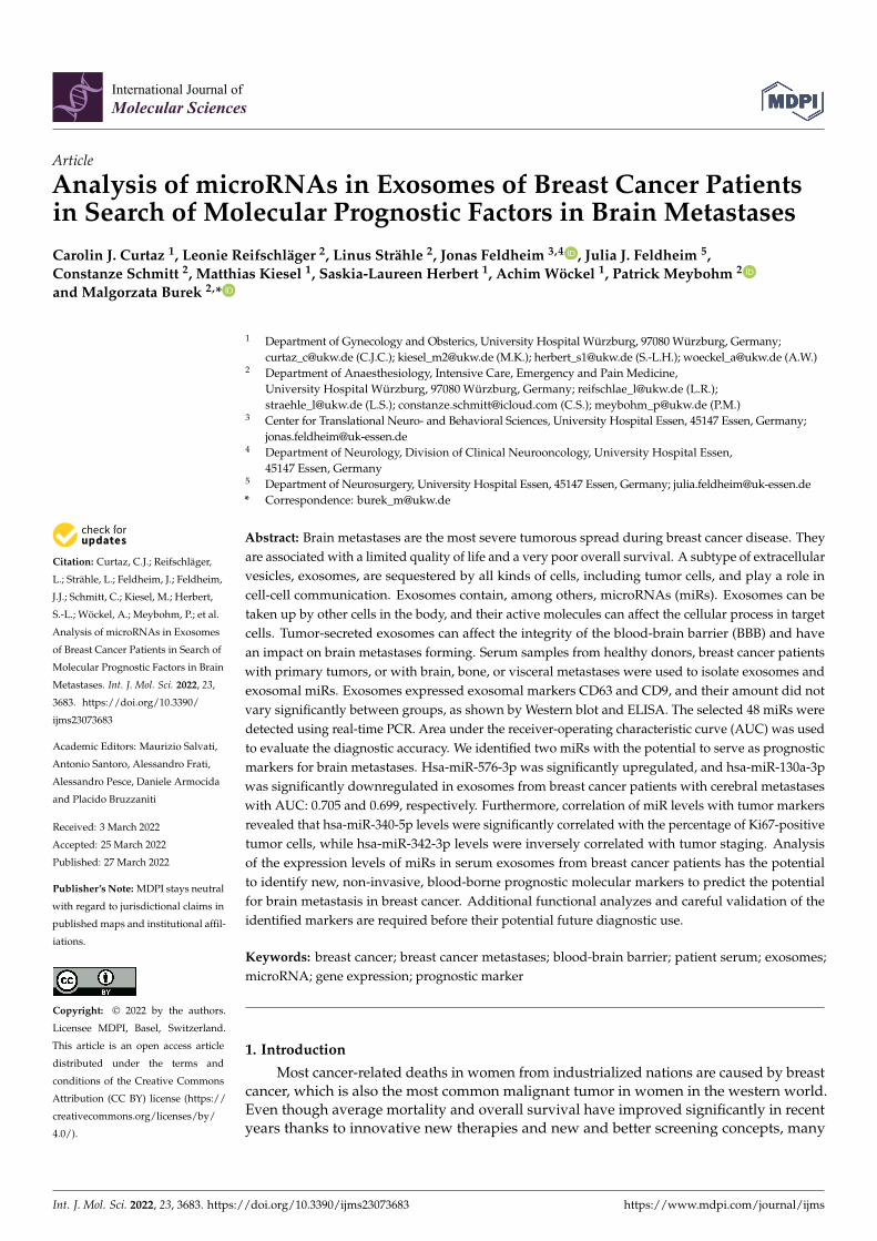
�����������������
Citation: Curtaz, C.J.; Reifschläger,
L.; Strähle, L.; Feldheim, J.; Feldheim,
J.J.; Schmitt, C.; Kiesel, M.; Herbert,
S.-L.; Wöckel, A.; Meybohm, P.; et al.
Analysis of microRNAs in Exosomes
of Breast Cancer Patients in Search of
Molecular Prognostic Factors in Brain
Metastases. Int. J. Mol. Sci. 2022, 23,
3683. https://doi.org/10.3390/
ijms23073683
Academic Editors: Maurizio Salvati,
Antonio Santoro, Alessandro Frati,
Alessandro Pesce, Daniele Armocida
and Placido Bruzzaniti
Received: 3 March 2022
Accepted: 25 March 2022
Published: 27 March 2022
Publisher’s Note: MDPI stays neutral
with regard to jurisdictional claims in
published maps and institutional affil-
iations.
Copyright: © 2022 by the authors.
Licensee MDPI, Basel, Switzerland.
This article is an open access article
distributed under the terms and
conditions of the Creative Commons
Attribution (CC BY) license (https://
creativecommons.org/licenses/by/
4.0/).
International Journal of
Molecular Sciences
Article
Analysis of microRNAs in Exosomes of Breast Cancer Patientsin Search of Molecular Prognostic Factors in Brain MetastasesCarolin J. Curtaz 1, Leonie Reifschläger 2, Linus Strähle 2, Jonas Feldheim 3,4 , Julia J. Feldheim 5,Constanze Schmitt 2, Matthias Kiesel 1, Saskia-Laureen Herbert 1, Achim Wöckel 1, Patrick Meybohm 2
and Malgorzata Burek 2,*
1 Department of Gynecology and Obsterics, University Hospital Würzburg, 97080 Würzburg, Germany;[email protected] (C.J.C.); [email protected] (M.K.); [email protected] (S.-L.H.); [email protected] (A.W.)
2 Department of Anaesthesiology, Intensive Care, Emergency and Pain Medicine,University Hospital Würzburg, 97080 Würzburg, Germany; [email protected] (L.R.);[email protected] (L.S.); [email protected] (C.S.); [email protected] (P.M.)
3 Center for Translational Neuro- and Behavioral Sciences, University Hospital Essen, 45147 Essen, Germany;[email protected]
4 Department of Neurology, Division of Clinical Neurooncology, University Hospital Essen,45147 Essen, Germany
5 Department of Neurosurgery, University Hospital Essen, 45147 Essen, Germany; [email protected]* Correspondence: [email protected]
Abstract: Brain metastases are the most severe tumorous spread during breast cancer disease. Theyare associated with a limited quality of life and a very poor overall survival. A subtype of extracellularvesicles, exosomes, are sequestered by all kinds of cells, including tumor cells, and play a role incell-cell communication. Exosomes contain, among others, microRNAs (miRs). Exosomes can betaken up by other cells in the body, and their active molecules can affect the cellular process in targetcells. Tumor-secreted exosomes can affect the integrity of the blood-brain barrier (BBB) and havean impact on brain metastases forming. Serum samples from healthy donors, breast cancer patientswith primary tumors, or with brain, bone, or visceral metastases were used to isolate exosomes andexosomal miRs. Exosomes expressed exosomal markers CD63 and CD9, and their amount did notvary significantly between groups, as shown by Western blot and ELISA. The selected 48 miRs weredetected using real-time PCR. Area under the receiver-operating characteristic curve (AUC) was usedto evaluate the diagnostic accuracy. We identified two miRs with the potential to serve as prognosticmarkers for brain metastases. Hsa-miR-576-3p was significantly upregulated, and hsa-miR-130a-3pwas significantly downregulated in exosomes from breast cancer patients with cerebral metastaseswith AUC: 0.705 and 0.699, respectively. Furthermore, correlation of miR levels with tumor markersrevealed that hsa-miR-340-5p levels were significantly correlated with the percentage of Ki67-positivetumor cells, while hsa-miR-342-3p levels were inversely correlated with tumor staging. Analysisof the expression levels of miRs in serum exosomes from breast cancer patients has the potentialto identify new, non-invasive, blood-borne prognostic molecular markers to predict the potentialfor brain metastasis in breast cancer. Additional functional analyzes and careful validation of theidentified markers are required before their potential future diagnostic use.
Keywords: breast cancer; breast cancer metastases; blood-brain barrier; patient serum; exosomes;microRNA; gene expression; prognostic marker
1. Introduction
Most cancer-related deaths in women from industrialized nations are caused by breastcancer, which is also the most common malignant tumor in women in the western world.Even though average mortality and overall survival have improved significantly in recentyears thanks to innovative new therapies and new and better screening concepts, many
Int. J. Mol. Sci. 2022, 23, 3683. https://doi.org/10.3390/ijms23073683 https://www.mdpi.com/journal/ijms

Int. J. Mol. Sci. 2022, 23, 3683 2 of 15
patients die prematurely due to a pronounced tumor infestation. Unfortunately, around10%–15% of all breast cancer patients suffer from brain metastases, resulting in poor overallsurvival but also a severe impairment in quality of life [1]. The key event for entry into thebrain is the migration of cancer cells across the blood-brain barrier (BBB) [2,3]. To date, theexact mechanism of metastatic progression of breast cancer to the brain and migration ofcancer cells across the BBB is not well understood in detail [4].
In the search for blood-based factors in breast cancer patients that could impairthe integrity of the BBB and thus provoke brain metastases, microRNA (miR) recentlycame into focus [5]. Only about 20 nucleotides in length, miRs are short, non-codingRNAs that regulate gene expression post-transcriptionally by downregulating mRNAor interfering with its translation [5]. It is known that only 2% of the human genomeconsists of protein-coding sequences since non-coding sequences predominate [6]. Thesenon-coding sequences are the least studied of the human genome. Their influence ontumor development is not really understood. In recent years, several research groupsdemonstrated that miRs can be detected in tissues but also in cell-free body fluids such asplasma or serum [7]. In addition, it could be shown that there is a prognostic connectionwith regard to cancer of different entities [8–10].
Extracellular vesicles (EVs) are secreted by eukaryotic cells and can be divided intothree categories based on their size: exosomes, activation-or apoptosis-induced microvesi-cles, and apoptotic bodies. While apoptotic bodies are vesicles with a diameter of only1–5 µM, microvesicles consist of membrane vesicles and have a diameter of 100–1000 nm.Exosomes, on the other hand, are defined by a diameter of 30–100 nm [11,12]. It was theirsmall size that brought exosomes into the focus of drug delivery but also of biomarkerresearch [13]. They are able to transfer proteins and genetic material [14]. The circulationof exosomes in body fluids allows them to transport a wide variety of active moleculesfar from their source, where they can absorb and release their contents. Due to their highstability, exosomes are therefore considered to be powerful non-invasive biomarkers [15].
Healthy cells usually release fewer exosomes than tumor cells [16,17]. By analyzingthe expression profiles of exosomes isolated from serum/plasma of cancer patients, it canbe shown that numerous miRs have different levels compared to healthy individuals [2].Tumor spread to distant organs of its origin indicated an advanced stage of cancer. Breastcancer is one of the types of cancer that induce metastases in the central nervous system(CNS) with a high incidence, along with lung cancer, melanoma, and colorectal cancer [18].It could be shown that the isolated exosomes of cancer patients with metastases havedifferent miR expression patterns compared to healthy individuals of patients with primaryneoplasia [19]. The development of new and innovative classification criteria for future on-cology therapies could help improve tumor therapies, overall patient survival, and qualityof life. In connection with the twelfth St. Gallen International Breast Cancer Conference2011, a classification using biological markers of the primary tumor was introduced [20].Four subtypes can be defined based on clinical and histological evidence such as expressionof estrogen (ER) and progesterone receptors (PR), human epidermal growth factor receptor2 positivity (HER2), and the proliferation factor Ki-67: luminal A (ER and/or PR+, HER2−,Ki-67 low); HER2− negative luminal B (HER2−, ER and/or PR+, Ki-67 high); HER2−positive luminal B (HER2+, ER and/or PR+, any Ki-67); HER2 overexpressed (any Ki-67,HER2+, ER and PR−, HER2 overexpressed); triple-negative breast cancer (TNBC) (ERand PR−, HER2−). The division into subgroups enables a better differentiation of theindividual types in order to make more accurate prognoses and to enable more individualtherapy [20].
In the present study, we used TaqMan Advanced miRNA Human Cards to identifythe differential expression profiles of miRs in sera from breast cancer patients and to evaluatetheir prognostic potential. We analyzed the miR levels in the sera of breast cancer patients withprimary cancer, bone, visceral or cerebral metastases compared to a healthy control group.
Int. J. Mol. Sci. 2022, 23, 3683 3 of 15
2. Results2.1. Patient Characteristics
We collected blood samples from healthy age and sex-matched donors (n = 18), breastcancer patients with primary cancer (n = 15), visceral metastases (n = 18), bone metastases(n = 16) and cerebral metastases (n = 16). The healthy donors had no tumor or a knowninfection at the time the blood was taken. Clinical data of breast cancer patients werecollected for each patient, as shown in Table 1. The median age of patients ranged from 61.1to 62.9. Most of the patients were postmenopausal. Tumor characteristics and classificationof tumors were collected for each patient. Patient serum was stored frozen until use.
Table 1. Patient characteristics. Summary of clinical data.
C PC CM VM BM
Patients characteristicsTotal number 18 15 16 18 16Median age 60.3 61.6 62.9 61.1 61.8
Deceased 12 9 2Pre-/postmenopausal 4/14 4/11 2/14 3/15 2/14Tumor characteristics
ER/PR-positive 8 4 8 10HER2/neu-positive 5 8 9 5
Triple-negative 2 4 1 1Grading
Well differentiated (G1) 1Moderately differentiated (G2) 10 9 9 11
Poorly differentiated (G3) 5 7 8 5% of Ki67-positive cells (median) 22% 43% 34.7% 33.9%
Other 1BM = bone metastases, C = control group of healthy donors, CM = cerebral metastases, ER = estrogen receptor,HER2/neu = human epidermal growth factor 2, PC = primary cancer, PR = progesteron receptor,VM = visceral metastases.
2.2. Isolation and Characterization of Exosomes Derived from Controls and Breast Cancer Patientswith Brain Metastases
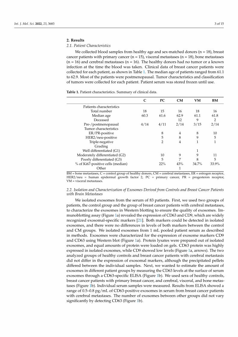
We isolated exosomes from the serum of 83 patients. First, we used two groups ofpatients, the control group and the group of breast cancer patients with cerebral metastases,to characterize the exosomes in Western blotting to ensure the quality of exosomes. Im-munoblotting assay (Figure 1a) revealed the expression of CD63 and CD9, which are widelyrecognized exosomal-specific markers [21]. Both markers could be detected in isolatedexosomes, and there were no differences in levels of both markers between the controland CM groups. We isolated exosomes from 1 mL pooled patient serum as describedin methods. Exosomes were characterized for the expression of exosome markers CD9and CD63 using Western blot (Figure 1a). Protein lysates were prepared out of isolatedexosomes, and equal amounts of protein were loaded on gels. CD63 protein was highlyexpressed in isolated exosomes, while CD9 showed low levels (Figure 1a, arrows). The twoanalyzed groups of healthy controls and breast cancer patients with cerebral metastasisdid not differ in the expression of exosomal markers, although the precipitated pelletsdiffered between the individual samples. Next, we wanted to estimate the amount ofexosomes in different patient groups by measuring the CD63 levels at the surface of serumexosomes through a CD63-specific ELISA (Figure 1b). We used sera of healthy controls,breast cancer patients with primary breast cancer, and cerebral, visceral, and bone metas-tases (Figure 1b). Individual serum samples were measured. Results from ELISA showed arange of 0.5–0.8 pg/mL of CD63-positive exosomes in serum from breast cancer patientswith cerebral metastases. The number of exosomes between other groups did not varysignificantly by detecting CD63 (Figure 1b).

Int. J. Mol. Sci. 2022, 23, 3683 4 of 15
Int. J. Mol. Sci. 2022, 23, 3683 4 of 16
patients with cerebral metastasis did not differ in the expression of exosomal markers, although the precipitated pellets differed between the individual samples. Next, we wanted to estimate the amount of exosomes in different patient groups by measuring the CD63 levels at the surface of serum exosomes through a CD63-specific ELISA (Figure 1b). We used sera of healthy controls, breast cancer patients with primary breast cancer, and cerebral, visceral, and bone metastases (Figure 1b). Individual serum samples were measured. Results from ELISA showed a range of 0.5–0.8 pg/mL of CD63-positive exosomes in serum from breast cancer patients with cerebral metastases. The number of exosomes between other groups did not vary significantly by detecting CD63 (Figure 1b).
Figure 1. Assessment of exosomal markers. (a) Western blot analysis of exosomal markers CD63 and CD9. Arrows indicate the bands corresponding to CD63 (53 kDa) and CD9 (28 kDa), respectively, C—control, CM—cerebral metastases. (b) Estimation of exosome levels in patient serum. CD63 protein levels in exosomes from breast cancer patients with primary breast cancer (PC) and breast cancer patients with cerebral (CM), viszeral (VM), and bone (BM) metastases were estimated using a CD63-specific ELISA.
2.3. miRNA Expression in Exosomes Isolated from Patient Serum by Site of Metastasis In order to first characterize the expression profile of the exosomal miRNA in our
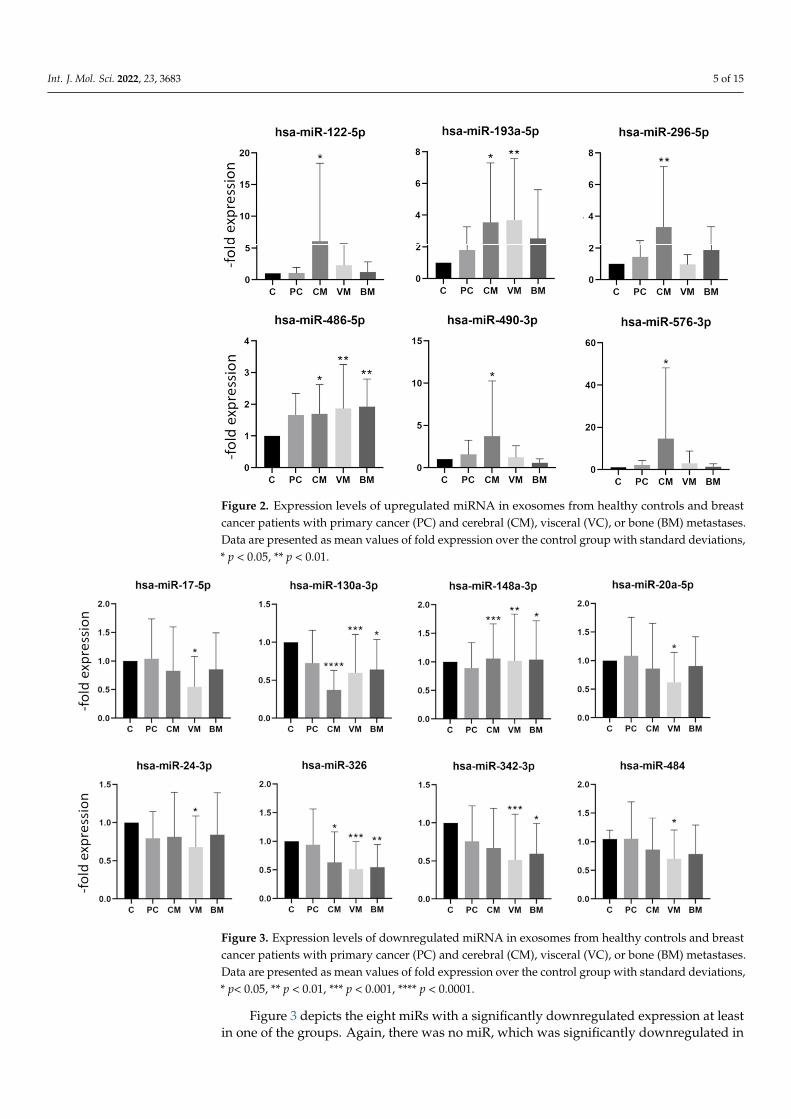
samples, we analyzed 384 miRs per sample using the Human TaqMan Advanced miRNA Array Cards A (Thermo Fisher Scientific, Waltham, MA, USA) and the control group and the breast cancer patients with brain metastases. MiRs expressed at levels with Ct values higher than 35 were considered absent from exosomes (results not shown). From the well-expressed miRs, 48 were selected for analysis with a larger patient cohort. Selected miRs and their sequences are presented in Supplementary Table S1. By comparing the exosomal miR expression levels found in serum exosomes of the control group, patients with primary breast cancer, and patients with breast cancer with cerebral, visceral, or bone metastases, several differentially expressed miRs were identified. First, the samples were divided by site of metastasis to the various body regions such as cerebral (n = 16), visceral (n = 18), bone (n = 16), and the group with primary cancer without metastasis (n = 15). The division into four subgroups made it possible to determine whether a specific miR dysregulation is associated with a specific form of metastasis. The comparison of each individual group to the control group revealed a significant difference in the expression of 14 miR in the metastasis groups. Increased expression was identified for six and a decreased expression for eight miRs, as shown in Figures 2 and 3, respectively. None of
Figure 1. Assessment of exosomal markers. (a) Western blot analysis of exosomal markers CD63and CD9. Arrows indicate the bands corresponding to CD63 (53 kDa) and CD9 (28 kDa), respec-tively, C—control, CM—cerebral metastases. (b) Estimation of exosome levels in patient serum.CD63 protein levels in exosomes from breast cancer patients with primary breast cancer (PC) andbreast cancer patients with cerebral (CM), viszeral (VM), and bone (BM) metastases were estimatedusing a CD63-specific ELISA.
2.3. miRNA Expression in Exosomes Isolated from Patient Serum by Site of Metastasis
In order to first characterize the expression profile of the exosomal miRNA in oursamples, we analyzed 384 miRs per sample using the Human TaqMan Advanced miRNAArray Cards A (Thermo Fisher Scientific, Waltham, MA, USA) and the control group andthe breast cancer patients with brain metastases. MiRs expressed at levels with Ct valueshigher than 35 were considered absent from exosomes (results not shown). From the well-expressed miRs, 48 were selected for analysis with a larger patient cohort. Selected miRsand their sequences are presented in Supplementary Table S1. By comparing the exosomalmiR expression levels found in serum exosomes of the control group, patients with primarybreast cancer, and patients with breast cancer with cerebral, visceral, or bone metastases,several differentially expressed miRs were identified. First, the samples were divided bysite of metastasis to the various body regions such as cerebral (n = 16), visceral (n = 18),bone (n = 16), and the group with primary cancer without metastasis (n = 15). The divisioninto four subgroups made it possible to determine whether a specific miR dysregulationis associated with a specific form of metastasis. The comparison of each individual groupto the control group revealed a significant difference in the expression of 14 miR in themetastasis groups. Increased expression was identified for six and a decreased expressionfor eight miRs, as shown in Figures 2 and 3, respectively. None of the six upregulated miRsshowed a significant upregulation in all four groups. Hsa-miR-122-5p, hsa-miR-296-5p,hsa-miR-490-3p, and hsa-miR-576-3p were in particular increased in the group with cerebralmetastases, while hsa-miR-486-5p showed increased expression in groups with cerebral,visceral, and bone metastases.
Int. J. Mol. Sci. 2022, 23, 3683 5 of 15
Int. J. Mol. Sci. 2022, 23, 3683 5 of 16
the six upregulated miRs showed a significant upregulation in all four groups. Hsa-miR-122-5p, hsa-miR-296-5p, hsa-miR-490-3p, and hsa-miR-576-3p were in particular increased in the group with cerebral metastases, while hsa-miR-486-5p showed increased expression in groups with cerebral, visceral, and bone metastases.
Figure 2. Expression levels of upregulated miRNA in exosomes from healthy controls and breast cancer patients with primary cancer (PC) and cerebral (CM), visceral (VC), or bone (BM) metastases. Data are presented as mean values of fold expression over the control group with standard deviations, * p < 0.05, ** p < 0.01.
Figure 3 depicts the eight miRs with a significantly downregulated expression at least in one of the groups. Again, there was no miR, which was significantly downregulated in all groups. The hsa-miR-130a-3p, hsa-miR-148b-3p, and hsa-miR-326 showed a significant decrease in expression in the group with cerebral, visceral, and bone metastases but were not decreased in the group with primary breast cancer. The hsa-miR-130a-3p showed a particularly strong decrease in the group with cerebral metastases (Figure 3). The fold expression changes of downregulated miRs were not as pronounced as those of upregulated miRs (Figures 2 and 3). Interestingly, none of the analyzed miRs was significantly deregulated in the group with primary breast cancer, which is in accordance with reports showing the involvement of miRs in metastasis development [22].
Figure 2. Expression levels of upregulated miRNA in exosomes from healthy controls and breastcancer patients with primary cancer (PC) and cerebral (CM), visceral (VC), or bone (BM) metastases.Data are presented as mean values of fold expression over the control group with standard deviations,* p < 0.05, ** p < 0.01.
Int. J. Mol. Sci. 2022, 23, 3683 6 of 16
Figure 3. Expression levels of downregulated miRNA in exosomes from healthy controls and breast cancer patients with primary cancer (PC) and cerebral (CM), visceral (VC), or bone (BM) metastases. Data are presented as mean values of fold expression over the control group with standard deviations, * p< 0.05, ** p < 0.01, *** p < 0.001, **** p < 0.0001.
2.4. miRNA Expression in Exosomes Isolated from Patient Serum Classified by Tumor Characteristics
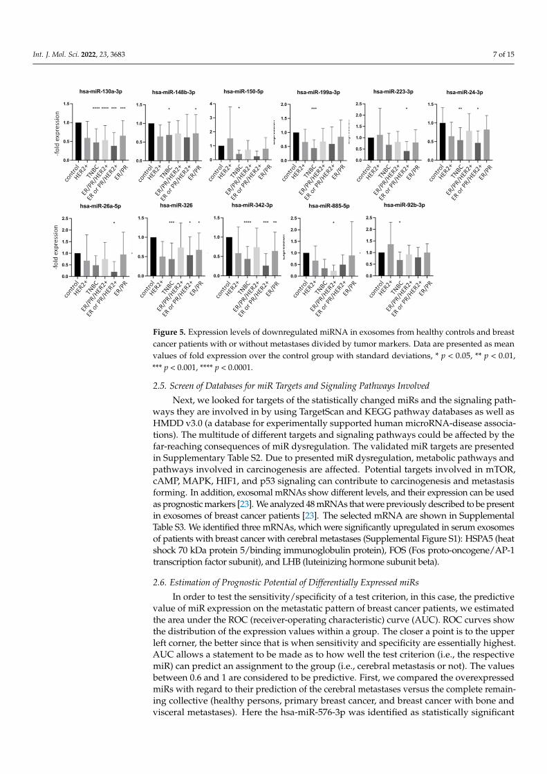
The expression of different tumor markers is used as a basis for the selection of therapy and correlates with the prognosis. To analyze whether there is a connection between specific miR dysregulation and a receptor status of tumor, we analyzed the same data sets presented in Figures 2 and 3 by sorting the groups by receptor status into five groups: (1) HER2 + (n = 5), (2) TNBC (n = 14), (3) ER/PR/HER2+ (n = 17), (4) ER or PR/HER2+ (n = 7) and (5) ER/PR+ (n = 31). According to previous reports, differential expression patterns of miRs were observed in groups with different tumor markers [20]. When comparing each individual group with the control group, a total of 21 miRs showed a significant dysregulation in the expression pattern. An increased expression profile was found for 10 miRs, while 11 miRs were decreased. Figures 4 and 5 show selected up and downregulated miRs, respectively, in at least one group. Again, none of the examined miRs showed a significant dysregulation in all subgroups simultaneously. The hsa-miR-197-3p, hsa-miR-410-3p, hsa-miR-490-3p showed increased expression in the HER2-positive groups (Figures 4 and 5). The hsa-miR-32-5p was only increased in a TNBC. Hsa-miR-125a-3p was increased in an ER/PR+ group.
Figure 3. Expression levels of downregulated miRNA in exosomes from healthy controls and breastcancer patients with primary cancer (PC) and cerebral (CM), visceral (VC), or bone (BM) metastases.Data are presented as mean values of fold expression over the control group with standard deviations,* p< 0.05, ** p < 0.01, *** p < 0.001, **** p < 0.0001.
Figure 3 depicts the eight miRs with a significantly downregulated expression at leastin one of the groups. Again, there was no miR, which was significantly downregulated in

Int. J. Mol. Sci. 2022, 23, 3683 6 of 15
all groups. The hsa-miR-130a-3p, hsa-miR-148b-3p, and hsa-miR-326 showed a significantdecrease in expression in the group with cerebral, visceral, and bone metastases but werenot decreased in the group with primary breast cancer. The hsa-miR-130a-3p showed aparticularly strong decrease in the group with cerebral metastases (Figure 3). The foldexpression changes of downregulated miRs were not as pronounced as those of upregu-lated miRs (Figures 2 and 3). Interestingly, none of the analyzed miRs was significantlyderegulated in the group with primary breast cancer, which is in accordance with reportsshowing the involvement of miRs in metastasis development [22].
2.4. miRNA Expression in Exosomes Isolated from Patient Serum Classified by Tumor Characteristics
The expression of different tumor markers is used as a basis for the selection oftherapy and correlates with the prognosis. To analyze whether there is a connection be-tween specific miR dysregulation and a receptor status of tumor, we analyzed the same datasets presented in Figures 2 and 3 by sorting the groups by receptor status into five groups:(1) HER2+ (n = 5), (2) TNBC (n = 14), (3) ER/PR/HER2+ (n = 17), (4) ER or PR/HER2+ (n = 7)and (5) ER/PR+ (n = 31). According to previous reports, differential expression patternsof miRs were observed in groups with different tumor markers [20]. When comparingeach individual group with the control group, a total of 21 miRs showed a significantdysregulation in the expression pattern. An increased expression profile was found for10 miRs, while 11 miRs were decreased. Figures 4 and 5 show selected up and down-regulated miRs, respectively, in at least one group. Again, none of the examined miRsshowed a significant dysregulation in all subgroups simultaneously. The hsa-miR-197-3p,hsa-miR-410-3p, hsa-miR-490-3p showed increased expression in the HER2-positive groups(Figures 4 and 5). The hsa-miR-32-5p was only increased in a TNBC. Hsa-miR-125a-3p wasincreased in an ER/PR+ group.
Int. J. Mol. Sci. 2022, 23, 3683 7 of 16
Figure 4. Expression levels of mostly upregulated miRNA in exosomes from healthy controls and breast cancer patients with or without metastases divided by tumor markers. Data are presented as mean values of fold expression over the control group with standard deviations, * p < 0.05, ** p < 0.01, *** p < 0.001, **** p < 0.0001.
Figure 5. Expression levels of downregulated miRNA in exosomes from healthy controls and breast cancer patients with or without metastases divided by tumor markers. Data are presented as mean values of fold expression over the control group with standard deviations, * p < 0.05, ** p < 0.01, *** p < 0.001, **** p < 0.0001.
Figure 4. Expression levels of mostly upregulated miRNA in exosomes from healthy controls andbreast cancer patients with or without metastases divided by tumor markers. Data are presented asmean values of fold expression over the control group with standard deviations, * p < 0.05, ** p < 0.01,*** p < 0.001, **** p < 0.0001.
Int. J. Mol. Sci. 2022, 23, 3683 7 of 15
Int. J. Mol. Sci. 2022, 23, 3683 7 of 16
Figure 4. Expression levels of mostly upregulated miRNA in exosomes from healthy controls and breast cancer patients with or without metastases divided by tumor markers. Data are presented as mean values of fold expression over the control group with standard deviations, * p < 0.05, ** p < 0.01, *** p < 0.001, **** p < 0.0001.
Figure 5. Expression levels of downregulated miRNA in exosomes from healthy controls and breast cancer patients with or without metastases divided by tumor markers. Data are presented as mean values of fold expression over the control group with standard deviations, * p < 0.05, ** p < 0.01, *** p < 0.001, **** p < 0.0001.
Figure 5. Expression levels of downregulated miRNA in exosomes from healthy controls and breastcancer patients with or without metastases divided by tumor markers. Data are presented as meanvalues of fold expression over the control group with standard deviations, * p < 0.05, ** p < 0.01,*** p < 0.001, **** p < 0.0001.
2.5. Screen of Databases for miR Targets and Signaling Pathways Involved
Next, we looked for targets of the statistically changed miRs and the signaling path-ways they are involved in by using TargetScan and KEGG pathway databases as well asHMDD v3.0 (a database for experimentally supported human microRNA-disease associa-tions). The multitude of different targets and signaling pathways could be affected by thefar-reaching consequences of miR dysregulation. The validated miR targets are presentedin Supplementary Table S2. Due to presented miR dysregulation, metabolic pathways andpathways involved in carcinogenesis are affected. Potential targets involved in mTOR,cAMP, MAPK, HIF1, and p53 signaling can contribute to carcinogenesis and metastasisforming. In addition, exosomal mRNAs show different levels, and their expression can be usedas prognostic markers [23]. We analyzed 48 mRNAs that were previously described to be presentin exosomes of breast cancer patients [23]. The selected mRNA are shown in SupplementalTable S3. We identified three mRNAs, which were significantly upregulated in serum exosomesof patients with breast cancer with cerebral metastases (Supplemental Figure S1): HSPA5 (heatshock 70 kDa protein 5/binding immunoglobulin protein), FOS (Fos proto-oncogene/AP-1transcription factor subunit), and LHB (luteinizing hormone subunit beta).
2.6. Estimation of Prognostic Potential of Differentially Expressed miRs
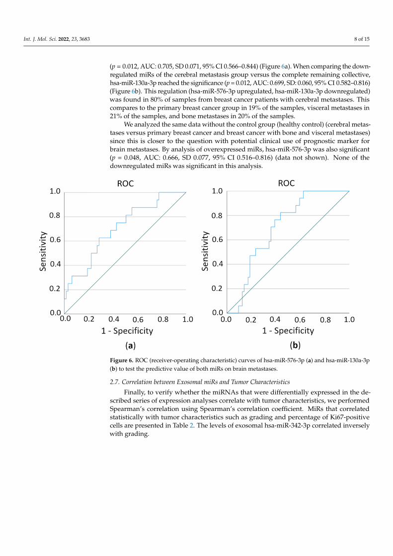
In order to test the sensitivity/specificity of a test criterion, in this case, the predictivevalue of miR expression on the metastatic pattern of breast cancer patients, we estimatedthe area under the ROC (receiver-operating characteristic) curve (AUC). ROC curves showthe distribution of the expression values within a group. The closer a point is to the upperleft corner, the better since that is when sensitivity and specificity are essentially highest.AUC allows a statement to be made as to how well the test criterion (i.e., the respectivemiR) can predict an assignment to the group (i.e., cerebral metastasis or not). The valuesbetween 0.6 and 1 are considered to be predictive. First, we compared the overexpressedmiRs with regard to their prediction of the cerebral metastases versus the complete remain-ing collective (healthy persons, primary breast cancer, and breast cancer with bone andvisceral metastases). Here the hsa-miR-576-3p was identified as statistically significant
Int. J. Mol. Sci. 2022, 23, 3683 8 of 15
(p = 0.012, AUC: 0.705, SD 0.071, 95% CI 0.566–0.844) (Figure 6a). When comparing the down-regulated miRs of the cerebral metastasis group versus the complete remaining collective,hsa-miR-130a-3p reached the significance (p = 0.012, AUC: 0.699, SD: 0.060, 95% CI 0.582–0.816)(Figure 6b). This regulation (hsa-miR-576-3p upregulated, hsa-miR-130a-3p downregulated)was found in 80% of samples from breast cancer patients with cerebral metastases. Thiscompares to the primary breast cancer group in 19% of the samples, visceral metastases in21% of the samples, and bone metastases in 20% of the samples.
We analyzed the same data without the control group (healthy control) (cerebral metas-tases versus primary breast cancer and breast cancer with bone and visceral metastases)since this is closer to the question with potential clinical use of prognostic marker forbrain metastases. By analysis of overexpressed miRs, hsa-miR-576-3p was also significant(p = 0.048, AUC: 0.666, SD 0.077, 95% CI 0.516–0.816) (data not shown). None of thedownregulated miRs was significant in this analysis.
Int. J. Mol. Sci. 2022, 23, 3683 9 of 16
Figure 6. ROC (receiver-operating characteristic) curves of hsa-miR-576-3p (a) and hsa-miR-130a-3p (b) to test the predictive value of both miRs on brain metastases.
2.7. Correlation Between Exosomal miRs and Tumor Characteristics Finally, to verify whether the miRNAs that were differentially expressed in the
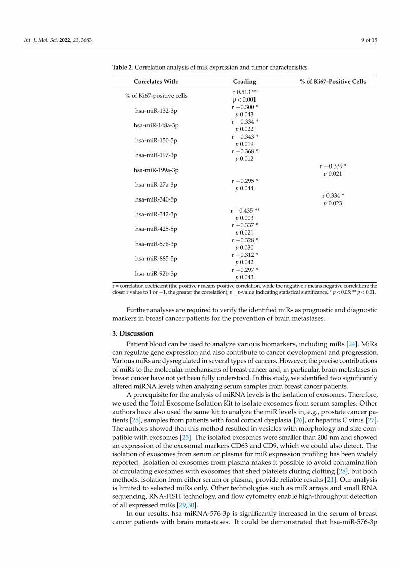
described series of expression analyses correlate with tumor characteristics, we performed Spearman’s correlation using Spearman’s correlation coefficient. MiRs that correlated statistically with tumor characteristics such as grading and percentage of Ki67-positive cells are presented in Table 2. The levels of exosomal hsa-miR-342-3p correlated inversely with grading.
Table 2. Correlation analysis of miR expression and tumor characteristics.
Correlates With: Grading % of Ki67-Positive
Cells
% of Ki67-positive cells r 0.513 ** p < 0.001
hsa-miR-132-3p r −0.300 * p 0.043
hsa-miR-148a-3p r −0.334 * p 0.022
hsa-miR-150-5p r −0.343 *
p 0.019
hsa-miR-197-3p r −0.368 * p 0.012
hsa-miR-199a-3p
r −0.339 * p 0.021
hsa-miR-27a-3p r −0.295 *
p 0.044
hsa-miR-340-5p
r 0.334 * p 0.023
hsa-miR-342-3p r −0.435 ** p 0.003
Figure 6. ROC (receiver-operating characteristic) curves of hsa-miR-576-3p (a) and hsa-miR-130a-3p(b) to test the predictive value of both miRs on brain metastases.
2.7. Correlation between Exosomal miRs and Tumor Characteristics
Finally, to verify whether the miRNAs that were differentially expressed in the de-scribed series of expression analyses correlate with tumor characteristics, we performedSpearman’s correlation using Spearman’s correlation coefficient. MiRs that correlatedstatistically with tumor characteristics such as grading and percentage of Ki67-positivecells are presented in Table 2. The levels of exosomal hsa-miR-342-3p correlated inverselywith grading.
Int. J. Mol. Sci. 2022, 23, 3683 9 of 15
Table 2. Correlation analysis of miR expression and tumor characteristics.
Correlates With: Grading % of Ki67-Positive Cells
% of Ki67-positive cells r 0.513 **p < 0.001
hsa-miR-132-3p r −0.300 *p 0.043
hsa-miR-148a-3p r −0.334 *p 0.022
hsa-miR-150-5p r −0.343 *p 0.019
hsa-miR-197-3p r −0.368 *p 0.012
hsa-miR-199a-3p r −0.339 *p 0.021
hsa-miR-27a-3p r −0.295 *p 0.044
hsa-miR-340-5p r 0.334 *p 0.023
hsa-miR-342-3p r −0.435 **p 0.003
hsa-miR-425-5p r −0.337 *p 0.021
hsa-miR-576-3p r −0.328 *p 0.030
hsa-miR-885-5p r −0.312 *p 0.042
hsa-miR-92b-3p r −0.297 *p 0.043
r = correlation coefficient (the positive r means positive correlation, while the negative r means negative correlation; thecloser r value to 1 or −1, the greater the correlation); p = p-value indicating statistical significance, * p < 0.05; ** p < 0.01.
Further analyses are required to verify the identified miRs as prognostic and diagnosticmarkers in breast cancer patients for the prevention of brain metastases.
3. Discussion
Patient blood can be used to analyze various biomarkers, including miRs [24]. MiRscan regulate gene expression and also contribute to cancer development and progression.Various miRs are dysregulated in several types of cancers. However, the precise contributionsof miRs to the molecular mechanisms of breast cancer and, in particular, brain metastases inbreast cancer have not yet been fully understood. In this study, we identified two significantlyaltered miRNA levels when analyzing serum samples from breast cancer patients.
A prerequisite for the analysis of miRNA levels is the isolation of exosomes. Therefore,we used the Total Exosome Isolation Kit to isolate exosomes from serum samples. Otherauthors have also used the same kit to analyze the miR levels in, e.g., prostate cancer pa-tients [25], samples from patients with focal cortical dysplasia [26], or hepatitis C virus [27].The authors showed that this method resulted in vesicles with morphology and size com-patible with exosomes [25]. The isolated exosomes were smaller than 200 nm and showedan expression of the exosomal markers CD63 and CD9, which we could also detect. Theisolation of exosomes from serum or plasma for miR expression profiling has been widelyreported. Isolation of exosomes from plasma makes it possible to avoid contaminationof circulating exosomes with exosomes that shed platelets during clotting [28], but bothmethods, isolation from either serum or plasma, provide reliable results [21]. Our analysisis limited to selected miRs only. Other technologies such as miR arrays and small RNAsequencing, RNA-FISH technology, and flow cytometry enable high-throughput detectionof all expressed miRs [29,30].
In our results, hsa-miRNA-576-3p is significantly increased in the serum of breastcancer patients with brain metastases. It could be demonstrated that hsa-miR-576-3p
Int. J. Mol. Sci. 2022, 23, 3683 10 of 15
targets PD-L1 and cyclin D1 [31]. The PD-1/PD-L1 pathway controls the induction andmaintenance of immune tolerance within the tumor microenvironment. The activity ofPD-1 and its ligands PD-L1/PD-L2 are responsible for T cell activation, proliferation, andcytotoxic secretion in cancer to degenerating anti-tumor immune responses [32]. D-typecyclins (D1, D2, and D3), along with their associated cyclin-dependent kinases CDK4 andCDK6, are components of the core cell cycle that are responsible for cell proliferation [33].Hsa-miR-576-3p influencing PD-L1 and cyclin D1 could therefore directly interfere withcancer progression.
Hsa-miR-576-3p has also been identified in association with non-melanoma skincancer [34]. Its downregulation was observed in plasma samples of non-melanoma skincancer patients. Other authors identified miR-576-3p as significantly reduced in lungadenocarcinoma, while overexpression of hsa-miR-576–3p in lung adenocarcinoma cellsreduced mesenchymal marker expression and inhibited migration and invasion [35]. Inaddition, the downregulation of hsa-miR-576-3p was observed in bladder cancer tissuesand correlated with a poor clinical outcome [36,37]. Those authors concluded, therefore,that hsa-miR-576-3p has a negative regulatory effect on carcinogenesis. In further studies,downregulation of hsa-miR-576-3p was associated with affecting the chemosensitivity ofovarian cancer [31], human teratoma [38], and also breast cancer cells [39].
Our results, in contrast to other published studies, showed a significant increase inhsa-miR-576-3p in serum exosomes from breast cancer patients with brain metastases,while it was at very low levels in all other patient groups, including the control group.Statistical analysis of this miR as a potential prognostic marker revealed a potential ofhsa-miR-576-3p as a predictor of cerebral metastases. To the best of our knowledge, this isa first report identifying hsa-miR-576-3p as a potential molecular prognostic factor in brainmetastases from breast cancer.
Next to the results of hsa-miR-576-3p, another miR could be identified as a potentialmolecular prognostic factor. In our statistical analysis, expression of hsa-miR-130a-3pwas significantly reduced in cerebral metastases compared to the control group. Hsa-miR-130a-3p seems to have a cancer-promoting function in connection with RAB5B [40].RAB5B belongs to the Ras family [41]. The RAB protein presumably plays a central rolein vesicular transport to the cell membrane but also acts as a tumor-suppressive factorinducing apoptosis and inhibiting angiogenesis [42]. Pan and colleagues could demonstratein their tumor samples that RAB5A was upregulated and miR-130a was downregulated inbreast cancer tissues and cells [43]. Next to that, it could be shown that the endogenous levelof RAB5A can be inhibited by the overexpression of hsa-miR-130a [43]. In addition, reducedhsa-miR-130a-3p levels are mentioned in connection with liver fibrosis [44], overexpressedlevels in metastatic colon cancer not responding to first-line chemotherapy [45], but alsogeneral downregulation in chronic inflammation and macrophagal activity [46]. MDM4,one of the hsa-miR-130a-3p targets, can affect the sensitivity of breast cancer cells tochemotherapy and modulate the p53 signaling pathway [47]. Low expression of hsa-miR-130a-3p correlated with p53 mutation in chronic myeloid leukemia [48]. In breast cancer, thep53 mutation is associated with more aggressive disease and poorer overall survival [49]. Adirect influence of hsa-miR-130a-3p on p53 could therefore play a role in the developmentof brain metastases in breast cancer.
We analyzed the hsa-miR-130a-3p in serum exosomes from breast cancer patients,where it showed significantly reduced levels in the cohort of patients with brain metastasescompared to the control group. One reason for this could be that with increasingly reducedhsa-miR-130-3p, the cells become more susceptible to metastatic spread, especially to theCNS. Reduced levels of hsa-miR-130-3p could therefore be a specific prognostic molecularmarker for brain metastases in breast cancer.
Changed amounts of miRs can affect the spread and metastasis of visceral tumors.However, the question remains whether and how elevated hsa-miR-576-3p or decreasedhsa-miR-130a-3p levels affect the BBB and thus promote its overcoming and brain metastasisformation. Therefore, additional patient samples and molecular studies on in vitro BBB
Int. J. Mol. Sci. 2022, 23, 3683 11 of 15
models should be conducted in further investigations. A major problem is the recruitmentof patients with brain metastases, as clinically normal patients are not routinely screenedfor brain metastases [50], while the incidence at autopsy is much higher [51]. There arerarely patients with only brain metastases, as most of them demonstrate visceral metastasesfirst and, in the further course of the disease, the formation of brain metastases. Thispoint is also often discussed critically in data analyses in reviews evaluating publishedresults [22,52]. McGuire states in his review that studies investigating circulating miRprofiles in patients with different metastatic diseases should correlate their data to differentmolecular subtypes of breast cancer [22]. While our study identified hsa-miR-576-3p andhsa-miR-130-3p as prognostic markers for brain metastases in breast cancer, other studiesdescribe hsa-miR-145, hsa-miR-155, hsa-miR-382, and hsa-miR-1910-3p to be present inserum exosomes and promote breast cancer progression [53,54].
Therefore, further analysis is needed to identify the corresponding targets involved inthe spread of cancer cells into the CNS.
4. Materials and Methods4.1. Patients and Samples
Serum samples were collected from donors after signing an informed consent form inaccordance with legislation rules [55]. Ethical guidelines in accordance with the HelsinkiDeclaration of 1975 and its revision of 1983 were strictly followed.
4.2. Exosome Isolation from Patient Serum
Exosomes were isolated as described previously [56]. Briefly, serum samples (0.5–1 mL)were centrifuged at 2000× g for 30 min at 4 ◦C to remove cells and cell debris. A 0.2-foldvolume of Total Exosome Isolation (from serum) Kit (Thermo Fisher Scientific) was addedfor 30 min at 4 ◦C. The samples were centrifuged at 10,000× g for 10 min at room tem-perature. The resulting pellet, which contained the exosomes, was dissolved in 200 µL ofExosome Resuspension Buffer. Exosomes were frozen at −80 ◦C or were used immediatelyfor RNA or protein isolation.
4.3. Protein Extraction and Western Blot Analysis
Exosomes were extracted as described above. The exosome pellet was shortly washedwith 1 mL PBS and centrifuged at 10,000× g for 10 min. Exosomes were resuspendedin 300 µL Exosome Resuspension Buffer. Protein concentration was estimated usingBCA Protein Assay (Thermo Fisher Scientific) following the manufacturer’s instructions.Western blot was performed as previously described [57–59]. Briefly, 120 µg of proteinextract was mixed with 2× Tris-Glycine Sample Buffer and Reducing Agent (ThermoFisher Scientific). The samples were loaded on 4%–12% Tris-Glycine Gel (Thermo FisherScientific). Separated proteins were transferred to PVDF membrane, blocked with 5% non-fat dry milk for 1 h at room temperature, and then incubated overnight with the primaryantibodies anti-CD63 (1:1000; SBI System Biosciences, Palo Alto, CA, USA) and anti-CD9(1:1000, SBI System Biosciences). After washing the membranes with TBST, incubation withhorseradish peroxidase-conjugated anti-rabbit (1:20,000, Cell Signaling, Danvers, MA, USA)was performed for 1 h at room temperature. Next, blots were developed using ECL andFluorChem FC2 Multi-Imager II (Alpha Innotech, San Leandro, CA, USA). The intensity ofprotein bands was measured with Image J software version 1.52a (NIH, New York, NY, USA).
4.4. CD63 Enzyme-Linked Immunosorbent Assay
Enzyme-linked immunosorbent assay (ELISA) was used to measure the CD63 levelson serum exosomes in accordance with the manufacturer’s instructions and as describedpreviously [60,61] (R&D Systems, Minneapolis, MN, USA).

Int. J. Mol. Sci. 2022, 23, 3683 12 of 15
4.5. RNA Extraction
RNA was extracted from exosomes using the Total Exosome RNA and Protein IsolationKit (Thermo Fisher Scientific) according to the manufacturer’s instructions and previousprotocols [56]. RNA was eluted in 20–50 µL of DNase/RNase-free water. RNA wasquantified using NanoDrop (Thermo Fisher Scientific). RNA was stored at −80 ◦C untilfurther processing.
4.6. cDNA Synthesis and miRNA Expression
The miRNA reverse transcription from 10 ng of RNA extracted from exosomes wasperformed using the TaqMan Advanced miRNA cDNA Synthesis Kit (Thermo FisherScientific) following the manufacturer’s instructions. First, a poly (A) tail was added toone end, and an adapter to the other end of each miRNA and cDNA was synthesizedusing a universal RT primer, which anneals to the poly (A) tail. Then, each cDNA waspreamplified for 14 cycles. The resulting cDNA was frozen for storage at −20 ◦C. Expressionlevels of selected miRNAs were investigated using custom-designed TaqMan AdvancedmiRNA Array Cards (Thermo Fisher Scientific) with 48 miRNAs per sample, including anendogenous control (has-miR-16). Selected miRNAs are reported in Supplementary TableS1. The 48 miRNAs were selected based on the pilot analysis of exosomal microRNA from18 samples of the control group and breast cancer patients with brain metastases groupusing Human TaqMan Advanced miRNA Array Cards A (Thermo Fisher Scientific). Themost deregulated miRNAs in this analysis were selected, and the analysis groups wereexpanded to patients with visceral and bone metastases as well as patients with primarybreast cancer. Microfluidics cards were run on the QuantStudio 7 flex Fast Real-TimePCR System (Thermo Fisher Scientific). The plates were incubated 10 min at 92 ◦C forenzyme activation, then amplified in 50 cycles of 1 s at 95 ◦C denaturation and 20 s at60 ◦C annealing/elongation step. QuantStudioTM Real-Time PCR Software (Thermo FisherScientific) was used to calculate cycle threshold (Ct) values (cutoff 35 cycles). MiR-320a wasused for normalization. Bioinformatic analyses with TargetScan 7.2, the Kyoto Encyclopediaof Genes and Genomes (KEGG) pathway enrichment analysis, and HMDD v3.0 databasefor experimentally supported human microRNA-disease associations were performed.
4.7. Statistical Analysis
Statistical analysis was conducted in GraphPad Prism v9.3 software (GraphPadSoftware, San Diego, CA, USA). Data are expressed as mean ± standard deviation (SD). Dif-ferences among groups were analyzed using the ANOVA with Dunnett’s multiple compar-ison test. Spearman’s correlation and receiver-operating characteristic (ROC) curves wereperformed using the IBM SPSS Statistics 23 Software (IBM Corporation, Armonk, NY, USA).p values lower than 0.05 were considered statistically significant.
Supplementary Materials: The following are available online at https://www.mdpi.com/article/10.3390/ijms23073683/s1.
Author Contributions: Conceptualization, M.B. and C.J.C.; methodology, L.R., L.S., C.S., J.F., J.J.F.and M.B.; software, J.F. and J.J.F.; validation, J.F., J.J.F. and M.B.; formal analysis, L.R., L.S., C.S. andM.B.; investigation, L.R., L.S., C.S. and M.B.; resources, C.J.C., A.W., P.M. and M.B.; data curation,C.J.C., L.R. and L.S.; writing—original draft preparation, C.J.C., L.R. and M.B.; writing—reviewand editing, C.J.C., L.R., L.S., J.F., J.J.F., C.S., M.K., S.-L.H., A.W., P.M. and M.B.; visualization, J.F.,J.J.F. and M.B.; supervision, A.W., P.M. and M.B.; project administration, C.J.C. and M.B.; fundingacquisition, C.J.C., A.W., P.M. and M.B. All authors have read and agreed to the published version ofthe manuscript.
Funding: This research was funded by Unibund Würzburg and Forschungsförderpreis der VogelStiftung Eckernkamp and internal funds. This publication was supported by the Open AccessPublication Fund of the University of Würzburg.

Int. J. Mol. Sci. 2022, 23, 3683 13 of 15
Institutional Review Board Statement: The study was conducted according to the guidelines ofthe Declaration of Helsinki and approved by the Ethics Committee of the University of Würzburg(protocol code 137/18-me, 26 February 2019).
Informed Consent Statement: Informed consent was obtained from all subjects involved in the study.
Data Availability Statement: Data are contained within the article or Supplementary Material.
Acknowledgments: Special thanks go to Elisabeth Wilken and Anja Neuhoff for excellent techni-cal assistance. We acknowledge the donors of blood samples for contributing to this study. Theauthors gratefully acknowledge the support of this work by the funds of Unibund Würzburg andForschungsförderpreis der Vogel Stiftung Eckernkamp.
Conflicts of Interest: The authors declare no conflict of interest.
References1. Rick, J.W.; Shahin, M.; Chandra, A.; Dalle Ore, C.; Yue, J.K.; Nguyen, A.; Yagnik, G.; Sagar, S.; Arfaie, S.; Aghi, M.K. Systemic
therapy for brain metastases. Crit. Rev. Oncol. Hematol. 2019, 142, 44–50. [CrossRef] [PubMed]2. Curtaz, C.J.; Schmitt, C.; Blecharz-Lang, K.G.; Roewer, N.; Wockel, A.; Burek, M. Circulating MicroRNAs and Blood-Brain-Barrier
Function in Breast Cancer Metastasis. Curr. Pharm. Des. 2020, 26, 1417–1427. [CrossRef] [PubMed]3. Figueira, I.; Godinho-Pereira, J.; Galego, S.; Maia, J.; Hasko, J.; Molnar, K.; Malho, R.; Costa-Silva, B.; Wilhelm, I.; Krizbai, I.A.;
et al. MicroRNAs and Extracellular Vesicles as Distinctive Biomarkers of Precocious and Advanced Stages of Breast Cancer BrainMetastases Development. Int. J. Mol. Sci. 2021, 22, 5214. [CrossRef]
4. Sereno, M.; Videira, M.; Wilhelm, I.; Krizbai, I.A.; Brito, M.A. miRNAs in Health and Disease: A Focus on the Breast CancerMetastatic Cascade towards the Brain. Cells 2020, 9, 1790. [CrossRef]
5. Lee, R.; Feinbaum, R.; Ambros, V. A short history of a short RNA. Cell 2004, S116, S89–S92. [CrossRef]6. Consortium, E.P. An integrated encyclopedia of DNA elements in the human genome. Nature 2012, 489, 57–74. [CrossRef]7. Lowery, A.J.; Miller, N.; Devaney, A.; McNeill, R.E.; Davoren, P.A.; Lemetre, C.; Benes, V.; Schmidt, S.; Blake, J.; Ball, G.; et al.
MicroRNA signatures predict oestrogen receptor, progesterone receptor and HER2/neu receptor status in breast cancer. BreastCancer Res. 2009, 11, R27. [CrossRef]
8. Huang, Z.; Huang, D.; Ni, S.; Peng, Z.; Sheng, W.; Du, X. Plasma microRNAs are promising novel biomarkers for early detectionof colorectal cancer. Int. J. Cancer 2010, 127, 118–126. [CrossRef]
9. Maierthaler, M.; Benner, A.; Hoffmeister, M.; Surowy, H.; Jansen, L.; Knebel, P.; Chang-Claude, J.; Brenner, H.; Burwinkel, B.Plasma miR-122 and miR-200 family are prognostic markers in colorectal cancer. Int. J. Cancer 2017, 140, 176–187. [CrossRef]
10. Zhang, C.; Wang, C.; Chen, X.; Yang, C.; Li, K.; Wang, J.; Dai, J.; Hu, Z.; Zhou, X.; Chen, L.; et al. Expression profile of microRNAsin serum: A fingerprint for esophageal squamous cell carcinoma. Clin. Chem. 2010, 56, 1871–1879. [CrossRef]
11. Andras, I.E.; Toborek, M. Extracellular vesicles of the blood-brain barrier. Tissue Barriers 2016, 4, e1131804. [CrossRef] [PubMed]12. Thery, C.; Ostrowski, M.; Segura, E. Membrane vesicles as conveyors of immune responses. Nat. Rev. Immunol. 2009, 9, 581–593.
[CrossRef] [PubMed]13. St-Denis-Bissonnette, F.; Khoury, R.; Mediratta, K.; El-Sahli, S.; Wang, L.; Lavoie, J.R. Applications of Extracellular Vesicles in
Triple-Negative Breast Cancer. Cancers 2022, 14, 451. [CrossRef]14. Teles, R.H.G.; Yano, R.S.; Villarinho, N.J.; Yamagata, A.S.; Jaeger, R.G.; Meybohm, P.; Burek, M.; Freitas, V.M. Advances in Breast
Cancer Management and Extracellular Vesicle Research, a Bibliometric Analysis. Curr. Oncol. 2021, 28, 4504–4520. [CrossRef][PubMed]
15. Alexandra, T.; Maria, G.; Charalampos, T.; Eleni, Z.; George, Z.C.; Nikolaos, M.V. Exosomes in breast cancer management: Wheredo we stand? A literature review. Biol. Cell 2022. [CrossRef]
16. Wang, M.; Yu, F.; Ding, H.; Wang, Y.; Li, P.; Wang, K. Emerging Function and Clinical Values of Exosomal MicroRNAs in Cancer.Mol. Ther. Nucleic Acids 2019, 16, 791–804. [CrossRef]
17. Sempere, L.F.; Keto, J.; Fabbri, M. Exosomal MicroRNAs in Breast Cancer towards Diagnostic and Therapeutic Applications.Cancers 2017, 9, 71. [CrossRef]
18. Nayak, L.; Lee, E.Q.; Wen, P.Y. Epidemiology of brain metastases. Curr. Oncol. Rep. 2012, 14, 48–54. [CrossRef]19. Piombino, C.; Mastrolia, I.; Omarini, C.; Candini, O.; Dominici, M.; Piacentini, F.; Toss, A. The Role of Exosomes in Breast Cancer
Diagnosis. Biomedicines 2021, 9, 312. [CrossRef]20. Goldhirsch, A.; Wood, W.C.; Coates, A.S.; Gelber, R.D.; Thurlimann, B.; Senn, H.J.; Panel, M. Strategies for subtypes—Dealing
with the diversity of breast cancer: Highlights of the St. Gallen International Expert Consensus on the Primary Therapy of EarlyBreast Cancer 2011. Ann. Oncol. 2011, 22, 1736–1747. [CrossRef]
21. Yi, Y.W.; Lee, J.H.; Kim, S.Y.; Pack, C.G.; Ha, D.H.; Park, S.R.; Youn, J.; Cho, B.S. Advances in Analysis of Biodistribution ofExosomes by Molecular Imaging. Int. J. Mol. Sci. 2020, 21, 665. [CrossRef] [PubMed]
22. McGuire, A.; Brown, J.A.; Kerin, M.J. Metastatic breast cancer: The potential of miRNA for diagnosis and treatment monitoring.Cancer Metastasis Rev. 2015, 34, 145–155. [CrossRef] [PubMed]
Int. J. Mol. Sci. 2022, 23, 3683 14 of 15
23. Conley, A.; Minciacchi, V.R.; Lee, D.H.; Knudsen, B.S.; Karlan, B.Y.; Citrigno, L.; Viglietto, G.; Tewari, M.; Freeman, M.R.;Demichelis, F.; et al. High-throughput sequencing of two populations of extracellular vesicles provides an mRNA signature thatcan be detected in the circulation of breast cancer patients. RNA Biol. 2017, 14, 305–316. [CrossRef] [PubMed]
24. Renz, A.; Burek, C.; Mier, W.; Mozoluk, M.; Schulze-Osthoff, K.; Los, M. Cytochrome c is rapidly extruded from apoptotic cellsand detectable in serum of anticancer-drug treated tumor patients. Adv. Exp. Med. Biol. 2001, 495, 331–334. [CrossRef] [PubMed]
25. Malla, B.; Aebersold, D.M.; Dal Pra, A. Protocol for serum exosomal miRNAs analysis in prostate cancer patients treated withradiotherapy. J. Transl. Med. 2018, 16, 223. [CrossRef]
26. Chen, S.D.; Pan, H.Y.; Huang, J.B.; Liu, X.P.; Li, J.H.; Ho, C.J.; Tsai, M.H.; Yang, J.L.; Chen, S.F.; Chen, N.C.; et al. CirculatingMicroRNAs from Serum Exosomes May Serve as a Putative Biomarker in the Diagnosis and Treatment of Patients with FocalCortical Dysplasia. Cells 2020, 9, 1867. [CrossRef]
27. Wang, L.; Cao, D.; Wang, L.; Zhao, J.; Nguyen, L.N.; Dang, X.; Ji, Y.; Wu, X.Y.; Morrison, Z.D.; Xie, Q.; et al. HCV-associatedexosomes promote myeloid-derived suppressor cell expansion via inhibiting miR-124 to regulate T follicular cell differentiationand function. Cell Discov. 2018, 4, 51. [CrossRef]
28. Palviainen, M.; Saraswat, M.; Varga, Z.; Kitka, D.; Neuvonen, M.; Puhka, M.; Joenvaara, S.; Renkonen, R.; Nieuwland, R.; Takatalo,M.; et al. Extracellular vesicles from human plasma and serum are carriers of extravesicular cargo-Implications for biomarkerdiscovery. PLoS ONE 2020, 15, e0236439. [CrossRef]
29. Porichis, F.; Hart, M.G.; Griesbeck, M.; Everett, H.L.; Hassan, M.; Baxter, A.E.; Lindqvist, M.; Miller, S.M.; Soghoian, D.Z.;Kavanagh, D.G.; et al. High-throughput detection of miRNAs and gene-specific mRNA at the single-cell level by flow cytometry.Nat. Commun. 2014, 5, 5641. [CrossRef]
30. Precazzini, F.; Detassis, S.; Imperatori, A.S.; Denti, M.A.; Campomenosi, P. Measurements Methods for the Development ofMicroRNA-Based Tests for Cancer Diagnosis. Int. J. Mol. Sci. 2021, 22, 1176. [CrossRef]
31. Zuo, Y.; Zheng, W.; Tang, Q.; Liu, J.; Wang, S.; Xin, C. miR5763p overexpression enhances cisplatin sensitivity of ovarian cancercells by dysregulating PDL1 and cyclin D1. Mol. Med. Rep. 2021, 23, 81. [CrossRef] [PubMed]
32. Han, Y.; Liu, D.; Li, L. PD-1/PD-L1 pathway: Current researches in cancer. Am. J. Cancer Res. 2020, 10, 727–742. [PubMed]33. Zhang, J.; Bu, X.; Wang, H.; Zhu, Y.; Geng, Y.; Nihira, N.T.; Tan, Y.; Ci, Y.; Wu, F.; Dai, X.; et al. Cyclin D-CDK4 kinase destabilizes
PD-L1 via cullin 3-SPOP to control cancer immune surveillance. Nature 2018, 553, 91–95. [CrossRef] [PubMed]34. Balci, S.; Ayaz, L.; Gorur, A.; Yildirim Yaroglu, H.; Akbayir, S.; Dogruer Unal, N.; Bulut, B.; Tursen, U.; Tamer, L. microRNA
profiling for early detection of nonmelanoma skin cancer. Clin. Exp. Dermatol. 2016, 41, 346–351. [CrossRef]35. Greenawalt, E.J.; Edmonds, M.D.; Jain, N.; Adams, C.M.; Mitra, R.; Eischen, C.M. Targeting of SGK1 by miR-576-3p Inhibits Lung
Adenocarcinoma Migration and Invasion. Mol. Cancer Res. 2019, 17, 289–298. [CrossRef]36. Meng, F.M.; Meng, F.M.; Song, X.L. MiR-576-3p is a novel marker correlated with poor clinical outcome in bladder cancer. Eur.
Rev. Med. Pharmacol. Sci. 2017, 21, 973–977.37. Liang, Z.; Li, S.; Xu, X.; Xu, X.; Wang, X.; Wu, J.; Zhu, Y.; Hu, Z.; Lin, Y.; Mao, Y.; et al. MicroRNA-576-3p inhibits proliferation in
bladder cancer cells by targeting cyclin D1. Mol. Cells 2015, 38, 130–137. [CrossRef]38. Port, M.; Glaesener, S.; Ruf, C.; Riecke, A.; Bokemeyer, C.; Meineke, V.; Honecker, F.; Abend, M. Micro-RNA expression in cisplatin
resistant germ cell tumor cell lines. Mol. Cancer 2011, 10, 52. [CrossRef]39. Lv, J.; Xia, K.; Xu, P.; Sun, E.; Ma, J.; Gao, S.; Zhou, Q.; Zhang, M.; Wang, F.; Chen, F.; et al. miRNA expression patterns in
chemoresistant breast cancer tissues. Biomed. Pharm. 2014, 68, 935–942. [CrossRef]40. Kong, X.; Zhang, J.; Li, J.; Shao, J.; Fang, L. MiR-130a-3p inhibits migration and invasion by regulating RAB5B in human breast
cancer stem cell-like cells. Biochem. Biophys. Res. Commun. 2018, 501, 486–493. [CrossRef]41. Wilson, D.B.; Wilson, M.P. Identification and subcellular localization of human rab5b, a new member of the ras-related superfamily
of GTPases. J. Clin. Investig. 1992, 89, 996–1005. [CrossRef] [PubMed]42. Gopal Krishnan, P.D.; Golden, E.; Woodward, E.A.; Pavlos, N.J.; Blancafort, P. Rab GTPases: Emerging Oncogenes and Tumor
Suppressive Regulators for the Editing of Survival Pathways in Cancer. Cancers 2020, 12, 259. [CrossRef] [PubMed]43. Pan, Y.; Wang, R.; Zhang, F.; Chen, Y.; Lv, Q.; Long, G.; Yang, K. MicroRNA-130a inhibits cell proliferation, invasion and migration
in human breast cancer by targeting the RAB5A. Int. J. Clin. Exp. Pathol. 2015, 8, 384–393. [PubMed]44. Liu, L.; Wang, P.; Wang, Y.S.; Zhang, Y.N.; Li, C.; Yang, Z.Y.; Liu, Z.H.; Zhan, T.Z.; Xu, J.; Xia, C.M. MiR-130a-3p Alleviates Liver
Fibrosis by Suppressing HSCs Activation and Skewing Macrophage to Ly6C(lo) Phenotype. Front. Immunol. 2021, 12, 696069.[CrossRef]
45. Kjersem, J.B.; Ikdahl, T.; Lingjaerde, O.C.; Guren, T.; Tveit, K.M.; Kure, E.H. Plasma microRNAs predicting clinical outcome inmetastatic colorectal cancer patients receiving first-line oxaliplatin-based treatment. Mol. Oncol. 2014, 8, 59–67. [CrossRef]
46. Su, S.; Zhao, Q.; He, C.; Huang, D.; Liu, J.; Chen, F.; Chen, J.; Liao, J.Y.; Cui, X.; Zeng, Y.; et al. miR-142-5p and miR-130a-3p areregulated by IL-4 and IL-13 and control profibrogenic macrophage program. Nat. Commun. 2015, 6, 8523. [CrossRef]
47. Lam, S.; Lodder, K.; Teunisse, A.F.; Rabelink, M.J.; Schutte, M.; Jochemsen, A.G. Role of Mdm4 in drug sensitivity of breast cancercells. Oncogene 2010, 29, 2415–2426. [CrossRef]
48. Zhu, X.; Zhao, H.; Lin, Z.; Zhang, G. Functional studies of miR-130a on the inhibitory pathways of apoptosis in patients withchronic myeloid leukemia. Cancer Gene Ther. 2015, 22, 573–580. [CrossRef]
49. Rivlin, N.; Brosh, R.; Oren, M.; Rotter, V. Mutations in the p53 Tumor Suppressor Gene: Important Milestones at the Various Stepsof Tumorigenesis. Genes Cancer 2011, 2, 466–474. [CrossRef]

Int. J. Mol. Sci. 2022, 23, 3683 15 of 15
50. Hadjipanteli, A.; Doolan, P.; Kyriacou, E.; Constantinidou, A. Breast Cancer Brain Metastasis: The Potential Role of MRI BeyondCurrent Clinical Applications. Cancer Manag. Res. 2020, 12, 9953–9964. [CrossRef]
51. Martin, A.M.; Cagney, D.N.; Catalano, P.J.; Warren, L.E.; Bellon, J.R.; Punglia, R.S.; Claus, E.B.; Lee, E.Q.; Wen, P.Y.; Haas-Kogan,D.A.; et al. Brain Metastases in Newly Diagnosed Breast Cancer: A Population-Based Study. JAMA Oncol. 2017, 3, 1069–1077.[CrossRef] [PubMed]
52. Adhami, M.; Haghdoost, A.A.; Sadeghi, B.; Malekpour Afshar, R. Candidate miRNAs in human breast cancer biomarkers: Asystematic review. Breast Cancer 2018, 25, 198–205. [CrossRef]
53. Gonzalez-Villasana, V.; Rashed, M.H.; Gonzalez-Cantu, Y.; Bayraktar, R.; Menchaca-Arredondo, J.L.; Vazquez-Guillen, J.M.;Rodriguez-Padilla, C.; Lopez-Berestein, G.; Resendez-Perez, D. Presence of Circulating miR-145, miR-155, and miR-382 inExosomes Isolated from Serum of Breast Cancer Patients and Healthy Donors. Dis. Markers 2019, 2019, 6852917. [CrossRef][PubMed]
54. Wang, B.; Mao, J.H.; Wang, B.Y.; Wang, L.X.; Wen, H.Y.; Xu, L.J.; Fu, J.X.; Yang, H. Exosomal miR-1910-3p promotes proliferation,metastasis, and autophagy of breast cancer cells by targeting MTMR3 and activating the NF-kappaB signaling pathway. CancerLett. 2020, 489, 87–99. [CrossRef] [PubMed]
55. Curtaz, C.J.; Schmitt, C.; Herbert, S.L.; Feldheim, J.; Schlegel, N.; Gosselet, F.; Hagemann, C.; Roewer, N.; Meybohm, P.; Wockel,A.; et al. Serum-derived factors of breast cancer patients with brain metastases alter permeability of a human blood-brain barriermodel. Fluids Barriers CNS 2020, 17, 31. [CrossRef]
56. Burek, M.; Konig, A.; Lang, M.; Fiedler, J.; Oerter, S.; Roewer, N.; Bohnert, M.; Thal, S.C.; Blecharz-Lang, K.G.; Woitzik, J.;et al. Hypoxia-Induced MicroRNA-212/132 Alter Blood-Brain Barrier Integrity Through Inhibition of Tight Junction-AssociatedProteins in Human and Mouse Brain Microvascular Endothelial Cells. Transl. Stroke Res. 2019, 10, 672–683. [CrossRef]
57. Kaiser, M.; Burek, M.; Britz, S.; Lankamp, F.; Ketelhut, S.; Kemper, B.; Forster, C.; Gorzelanny, C.; Goycoolea, F.M. The Influence ofCapsaicin on the Integrity of Microvascular Endothelial Cell Monolayers. Int. J. Mol. Sci. 2018, 20, 122. [CrossRef]
58. Salvador, E.; Burek, M.; Lohr, M.; Nagai, M.; Hagemann, C.; Forster, C.Y. Senescence and associated blood-brain barrier alterationsin vitro. Histochem. Cell Biol. 2021, 156, 283–292. [CrossRef]
59. Burek, M.; Burmester, S.; Salvador, E.; Moller-Ehrlich, K.; Schneider, R.; Roewer, N.; Nagai, M.; Forster, C.Y. Kidney Is-chemia/Reperfusion Injury Induces Changes in the Drug Transporter Expression at the Blood-Brain Barrier in vivo and in vitro.Front. Physiol. 2020, 11, 569881. [CrossRef]
60. Blecharz-Lang, K.G.; Prinz, V.; Burek, M.; Frey, D.; Schenkel, T.; Krug, S.M.; Fromm, M.; Vajkoczy, P. Gelatinolytic activity of autocrinematrix metalloproteinase-9 leads to endothelial de-arrangement in Moyamoya disease. J. Cereb. Blood Flow Metab. 2018, 38, 1940–1953.[CrossRef]
61. Gabbert, L.; Dilling, C.; Meybohm, P.; Burek, M. Deletion of Protocadherin Gamma C3 Induces Phenotypic and FunctionalChanges in Brain Microvascular Endothelial Cells In Vitro. Front. Pharmacol. 2020, 11, 590144. [CrossRef] [PubMed]
Copyright © 2022 FDOKUMEN