The Malignant Role of Exosomes as Nanocarriers of Rare ...
19
International Journal of Molecular Sciences Review The Malignant Role of Exosomes as Nanocarriers of Rare RNA Species Alina-Andreea Zimta 1 , Olafur Eysteinn Sigurjonsson 2,3 , Diana Gulei 1, * and Ciprian Tomuleasa 1,4 1 Research Center for Advanced Medicine-Medfuture, Iuliu Hatieganu University of Medicine and Pharmacy, 400012 Cluj-Napoca, Romania; [email protected] (A.-A.Z.); [email protected] (C.T.) 2 The Blood Bank, Landspitali University Hospital, 121 Reykjavik, Iceland; [email protected] 3 School of Science and Engineering, Reykjavik University, 107 Reykjavik, Iceland 4 Department of Hematology, Oncology Institute Prof. Dr. Ion Chiricuta, 400015 Cluj-Napoca, Romania * Correspondence: [email protected] or [email protected] Received: 10 July 2020; Accepted: 13 August 2020; Published: 15 August 2020 Abstract: Nowadays, advancements in the oncology sector regarding diagnosis methods allow us to specifically detect an increased number of cancer patients, some of them in incipient stages. However, one of the main issues consists of the invasive character of most of the diagnosis protocols or complex medical procedures associated with it, that impedes part of the patients to undergo routine checkups. Therefore, in order to increase the number of cancer cases diagnosed in incipient stages, other minimally invasive alternatives must be considered. The current review paper presents the value of rare RNA species isolated from circulatory exosomes as biomarkers of diagnosis, prognosis or even therapeutic intervention. Rare RNAs are most of the time overlooked in current research in favor of the more abundant RNA species like microRNAs. However, their high degree of stability, low variability and, for most of them, conservation across species could shift the interest toward these types of RNAs. Moreover, due to their low abundance, the variation interval in terms of the number of sequences with differential expression between samples from healthy individuals and cancer patients is significantly diminished and probably easier to interpret in a clinical context. Keywords: exosomes; snRNA; snoRNA; piRNA; vRNA; yRNA; tRNA; cancer 1. Introduction Cancer represents the second leading cause of death caused by non-communicable diseases. Due to the immense burden that this pathology places on modern society, many of the latest discoveries in molecular and cellular biology have focused on cancer [1]. Exosomes (30–150 nm diameter) are membrane-bound vesicles framed within the group of extracellular vesicles (EVs) and are distinguished based on their density, size and origin. These vehicles are loaded with proteins, lipids and a heterogenous profile of ribonucleic acids and are ubiquitously present in most bodily fluids (e.g., plasma, serum, urine, saliva, cerebrospinal fluid and breast milk). Exosomes are able to survive in circulation and even cross anatomical barriers of blood vessels in order to exchange molecular information between different types of secretory and recipient cells, with the final purpose of maintaining the homeostatic balance [2]. In cancer, this mechanism is recapitulated under a pathological context, where exosomes function as active shuttles that influence the behavior of recipient cells in the context of enhanced carcinogenesis, invasion and metastasis, along with drug resistance. In addition to oncogenic cargo, in malignant pathologies, the rate of exosome production and trafficking is significantly increased [3]. Their wide availability in body fluids concomitant with Int. J. Mol. Sci. 2020, 21, 5866; doi:10.3390/ijms21165866 www.mdpi.com/journal/ijms
-
Upload
khangminh22 -
Category
Documents
-
view
2 -
download
0
Transcript of The Malignant Role of Exosomes as Nanocarriers of Rare ...

International Journal of
Molecular Sciences
Review
The Malignant Role of Exosomes as Nanocarriers ofRare RNA Species
Alina-Andreea Zimta 1 , Olafur Eysteinn Sigurjonsson 2,3 , Diana Gulei 1,* andCiprian Tomuleasa 1,4
1 Research Center for Advanced Medicine-Medfuture, Iuliu Hatieganu University of Medicine and Pharmacy,400012 Cluj-Napoca, Romania; [email protected] (A.-A.Z.);[email protected] (C.T.)
2 The Blood Bank, Landspitali University Hospital, 121 Reykjavik, Iceland; [email protected] School of Science and Engineering, Reykjavik University, 107 Reykjavik, Iceland4 Department of Hematology, Oncology Institute Prof. Dr. Ion Chiricuta, 400015 Cluj-Napoca, Romania* Correspondence: [email protected] or [email protected]
Received: 10 July 2020; Accepted: 13 August 2020; Published: 15 August 2020�����������������
Abstract: Nowadays, advancements in the oncology sector regarding diagnosis methods allowus to specifically detect an increased number of cancer patients, some of them in incipient stages.However, one of the main issues consists of the invasive character of most of the diagnosis protocolsor complex medical procedures associated with it, that impedes part of the patients to undergo routinecheckups. Therefore, in order to increase the number of cancer cases diagnosed in incipient stages,other minimally invasive alternatives must be considered. The current review paper presents thevalue of rare RNA species isolated from circulatory exosomes as biomarkers of diagnosis, prognosisor even therapeutic intervention. Rare RNAs are most of the time overlooked in current research infavor of the more abundant RNA species like microRNAs. However, their high degree of stability,low variability and, for most of them, conservation across species could shift the interest towardthese types of RNAs. Moreover, due to their low abundance, the variation interval in terms of thenumber of sequences with differential expression between samples from healthy individuals andcancer patients is significantly diminished and probably easier to interpret in a clinical context.
Keywords: exosomes; snRNA; snoRNA; piRNA; vRNA; yRNA; tRNA; cancer
1. Introduction
Cancer represents the second leading cause of death caused by non-communicable diseases.Due to the immense burden that this pathology places on modern society, many of the latest discoveriesin molecular and cellular biology have focused on cancer [1].
Exosomes (30–150 nm diameter) are membrane-bound vesicles framed within the group ofextracellular vesicles (EVs) and are distinguished based on their density, size and origin. These vehiclesare loaded with proteins, lipids and a heterogenous profile of ribonucleic acids and are ubiquitouslypresent in most bodily fluids (e.g., plasma, serum, urine, saliva, cerebrospinal fluid and breast milk).Exosomes are able to survive in circulation and even cross anatomical barriers of blood vessels in orderto exchange molecular information between different types of secretory and recipient cells, with thefinal purpose of maintaining the homeostatic balance [2]. In cancer, this mechanism is recapitulatedunder a pathological context, where exosomes function as active shuttles that influence the behavior ofrecipient cells in the context of enhanced carcinogenesis, invasion and metastasis, along with drugresistance. In addition to oncogenic cargo, in malignant pathologies, the rate of exosome productionand trafficking is significantly increased [3]. Their wide availability in body fluids concomitant with
Int. J. Mol. Sci. 2020, 21, 5866; doi:10.3390/ijms21165866 www.mdpi.com/journal/ijms
Int. J. Mol. Sci. 2020, 21, 5866 2 of 19
signature cargo for pathological states is promoting the use of these EVs as diagnostic/prognosticbiomarkers in oncology and as possible therapeutic targets [4].
One of the most studied types of exosome cargo is represented by RNA species, coding andnon-coding, that are captured in a controlled and differential manner in these vehicles with furtheractive influence upon the recipient cell. In this context, it was shown that exosomal-shuttle RNAs(esRNAs) are packed into exosomes via RNA binding proteins (RBPs) that form complexes with theRNA species and mediate the transfer during exosome biosynthesis. Exosome analysis showed thepresence of 30 RBPs that interact with both exosomal and cellular RNAs; silencing of some of the genesresponsible for the translation of RBPs, especially the major vault protein (MVP) transcript, correlateswith a reduced presence of esRNAs, showing the functionality of RBP–RNA complexes in exosomeshuttling [5].
Nowadays, multiple studies on non-coding RNAs are focusing on microRNAs (miRNAs),long-noncoding RNAs (lncRNAs), small-interfering RNAs (siRNAs) and, lately, circular RNAs(circRNAs) [6–8]. However, this approach narrows the perspective of the molecular changes incancer and ignores many other RNA species with higher potential of becoming future biomarkers ortherapeutic targets. The term “rare RNAs”, which we use in this manuscript, refers to RNA species ofeither long or short sequence length that are less frequent in the human genome or that are largelyignored by experts analyzing cancer transcriptomics. Each subtype of rare RNA has specific functionsand was proven to be essential in the progression of various cancer types, such as lung cancer [9],head and neck cancer [10,11], breast cancer [12] and colon cancer [13,14] (Table 1).
The rare RNAs represent a variety of RNA species in terms of biogenesis and mechanisms ofaction. For our review, we decided to focus on the RNAs that fall into the above-mentioned categoryand that are found in exosomes. These are yRNAs, transfer RNA (tRNA) fragments (tRF), tRNAhalves (tiRNA), small nucleolar RNA (snoRNA), small nuclear RNA (snRNA), vault RNA (vRNA)and piwi-interacting RNA (piRNA) (Table 1). These RNAs are increasingly correlated with cancerexosomal loading, they are generally highly conserved across species, and they participate in essentialbiological processes.
yRNAs consist of a group of four RNAs that are transcribed, in humans, from specific yDNAregions, named hY1, hY3, hY4 and hY5. As single units, yRNAs are involved in DNA replication.When bound to the Ro60 protein, they participate in maintaining RNA stability [15]. yRNAs can befragmented into smaller particles, named small yRNAs (s-RNYs), that have the function of translationrepression [16].
tRNA is a well-known non-coding RNA with an essential role in translation; however, it waslater discovered that this RNA species can be fragmented into tiRNAs and tRFs that regulate geneexpression at the epigenetic level and participate in translation repression [17].
snRNAs are encoded by special regions of the DNA called U1, U2, U4, U5 and U6 that also givetheir name. snRNAs have low variability and are an essential part of the spliceosome machineryduring processing of primary messenger RNA (mRNA) [18]. snoRNAs are encoded in the introns andintergenic regions of the DNA. snoRNAs are also part of the spliceosome machinery. In addition tothis, snoRNAs can also participate in the 2′-O-methylation or pseudouridylation of ribosomal RNA(rRNA) [19,20].
vRNAs are found in the cytoplasm in ribonucleoprotein complexes called vaults. These are highlyconserved sequences across species and have been known for many years to be involved in viralreplication and cytoplasmic/plasma membrane trafficking of biomolecules. In cancer, vRNAs areinvolved specifically in autophagy and in the extracellular export of cytotoxic therapeutics during theprocess of acquired drug resistance [21,22].
Alu-containing RNAs (Alu-RNAs) are transcribed from transposable element, containing Alurepeats that offer new promoter/enhancer sites for transcription. Alu-RNAs have also been found toparticipate in the modulation of miRNA expression [23–26].
Int. J. Mol. Sci. 2020, 21, 5866 3 of 19
piRNAs have been initially discovered in the male gonadal cells, with the primary role of protectinggerminal cells from transposable elements, especially those of viral origin [27]. piRNAs are specificallybound to piwi proteins and form the ping pong pathway. In malignant cells, they participate in theepigenetic regulation of DNA [28] and are essential for maintaining cancer stemness [29].
The pseudogenes are formed from protein-coding genes that have acquired new loss-of-functionmutations. These DNA areas were for a long time believed to be transcriptionally inactive; however,the transcripts of these pseudogenes are now known to form endogenous siRNAs or act as decoys formiRNA-mediated repression of translation, especially miRNA interaction with their gene-of-origintranscripts [30].
In the present manuscript, we decided to focus on the role of rare RNA species as biomarkers ofvarious cancers, due to their presence in the circulating exosomes and differential expression betweenhealthy controls and cancer patients.
2. Exosomal Loading of Rare RNAs in Cancer
2.1. Exosomal Loading of Rare RNAs in Breast Cancer and Their Roles in Development and Progression
Breast cancer is the second most common type of cancer in women and the leading cause ofcancer-related death in women [31]. This type of cancer has both genetic [32] and lifestyle causes [33].
In the case of RNASeq analysis of MCF-7 (human double positive breast cancer) and MCF-10A(human mammary epithelial cells) cell lines, miRNAs were the most common RNA species, comprising43% of the total analyzed small reads from the intracellular environment and approximately 11% ofthe total RNA species in the intracellular environment. In contrast to this, in exosomes released bythese two cell lines, miRNAs represent less than 1% of the analyzed transcripts. In exosomes, rRNAand tRNA fragments are the dominant components of the mapped reads, constituting approximately10% and 62%, respectively. yRNAs have a uniform distribution in the two compartments: intracellularand extracellular vesicles. vRNAs are present in very low levels in the intracellular space and onlyhave modest representation in the exosomes; the difference was not significant in this case [34].RNA profiles of MCF-7 and MCF-10 cell lines identified over 100 piRNAs, with some of them beingdifferentially expressed between the breast cancer cell line and the immortalized breast epithelial cellline, MCF-10A [35]. The existence of these RNA species within the intercellular space is raising thepossibility of piRNA transfer in exosomes derived from breast cancer cells and dynamic differentialexpression between healthy and malignant tissue [35,36]; however, further investigations are necessaryto confirm this hypothesis. tRNA fragments (tiRNAs and tRFs) can form dimers or G-quadruplexstructures that confer on them more stability during exosomal transport. In a study by Gámbaro F. et al.,the double positive breast cancer cell line MCF-7 was transfected with 5′ tiRNAGly. This tiRNA was laterfound to be exported through exosomes and successfully received in the recipient cells. The intracellularlevel of 5′ tiRNAGly was positively correlated with the exosomal level in a dose-dependent manner,thus showing specificity for the exosomal loading of this non-coding RNA [37].
2.2. Exosomal Loading of Rare RNAs in Lung Tumors and Their Roles in Development and Progression
Lung cancer represents the cancer with the highest mortality rate, affecting especially the malepopulation, with more than 1.7 million deaths worldwide [31]. This disease is very hard to diagnosein incipient stages, even in the context of modern day advances in clinical diagnostics. Even a smalltumor can rapidly migrate and metastasize to novel sites within the body [38]. Therefore, there is anurgent need to discover reliable biomarkers that are detectable through minimally invasive procedures.
Savelyeva A.V et al., through differential centrifugation of blood samples from patients, revealedan array of biomarkers. In the 160,000× g fraction pellet enriched with EVs isolated from theblood of non-small cell lung cancer (NSCLC) patients and healthy donors, in NSCLC, the followingsnoRNAs were upregulated: SNORD113 and SNORD78. Meanwhile, the following snoRNAs weredownregulated: SNORA33 and SNORD1B. In the supernatant of the 160,000× g fraction of NSCLC
Int. J. Mol. Sci. 2020, 21, 5866 4 of 19
samples, SNORD113 was again upregulated and SNORD50D, SNORD18A, SNORD59B, SNORD48,SNORD81 and SNORD29 were downregulated [39]. snRNAs U1, U5 and U6 were also enriched in the160,000× g fraction containing circulating exosomes from NSCLC patients [39].
2.3. Exosomal Loading of Rare RNAs in Oral Tumors and Their Roles in Development and Progression
Oral cancer comprises the malignant transformation of epithelial cells from the oral cavity.The main risk factors for this disease are betel quid chewing, alcohol consumption, smoking tobaccoand human papilloma virus (HPV) infection [40,41].
Human whole saliva (WS) is abundant in clinically discriminatory RNA species that can becomeuseful in the minimally invasive diagnosis of oral cancers; these RNA species are found in free form,inside cancer circulating cells or loaded into exosomes [42]. RNA sequencing and genome mappingderived from WS exosomes versus WS showed a high abundance of miRNA species but also highlightedthe presence of piRNAs and snoRNAs. Interestingly, exosomes were associated with a significantabundance of piRNAs in comparison with the other biological samples. piR-39980 is upregulated inexosomes, while piR-61648 is overexpressed in WS. In the WS, two types of exosome (exosome 1 andexosome 2) with different sizes and proteome profiles were identified. piRNAs and snoRNAs alsohave specific profiles between these two types of exosome. piR-39980, piR-48209, piR-52207, piR-38581and piR-36095 and U78, U44, U21, U31 and U104 are the most abundant piRNAs and snoRNAs fromexosome 1. piR-39980, piR-59293, piR-61648, piR-55361 and piR-41405 and U78, U21, U44, U31 andU27 have the highest levels in exosome 2 [42].
These initial observations demonstrate that WS is abundant in exosomes that contain rare RNAspecies, a fact that can be extrapolated to possible studies regarding the minimally invasive diagnosis oforal malignancies through assessment of differential types of exosomal rare RNA cargo. A later studyshowed that piRNAs are surprisingly predominant in human cell-free saliva (CFS) in comparison withother fluid samples or intercellular ones, while the level of miRNAs was found to be similar betweenCFS, serum and cerebrospinal fluid [43].
2.4. Exosomal Loading of Rare RNAs in Gastrointestinal Tumors and Their Roles in Developmentand Progression
Gastrointestinal tumors comprise 26% of new cases globally and 35% of cancer-related deathsworldwide. This group of malignancies places a major burden on public health and, often, cases arepreventable through lifestyle changes [44].
In hepatocellular carcinoma (HCC), miRNAs and yRNAs are the most abundant RNA typesin plasma exosomes; however, the most differentially expressed RNA species in HCC patientsversus healthy controls were lncRNAs and snoRNAs. SNORD3A, SNORD91B, SNORD65 andSNORD55 were upregulated in HCC, while SNORD116-3 and SNORD116-24 were downregulated [45].The signal recognition particle RNA (srpRNA) is the RNA component of the signal recognition particle,a ribonucleoprotein (RNP) that control cotranslational protein targeting, through which proteinsare send to the endoplasmic reticulum for processing [46]. srpRNA RN7SL1S was identified as themost upregulated RNA species in the extracellular vesicles from HCC patients. The overexpressionof this srpRNA enhances the proliferation and colony formation capacity of HCC cells in vitro [45].In the culture media from the SK-Hep1 liver cancer cell line, in the exosomes, the predominantnon-coding RNA type is rRNA (30%), followed by miRNA (22%), snRNA (9%), tRNA (5%) andsnoRNA with less than 1%. tRNA fragments show differential expression between liver cancer patients,with 35 upregulated and 11 downregulated transcripts. Liver cancer patients have higher expressionof tRNA-ValTAC-3, tRNA-GlyTCC-5, tRNA-ValAAC-5 and tRNA-GluCTC-5, in comparison withexosomes from plasma of healthy donors [47].
Five snoRNAs (SNORA14B, SNORA18, SNORA25, SNORA74A and SNORD22), present inthe culture media from pancreatic cell lines, were encapsulated in the exosomes. During anobservational clinical trial comparing the expression of circulating encapsulated RNAs between

Int. J. Mol. Sci. 2020, 21, 5866 5 of 19
27 pancreatic cancer patients and healthy controls, it was discovered that SNORA74A and SNORA25have greater power to distinguish between the two groups compared with the commonly usedbiomarker, CA19-9, thus rendering these two snoRNAs potential biomarkers of pancreatic cancer [48].piRNAs and tRNAs have a low level of representation in plasma from pancreatic cancer patients.However, they have a higher potential of becoming disease biomarkers that could be morespecific than miRNAs. tRNAs and piRNAs from plasma exosomes can differentiate betweenhealthy individuals and pancreatic cancer patients and between different types of pancreatic cancer,such as intraductal papillary mucinous neoplasm (IPMN) and pancreatic ductal adenocarcinoma(PDAC). The following tRNAs are overexpressed in pancreatic cancer versus healthy individuals:tRNA125-Thr CGT, tRNA21- Ser TGA, tRNA15-Cys GCA, tRNA55-Ile-TAT, tRNA5-ILE TAT. With afold change (FC) above 15, in pancreatic cancer versus healthy individuals, tRNA125-Thr CGT can bea biomarker for this disease. At the same time, tRNA21-Ser TGA can discriminate between the twopancreatic cancer types (PDAC and IPMN). It is significantly overexpressed in PDAC versus IPMN(FC = 6.73). The same study also found that the following piRNAs are upregulated in pancreaticcancer: hsa-piR-52959, hsa-piR-53108, hsa-piR-30690, hsa-piR-54479, hsa-piR-56621, hsa-piR-54888,hsa-piR-42185, hsa-piR-46410, hsa-piR-58897, hsa-piR-58897. The overexpression of these piRNAs ismore specific for PDAC than IPMN [49].
In a study comparing the exosomal RNA content from plasma of gastric cancer patients versushealthy controls, it was proven that tRF-25, tRF-38 and tRF-18 have significantly higher expression ingastric cancer; it follows that these RNAs can accurately predict the diagnosis of this malignancy [50].The exosomes from four gastric cancer cell lines, MKN45, SGC7901, NCI-N87 and AGS, compared tothe ones obtained from a normal cell line, GES-1, were analyzed through deep sequencing in order toclassify the abundance of RNA species found in extracellular vesicles. As expected, miRNAs accountedfor approximately 25% of the mappable counts, followed by rRNA, tRNA, snRNA, snoRNA, piRNAand yRNA [51]. In the context of a diagnosis proposal, the limited range of variation of specific rareRNA, implication in various molecular processes and high stability (due to secondary structures or bybeing part of RNPs) in circulation favor the use of these RNAs as minimally invasive disease biomarkers.For instance, the use of piRNAs (piR_651 overexpression) [52] or tiRNAs (tiRNA-5034-GluTTC-2downregulation) [53] as circulating gastric cancer biomarkers can be proposed.
Even the same cell line of colon cancer can produce different kinds of extracellular vesicles (EVs)in terms of RNA profile. LIM1863 cell line releases two types of exosome: from the apical part(Ep-CAM-Exo) and from the basolateral part (A33-Exo). The pseudogene transcripts are especiallylocated in the A33-Exo. Of these, the most abundant are RPL41P1, RPL39P3, EEF1A1P5, CTD-2031P19.4,CTB-63M22.1, RP11-742N3.1, RP11-122C9.1, RPL9P8, RPL9P9 and RP11-466H18.1. In these exosomes,a type of RNA with a yRNA-like sequence was enriched; however, its exact function is still unknown.snRNA U6 has a significantly higher level in EpCAM-Exos compared with A33-Exos. In regard tosnoRNAs, SNORA18, SNORA27, SNORA57, SNORA62, SNORA68, SNORA70, SNORA77, SNORD23and snoU13 are overexpressed in the A33-Exos, while SNORA14B, SNORA31, SNORA76, SNORA77,U3 and snoU13 are enriched in the Ep-CAM-Exos [54].
2.5. Exosomal Loading of Rare RNAs in Billary Tract Tumors and Their Roles in Development and Progression
Gall bladder carcinoma (GBC) and cholangiocarcinoma (CCA), both cancers of the biliary tract,are associated with poor prognosis and a high recurrence rate. In this sense, the identification ofminimally invasive diagnosis strategies by means of circulatory biomarkers able to discriminateincipient stages becomes especially important. RNA sequencing of exosomes from plasma of GBC(n = 4) and CCA (n = 5) patients compared to healthy ones shows 514 and 730, respectively, alteredpiRNAs, with most of the sequences found to be overexpressed. Some of the rare RNAs wereupregulated in both malignancies, demonstrating a general role of these piRNAs in carcinogenesis orspecificity for biliary tract cancers. Importantly, piR-10506469 was further validated in blood samplesfrom a large cohort of patients with GBC and CCA as being significantly increased compared to healthy

Int. J. Mol. Sci. 2020, 21, 5866 6 of 19
controls. The same analysis of patients that underwent resection therapy showed that this specificpiRNA decreases after tumor removal, together with piR-20548188 [55].
2.6. Exosomal Loading of Rare RNAs in Brain Tumors and Their Roles in Development and Progression
The most common brain tumors originate in glial cells and form gliomas, among which the mostaggressive forms are represented by glioblastomas. The tumors do not invade new sites of the body butare highly aggressive due to their location and the rapid degradation of brain function, with associatedaccelerated decrease in the quality of life [56,57].
There is a difference in the non-coding RNAs’ cargo from microvesicles and parent glioma celllines (U251) that alters the behavior of recipient cells (vascular endothelial cells). Besides miRNAs,which represent more than 90% of exosome cargo, there are vRNAs and antisense intron-derived RNAs.EVs are enriched in the following vRNAs: VTRNA1-1 (the most abundant), VTRNA2-1, VTRNA1-2,VTRNA1-3, VTRNA3-1p [58].
From the total levels of RNA cargo from glioma cells, piRNAs represent the second most frequentRNA species, followed by snRNAs, snoRNAs and rRNAs. From these, the following piRNAswere enriched in EVs from glioma cell lines: hsa_piR_019675, hsa-piR_020388, hsa_piR-020829,hsa_piR_020381, has_piR_016735, has_piR_020365, ha_piR_004153, hsa-piR_019825, has_piR_015249,has_piR_009059, has_piR_000753, has_piR_008488. SNORD20 occupies the eighth place among themost expressed RNAs in glioblastoma cell lines [59]. When comparing the RNA content of exosomesin four glioma cell lines, it was proven that their exosomes and extracellular ribonucleoproteins havedifferent RNA species patterns. yRNAs are among the dominant RNA types (along with snRNA andrepeat RNA) in exosomes isolated from GBM4 (grade 4 glioblastoma); however, they were almost absentin the exosomes from GBM8 (glioblastoma stem cells) and had overall low expression compared to themean expression of all exo-glioma cell lines. Upon analysis of RNPs versus intracellular components,yRNAs were significantly overexpressed in the RNPs versus intracellular components and showeduniform distribution throughout all cell lines. snRNA had uniform overrepresentation in all gliomacell exosomes, except for MGG75 (intracranial glioma), which showed slight underrepresentation.tRNAs were also very underrepresented in glioma exosomes, whereas in RNPs, they were the mostabundant RNA species [60].
Glioblastoma cells secrete, in vitro and under hypoxic conditions, exosomes that containprotein-lysine 6-oxidase (LOX), thrombospondin-1 (TSP1), vascular endothelial growth factor (VEGF)and metalloproteinase with thrombospondin motifs 1 (ADAMTS1). Upon delivery in targetedglioblastoma cells, these exosomes induce upregulation of SNORD1 (SNORD116-21), along with otherlncRNAs. This causes increased viability of glioblastoma cells and, in endothelial cells receiving thesehypoxic exosomes, causes increased ability of tube formation, thus showing the malignant role ofexosomes containing SNORD1 [61]. yRNAs are many times enriched in exosomes because they areinvolved in the degradation of misfolded RNAs. SnoRNAs are overall more common in cells thanin exosomes; however, some snoRNAs were found to be more abundant in exosomes isolated fromendothelial cells than in the cells of origin. These are SNORD78, SNORD93, SNORD114-22, SNORD43,SNORD114-9, SNORD104, SNORD119, SNORD114-24, SNORD100, SNORD82, SNORD12, SNORD99,SNORD69, SNORD66, SNORD12B [62]. vRNAs and yRNAs are the most common types of RNA in theexosomes of endothelial cells, besides miRNAs. VRNA1-3 and VRNA2-1 are abundant in exosomesand the 5′ fragment of RNY5 [62].
Medulloblastoma is a very aggressive type of brain tumor that originates in neuronal stem cells.Medulloblastoma cells secrete exosomes in larger quantities than normal fibroblasts. In these exosomes,transposable RNA/DNA elements, besides DNA fragments, were found. The retrotransposon elementsfrom HERV, Alu and L1 repeats had higher levels in exosomes than in the cells of origin [63].

Int. J. Mol. Sci. 2020, 21, 5866 7 of 19
2.7. Exosomal Loading of Rare RNAs in Melanoma and Their Roles in Development and Progression
Melanoma develops from the malignization of melanocytes and is the most aggressive type ofskin cancer, with rapid progression toward metastatic phases and reduced survival rate. Half of themetastatic patients have the BRAF somatic missense mutation, especially at the amino acid residueV600 (BRAFV600) [64]. Development of BRAF inhibitors vemurafenib and dabrafenib has significantlyincreased the survival rate of melanoma patients; however, the installation of drug resistance oftenoccurs. To assess the dynamic changes upon vemurafenib treatment, researchers analyzed the RNAand protein content in EVs, including exosomes, after vemurafenib administration in melanoma celllines and patient-derived xenografts (PDX) [65]. Exosomes isolated from the cell media of treatedcells showed increased concentrations of total RNA and also misbalanced RNA/protein ratios (theRNA content was also found to be increased in other EVs, namely microvesicles and apoptotic bodies,but the RNA/protein ratio was not affected). Small RNA deep sequencing of exosomes, microvesiclesand apoptotic bodies showed the presence of yRNA, snRNA, tRNA, snoRNA, rRNA, lincRNA,piRNA, miRNA and mRNA. The small RNA profile from exosomes was characterized by an increasedconcentration of miRNAs compared with the other two EVs and a unique set of snoRNAs distinctfrom microvesicles, apoptotic bodies and cells. Moreover, tRNAs were found to be mainly associatedwith EV content, with valine tRNA increased in exosomes compared to cells and the other two typesof analyzed EVs. Finally, although the proportion of the ncRNA species did not significantly changeupon vemurafenib administration, the expression of individual ncRNAs was altered, e.g., miR-211–5pupregulation in EVs [65].
2.8. Exosomal Loading of Rare RNAs in Bone Cancer and Their Roles in Development and Progression
Osteosarcoma is the most common and aggressive forms of bone cancer. It develops fromosteoblasts found in bones. One of the major challenges that this disease poses is frequent recurrenceand metastasis formation [66]. A theory was recently formed that, in p53 mutated (p53R172H)osteosarcoma cells, the Ets2 transcription factor stimulates the transcription of over 24 snoRNAs,which leads to further mutations in the tumor cells or, through exosomal transfer, they may play asignificant role in metastatic niche formation in lung tissue [67]. Ewing sarcoma is a type of rare boneor soft tissue cancer that originates in mesenchymal stem cells. It was proven that the cancer cells ofEwing sarcoma secrete exosomes loaded with retroelement RNA, which sustains inflammation andmigration [68].
2.9. Exosomal Loading of Rare RNAs in Hematological Malignancies and Their Roles in Developmentand Progression
Hematological malignancies comprise a group of cancers that arise from the different progenitorcells of hematopoiesis in the bone marrow and lymphoid organs [69,70]. The malignancies that originatein the lymphoid lineage are represented by acute lymphoblastic leukemia (ALL), chronic lymphoblasticleukemia (CLL), lymphoma and multiple myeloma (MM). The hematological cancers that arise frommyelogenous lineage are named myelodysplastic syndromes (MS), acute myelogenous leukemia(AML), chronic myelogenous leukemia (CML) and myeloproliferative neoplasms. As opposed tosolid tumors, these groups of malignancies present high heterogeneity in terms of development andtreatment approaches [71].
yRNAs are the predominant small RNA species, besides miRNAs, found in exosomes isolatedfrom pediatric patients diagnosed with anaplastic large cell lymphoma (ALCL). A comparison wasmade between the plasma of pediatric patients with NPM-ALK fusion gene and the plasma fromhealthy donors. Five ALCL cell lines were also taken into consideration. Moreover, in relapsed patients,the levels of full length RNY4 are significantly more abundant than in the case of ALCL patients incomplete remission [72].
Analysis of exosomes isolated from bone marrow supernatant of MM patients highlighted theabundant presence of another rare RNA species: piR-004800 (piRNA exosomal content represents
Int. J. Mol. Sci. 2020, 21, 5866 8 of 19
approximately 20–30% of that of the total exosomal RNA). The same piRNA was found to be upregulatedalso in primary MM cells. Functional studies involving the experimental inhibition of the piRNAreduced cell proliferation rate both in in vitro and in vivo models, accompanied by increased apoptosisrate. The pathological mechanism was positively associated with the sphingosine-1-phosphate receptor(S1PR) signaling pathway, where S1PR is concomitantly expressed and functionally interconnectedwith piR-004800, with further positive regulation of the PI3K/Akt/mTOR signaling pathway [73].
Stem cell isolation and manipulation is currently being explored in immunotherapy after allogeneicstem cell transplantation in hematological malignancies. Importantly, researchers are investigating themost suitable stem cells for specific applications by analyzing their molecular content and phenotypicalbehavior. Bone marrow mesenchymal stem cells (BMMSCs) and stem cells from the apical papilla(SCAP) of teeth were compared in terms of their piRNAs exosomal cargo content in order to highlightthe potential signaling pathways modulated by the secreted vesicles [74]. Previous studies haveshown that the exosomes secreted by MSCs exhibit synonymous functions with the cell itself in termsof regeneration, inhibition of inflammation, immunoregulation and tissue repair [75–77]. piRNAexpression profiling revealed 593 and 920 piRNAs, with a median length of 21 nucleotides derivedfrom exosomes secreted by SCAP and BMMSCs, respectively. hsa-piR-020326, hsa-piR-016735 andhsa-piR-017716 were identified as being highly expressed in SCAP, while hsa-piR-016735 demonstratedhigh expression in BMMSCs; moreover, 21 miRNAs were identified as being differentially expressedbetween the exosomes secreted by the two types of cells. Gene enrichment analysis associated thedifferentially expressed piRNAs with metabolic processes, biological regulation, binding and catalyticactivity and cellular processes, all pathways being previously connected with the function of MSCs [74].The RNA deep sequencing of plasma EVs revealed that indeed the miRNAs are the most abundantRNA species loaded into these vesicles: they constituted 76.20% of mappable reads, followed bypiRNA (1.31%), tRNA (1.24%), snRNA (0.18%) and snoRNA (0.01%) [78]. However, these profilesare highly dependent on the source cell. For instance, in the exosomes from mesenchymal stem cells(MSCs), miRNAs represent only 2–5% of loaded RNAs. Other RNA species have significant foldchange differences between MSCs and their secreted exosomes: pseudogenes (3.83 FC), rRNA (3.13 FC),tRNA (2.46 FC), snRNA (−3.41 FC), snoRNA (−6.65 FC). tRNA halves represent the dominant parts ofexosomes from MSCs from adipose tissue, while miscRNA is the dominant component in bone marrowMSCs; it follows that this profile is dependent on the cell of origin [79]. MiscRNA is the term usedfor miscellaneous RNA, referring to small RNA sequences that function as enzymes and translationrepressors and induce RNA degradation [80].
circRNAs were also considered one of the rare RNA species until recently, when the interestin these RNA species was greatly enhanced. As a result of this, the focus of this review was notcircRNAs; however, there is a type of circRNA that is still widely ignored. The majority of circRNAsstudied are of nuclear origin, meaning that they are encoded by the nuclear genome. There is, however,a type of circRNA that is encoded by the mitochondrial genome, namely the mitochondrial circRNA(mcRNA). In chronic lymphocytic leukemia (CLL), the mcRNA, mcCOX2, left after the processing ofthe COX2 gene, was found to be overexpressed in plasma exosomes from CLL patients compared withnormal controls. During in vitro simulation, the CLL cells overexpressing mcCOX2 have enhancedproliferation and apoptosis resistance ability; thus, mcCOX2 stimulates leukemogenesis [81].
3. The Clinical Impact for New Biomarker and Therapeutic Target Discovery—Importance ofDeep Sequencing and Data Mining
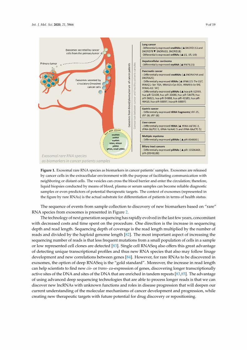
All of the studies presented in this review, which discovered differential patterns of RNA speciesloading in exosomes versus donor cells or exosomes from patients versus healthy controls (Figure 1),have in common the use of next generation sequencing, especially deep sequencing, of the RNA anddata mining of publicly available databases.
Int. J. Mol. Sci. 2020, 21, 5866 9 of 19Int. J. Mol. Sci. 2020, 21, x FOR PEER REVIEW 9 of 21
Figure 1. Exosomal rare RNA species as biomarkers in cancer patients’ samples. Exosomes are released by cancer cells in the extracellular environment with the purpose of facilitating communication with neighboring or distant cells. The vesicles can cross the blood barrier and enter the circulation; therefore, liquid biopsies conducted by means of blood, plasma or serum samples can become reliable diagnostic samples or even predictors of potential therapeutic targets. The context of exosomes (represented in the figure by rare RNAs) is the actual substrate for differentiation of patients in terms of health status.
Figure 1. Exosomal rare RNA species as biomarkers in cancer patients’ samples. Exosomes are releasedby cancer cells in the extracellular environment with the purpose of facilitating communication withneighboring or distant cells. The vesicles can cross the blood barrier and enter the circulation; therefore,liquid biopsies conducted by means of blood, plasma or serum samples can become reliable diagnosticsamples or even predictors of potential therapeutic targets. The context of exosomes (represented inthe figure by rare RNAs) is the actual substrate for differentiation of patients in terms of health status.
The sequence of events from sample collection to discovery of new biomarkers based on “rare”RNA species from exosomes is presented in Figure 2.
The technology of next generation sequencing has rapidly evolved in the last few years, concomitantwith decreased costs and time spent on the procedure. One direction is the increase in sequencingdepth and read length. Sequencing depth of coverage is the read length multiplied by the number ofreads and divided by the haploid genome length [82]. The most important aspect of increasing thesequencing number of reads is that less frequent mutations from a small population of cells in a sampleor low represented cell clones are detected [83]. Single cell RNASeq also offers this great advantageof detecting unique transcriptional profiles and thus new RNA species that also may follow linagedevelopment and new correlations between genes [84]. However, for rare RNAs to be discovered inexosomes, the option of deep RNASeq is the “gold standard”. Moreover, the increase in read lengthcan help scientists to find new cis- or trans- co-expression of genes, discovering longer transcriptionallyactive sites of the DNA and sites of the DNA that are enriched in tandem repeats [83,85]. The advantageof using advanced deep sequencing technologies that are able to process longer reads is that we candiscover new lncRNAs with unknown functions and roles in disease progression that will deepen ourcurrent understanding of the molecular mechanisms of cancer development and progression, whilecreating new therapeutic targets with future potential for drug discovery or repositioning.

Int. J. Mol. Sci. 2020, 21, 5866 10 of 19Int. J. Mol. Sci. 2020, 21, x FOR PEER REVIEW 10 of 21
Figure 2. From the collection of samples from patients undergoing minimally invasive procedures until the discovery of new biomarkers of a cancer type or for the assessment of cancer progression, there are 7 steps to be taken. (1) The samples are collected from patients and processed separately by centrifugation at low speed for the separation of exosomes from other heavier particles, such as cells. Then, through high speed ultracentrifugation or differential centrifugation, the next step is taken. (2) Exosome isolation. (3) The purified exosomes are sequenced during RNASeq: first, the total or small RNA is purified; then, it is reversed transcribed into cDNA, fragmented and labeled. The cDNA fragments are sequenced, and the reads are mapped to the genome of reference. During deep sequencing technologies, the repetition of sequencing is greater; thus, the genes that have a very low level of expression can be more easily identified. (4) The RNA Seq or microarray data (the microarray technology was not presented) should be made public by uploading the sequencing data along with clinical characteristics of the analyzed samples on publicly available databases, such as the Cancer Genome Atlas (TCGA) or Gene Expression Ominibus (GEO). In addition, if required, the authors should provide the RNASeq raw data. (5) Data mining. By accessing the above-mentioned databases, other scientific groups may find new associations between gene expression and clinical data. (6) In order to test for a causative correlation between RNA species expression and function, the in vitro testing and collection of exosomes from supernatant are needed. After definitive results in pre-clinical data, if the new RNA species show high sensitivity and specificity, comparable with that of the current biomarkers, they will be introduced in common clinical practice. (7) New biomarker discovery for wide application of a new diagnostic tool.
The technology of next generation sequencing has rapidly evolved in the last few years, concomitant with decreased costs and time spent on the procedure. One direction is the increase in sequencing depth and read length. Sequencing depth of coverage is the read length multiplied by the number of reads and divided by the haploid genome length [82]. The most important aspect of increasing the sequencing number of reads is that less frequent mutations from a small population of cells in a sample or low represented cell clones are detected [83]. Single cell RNASeq also offers this great advantage of detecting unique transcriptional profiles and thus new RNA species that also may follow linage development and new correlations between genes [84]. However, for rare RNAs to be discovered in exosomes, the option of deep RNASeq is the “gold standard”. Moreover, the increase in read length can help scientists to find new cis- or trans- co-expression of genes, discovering longer transcriptionally active sites of the DNA and sites of the DNA that are enriched
Figure 2. From the collection of samples from patients undergoing minimally invasive procedures untilthe discovery of new biomarkers of a cancer type or for the assessment of cancer progression, there are 7steps to be taken. (1) The samples are collected from patients and processed separately by centrifugationat low speed for the separation of exosomes from other heavier particles, such as cells. Then, throughhigh speed ultracentrifugation or differential centrifugation, the next step is taken. (2) Exosome isolation.(3) The purified exosomes are sequenced during RNASeq: first, the total or small RNA is purified; then,it is reversed transcribed into cDNA, fragmented and labeled. The cDNA fragments are sequenced, andthe reads are mapped to the genome of reference. During deep sequencing technologies, the repetitionof sequencing is greater; thus, the genes that have a very low level of expression can be more easilyidentified. (4) The RNA Seq or microarray data (the microarray technology was not presented) shouldbe made public by uploading the sequencing data along with clinical characteristics of the analyzedsamples on publicly available databases, such as the Cancer Genome Atlas (TCGA) or Gene ExpressionOminibus (GEO). In addition, if required, the authors should provide the RNASeq raw data. (5) Datamining. By accessing the above-mentioned databases, other scientific groups may find new associationsbetween gene expression and clinical data. (6) In order to test for a causative correlation between RNAspecies expression and function, the in vitro testing and collection of exosomes from supernatant areneeded. After definitive results in pre-clinical data, if the new RNA species show high sensitivity andspecificity, comparable with that of the current biomarkers, they will be introduced in common clinicalpractice. (7) New biomarker discovery for wide application of a new diagnostic tool.
However, large quantities of data are difficult to process; thus, more and more advancedbioinformatic tools that can keep up with this information will also be needed. The first step towardunderstanding the newly discovered lncRNAs is to look at the RNAs that are the origin of shortnon-coding RNAs and whose functions are easier to determine [86]. The sequencing technology canprovide valuable information, but it will be difficult to assess a single type of molecular interaction,considering that cancer is a polyfactorial disease. Moreover, what is known so far is that these sequenceshave very low levels of expression; thus, it is easy to assume that their function is not fundamentalfor cancer cell survival. In addition to these, there is the financial aspect; if all this technologicaldevelopment and financial investment in discovering the role of rare RNAs will lead to a dead end,it will not be profitable to obtain funding for this area of research. To overcome this challenge, thesecond approach to “rare RNA” function discovery, namely data mining, is important.
Data mining from public datasets, such as the Cancer Genome Atlas (TCGA) or Gene expressionomnibus (GEO), is essential for understanding the role of these rare RNAs and their specificity towardcertain types of cancer. For instance, the pan-cancer analysis of TCGA RNASeq data reveled a
Int. J. Mol. Sci. 2020, 21, 5866 11 of 19
significant implication of snRNAs especially in cancers of the digestive tract. For instance, RNU6-101 Pis a risk factor for esophageal cancer, while RNVU1-4 is a protective factor for stomach cancer [87].In prostate cancer, with the help of data mining from GEO, by means of re-analyzing raw data andapplying differential expression analysis between seminal fluid from controls and prostate cancerpatients, it was proven that 5′ tRNA halves have increased expression levels in seminal fluid fromprostate cancer patients, thus creating a new path for non-invasive biomarker discovery [88].
4. Final Remarks and Conclusions
The cancer-driven role of rare RNA loading in exosomes is becoming increasingly recognized withthe development of databases containing the specific expression of these RNAs (http://bioinformatics.zju.edu.cn/OncotRF/index.html), such as OncotRF, a database based on TCGA analysis of variouscancer samples that can show the expression of tRFs in various cancers [89]. The “rare” RNA speciesare often among the dominant RNAs in exosomes and some of them show clear specificity to a cancertype. Due to the high degree of stability of these RNAs, low variability and, in most cases, conservationacross species, rare RNAs can be considered a more reliable source of biomarkers for non-invasiveinvestigation of cancer than commonly used miRNAs or lncRNAs.
Int. J. Mol. Sci. 2020, 21, 5866 12 of 19
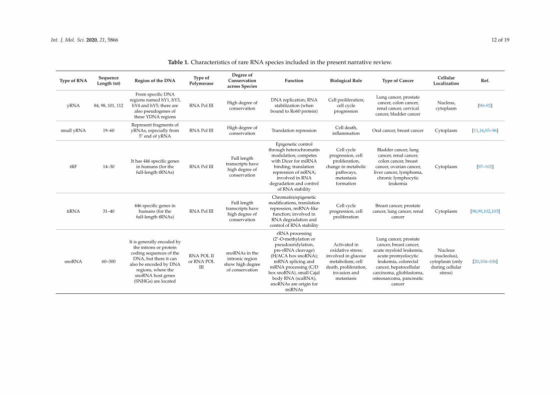
Table 1. Characteristics of rare RNA species included in the present narrative review.
Type of RNA SequenceLength (nt) Region of the DNA Type of
Polymerase
Degree ofConservationacross Species
Function Biological Role Type of Cancer CellularLocalization Ref.
yRNA 84, 98, 101, 112
From specific DNAregions named hY1, hY3,hY4 and hY5; there are
also pseudogenes ofthese YDNA regions
RNA Pol III High degree ofconservation
DNA replication; RNAstabilization (when
bound to Ro60 protein)
Cell proliferation;cell cycle
progression
Lung cancer, prostatecancer, colon cancer,renal cancer, cervical
cancer, bladder cancer
Nucleus,cytoplasm [90–92]
small yRNA 19–60Represent fragments ofyRNAs, especially from
5′ end of yRNARNA Pol III High degree of
conservation Translation repression Cell death,inflammation Oral cancer, breast cancer Cytoplasm [11,16,93–96]
tRF 14–30It has 446 specific genes
in humans (for thefull-length tRNAs)
RNA Pol III
Full lengthtranscripts havehigh degree ofconservation
Epigenetic controlthrough heterochromatin
modulation; competeswith Dicer for miRNA
binding; translationrepression of mRNA;
involved in RNAdegradation and control
of RNA stability
Cell cycleprogression, cell
proliferation,change in metabolic
pathways,metastasisformation
Bladder cancer, lungcancer, renal cancer,colon cancer, breast
cancer, ovarian cancer,liver cancer, lymphoma,
chronic lymphocyticleukemia
Cytoplasm [97–102]
tiRNA 31–40446 specific genes in
humans (for thefull-length tRNAs)
RNA Pol III
Full lengthtranscripts havehigh degree ofconservation
Chromatin/epigeneticmodifications, translationrepression, miRNA-like
function; involved inRNA degradation and
control of RNA stability
Cell cycleprogression, cell
proliferation
Breast cancer, prostatecancer, lung cancer, renal
cancerCytoplasm [98,99,102,103]
snoRNA 60–300
It is generally encoded bythe introns or protein
coding sequences of theDNA, but there it can
also be encoded by DNAregions, where the
snoRNA host genes(SNHGs) are located
RNA POL IIor RNA POL
III
snoRNAs in theintronic region
show high degreeof conservation
rRNA processing(2′-O-methylation orpseudouridylation,pre-rRNA cleavage)
(H/ACA box snoRNA);mRNA splicing and
mRNA processing (C/Dbox snoRNA), small Cajal
body RNA (scaRNA),snoRNAs are origin for
miRNAs
Activated inoxidative stress;
involved in glucosemetabolism; cell
death, proliferation,invasion and
metastasis
Lung cancer, prostatecancer, breast cancer,
acute myeloid leukemia,acute promyelocyticleukemia, colorectal
cancer, hepatocellularcarcinoma, glioblastoma,osteosarcoma, pancreatic
cancer
Nucleus(nucleolus),
cytoplasm (onlyduring cellular
stress)
[20,104–106]

Int. J. Mol. Sci. 2020, 21, 5866 13 of 19
Table 1. Cont.
Type of RNA SequenceLength (nt) Region of the DNA Type of
Polymerase
Degree ofConservationacross Species
Function Biological Role Type of Cancer CellularLocalization Ref.
snRNA 100–300Encoded by 5 DNA
regions: U1, U2, U4, U5,and U6
RNA POL III(U6), RNA
POL II(U1-U5)
High degree ofconservation pre-mRNA splicing
Cell cycleprogression,
Tumorigenesis,oncogenic
development
Breast cancer, lung cancer
Nucleus (mainfunction), can
also be exportedinto the
cytoplasm (U6)
[107–112]
vtRNA 88–100
Special sequence fromthe DNA, vault DNA
sequences are located onchromosome 5
RNA POL III High degree ofconservation
Associated with specificproteins vault proteins inthe cytoplasm, forming
vault RNPs in thecytoplasm
Autophagy,intracellular and
membranetrafficking,multidrug
resistance (drugexport from the
cytoplasm)
Breast cancer, lymphoma,lung cancer, multiple
myelomaCytoplasm [22,113–116]
Alu-elementRNA around 250
Alu-repeat containingtransposable elements,
comprises around 10% ofhuman genome
RNA POL II,RNA POL III
The main genescontaining ALU
repeats areconserved, butthere are also a
number of evolvedpseudogenes
through single basesubstitution
They are involved inprotein translation, are
the ancestors ofepigenetic enhancers,
they offer new bindingsites for transcription
factors, impair mRNA ormiRNA transcription
Increasedexpression in stress
conditions,involved in the
epithelial-to-mesenchymaltransition, cell cycle
progression
Breast cancer, colorectalcancer Nucleus [23–26]
piRNA 21–35
Encoded byprotein-coding genes
(untranslated regions ofmessenger RNAs),
sequences formintergenic regions (long
intergenic regions)
RNA POL III High degree ofconservation
It has a close interactionwith piwi protein and
together are involved inRNA cleavage. It also has
epigenetic functionsthrough heterochromatinregulation and induced
changes in DNAmethylation pattern.
Protects againstgermline genomestability and DNA
integrity; maintainscancer stemness,
apoptosisimpairment,involved in
telomerase activity,cell cycle
progression andmetastasis
Gastric cancer, lungcancer, cervical cancer,hepatocellular cancer,
breast cancer, colorectalcancer, ovarian cancer
Cytoplasm [117–120]
Int. J. Mol. Sci. 2020, 21, 5866 14 of 19
Funding: This research was funded by three national research grants from the Romanian Government: the first oneawarded for Frontiers Research Projects 2018–2022 (grant number PN-III-P4-ID-PCCF-2016-112) to Babes BolyaiUniversity, Cluj-Napoca, in collaboration with the Ion Chiricuta Oncology Institute, Cluj- Napoca; the secondone awarded for Young Research Teams 2020–2022 (grant number PN-III-P1-1.1-TE2019-0271) to Iuliu HatieganuUniversity of Medicine and Pharmacy, Cluj-Napoca, and the third one awarded for Postdoctoral Research Projects2020–2022 (grant number PN-III-P1-1.1-PD-2019-0805) to Iuliu Hatieganu University of Medicine and Pharmacy,Cluj-Napoca. The APC was funded by an international collaborative grant from the European Economic Spacebetween Romania and Iceland 2020–2022 (grant number 19-COP-0031).
Acknowledgments: The figures of the article were done with the help of illustrations from Servier Medical Art(https://smart.servier.com).
Conflicts of Interest: The authors have no conflict of interest.
References
1. Nagai, H.; Kim, Y.H. Cancer prevention from the perspective of global cancer burden patterns. J. Thorac. Dis.2017, 9, 448–451. [CrossRef]
2. Jayaseelan, V.P. Emerging role of exosomes as promising diagnostic tool for cancer. Cancer Gene Ther. 2020,27, 395–398. [CrossRef] [PubMed]
3. Gulei, D.; Petrut, B.; Tigu, A.B.; Onaciu, A.; Fischer-Fodor, E.; Atanasov, A.G.; Ionescu, C.; Berindan-Neagoe, I.Exosomes at a glance—Common nominators for cancer hallmarks and novel diagnosis tools. Crit. Rev.Biochem. Mol. Biol. 2018, 53, 564–577. [CrossRef] [PubMed]
4. Nawaz, M.; Camussi, G.; Valadi, H.; Nazarenko, I.; Ekström, K.; Wang, X.; Principe, S.; Shah, N.; Ashraf, N.M.;Fatima, F.; et al. The emerging role of extracellular vesicles as biomarkers for urogenital cancers. Nat. Rev.Urol. 2014, 11, 688–701. [CrossRef] [PubMed]
5. Statello, L.; Maugeri, M.; Garre, E.; Nawaz, M.; Wahlgren, J.; Papadimitriou, A.; Lundqvist, C.; Lindfors, L.;Collén, A.; Sunnerhagen, P.; et al. Identification of RNA-binding proteins in exosomes capable of interactingwith different types of RNA: RBP-facilitated transport of RNAs into exosomes. PLoS ONE 2018, 13, e0195969.[CrossRef] [PubMed]
6. Fatima, F.; Nawaz, M. Vesiculated Long Non-Coding RNAs: Offshore Packages Deciphering Trans-Regulationbetween Cells, Cancer Progression and Resistance to Therapies. Noncoding RNA 2017, 3, 10. [CrossRef][PubMed]
7. Silva, M.; Melo, S.A. Non-coding RNAs in Exosomes: New Players in Cancer Biology. Curr. Genom. 2015, 16,295–303. [CrossRef]
8. Xie, Y.; Dang, W.; Zhang, S.; Yue, W.; Yang, L.; Zhai, X.; Yan, Q.; Lu, J. The role of exosomal noncoding RNAsin cancer. Mol. Cancer 2019, 18, 37. [CrossRef]
9. Braicu, C.; Zimta, A.A.; Harangus, A.; Iurca, I.; Irimie, A.; Coza, O.; Berindan-Neagoe, I. The Function ofNon-Coding RNAs in Lung Cancer Tumorigenesis. Cancers 2019, 11, 605. [CrossRef]
10. Irimie, A.I.; Zimta, A.A.; Ciocan, C.; Mehterov, N.; Dudea, D.; Braicu, C.; Berindan-Neagoe, I. The UnforeseenNon-Coding RNAs in Head and Neck Cancer. Genes 2018, 9, 134. [CrossRef]
11. Dhahbi, J.; Nunez Lopez, Y.O.; Schneider, A.; Victoria, B.; Saccon, T.; Bharat, K.; McClatchey, T.; Atamna, H.;Scierski, W.; Golusinski, P.; et al. Profiling of tRNA Halves and YRNA Fragments in Serum and Tissue FromOral Squamous Cell Carcinoma Patients Identify Key Role of 5′ tRNA-Val-CAC-2-1 Half. Front. Oncol. 2019,9. [CrossRef] [PubMed]
12. Guo, Y.; Yu, H.; Wang, J.; Sheng, Q.; Zhao, S.; Zhao, Y.Y.; Lehmann, B.D. The Landscape of Small Non-CodingRNAs in Triple-Negative Breast Cancer. Genes 2018, 9, 29. [CrossRef]
13. Zhang, D.; Zhou, J.; Gao, J.; Wu, R.Y.; Huang, Y.L.; Jin, Q.W.; Chen, J.S.; Tang, W.Z.; Yan, L.H. TargetingsnoRNAs as an emerging method of therapeutic development for cancer. Am. J. Cancer Res. 2019, 9,1504–1516. [PubMed]
14. Wang, X.; Xu, M.; Yan, Y.; Kuang, Y.; Li, P.; Zheng, W.; Liu, H.; Jia, B. Identification of Eight Small NucleolarRNAs as Survival Biomarkers and Their Clinical Significance in Gastric Cancer. Front. Oncol. 2019, 9.[CrossRef] [PubMed]
15. Kowalski, M.P.; Krude, T. Functional roles of non-coding Y RNAs. Int. J. Biochem. Cell Biol. 2015, 66, 20–29.[CrossRef] [PubMed]
Int. J. Mol. Sci. 2020, 21, 5866 15 of 19
16. Hizir, Z.; Bottini, S.; Grandjean, V.; Trabucchi, M.; Repetto, E. RNY (YRNA)-derived small RNAs regulate celldeath and inflammation in monocytes/macrophages. Cell Death Dis. 2017, 8, e2530. [CrossRef]
17. Anderson, P.; Ivanov, P. tRNA fragments in human health and disease. FEBS Lett. 2014, 588, 4297–4304.[CrossRef]
18. Valadkhan, S.; Gunawardane, L.S. Role of small nuclear RNAs in eukaryotic gene expression. Essays Biochem.2013, 54, 79–90. [CrossRef]
19. Kiss, T. Small Nucleolar RNAs: An Abundant Group of Noncoding RNAs with Diverse Cellular Functions.Cell 2002, 109, 145–148. [CrossRef]
20. Liang, J.; Wen, J.; Huang, Z.; Chen, X.P.; Zhang, B.X.; Chu, L. Small Nucleolar RNAs: Insight Into TheirFunction in Cancer. Front. Oncol. 2019, 9. [CrossRef]
21. Horos, R.; Büscher, M.; Kleinendorst, R.; Alleaume, A.-M.; Tarafder, A.K.; Schwarzl, T.; Dziuba, D.; Tischer, C.;Zielonka, E.M.; Adak, A.; et al. The Small Non-coding Vault RNA1-1 Acts as a Riboregulator of Autophagy.Cell 2019, 176, 1054–1067.e12. [CrossRef] [PubMed]
22. Chen, J.; OuYang, H.; An, X.; Liu, S. Vault RNAs partially induces drug resistance of human tumor cellsMCF-7 by binding to the RNA/DNA-binding protein PSF and inducing oncogene GAGE6. PLoS ONE 2018,13, e0191325. [CrossRef] [PubMed]
23. Chen, L.-L.; Yang, L. ALUternative Regulation for Gene Expression. Trends Cell Biol. 2017, 27, 480–490.[CrossRef] [PubMed]
24. Caudron-Herger, M.; Pankert, T.; Seiler, J.; Németh, A.; Voit, R.; Grummt, I.; Rippe, K. Alu element-containingRNAs maintain nucleolar structure and function. EMBO J. 2015, 34, 2758–2774. [CrossRef] [PubMed]
25. Di Ruocco, F.; Basso, V.; Rivoire, M.; Mehlen, P.; Ambati, J.; De Falco, S.; Tarallo, V. Alu RNA accumulationinduces epithelial-to-mesenchymal transition by modulating miR-566 and is associated with cancerprogression. Oncogene 2018, 37, 627–637. [CrossRef] [PubMed]
26. Cantarella, S.; Carnevali, D.; Morselli, M.; Conti, A.; Pellegrini, M.; Montanini, B.; Dieci, G. Alu RNAModulates the Expression of Cell Cycle Genes in Human Fibroblasts. Int. J. Mol. Sci. 2019, 20, 3315.[CrossRef] [PubMed]
27. Tóth, K.F.; Pezic, D.; Stuwe, E.; Webster, A. The piRNA Pathway Guards the Germline Genome AgainstTransposable Elements. Adv. Exp. Med. Biol. 2016, 886, 51–77. [CrossRef] [PubMed]
28. Siddiqi, S.; Matushansky, I. Piwis and piwi-interacting RNAs in the epigenetics of cancer. J. Cell. Biochem.2012, 113, 373–380. [CrossRef]
29. Cheng, Y.; Wang, Q.; Jiang, W.; Bian, Y.; Zhou, Y.; Gou, A.; Zhang, W.; Fu, K.; Shi, W. Emerging roles ofpiRNAs in cancer: Challenges and prospects. Aging 2019, 11, 9932–9946. [CrossRef]
30. Pink, R.C.; Wicks, K.; Caley, D.P.; Punch, E.K.; Jacobs, L.; Carter, D.R.F. Pseudogenes: Pseudo-functional orkey regulators in health and disease? RNA (N.Y.) 2011, 17, 792–798. [CrossRef]
31. Ferlay, J.; Colombet, M.; Soerjomataram, I.; Mathers, C.; Parkin, D.M.; Piñeros, M.; Znaor, A.; Bray, F.Estimating the global cancer incidence and mortality in 2018: GLOBOCAN sources and methods. Int. J.Cancer 2019, 144, 1941–1953. [CrossRef] [PubMed]
32. King, M.-C.; Marks, J.H.; Mandell, J.B. Breast and Ovarian Cancer Risks Due to Inherited Mutations inBRCA1 and BRCA2. Science 2003, 302, 643–646. [CrossRef] [PubMed]
33. Zimta, A.A.; Tigu, A.B.; Muntean, M.; Cenariu, D.; Slaby, O.; Berindan-Neagoe, I. Molecular Links betweenCentral Obesity and Breast Cancer. Int. J. Mol. Sci. 2019, 20, 5364. [CrossRef] [PubMed]
34. Tosar, J.P.; Gámbaro, F.; Sanguinetti, J.; Bonilla, B.; Witwer, K.W.; Cayota, A. Assessment of small RNAsorting into different extracellular fractions revealed by high-throughput sequencing of breast cell lines.Nucleic Acids Res. 2015, 43, 5601–5616. [CrossRef] [PubMed]
35. Hashim, A.; Rizzo, F.; Marchese, G.; Ravo, M.; Tarallo, R.; Nassa, G.; Giurato, G.; Santamaria, G.; Cordella, A.;Cantarella, C.; et al. RNA sequencing identifies specific PIWI-interacting small non-coding RNA expressionpatterns in breast cancer. Oncotarget 2014, 5, 9901–9910. [CrossRef]
36. Fu, A.; Jacobs, D.I.; Hoffman, A.E.; Zheng, T.; Zhu, Y. PIWI-interacting RNA 021285 is involved in breasttumorigenesis possibly by remodeling the cancer epigenome. Carcinogenesis 2015, 36, 1094–1102. [CrossRef]
37. Gámbaro, F.; Li Calzi, M.; Fagúndez, P.; Costa, B.; Greif, G.; Mallick, E.; Lyons, S.; Ivanov, P.; Witwer, K.;Cayota, A.; et al. Stable tRNA halves can be sorted into extracellular vesicles and delivered to recipient cellsin a concentration-dependent manner. RNA Biol. 2019, 1–15. [CrossRef]
38. Ellis, P.M.; Vandermeer, R. Delays in the diagnosis of lung cancer. J. Thorac. Dis. 2011, 3, 183–188. [CrossRef]
Int. J. Mol. Sci. 2020, 21, 5866 16 of 19
39. Savelyeva, A.V.; Kuligina, E.V.; Bariakin, D.N.; Kozlov, V.V.; Ryabchikova, E.I.; Richter, V.A.; Semenov, D.V.Variety of RNAs in Peripheral Blood Cells, Plasma, and Plasma Fractions. Biomed. Res. Int. 2017, 2017,7404912. [CrossRef]
40. Kumar, M.; Nanavati, R.; Modi, T.G.; Dobariya, C. Oral cancer: Etiology and risk factors: A review. J. CancerRes. Ther. 2016, 12, 458–463. [CrossRef]
41. Auguste, A.; Deloumeaux, J.; Joachim, C.; Gaete, S.; Michineau, L.; Herrmann-Storck, C.; Duflo, S.; Luce, D.Joint effect of tobacco, alcohol, and oral HPV infection on head and neck cancer risk in the French WestIndies. Cancer Med. 2020. [CrossRef] [PubMed]
42. Ogawa, Y.; Taketomi, Y.; Murakami, M.; Tsujimoto, M.; Yanoshita, R. Small RNA transcriptomes of two typesof exosomes in human whole saliva determined by next generation sequencing. Biol. Pharm. Bull. 2013, 36,66–75. [CrossRef]
43. Bahn, J.H.; Zhang, Q.; Li, F.; Chan, T.M.; Lin, X.; Kim, Y.; Wong, D.T.; Xiao, X. The landscape of microRNA,Piwi-interacting RNA, and circular RNA in human saliva. Clin. Chem. 2015, 61, 221–230. [CrossRef]
44. Arnold, M.; Abnet, C.C.; Neale, R.E.; Vignat, J.; Giovannucci, E.L.; McGlynn, K.A.; Bray, F. Global Burden of5 Major Types of Gastrointestinal Cancer. Gastroenterology 2020, 159, 335–349.e15. [CrossRef] [PubMed]
45. Tan, C.; Cao, J.; Chen, L.; Xi, X.; Wang, S.; Zhu, Y.; Yang, L.; Ma, L.; Wang, D.; Yin, J.; et al. Noncoding RNAsServe as Diagnosis and Prognosis Biomarkers for Hepatocellular Carcinoma. Clin. Chem. 2019, 65, 905–915.[CrossRef] [PubMed]
46. Bradshaw, N.; Walter, P. The signal recognition particle (SRP) RNA links conformational changes in the SRPto protein targeting. Mol. Biol. Cell 2007, 18, 2728–2734. [CrossRef] [PubMed]
47. Zhu, L.; Li, J.; Gong, Y.; Wu, Q.; Tan, S.; Sun, D.; Xu, X.; Zuo, Y.; Zhao, Y.; Wei, Y.Q.; et al. ExosomaltRNA-derived small RNA as a promising biomarker for cancer diagnosis. Mol. Cancer 2019, 18, 74. [CrossRef]
48. Kitagawa, T.; Taniuchi, K.; Tsuboi, M.; Sakaguchi, M.; Kohsaki, T.; Okabayashi, T.; Saibara, T. Circulatingpancreatic cancer exosomal RNAs for detection of pancreatic cancer. Mol. Oncol. 2019, 13, 212–227. [CrossRef]
49. Kumar, S.R.; Kimchi, E.T.; Manjunath, Y.; Gajagowni, S.; Stuckel, A.J.; Kaifi, J.T. RNA cargos in extracellularvesicles derived from blood serum in pancreas associated conditions. Sci. Rep. 2020, 10, 2800. [CrossRef]
50. Lin, C.; Zheng, L.; Huang, R.; Yang, G.; Chen, J.; Li, H. tRFs as Potential Exosome tRNA-Derived FragmentBiomarkers for Gastric Carcinoma. Clin. Lab. 2020, 66. [CrossRef]
51. Ren, J.; Zhou, Q.; Li, H.; Li, J.; Pang, L.; Su, L.; Gu, Q.; Zhu, Z.; Liu, B. Characterization of exosomal RNAsderived from human gastric cancer cells by deep sequencing. Tumour Biol. 2017, 39, 1010428317695012.[CrossRef] [PubMed]
52. Fonseca Cabral, G.; Azevedo Dos Santos Pinheiro, J.; Vidal, A.F.; Santos, S.; Ribeiro-Dos-Santos, Â. piRNAs inGastric Cancer: A New Approach Towards Translational Research. Int. J. Mol. Sci. 2020, 21, 2126. [CrossRef][PubMed]
53. Zhu, L.; Li, T.; Shen, Y.; Yu, X.; Xiao, B.; Guo, J. Using tRNA halves as novel biomarkers for the diagnosis ofgastric cancer. Cancer Biomark. 2019, 25, 169–176. [CrossRef] [PubMed]
54. Chen, M.; Xu, R.; Ji, H.; Greening, D.W.; Rai, A.; Izumikawa, K.; Ishikawa, H.; Takahashi, N.; Simpson, R.J.Transcriptome and long noncoding RNA sequencing of three extracellular vesicle subtypes released from thehuman colon cancer LIM1863 cell line. Sci. Rep. 2016, 6, 38397. [CrossRef] [PubMed]
55. Gu, X.; Wang, C.; Deng, H.; Qing, C.; Liu, R.; Liu, S.; Xue, X. Exosomal piRNA profiling revealed uniquecirculating piRNA signatures of cholangiocarcinoma and gallbladder carcinoma. Acta Biochim. Biophys. Sin.2020, 52, 475–484. [CrossRef] [PubMed]
56. Bosma, I.; Vos, M.J.; Heimans, J.J.; Taphoorn, M.J.B.; Aaronson, N.K.; Postma, T.J.; van der Ploeg, H.M.;Muller, M.; Vandertop, W.P.; Slotman, B.J.; et al. The course of neurocognitive functioning in high-gradeglioma patients. Neuro Oncol. 2007, 9, 53–62. [CrossRef] [PubMed]
57. van Kessel, E.; Baumfalk, A.E.; van Zandvoort, M.J.E.; Robe, P.A.; Snijders, T.J. Tumor-related neurocognitivedysfunction in patients with diffuse glioma: A systematic review of neurocognitive functioning prior toanti-tumor treatment. J. Neurooncol. 2017, 134, 9–18. [CrossRef]
58. Li, C.C.; Eaton, S.A.; Young, P.E.; Lee, M.; Shuttleworth, R.; Humphreys, D.T.; Grau, G.E.; Combes, V.;Bebawy, M.; Gong, J.; et al. Glioma microvesicles carry selectively packaged coding and non-coding RNAswhich alter gene expression in recipient cells. RNA Biol. 2013, 10, 1333–1344. [CrossRef]
59. de Mooij, T.; Peterson, T.E.; Evans, J.; McCutcheon, B.; Parney, I.F. Short non-coding RNA sequencing ofglioblastoma extracellular vesicles. J. Neurooncol. 2020, 146, 253–263. [CrossRef]
Int. J. Mol. Sci. 2020, 21, 5866 17 of 19
60. Wei, Z.; Batagov, A.O.; Schinelli, S.; Wang, J.; Wang, Y.; El Fatimy, R.; Rabinovsky, R.; Balaj, L.; Chen, C.C.;Hochberg, F.; et al. Coding and noncoding landscape of extracellular RNA released by human glioma stemcells. Nat. Commun. 2017, 8, 1145. [CrossRef]
61. Kore, R.A.; Edmondson, J.L.; Jenkins, S.V.; Jamshidi-Parsian, A.; Dings, R.P.M.; Reyna, N.S.; Griffin, R.J.Hypoxia-derived exosomes induce putative altered pathways in biosynthesis and ion regulatory channels inglioblastoma cells. Biochem. Biophys. Rep. 2018, 14, 104–113. [CrossRef] [PubMed]
62. van Balkom, B.W.; Eisele, A.S.; Pegtel, D.M.; Bervoets, S.; Verhaar, M.C. Quantitative and qualitative analysisof small RNAs in human endothelial cells and exosomes provides insights into localized RNA processing,degradation and sorting. J. Extracell. Vesicles 2015, 4, 26760. [CrossRef] [PubMed]
63. Balaj, L.; Lessard, R.; Dai, L.; Cho, Y.J.; Pomeroy, S.L.; Breakefield, X.O.; Skog, J. Tumour microvesiclescontain retrotransposon elements and amplified oncogene sequences. Nat. Commun. 2011, 2, 180. [CrossRef][PubMed]
64. Davies, H.; Bignell, G.R.; Cox, C.; Stephens, P.; Edkins, S.; Clegg, S.; Teague, J.; Woffendin, H.; Garnett, M.J.;Bottomley, W.; et al. Mutations of the BRAF gene in human cancer. Nature 2002, 417, 949–954. [CrossRef]
65. Lunavat, T.R.; Cheng, L.; Einarsdottir, B.O.; Olofsson Bagge, R.; Veppil Muralidharan, S.; Sharples, R.A.;Lässer, C.; Gho, Y.S.; Hill, A.F.; Nilsson, J.A.; et al. BRAFV600 inhibition alters the microRNA cargo in thevesicular secretome of malignant melanoma cells. Proc. Natl. Acad. Sci. USA 2017. [CrossRef]
66. Luetke, A.; Meyers, P.A.; Lewis, I.; Juergens, H. Osteosarcoma treatment—Where do we stand? A state of theart review. Cancer Treat. Rev. 2014, 40, 523–532. [CrossRef]
67. Liu, D.D.; Kang, Y. Ets2 anchors the prometastatic function of mutant p53 in osteosarcoma. Genes Dev. 2017,31, 1823–1824. [CrossRef]
68. Evdokimova, V.; Ruzanov, P.; Gassmann, H.; Zaidi, S.H.; Peltekova, V.; Heisler, L.E.; McPherson, J.D.;Orlic-Milacic, M.; Specht, K.; Steiger, K.; et al. Exosomes transmit retroelement RNAs to drive inflammationand immunosuppression in Ewing Sarcoma. bioRxiv 2019, 806851. [CrossRef]
69. Vardiman, J.W.; Thiele, J.; Arber, D.A.; Brunning, R.D.; Borowitz, M.J.; Porwit, A.; Harris, N.L.; Le Beau, M.M.;Hellström-Lindberg, E.; Tefferi, A.; et al. The 2008 revision of the World Health Organization (WHO)classification of myeloid neoplasms and acute leukemia: Rationale and important changes. Blood 2009, 114,937–951. [CrossRef]
70. Baba AI, C.C. Chapter 17, TUMORS OF HEMATOPOIETIC AND LYMPHOID TISSUES. In ComparativeOncology; The Publishing House of the Romanian Academy: Bucharest, Romania, 2007.
71. Ireland, R. Haematological malignancies: The rationale for integrated haematopathology services,key elements of organization and wider contribution to patient care. Histopathology 2011, 58, 145–154.[CrossRef]
72. Lovisa, F.; Di Battista, P.; Gaffo, E.; Damanti, C.C.; Garbin, A.; Gallingani, I.; Carraro, E.; Pillon, M.; Biffi, A.;Bortoluzzi, S.; et al. RNY4 in Circulating Exosomes of Patients With Pediatric Anaplastic Large CellLymphoma: An Active Player? Front. Oncol. 2020, 10, 238. [CrossRef] [PubMed]
73. Ma, H.; Wang, H.; Tian, F.; Zhong, Y.; Liu, Z.; Liao, A. PIWI-Interacting RNA-004800 Is Regulated by S1PReceptor Signaling Pathway to Keep Myeloma Cell Survival. Front. Oncol. 2020, 10, 438. [CrossRef][PubMed]
74. Wang, A.; Liu, J.; Zhuang, X.; Yu, S.; Zhu, S.; Liu, Y.; Chen, X. Identification and Comparison of piRNAExpression Profiles of Exosomes Derived from Human Stem Cells from the Apical Papilla and Bone MarrowMesenchymal Stem Cells. Stem Cells Dev. 2020, 29, 511–520. [CrossRef] [PubMed]
75. Wu, Y.; Qiu, W.; Xu, X.; Kang, J.; Wang, J.; Wen, Y.; Tang, X.; Yan, Y.; Qian, H.; Zhang, X.; et al. Exosomesderived from human umbilical cord mesenchymal stem cells alleviate inflammatory bowel disease in micethrough ubiquitination. Am. J. Transl. Res. 2018, 10, 2026–2036. [PubMed]
76. He, J.G.; Xie, Q.L.; Li, B.B.; Zhou, L.; Yan, D. Exosomes Derived from IDO1-Overexpressing Rat BoneMarrow Mesenchymal Stem Cells Promote Immunotolerance of Cardiac Allografts. Cell Transplant. 2018, 27,1657–1683. [CrossRef] [PubMed]
77. Hu, L.; Wang, J.; Zhou, X.; Xiong, Z.; Zhao, J.; Yu, R.; Huang, F.; Zhang, H.; Chen, L. Exosomes derivedfrom human adipose mensenchymal stem cells accelerates cutaneous wound healing via optimizing thecharacteristics of fibroblasts. Sci. Rep. 2016, 6, 32993. [CrossRef]

Int. J. Mol. Sci. 2020, 21, 5866 18 of 19
78. Huang, X.; Yuan, T.; Tschannen, M.; Sun, Z.; Jacob, H.; Du, M.; Liang, M.; Dittmar, R.L.; Liu, Y.; Liang, M.;et al. Characterization of human plasma-derived exosomal RNAs by deep sequencing. BMC Genom. 2013,14, 319. [CrossRef]
79. Baglio, S.R.; Rooijers, K.; Koppers-Lalic, D.; Verweij, F.J.; Pérez Lanzón, M.; Zini, N.; Naaijkens, B.; Perut, F.;Niessen, H.W.; Baldini, N.; et al. Human bone marrow- and adipose-mesenchymal stem cells secreteexosomes enriched in distinctive miRNA and tRNA species. Stem Cell Res. Ther. 2015, 6, 127. [CrossRef]
80. Fan, G.; Li, J. Regions identity between the genome of vertebrates and non-retroviral families of insect viruses.Virol. J. 2011, 8, 511. [CrossRef]
81. Wu, Z.; Sun, H.; Wang, C.; Liu, W.; Liu, M.; Zhu, Y.; Xu, W.; Jin, H.; Li, J. Mitochondrial Genome-DerivedcircRNA mc-COX2 Functions as an Oncogene in Chronic Lymphocytic Leukemia. Mol. Ther. Nucleic Acids2020, 20, 801–811. [CrossRef]
82. Sims, D.; Sudbery, I.; Ilott, N.E.; Heger, A.; Ponting, C.P. Sequencing depth and coverage: Key considerationsin genomic analyses. Nat. Rev. Genet. 2014, 15, 121–132. [CrossRef] [PubMed]
83. Goldman, D.; Domschke, K. Making sense of deep sequencing. Int. J. Neuropsychopharmacol. 2014, 17,1717–1725. [CrossRef] [PubMed]
84. Hwang, B.; Lee, J.H.; Bang, D. Single-cell RNA sequencing technologies and bioinformatics pipelines.Exp. Mol. Med. 2018, 50, 96. [CrossRef] [PubMed]
85. Wang, Z.; Gerstein, M.; Snyder, M. RNA-Seq: A revolutionary tool for transcriptomics. Nat. Rev. Genet. 2009,10, 57–63. [CrossRef] [PubMed]
86. Umbach, N.; Beißbarth, T.; Bleckmann, A.; Duttge, G.; Flatau, L.; König, A.; Kuhn, J.; Perera-Bel, J.;Roschauer, J.; Schulze, T.G.; et al. Clinical application of genomic high-throughput data: Infrastructural,ethical, legal and psychosocial aspects. Eur. Neuropsychopharmacol. J. Eur. Coll. Neuropsychopharmacol. 2020,31, 1–15. [CrossRef] [PubMed]
87. Qin, X.G.; Zeng, J.H.; Lin, P.; Mo, W.J.; Li, Q.; Feng, Z.B.; Luo, D.Z.; Yang, H.; Chen, G.; Zeng, J.J. Prognosticvalue of small nuclear RNAs (snRNAs) for digestive tract pan- adenocarcinomas identified by RNAsequencing data. Pathol. Res. Pract. 2019, 215, 414–426. [CrossRef]
88. Dhahbi, J.M.; Atamna, H.; Selth, L.A. Data Mining of Small RNA-Seq Suggests an Association BetweenProstate Cancer and Altered Abundance of 5′ Transfer RNA Halves in Seminal Fluid and Prostatic Tissues.Biomark. Cancer 2018, 10, 1179299x18759545. [CrossRef]
89. Yao, D.; Sun, X.; Zhou, L.; Amanullah, M.; Pan, X.; Liu, Y.; Liang, M.; Liu, P.; Lu, Y. OncotRF: An onlineresource for exploration of tRNA-derived fragments in human cancers. RNA Biol. 2020, 1–11. [CrossRef]
90. Wolin, S.L.; Steitz, J.A. Genes for two small cytoplasmic Ro RNAs are adjacent and appear to be single-copyin the human genome. Cell 1983, 32, 735–744. [CrossRef]
91. Sacks, D.; Baxter, B.; Campbell, B.C.V.; Carpenter, J.S.; Cognard, C.; Dippel, D.; Eesa, M.; Fischer, U.;Hausegger, K.; Hirsch, J.A.; et al. Multisociety Consensus Quality Improvement Revised ConsensusStatement for Endovascular Therapy of Acute Ischemic Stroke. Int. J. Stroke 2018, 13, 612–632. [CrossRef]
92. Christov, C.P.; Trivier, E.; Krude, T. Noncoding human Y RNAs are overexpressed in tumours and requiredfor cell proliferation. Br. J. Cancer 2008, 98, 981–988. [CrossRef] [PubMed]
93. Sim, S.; Wolin, S.L. Emerging roles for the Ro 60-kDa autoantigen in noncoding RNA metabolism. WileyInterdiscip. Rev. RNA 2011, 2, 686–699. [CrossRef]
94. Dhahbi, J.M.; Spindler, S.R.; Atamna, H.; Boffelli, D.; Mote, P.; Martin, D.I.K. 5′-YRNA fragments derived byprocessing of transcripts from specific YRNA genes and pseudogenes are abundant in human serum andplasma. Physiol. Genomics 2013, 45, 990–998. [CrossRef]
95. Gulìa, C.; Signore, F.; Gaffi, M.; Gigli, S.; Votino, R.; Nucciotti, R.; Bertacca, L.; Zaami, S.; Baffa, A.; Santini, E.;et al. Y RNA: An Overview of Their Role as Potential Biomarkers and Molecular Targets in Human Cancers.Cancers 2020, 12, 1238. [CrossRef]
96. Dhahbi, J.M.; Spindler, S.R.; Atamna, H.; Boffelli, D.; Martin, D.I. Deep Sequencing of Serum SmallRNAs Identifies Patterns of 5′ tRNA Half and YRNA Fragment Expression Associated with Breast Cancer.Biomark. Cancer 2014, 6, 37–47. [CrossRef]
97. Boskovic, A.; Bing, X.Y.; Kaymak, E.; Rando, O.J. Control of noncoding RNA production and histone levelsby a 5′ tRNA fragment. Genes Dev. 2020, 34, 118–131. [CrossRef]
98. Goodenbour, J.M.; Pan, T. Diversity of tRNA genes in eukaryotes. Nucleic Acids Res. 2006, 34, 6137–6146.[CrossRef]
Int. J. Mol. Sci. 2020, 21, 5866 19 of 19
99. Xie, Y.; Yao, L.; Yu, X.; Ruan, Y.; Li, Z.; Guo, J. Action mechanisms and research methods of tRNA-derivedsmall RNAs. Signal Transduct. Target. Ther. 2020, 5, 109. [CrossRef]
100. Kutsuna, T.; Hiyama, Y.; Kusaka, S.; Kusumoto, Y.; Tsuchiya, J.; Umeda, M.; Takahashi, T. The effect ofshort-term health promotion intervention on motor function in community-dwelling older adults. Aging Clin.Exp. Res. 2019, 31, 475–481. [CrossRef]
101. Telonis, A.G.; Loher, P.; Magee, R.; Pliatsika, V.; Londin, E.; Kirino, Y.; Rigoutsos, I. tRNA Fragments ShowIntertwining with mRNAs of Specific Repeat Content and Have Links to Disparities. Cancer Res. 2019, 79,3034–3049. [CrossRef]
102. Zhu, P.; Yu, J.; Zhou, P. Role of tRNA-derived fragments in cancer: Novel diagnostic and therapeutic targetstRFs in cancer. Am. J. Cancer Res. 2020, 10, 393–402.
103. Lee, Y.S.; Shibata, Y.; Malhotra, A.; Dutta, A. A novel class of small RNAs: tRNA-derived RNA fragments(tRFs). Genes Dev. 2009, 23, 2639–2649. [CrossRef]
104. Holley, C.L.; Li, M.W.; Scruggs, B.S.; Matkovich, S.J.; Ory, D.S.; Schaffer, J.E. Cytosolic accumulation ofsmall nucleolar RNAs (snoRNAs) is dynamically regulated by NADPH oxidase. J. Biol. Chem. 2015, 290,11741–11748. [CrossRef]
105. Scott, M.S.; Ono, M. From snoRNA to miRNA: Dual function regulatory non-coding RNAs. Biochimie 2011,93, 1987–1992. [CrossRef]
106. Hoeppner, M.P.; White, S.; Jeffares, D.C.; Poole, A.M. Evolutionarily stable association of intronic snoRNAsand microRNAs with their host genes. Genome Biol. Evol. 2009, 1, 420–428. [CrossRef]
107. Kiss, T. Biogenesis of small nuclear RNPs. J. Cell Sci. 2004, 117, 5949–5951. [CrossRef]108. Jády, B.E.; Darzacq, X.; Tucker, K.E.; Matera, A.G.; Bertrand, E.; Kiss, T. Modification of Sm small nuclear
RNAs occurs in the nucleoplasmic Cajal body following import from the cytoplasm. EMBO J. 2003, 22,1878–1888. [CrossRef]
109. Matera, A.G.; Terns, R.M.; Terns, M.P. Non-coding RNAs: Lessons from the small nuclear and small nucleolarRNAs. Nat. Rev. Mol. Cell Biol. 2007, 8, 209–220. [CrossRef]
110. Roy, K.; Chanfreau, G.F. Chapter Ten—The Diverse Functions of Fungal RNase III Enzymes in RNAMetabolism. In The Enzymes; Chanfreau, G.F., Tamanoi, F., Eds.; Academic Press: Cambridge, MA, USA,2012; Volume 31, pp. 213–235.
111. Dvinge, H.; Guenthoer, J.; Porter, P.L.; Bradley, R.K. RNA components of the spliceosome regulate tissue-and cancer-specific alternative splicing. Genome Res. 2019, 29, 1591–1604. [CrossRef]
112. Oh, J.-M.; Venters, C.C.; Di, C.; Pinto, A.M.; Wan, L.; Younis, I.; Cai, Z.; Arai, C.; So, B.R.; Duan, J.; et al. U1snRNP regulates cancer cell migration and invasion in vitro. Nat. Commun. 2020, 11, 1–8. [CrossRef]
113. Büscher, M.; Horos, R.; Hentze, M.W. ‘High vault-age’: Non-coding RNA control of autophagy. Open Biol.2020, 10, 190307. [CrossRef]
114. Amort, M.; Nachbauer, B.; Tuzlak, S.; Kieser, A.; Schepers, A.; Villunger, A.; Polacek, N. Expression of thevault RNA protects cells from undergoing apoptosis. Nat. Commun. 2015, 6, 7030. [CrossRef]
115. van Zon, A.; Mossink, M.H.; Houtsmuller, A.B.; Schoester, M.; Scheffer, G.L.; Scheper, R.J.; Sonneveld, P.;Wiemer, E.A. Vault mobility depends in part on microtubules and vaults can be recruited to the nuclearenvelope. Exp. Cell Res. 2006, 312, 245–255. [CrossRef]
116. Labuda, D.; Striker, G. Sequence conservation in Alu evolution. Nucleic Acids Res. 1989, 17, 2477–2491.[CrossRef]
117. Ozata, D.M.; Gainetdinov, I.; Zoch, A.; O’Carroll, D.; Zamore, P.D. PIWI-interacting RNAs: Small RNAs withbig functions. Nat. Rev. Genet. 2019, 20, 89–108. [CrossRef]
118. Sun, T.; Han, X. The disease-related biological functions of PIWI-interacting RNAs (piRNAs) and underlyingmolecular mechanisms. ExRNA 2019, 1. [CrossRef]
119. Han, B.W.; Zamore, P.D. piRNAs. Curr. Biol. 2014, 24, R730–R733. [CrossRef]120. Lim, S.L.; Tsend-Ayush, E.; Kortschak, R.D.; Jacob, R.; Ricciardelli, C.; Oehler, M.K.; Grützner, F. Conservation
and Expression of PIWI-Interacting RNA Pathway Genes in Male and Female Adult Gonad of Amniotes.Biol. Reprod. 2013, 89. [CrossRef]
© 2020 by the authors. Licensee MDPI, Basel, Switzerland. This article is an open accessarticle distributed under the terms and conditions of the Creative Commons Attribution(CC BY) license (http://creativecommons.org/licenses/by/4.0/).


![Nanocarriers for skin delivery of cosmetic antioxidants. [Nanovehículos para la liberación en piel de cosméticos antioxidantes]](https://static.fdokumen.com/doc/165x107/631cc2205a0be56b6e0e5bcd/nanocarriers-for-skin-delivery-of-cosmetic-antioxidants-nanovehiculos-para-la.jpg)