Exosomes as Powerful Engines in Cancer: Isolation ... - MDPI
37
biosensors Review Exosomes as Powerful Engines in Cancer: Isolation, Characterization and Detection Techniques Marwa Gamal Saad, Haluk Beyenal and Wen-Ji Dong * Citation: Saad, M.G.; Beyenal, H.; Dong, W.-J. Exosomes as Powerful Engines in Cancer: Isolation, Characterization and Detection Techniques. Biosensors 2021, 11, 518. https://doi.org/10.3390/bios11120518 Received: 17 October 2021 Accepted: 2 December 2021 Published: 16 December 2021 Publisher’s Note: MDPI stays neutral with regard to jurisdictional claims in published maps and institutional affil- iations. Copyright: © 2021 by the authors. Licensee MDPI, Basel, Switzerland. This article is an open access article distributed under the terms and conditions of the Creative Commons Attribution (CC BY) license (https:// creativecommons.org/licenses/by/ 4.0/). The Gene and Linda Voiland School of Chemical Engineering and Bioengineering, Washington State University, Pullman, WA 99164, USA; [email protected] (M.G.S.); [email protected] (H.B.) * Correspondence: [email protected] Abstract: Exosomes, powerful extracellular nanovesicles released from almost all types of living cells, are considered the communication engines (messengers) that control and reprogram physiological pathways inside target cells within a community or between different communities. The cell-like structure of these extracellular vesicles provides a protective environment for their proteins and DNA/RNA cargos, which serve as biomarkers for many malicious diseases, including infectious diseases and cancers. Cancer-derived exosomes control cancer metastasis, prognosis, and devel- opment. In addition to the unique structure of exosomes, their nanometer size and tendency of interacting with cells makes them a viable novel drug delivery solution. In recent years, numerous research efforts have been made to quantify and characterize disease-derived exosomes for diagnosis, monitoring, and therapeutic purposes. This review aims to (1) relate exosome biomarkers to their origins, (2) focus on current isolation and detection methods, (3) discuss and evaluate the proposed technologies deriving from exosome research for cancer treatment, and (4) form a conclusion about the prospects of the current exosome research. Keywords: exosomes; isolation; characterization; detection; biomarkers; communication; cancer 1. Introduction There are two types of extracellular vesicles released from cells: exosomes and ec- tosomes. Exosomes have smaller diameter sizes than ectosomes [1] and are released via the fusion of multivesicular bodies with the plasma membrane, whereas ectosomes are shed directly from the plasma membrane [2]. Exosomes are endosome-derived membrane vesicles with a size range of 20–150 nm [3–12] and are derived naturally from nearly all cell types [1,13–15]. When exosomes were first discovered in 1981 in mammalian cells, they were believed to act as discharged vesicles of obsolete molecules [16,17]. After their roles in communication were revealed, they were thought to communicate only within one species, but they were later found to engage in interkingdom communication [10]. Due to their roles in cellular communications, exosomes are carriers for vital biomarkers that originate from parental cells. These biomarkers include nucleic acids, e.g., miR-21, which is considered a reference biomarker for ovarian [18], prostate [19], and breast cancers [20]. As a result, exosomes are considered unique biomarkers for the diagnosis and prognosis of various malicious diseases, such as cancer. Cancer is one of the deadliest diseases worldwide. Prostate cancer ranks as the most widespread cancer, and it causes a significant percentage of death [21]. Lung cancer, the second-most deadly cancer, causes 25% of cancer deaths with either small cell carcinoma or non-small-cell carcinoma [22]. Cortical cancer is the second-most fatal cancer and the third-most widespread one [23], and breast cancer is a hostile tumor among women over 40 [24,25]. Due to the high fatality of cancers and complicated treatment procedures, it is highly desired to find a reliable cancer biomarker to improve cancer detection for early diagnosis and treatment [24–26]. Since cancer-derived exosomes are involved in both the development and metastasis of cancer through intracellular communication [27,28], are Biosensors 2021, 11, 518. https://doi.org/10.3390/bios11120518 https://www.mdpi.com/journal/biosensors
-
Upload
khangminh22 -
Category
Documents
-
view
1 -
download
0
Transcript of Exosomes as Powerful Engines in Cancer: Isolation ... - MDPI

biosensors
Review
Exosomes as Powerful Engines in Cancer: Isolation,Characterization and Detection Techniques
Marwa Gamal Saad, Haluk Beyenal and Wen-Ji Dong *
�����������������
Citation: Saad, M.G.; Beyenal, H.;
Dong, W.-J. Exosomes as Powerful
Engines in Cancer: Isolation,
Characterization and Detection
Techniques. Biosensors 2021, 11, 518.
https://doi.org/10.3390/bios11120518
Received: 17 October 2021
Accepted: 2 December 2021
Published: 16 December 2021
Publisher’s Note: MDPI stays neutral
with regard to jurisdictional claims in
published maps and institutional affil-
iations.
Copyright: © 2021 by the authors.
Licensee MDPI, Basel, Switzerland.
This article is an open access article
distributed under the terms and
conditions of the Creative Commons
Attribution (CC BY) license (https://
creativecommons.org/licenses/by/
4.0/).
The Gene and Linda Voiland School of Chemical Engineering and Bioengineering, Washington State University,Pullman, WA 99164, USA; [email protected] (M.G.S.); [email protected] (H.B.)* Correspondence: [email protected]
Abstract: Exosomes, powerful extracellular nanovesicles released from almost all types of living cells,are considered the communication engines (messengers) that control and reprogram physiologicalpathways inside target cells within a community or between different communities. The cell-likestructure of these extracellular vesicles provides a protective environment for their proteins andDNA/RNA cargos, which serve as biomarkers for many malicious diseases, including infectiousdiseases and cancers. Cancer-derived exosomes control cancer metastasis, prognosis, and devel-opment. In addition to the unique structure of exosomes, their nanometer size and tendency ofinteracting with cells makes them a viable novel drug delivery solution. In recent years, numerousresearch efforts have been made to quantify and characterize disease-derived exosomes for diagnosis,monitoring, and therapeutic purposes. This review aims to (1) relate exosome biomarkers to theirorigins, (2) focus on current isolation and detection methods, (3) discuss and evaluate the proposedtechnologies deriving from exosome research for cancer treatment, and (4) form a conclusion aboutthe prospects of the current exosome research.
Keywords: exosomes; isolation; characterization; detection; biomarkers; communication; cancer
1. Introduction
There are two types of extracellular vesicles released from cells: exosomes and ec-tosomes. Exosomes have smaller diameter sizes than ectosomes [1] and are released viathe fusion of multivesicular bodies with the plasma membrane, whereas ectosomes areshed directly from the plasma membrane [2]. Exosomes are endosome-derived membranevesicles with a size range of 20–150 nm [3–12] and are derived naturally from nearly allcell types [1,13–15]. When exosomes were first discovered in 1981 in mammalian cells,they were believed to act as discharged vesicles of obsolete molecules [16,17]. After theirroles in communication were revealed, they were thought to communicate only within onespecies, but they were later found to engage in interkingdom communication [10]. Dueto their roles in cellular communications, exosomes are carriers for vital biomarkers thatoriginate from parental cells. These biomarkers include nucleic acids, e.g., miR-21, whichis considered a reference biomarker for ovarian [18], prostate [19], and breast cancers [20].As a result, exosomes are considered unique biomarkers for the diagnosis and prognosis ofvarious malicious diseases, such as cancer.
Cancer is one of the deadliest diseases worldwide. Prostate cancer ranks as the mostwidespread cancer, and it causes a significant percentage of death [21]. Lung cancer, thesecond-most deadly cancer, causes 25% of cancer deaths with either small cell carcinomaor non-small-cell carcinoma [22]. Cortical cancer is the second-most fatal cancer and thethird-most widespread one [23], and breast cancer is a hostile tumor among women over40 [24,25]. Due to the high fatality of cancers and complicated treatment procedures, it ishighly desired to find a reliable cancer biomarker to improve cancer detection for earlydiagnosis and treatment [24–26]. Since cancer-derived exosomes are involved in both thedevelopment and metastasis of cancer through intracellular communication [27,28], are
Biosensors 2021, 11, 518. https://doi.org/10.3390/bios11120518 https://www.mdpi.com/journal/biosensors
Biosensors 2021, 11, 518 2 of 37
stable with small size phenotypes, and are accessible in most biological fluids [29,30], theyare considered excellent candidates for early cancer diagnosis and vehicles for cell-basedtherapy and drug delivery [10].
This review will start with a brief description of exosome structure, release, andbiogenesis mechanisms, followed by a description of the known techniques for isolationand detection. The exosome applications for cancer will be specified. The multiple roles ofexosomes, such as (1) biomarkers, (2) diagnostic agents, and (3) signal transduction factors,will be included. Finally, future research efforts to address the technical challenges relatedto exosome study will be discussed.
2. Exosome Structure, Release, and Biogenesis2.1. Exosome Structure
In this section, we will emphasize the structure of exosomes in mammalian popu-lations. Exosomes derived from mammalian populations contain a lipid bilayer mem-brane [31], intracellular components, and extracellular components bound to the outermembrane (Figure 1).
Biosensors 2021, 11, x FOR PEER REVIEW 2 of 40
diagnosis and treatment [24–26]. Since cancer-derived exosomes are involved in both the development and metastasis of cancer through intracellular communication [27,28], are stable with small size phenotypes, and are accessible in most biological fluids [29,30], they are considered excellent candidates for early cancer diagnosis and vehicles for cell-based therapy and drug delivery [10].
This review will start with a brief description of exosome structure, release, and bio-genesis mechanisms, followed by a description of the known techniques for isolation and detection. The exosome applications for cancer will be specified. The multiple roles of ex-osomes, such as (1) biomarkers, (2) diagnostic agents, and (3) signal transduction factors, will be included. Finally, future research efforts to address the technical challenges related to exosome study will be discussed.
2. Exosome Structure, Release, and Biogenesis 2.1. Exosome Structure
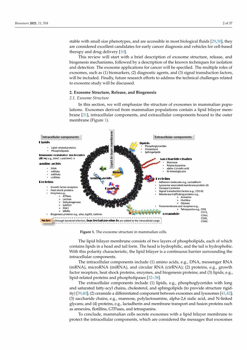
In this section, we will emphasize the structure of exosomes in mammalian popula-tions. Exosomes derived from mammalian populations contain a lipid bilayer membrane [31], intracellular components, and extracellular components bound to the outer mem-brane (Figure 1).
Figure 1. The exosome structure in mammalian cells.
The lipid bilayer membrane consists of two layers of phospholipids, each of which contains lipids in a head and tail form. The head is hydrophilic, and the tail is hydropho-bic. With this polarity characteristic, the lipid bilayer is a continuous barrier surrounding the intracellular components.
The intracellular components include (1) amino acids, e.g., DNA, messenger RNA (mRNA), microRNA (miRNA), and circular RNA (cirRNA); (2) proteins, e.g., growth fac-tor receptors, heat shock proteins, enzymes, and biogenesis proteins; and (3) lipids, e.g., lipid-related proteins and phospholipases [32–38].
The extracellular components include (1) lipids, e.g., phosphoglycerides with long and saturated fatty-acyl chains, cholesterol, and sphingolipids (to provide structure rigid-ity) [39,40]; (2) ceramide a differentiated component between exosomes and lysosomes [41,42]; (3) saccharide chains, e.g., mannose, polylactosamine, alpha-2,6 sialic acid, and N-linked glycans; and (4) proteins, e.g., lactadherin and membrane transport and fusion pro-teins such as annexins, flotillins, GTPases, and tetraspanins.
Figure 1. The exosome structure in mammalian cells.
The lipid bilayer membrane consists of two layers of phospholipids, each of whichcontains lipids in a head and tail form. The head is hydrophilic, and the tail is hydrophobic.With this polarity characteristic, the lipid bilayer is a continuous barrier surrounding theintracellular components.
The intracellular components include (1) amino acids, e.g., DNA, messenger RNA(mRNA), microRNA (miRNA), and circular RNA (cirRNA); (2) proteins, e.g., growthfactor receptors, heat shock proteins, enzymes, and biogenesis proteins; and (3) lipids, e.g.,lipid-related proteins and phospholipases [32–38].
The extracellular components include (1) lipids, e.g., phosphoglycerides with longand saturated fatty-acyl chains, cholesterol, and sphingolipids (to provide structure rigid-ity) [39,40]; (2) ceramide a differentiated component between exosomes and lysosomes [41,42];(3) saccharide chains, e.g., mannose, polylactosamine, alpha-2,6 sialic acid, and N-linkedglycans; and (4) proteins, e.g., lactadherin and membrane transport and fusion proteins suchas annexins, flotillins, GTPases, and tetraspanins.
To conclude, mammalian cells secrete exosomes with a lipid bilayer membrane toprotect the intracellular components, which are considered the messages that exosomes
Biosensors 2021, 11, 518 3 of 37
carry and transfer between parent and recipient cells. The extracellular components arethe signals used for specific cell receptors to facilitate cellular communication betweenexosomes and receiving cells.
2.2. Exosome Biogenesis
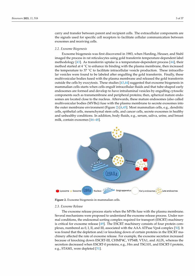
Exosome biogenesis was first discovered in 1983, when Harding, Heuser, and Stahlimaged the process in rat reticulocytes using gold transferrin temperature-dependent labelmethodology [43]. As transferrin uptake is a temperature-dependent process [44], theirmethod started at 4 ◦C to enhance its binding with the plasma membrane, then increasedthe temperature to 37 ◦C to facilitate intracellular vesicle production. These intracellu-lar vesicles were found to be labeled after engulfing the gold transferrin. Finally, thesemultivesicular bodies fused with the plasma membrane and released the gold transferrinoutside the cells by exocytosis. These studies [43,44] suggested that exosome biogenesis inmammalian cells starts when cells engulf intracellular fluids and that tube-shaped earlyendosomes are formed and develop to have intraluminal vesicles by engulfing cytosoliccomponents such as transmembrane and peripheral proteins; then, spherical mature endo-somes are located close to the nucleus. Afterwards, these mature endosomes (also calledmultivesicular bodies (MVBs)) fuse with the plasma membrane to secrete exosomes intothe outer membrane environment (Figure 2) [4,45]. Most mammalian cells, e.g., dendriticcells, epithelial cells, mesenchymal stem cells, and cancer cells, secrete exosomes in healthyand unhealthy conditions. In addition, body fluids, e.g., serum, saliva, urine, and breastmilk, contain exosomes [46–48].
Biosensors 2021, 11, x FOR PEER REVIEW 3 of 40
To conclude, mammalian cells secrete exosomes with a lipid bilayer membrane to protect the intracellular components, which are considered the messages that exosomes carry and transfer between parent and recipient cells. The extracellular components are the signals used for specific cell receptors to facilitate cellular communication between exosomes and receiving cells.
2.2. Exosome Biogenesis Exosome biogenesis was first discovered in 1983, when Harding, Heuser, and Stahl
imaged the process in rat reticulocytes using gold transferrin temperature-dependent la-bel methodology [43]. As transferrin uptake is a temperature-dependent process [44], their method started at 4 °C to enhance its binding with the plasma membrane, then increased the temperature to 37 °C to facilitate intracellular vesicle production. These intracellular vesicles were found to be labeled after engulfing the gold transferrin. Finally, these mul-tivesicular bodies fused with the plasma membrane and released the gold transferrin out-side the cells by exocytosis. These studies [43,44] suggested that exosome biogenesis in mammalian cells starts when cells engulf intracellular fluids and that tube-shaped early endosomes are formed and develop to have intraluminal vesicles by engulfing cytosolic components such as transmembrane and peripheral proteins; then, spherical mature en-dosomes are located close to the nucleus. Afterwards, these mature endosomes (also called multivesicular bodies (MVBs)) fuse with the plasma membrane to secrete exosomes into the outer membrane environment (Figure 2) [4,45]. Most mammalian cells, e.g., den-dritic cells, epithelial cells, mesenchymal stem cells, and cancer cells, secrete exosomes in healthy and unhealthy conditions. In addition, body fluids, e.g., serum, saliva, urine, and breast milk, contain exosomes [46–48].
Figure 2. Exosome biogenesis in mammalian cells.
2.3. Exosome Release The exosome release process starts when the MVBs fuse with the plasma membrane.
Several mechanisms were proposed to understand the exosome release process. Under normal conditions, the endosomal sorting complex required for transport (ESCRT) ma-chinery is critical for exosome release [49]. The ESCRT machinery consists of four protein complexes, numbered as 0, I, II, and III, associated with the AAA ATPase Vps4 complex [50]. It was found that the depletion and/or knocking down of certain proteins in the ESCRT machinery affected the rate of exosome release. For example, the exosome secre-tion increased because of knocking down ESCRT-III, CHMP4C, VPS4B, VTA1, and ALIX,
Figure 2. Exosome biogenesis in mammalian cells.
2.3. Exosome Release
The exosome release process starts when the MVBs fuse with the plasma membrane.Several mechanisms were proposed to understand the exosome release process. Under nor-mal conditions, the endosomal sorting complex required for transport (ESCRT) machineryis critical for exosome release [49]. The ESCRT machinery consists of four protein com-plexes, numbered as 0, I, II, and III, associated with the AAA ATPase Vps4 complex [50]. Itwas found that the depletion and/or knocking down of certain proteins in the ESCRT ma-chinery affected the rate of exosome release. For example, the exosome secretion increasedbecause of knocking down ESCRT-III, CHMP4C, VPS4B, VTA1, and ALIX, whereas thesecretion decreased when ESCRT-0 proteins, e.g., Hrs and TSG101, and ESCRT-I protein,e.g., STAM1, were depleted [51].

Biosensors 2021, 11, 518 4 of 37
In addition, ESCRT-independent mechanisms were proposed for exosome release inthe case of ESCRT machinery being knocked down [52]. Tetraspanin proteins, e.g., CD9,CD63, and CD82, enhanced the exosome secretion of β-catenin from HEK293 cells [53–55].In addition, targeting specific lipid enzymes such as neutral sphingomyelinase 2 to modifythe plasma membrane lipid configuration (size of the headgroup, length, and saturation ofthe acyl chains) inhibited exosome secretion [41]. Exosome release is also regulated andstimulated by multiple factors, such as Ca2+ [56], ceramide synthesis [41], and acidosis [57].
The p53-based mechanism was proposed to operate under stress conditions. It wasfound that the production of exosomes under stress conditions was regulated by the p53protein to communicate to other cells to respond to stress in a phenomenon called the“bystander effect” [58,59]. TSAP6 is upregulated and transcribed in response to stress [60].TSAP6 is a p53-regulated gene. Then, p53 induces cells to secrete specific proteins withinexosomes to migrate to other cells, communicate, and face the stress [61]. Yu and col-leagues [62] examined the protein secretion in exosomes after a p53-mediated stress re-sponse to lung cancer cells in culture. They tested cells containing a wild-type p53 gene(H460) and mutated cells (having a mutant p53 allele). The cells were irradiated withgamma irradiation to induce p53 and apoptosis. They observed a dramatic increase inexosome production as a response to the p53-regulated mechanism due to irradiation.Exosomes were not detected in the cases of the mutant p53 allele or nonirradiated cells.
The other mechanisms involved a variety of stress stimuli. For example, researchersat the School of Medicine at Flinders University showed the enhancement effect of hypoxiaon a percentage of cancer-derived exosomes. In their study, after hypoxia exposure,the exosomes were isolated and quantified using a nanoparticle tracking analysis andimmunoblotting for the CD63 and miRNA-210 assays by RT-PCR. They demonstratedthat hypoxia enhanced the release of breast cancer-derived exosomes [63]. Additionally,Németh’s research team investigated the effect of the antibiotic ciprofloxacin on exosomerelease and showed that a low concentration of ciprofloxacin caused the release of DNAproteins on exosome surfaces and blocked them from further cellular processes [64]. Finally,Rab proteins, e.g., Rab 11, Rab 27a,b, and Rab 35, were found to play key regulatory rolesin exosomes released in mammalian cells [56,65,66].
These mechanistic studies strongly suggest that exosome release is a stimuli-basedprocess. Further research studies are needed for verification. Once these mechanisms areverified, it will be a starting point for maximizing the production of the desired exosomesand improving the exosome applications.
3. General Techniques for Exosome Isolation, Characterization, and Detection
Exosome isolation and detection are a challenge because of the low concentrations and cellline-dependent heterogeneity of the exosomes [67–69]. Accordingly, developing and improvingreliable methods to prepare, detect, and analyze exosomes is critical for exosome researchand will have a great impact on the development of exosome-based disease diagnoses andtherapeutics. Figure 3 summarizes the general methods for exosome isolation and detection.
3.1. Isolation Techniques
The isolation of exosomes from a cell culture depends mainly on the physical andchemical properties of the exosomes. Ultracentrifugation and ultrafiltration target the sizeand density of exosomes, and chemical precipitation and immune affinity target specificextracellular proteins. To choose the suitable isolation technique, the number and volume ofthe samples, the available instruments, and the aim of the analysis must be considered [70].Here, we will discuss the isolation methods and the advantages and disadvantages associatedwith each method (Table 1). The isolation methods are classified according to (1) specificity,as specific or nonspecific, and (2) sample volume, as high-throughput or low-throughput.All methods of isolation are considered nonspecific, except for the immune affinity-basedtechniques. The immune affinity-based techniques and microfluidics are considered low-throughput methods because of their small sample volumes.
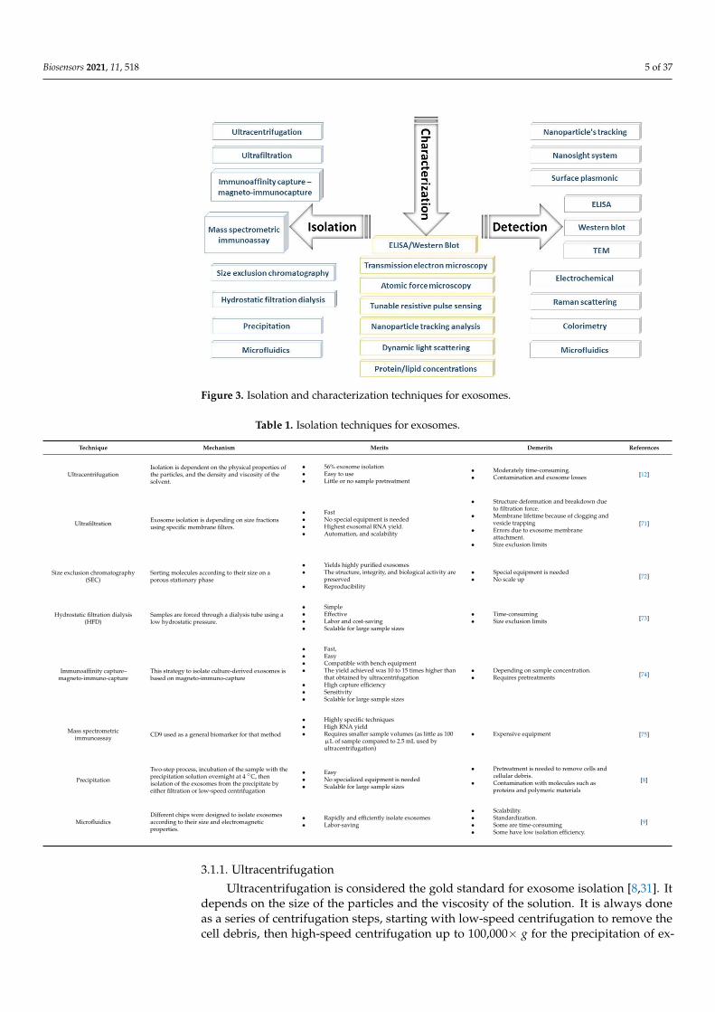
Biosensors 2021, 11, 518 5 of 37Biosensors 2021, 11, x FOR PEER REVIEW 5 of 40
Figure 3. Isolation and characterization techniques for exosomes.
3.1. Isolation Techniques The isolation of exosomes from a cell culture depends mainly on the physical and
chemical properties of the exosomes. Ultracentrifugation and ultrafiltration target the size and density of exosomes, and chemical precipitation and immune affinity target specific extracellular proteins. To choose the suitable isolation technique, the number and volume of the samples, the available instruments, and the aim of the analysis must be considered [70]. Here, we will discuss the isolation methods and the advantages and disadvantages associated with each method (Table 1). The isolation methods are classified according to (1) specificity, as specific or nonspecific, and (2) sample volume, as high-throughput or low-throughput. All methods of isolation are considered nonspecific, except for the im-mune affinity-based techniques. The immune affinity-based techniques and microfluidics are considered low-throughput methods because of their small sample volumes.
Table 1. Isolation techniques for exosomes.
Technique Mechanism Merits Demerits References
Ultracentrifugation
Isolation is dependent on the physical properties of the par-ticles, and the density and vis-cosity of the solvent.
• 56% exosome isolation • Easy to use • Little or no sample pretreatment
Moderately time-consuming. Contamination and exosome losses [12]
Ultrafiltration Exosome isolation is depend-ing on size fractions using specific membrane filters.
• Fast • No special equipment is needed • Highest exosomal RNA yield. • Automation, and scalability
Structure deformation and break-down due to filtration force.
Membrane lifetime because of clogging and vesicle trapping
Errors due to exosome membrane attachment.
Size exclusion limits
[71]
Size exclusion chromatog-raphy (SEC)
Sorting molecules according to their size on a porous sta-tionary phase
• Yields highly purified exosomes • The structure, integrity, and bio-
logical activity are preserved • Reproducibility
Special equipment is needed No scale up [72]
Hydrostatic filtration dial-ysis (HFD)
Samples are forced through a dialysis tube using a low hy-drostatic pressure.
• Simple • Effective • Labor and cost-saving • Scalable for large sample sizes
Time-consuming Size exclusion limits [73]
Immunoaffinity capture–magneto-immuno-capture
This strategy to isolate cul-ture-derived exosomes is
• Fast, • Easy • Compatible with bench equipment
• Depending on sample concentration. • Requires pretreatments [74]
Figure 3. Isolation and characterization techniques for exosomes.
Table 1. Isolation techniques for exosomes.
Technique Mechanism Merits Demerits References
UltracentrifugationIsolation is dependent on the physical properties ofthe particles, and the density and viscosity of thesolvent.
• 56% exosome isolation• Easy to use• Little or no sample pretreatment
• Moderately time-consuming.• Contamination and exosome losses [12]
Ultrafiltration Exosome isolation is depending on size fractionsusing specific membrane filters.
• Fast• No special equipment is needed• Highest exosomal RNA yield.• Automation, and scalability
• Structure deformation and breakdown dueto filtration force.
• Membrane lifetime because of clogging andvesicle trapping
• Errors due to exosome membraneattachment.
• Size exclusion limits
[71]
Size exclusion chromatography(SEC)
Sorting molecules according to their size on aporous stationary phase
• Yields highly purified exosomes• The structure, integrity, and biological activity are
preserved• Reproducibility
• Special equipment is needed• No scale up [72]
Hydrostatic filtration dialysis(HFD)
Samples are forced through a dialysis tube using alow hydrostatic pressure.
• Simple• Effective• Labor and cost-saving• Scalable for large sample sizes
• Time-consuming• Size exclusion limits [73]
Immunoaffinity capture–magneto-immuno-capture
This strategy to isolate culture-derived exosomes isbased on magneto-immuno-capture
• Fast,• Easy• Compatible with bench equipment• The yield achieved was 10 to 15 times higher than
that obtained by ultracentrifugation• High capture efficiency• Sensitivity• Scalable for large sample sizes
• Depending on sample concentration.• Requires pretreatments [74]
Mass spectrometricimmunoassay CD9 used as a general biomarker for that method
• Highly specific techniques• High RNA yield• Requires smaller sample volumes (as little as 100
µL of sample compared to 2.5 mL used byultracentrifugation)
• Expensive equipment [75]
Precipitation
Two-step process, incubation of the sample with theprecipitation solution overnight at 4 ◦C, thenisolation of the exosomes from the precipitate byeither filtration or low-speed centrifugation
• Easy• No specialized equipment is needed• Scalable for large sample sizes
• Pretreatment is needed to remove cells andcellular debris.
• Contamination with molecules such asproteins and polymeric materials
[8]
MicrofluidicsDifferent chips were designed to isolate exosomesaccording to their size and electromagneticproperties.
• Rapidly and efficiently isolate exosomes• Labor-saving
• Scalability.• Standardization.• Some are time-consuming• Some have low isolation efficiency.
[9]
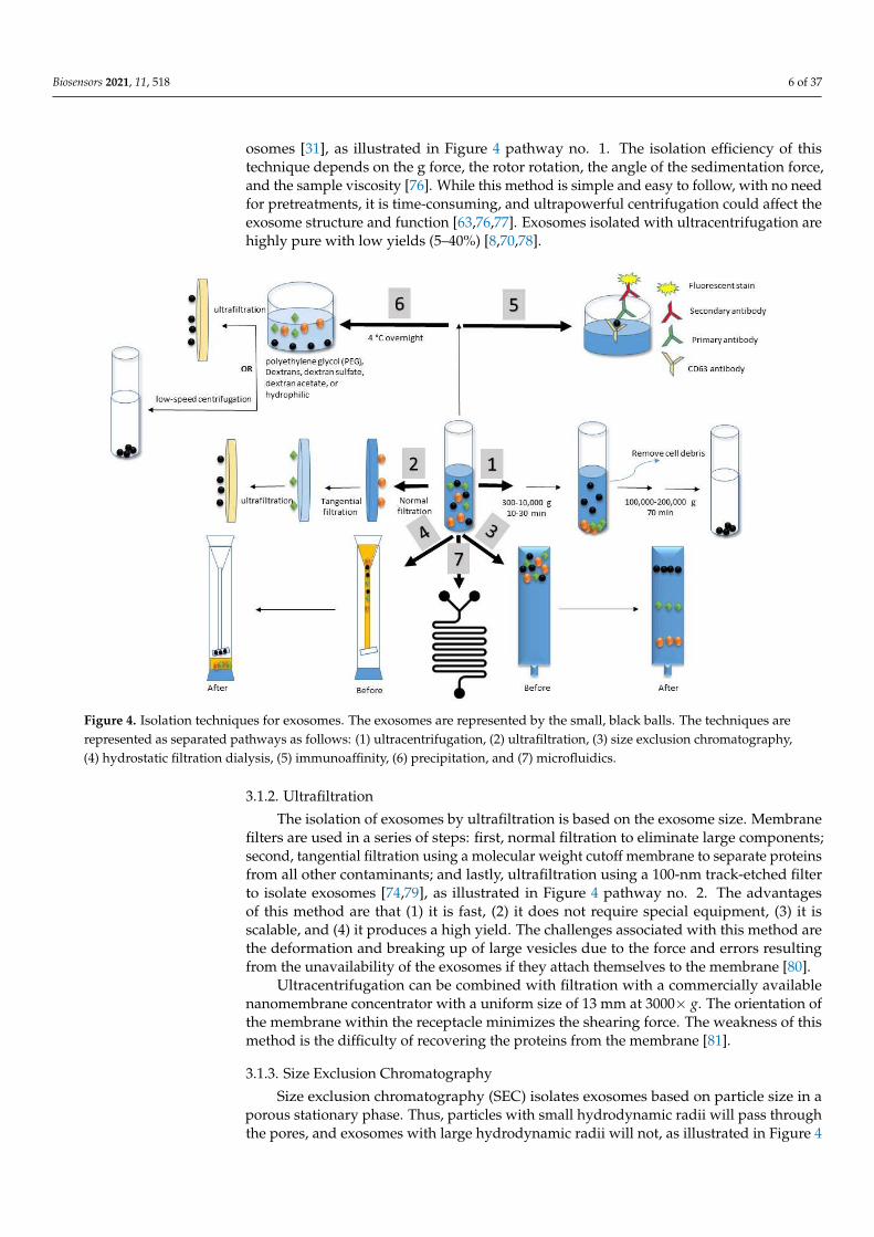
3.1.1. Ultracentrifugation
Ultracentrifugation is considered the gold standard for exosome isolation [8,31]. Itdepends on the size of the particles and the viscosity of the solution. It is always doneas a series of centrifugation steps, starting with low-speed centrifugation to remove thecell debris, then high-speed centrifugation up to 100,000× g for the precipitation of ex-
Biosensors 2021, 11, 518 6 of 37
osomes [31], as illustrated in Figure 4 pathway no. 1. The isolation efficiency of thistechnique depends on the g force, the rotor rotation, the angle of the sedimentation force,and the sample viscosity [76]. While this method is simple and easy to follow, with no needfor pretreatments, it is time-consuming, and ultrapowerful centrifugation could affect theexosome structure and function [63,76,77]. Exosomes isolated with ultracentrifugation arehighly pure with low yields (5–40%) [8,70,78].
Biosensors 2021, 11, x FOR PEER REVIEW 7 of 40
Figure 4. Isolation techniques for exosomes. The exosomes are represented by the small, black balls. The techniques are represented as separated pathways as follows: (1) ultracentrifugation, (2) ultrafiltration, (3) size exclusion chromatog-raphy, (4) hydrostatic filtration dialysis, (5) immunoaffinity, (6) precipitation, and (7) microfluidics.
3.1.2. Ultrafiltration The isolation of exosomes by ultrafiltration is based on the exosome size. Membrane
filters are used in a series of steps: first, normal filtration to eliminate large components; second, tangential filtration using a molecular weight cutoff membrane to separate pro-teins from all other contaminants; and lastly, ultrafiltration using a 100-nm track-etched filter to isolate exosomes [74,79], as illustrated in Figure 4 pathway no. 2. The advantages of this method are that (1) it is fast, (2) it does not require special equipment, (3) it is scal-able, and (4) it produces a high yield. The challenges associated with this method are the deformation and breaking up of large vesicles due to the force and errors resulting from the unavailability of the exosomes if they attach themselves to the membrane [80].
Ultracentrifugation can be combined with filtration with a commercially available nanomembrane concentrator with a uniform size of 13 mm at 3000× g. The orientation of the membrane within the receptacle minimizes the shearing force. The weakness of this method is the difficulty of recovering the proteins from the membrane [81].
3.1.3. Size Exclusion Chromatography Size exclusion chromatography (SEC) isolates exosomes based on particle size in a
porous stationary phase. Thus, particles with small hydrodynamic radii will pass through the pores, and exosomes with large hydrodynamic radii will not, as illustrated in Figure 4 pathway no. 3. Early research was done by Baranyai et al. to isolate exosomes from rates and human plasma samples using SEC [82]. Plasma samples were diluted and loaded onto the system. The authors tested various column matrices, e.g., Sepharose 2B, Sepharose CL-4B, and Sephacryl S-400. Their results indicated that the Sepharose CL-4B and Sephacryl S-400 columns were sufficient for significantly reducing the albumin contamination. Their
Figure 4. Isolation techniques for exosomes. The exosomes are represented by the small, black balls. The techniques arerepresented as separated pathways as follows: (1) ultracentrifugation, (2) ultrafiltration, (3) size exclusion chromatography,(4) hydrostatic filtration dialysis, (5) immunoaffinity, (6) precipitation, and (7) microfluidics.
3.1.2. Ultrafiltration
The isolation of exosomes by ultrafiltration is based on the exosome size. Membranefilters are used in a series of steps: first, normal filtration to eliminate large components;second, tangential filtration using a molecular weight cutoff membrane to separate proteinsfrom all other contaminants; and lastly, ultrafiltration using a 100-nm track-etched filterto isolate exosomes [74,79], as illustrated in Figure 4 pathway no. 2. The advantagesof this method are that (1) it is fast, (2) it does not require special equipment, (3) it isscalable, and (4) it produces a high yield. The challenges associated with this method arethe deformation and breaking up of large vesicles due to the force and errors resultingfrom the unavailability of the exosomes if they attach themselves to the membrane [80].
Ultracentrifugation can be combined with filtration with a commercially availablenanomembrane concentrator with a uniform size of 13 mm at 3000× g. The orientation ofthe membrane within the receptacle minimizes the shearing force. The weakness of thismethod is the difficulty of recovering the proteins from the membrane [81].
3.1.3. Size Exclusion Chromatography
Size exclusion chromatography (SEC) isolates exosomes based on particle size in aporous stationary phase. Thus, particles with small hydrodynamic radii will pass throughthe pores, and exosomes with large hydrodynamic radii will not, as illustrated in Figure 4
Biosensors 2021, 11, 518 7 of 37
pathway no. 3. Early research was done by Baranyai et al. to isolate exosomes from ratesand human plasma samples using SEC [82]. Plasma samples were diluted and loaded ontothe system. The authors tested various column matrices, e.g., Sepharose 2B, Sepharose CL-4B, and Sephacryl S-400. Their results indicated that the Sepharose CL-4B and SephacrylS-400 columns were sufficient for significantly reducing the albumin contamination. Theirprotocol helps to isolate highly purified exosomes with preserved biological activities.Research efforts such as combining SEC with ultracentrifugation to enrich the yield [83]and combining it with ultrafiltration to enhance its efficacy and speed [84] have been doneto overcome SEC challenges such as slowness, the need for dedicated equipment, low yield,and difficulty in scaling up [72].
3.1.4. Hydrostatic Filtration Dialysis
Exosomes have been isolated according to their size using a hydrostatic filtrationdialysis (HFD) system forced with a low hydrostatic pressure. In 2014, Musante et al. effi-ciently isolated diabetic nephropathy biomarker-based exosomes from urine samples usinga dialysis system [85]. Their dialysis system consisted of a defined 1000-kDa cutoff dialysismembrane connected to a funnel with a long, sheer column that created a hydrostaticpressure to push the solution through the dialysis membrane, as illustrated in Figure 4pathway no. 4. The system was refilled with pure water until all the pigments were washedout from the dialysis part. This method was found to be simple, fast, and effective: Itreduced the labor, maintained the protein pattern, and was capable of processing largesample volumes: 10 mL–1 L with a rate of 75 mL/h.
3.1.5. Immunoaffinity
Antigen–antibody linkage is the main mechanism for the immunoaffinity method, inwhich specific antigens are used to target specific extracellular proteins on exosome mem-branes. Technically, the immunoaffinity method can be considered an upgrade of the mainenzyme-linked immunosorbent assay (ELISA) mechanism, in which two antibodies areused to detect a specific antigen, as illustrated in Figure 4 pathway no. 5. The first antibodyis the antigen-trapping molecule, and the second antibody is the fluorescence-detectingmolecule. To enhance the proficiency of this method, two techniques were proposed: themicroplate-based immunocapture technique and the immunoaffinity capture/magneto-immunocapture technique. These techniques are further illustrated in the next subsections.
Microplate-Based Immunocapture Technique
Briefly, in the microplate-based immunocapture technique, the exosomes are attacheddirectly to a microplate surface. The surface of this microplate is immobilized with therequired antibodies to capture exosomes, leading to the exosomes precipitating fromthe culture [86]. However, the samples must be prepared before treatment, and it isrequired that there be at least 20 µg of protein content in the exosomes. This techniqueis highly specific and yields a high RNA content from a low sample volume, as little as100 µL of sample, compared to the 2.5 mL needed for ultracentrifugation [87]. A noveldendrimer–PEG antibody dual-layer platform was proposed to significantly capture andisolate tumor exosomes from serum samples. This platform was assembled as a sandwichwith two layers of carboxylated generation 7 poly amidoamine dendrimers and wasstuffed with polyethylene glycol (PEG) (2, 5, and 20 kDa) conjugated with dendrimers.The dendrimers for the bottom layer coated an epoxide-functionalized glass slide. Thisstructure facilitated the multivalent capture ability by applying multiple antibodies andminimizing the nonspecific bindings. This platform possesses high avidity, specificity,antibody orientation flexibility, and tumor-derived exosome yield [88].
Immunoaffinity Capture/Magneto-Immunocapture
To add value to the microplate-based technique, magnetic beads, such as latex beads andnano-sized beads, have been conjugated with antibodies [5,35]. One example is Dynabeads.

Biosensors 2021, 11, 518 8 of 37
Dynabeads® are superparamagnetic polystyrene beads with a diameter of 1–4.5 µm. Thesebeads are specified to conjugates with the anti-human CD63 antibody, either directly or viaa secondary linker such as anti-mouse IgG [8]. Using this new combination of antibody andmagnetic particles increases the capture affinity and sensitivity and makes it easy and rapidto proceed. The efficiency of this method depends on the interaction between the antigen andantibody, temperature, concentration of exosomes, and incubation time [5]. Sample volumes couldbe scaled up or down without any restriction. The isolation yield is 15 times higher than withultracentrifugation [12]. Although this method is considered the superior strategy for isolatingexosomes from cell culture media, it depends on the quality of the pre-enriched exosomes [5].
An ideal example is T-cell Immunoglobulin Mucin Protein (Tim4) binding with phos-phatidylserine molecules on the surfaces of exosomes. Tim4 immobilized on magneticbeads has Ca2+-dependent binding to phosphatidylserine. Moreover, exosomes can be re-leased from the Tim4 surface by adding a complexing agent to remove Ca2+ [89]. Greeningand coworkers (2015) [12] evaluated the efficacy of three isolation techniques: ultracen-trifugation (UC-Exos), OptiPrep™ density gradient centrifugation (DG-Exos), and immuneisolation using EpCAM (CD326) antibodies coupled to magnetic beads (IAC-Exos) target-ing markers Alix, TSG101, and HSP70 to enrich exosomes released from LIM1863 humancolon cancer cells. The isolated exosomes had a uniform size of 40–150 nm, and theyverified that the IAC method was the most efficient for exosome isolation.
3.1.6. Precipitation
Based on the chemical properties of exosomes: (1) a water-excluding polymer, e.g.,polyethylene glycol (PEG); (2) dextran derivatives, e.g., dextran sulfate and dextran acetate;and (3) hydrophilic polymers such as polyvinyl alcohol, polyvinyl acetate, and polyvinylsulfate were used to chemically precipitate exosomes from the culture [8]. After a mixedsample was incubated at 4 ◦C overnight with the precipitation solution, exosomes could beisolated from the precipitate either by low-speed centrifugation or filtration, as illustratedin Figure 4 pathway no. 6. This method is easy-to-handle, does not require specificequipment, and can be scalable for large sample volumes. However, if the samples are notprecleaned of cells and cellular debris, proteins and polymeric materials will be found ascoprecipitates [8].
A modified protocol was proposed by Alvarez et al. 2012 [70]. The authors usedExoQuick-TC to precipitate exosomes [63]. Their protocol is perfect for proceeding withmultiple samples in the absence of an ultracentrifuge and for targeting RNAs and mRNAsfor biomarker identification [70].
3.1.7. Microfluidics
Multiple microfluidic chips have been designed to isolate exosomes rapidly and effi-ciently with significant reductions in the sample volume, reagent consumption, and isolationtime, as illustrated in Figure 4 pathway no. 7. However, scalability, validation, samplepretreatments, and standardization are considered disadvantages for these devices [90].
Wang and colleagues (2017) [9] fabricated an acoustofluidics device to isolate exosomesdirectly from undiluted blood samples based on their size and density using ultrasoundstanding waves. With respect to the channel orientation, particles are subjected to acousticforce and pushed toward the pressure node. The device consists of two modules. Thefirst separates larger components, >1 µm in diameter, such as red and white blood cells,and platelets with 99% efficiency. The second module isolates exosomes to 98.4% purity.This device offers continuous flow exosome isolation while maintaining the structures,characteristics, and functions of the exosomes. Additionally, it enables short processingtimes with decreasing human intervention.
In another device, exosomes with diameters of 40–100 nm were preferentially trappedon a ciliated micropillar with a porous silicon nanowire. Proteins and other cellular debriswere filtered out. Exosomes were released from the porous silicon nanowires by dissolvingthem in a phosphate buffer solution. ExoChip is a commercial immune-microfluidics
Biosensors 2021, 11, 518 9 of 37
chip that is functionalized with a commonly expressed antigen, CD63 (a member of thetetraspanin family). The specific interaction between the CD63 and antibodies immobilizedon the chip allowed the isolation of exosomes from mixed cultures. While, in the integratedmicrofluidic exosome device, the sample was mixed with antibody-labeled magneticbeads; then, a lysis buffer was added, and detection reagents were introduced in a separatechamber. To improve the scalability of the integrated device, in-line ultraviolet and dynamiclight scattering detectors were coupled with the field–flow fraction system to isolate andcharacterize exosomes rapidly [91].
Dr. Chang’s research group fabricated microfluidics chips to isolate exosomes fromplasma and cell culture samples based on an ionic exchange property. In 2017, theyfabricated an integrated platform to isolate exosomes using an ion-selective membrane [92].One year later, they upgraded their system by adding a pressure-driven flow force to filterout unwanted debris before concentrating exosomes on the ion-selective membrane [93].This system was fast and sensitive and recovered 60–80% of the exosomes from the serumand cell culture compared to 25% for other systems.
To summarize this section, multiple techniques have been recognized for isolatingexosomes from the culture, e.g., ultracentrifugation, ultrafiltration, size exclusion chro-matography, hydrostatic filtration dialysis, immunoaffinity, precipitation, and microfluidics.These techniques have been modified and/or combined to improve the isolation procedure.
3.2. Characterization and Detection Techniques
Generally, analyses of the characteristics of purified exosomes fall into four basic cat-egories: size, concentration, purity, and content. For size and purity, transmission electronmicroscopy (TEM) is the standard, with a very low-throughput method for taking and analyz-ing data [81]. More recently, the NanoSight system [94] has been used to image and determineparticle sizes and concentrations. A promising approach to assessing the purity is combiningNanoSight with a protein assay [95], but this is a source-dependent method. Exosome contentscan be examined using the “-omics” methods, such as proteomics, transcriptomics (miRNA ormRNA), lipidomics, and glycomics (glycoproteins), or analyzed using more focused methods,such as Western blot, RT-qPCR, and GC-MS [76]. Recently, protein and/or lipid concentrationassays using simple spectrophotometer protocols have been considered promising methodsfor characterizing the protein and lipids contents of exosomes [96].
The method of characterization is chosen according to the purpose of the analysis. Ifthe purpose is to identify the morphology and confirm the sample purity, then TEM is thestandard method to follow. If the purpose is to determine the size and morphology of theparticles, then a nanoparticle tracking analysis is sufficient. Western blot and ELISA canbe used to detect and identify proteins with respect to their role (up- or downregulation).Spectrophotometry is a standard method for determining the concentrations of the particles.Table 2 details these methods with respect to their targets, advantages, and disadvantages.
Table 2. Physical and chemical characterization techniques for exosomal samples.
Technique Target Merits Demerits References
Transmission electron microscopy Phenotype as shape anddimension
• Direct method• Requires a small sample amount
• Expensive• Sample preparation may lead to shape modifications [97]
Nanoparticle tracking analysis Size distribution andconcentration • Fast
• No preparation steps
• Inaccurate if samples are aggregated and/or have different sizedistributions, or the same instruments are in different geographical areas.
• Non specificity
[98]
Dynamic light scattering Size distribution [17,98]
Tunable resistive pulse sensing Size distribution, concentration,and surface charge
• Hard to select the appropriate nanopore setup [99,100]
Atomic force microscopy 3-D topography• No fixation or staining steps• Requires a small sample amount • Sample dehydration may lead to topography modifications [101]
ELISA/Western Blot Protein profile• Simple• Specific• Low sample volume
• Sample preparation• Time-consuming• Inaccurate if detecting non-exosomal contents
[75]
Spectrophotometer Protein and/or lipidconcentration
• Simple• Fast• No fixation or staining steps• Requires a small sample amount
• Sample preparation• Inaccurate if detecting non-exosomal contents [102]

Biosensors 2021, 11, 518 10 of 37
Recently, several individual approaches and combined methods, such as surfaceplasmonic biosensors [29,103,104], microchip-based technologies [14,105], electrochemicaltechniques [106], fluorescence [107], and colorimetry [108], have been proposed for exosomedetection; some of them have been upgraded over time to reach the high-throughput, highsensitivity, and real-time detection and quantification of disease-based exosomes. Thesemethods are summarized in Table 3.
Table 3. Detection techniques for exosomal samples.
Method ApproachType
TargetComponent Mechanism Merits Demerits References
Surfaceplasmonicbiosensor
Quantitative Biomarkers
The electromagneticfield of surfaceplasmon, and theoptical wavesoriginate from themass oscillations ofelectronic chargedensity of thin(nanoscale) metallicfilms
Integration,miniaturization,multiparameter,real-time, andlabel-free detection,Sensitivity
Not capable ofidentifying thepost-transcriptionalmodifications ofmiRNA
[29,103,104]
Microchips-basedTechniques Quantitative Various
Various designsaccording to thepurpose and target
High-throughput fornonpurified samplesFast detectionEasy to use,reagent-saving, andpossessing highefficacy
Low masstransfer scale andinterference withexosomal binding
[105,109–111]
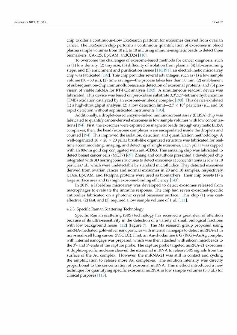
Specific RamanScattering
TechniquesQuantitative miRNA
Detection of capturedexosomes withidentified hairpins
Ultra-sensitiveLow backgroundnoise.
Contaminationissue [112,113]
ElectrochemicalTechniques Quantitative Biomarkers
Decrease of theelectrochemical signalbecause of the releaseof the pre-labeledstands from thefunctionalized surfaceof a gold electrodewhen the exosomeswere captured by theanti-marker beads.
Reliable, fastCost-effectiveLow sampleconcentrationSensitivityEasy to handleSaves timeNontoxic materialsLow background, andsimpleinstrumentation
Indictmeasurements [75]
Fluorescent andColorimetricTechniques
Qualitative/quantitative miRNA
Label capturedexosomes with staine.g., Cy3
Fast, simpleNeeds highsampleconcentration
[114]
Examining all this information on the characterization and detection techniques usedfor exosome samples indicates that visualizing the procedure with respect to the chemicalcomposition of the particles and preparing the samples are the main factors in choosingthe suitable technique. However, it is better to combine two or more techniques to confirmthe results.
4. Tumor Exosomes for Cancer Detections
Exosomes are found to promote cancer angiogenesis, generate the premetastatic niche,and modulate the host immune system [49]. In the next few sections, we will summarizethe cases in which exosomes have been used for diagnosis and for monitoring canceragents.
4.1. Exosomes as Disease Biomarkers with Diagnostic Potential
With respect to cancer, exosomes have potential effects on cancer development andtissue reprogramming [115]. Exosome nucleic acids pool and proteins act as the primarybiomarkers for early cancer early detection and diagnosis [1,116,117].

Biosensors 2021, 11, 518 11 of 37
Exosomes have a cell line-based structure that suggests a subpopulation distributionon a cell line basis. The role of these subpopulations is likely related to the normaland cancer cells and helps in diagnostic purposes. Exosomes released from normal andcancer cell lines have different (1) nucleic acid contents and (2) membrane structures inaccordance with their cholesterol contents, surface proteins, and cholesterol: phospholipidratios [67,118].
Exosomes contain functional components, e.g., RNA, DNA, and proteins, which canbe used as biomarkers for diagnostic and monitoring purposes and can be easily transferredto recipient cells. Exosomes derived from infected cells mimic special elements spanningthe normal cells that provide a blueprint of tumor cells for medical purposes [119].
The miRNA is one of the critical intracellular components in exosomes. It is a class ofnoncoding RNA with 18–25 nucleotides that plays vital roles in cell-to-cell communicationpathways in carcinogenesis [120]. These noncoding RNAs can facilitate metastasis by enhanc-ing the molecular pathways associated with cancer [121]. Systemically, miRNAs are the mostabundant species, with around 42.3% of the exosome RNA pool [122]. Other RNA fractionsinclude rRNA, tRNA, noncoding RNA, piwi-interacting RNA, small nuclear RNA, and smallnucleolar RNA. The common miRNAs are miR-22-3p, miR-99a-5p, miR-99b-5p, miR-124-3p,and miR-128 [13]. It was suggested that miRNAs play vital roles in physiological processessuch as RNA splicing, protein phosphorylation, chromosomal abnormality, and angiogene-sis [13,122]. The exosomal miRNA profiles can potentially be used as cancer biomarkers, e.g.,miRNA-141, miRNA-200a, and miRNA-200c [18,40,123,124].
Deoxy ribonucleic acid (DNA) is another critical component of exosome structures.ExoDNA is poorly studied compared to ExoRNA. Previously, it was believed that mi-crovesicles have intracellular single-stranded DNA (ssDNA) and mitochondrial DNA [125].Thakur et al. 2014 found evidence that exosomes have intra- and extracellular double-stranded DNA (ddDNA) as a whole genomic material [126]. They compared the types andconcentrations of the DNA loops in pretreated exosome samples with dsDNase and un-treated samples. They observed a significant reduction in the concentration of the DNA loopin the treated samples, which means that exosomes carry high concentrations of ddDNA.They also found that the circulating exosomal ddDNA was a promising tumor-basedmutation biomarker that could be used to validate cancer diagnostics and prognostics byidentifying multiple genes, such as EGFR, BRAF, RAS, IDH, and HER2, because (1) it is sta-ble, (2) it is biocompatible, and (3) its functional group can be modified [127,128]. ExoDNAis a key regulator for tumor immunity [128]. Cancer cells secrete harmful fragmentedDNA through their exosomes to avoid senescence (cell death) and avoid the stimulatorof interferon genes (STING) and cyclic GMP–AMP synthase (cGAS) resulting from DNAaccumulation [129]. STING and cGAS are two machineries that are activated by DNAaccumulating in the cytoplasm. These DNA machineries act against tumorigenesis [130].The therapeutic efficacy of the tumor is based on the STING mechanism [131]. The loopsof DNA fragments that have accumulated in the cytoplasm because of radiotherapy andchemotherapy induce the antitumor response and STING activation in dendritic cells toprevent further tumor growth and promote inflammation. The mechanism of packing theDNA inside the exosomes is still unclear. Exosome biogenesis is enhanced in infected cellsbecause of the hypoxia and the low pH [132].
The third critical diagnostic biomarker component of the exosomes is their proteins,as they are protected from the proteinases and stable in plasma and serum circulation.There are specific types of exosomal proteins that act to discriminate between different celltypes; for example, the epithelial cell adhesion molecule (EpCAM) differentiates betweencancer cells and normal cells. Other types of proteins that differentiate exosomes fromother vesicles are secreted by all cell types. These biomarkers include CD9; CD63; CD81;LAMP1; heat shock proteins (Hsp25, Hsp60, Hsp70, and Hsp90); synthenins; endosomes;and calnexin [5,12,133–136].
To conclude this section, lists of specific biomarkers according to either the disease orthe cell type is given in Tables 4 and 5 [137,138]. Interestingly, it was found that the same
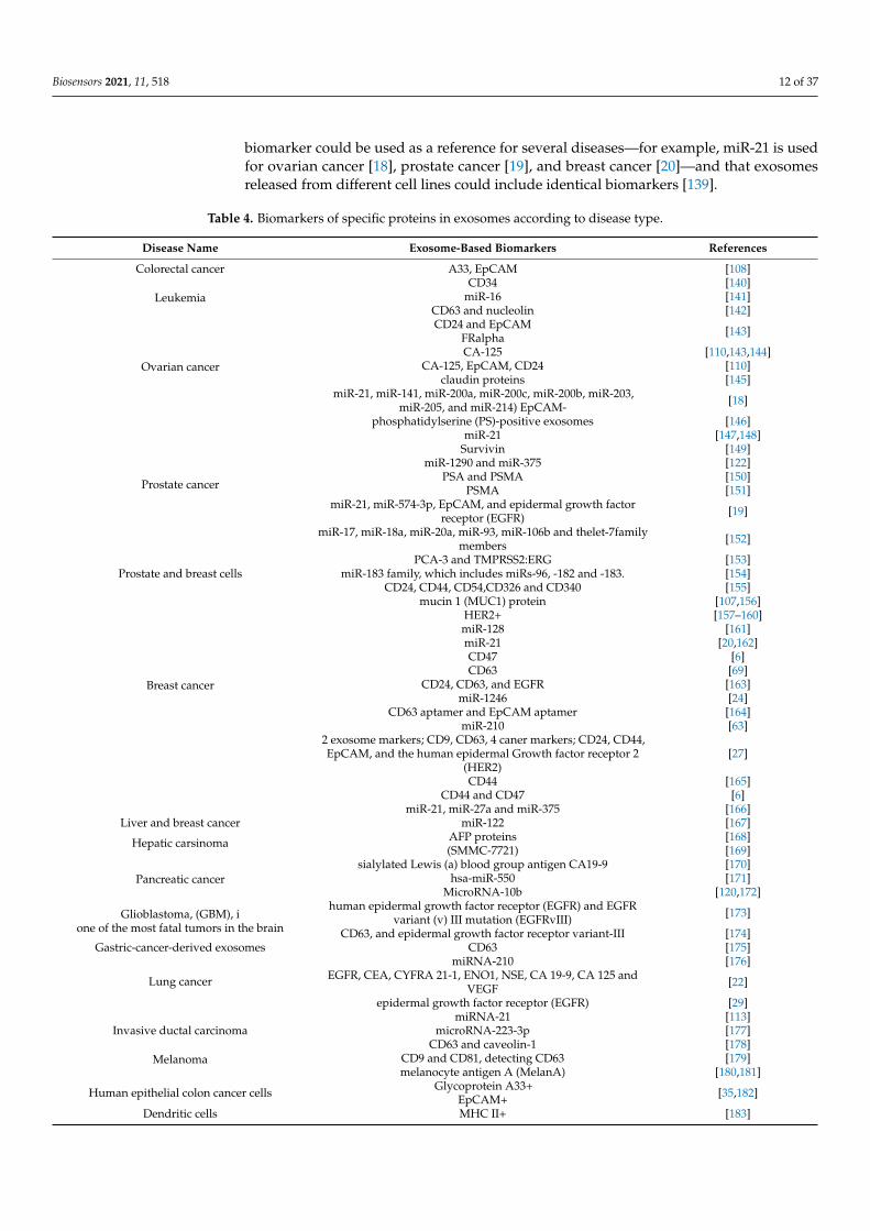
Biosensors 2021, 11, 518 12 of 37
biomarker could be used as a reference for several diseases—for example, miR-21 is usedfor ovarian cancer [18], prostate cancer [19], and breast cancer [20]—and that exosomesreleased from different cell lines could include identical biomarkers [139].
Table 4. Biomarkers of specific proteins in exosomes according to disease type.
Disease Name Exosome-Based Biomarkers References
Colorectal cancer A33, EpCAM [108]
LeukemiaCD34 [140]
miR-16 [141]CD63 and nucleolin [142]
Ovarian cancer
CD24 and EpCAMFRalpha [143]
CA-125 [110,143,144]CA-125, EpCAM, CD24 [110]
claudin proteins [145]miR-21, miR-141, miR-200a, miR-200c, miR-200b, miR-203,
miR-205, and miR-214) EpCAM- [18]
phosphatidylserine (PS)-positive exosomes [146]
Prostate cancer
miR-21 [147,148]Survivin [149]
miR-1290 and miR-375 [122]PSA and PSMA [150]
PSMA [151]miR-21, miR-574-3p, EpCAM, and epidermal growth factor
receptor (EGFR) [19]
miR-17, miR-18a, miR-20a, miR-93, miR-106b and thelet-7familymembers [152]
PCA-3 and TMPRSS2:ERG [153]Prostate and breast cells miR-183 family, which includes miRs-96, -182 and -183. [154]
Breast cancer
CD24, CD44, CD54,CD326 and CD340 [155]mucin 1 (MUC1) protein [107,156]
HER2+ [157–160]miR-128 [161]miR-21 [20,162]CD47 [6]CD63 [69]
CD24, CD63, and EGFR [163]miR-1246 [24]
CD63 aptamer and EpCAM aptamer [164]miR-210 [63]
2 exosome markers; CD9, CD63, 4 caner markers; CD24, CD44,EpCAM, and the human epidermal Growth factor receptor 2
(HER2)[27]
CD44 [165]CD44 and CD47 [6]
miR-21, miR-27a and miR-375 [166]Liver and breast cancer miR-122 [167]
Hepatic carsinoma AFP proteins [168](SMMC-7721) [169]
Pancreatic cancersialylated Lewis (a) blood group antigen CA19-9 [170]
hsa-miR-550 [171]MicroRNA-10b [120,172]
Glioblastoma, (GBM), ione of the most fatal tumors in the brain
human epidermal growth factor receptor (EGFR) and EGFRvariant (v) III mutation (EGFRvIII) [173]
CD63, and epidermal growth factor receptor variant-III [174]Gastric-cancer-derived exosomes CD63 [175]
Lung cancer
miRNA-210 [176]EGFR, CEA, CYFRA 21-1, ENO1, NSE, CA 19-9, CA 125 and
VEGF [22]
epidermal growth factor receptor (EGFR) [29]miRNA-21 [113]
Invasive ductal carcinoma microRNA-223-3p [177]
MelanomaCD63 and caveolin-1 [178]
CD9 and CD81, detecting CD63 [179]melanocyte antigen A (MelanA) [180,181]
Human epithelial colon cancer cells Glycoprotein A33+EpCAM+ [35,182]
Dendritic cells MHC II+ [183]
Biosensors 2021, 11, 518 13 of 37
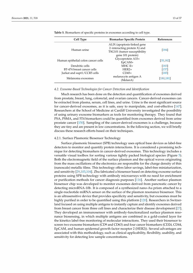
Table 5. Biomarkers of specific proteins in exosomes according to cell type.
Cell Type Biomarker Specific Protein References
Human urine
ALIX (apoptosis-linked gene2–interacting protein X) andTSG101 (tumor susceptibility
gene 101 protein)
[184]
Human epithelial colon cancer cells Glycoprotein A33+EpCAM+ [35,182]
Dendritic cells MHC II+ [183]BT-474 breast cancer cells HER2+ [157]
Jurkat and supt1/CCR5 cells CD45+ [185]
Melanoma exosomes melanocyte antigen A(MelanA) [180,181]
4.2. Exosome-Based Technologies for Cancer Detection and Identification
Much research has been done on the detection and quantification of exosomes derivedfrom prostate, breast, lung, colorectal, and ovarian cancers. Cancer-derived exosomes canbe extracted from plasma, serum, cell lines, and urine. Urine is the most significant sourcefor cancer-derived exosomes, as it is safe, easy to manipulate, and cost-effective [147].Researchers at the School of Medicine at Cardiff University investigated the possibilityof using urinary exosome biomarkers as tools for monitoring therapy. They found thatPSA, PSMA, and 5T4 biomarkers could be quantified from exosomes derived from urineprostate cancer [150]. Sampling of the cancer-derived exosomes is a challenge, becausethey are tiny and are present in low concentrations. In the following section, we will brieflydiscuss these research efforts based on their techniques.
4.2.1. Surface Plasmonic Biosensor Technology
Surface plasmonic biosensor (SPB) technology uses optical base devices as label-freedetectors to monitor and quantify protein interactions. It is considered a promising tech-nique for detecting biomarkers in cancer-derived exosomes. This technology includes aversatile visual toolbox for sorting various tightly packed biological species (Figure 5).Both the electromagnetic field of the surface plasmon and the optical waves originatingfrom the mass oscillations of the electronics are responsible for the charge density of thin(nanoscale) metallic films. This technology offers labor savings, label-free miniaturization,and sensitivity [29,103,104]. Zhu fabricated a biosensor based on detecting exosome surfaceproteins using SPB technology with antibody microarrays with no need for enrichmentor purification methods for cancer diagnosis purposes [116]. Another surface plasmonbiosensor chip was developed to monitor exosomes derived from pancreatic cancer bydetecting microRNA-10b. It is composed of a synthesized nano-Au prism attached to asingle-nucleotide miRNA sensor on the surface of the plasmon resonance biosensor. Thisis an ultrasensitive device that provides specificity and sensitivity, but exosomes must behighly purified in order to be quantified using this platform [120]. Researchers in Switzer-land focused on using multiple antigens to instantly capture and identify exosomes derivedfrom breast cancer from three cell lines and characterize their disease development [27].They developed an immunosensor with antibody-functionalized surface plasmon reso-nance biosensing, in which multiple antigens are combined in a gold-coated layer forthe kinetics label-free monitoring of molecular interactions. They used their biosensor toscreen two exosome biomarkers (CD9 and CD63) and four cancer biomarkers (CD24, CD44,EpCAM, and human epidermal growth factor receptor 2 (HER2)). Several advantages areassociated with this methodology, such as clinical applicability, flexibility, usability, andsensitivity for detecting low sample concentrations.

Biosensors 2021, 11, 518 14 of 37Biosensors 2021, 11, x FOR PEER REVIEW 16 of 40
Figure 5. Examples of surface plasmonic biosensors for exosome detection and quantification. The figure permissions are as follows: (A) License number 5071041050010, license date 16 May 2021, licensed content publisher John Wiley and Sons. Licensed content publication of “Advanced Func-tional Materials”, licensed content title “Detection of Glioma-Derived Exosomes with the Biotinyl-ated Antibody-Functionalized Titanium Nitride Plasmonic Biosensor”. (B) Adapted with permis-sion from Liu, C. et al. 2018. Sensitive detection of exosomal proteins via a compact surface plasmon resonance biosensor for cancer diagnosis. ACS sensors, 3(8), pp. 1471–1479. Copyright (2021) Amer-ican Chemical Society, Rightslink® by Copyright Clearance Center. (C) Adapted with permission from Zhu, L. et al. 2014. Label-free quantitative detection of tumor-derived exosomes through sur-face plasmon resonance imaging. Analytical chemistry, 86(17), pp. 8857–8864. Copyright (2021) La-bel-Free Quantitative Detection of Tumor-Derived Exosomes through Surface Plasmon Resonance Imaging. Analytical Chemistry (acs.org) and further permissions related to the excerpted material should be directed to ACS. (D) Adapted from Raghu, D. et al. 2018. Nanoplasmonic pillars engi-neered for single exosome detection. PloS ONE, 13(8), p. e0202773, with no permission required for reuse, as the work was made available under the Creative Commons CC0 public domain dedication. (E) Adapted with permission from Joshi, G.K et al. 2015. Label-free nanoplasmonic-based short noncoding RNA sensing at attomolar concentrations allows for the quantitative and highly specific assay of microRNA-10b in biological fluids and circulating exosomes. ACS nano, 9(11), pp. 11075–11089, hps://pubs.acs.org/doi/abs/10.1021/acsnano.5b04527, 10/2021, and further permissions re-lated to the excerpted material should be directed to ACS.
Liu updated a similar platform to detect exosomes derived from lung cancer and overcome the high cost associated with the fabrication process. His platform is composed of an Au-coated glass layer conjugated with a prism and a NeutrAvidin–polyethylene glycol (PEG)–thiol–biotin PEG mixture. Exosomes are captured on the surface of that mix-ture, and then, the laser signals are reflected through the prism. This platform offers high sensitivity and simplicity, but the detection of biomarkers differs according to the sample origin for the same disease, e.g., the signals of the exosomal epidermal growth factor re-ceptor (EGFR) were not the same in human serum as in the cell lines [30]. Another surface plasmon biosensor chip was developed with a nonfactionalized nanogold layer to distin-guish between exosomes and extracellular macrovesicles associated with lung cancer in mice, which revealed the importance of surface [29] exosome properties [186]. A surface biotinylated antibody-functionalized titanium nitride plasmon resonance biosensor was fabricated to detect glioma-derived exosomes (glioma is a brain cancer that starts in the glial cells of the brain or the spine). The titanium nitride biosensor was able to detect both CD63 and epidermal growth factor receptor variant-III with detection limits of 10−3 μg mL−1 and 2.75 × 10⁻³ μg mL−1, respectively. In addition, it had excellent performance, sta-bility level, and biocompatibility with titanium nitride [174]. Research groups in Australia
Figure 5. Examples of surface plasmonic biosensors for exosome detection and quantification. Thefigure permissions are as follows: (A) License number 5071041050010, license date 16 May 2021,licensed content publisher John Wiley and Sons. Licensed content publication of “Advanced Func-tional Materials”, licensed content title “Detection of Glioma-Derived Exosomes with the BiotinylatedAntibody-Functionalized Titanium Nitride Plasmonic Biosensor”. (B) Adapted with permission fromLiu, C. et al. 2018. Sensitive detection of exosomal proteins via a compact surface plasmon reso-nance biosensor for cancer diagnosis. ACS sensors, 3(8), pp. 1471–1479. Copyright (2021) AmericanChemical Society, Rightslink® by Copyright Clearance Center. (C) Adapted with permission fromZhu, L. et al. 2014. Label-free quantitative detection of tumor-derived exosomes through surfaceplasmon resonance imaging. Analytical chemistry, 86(17), pp. 8857–8864. Copyright (2021) Label-FreeQuantitative Detection of Tumor-Derived Exosomes through Surface Plasmon Resonance Imaging.Analytical Chemistry (acs.org) and further permissions related to the excerpted material should bedirected to ACS. (D) Adapted from Raghu, D. et al. 2018. Nanoplasmonic pillars engineered forsingle exosome detection. PloS ONE, 13(8), p. e0202773, with no permission required for reuse, as thework was made available under the Creative Commons CC0 public domain dedication. (E) Adaptedwith permission from Joshi, G.K et al. 2015. Label-free nanoplasmonic-based short noncodingRNA sensing at attomolar concentrations allows for the quantitative and highly specific assay ofmicroRNA-10b in biological fluids and circulating exosomes. ACS nano, 9(11), pp. 11075–11089,hps://pubs.acs.org/doi/abs/10.1021/acsnano.5b04527, 10/2021, and further permissions related tothe excerpted material should be directed to ACS.
Liu updated a similar platform to detect exosomes derived from lung cancer andovercome the high cost associated with the fabrication process. His platform is composedof an Au-coated glass layer conjugated with a prism and a NeutrAvidin–polyethyleneglycol (PEG)–thiol–biotin PEG mixture. Exosomes are captured on the surface of thatmixture, and then, the laser signals are reflected through the prism. This platform offershigh sensitivity and simplicity, but the detection of biomarkers differs according to thesample origin for the same disease, e.g., the signals of the exosomal epidermal growthfactor receptor (EGFR) were not the same in human serum as in the cell lines [30]. Anothersurface plasmon biosensor chip was developed with a nonfactionalized nanogold layerto distinguish between exosomes and extracellular macrovesicles associated with lungcancer in mice, which revealed the importance of surface [29] exosome properties [186]. Asurface biotinylated antibody-functionalized titanium nitride plasmon resonance biosensorwas fabricated to detect glioma-derived exosomes (glioma is a brain cancer that startsin the glial cells of the brain or the spine). The titanium nitride biosensor was able todetect both CD63 and epidermal growth factor receptor variant-III with detection limitsof 10−3 µg mL−1 and 2.75 × 10⁻³ µg mL−1, respectively. In addition, it had excellent

Biosensors 2021, 11, 518 15 of 37
performance, stability level, and biocompatibility with titanium nitride [174]. Researchgroups in Australia and Singapore have developed a real-time-functionalized ani-HER2surface plasmon biosensor to detect breast cancer cells. Their platform is simple, label-free,and sensitive, with a detection limit of 8.2 × 10⁻³ particles/µL [159].
In 2020, Portela created an upgraded nanoplasmonic biosensor featuring nanogapantennas by employing the colloidal lithography process. Gaps that had a size of ~11.6–4.7 nm formed between gold nano-disk pairs. This antenna biosensor detected lungcancer biomarker miRNA-210 via a hybridization assay of DNA/miRNA. Several advan-tages were reported for this platform, including (1) high performance, (2) high sensitivity,(3) simplicity, (4) cost-effectiveness, (5) a low detection limit (5.1 ng mL−1), and (6) thedirect detection of miRNAs [176]. Recently, a simple plasmonic surface polydopamine-functionalized Au nanobiosensor with two aptamers was invented. The DNA tetrahedronprobes were immobilized on the gold nanoparticle samples under alkaline pH. Afterwards,a covalent bond between aptamer 1 and aptamer 2 was structured as a NH2–COOH bond.In the first step, SMMC-7721 exosomes were captured on the surface of the first aptamer,which was complementary to the DNA tetrahedron probes. Then, the second aptamer rec-ognized the SMMC-7721 on the captured exosomes and enhanced the signal amplification.Consequently, signal amplification improved when the first aptamer reduced the HAuCl4.This platform offers specificity, a low detection limit of 5.6 × 10⁵ particles/mL, and noneed for pretreatments [169].
Exosome–antibody kinetics were studied and described as the hit–stay–run reactionby Yang and coauthors [187]. They created an interferometric plasmonic microscopy withwhich they were able to image single exosomes, monitor the adsorption of exosomesonto Au surfaces, and determine the exosomal size distribution. This offers the abilityto distinguish between exosomes and liposomes [187]. A second real-time detectionprotocol was designed to detect circulating proteins on exosome surfaces. This mechanismstarts with antibodies capturing exosomes on the surface of a plasmonic sensor, whichcauses a change in the refractive index between the central aperture and nanogroove rings,which changes the intensity of the transmitted light. This technique can detect a sampleconcentration of 3.86 × 10⁸ exosomes/mL, providing the opportunity to monitor andanalyze biomolecular binding kinetics. It can be coupled to a smartphone as a healthcaredevice [188]. Recently, a smartphone-based sensor was applied to detect single exosomesdirectly based on their physical and biomolecular structures. This plasmonic biosensoris structured with gold nanoshells at which the same exosomes will be captured andidentified by their dimensions and biomolecular structures, such as miRNAs and proteins.This platform is fast, sensitive, and wash-free [189].
4.2.2. Microchip-Based Technology
Microchip-based technology is used for circulating, capturing, and detecting exo-somes, because it is (1) easy to use, (2) reagent-saving, and (3) highly efficacious [105].Microchip-based technology has all these advantages, but it also has operating challenges,such as a low-mass transfer scale and interference with exosomal binding [143] (Figure 6).Starting in the USA in 2014, a group of researchers fabricated a multiple-channel chip basedon sample transmission through antibody-functionalized arrays with periodic holes and asimaging setup for simultaneous density detection. By applying the fabricated microfluidicschip, they were able to detect CD24 and EpCAM in 20 ovarian cancer patient samplescompared to 10 noncancer samples [144]. Their chip provides (1) quantitative analysesof specific exosomes, (2) high sensitivity, (3) retrievability for further analysis, and (4) aone-step process. A research group at the Australian Institute for Bioengineering andNanotechnology fabricated a multichannel device for multiplexed simultaneous nakedeye readouts and UV spectrophotometer quantifications of cancer-derived exosomes [190].Their methodology was based on generating a shear force on functionalized-antibodysurfaces of nanoelectrodes, which improved the specificity to capture targeted exosomeswithout interfering with other types of similar-sized vesicles. One year later, two mi-
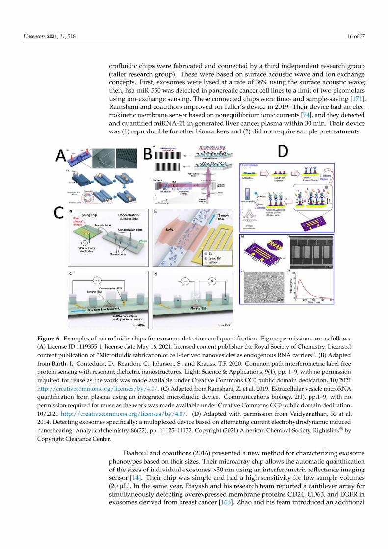
Biosensors 2021, 11, 518 16 of 37
crofluidic chips were fabricated and connected by a third independent research group(taller research group). These were based on surface acoustic wave and ion exchangeconcepts. First, exosomes were lysed at a rate of 38% using the surface acoustic wave;then, hsa-miR-550 was detected in pancreatic cancer cell lines to a limit of two picomolarsusing ion-exchange sensing. These connected chips were time- and sample-saving [171].Ramshani and coauthors improved on Taller′s device in 2019. Their device had an elec-trokinetic membrane sensor based on nonequilibrium ionic currents [74], and they detectedand quantified miRNA-21 in generated liver cancer plasma within 30 min. Their devicewas (1) reproducible for other biomarkers and (2) did not require sample pretreatments.
Biosensors 2021, 11, x FOR PEER REVIEW 18 of 40
(taller research group). These were based on surface acoustic wave and ion exchange con-cepts. First, exosomes were lysed at a rate of 38% using the surface acoustic wave; then, hsa-miR-550 was detected in pancreatic cancer cell lines to a limit of two picomolars using ion-exchange sensing. These connected chips were time- and sample-saving [171]. Ramshani and coauthors improved on Taller`s device in 2019. Their device had an elec-trokinetic membrane sensor based on nonequilibrium ionic currents [74], and they de-tected and quantified miRNA-21 in generated liver cancer plasma within 30 min. Their device was (1) reproducible for other biomarkers and (2) did not require sample pretreat-ments.
Figure 6. Examples of microfluidic chips for exosome detection and quantification. Figure permissions are as follows: (A) License ID 1119355-1, license date May 16, 2021, licensed content publisher the Royal Society of Chemistry. Licensed con-tent publication of “Microfluidic fabrication of cell-derived nanovesicles as endogenous RNA carriers”. (B) Adapted from Barth, I., Conteduca, D., Reardon, C., Johnson, S., and Krauss, T.F. 2020. Common path interferometric label-free protein sensing with resonant dielectric nanostructures. Light: Science & Applications, 9(1), pp. 1–9, with no permission required for reuse as the work was made available under Creative Commons CC0 public domain dedication, 10/2021 http://crea-tivecommons.org/licenses/by/4.0/. (C) Adapted from Ramshani, Z. et al. 2019. Extracellular vesicle microRNA quantifica-tion from plasma using an integrated microfluidic device. Communications biology, 2(1), pp.1–9, with no permission re-quired for reuse as the work was made available under Creative Commons CC0 public domain dedication, 10/2021 http://creativecommons.org/licenses/by/4.0/. (D) Adapted with permission from Vaidyanathan, R. at al. 2014. Detecting exosomes specifically: a multiplexed device based on alternating current electrohydrodynamic induced nanoshearing. Analytical chemistry, 86(22), pp. 11125–11132. Copyright (2021) American Chemical Society. Rightslink® by Copyright Clearance Center.
Daaboul and coauthors (2016) presented a new method for characterizing exosome phenotypes based on their sizes. Their microarray chip allows the automatic quantifica-tion of the sizes of individual exosomes >50 nm using an interferometric reflectance im-aging sensor [14]. Their chip was simple and had a high sensitivity for low sample vol-umes (20 μL). In the same year, Etayash and his research team reported a cantilever array for simultaneously detecting overexpressed membrane proteins CD24, CD63, and EGFR in exosomes derived from breast cancer [163]. Zhao and his team introduced an additional chip to offer a continuous-flow ExoSearch platform for exosomes derived from ovarian
Figure 6. Examples of microfluidic chips for exosome detection and quantification. Figure permissions are as follows:(A) License ID 1119355-1, license date May 16, 2021, licensed content publisher the Royal Society of Chemistry. Licensedcontent publication of “Microfluidic fabrication of cell-derived nanovesicles as endogenous RNA carriers”. (B) Adaptedfrom Barth, I., Conteduca, D., Reardon, C., Johnson, S., and Krauss, T.F. 2020. Common path interferometric label-freeprotein sensing with resonant dielectric nanostructures. Light: Science & Applications, 9(1), pp. 1–9, with no permissionrequired for reuse as the work was made available under Creative Commons CC0 public domain dedication, 10/2021http://creativecommons.org/licenses/by/4.0/. (C) Adapted from Ramshani, Z. et al. 2019. Extracellular vesicle microRNAquantification from plasma using an integrated microfluidic device. Communications biology, 2(1), pp.1–9, with nopermission required for reuse as the work was made available under Creative Commons CC0 public domain dedication,10/2021 http://creativecommons.org/licenses/by/4.0/. (D) Adapted with permission from Vaidyanathan, R. at al.2014. Detecting exosomes specifically: a multiplexed device based on alternating current electrohydrodynamic inducednanoshearing. Analytical chemistry, 86(22), pp. 11125–11132. Copyright (2021) American Chemical Society. Rightslink® byCopyright Clearance Center.
Daaboul and coauthors (2016) presented a new method for characterizing exosomephenotypes based on their sizes. Their microarray chip allows the automatic quantificationof the sizes of individual exosomes >50 nm using an interferometric reflectance imagingsensor [14]. Their chip was simple and had a high sensitivity for low sample volumes(20 µL). In the same year, Etayash and his research team reported a cantilever array forsimultaneously detecting overexpressed membrane proteins CD24, CD63, and EGFR inexosomes derived from breast cancer [163]. Zhao and his team introduced an additional
Biosensors 2021, 11, 518 17 of 37
chip to offer a continuous-flow ExoSearch platform for exosomes derived from ovariancancer. The ExoSearch chip performs a continuous quantification of exosomes in bloodplasma sample volumes from 10 µL to 10 mL using immune-magnetic beads to detect threebiomarkers: CA-125, EpCAM, andCD24 [110].
To overcome the challenges of exosome-based methods for cancer diagnosis, suchas (1) low density, (2) tiny size, (3) difficulty of isolation from plasma, (4) lab-consumingsteps, and (5) enrichment and purification issues [116,191], an electrokinetic microarraychip was fabricated [192]. This chip provides several advantages, such as (1) a low samplevolume (30−50 µL), (2) time savings—the process takes less than 30 min, (2) enablementof subsequent on-chip immunofluorescence detection of exosomal proteins, and (3) pro-vision of viable mRNA for RT-PCR analysis [192]. A simultaneous readout device wasfabricated. This device was based on peroxidase substrate 3,3′,5,5′-tetramethylbenzidine(TMB) oxidation catalyzed by an exosome–antibody complex [193]. This device exhibited(1) a high-throughput analysis, (2) a low detection limit—2.7 × 10³ particles/µL, and (3)rapid detection without sophisticated instruments [193].
Additionally, a droplet-based enzyme-linked immunosorbent assay (ELISA) chip wasfabricated to quantify cancer-derived exosomes in low sample volumes with low concentra-tions [194]. First, the exosomes were captured on magnetic beads through enzymatic ELISAcomplexes; then, the bead/exosome complexes were encapsulated inside the droplets andcounted [194]. This improved the isolation, detection, and quantification methodology. Awell-organized 16 × 20 × 20 pillar brush-like organized structure was fabricated for real-time accommodating, imaging, and detecting of single exosomes. Each pillar was cappedwith an 80-nm gold cap conjugated with anti-CD63. This amazing chip was fabricated todetect breast cancer cells (MCF7) [69]. Zhang and coauthors presented a developed chipintegrated with 3D herringbone structures to detect exosomes at concentrations as low as 10particles/µL, which were undetectable by standard microfluidics. They detected exosomesderived from ovarian cancer and normal exosomes in 20 and 10 samples, respectively.CD24, EpCAM, and FRalpha proteins were used as biomarkers. Their chip boasts (1) alarge surface area and (2) high exosome-binding efficiency [143].
In 2019, a label-free microarray was developed to detect exosomes released frommacrophages to evaluate the immune response. The chip had seven exosomal-specificantibodies fabricated on a photonic crystal biosensor surface. This chip (1) was cost-effective, (2) fast, and (3) required a low sample volume of 1 µL [111].
4.2.3. Specific Raman Scattering Technology
Specific Raman scattering (SRS) technology has received a great deal of attentionbecause of its ultra-sensitivity in the detection of a variety of small biological fractionswith low background noise [112] (Figure 7). The Ma research group proposed usingmiRNA-mediated gold–silver nanoparticles with internal nanogaps to detect miRNA-21 innon-small-cell lung cancer (NSCLC). First, an Au-rhodamine 6 G (R6G)–AuAg complexwith internal nanogaps was prepared, which was then attached with silicon microbeads tothe 3′- and 5′-ends of the capture probe. The capture probe targeted miRNA-21 exosomes.A duplex-specific nuclease cleaved the exosomal miRNA to release SRS signals from thesurface of the Au complex. However, the miRNA-21 was still in contact and cyclingthe amplification to release more Au complexes. The solution intensity was directlyproportional to the concentration of exosomal miRNA. This method introduced a newtechnique for quantifying specific exosomal miRNA in low sample volumes (5.0 µL) forclinical purposes [113].
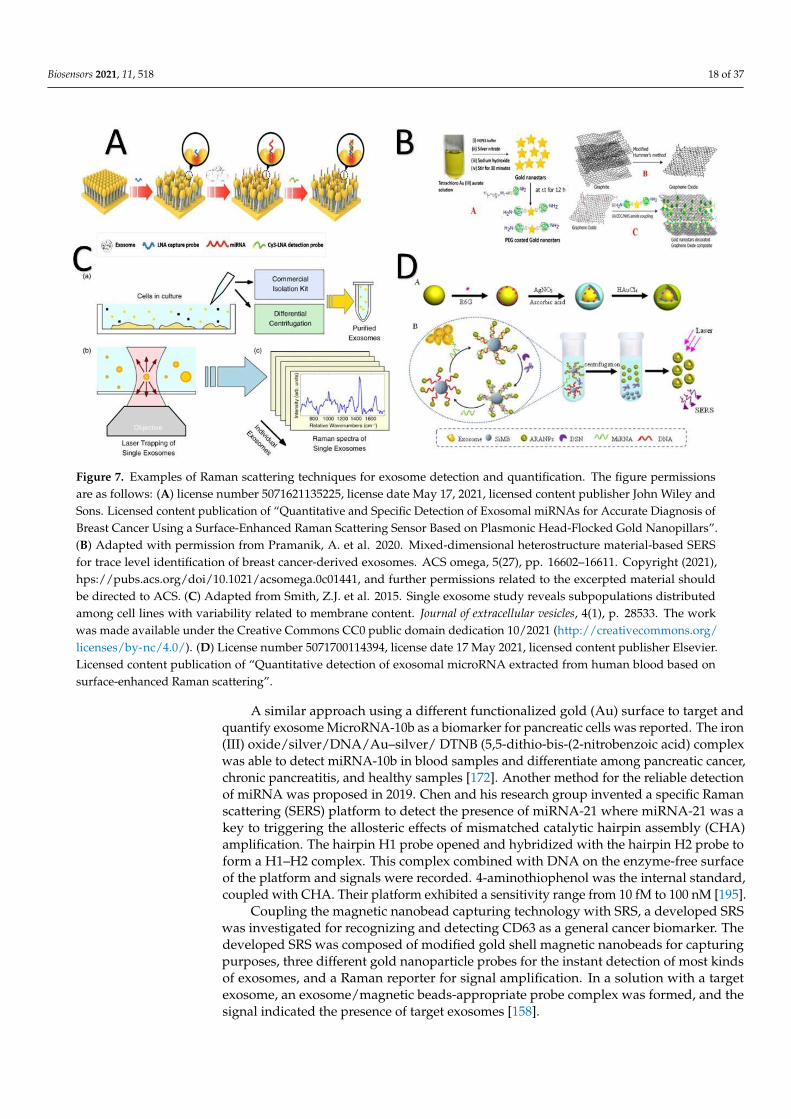
Biosensors 2021, 11, 518 18 of 37Biosensors 2021, 11, x FOR PEER REVIEW 20 of 40
Figure 7. Examples of Raman scattering techniques for exosome detection and quantification. The figure permissions are as follows: (A) license number 5071621135225, license date May 17, 2021, licensed content publisher John Wiley and Sons. Licensed content publication of “Quantitative and Specific Detection of Exosomal miRNAs for Accurate Diagnosis of Breast Cancer Using a Surface-Enhanced Raman Scattering Sensor Based on Plasmonic Head-Flocked Gold Nanopillars”. (B) Adapted with permission from Pramanik, A. et al. 2020. Mixed-dimensional heterostructure material-based SERS for trace level identification of breast cancer-derived exosomes. ACS omega, 5(27), pp. 16602–16611. Copyright (2021), hps://pubs.acs.org/doi/10.1021/acsomega.0c01441, and further permissions related to the excerpted material should be di-rected to ACS. (C) Adapted from Smith, Z.J. et al. 2015. Single exosome study reveals subpopulations distributed among cell lines with variability related to membrane content. Journal of extracellular vesicles, 4(1), p. 28533. The work was made available under the Creative Commons CC0 public domain dedication 10/2021 (http://creativecommons.org/licenses/by-nc/4.0/). (D) License number 5071700114394, license date 17 May 2021, licensed content publisher Elsevier. Licensed con-tent publication of “Quantitative detection of exosomal microRNA extracted from human blood based on surface-en-hanced Raman scattering”.
A similar approach using a different functionalized gold (Au) surface to target and quantify exosome MicroRNA-10b as a biomarker for pancreatic cells was reported. The iron (III) oxide/silver/DNA/Au–silver/ DTNB (5,5-dithio-bis-(2-nitrobenzoic acid) com-plex was able to detect miRNA-10b in blood samples and differentiate among pancreatic cancer, chronic pancreatitis, and healthy samples [172]. Another method for the reliable detection of miRNA was proposed in 2019. Chen and his research group invented a spe-cific Raman scattering (SERS) platform to detect the presence of miRNA-21 where miRNA-21 was a key to triggering the allosteric effects of mismatched catalytic hairpin assembly (CHA) amplification. The hairpin H1 probe opened and hybridized with the hairpin H2 probe to form a H1–H2 complex. This complex combined with DNA on the enzyme-free surface of the platform and signals were recorded. 4-aminothiophenol was the internal standard, coupled with CHA. Their platform exhibited a sensitivity range from 10 fM to 100 nM [195].
Coupling the magnetic nanobead capturing technology with SRS, a developed SRS was investigated for recognizing and detecting CD63 as a general cancer biomarker. The developed SRS was composed of modified gold shell magnetic nanobeads for capturing purposes, three different gold nanoparticle probes for the instant detection of most kinds of exosomes, and a Raman reporter for signal amplification. In a solution with a target
Figure 7. Examples of Raman scattering techniques for exosome detection and quantification. The figure permissionsare as follows: (A) license number 5071621135225, license date May 17, 2021, licensed content publisher John Wiley andSons. Licensed content publication of “Quantitative and Specific Detection of Exosomal miRNAs for Accurate Diagnosis ofBreast Cancer Using a Surface-Enhanced Raman Scattering Sensor Based on Plasmonic Head-Flocked Gold Nanopillars”.(B) Adapted with permission from Pramanik, A. et al. 2020. Mixed-dimensional heterostructure material-based SERSfor trace level identification of breast cancer-derived exosomes. ACS omega, 5(27), pp. 16602–16611. Copyright (2021),hps://pubs.acs.org/doi/10.1021/acsomega.0c01441, and further permissions related to the excerpted material shouldbe directed to ACS. (C) Adapted from Smith, Z.J. et al. 2015. Single exosome study reveals subpopulations distributedamong cell lines with variability related to membrane content. Journal of extracellular vesicles, 4(1), p. 28533. The workwas made available under the Creative Commons CC0 public domain dedication 10/2021 (http://creativecommons.org/licenses/by-nc/4.0/). (D) License number 5071700114394, license date 17 May 2021, licensed content publisher Elsevier.Licensed content publication of “Quantitative detection of exosomal microRNA extracted from human blood based onsurface-enhanced Raman scattering”.
A similar approach using a different functionalized gold (Au) surface to target andquantify exosome MicroRNA-10b as a biomarker for pancreatic cells was reported. The iron(III) oxide/silver/DNA/Au–silver/ DTNB (5,5-dithio-bis-(2-nitrobenzoic acid) complexwas able to detect miRNA-10b in blood samples and differentiate among pancreatic cancer,chronic pancreatitis, and healthy samples [172]. Another method for the reliable detectionof miRNA was proposed in 2019. Chen and his research group invented a specific Ramanscattering (SERS) platform to detect the presence of miRNA-21 where miRNA-21 was akey to triggering the allosteric effects of mismatched catalytic hairpin assembly (CHA)amplification. The hairpin H1 probe opened and hybridized with the hairpin H2 probe toform a H1–H2 complex. This complex combined with DNA on the enzyme-free surfaceof the platform and signals were recorded. 4-aminothiophenol was the internal standard,coupled with CHA. Their platform exhibited a sensitivity range from 10 fM to 100 nM [195].
Coupling the magnetic nanobead capturing technology with SRS, a developed SRSwas investigated for recognizing and detecting CD63 as a general cancer biomarker. Thedeveloped SRS was composed of modified gold shell magnetic nanobeads for capturingpurposes, three different gold nanoparticle probes for the instant detection of most kindsof exosomes, and a Raman reporter for signal amplification. In a solution with a targetexosome, an exosome/magnetic beads-appropriate probe complex was formed, and thesignal indicated the presence of target exosomes [158].
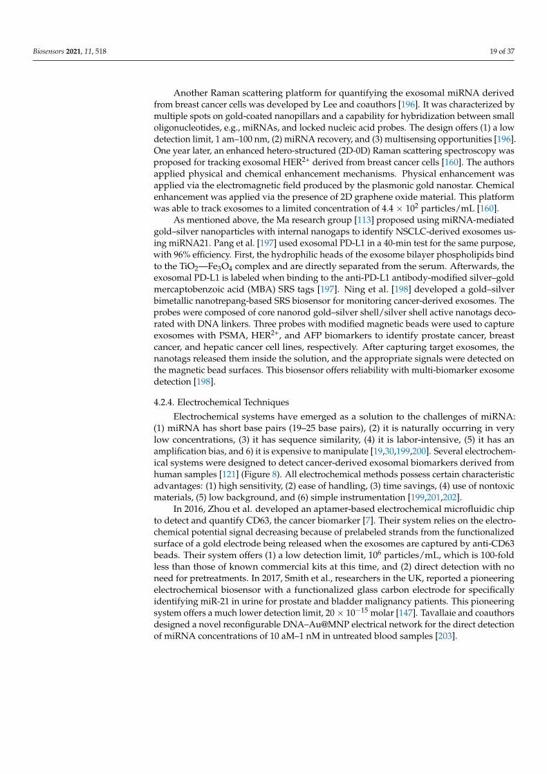
Biosensors 2021, 11, 518 19 of 37
Another Raman scattering platform for quantifying the exosomal miRNA derivedfrom breast cancer cells was developed by Lee and coauthors [196]. It was characterized bymultiple spots on gold-coated nanopillars and a capability for hybridization between smalloligonucleotides, e.g., miRNAs, and locked nucleic acid probes. The design offers (1) a lowdetection limit, 1 am–100 nm, (2) miRNA recovery, and (3) multisensing opportunities [196].One year later, an enhanced hetero-structured (2D-0D) Raman scattering spectroscopy wasproposed for tracking exosomal HER2+ derived from breast cancer cells [160]. The authorsapplied physical and chemical enhancement mechanisms. Physical enhancement wasapplied via the electromagnetic field produced by the plasmonic gold nanostar. Chemicalenhancement was applied via the presence of 2D graphene oxide material. This platformwas able to track exosomes to a limited concentration of 4.4 × 10² particles/mL [160].
As mentioned above, the Ma research group [113] proposed using miRNA-mediatedgold–silver nanoparticles with internal nanogaps to identify NSCLC-derived exosomes us-ing miRNA21. Pang et al. [197] used exosomal PD-L1 in a 40-min test for the same purpose,with 96% efficiency. First, the hydrophilic heads of the exosome bilayer phospholipids bindto the TiO2—Fe3O4 complex and are directly separated from the serum. Afterwards, theexosomal PD-L1 is labeled when binding to the anti-PD-L1 antibody-modified silver–goldmercaptobenzoic acid (MBA) SRS tags [197]. Ning et al. [198] developed a gold–silverbimetallic nanotrepang-based SRS biosensor for monitoring cancer-derived exosomes. Theprobes were composed of core nanorod gold–silver shell/silver shell active nanotags deco-rated with DNA linkers. Three probes with modified magnetic beads were used to captureexosomes with PSMA, HER2+, and AFP biomarkers to identify prostate cancer, breastcancer, and hepatic cancer cell lines, respectively. After capturing target exosomes, thenanotags released them inside the solution, and the appropriate signals were detected onthe magnetic bead surfaces. This biosensor offers reliability with multi-biomarker exosomedetection [198].
4.2.4. Electrochemical Techniques
Electrochemical systems have emerged as a solution to the challenges of miRNA:(1) miRNA has short base pairs (19–25 base pairs), (2) it is naturally occurring in verylow concentrations, (3) it has sequence similarity, (4) it is labor-intensive, (5) it has anamplification bias, and 6) it is expensive to manipulate [19,30,199,200]. Several electrochem-ical systems were designed to detect cancer-derived exosomal biomarkers derived fromhuman samples [121] (Figure 8). All electrochemical methods possess certain characteristicadvantages: (1) high sensitivity, (2) ease of handling, (3) time savings, (4) use of nontoxicmaterials, (5) low background, and (6) simple instrumentation [199,201,202].
In 2016, Zhou et al. developed an aptamer-based electrochemical microfluidic chipto detect and quantify CD63, the cancer biomarker [7]. Their system relies on the electro-chemical potential signal decreasing because of prelabeled strands from the functionalizedsurface of a gold electrode being released when the exosomes are captured by anti-CD63beads. Their system offers (1) a low detection limit, 106 particles/mL, which is 100-foldless than those of known commercial kits at this time, and (2) direct detection with noneed for pretreatments. In 2017, Smith et al., researchers in the UK, reported a pioneeringelectrochemical biosensor with a functionalized glass carbon electrode for specificallyidentifying miR-21 in urine for prostate and bladder malignancy patients. This pioneeringsystem offers a much lower detection limit, 20× 10−15 molar [147]. Tavallaie and coauthorsdesigned a novel reconfigurable DNA–Au@MNP electrical network for the direct detectionof miRNA concentrations of 10 aM–1 nM in untreated blood samples [203].
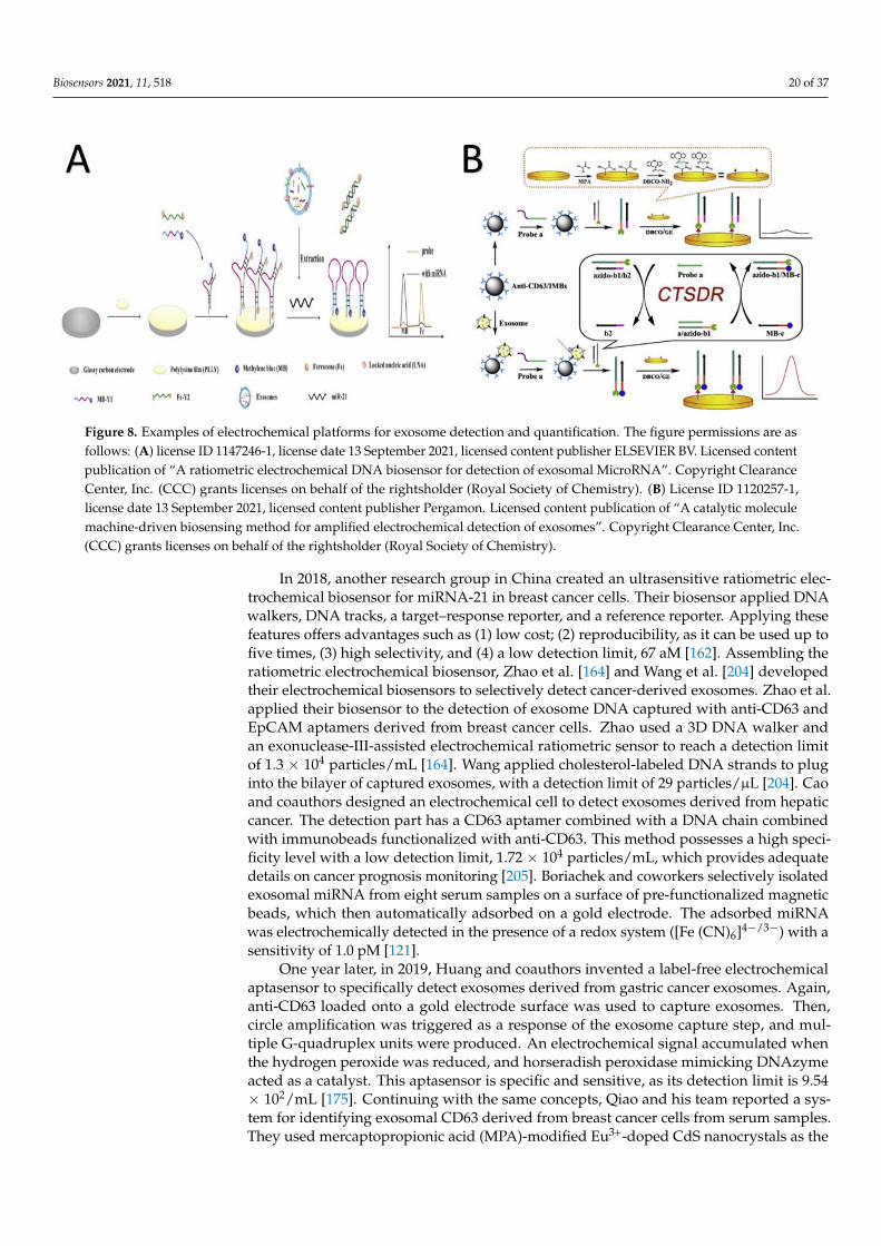
Biosensors 2021, 11, 518 20 of 37Biosensors 2021, 11, x FOR PEER REVIEW 22 of 40
Figure 8. Examples of electrochemical platforms for exosome detection and quantification. The figure permissions are as follows: (A) license ID 1147246-1, license date 13 September 2021, licensed content publisher ELSEVIER BV. Licensed con-tent publication of “A ratiometric electrochemical DNA biosensor for detection of exosomal MicroRNA”. Copyright Clear-ance Center, Inc. (CCC) grants licenses on behalf of the rightsholder (Royal Society of Chemistry). (B) License ID 1120257-1, license date 13 September 2021, licensed content publisher Pergamon. Licensed content publication of “A catalytic mol-ecule machine-driven biosensing method for amplified electrochemical detection of exosomes”. Copyright Clearance Cen-ter, Inc. (CCC) grants licenses on behalf of the rightsholder (Royal Society of Chemistry).
In 2016, Zhou et al. developed an aptamer-based electrochemical microfluidic chip to detect and quantify CD63, the cancer biomarker [7]. Their system relies on the electro-chemical potential signal decreasing because of prelabeled strands from the functional-ized surface of a gold electrode being released when the exosomes are captured by anti-CD63 beads. Their system offers (1) a low detection limit, 106 particles/mL, which is 100-fold less than those of known commercial kits at this time, and (2) direct detection with no need for pretreatments. In 2017, Smith et al., researchers in the UK, reported a pioneer-ing electrochemical biosensor with a functionalized glass carbon electrode for specifically identifying miR-21 in urine for prostate and bladder malignancy patients. This pioneering system offers a much lower detection limit, 20 × 10−15 molar [147]. Tavallaie and coauthors designed a novel reconfigurable DNA–Au@MNP electrical network for the direct detec-tion of miRNA concentrations of 10 aM–1 nM in untreated blood samples [203].
In 2018, another research group in China created an ultrasensitive ratiometric elec-trochemical biosensor for miRNA-21 in breast cancer cells. Their biosensor applied DNA walkers, DNA tracks, a target–response reporter, and a reference reporter. Applying these features offers advantages such as (1) low cost; (2) reproducibility, as it can be used up to five times, (3) high selectivity, and (4) a low detection limit, 67 aM [162]. Assembling the ratiometric electrochemical biosensor, Zhao et al. [164] and Wang et al. [204] developed their electrochemical biosensors to selectively detect cancer-derived exosomes. Zhao et al. applied their biosensor to the detection of exosome DNA captured with anti-CD63 and EpCAM aptamers derived from breast cancer cells. Zhao used a 3D DNA walker and an exonuclease-III-assisted electrochemical ratiometric sensor to reach a detection limit of 1.3 × 10⁴ particles/mL [164]. Wang applied cholesterol-labeled DNA strands to plug into the bilayer of captured exosomes, with a detection limit of 29 particles/μL [204]. Cao and co-authors designed an electrochemical cell to detect exosomes derived from hepatic cancer. The detection part has a CD63 aptamer combined with a DNA chain combined with im-munobeads functionalized with anti-CD63. This method possesses a high specificity level with a low detection limit, 1.72 × 10⁴ particles/mL, which provides adequate details on cancer prognosis monitoring [205]. Boriachek and coworkers selectively isolated exoso-mal miRNA from eight serum samples on a surface of pre-functionalized magnetic beads,
Figure 8. Examples of electrochemical platforms for exosome detection and quantification. The figure permissions are asfollows: (A) license ID 1147246-1, license date 13 September 2021, licensed content publisher ELSEVIER BV. Licensed contentpublication of “A ratiometric electrochemical DNA biosensor for detection of exosomal MicroRNA”. Copyright ClearanceCenter, Inc. (CCC) grants licenses on behalf of the rightsholder (Royal Society of Chemistry). (B) License ID 1120257-1,license date 13 September 2021, licensed content publisher Pergamon. Licensed content publication of “A catalytic moleculemachine-driven biosensing method for amplified electrochemical detection of exosomes”. Copyright Clearance Center, Inc.(CCC) grants licenses on behalf of the rightsholder (Royal Society of Chemistry).
In 2018, another research group in China created an ultrasensitive ratiometric elec-trochemical biosensor for miRNA-21 in breast cancer cells. Their biosensor applied DNAwalkers, DNA tracks, a target–response reporter, and a reference reporter. Applying thesefeatures offers advantages such as (1) low cost; (2) reproducibility, as it can be used up tofive times, (3) high selectivity, and (4) a low detection limit, 67 aM [162]. Assembling theratiometric electrochemical biosensor, Zhao et al. [164] and Wang et al. [204] developedtheir electrochemical biosensors to selectively detect cancer-derived exosomes. Zhao et al.applied their biosensor to the detection of exosome DNA captured with anti-CD63 andEpCAM aptamers derived from breast cancer cells. Zhao used a 3D DNA walker andan exonuclease-III-assisted electrochemical ratiometric sensor to reach a detection limitof 1.3 × 10⁴ particles/mL [164]. Wang applied cholesterol-labeled DNA strands to pluginto the bilayer of captured exosomes, with a detection limit of 29 particles/µL [204]. Caoand coauthors designed an electrochemical cell to detect exosomes derived from hepaticcancer. The detection part has a CD63 aptamer combined with a DNA chain combinedwith immunobeads functionalized with anti-CD63. This method possesses a high speci-ficity level with a low detection limit, 1.72 × 10⁴ particles/mL, which provides adequatedetails on cancer prognosis monitoring [205]. Boriachek and coworkers selectively isolatedexosomal miRNA from eight serum samples on a surface of pre-functionalized magneticbeads, which then automatically adsorbed on a gold electrode. The adsorbed miRNAwas electrochemically detected in the presence of a redox system ([Fe (CN)6]4−/3−) with asensitivity of 1.0 pM [121].
One year later, in 2019, Huang and coauthors invented a label-free electrochemicalaptasensor to specifically detect exosomes derived from gastric cancer exosomes. Again,anti-CD63 loaded onto a gold electrode surface was used to capture exosomes. Then,circle amplification was triggered as a response of the exosome capture step, and mul-tiple G-quadruplex units were produced. An electrochemical signal accumulated whenthe hydrogen peroxide was reduced, and horseradish peroxidase mimicking DNAzymeacted as a catalyst. This aptasensor is specific and sensitive, as its detection limit is 9.54× 10²/mL [175]. Continuing with the same concepts, Qiao and his team reported a sys-tem for identifying exosomal CD63 derived from breast cancer cells from serum samples.They used mercaptopropionic acid (MPA)-modified Eu3+-doped CdS nanocrystals as the

Biosensors 2021, 11, 518 21 of 37
electrochemiluminescent emitters and hydrogen peroxide as the core actant. After rec-ognizing the exosomes, the H2O2 decomposed, and the electrochemiluminescence (ECL)signals decreased [206]. The limit of detection (7.41 × 10⁴ particles/mL) was twice aslow as that of the Rongrong design [175]. Other efforts produced a CD63-fuctionalizedreduced graphene oxide field effect transistor biosensor to quantify cancer-derived exo-somes in low-concentration samples (33 particles/µL) electrically. This device was capableof distinguishing prostate cancer samples from control samples without labeling [11].
A peptide nucleic acid (±)/microRNA/spherical nucleic acid nanoprobe (+) sandwichon an electrochemical sensor was developed by the Liu research team to detect exosomalmiRNA in blood samples from breast cancer patients. This sandwich sensor was (1) spe-cific because of the hybridization opportunity between the neutral and the negative sidesof the sandwich, which enhanced the detection process, (2) sensitive enough to detecta single base mismatch, (3) label-free, and (4) an enzyme-free process [207]. Recently asandwich structure for diagnosing ovarian malignancy early by detecting phosphatidylser-ine (PS)-positive exosomes was created by Liu. Exosomes were captured on the surfacesof functionalized gold nanoflowers that capped g–C3N4 nanosheets. When exosomeswere captured, the nanosheets catalyzed the decomposition of H2O2, and the ECL signalswere amplified. In addition to its sensitivity and low cost, this system possesses the lowdetection limit of 39 particles/µL [146].
Applying 2D Ti3C2 MXenes nanosheets and anti-EpCAM as the ECL probe and thecapture aptamer, Zhang designed a reliable, sensitive biosensor to detect breast cancerin serum samples with a detection limit of 125 particles/µL [137]. A novel ECL systemconjugated with polymerase amplification was constructed to detect miRNA-16 derivedfrom leukemia [141]. A magnet-controlled glassy carbon electrode functionalized witha nano-Au surface that adsorbs assistance DNA with the pyridine–ruthenium complexincluded the chemical components of the system. When the target miRNA was detected,the capture DNA immobilized on the electrode opened and was conjugated with thattarget to form double-stranded DNA. Then, the hairpin structure opened, and the DNAprimers conjugated with the complementary sequence occurring at the hairpin neck withthe assistance of the Klenow fragment of DNA polymerase and the ECL signals wererecorded. This biosensor is (1) label-free, (2) ultrasensitive, with the low detection limit of4.3 × 10−17 mol/L, (3) stable, and (4) reproducible [141].
In 2020, an electrochemical biosensor for exosomal microRNA detection was realizedthat has two steps: (1) the induction of miRNA signal amplification and (2) inductionof silver nanoparticle deposition. This method is simple, inexpensive, and ultrasensitive(limit of 0.4 fM) [200]. An improved electrochemical system for exosome detection wasfabricated by Fang and coauthors. They enhanced their system by combining the benefitsof (1) MXenes, which act as a probe supporter to improve the signals, (2) black phosphorousquantum dots (BPQDs), which act as an oxidation catalyst for tris (4,4′-dicarboxylicacid-2,2′-bipyridyl) ruthenium (II) dichloride (Ru(dcbpy)3)2+ and also improves signals, and (3)SiO2 nanourchin, which acts as a sensing platform for the aptamer. Both the MXenes andthe BPQDs provide extraordinary photothermal properties. Their systems operate in twoforward steps: First, EpCAM is detected on an immobilized aptamer; second, the modifiedexosomes are combined with the CD63 antibody on the MXenes-BPQDs–Ru(dcbpy3)2+
complex [115]. This dual ECL system is specific and effective. Another electrochemicalassay was fabricated and hybridized with the chain reaction method to detect miR-122 inliver and breast cancers [167]. The presence of miR-122 opens hairpin DNA immobilized ona gold electrode surface and triggers a hybridization chain reaction to generate long doublehelixes that capture more [Ru (NH3)6]3+ and increases the differential pulse voltammetry.The structure and controlling features of this method offer a promising amplificationefficiency and high sensitivity. A self-reductant ECL biosensor was developed to derivebenefits from the hybridization of Ti3C2–MXenes/gold nanoparticle complexes and CD63aptamer to capture and detect tumor-derived exosomes. This naked catalytic surfacebiosensor delivers high sensitivity, excellent conductivity, a large surface area, and the

Biosensors 2021, 11, 518 22 of 37
low detection limit of 30 particles/µL, which is 1000-fold lower than that of ELISA [208].A ratiometric electrochemical DNA biosensor was also fabricated and modified witha Y-shaped locked nucleic acid to detect exosome miR-21 derived from breast cancer.This modification increased the selectivity and accuracy and brought the sensitivity to adetection limit of 2.3 × 10⁻¹⁵ moles [209].
Human epidermal growth factor receptor (EGFR) and the EGFR variant (v) III muta-tion (EGFRvIII) are exosomal biomarkers that have been used for the early detection offatal brain tumor glioblastoma from circulating blood. In the designed electrochemicalsystem, (1) a peptide ligand binds to the EGFR and EGFRvIII, (2) Zr4+ from the metalorganic framework/electroactive methylene blue complex binds to the intrinsic phosphategroups on exosome surfaces, and (3) the exosome concentrations are quantified directlyfrom the concentration of electroactive molecules on the exosome surfaces. The detectionlimit is 7.83 × 10³ particles/µL [173].
Applying the strategies of locked nucleic acids, a G-quadruplex, and rolling circleamplification and using a gold electrode, Xiaoqi and coauthors designed an electrochemicalsystem to detect exosomal miRNA-21 with a detection limit of 2.75 × 10⁻¹⁵ M. Theirdesign was comparable to the RT-PCR strategy because of its stability, consistency, andreproducibility [210].
A dual-signal self-calibrating biosensor for detecting cancer-derived exosomes wasdeveloped from previous electrochemical protocols [211]. The authors designed theirplatform using black phosphorus nanosheets associated with a metal–organic framework,which hybridized with antibody thin films to capture exosome biomarkers. Both thenanosheets and the framework were assembled on an indium tin oxide slice, which wasattached to a ssDNA aptamer labeled with methylene blue. The redox current of themethylene blue was reduced in the presence of the desired exosomes. This workstationoffered a low detection limit—100 particles/mL, specificity, and the capability of detectingmultiple biomarkers [211].
Meng and colleagues designed a novel on/off photoelectrochemical biosensor apply-ing an organic dye (toluidine blue O dye) to detect cancer biomarker miRNA 21. A lowconcentration of miRNA 21 (1017–1011 mol/L) could be detected when transformed intoabundant p-type copper sulfide (CuS)-labeled signal tags and combined with TBO@Bi2S3-ZnS. The photoelectrochemical signals could then be used to detect and quantify miRNA21 [212].
Electrochemical techniques, which have been the subject of research efforts since 2016,have many applications and are considered the most promising techniques for exosomedetection and cancer diagnosis, because all the electrochemical techniques are easy tohandle, rely on nontoxic materials, are timesaving, and have high sensitivity with a lowbackground.
4.2.5. Fluorescence and Colorimetric Techniques
The main differences between fluorescence immunoassays and colorimetric techniquesare the high sensitivity and accuracy of the fluorescence immunoassays [213]. Here,we focused on the published designs (37 models) using either one of the methods todetect/quantify cancer-derived exosomes for diagnostic purposes (Figure 9).
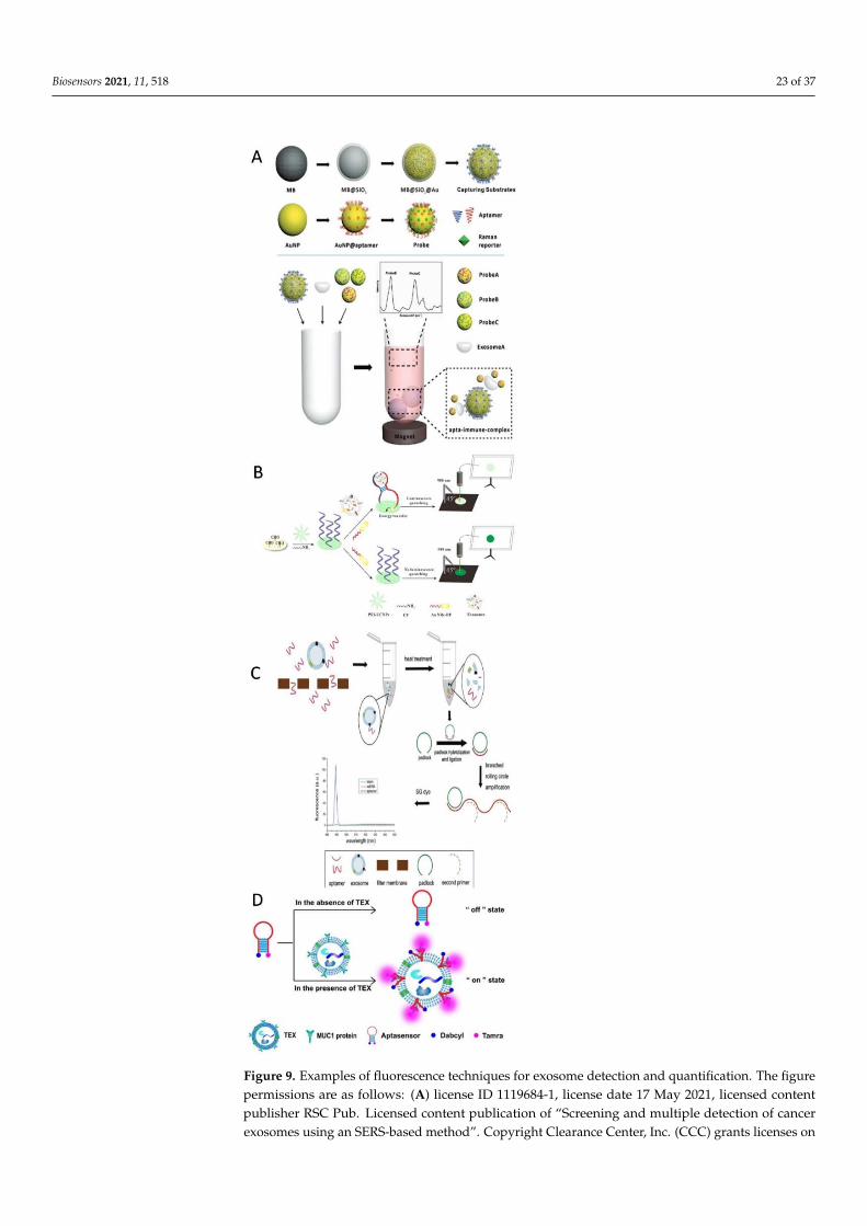
Biosensors 2021, 11, 518 23 of 37
Biosensors 2021, 11, x FOR PEER REVIEW 25 of 40
4.2.5. Fluorescence and Colorimetric Techniques The main differences between fluorescence immunoassays and colorimetric tech-
niques are the high sensitivity and accuracy of the fluorescence immunoassays [213]. Here, we focused on the published designs (37 models) using either one of the methods to detect/quantify cancer-derived exosomes for diagnostic purposes (Figure 9).
Figure 9. Examples of fluorescence techniques for exosome detection and quantification. The figurepermissions are as follows: (A) license ID 1119684-1, license date 17 May 2021, licensed contentpublisher RSC Pub. Licensed content publication of “Screening and multiple detection of cancerexosomes using an SERS-based method”. Copyright Clearance Center, Inc. (CCC) grants licenses on

Biosensors 2021, 11, 518 24 of 37
behalf of the rightsholder (Royal Society of Chemistry). (B) License ID 1147245-1, license date13 September 2021, licensed content publisher Pergamon. Licensed content publication of “Apaper-supported aptasensor based on upconversion luminescence resonance energy transfer for theaccessible determination of exosomes”. Copyright Clearance Center, Inc. (CCC) grants licenses onbehalf of the rightsholder (Royal Society of Chemistry). (C) License ID 1119685-1, license date 17 May2021, licensed content publisher RSC Pub. Licensed content publication of “A simple fluorescenceaptasensor for gastric cancer exosome detection based on branched rolling circle amplification”.Copyright Clearance Center, Inc. (CCC) grants licenses on behalf of the rightsholder (Royal Society ofChemistry). (D) License Number 5071720075356, license date 18 May 2021, licensed content publisherElsevier. Licensed content publication of “A simple, specific and “on-off” type MUC1 fluorescenceaptasensor based on exosomes for detection of breast cancer”.
As miRNAs are responsible for regulating gene expression and are protected fromribonuclease degradation, they are excellent biomarkers for the diagnosis and prognosisof cancer [195,199,200]. In 2008, Taylor and Gercel-Taylor were able to separate exosomesderived from ovarian cancer from serum samples by applying a modified magneticallyactivated cell sorting technique based on identifying the exosomal EpCAM protein using aLD microcolumn. The exosomes and beads were separated, and the exosomes were recov-ered in PBS by ultracentrifugation. The miRNAs were isolated using a mirVanamicroRNAisolation kit, then labeled with Cy3, hybridized using microarrays, and scanned with anarray scanner. Perfect matches for miR-27a, miR-93, and miR-152 plus two mismatchesfor each one was applied. According to Taylor’s procedure, detecting certain miRNAs(miR-21, miR-141, miR-200a, miR-200c, miR-200b, miR-203, miR-205, and miR-214) arethe key to discriminating between ovarian cancer samples and benign disease or healthysamples [18].
Ovarian cancer diagnosis research has been extended to screening exosome claudinproteins, e.g., claudin-4, as biomarkers. In 2009, Li et al. found full-length claudin-4shed from a plasma culture in a conditioned culture medium following immunoblotting.They detected it using sucrose gradient separation and immunogold electron microscopyexperiments. The authors presented their procedure as a step for confirming ovariancancer detection combined with further analysis [145]. Additionally, in 2009, Logozzipresented a novel tool for screening for and following-up on melanoma-derived exosomes.Logozzi invented a home-based sandwich ELISA to capture exosomes CD63 and Rab-5band to quantify caveolin-1 from mice plasma samples as follows: (1) isolate exosomesusing ultracentrifugation, (2) coat 96-well plates with anti-Rab-5b, (3) incubate exosomesovernight inside the functionalized 96-well plates, (4) add anti-CD63 Mab or anticaveolin-1Mab, and (5) record the density at 450 nm. The results collected from the invented tool wereconfirmed using Western blot and flow cytometry. Melanoma samples: n = 0 compared tohealthy cases: n = 58 [178].
By 2015, several research efforts were published. It was found that exosome c-Metpromotes the progression of melanoma [214]. A fast, simple, and sensitive procedure forcapturing, detecting, and quantifying exosomal miRNA-21 from exosomes derived frombreast cancer was reported. Simply by functionalizing molecular beads with streptolysin Oand fluorescent dye Cys3, Lee and his team were able to selectively identify and quantifymiRNA-21 mixed into human serum [20].
Melanoma-derived exosomes inside lymph nodes pulse signals to control cell re-cruitment, the spread of vascular tissues inside the lymph nodes, and the deposition offluids [215]. For the first time, melanoma exosomes were imaged in vitro and within lymphnodes in vivo, using magnetic resonance imaging to prove those exosomes can be moni-tored in vivo. The exosomes can be tracked after they are loaded onto superparamagneticiron oxide nanoparticles [194].
To report the progression of tumors implanted in mice, microtoroid optical resonatorswere applied. The target exosomes were only able to land on a functionalized silica

Biosensors 2021, 11, 518 25 of 37
microtoroid and change its resonance frequency. This method was label-free, applicable toa range of exosome sizes, and sensitive [216].
In 2016, Ahadi and coauthors published a dataset identifying the long noncodingRNA exosomes related to prostate cancer. After testing the exosomes from five cell lines,they concluded that there was a significant expression difference between the exosomesand their original cells [217]. They found that miR-17, miR-18a, miR-20a, miR-93, miR-106b,and thelet-7 were highly expressed in prostate cancer samples. A rapid, one-step, and novelflow immunoassay strip was invented in 2016 for capturing CD9 and CD81, detectingCD63 labeled with nano-Au, and quantifying exosomes derived from melanoma cancer inplasma and urine samples based on the use of tetraspanins as targets [179]. In addition,Kibria et al. (2016) were able to distinguish a single circulating exosome biomarker inhuman cell lines and blood samples using a microflow cytometer. This captured exosome,CD63, then applied fast staining and automated counting steps. They were also able tocompare the expression of CD44 and CD47 between breast cancer and healthy samples [6].miR 182, which is dose-independent in breast cancer, was detected in both the cell culturesupernatant and fresh serum. Higginbotham and coauthors developed a fluorescence-activated vesicle-sorting platform for detecting and sorting individual exosomes from celllines based on specific exosome-surface biomarkers. Their protocol is based on (1) sequen-tial ultracentrifugation to isolate exosomes, (2) the capturing and labeling of exosomesusing labeled fluorescent antibodies, and (3) the detection of individual exosomes using acommercial flow cytometer. They were able to detect EGFR and CD9 as biomarkers forhuman colorectal cancer and recognized the activation state of EGFR using monoclonalantibody 806 [218]. A capillary electrophoresis/mass spectrometry method was appliedto isolate and analyze iso-miR-16-5p and miR-21-5p in serum samples from leukemiapatients [114].
In 2017, for the first time, carcinoembryonic antigen was detected and quantified incolorectal cancer-derived exosomes in serum. At Wakayama Medical University Hospital,116 patients were tested through one year using an ELISA protocol, and the protocolwas optimized [219]. In the same year, single-stranded DNA was found to improve theperoxidase activity of g–C3N4 nanosheets, yielding a four-fold increase in the colorimetricanalysis of exosomal CD63 in breast cancer cells. The reason for this was the electrostaticattraction and aromatic stacking between the DNA and the nanosheets [220]. An on-offaptamer was designed to detect MUC1 on breast cancer-derived exosomes. The aptasensorhad two ends, one representing TAMRA (luminophore) and the other representing Dabcyl(quenching group). In the presence of exosomes, the aptasensor was locked, and MUCIwas recognized by the aptasensor, which turned on and emitted fluorescence signals. Thisprotocol offers applicability, sensitivity, and specificity [107].
Three microchip and electrochemical technologies allowing the detection of exosomeswith the naked eye were mentioned above [190,193,208]. Other published platforms allow-ing detection with the naked eye applied calorimetric and fluorescence technologies. Thefirst one was designed by Reference [151]. The mechanism was based on substituting theaptamer/Au nanoparticle complex with aptamer/exosome surface proteins and releasingAu nanoparticles, which aggregate in the ultra-salty solution and change the solutioncolor [151]. Additionally, Zhang and his team initiated a novel protocol for virtually rec-ognizing cancer-derived exosomes. Their protocol was based on the following steps: (1)capture exosomes through binding with anti-CD63 on a magnetic bead aptamer, and (2)immediately insert a modified DNA probe into the exosome membrane to trigger thehybridization chain reaction, transduce the signal, and amplify the signal through alkalinephosphatase. As a result, silver ions are reduced by ascorbic acid and form a shell aroundgold nanoparticles, causing a vivid color change. This protocol offered limits of detectionof 1.6 × 10² particles/µL with UV−Visible spectroscopy and 9 × 10³ particles/µL with thenaked eye [221]. The imaging protocol recently designed by Zhou [156] targets simplicity,rapidity, and low cost and can identify exosomes derived from breast cancer. This protocol,like the previous protocol published by Reference [107], is aimed at creating an on–off

Biosensors 2021, 11, 518 26 of 37
switch with a low limit of detection, 3.9 × 10⁵ particles/mL. The two ends of the aptasen-sor represent MUC1 aptamer as a luminophore and mimicking DNAzyme in a hairpinstructure as a quenching group. If there are exosomes, they bind to the MUC1 aptamer;then, the DNA hairpin unfolds and triggers the conformation of G-quadruplex–DNAzyme.As a result, a clear blue color originates from the reduction of hydrogen peroxide [156].
Efforts have been made to fabricate a self-referenceable platform specifically for detect-ing and quantifying cancer-derived exosomal biomarker CD63. Researchers constructed aCy3–anti-CD63 aptamer that adsorbed on Ti3C2 MXene nanosheets via metal interactionsand hydrogen bonds. In the presence of the desired exosomes, the aptamer highly bondedwith exosome CD63 released from the nanosheet surface and produced fluorescence signalsthat were referenced through the fluorescence resonance energy transfer nanoprobe. Thisself-referenced platform can be applied to detect several biomarkers at once, and it offers alimited range of detection, 1000-fold lower than that of ELISA [222]. Since the expressionprofiles of exosomal miRNA make them potential candidates for cancer diagnosis, Zhaiand coauthors developed a 4-h detection and quantification protocol for detecting miR-1246 and identifying breast cancer samples in plasma. They constructed a functionalizedAu nanoflare probe with nucleic acids that were able to enter the exosomal plasma andgenerate fluorescence signals. This in situ detection protocol is (1) inexpensive, (2) fast,(3) simple, and (4) specific [24]. Another miRNA was investigated as a biomarker forductal carcinoma, which is sometimes a result of breast cancer. Yoshikawa investigatedmiR-223-3p as a biomarker for diagnosing ductal carcinoma in 185 plasma samples using amiRNA microarray and TaqMan miR assays [177]. Both CD63 and nucleolin were usedas targets to capture exosomes derived from leukemia cells using anti-CD63 antibodyconjugated with magnetic beads. After that, a DNA primer–nucleolin–recognition aptamerbound the exosomes, initiating the amplification reaction and generating gold nanopar-ticle/DNA/fluorescent dye complexes. Through the help of nicking endonuclease, thefluorescent dye was released, emission started, and signals accumulated [142]. This dualfluorescence platform provides sensitivity for sample concentrations as low as 1 × 10²
particles/µL. Another fluorescent biosensor for detecting miRNA-21 was fabricated byReference [148]. This biosensor couples the target-catalyzing signal amplification withDNA-labeled carbon dots and a 5,7-dinitro-2-sulfo-acridone probe to reach high fluores-cence resonance energy transfer when the probe is assembled. This ratiometric biosensoris (1) sensitive, with the low detection limit of 3 × 10⁻¹⁵ M, (2) stable, (3) capable of re-moving environmental fluctuations, (4) selective of a single base mismatch sequence, (5)cost-effective, and (6) usable for monitoring purposes [148].
In 2019, a direct method for exosomal DNA detection was published. When exosomeswere captured on the CD63 aptamer surface, the DNA probe initiated a hairpin DNAcascade reaction, and the open DNA hairpin bound to gold nanoparticles. The signalsof the labeled DNA dendrimers were recorded. The limit of detection was 1.16 × 10³
particles/µL [223].Another way to overcome the disadvantages of exosomal miRNA detection for early
cancer diagnosis and monitoring is to combine the detection of the miRNA pool withsurface proteins to reflect the tumor heterogeneity. Cho and coworkers (2019) found thatmiR-21, miR-574-3p, EpCAM, and epidermal growth factor receptor (EGFR) were morehighly expressed in exosomal cancer-derived cells than in noncancer cells. Exosomesderived from a prostate cancer biopsy were first isolated using anti-CD63 antibody. Thentheir miR-21, miR-574-3p, and EpCAM surface proteins were simultaneously detected andquantified in a single step using CD63/antibody/nanomolecular beacon/oligonucleotideprobes at 37 ◦C for 60 min after normalization [19]. This multitarget detection method is(1) simple, (2) inexpensive, and (3) labor-saving. Like Cho, Shi proposed a protocol usingmagnetic beads, antibodies, and aptamer hybridized with chain reaction probes to detectexosomes derived from hepatic carcinoma at a limiting rate of 100 particles/mL. Threetypes of antibodies were used: prostate-specific antigen, D-dimer, and anti-CD63. Thisprotocol requires the pretreatment of exosomes before manipulation [224].

Biosensors 2021, 11, 518 27 of 37
Turning their attention from the miRNA biomarkers to other RNAs to use as biomark-ers, researchers in Japan investigated the possibility of relating the expression of fourmRNAs and five small nucleolar RNAs for the early diagnosis of pancreatic cancer. Theyfound that the mRNAs (ARF6 and WASF2) and snRNAs (SNORA25 and SNORA74A) werehighly expressed in patient samples and could be used as biomarkers [170].
Another on/off detector has been designed in which an afterglow semiconductingpolyelectrolyte switches between the on and off modes according to the presence of targetexosomes [225]. Different exosome proteins can be detected using different aptamer se-quences. This design offers a detection limit that is twice as low as that of other fluorescencedetection methods.
Recently, the Huang research group developed an assay specifically for detectingexosomes derived from gastric cancer. The desired exosomes bind to the aptamer, andbranched rolling circle amplification is triggered when a second primer is added. SYBRGreen I fluorescent dye is used to detect long, double-stranded DNA. This assay possessesthroughput specificity, a detection limit of 4.27 × 10⁴ particles/mL, and a probability of anearly diagnosis of gastric cancer [226].
A new colorimetric method for detecting exosomes derived from colorectal cancerpatients (n = 16) and comparing them to healthy ones (n = 9) was invented by Huang et al.2020. Their detection method was based on the enzymatic activity of deoxynucleotidyltransferase. Two probes were selected: anti-A33 as a capture probe and the EpCAM ap-tamer/Au/primer complex as a signal probe. After capturing exosomes, deoxynucleotidyltransferase helps the signal probe to reach the biotin adenine chains and bind them toavidin-modified horseradish peroxidase for the hydrogen peroxide-mediated oxidation of3,3′,5,5′-tetramethyl benzidine in an enzyme-linked aptamer-sorbent assay (ELASA). Theresults showed high sensitivity, with the method detecting 1.95 × 10⁶ particles/µL. Thisplatform also provides flexibility; the probes can be changed to detect different biomark-ers [108]. In a new sandwich-type biosensor developed for cancer diagnosis, functionalizedmagnetic beads with anti-CD63, anti-EpCAM, and horseradish peroxidase catalysis wereapplied. After capturing the exosomes with anti-CD63, anti-EpCAM captured the exo-somes derived from hepatic cancer. At this point, the oxidation–reduction reaction wastriggered, and the fluorescent spectra at 370–550 nm were detected with a detection limitof 200 particles/mL [227].
A magnet-based immunoassay for characterizing and quantifying general and specificbiomarkers for breast cancer patients was presented recently [155]. The exosomes werepreconcentrated using anti-CD9, CD63, and CD-81-functionalized magnetic beads, thenlabeled with a second antibody for detecting CD24, CD44, CD54, CD326, and CD340.The second group of antibodies was conjugated with ELISA to quantify the signals. Thedetection limit of this procedure was 105 particles/µL, and there was no need for pretreat-ments [155]. Immuno-translating exosomal proteins helps us to determine their structureand physiological roles. An immunosensor was developed to exploit CD63 on exosomesurfaces. First, the desired exosomes are isolated using a size-exclusion chromatography.Then, the desired exosomes are detected using an anti-CD63-functionalized quartz crystalmicrobalance with dissipation monitoring. The detection limit of this technique (2.9 × 10⁸
particles/mL) is much higher than those of other techniques [228].Xia and coauthors developed a cactus-like biosensor in which anti-CD63 mimicked
the roots, captured exosomes mimicked the stem, and modified streptavidin/horseradishperoxidase/cholesterol-labeled DNA mimicked the thorns when inserted into an exosomemembrane. The signals were generated from fluorescein because of the catalyzed oxidationof 1,4-phenylenediamine. This biosensor is comparable to known methods, because it issensitive with no interference from proteins, has a detection limit of ~3 × 10³ particles/µL,and quantitatively measures the exosome extraction [229]. An advanced all-in-one biosen-sor was fabricated to simultaneously detect multiple biomarkers derived from breast cancer(miR-21, miR-27a, and miR-375) based on competitive strand displacement. This biosensorhas three Y-type scaffold oligonucleotides, each of which has ss-recognition sequences
Biosensors 2021, 11, 518 28 of 37
associated with three quenchers and three labeled reporters conjugated with fluorophoresFAM, Cy3, and Cy5. The protocol for this biosensor is based on intra-communicationbetween the biosensor and the exosomes: the oligonucleotides complementarily hybridizewith miRNA inside exosomes and release the reporter to activate the fluorescent signals.The limit of detection for this biosensor was found to vary according to the target miRNAand ranges from 0.116 to 0.287 µg/mL. This biosensor could be a routine bioassay forclinical purposes [166].
Fluorescence immunoassays and colorimetric techniques for detecting/quantifyingcancer-derived exosomes are highly sensitive and easy to manipulate and have beenupgraded to allow detection with the naked eye, which makes them fast and accurate tofollow.
Many published research efforts have indicated the suitability of methods charac-terizing and detecting exosomes for procedures for monitoring cancer biogenesis. Thesemethods include surface plasmonic biosensors; microchips; specific Raman scattering; andelectrochemical, fluorescence, and colorimetric techniques. In addition, these methods canbe used in applications that provide results that can be seen with the naked eye.
5. Conclusions and Future Prospects
Exosomes have been investigated as (1) disease biomarkers, (2) therapeutic agents,(3) activation motors, and (4) diagnostic tools. Exosomes are considered promising candi-dates for human disease and therapeutics biomarkers because they are easy to circulatethrough the blood, represent their parent cells, and are stable. Further exosome researchis needed because of their heterogeneity and purification requirements and the need toattain supersensitive molecular profiling for individual exosomes with sizes of 30–50nm [6,163,230].
Exosomes as disease-monitoring nanovesicles have been applied as rate-dependentbiomarkers of disease stages [231]. We expect and suggest testing the informative role ofexosomes as tools to predict the development of cancer diseases like Alzheimer’s disease.Certain exosome biomarkers (P-S396-tau, P-T181-tau, and Ab1–42) found to predict thedevelopment of Alzheimer’s disease up to 10 years in advance [232].
For another prospect, using exosomes to activate cells such as sperm [233] couldbe a start towards investigating the possibility of using exosomes as engines to activateimmune-defense cells (T cells) and protect cells from cancer diseases. The future prospectsfor exosome research include, besides investigating exosomes for defense and predictionpurposes, investigating the roles of exosomes derived from microorganisms such as bacteriaand algae, as these organisms naturally produce multiple bioactive compounds.
Author Contributions: Conceptualization, W.-J.D. and M.G.S.; writing—original draft preparation,M.G.S.; writing—review and editing, W.-J.D. and H.B.; funding acquisition, W.-J.D. All authors haveread and agreed to the published version of the manuscript.
Funding: This work was partially supported by NIH/CA248734 and NSF 1934227 to W.-J.D.
Institutional Review Board Statement: Not applicable.
Informed Consent Statement: Not applicable.
Data Availability Statement: Not applicable.
Conflicts of Interest: The authors declare no conflict of interest.
References1. Hood, J.L.; Wickline, S.A. A systematic approach to exosome-based translational nanomedicine. Wiley Interdiscip. Rev. Nanomed.
Nanobiotechnol. 2012, 4, 458–467. [CrossRef]2. Choi, D.-S.; Kim, D.-K.; Kim, Y.-K.; Gho, Y.S. Proteomics, transcriptomics and lipidomics of exosomes and ectosomes. Proteomics
2013, 13, 1554–1571. [CrossRef]3. Fernández-Llama, P.; Khositseth, S.; Gonzales, P.A.; Star, R.A.; Pisitkun, T.; Knepper, M.A. Tamm-Horsfall protein and urinary
exosome isolation. Kidney Int. 2010, 77, 736–742. [CrossRef] [PubMed]

Biosensors 2021, 11, 518 29 of 37
4. Théry, C.; Zitvogel, L.; Amigorena, S. Exosomes: Composition, biogenesis and function. Nat. Rev. Immunol. 2002, 2, 569–579.[CrossRef] [PubMed]
5. Morten, P.; Oksvold, A.N.; Ketil, W. Pedersen Magnetic Bead-Based Isolation of Exosomes; Humana Press: New York, NY, USA, 2015;ISBN 978-1-4939-1537-8.
6. Kibria, G.; Ramos, E.K.; Lee, K.E.; Bedoyan, S.; Huang, S.; Samaeekia, R.; Athman, J.J.; Harding, C.V.; Lötvall, J.; Harris, L.; et al. Arapid, automated surface protein profiling of single circulating exosomes in human blood. Sci. Rep. 2016, 6, 36502. [CrossRef]
7. Zhou, Q.; Rahimian, A.; Son, K.; Shin, D.-S.; Patel, T.; Revzin, A. Development of an aptasensor for electrochemical detection ofexosomes. Methods 2016, 97, 88–93. [CrossRef]
8. Vlassov, A.V.; Magdaleno, S.; Setterquist, R.; Conrad, R. Exosomes: Current knowledge of their composition, biological functions,and diagnostic and therapeutic potentials. Biochim. Biophys. Acta Gen. Subj. 2012, 1820, 940–948. [CrossRef] [PubMed]
9. Wang, Z.; Zhang, R.; Huang, P.; Chen, C.; Li, H.; Li, P.; Quinn, D.; Dao, M.; Suresh, S.; Wu, M.; et al. Isolation of exosomes fromwhole blood by integrating acoustics and microfluidics. Proc. Natl. Acad. Sci. USA 2017, 114, 10584–10589, Erratum in Proc. Natl.Acad. Sci. USA 2020, 117, 28525. [CrossRef]
10. Schuh, C.M.; Cuenca, J.; Alcayaga-Miranda, F.; Khoury, M. Exosomes on the border of species and kingdom intercommunication.Transl. Res. 2019, 210, 80–98. [CrossRef] [PubMed]
11. Yu, Y.; Li, Y.-T.; Jin, D.; Yang, F.; Wu, D.; Xiao, M.-M.; Zhang, H.; Zhang, Z.-Y.; Zhang, G.-J. Electrical and Label-Free Quantificationof Exosomes with a Reduced Graphene Oxide Field Effect Transistor Biosensor. Anal. Chem. 2019, 91, 10679–10686. [CrossRef][PubMed]
12. Greening, D.; Gopal, S.K.; Xu, R.; Simpson, R.; Chen, W. Exosomes and their roles in immune regulation and cancer. Semin. CellDev. Biol. 2015, 40, 72–81. [CrossRef] [PubMed]
13. Huang, X.; Yuan, T.; Tschannen, M.; Sun, Z.; Jacob, H.; Du, M.; Liang, M.; Dittmar, R.L.; Liu, Y.; Liang, M.; et al. Characterizationof human plasma-derived exosomal RNAs by deep sequencing. BMC Genom. 2013, 14, 319. [CrossRef] [PubMed]
14. Daaboul, G.G.; Gagni, P.; Benussi, L.; Bettotti, P.; Ciani, M.; Cretich, M.; Freedman, D.S.; Ghidoni, R.; Ozkumur, A.Y.; Piotto, C.;et al. Digital Detection of Exosomes by Interferometric Imaging. Sci. Rep. 2016, 6, 37246. [CrossRef]
15. Cheng, L.; Wang, Y.; Huang, L. Exosomes from M1-Polarized Macrophages Potentiate the Cancer Vaccine by Creating aPro-inflammatory Microenvironment in the Lymph Node. Mol. Ther. 2017, 25, 1665–1675. [CrossRef] [PubMed]
16. Trams, E.G.; Lauter, C.J.; Salem, N., Jr.; Heine, U. Exfoliation of membrane ecto-enzymes in the form of micro-vesicles. Biochim.Biophys. Acta 1981, 645, 63–70. [CrossRef]
17. Brown, H.L.; Clayton, A.; Stephens, P. The role of bacterial extracellular vesicles in chronic wound infections: Current knowledgeand future challenges. Wound Repair Regen. 2021, 29, 864–880. [CrossRef]
18. MicroRNA signatures of tumor-derived exosomes as diagnostic biomarkers of ovarian cancer. Gynecol. Oncol. 2008, 110, 13–21.[CrossRef]
19. Cho, S.; Yang, H.C.; Rhee, W.J. Simultaneous multiplexed detection of exosomal microRNAs and surface proteins for prostatecancer diagnosis. Biosens. Bioelectron. 2019, 146, 111749. [CrossRef]
20. Lee, J.H.; Kim, J.A.; Kwon, M.H.; Kang, J.Y.; Rhee, W.J. In situ single step detection of exosome microRNA using molecularbeacon. Biomaterials 2015, 54, 116–125. [CrossRef] [PubMed]
21. Siegel, R.L.; Miller, K.D.; Jemal, A. Cancer statistics, 2018. CA Cancer J. Clin. 2018, 68, 7–30. [CrossRef] [PubMed]22. Khanmohammadi, A.; Aghaie, A.; Vahedi, E.; Qazvini, A.; Ghanei, M.; Afkhami, A.; Hajian, A.; Bagheri, H. Electrochemical
biosensors for the detection of lung cancer biomarkers: A review. Talanta 2020, 206, 120251. [CrossRef]23. Xie, Y.-H.; Chen, Y.-X.; Fang, J.-Y. Comprehensive review of targeted therapy for colorectal cancer. Signal Transduct. Target. Ther.
2020, 5, 22. [CrossRef] [PubMed]24. Zhai, L.-Y.; Li, M.-X.; Pan, W.-L.; Chen, Y.; Pang, J.-X.; Zheng, L.; Chen, J.-X.; Duan, W.-J. In Situ Detection of Plasma Exosomal
MicroRNA-1246 for Breast Cancer Diagnostics by a Au Nanoflare Probe. ACS Appl. Mater. Interfaces 2018, 10, 39478–39486.[CrossRef] [PubMed]
25. Ezzati, M.; Yousefi, B.; Velaei, K.; Safa, A. A review on anti-cancer properties of Quercetin in breast cancer. Life Sci. 2020, 248,117463. [CrossRef] [PubMed]
26. Khalili, T.M.; Fleshner, P.R.; Hiatt, J.R.; Sokol, T.P.; Manookian, C.; Tsushima, G.; Phillips, E.H. Colorectal cancer. Dis. ColonRectum 1998, 41, 832–838. [CrossRef]
27. Grasso, L.; Wyss, R.; Weidenauer, L.; Thampi, A.; Demurtas, D.; Prudent, M.; Lion, N.; Vogel, H. Molecular screening ofcancer-derived exosomes by surface plasmon resonance spectroscopy. Anal. Bioanal. Chem. 2015, 407, 5425–5432. [CrossRef][PubMed]
28. Desplantes, R.; Lévêque, C.; Muller, B.; Lotierzo, M.; Ferracci, G.; Popoff, M.; Seagar, M.; Mamoun, R.; El Far, O. Affinitybiosensors using recombinant native membrane proteins displayed on exosomes: Application to botulinum neurotoxin B receptor.Sci. Rep. 2017, 7, 1032. [CrossRef]
29. Liu, C.; Zeng, X.; An, Z.; Yang, Y.; Eisenbaum, M.; Gu, X.; Jornet, J.M.; Dy, G.K.; Reid, M.E.; Gan, Q.; et al. Sensitive Detection ofExosomal Proteins via a Compact Surface Plasmon Resonance Biosensor for Cancer Diagnosis. ACS Sens. 2018, 3, 1471–1479.[CrossRef]
30. Bu, H.; He, D.; He, X.; Wang, K. Exosomes: Isolation, Analysis, and Applications in Cancer Detection and Therapy. ChemBioChem2019, 20, 451–461. [CrossRef]
Biosensors 2021, 11, 518 30 of 37
31. Obermaier, C.; Griebel, A.; Westermeier, R. Principles of Protein Labeling Techniques; Humana: New York, NY, USA, 2021; Volume2261, ISBN 9781493925490.
32. Schorey, J.S.; Bhatnagar, S. Exosome Function: From Tumor Immunology to Pathogen Biology. Traffic 2008, 9, 871–881. [CrossRef]33. Ji, H.; Erfani, N.; Tauro, B.J.; Kapp, E.A.; Zhu, H.J.; Moritz, R.L.; Lim, J.W.E.; Simpson, R.J. Difference gel electrophoresis analysis
of Ras-transformed fibroblast cell-derived exosomes. Electrophoresis 2008, 29, 2660–2671. [CrossRef]34. Simpson, R.J.; Lim, J.W.; Moritz, R.L.; Mathivanan, S. Exosomes: Proteomic insights and diagnostic potential. Expert Rev. Proteom.
2009, 6, 267–283. [CrossRef] [PubMed]35. Tauro, B.J.; Greening, D.W.; Mathias, R.A.; Ji, H.; Mathivanan, S.; Scott, A.M.; Simpson, R.J. Comparison of ultracentrifugation,
density gradient separation, and immunoaffinity capture methods for isolating human colon cancer cell line LIM1863-derivedexosomes. Methods 2012, 56, 293–304. [CrossRef] [PubMed]
36. Bellingham, S.A.; Guo, B.B.; Coleman, B.M.; Hill, A.F. Exosomes: Vehicles for the Transfer of Toxic Proteins Associated withNeurodegenerative Diseases? Front. Physiol. 2012, 3, 124. [CrossRef] [PubMed]
37. Tauro, B.J.; Greening, D.; Mathias, R.; Mathivanan, S.; Ji, H.; Simpson, R.J. Two Distinct Populations of Exosomes Are Releasedfrom LIM1863 Colon Carcinoma Cell-derived Organoids. Mol. Cell. Proteom. 2013, 12, 587–598. [CrossRef] [PubMed]
38. Ji, H.; Greening, D.; Barnes, T.W.; Lim, J.W.; Tauro, B.J.; Rai, A.; Xu, R.; Adda, C.; Mathivanan, S.; Zhao, W.; et al. Proteomeprofiling of exosomes derived from human primary and metastatic colorectal cancer cells reveal differential expression of keymetastatic factors and signal transduction components. Proteomics 2013, 13, 1672–1686. [CrossRef] [PubMed]
39. Wubbolts, R.; Leckie, R.S.; Veenhuizen, P.T.M.; Schwarzmann, G.; Möbius, W.; Hoernschemeyer, J.; Slot, J.-W.; Geuze, H.J.;Stoorvogel, W. Proteomic and Biochemical Analyses of Human B Cell-derived Exosomes. J. Biol. Chem. 2003, 278, 10963–10972.[CrossRef] [PubMed]
40. Yáñez-Mó, M.; Siljander, P.R.-M.; Andreu, Z.; Zavec, A.B.; Borràs, F.E.; Buzas, E.I.; Buzas, K.; Casal, E.; Cappello, F.; Carvalho, J.;et al. Biological properties of extracellular vesicles and their physiological functions. J. Extracell. Vesicles 2015, 4, 27066. [CrossRef]
41. Trajkovic, K.; Hsu, C.; Chiantia, S.; Rajendran, L.; Wenzel, D.; Wieland, F.; Schwille, P.; Brügger, B.; Simons, M. Ceramide TriggersBudding of Exosome Vesicles into Multivesicular Endosomes. Science 2008, 319, 1244–1247. [CrossRef]
42. De Gassart, A.; Geminard, C.; Fevrier, B.; Raposo, G.; Vidal, M. Lipid raft-associated protein sorting in exosomes. Blood 2003, 102,4336–4344. [CrossRef] [PubMed]
43. Harding, C.; Heuser, J.; Stahl, P. Receptor-mediated endocytosis of transferrin and recycling of the transferrin receptor in ratreticulocytes. J. Cell Biol. 1983, 97, 329–339. [CrossRef] [PubMed]
44. Dixon, L. The WSU Libraries’ Goal Is to Provide Excellent Customer Service. Let Us Know How We Are Doing by Responding toThis Short Survey. 1991. Available online: http://fluidsurveys.com/s/AccessServicesSurvey/ (accessed on 17 October 2021).
45. Keller, S.; Sanderson, M.; Stoeck, A.; Altevogt, P. Exosomes: From biogenesis and secretion to biological function. Immunol. Lett.2006, 107, 102–108. [CrossRef] [PubMed]
46. Henderson, M.C.; Azorsa, D.O. The Genomic and Proteomic Content of Cancer Cell-Derived Exosomes. Front. Oncol. 2012, 2, 38.[CrossRef] [PubMed]
47. Akers, J.C.; Gonda, D.; Kim, R.; Carter, B.S.; Chen, C.C. Biogenesis of extracellular vesicles (EV): Exosomes, microvesicles,retrovirus-like vesicles, and apoptotic bodies. J. Neuro-Oncol. 2013, 113, 1–11. [CrossRef] [PubMed]
48. Beach, A.; Zhang, H.-G.; Ratajczak, M.Z.; Kakar, S.S. Exosomes: An overview of biogenesis, composition and role in ovariancancer. J. Ovarian Res. 2014, 7, 14. [CrossRef] [PubMed]
49. Hessvik, N.P.; Llorente, A. Current knowledge on exosome biogenesis and release. Cell. Mol. Life Sci. 2018, 75, 193–208. [CrossRef][PubMed]
50. Henne, W.M.; Stenmark, H.; Emr, S.D. Molecular Mechanisms of the Membrane Sculpting ESCRT Pathway. Cold Spring Harb.Perspect. Biol. 2013, 5, a016766. [CrossRef]
51. Colombo, M.; Moita, C.F.; van Niel, G.; Kowal, J.; Vigneron, J.; Benaroch, P.; Manel, N.; Moita, L.F.; Théry, C.; Raposo, G. Analysisof ESCRT functions in exosome biogenesis, composition and secretion highlights the heterogeneity of extracellular vesicles. J. CellSci. 2013, 126, 5553–5565. [CrossRef]
52. Stuffers, S.; Wegner, C.S.; Stenmark, H.; Brech, A. Multivesicular endosome biogenesis in the absence of ESCRTs. Traffic 2009, 10,925–937. [CrossRef]
53. Escola, J.-M.; Kleijmeer, M.J.; Stoorvogel, W.; Griffith, J.M.; Yoshie, O.; Geuze, H.J. Selective Enrichment of Tetraspan Proteins onthe Internal Vesicles of Multivesicular Endosomes and on Exosomes Secreted by Human B-lymphocytes. J. Biol. Chem. 1998, 273,20121–20127. [CrossRef]
54. Chairoungdua, A.; Smith, D.L.; Pochard, P.; Hull, M.; Caplan, M.J. Exosome release of β-catenin: A novel mechanism thatantagonizes Wnt signaling. J. Cell Biol. 2010, 190, 1079–1091. [CrossRef] [PubMed]
55. Hurwitz, S.N.; Conlon, M.M.; Rider, M.A.; Brownstein, N.; Meckes, D.G. Nanoparticle analysis sheds budding insights intogenetic drivers of extracellular vesicle biogenesis. J. Extracell. Vesicles 2016, 5, 31295. [CrossRef]
56. Savina, A.; Vidal, M.; Colombo, M.I. The exosome pathway in K562 cells is regulated by Rab11. J. Cell Sci. 2002, 115, 2505–2515.[CrossRef] [PubMed]
57. Parolini, I.; Federici, C.; Raggi, C.; Lugini, L.; Palleschi, S.; De Milito, A.; Coscia, C.; Iessi, E.; Logozzi, M.; Molinari, A.; et al.Microenvironmental pH Is a Key Factor for Exosome Traffic in Tumor Cells. J. Biol. Chem. 2009, 284, 34211–34222. [CrossRef]

Biosensors 2021, 11, 518 31 of 37
58. Goldberg, Z.; Lehnert, B. Radiation-induced effects in unirradiated cells: A review and implications in cancer. Int. J. Oncol. 2002,21, 337–349. [CrossRef]
59. Azzam, E.I.; Little, J.B. The radiation-induced bystander effect: Evidence and significance. Hum. Exp. Toxicol. 2004, 23, 61–65.[CrossRef]
60. Amzallag, N.; Passer, B.J.; Allanic, D.; Segura, E.; Théry, C.; Goud, B.; Amson, R.; Telerman, A. TSAP6 Facilitates the Secretionof Translationally Controlled Tumor Protein/Histamine-releasing Factor via a Nonclassical Pathway. J. Biol. Chem. 2004, 279,46104–46112. [CrossRef] [PubMed]
61. Passer, B.J.; Nancy-Portebois, V.; Amzallag, N.; Prieur, S.; Cans, C.; de Climens, A.R.; Fiucci, G.; Bouvard, V.; Tuynder, M.; Susini,L.; et al. The p53-inducible TSAP6 gene product regulates apoptosis and the cell cycle and interacts with Nix and the Myt1 kinase.Proc. Natl. Acad. Sci. USA 2003, 100, 2284–2289. [CrossRef]
62. Yu, X.; Harris, S.L.; Levine, A.J. The Regulation of Exosome Secretion: A Novel Function of the p53 Protein. Cancer Res. 2006, 66,4795–4801. [CrossRef] [PubMed]
63. King, H.W.; Michael, M.Z.; Gleadle, J.M. Hypoxic enhancement of exosome release by breast cancer cells. BMC Cancer 2012, 12,421. [CrossRef] [PubMed]
64. Németh, A.; Orgovan, N.; Sódar, B.W.; Osteikoetxea, X.; Pálóczi, K.; Szabó-Taylor, K.É.; Vukman, K.V.; Kittel, Á.; Turiák, L.;Wiener, Z.; et al. Antibiotic-induced release of small extracellular vesicles (exosomes) with surface-associated DNA. Sci. Rep.2017, 7, 8202. [CrossRef]
65. Ostrowski, M.; Carmo, N.B.; Krumeich, S.; Fanget, I.; Raposo, G.; Savina, A.; Moita, C.F.; Schauer, K.; Hume, A.N.; Freitas, R.P.;et al. Rab27a and Rab27b control different steps of the exosome secretion pathway. Nat. Cell Biol. 2010, 12, 19–30. [CrossRef][PubMed]
66. Hsu, C.; Morohashi, Y.; Yoshimura, S.-I.; Manrique-Hoyos, N.; Jung, S.; Lauterbach, M.A.; Bakhti, M.; Grønborg, M.; Möbius, W.;Rhee, J.; et al. Regulation of exosome secretion by Rab35 and its GTPase-activating proteins TBC1D10A–C. J. Cell Biol. 2010, 189,223–232. [CrossRef] [PubMed]
67. Smith, Z.; Lee, C.; Rojalin, T.; Carney, R.P.; Hazari, S.; Knudson, A.; Lam, K.; Saari, H.; Ibañez, E.L.; Viitala, T.; et al. Singleexosome study reveals subpopulations distributed among cell lines with variability related to membrane content. J. Extracell.Vesicles 2015, 4, 28533. [CrossRef] [PubMed]
68. Jeong, S.; Park, J.; Pathania, D.; Castro, C.M.; Weissleder, R.; Lee, H. Integrated Magne-to-Electrochemical Sensor for ExosomeAnalysis. ACS Nano 2016, 10, 1802–1809. [CrossRef] [PubMed]
69. Raghu, D.; Christodoulides, J.A.; Christophersen, M.; Liu, J.L.; Anderson, G.P.; Robitaille, M.; Byers, J.M.; Raphael, M.P.Nanoplasmonic pillars engineered for single exosome detection. PLoS ONE 2018, 13, e0202773. [CrossRef] [PubMed]
70. Alvarez, M.L.; Khosroheidari, M.; Ravi, R.K.; DiStefano, J.K. Comparison of protein, microRNA, and mRNA yields using differentmethods of urinary exosome isolation for the discovery of kidney disease biomarkers. Kidney Int. 2012, 82, 1024–1032. [CrossRef]
71. Vergauwen, G.; Dhondt, B.; Van Deun, J.; De Smedt, E.; Berx, G.; Timmerman, E.; Gevaert, K.; Miinalainen, I.; Cocquyt, V.; Braems,G.; et al. Confounding factors of ultrafiltration and protein analysis in extracellular vesicle research. Sci. Rep. 2017, 7, 2704.[CrossRef]
72. Gámez-Valero, A.; Monguió-Tortajada, M.; Carreras-Planella, L.; Franquesa, M.; Beyer, K.; Borràs, F.E. Size-ExclusionChromatography-based isolation minimally alters Extracellular Vesicles’ characteristics compared to precipitating agents. Sci.Rep. 2016, 6, srep33641. [CrossRef]
73. Barreiro, K.; Huber, T.B.; Holthofer, H. Isolating Urinary Extracellular Vesicles as Biomarkers for Diabetic Disease. Diabet. Nephrop.2020, 2067, 175–188. [CrossRef]
74. Li, P.; Kaslan, M.; Lee, S.H.; Yao, J.; Gao, Z. Progress in Exosome Isolation Techniques. Theranostics 2017, 7, 789–804. [CrossRef][PubMed]
75. Doldán, X.; Fagúndez, P.; Cayota, A.; Laíz, J.; Tosar, J.P. Electrochemical Sandwich Immunosensor for Determination of ExosomesBased on Surface Marker-Mediated Signal Amplification. Anal. Chem. 2016, 88, 10466–10473. [CrossRef]
76. Taylor, D.D.; Shah, S. Methods of isolating extracellular vesicles impact down-stream analyses of their cargoes. Methods 2015, 87,3–10. [CrossRef] [PubMed]
77. Linares, R.; Tan, S.; Gounou, C.; Brisson, A.R. Imaging and Quantification of Extracellular Vesicles by Transmission ElectronMicroscopy. In Exosomes and Microvesicles; Humana Press: New York, NY, USA, 2016; ISBN 978-1-4939-6728-5.
78. Gad, S.C. Drug Safety Evaluation; John Wiley & Sons: Hoboken, NJ, USA, 2011; ISBN 9780470253168.79. Heinemann, M.L.; Ilmer, M.; Silva, L.P.; Hawke, D.H.; Recio, A.; Vorontsova, M.A.; Alt, E.; Vykoukal, J. Benchtop isolation and
characterization of functional exosomes by sequential filtration. J. Chromatogr. A 2014, 1371, 125–135. [CrossRef]80. Petersen, K.E.; Manangon, E.; Hood, J.; Wickline, S.A.; Fernandez, D.P.; Johnson, W.P.; Gale, B. A review of exosome separation
techniques and characterization of B16-F10 mouse melanoma exosomes with AF4-UV-MALS-DLS-TEM. Anal. Bioanal. Chem.2014, 406, 7855–7866. [CrossRef]
81. Cheruvanky, A.; Zhou, H.; Pisitkun, T.; Kopp, J.B.; Knepper, M.A.; Yuen, P.S.T.; Star, R.A. Rapid isolation of urinary exosomalbiomarkers using a nanomembrane ultrafiltration concentrator. Am. J. Physiol. Ren. Physiol. 2007, 292, F1657–F1661. [CrossRef][PubMed]

Biosensors 2021, 11, 518 32 of 37
82. Baranyai, T.; Herczeg, K.; Onódi, Z.; Voszka, I.; Módos, K.; Marton, N.; Nagy, G.; Mäger, I.; Wood, M.J.; El Andaloussi, S.; et al.Isolation of Exosomes from Blood Plasma: Qualitative and Quantitative Comparison of Ultracentrifugation and Size ExclusionChromatography Methods. PLoS ONE 2015, 10, e0145686. [CrossRef]
83. Koh, Y.Q.; Almughlliq, F.B.; Vaswani, K.; Peiris, H.N.; Mitchell, M.D. Exosome enrichment by ultracentrifugation and sizeexclusion chromatography. Front. Biosci. 2018, 23, 865–874. [CrossRef]
84. Freitas, J.T.; Jozic, I.; Bedogni, B. Wound Healing Assay for Melanoma Cell Migration. Methods Mol. Biol. 2021, 2265, 65–71.85. Musante, L.; Tataruch, D.E.; Holthofer, H. Use and Isolation of Urinary Exosomes as Biomarkers for Diabetic Nephropathy. Front.
Endocrinol. 2014, 5, 149. [CrossRef]86. Popovic, M.; Mazzega, E.; Toffoletto, B.; De Marco, A. Isolation of anti-extra-cellular vesicle single-domain antibodies by direct
panning on vesicle-enriched fractions. Microb. Cell Factories 2018, 17, 6. [CrossRef]87. Zarovni, N.; Corrado, A.; Guazzi, P.; Zocco, D.; Lari, E.; Radano, G.; Muhhina, J.; Fondelli, C.; Gavrilova, J.; Chiesi, A. Integrated
isolation and quantitative analysis of exosome shuttled proteins and nucleic acids using immunocapture approaches. Methods2015, 87, 46–58. [CrossRef]
88. Poellmann, M.J.; Nair, A.; Bu, J.; Kim, J.K.H.; Kimple, R.J.; Hong, S. Immunoavidity-Based Capture of Tumor Exosomes UsingPoly(amidoamine) Dendrimer Surfaces. Nano Lett. 2020, 20, 5686–5692. [CrossRef]
89. Nakai, W.; Yoshida, T.; Diez, D.; Miyatake, Y.; Nishibu, T.; Imawaka, N.; Naruse, K.; Sadamura, Y.; Hanayama, R. A novelaffinity-based method for the isolation of highly purified extracellular vesicles. Sci. Rep. 2016, 6, 1–11. [CrossRef]
90. Santos, H.A.; Liu, D.; Zhang, H. Microfluidics for Pharmaceutical Applications: From Nano/Micro Systems Fabrication to ControlledDrug Delivery; William Andrew: Norwich, NY, USA, 2018; ISBN 9780128126592.
91. Wang, Z.; Wu, H.-J.; Fine, D.; Schmulen, J.; Hu, T.; Godin, B.; Zhang, J.X.J.; Liu, X. Ciliated micropillars for the microfluidic-basedisolation of nanoscale lipid vesicles. Lab Chip 2013, 13, 2879–2882. [CrossRef]
92. Sun, G.; Pan, Z.; Senapati, S.; Chang, H.-C. Concentration-Gradient Stabilization with Segregated Counter- and Co-Ion Paths: AQuasistationary Depletion Front for Robust Molecular Isolation or Concentration. Phys. Rev. Appl. 2017, 7, 064024. [CrossRef]
93. Marczak, S.; Richards, K.; Ramshani, Z.; Smith, E.; Senapati, S.; Hill, R.; Go, D.B.; Chang, H.-C. Simultaneous isolation andpreconcentration of exosomes by ion concentration polarization. Electrophoresis 2018, 39, 2029–2038. [CrossRef]
94. Conde-Vancells, J.; Falcón-Pérez, J.M. Isolation of Urinary Exosomes from Animal Models to Unravel Noninvasive DiseaseBiomarkers. Liver Proteom. 2012, 909, 321–340. [CrossRef]
95. Webber, J.; Clayton, A. How pure are your vesicles? J. Extracell. Vesicles 2013, 2. [CrossRef] [PubMed]96. Rice, K.C.; Mann, E.E.; Endres, J.L.; Weiss, E.C.; Cassat, J.E.; Smeltzer, M.; Bayles, K.W. The cidA murein hydrolase regulator
contributes to DNA release and biofilm development in Staphylococcus aureus. Proc. Natl. Acad. Sci. USA 2007, 104, 8113–8118.[CrossRef]
97. Jung, M.K.; Mun, J.Y. Sample Preparation and Imaging of Exosomes by Transmission Electron Microscopy. J. Vis. Exp. 2018,e56482. [CrossRef]
98. Tatischeff, I.; Larquet, E.; Falcon-Perez, J.M.; Turpin, P.-Y.; Kruglik, S.G. Fast characterisation of cell-derived extracellular vesiclesby nanoparticles tracking analysis, cryo-electron microscopy, and Raman tweezers microspectroscopy. J. Extracell. Vesicles 2012, 1,19179. [CrossRef]
99. Coumans, F.A.W.; van der Pol, E.; Böing, A.N.; Hajji, N.; Sturk, G.; van Leeuwen, T.; Nieuwland, R. Reproducible extracellularvesicle size and concentration determination with tunable resistive pulse sensing. J. Extracell. Vesicles 2014, 3, 25922. [CrossRef]
100. Vogel, R.; Coumans, F.A.W.; Maltesen, R.G.; Böing, A.N.; Bonnington, K.E.; Broekman, M.L.; Broom, M.F.; Buzás, E.I.; Christiansen,G.; Hajji, N.; et al. A standardized method to determine the concentration of extracellular vesicles using tunable resistive pulsesensing. J. Extracell. Vesicles 2016, 5, 31242. [CrossRef]
101. Ashcroft, B.A.; De Sonneville, J.; Yuana, Y.; Osanto, S.; Bertina, R.; Kuil, M.E.; Oosterkamp, T.H. Determination of the sizedistribution of blood microparticles directly in plasma using atomic force microscopy and microfluidics. Biomed. Microdevices2012, 14, 641–649. [CrossRef] [PubMed]
102. Klimentová, J.; Stulík, J. Methods of isolation and purification of outer membrane vesicles from gram-negative bacteria. Microbiol.Res. 2015, 170, 1–9. [CrossRef] [PubMed]
103. Reiner, A.T.; Toma, K.; Brisson, A.R.; Pils, D.; Knoll, W.; Dostalek, J. Plasmonic exosome biosensors for medical diagnostics. InFrontiers in Biophotonics for Translational Medicine: In the Celebration of Year of Light; Springer: Singapore, 2015; pp. 249–272. ISBN978-981-287-627-0.
104. Lv, X.; Geng, Z.; Su, Y.; Fan, Z.; Wang, S.; Fang, W.; Chen, H. Label-Free Exosome Detection Based on a Low-Cost PlasmonicBiosensor Array Integrated with Microfluidics. Langmuir 2019, 35, 9816–9824. [CrossRef]
105. Tadimety, A.; Closson, A.; Li, C.; Yi, S.; Shen, T.; Zhang, J.X.J. Advances in liquid biopsy on-chip for cancer management:Technologies, biomarkers, and clinical analysis. Crit. Rev. Clin. Lab. Sci. 2018, 55, 140–162. [CrossRef]
106. Wang, Q.; Chu, M.; Wang, X.; Niu, Q.; Ma, G.; Fang, Y.; Liu, J.; Lv, W.; Zhao, W. Electrochemical cytosensor for detection of cellsurface sialic acids based on 3D biointerface. Electrochimica Acta 2018, 282, 923–930. [CrossRef]
107. Zhang, J.; Shi, J.; Liu, W.; Zhang, K.; Zhao, H.; Zhang, H.; Zhang, Z. A simple, specific and “on-off” type MUC1 fluorescenceaptasensor based on exosomes for detection of breast cancer. Sens. Actuators B Chem. 2018, 276, 552–559. [CrossRef]
108. Huang, Z.; Lin, Q.; Ye, X.; Yang, B.; Zhang, R.; Chen, H.; Weng, W.; Kong, J. Terminal deoxynucleotidyl transferase based signalamplification for enzyme-linked aptamer-sorbent assay of colorectal cancer exosomes. Talanta 2020, 218, 121089. [CrossRef]

Biosensors 2021, 11, 518 33 of 37
109. de Vrij, J.; Maas, S.L.; van Nispen, M.; Sena-Esteves, M.; Limpens, R.W.; Koster, A.J.; Leenstra, S.; Lamfers, M.L.; Broekman,M.L. Quantification of nanosized extracellular membrane vesicles with scanning ion occlusion sensing. Nanomedicine 2013, 8,1443–1458. [CrossRef]
110. Zhao, Z.; Yang, Y.; Zeng, Y.; He, M. A microfluidic ExoSearch chip for multiplexed exosome detection towards blood-basedovarian cancer diagnosis. Lab Chip 2016, 16, 489–496. [CrossRef]
111. Wang, Y.; Yuan, W.; Zhang, Q.; Wang, Y.; Kimber, M.; Dong, L.; Lu, M. Exosome microarray based on label-free imaging biosensor.In Proceedings of the IEEE Sensors, Montreal, QC, Canada, 27–30 October 2019.
112. Ye, S.; Wu, Y.; Zhai, X.; Tang, B. Asymmetric Signal Amplification for Simultaneous SERS Detection of Multiple Cancer Markerswith Significantly Different Levels. Anal. Chem. 2015, 87, 8242–8249. [CrossRef]
113. Ma, D.; Huang, C.; Zheng, J.; Tang, J.; Li, J.; Yang, J.; Yang, R. Quantitative detection of exosomal microRNA extracted fromhuman blood based on surface-enhanced Raman scattering. Biosens. Bioelectron. 2018, 101, 167–173. [CrossRef]
114. Khan, N.; Mironov, G.; Berezovski, M.V. Direct detection of endogenous MicroRNAs and their post-transcriptional modificationsin cancer serum by capillary electrophoresis-mass spectrometry Young Investigators in Analytical and Bioanalytical Science. Anal.Bioanal. Chem. 2016, 408, 2891–2899. [CrossRef]
115. Fang, D.; Zhao, D.; Zhang, S.; Huang, Y.; Dai, H.; Lin, Y. Black phosphorus quantum dots functionalized MXenes as the enhanceddual-mode probe for exosomes sensing. Sens. Actuators B Chem. 2020, 305, 127544. [CrossRef]
116. Zhu, L.; Wang, K.; Cui, J.; Liu, H.; Bu, X.; Ma, H.; Wang, W.; Gong, H.; Lausted, C.; Hood, L.; et al. Label-Free QuantitativeDetection of Tumor-Derived Exosomes through Surface Plasmon Resonance Imaging. Anal. Chem. 2014, 86, 8857–8864. [CrossRef][PubMed]
117. Jalalian, S.H.; Ramezani, M.; Jalalian, S.A.; Abnous, K.; Taghdisi, S.M. Exosomes, new biomarkers in early cancer detection. Anal.Biochem. 2019, 571, 1–13. [CrossRef]
118. Ren, J.; Zhou, Q.; Li, H.; Li, J.; Pang, L.; Su, L.; Gu, Q.; Zhu, Z.; Liu, B. Characterization of exosomal RNAs derived from humangastric cancer cells by deep sequencing. Tumor Biol. 2017, 39, 1010428317695012. [CrossRef]
119. Wang, L.; Li, Y.; Guan, X.; Zhao, J.; Shen, L.; Liu, J. Exosomal double-stranded DNA as a biomarker for the diagnosis andpreoperative assessment of pheochromocytoma and paraganglioma. Mol. Cancer 2018, 17, 128. [CrossRef] [PubMed]
120. Joshi, G.; Deitz-McElyea, S.; Liyanage, T.U.L.H.; Lawrence, K.N.; Mali, S.; Sardar, R.; Korc, M. Label-Free Nanoplasmonic-BasedShort Noncoding RNA Sensing at Attomolar Concentrations Allows for Quantitative and Highly Specific Assay of MicroRNA-10bin Biological Fluids and Circulating Exosomes. ACS Nano 2015, 9, 11075–11089. [CrossRef]
121. Boriachek, K.; Umer, M.; Islam, N.; Gopalan, V.; Lam, A.K.; Nguyen, N.-T.; Shiddiky, M.J.A. An amplification-free electrochemicaldetection of exosomal miRNA-21 in serum samples. Analyst 2018, 143, 1662–1669. [CrossRef] [PubMed]
122. Huang, X.; Yuan, T.; Liang, M.; Du, M.; Xia, S.; Dittmar, R.; Wang, D.; See, W.; Costello, B.A.; Quevedo, F.; et al. ExosomalmiR-1290 and miR-375 as Prognostic Markers in Castration-resistant Prostate Cancer. Eur. Urol. 2015, 67, 33–41. [CrossRef]
123. He, L.; Hannon, G.J. MicroRNAs: Small RNAs with a big role in gene regulation. Nat. Rev. Genet. 2004, 5, 522–531. [CrossRef]124. Ha, D.; Yang, N.; Nadithe, V. Exosomes as therapeutic drug carriers and delivery vehicles across biological membranes: Current
perspectives and future challenges. Acta Pharm. Sin. B 2016, 6, 287–296. [CrossRef]125. Balaj, L.; Lessard, R.; Dai, L.; Cho, Y.-J.; Pomeroy, S.L.; Breakefield, X.O.; Skog, J. Tumour microvesicles contain retrotransposon
elements and amplified oncogene sequences. Nat. Commun. 2011, 2, 180. [CrossRef] [PubMed]126. Thakur, B.K.; Zhang, H.; Becker, A.; Matei, I.; Huang, Y.; Costa-Silva, B.; Zheng, Y.; Hoshino, A.; Brazier, H.; Xiang, J.; et al.
Double-stranded DNA in exosomes: A novel biomarker in cancer detection. Cell Res. 2014, 24, 766–769. [CrossRef]127. Huang, R.; He, N.; Li, Z. Recent progresses in DNA nanostructure-based biosensors for detection of tumor markers. Biosens.
Bioelectron. 2018, 109, 27–34. [CrossRef]128. Sharma, A.; Johnson, A. Exosome DNA: Critical regulator of tumor immunity and a diagnostic biomarker. J. Cell. Physiol. 2020,
235, 1921–1932. [CrossRef]129. Takahashi, A.; Okada, R.; Nagao, K.; Kawamata, Y.; Hanyu, A.; Yoshimoto, S.; Takasugi, M.; Watanabe, S.; Kanemaki, M.T.; Obuse,
C.; et al. Exosomes maintain cellular homeostasis by excreting harmful DNA from cells. Nat. Commun. 2017, 8, 15287. [CrossRef][PubMed]
130. Vanpouille-Box, C.; Demaria, S.; Formenti, S.C.; Galluzzi, L. Cytosolic DNA Sensing in Organismal Tumor Control. Cancer Cell2018, 34, 361–378. [CrossRef]
131. Vatner, R.E.; Janssen, E.M. STING, DCs and the link between innate and adaptive tumor immunity. Mol. Immunol. 2019, 110,13–23. [CrossRef]
132. Panigrahi, G.; Praharaj, P.P.; Peak, T.; Long, J.; Singh, R.; Rhim, J.S.; Elmageed, Z.Y.A.; Deep, G. Hypoxia-induced exosomesecretion promotes survival of African-American and Caucasian prostate cancer cells. Sci. Rep. 2018, 8, 3853. [CrossRef]
133. Shekhawat, S.D.; Purohit, H.J.; Taori, G.M.; Daginawala, H.F.; Kashyap, R.S. Evaluation of heat shock proteins for discriminatingbetween latent tuberculosis infection and active tuberculosis: A preliminary report. J. Infect. Public Health 2016, 9, 143–152.[CrossRef] [PubMed]
134. György, B.; Szabó, T.G.; Pásztói, M.; Pál, Z.; Misják, P.; Aradi, B.; László, V.; Pállinger, É.; Pap, E.; Kittel, Á.; et al. Membranevesicles, current state-of-the-art: Emerging role of extracellular vesicles. Cell. Mol. Life Sci. 2011, 68, 2667–2688. [CrossRef]

Biosensors 2021, 11, 518 34 of 37
135. Lötvall, J.; Hill, A.F.; Hochberg, F.; Buzás, E.I.; Di Vizio, D.; Gardiner, C.; Gho, Y.S.; Kurochkin, I.V.; Mathivanan, S.; Quesenberry,P.; et al. Minimal experimental requirements for definition of extracellular vesicles and their functions: A position statement fromthe International Society for Extracellular Vesicles. J. Extracell. Vesicles 2014, 3, 26913. [CrossRef]
136. Théry, C.; Witwer, K.W.; Aikawa, E.; Alcaraz, M.J.; Anderson, J.D.; Andriantsitohaina, R.; Antoniou, A.; Arab, T.; Archer, F.;Atkin-Smith, G.K.; et al. Minimal information for studies of extracellular vesicles 2018 (MISEV2018): A position statement of theInternational Society for Extracellular Vesicles and update of the MISEV2014 guidelines. J. Extracell. Vesicles 2018, 7, 1535750.[CrossRef]
137. Zhang, H.; Wang, Z.; Zhang, Q.; Wang, F.; Liu, Y. Ti3C2 MXenes nanosheets catalyzed highly efficient electrogeneratedchemiluminescence biosensor for the detection of exosomes. Biosens. Bioelectron. 2019, 124-125, 184–190. [CrossRef]
138. Tkach, M.; Théry, C. Communication by Extracellular Vesicles: Where We Are and Where We Need to Go. Cell 2016, 164,1226–1232. [CrossRef]
139. Picciolini, S.; Gualerzi, A.; Vanna, R.; Sguassero, A.; Gramatica, F.; Bedoni, M.; Masserini, M.; Morasso, C.F. Detection andCharacterization of Different Brain-Derived Subpopulations of Plasma Exosomes by Surface Plasmon Resonance Imaging. Anal.Chem. 2018, 90, 8873–8880. [CrossRef]
140. Hong, C.S.; Muller, L.; Boyiadzis, M.; Whiteside, T.L. Isolation and Characterization of CD34+ Blast-Derived Exosomes in AcuteMyeloid Leukemia. PLoS ONE 2014, 9, e103310. [CrossRef]
141. Zhang, M.; Zhou, F.; Zhou, D.; Chen, D.; Hai, H.; Li, J. An aptamer biosensor for leukemia marker mRNA detection based onpolymerase-assisted signal amplification and aggregation of illuminator. Anal. Bioanal. Chem. 2018, 411, 139–146. [CrossRef][PubMed]
142. Huang, L.; Wang, D.-B.; Singh, N.; Yang, F.; Gu, N.; Zhang, X.-E. A dual-signal amplification platform for sensitive fluorescencebiosensing of leukemia-derived exosomes. Nanoscale 2018, 10, 20289–20295. [CrossRef] [PubMed]
143. Zhang, P.; Zhou, X.; He, M.; Shang, Y.; Tetlow, A.L.; Godwin, A.K.; Zeng, Y. Ultrasensitive detection of circulating exosomes witha 3D-nanopatterned microfluidic chip. Nat. Biomed. Eng. 2019, 3, 438–451. [CrossRef]
144. Im, H.; Shao, H.; Park, Y.I.; Peterson, V.M.; Castro, C.M.; Weissleder, R.; Lee, H. Label-free detection and molecular profiling ofexosomes with a nano-plasmonic sensor. Nat. Biotechnol. 2014, 32, 490–495. [CrossRef]
145. Li, J.; Sherman-Baust, C.A.; Tsai-Turton, M.; E Bristow, R.; Roden, R.B.; Morin, P.J. Claudin-containing exosomes in the peripheralcirculation of women with ovarian cancer. BMC Cancer 2009, 9, 244. [CrossRef]
146. Liu, X.; Wang, Q.; Chen, J.; Chen, X.; Yang, W. Ultrasensitive electrochemiluminescence biosensor for the detection of tumorexosomes based on peptide recognition and luminol-AuNPs@g-C3N4 nanoprobe signal amplification. Talanta 2021, 221, 121379.[CrossRef]
147. Smith, D.A.; Newbury, L.J.; Drago, G.; Bowen, T.; Redman, J.E. Electrochemical detection of urinary microRNAs via sulfonamide-bound antisense hybridisation. Sens. Actuators B Chem. 2017, 253, 335–341. [CrossRef]
148. Xia, Y.; Wang, L.; Li, J.; Chen, X.; Lan, J.; Yan, A.; Lei, Y.; Yang, S.; Yang, H.-H.; Chen, J.-H. A Ratiometric Fluorescent BioprobeBased on Carbon Dots and Acridone Derivate for Signal Amplification Detection Exosomal microRNA. Anal. Chem. 2018, 90,8969–8976. [CrossRef]
149. Khan, S.; Jutzy, J.; Valenzuela, M.M.A.; Turay, D.; Aspe, J.R.; Ashok, A.; Mirshahidi, S.; Mercola, D.; Lilly, M.B.; Wall, N.R.Plasma-Derived Exosomal Survivin, a Plausible Biomarker for Early Detection of Prostate Cancer. PLoS ONE 2012, 7, e46737.[CrossRef] [PubMed]
150. Mitchell, P.J.; Welton, J.; Staffurth, J.; Court, J.; Mason, M.D.; Tabi, Z.; Clayton, A. Can urinary exosomes act as treatment responsemarkers in prostate cancer? J. Transl. Med. 2009, 7, 4. [CrossRef]
151. Jiang, Y.; Shi, M.; Liu, Y.; Wan, S.; Cui, C.; Zhang, L.; Tan, W. Aptamer/AuNP Biosensor for Colorimetric Profiling of ExosomalProteins. Angew. Chem. Int. Ed. 2017, 56, 11916–11920. [CrossRef]
152. Ahadi, A.; Brennan, S.; Kennedy, P.J.; Hutvagner, G.; Tran, N. Long non-coding RNAs harboring miRNA seed regions are enrichedin prostate cancer exosomes. Sci. Rep. 2016, 6, 24922. [CrossRef] [PubMed]
153. Dekker, E.; Tanis, P.J.; Vleugels, J.L.A.; Kasi, P.M.; Wallace, M.B. Colorectal cancer. Lancet 2019, 13, 941–947. [CrossRef]154. Mihelich, B.L.; Dambal, S.; Lin, S.; Nonn, L. miR-182, of the miR-183 cluster family, is packaged in exosomes and is detected in
human exosomes from serum, breast cells and prostate cells. Oncol. Lett. 2016, 12, 1197–1203. [CrossRef]155. Moura, S.L.; Martín, C.G.; Martí, M.; Pividori, M.I. Multiplex detection and characterization of breast cancer exosomes by
magneto-actuated immunoassay. Talanta 2020, 211, 120657. [CrossRef]156. Zhou, Y.; Xu, H.; Wang, H.; Ye, B.-C. Detection of breast cancer-derived exosomes using the horseradish peroxidase-mimicking
DNAzyme as an aptasensor. Analyst 2020, 145, 107–114. [CrossRef] [PubMed]157. Koga, K.; Matsumoto, K.; Akiyoshi, T.; Kubo, M.; Yamanaka, N.; Tasaki, A.; Nakashima, H.; Nakamura, M.; Kuroki, S.; Tanaka, M.;
et al. Purification, characterization and biological significance of tumor-derived exosomes. Anticancer Res. 2005, 25, 3703–3707.158. Wang, Z.; Zong, S.; Wang, Y.; Li, N.; Li, L.; Lu, J.; Wang, Z.; Chen, B.; Cui, Y. Screening and multiple detection of cancer exosomes
using an SERS-based method. Nanoscale 2018, 10, 9053–9062. [CrossRef]159. Ibn Sina, A.A.; Vaidyanathan, R.; Wuethrich, A.; Carrascosa, L.G.; Trau, M. Label-free detection of exosomes using a surface
plasmon resonance biosensor. Anal. Bioanal. Chem. 2019, 411, 1311–1318. [CrossRef] [PubMed]

Biosensors 2021, 11, 518 35 of 37
160. Pramanik, A.; Mayer, J.; Patibandla, S.; Gates, K.; Gao, Y.; Davis, D.; Seshadri, R.; Ray, P.C. Mixed-Dimensional HeterostructureMaterial-Based SERS for Trace Level Identification of Breast Cancer-Derived Exosomes. ACS Omega 2020, 5, 16602–16611.[CrossRef]
161. Wei, Y.; Li, M.; Cui, S.; Wang, D.; Zhang, C.-Y.; Zen, K.; Li, L. Shikonin Inhibits the Proliferation of Human Breast Cancer Cells byReducing Tumor-Derived Exosomes. Molecules 2016, 21, 777. [CrossRef]
162. Zhang, J.; Wang, L.-L.; Hou, M.-F.; Xia, Y.-K.; He, W.-H.; Yan, A.; Weng, Y.-P.; Zeng, L.-P.; Chen, J.-H. A ratiometric electrochemicalbiosensor for the exosomal microRNAs detection based on bipedal DNA walkers propelled by locked nucleic acid modifiedtoehold mediate strand displacement reaction. Biosens. Bioelectron. 2018, 102, 33–40. [CrossRef] [PubMed]
163. Etayash, H.; McGee, A.R.; Kaur, K.; Thundat, T. Nanomechanical sandwich assay for multiple cancer biomarkers in breast cancercell-derived exosomes. Nanoscale 2016, 8, 15137–15141. [CrossRef] [PubMed]
164. Zhao, L.; Sun, R.; He, P.; Zhang, X. Ultrasensitive Detection of Exosomes by Target-Triggered Three-Dimensional DNA WalkingMachine and Exonuclease III-Assisted Electrochemical Ratiometric Biosensing. Anal. Chem. 2019, 91, 14773–14779. [CrossRef][PubMed]
165. Al-Hajj, M.; Wicha, M.S.; Benito-Hernandez, A.; Morrison, S.J.; Clarke, M.F. Prospective identification of tumorigenic breastcancer cells. Proc. Natl. Acad. Sci. USA 2003, 100, 3983–3988. [CrossRef]
166. Wang, H.; He, D.; Wan, K.; Sheng, X.; Cheng, H.; Huang, J.; Zhou, X.; He, X.; Wang, K. In situ multiplex detection of serumexosomal microRNAs using an all-in-one biosensor for breast cancer diagnosis. Analyst 2020, 145, 3289–3296. [CrossRef]
167. Guo, Q.; Yu, Y.; Zhang, H.; Cai, C.; Shen, Q. Electrochemical Sensing of Exosomal MicroRNA Based on Hybridization ChainReaction Signal Amplification with Reduced False-Positive Signals. Anal. Chem. 2020, 92, 5302–5310. [CrossRef]
168. Jin, D.; Yang, F.; Zhang, Y.L.; Liu, L.; Zhou, Y.; Wang, F.; Zhang, G.-J. ExoAPP: Exosome-Oriented, Aptamer Nanoprobe-EnabledSurface Proteins Profiling and Detection. Anal. Chem. 2018, 90, 14402–14411. [CrossRef]
169. Liao, G.; Liu, X.; Yang, X.; Wang, Q.; Geng, X.; Zou, L.; Liu, Y.; Li, S.; Zheng, Y.; Wang, K. Surface plasmon resonance assay forexosomes based on aptamer recognition and polydopamine-functionalized gold nanoparticles for signal amplification. Microchim.Acta 2020, 187, 251. [CrossRef]
170. Kitagawa, T.; Taniuchi, K.; Tsuboi, M.; Sakaguchi, M.; Kohsaki, T.; Okabayashi, T.; Saibara, T. Circulating pancreatic cancerexosomal RNA s for detection of pancreatic cancer. Mol. Oncol. 2019, 13, 212–227. [CrossRef] [PubMed]
171. Taller, D.; Richards, K.; Slouka, Z.; Senapati, S.; Hill, R.; Go, D.B.; Chang, H.-C. On-chip surface acoustic wave lysis and ion-exchange nanomembrane detection of exosomal RNA for pancreatic cancer study and diagnosis. Lab Chip 2015, 15, 1656–1666.[CrossRef] [PubMed]
172. Pang, Y.; Wang, C.; Lu, L.; Wang, C.; Sun, Z.; Xiao, R. Dual-SERS biosensor for one-step detection of microRNAs in exosome andresidual plasma of blood samples for diagnosing pancreatic cancer. Biosens. Bioelectron. 2019, 130, 204–213. [CrossRef]
173. Sun, Z.; Wang, L.; Wu, S.; Pan, Y.; Dong, Y.; Zhu, S.; Yang, J.; Yin, Y.; Li, G. An Electrochemical Biosensor Designed by UsingZr-Based Metal–Organic Frameworks for the Detection of Glioblastoma-Derived Exosomes with Practical Application. Anal.Chem. 2020, 92, 3819–3826. [CrossRef]
174. Qiu, G.; Thakur, A.; Xu, C.; Ng, S.-P.; Lee, Y.; Wu, C.-M.L. Detection of Glioma-Derived Exosomes with the BiotinylatedAntibody-Functionalized Titanium Nitride Plasmonic Biosensor. Adv. Funct. Mater. 2019, 29, 1806761. [CrossRef]
175. Huang, R.; He, L.; Xia, Y.; Xu, H.; Liu, C.; Xie, H.; Wang, S.; Peng, L.; Liu, Y.; Liu, Y.; et al. A Sensitive Aptasensor Based on aHemin/G-Quadruplex-Assisted Signal Amplification Strategy for Electrochemical Detection of Gastric Cancer Exosomes. Small2019, 15, e1900735. [CrossRef] [PubMed]
176. Portela, A.; Calvo-Lozano, O.; Estevez, M.-C.; Escuela, A.M.; Lechuga, L.M. Optical nanogap antennas as plasmonic biosensorsfor the detection of miRNA biomarkers. J. Mater. Chem. B 2020, 8, 4310–4317. [CrossRef] [PubMed]
177. Yoshikawa, M.; Iinuma, H.; Umemoto, Y.; Yanagisawa, T.; Matsumoto, A.; Jinno, H. Exosome-encapsulated microRNA-223-3p asa minimally invasive biomarker for the early detection of invasive breast cancer. Oncol. Lett. 2018, 15, 9584–9592. [CrossRef]
178. Logozzi, M.; De Milito, A.; Lugini, L.; Borghi, M.; Calabro’, L.; Spada, M.; Perdicchio, M.; Marino, M.L.; Federici, C.; Iessi, E.; et al.High Levels of Exosomes Expressing CD63 and Caveolin-1 in Plasma of Melanoma Patients. PLoS ONE 2009, 4, e5219. [CrossRef]
179. Oliveira-Rodríguez, M.; López-Cobo, S.; Reyburn, H.T.; Costa-García, A.; López-Martín, S.; Yáñez-Mó, M.; Morollón, E.M.C.;Paschen, A.; Vales-Gomez, M.; López, M.D.C.B. Development of a rapid lateral flow immunoassay test for detection of exosomespreviously enriched from cell culture medium and body fluids. J. Extracell. Vesicles 2016, 5, 31803. [CrossRef]
180. Théry, C.; Amigorena, S.; Raposo, G.; Clayton, A. Isolation and Characterization of Exosomes from Cell Culture Supernatants andBiological Fluids. Curr. Protoc. Cell Biol. 2006, 30, 3–22. [CrossRef]
181. Mears, R.; Craven, R.A.; Hanrahan, S.; Totty, N.; Upton, C.; Young, S.L.; Patel, P.; Selby, P.J.; Banks, R.E. Proteomic analysis ofmelanoma-derived exosomes by two-dimensional polyacrylamide gel electrophoresis and mass spectrometry. Proteomics 2004, 4,4019–4031. [CrossRef] [PubMed]
182. Mathivanan, S.; Lim, J.W.E.; Tauro, B.J.; Ji, H.; Moritz, R.L.; Simpson, R.J. Proteomics Analysis of A33 Immunoaffinity-purifiedExosomes Released from the Human Colon Tumor Cell Line LIM1215 Reveals a Tissue-specific Protein Signature. Mol. Cell.Proteom. 2010, 9, 197–208. [CrossRef] [PubMed]
183. Irla, M.; Küpfer, N.; Suter, T.; Lissilaa, R.; Benkhoucha, M.; Skupsky, J.; Lalive, P.H.; Fontana, A.; Reith, W.; Hugues, S. MHC classII–restricted antigen presentation by plasmacytoid dendritic cells inhibits T cell–mediated autoimmunity. J. Exp. Med. 2010, 207,1891–1905. [CrossRef] [PubMed]

Biosensors 2021, 11, 518 36 of 37
184. Pisitkun, T.; Shen, R.-F.; Knepper, M.A. Identification and proteomic profiling of exosomes in human urine. Proc. Natl. Acad. Sci.USA 2004, 101, 13368–13373. [CrossRef] [PubMed]
185. Coren, L.V.; Shatzer, T.; E Ott, D. CD45 immunoaffinity depletion of vesicles from Jurkat T cells demonstrates that exosomescontain CD45: No evidence for a distinct exosome/HIV-1 budding pathway. Retrovirology 2008, 5, 64. [CrossRef] [PubMed]
186. Thakur, A.; Qiu, G.; Ng, S.P.; Guan, J.; Yue, J.; Lee, Y.; Wu, C.-M.L. Direct detection of two different tumor-derived extracellularvesicles by SAM-AuNIs LSPR biosensor. Biosens. Bioelectron. 2017, 94, 400–407. [CrossRef]
187. Yang, Y.; Shen, G.; Wang, H.; Li, H.; Zhang, T.; Tao, N.; Ding, X.; Yu, H. Interferometric plasmonic imaging and detection of singleexosomes. Proc. Natl. Acad. Sci. USA 2018, 115, 10275–10280. [CrossRef]
188. Zeng, X.; Yang, Y.; Zhang, N.; Ji, D.; Gu, X.; Jornet, J.M.; Wu, Y.; Gan, Q. Plasmonic Interferometer Array Biochip as a New MobileMedical Device for Cancer Detection. IEEE J. Sel. Top. Quantum Electron. 2019, 25, 1–7. [CrossRef]
189. Wu, X.; Zhao, H.; Natalia, A.; Lim, C.Z.J.; Ho, N.R.Y.; Ong, C.-A.J.; Teo, M.C.C.; So, J.B.Y.; Shao, H. Exosome-templatednanoplasmonics for multiparametric molecular profiling. Sci. Adv. 2020, 6, eaba2556. [CrossRef]
190. Vaidyanathan, R.; Naghibosadat, M.; Rauf, S.; Korbie, D.; Carrascosa, L.G.; Shiddiky, M.J.A.; Trau, M. Detecting ExosomesSpecifically: A Multiplexed Device Based on Alternating Current Electrohydrodynamic Induced Nanoshearing. Anal. Chem. 2014,86, 11125–11132. [CrossRef]
191. Jo, W.; Jeong, D.; Kim, J.; Cho, S.; Jang, S.C.; Han, C.; Kang, J.Y.; Gho, Y.S.; Park, J. Microfluidic fabrication of cell-derivednanovesicles as endogenous RNA carriers. Lab Chip 2014, 14, 1261–1269. [CrossRef] [PubMed]
192. Ibsen, S.D.; Wright, J.; Lewis, J.M.; Kim, S.; Ko, S.-Y.; Ong, J.; Manouchehri, S.; Vyas, A.; Akers, J.; Chen, C.C.; et al. Rapid Isolationand Detection of Exosomes and Associated Biomarkers from Plasma. ACS Nano 2017, 11, 6641–6651. [CrossRef] [PubMed]
193. Forbes, L.V.; Kettle, A.J. Myeloperoxidase: Unleashing the power of hydrogen peroxide. In Hydrogen Peroxide Metabolism in Healthand Disease; CRC Press: Boca Raton, FL, USA, 2017; ISBN 9781498776165.
194. Liu, C.; Xu, X.; Li, B.; Situ, B.; Pan, W.; Hu, Y.; An, T.; Yao, S.; Zheng, L. Single-Exosome-Counting Immunoassays for CancerDiagnostics. Nano Lett. 2018, 18, 4226–4232. [CrossRef]
195. Chen, J.; Wu, Y.; Fu, C.; Cao, H.; Tan, X.; Shi, W.; Wu, Z. Ratiometric SERS biosensor for sensitive and reproducible detection ofmicroRNA based on mismatched catalytic hairpin assembly. Biosens. Bioelectron. 2019, 143, 111619. [CrossRef]
196. Lee, J.U.; Kim, W.H.; Lee, H.S.; Park, K.H.; Sim, S.J. Quantitative and Specific Detection of Exosomal miRNAs for AccurateDiagnosis of Breast Cancer Using a Surface-Enhanced Raman Scattering Sensor Based on Plasmonic Head-Flocked GoldNanopillars. Small 2019, 15, e1804968. [CrossRef]
197. Panga, Y.; Shia, J.; Yangc, X.; Wangc, C.; Suna, Z.; Xiaob, R. Personalized detection of circling exosomal PD-L1 based onFe3O4@TiO2 isolation and SERS immunoassay. Biosens. Bioelectron. 2020, 148, 111800. [CrossRef] [PubMed]
198. Ning, C.-F.; Wang, L.; Tian, Y.-F.; Yin, B.-C.; Ye, B.-C. Multiple and sensitive SERS detection of cancer-related exosomes based ongold–silver bimetallic nanotrepangs. Analyst 2020, 145, 2795–2804. [CrossRef]
199. Campuzano, S.; Pedrero, M.; Pingarrón, J.M.; Ruiz, S.C. Electrochemical genosensors for the detection of cancer-related miRNAs.Anal. Bioanal. Chem. 2013, 406, 27–33. [CrossRef]
200. Cheng, W.; Ma, J.; Cao, P.; Zhang, Y.; Xu, C.; Yi, Y.; Li, J. Enzyme-free electrochemical biosensor based on double signalamplification strategy for the ultra-sensitive detection of exosomal microRNAs in biological samples. Talanta 2020, 219, 121242.[CrossRef]
201. Wu, Q.; Wang, P.; Yang, X.; Wei, M.; Zhou, M.; Pu, Y.; Zhang, M. Fe-Co-Co prussian blue analogues as a novel co-reactionaccelerator for ultrasensitive electrochemiluminescent biosensor construction. Sens. Actuators B Chem. 2019, 297, 126767.[CrossRef]
202. Liu, Z.; Zhang, X.; Ge, X.; Hu, L.; Hu, Y. Electrochemiluminescence sensing platform for ultrasensitive DNA analysis based onresonance energy transfer between graphitic carbon nitride quantum dots and gold nanoparticles. Sens. Actuators B Chem. 2019,297, 126790. [CrossRef]
203. Tavallaie, R.; McCarroll, J.; Le Grand, M.; Ariotti, N.; Schuhmann, W.; Bakker, E.; Tilley, R.D.; Hibbert, D.B.; Kavallaris, M.;Gooding, J.J. Nucleic acid hybridization on an electrically reconfigurable network of gold-coated magnetic nanoparticles enablesmicroRNA detection in blood. Nat. Nanotechnol. 2018, 13, 1066–1071. [CrossRef]
204. Wang, L.; Wang, Y.; Li, J.; Zeng, L.; Liao, Y.; Mao, H.; Chen, W.; Zhang, J.; Yang, H.; Chen, J.-H. Retraction of “MultileggedExosomal Walker for Electrochemical Detection of Exosomes”. Anal. Chem. 2019, 91, 16023. [CrossRef] [PubMed]
205. Cao, Y.; Li, L.; Han, B.; Wang, Y.; Dai, Y.; Zhao, J. A catalytic molecule machine-driven biosensing method for amplifiedelectrochemical detection of exosomes. Biosens. Bioelectron. 2019, 141, 111397. [CrossRef] [PubMed]
206. Qiao, B.; Guo, Q.; Jiang, J.; Qi, Y.; Zhang, H.; He, B.; Cai, C.; Shen, J. An electrochemiluminescent aptasensor for amplifieddetection of exosomes from breast tumor cells (MCF-7 cells) based on G-quadruplex/hemin DNAzymes. Analyst 2019, 144,3668–3675. [CrossRef]
207. Liu, L.; Lu, H.; Shi, R.; Peng, X.-X.; Xiang, Q.; Wang, B.; Wan, Q.-Q.; Sun, Y.; Yang, F.; Zhang, G.-J. Synergy of Peptide–NucleicAcid and Spherical Nucleic Acid Enabled Quantitative and Specific Detection of Tumor Exosomal MicroRNA. Anal. Chem. 2019,91, 13198–13205. [CrossRef]
208. Zhang, H.; Wang, Z.; Wang, F.; Zhang, Y.; Wang, H.; Liu, Y. In Situ Formation of Gold Nanoparticles Decorated Ti3C2 MXenesNanoprobe for Highly Sensitive Electrogenerated Chemiluminescence Detection of Exosomes and Their Surface Proteins. Anal.Chem. 2020, 92, 5546–5553. [CrossRef] [PubMed]
Biosensors 2021, 11, 518 37 of 37
209. Luo, L.; Wang, L.; Zeng, L.; Wang, Y.; Weng, Y.; Liao, Y.; Chen, T.; Xia, Y.; Zhang, J.; Chen, J. A ratiometric electrochemical DNAbiosensor for detection of exosomal MicroRNA. Talanta 2020, 207, 120298. [CrossRef]
210. Tang, X.; Wang, Y.; Zhou, L.; Zhang, W.; Yang, S.; Yu, L.; Zhao, S.; Chang, K.; Chen, M. Strand displacement-triggeredG-quadruplex/rolling circle amplification strategy for the ultra-sensitive electrochemical sensing of exosomal microRNAs.Microchim. Acta 2020, 187, 172. [CrossRef] [PubMed]
211. Sun, Y.; Jin, H.; Jiang, X.; Gui, R. Assembly of Black Phosphorus Nanosheets and MOF to Form Functional Hybrid Thin-Film forPrecise Protein Capture, Dual-Signal and Intrinsic Self-Calibration Sensing of Specific Cancer-Derived Exosomes. Anal. Chem.2020, 92, 2866–2875. [CrossRef] [PubMed]
212. Meng, H.; Liu, P.; Mo, F.; Chen, M.; Fu, Y. A novel ultrasensitive photoelectrochemical biosensor for detecting microRNA 21based on cosensitization strategy and p-n heterojunction quenching mode. Sens. Actuators B Chem. 2020, 325, 128782. [CrossRef]
213. Gibbs, J.; Vessels, M.; Rothenberg, M. Selecting the Detection System—Colorimetric, Fluorescent, Luminescent Methods for ELISAAssays; ELISA Technical Bulletin No 5; Corning Life Science: Singapore, 2017.
214. Peinado, H.; Aleckovic, M.; Lavotshkin, S.; Matei, I.; Costa-Silva, B.; Moreno-Bueno, G.; Hergueta-Redondo, M.; Williams, C.;García-Santos, G.; Ghajar, C.M.; et al. Melanoma exosomes educate bone marrow progenitor cells toward a pro-metastaticphenotype through MET. Nat. Med. 2012, 18, 883–891. [CrossRef]
215. Hood, J.L.; San, R.S.; Wickline, S.A. Exosomes Released by Melanoma Cells Prepare Sentinel Lymph Nodes for Tumor Metastasis.Cancer Res. 2011, 71, 3792–3801. [CrossRef] [PubMed]
216. Su, J. Label-Free Single Exosome Detection Using Frequency-Locked Microtoroid Optical Resonators. ACS Photon. 2015, 2,1241–1245. [CrossRef]
217. Ahadi, A.; Khoury, S.; Losseva, M.; Tran, N. A comparative analysis of lncRNAs in prostate cancer exosomes and their parentalcell lines. Genom. Data 2016, 9, 7–9. [CrossRef]
218. Higginbotham, J.N.; Zhang, Q.; Jeppesen, D.; Scott, A.; Manning, H.C.; Ochieng, J.; Franklin, J.L.; Coffey, R.J. Identification andcharacterization of EGF receptor in individual exosomes by fluorescence-activated vesicle sorting. J. Extracell. Vesicles 2016, 5,29254. [CrossRef] [PubMed]
219. Yokoyama, S.; Takeuchi, A.; Yamaguchi, S.; Mitani, Y.; Watanabe, T.; Matsuda, K.; Hotta, T.; Shively, J.E.; Yamaue, H. Clinicalimplications of carcinoembryonic antigen distribution in serum exosomal fraction—Measurement by ELISA. PLoS ONE 2017, 12,e0183337. [CrossRef]
220. Wang, Y.-M.; Liu, J.-W.; Adkins, G.; Shen, W.; Trinh, M.P.; Duan, L.-Y.; Jiang, J.-H.; Zhong, W. Enhancement of the IntrinsicPeroxidase-Like Activity of Graphitic Carbon Nitride Nanosheets by ssDNAs and Its Application for Detection of Exosomes.Anal. Chem. 2017, 89, 12327–12333. [CrossRef]
221. Zhang, Y.-Z.; Wang, D.-N.; Yue, S.; Lu, Y.-B.; Yang, C.; Fang, J.; Xu, Z.-R. Sensitive Multicolor Visual Detection of Exosomes viaDual Signal Amplification Strategy of Enzyme-Catalyzed Metallization of Au Nanorods and Hybridization Chain Reaction. ACSSensors 2019, 4, 3210–3218. [CrossRef] [PubMed]
222. Zhang, Q.; Wang, F.; Zhang, H.; Zhang, Y.; Liu, M.; Liu, Y. Universal Ti3C2 MXenes Based Self-Standard Ratiometric FluorescenceResonance Energy Transfer Platform for Highly Sensitive Detection of Exosomes. Anal. Chem. 2018, 90, 12737–12744. [CrossRef]
223. Gao, M.-L.; He, F.; Yin, B.-C.; Ye, B.-C. A dual signal amplification method for exosome detection based on DNA dendrimerself-assembly. Anal. 2019, 144, 1995–2002. [CrossRef] [PubMed]
224. Shi, L.; Ba, L.; Xiong, Y.; Peng, G. A hybridization chain reaction based assay for fluorometric determination of exosomes usingmagnetic nanoparticles and both aptamers and antibody as recognition elements. Microchim. Acta 2019, 186, 796. [CrossRef]
225. Lyu, Y.; Cui, D.; Huang, J.; Fan, W.; Miao, Y.; Pu, K. Near-Infrared Afterglow Semiconducting Nano-Polycomplexes for theMultiplex Differentiation of Cancer Exosomes. Angew. Chem. 2019, 131, 5037–5041. [CrossRef]
226. Huang, R.; He, L.; Li, S.; Liu, H.; Jin, L.; Chen, Z.; Zhao, Y.; Li, Z.; Deng, Y.; He, N. A simple fluorescence aptasensor for gastriccancer exosome detection based on branched rolling circle amplification. Nanoscale 2020, 12, 2445–2451. [CrossRef] [PubMed]
227. Chen, H.; Luo, D.; Shang, B.; Cao, J.; Wei, J.; Chen, Q.; Chen, J. Immunoassay-type biosensor based on magnetic nanoparticlecapture and the fluorescence signal formed by horseradish peroxidase catalysis for tumor-related exosome determination.Microchim. Acta 2020, 187, 1–8. [CrossRef]
228. Suthar, J.; Parsons, E.S.; Hoogenboom, B.W.; Williams, G.R.; Guldin, S. Acoustic Immunosensing of Exosomes Using a QuartzCrystal Microbalance with Dissipation Monitoring. Anal. Chem. 2020, 92, 4082–4093. [CrossRef]
229. Xia, Y.; Chen, T.; Chen, G.; Weng, Y.; Zeng, L.; Liao, Y.; Chen, W.; Lan, J.; Zhang, J.; Chen, J. A nature-inspired colorimetric andfluorescent dual-modal biosensor for exosomes detection. Talanta 2020, 214, 120851. [CrossRef]
230. Xu, L.; Shoaie, N.; Jahanpeyma, F.; Zhao, J.; Azimzadeh, M.; Al−Jamal, K.T. Optical, electrochemical and electrical(nano)biosensors for detection of exosomes: A comprehensive overview. Biosens. Bioelectron. 2020, 161, 112222. [CrossRef]
231. Yu, J.; Lin, Y.; Xiong, X.; Li, K.; Yao, Z.; Dong, H.; Jiang, Z.; Yu, D.; Yeung, S.-C.J.; Zhang, H. Detection of Exosomal PD-L1 RNA inSaliva of Patients with Periodontitis. Front. Genet. 2019, 10, 202. [CrossRef] [PubMed]
232. Fiandaca, M.S.; Kapogiannis, D.; Mapstone, M.; Boxer, A.; Eitan, E.; Schwartz, J.B.; Abner, E.L.; Petersen, R.C.; Federoff, H.J.;Miller, B.L.; et al. Identification of preclinical Alzheimer’s disease by a profile of pathogenic proteins in neurally derived bloodexosomes: A case-control study. Alzheimer’s Dement. 2015, 11, 600–607. [CrossRef] [PubMed]
233. Piehl, L.L.; Fischman, M.L.; Hellman, U.; Cisale, H.; Miranda, P.V. Boar seminal plasma exosomes: Effect on sperm function andprotein identification by sequencing. Theriogenology 2013, 79, 1071–1082. [CrossRef] [PubMed]