
miRNome traits analysis on endothelial lineage cells discloses biomarker potential circulating...
12
RESEARCH ARTICLE Open Access miRNome traits analysis on endothelial lineage cells discloses biomarker potential circulating microRNAs which affect progenitor activities Ting-Yu Chang 1† , Tse-Shun Huang 1,2† , Hsei-Wei Wang 1,3,4 , Shing-Jyh Chang 5 , Hung-Hao Lo 1 , Ya-Lin Chiu 1 , Yen-Li Wang 1 , Chung-Der Hsiao 6 , Chin-Han Tsai 5 , Chia-Hao Chan 5 , Ren-In You 7 , Chun-Hsien Wu 8 , Tsung-Neng Tsai 8 , Shu-Meng Cheng 8 and Cheng-Chung Cheng 8* Abstract Background: Endothelial progenitor cells (EPCs) play a fundamental role in not only blood vessel development but also post-natal vascular repair. Currently EPCs are defined as early and late EPCs based on their biological properties and their time of appearance during in vitro culture. Both EPC types assist angiogenesis and have been linked to ischemia-related disorders, including coronary artery disease (CAD). Results: We found late EPCs are more mobile than early EPCs and matured endothelial cells (ECs). To pinpoint the mechanism, microRNA profiles of early EPCs late EPCs, and ECs were deciphered by small RNA sequencing. Obtained signatures made up of both novel and known microRNAs, in which anti-angiogenic microRNAs such as miR-221 and miR-222 are more abundant in matured ECs than in late EPCs. Overexpression of miR-221 and miR-222 resulted in the reduction of genes involved in hypoxia response, metabolism, TGF-beta signalling, and cell motion. Not only hamper late EPC activities in vitro, both microRNAs (especially miR-222) also hindered in vivo vasculogenesis in a zebrafish model. Reporter assays showed that miR-222, but not miR-221, targets the angiogenic factor ETS1. In contrast, PIK3R1 is the target of miR-221, but not miR-222 in late EPCs. Clinically, both miR-221-PIK3R1 and miR-222-ETS1 pairs are deregulated in late EPCs of CAD patients. Conclusions: Our results illustrate EPCs and ECs exploit unique miRNA modalities to regulate angiogenic features, and explain why late EPC levels and activities are reduced in CAD patients. These data will further help to develop new plasma biomarkers and therapeutic approaches for ischemia-related diseases or tumor angiogenesis. Keywords: Endothelial progenitor cell, smRNA-seq, Circulating microRNA, Coronary artery disease, MicroRNA-221/222 Background Defect in angiogenesis or blood vessel repair is the major cause of complications of cardiovascular disorder (CVD) and many ischemia-related diseases, such as diabetes and stroke [1,2]. Post-natal angiogenesis and vasculogen- esis relies, at least partly, on circulating endothelial pro- genitor cells (EPCs), which may be derived from bone marrow stem cells [3-5]. The regulation of vessel repair depends not only on the number of circulating EPCs but also on the activity of these EPCs [6,7]. There is a paucity of data examining the EPC status, especially in terms of their functionality, in subjects with metabolic syndrome without diabetes or cardiovascular disease [8]. EPC activities also affect the efficacy of EPC-based trans- plantation therapy, specifically if only a low number of engraftment migrate to the injured site after infusion, which will limit the clinical applications of EPCs. Currently EPCs are defined into two distinct popula- tions based on phenotype and biological properties: early EPCs (eEPCs) appear early (<1 week) in culture dishes, whereas late EPCs appear late in culture (2–4 weeks) and have a cobblestone-like morphology [3]. As a result, Yoder’ s group denominates late and early EPC as ECFC (colony-forming cell) and CAC (circulating angiogenic * Correspondence: [email protected] † Equal contributors 8 Division of Cardiology, Department of Internal Medicine, Tri-Service General Hospital, National Defense Medical Center, Taipei, Taiwan Full list of author information is available at the end of the article © 2014 Chang et al.; licensee BioMed Central Ltd. This is an Open Access article distributed under the terms of the Creative Commons Attribution License (http://creativecommons.org/licenses/by/4.0), which permits unrestricted use, distribution, and reproduction in any medium, provided the original work is properly credited. The Creative Commons Public Domain Dedication waiver (http://creativecommons.org/publicdomain/zero/1.0/) applies to the data made available in this article, unless otherwise stated. Chang et al. BMC Genomics 2014, 15:802 http://www.biomedcentral.com/1471-2164/15/802
Transcript of miRNome traits analysis on endothelial lineage cells discloses biomarker potential circulating...
Chang et al. BMC Genomics 2014, 15:802http://www.biomedcentral.com/1471-2164/15/802
RESEARCH ARTICLE Open Access
miRNome traits analysis on endothelial lineagecells discloses biomarker potential circulatingmicroRNAs which affect progenitor activitiesTing-Yu Chang1†, Tse-Shun Huang1,2†, Hsei-Wei Wang1,3,4, Shing-Jyh Chang5, Hung-Hao Lo1, Ya-Lin Chiu1,Yen-Li Wang1, Chung-Der Hsiao6, Chin-Han Tsai5, Chia-Hao Chan5, Ren-In You7, Chun-Hsien Wu8,Tsung-Neng Tsai8, Shu-Meng Cheng8 and Cheng-Chung Cheng8*
Abstract
Background: Endothelial progenitor cells (EPCs) play a fundamental role in not only blood vessel development butalso post-natal vascular repair. Currently EPCs are defined as early and late EPCs based on their biological propertiesand their time of appearance during in vitro culture. Both EPC types assist angiogenesis and have been linked toischemia-related disorders, including coronary artery disease (CAD).
Results: We found late EPCs are more mobile than early EPCs and matured endothelial cells (ECs). To pinpoint themechanism, microRNA profiles of early EPCs late EPCs, and ECs were deciphered by small RNA sequencing. Obtainedsignatures made up of both novel and known microRNAs, in which anti-angiogenic microRNAs such as miR-221 andmiR-222 are more abundant in matured ECs than in late EPCs. Overexpression of miR-221 and miR-222 resulted in thereduction of genes involved in hypoxia response, metabolism, TGF-beta signalling, and cell motion. Not only hamperlate EPC activities in vitro, both microRNAs (especially miR-222) also hindered in vivo vasculogenesis in a zebrafishmodel. Reporter assays showed that miR-222, but not miR-221, targets the angiogenic factor ETS1. In contrast, PIK3R1 isthe target of miR-221, but not miR-222 in late EPCs. Clinically, both miR-221-PIK3R1 and miR-222-ETS1 pairs arederegulated in late EPCs of CAD patients.
Conclusions: Our results illustrate EPCs and ECs exploit unique miRNA modalities to regulate angiogenic features, andexplain why late EPC levels and activities are reduced in CAD patients. These data will further help to develop newplasma biomarkers and therapeutic approaches for ischemia-related diseases or tumor angiogenesis.
Keywords: Endothelial progenitor cell, smRNA-seq, Circulating microRNA, Coronary artery disease, MicroRNA-221/222
BackgroundDefect in angiogenesis or blood vessel repair is the majorcause of complications of cardiovascular disorder (CVD)and many ischemia-related diseases, such as diabetesand stroke [1,2]. Post-natal angiogenesis and vasculogen-esis relies, at least partly, on circulating endothelial pro-genitor cells (EPCs), which may be derived from bonemarrow stem cells [3-5]. The regulation of vessel repairdepends not only on the number of circulating EPCs butalso on the activity of these EPCs [6,7]. There is a
* Correspondence: [email protected]†Equal contributors8Division of Cardiology, Department of Internal Medicine, Tri-Service GeneralHospital, National Defense Medical Center, Taipei, TaiwanFull list of author information is available at the end of the article
© 2014 Chang et al.; licensee BioMed CentralCommons Attribution License (http://creativecreproduction in any medium, provided the orDedication waiver (http://creativecommons.orunless otherwise stated.
paucity of data examining the EPC status, especially interms of their functionality, in subjects with metabolicsyndrome without diabetes or cardiovascular disease [8].EPC activities also affect the efficacy of EPC-based trans-plantation therapy, specifically if only a low number ofengraftment migrate to the injured site after infusion,which will limit the clinical applications of EPCs.Currently EPCs are defined into two distinct popula-
tions based on phenotype and biological properties: earlyEPCs (eEPCs) appear early (<1 week) in culture dishes,whereas late EPCs appear late in culture (2–4 weeks)and have a cobblestone-like morphology [3]. As a result,Yoder’s group denominates late and early EPC as ECFC(colony-forming cell) and CAC (circulating angiogenic
Ltd. This is an Open Access article distributed under the terms of the Creativeommons.org/licenses/by/4.0), which permits unrestricted use, distribution, andiginal work is properly credited. The Creative Commons Public Domaing/publicdomain/zero/1.0/) applies to the data made available in this article,
Chang et al. BMC Genomics 2014, 15:802 Page 2 of 12http://www.biomedcentral.com/1471-2164/15/802
cell), respectively [9,10]. The distinct angiogenic propertiesof these two EPC subpopulations have been exploredusing angiogenesis assays: both late EPC and maturedendothelial cell (EC), but not early EPC, form capillarymicrovasculate structures at a similar efficiency [11].However, early EPC expresses abundant inflammatory cy-tokines and paracrine angiogenic factors to promoteangiogenesis in a paracrine manner [11]. The detailedmRNA expression profiles and functional module analysisfor different EPCs have been deciphered [11]. However,detail microRNA (miRNA) expression patterns of earlyand EPCs are awaited to be explored.miRNAs are small RNAs of 18–24 nucleotides in length
that have emerged as master regulators of angiogenesis[12]. These single-strand small RNAs act by silencing geneexpression through imperfect base pairing with cognatetranscripts, and one miRNA is able to target multiple dif-ferent mRNAs since RNA silencing by miRNA does notrequire perfect sequence complementarity [13]. In the hu-man genome, more than 2500 miRNAs have been discov-ered (until June 2013, miRBase R20). Tissue miRNAs, aswell as circulating miRNAs in body fluids, have recentlybeen considered as a potential approach to identifyingnew disease biomarkers, and novel drug targets [14-16].Many pro-angiogenic and anti-angiogenic miRNAs havebeen pinpointed. Examples like miR-221 and miR-222 aretranscribed from the same miRNA cluster and are able tomodulate the angiogenic properties of HUVECs by target-ing c-Kit and endothelial nitric oxide synthase (eNOS)[17,18]. Furthermore, levels of EPC miR-221/222 are sig-nificantly higher in patients with coronary artery disease(CAD) [19,20]. Conversely, miR-31-5p is a pro-angiogenicmiRNA that induces EPC migration/invasion by targetingFAT4 [21,22]. miR-10b and miR-196b, which are key regu-lators of HOX signaling and adult stem cell differentiation,have also been identified as upregulated miRNAs in circu-lating tumor EPCs and are responsive to vascular endo-thelial growth factor (VEGF) stimulation [23]. Thesefindings indicate that targeting miRNAs may constitute anovel strategy for manipulating EPC activities for thera-peutic purposes.Up to the present, thousands of miRNAs have been
identified in animals and plants, and many more miRNAsare continuously being identified by newly available tech-nologies including small RNA sequencing (smRNA-seq).High-throughput sequencing is not only able to reveal theexpression profiles of known miRNAs, but also is able todiscover new miRNAs that have not been recorded previ-ously in any databases, in particular the miRBase reposi-tory. Small RNA sequencing has been used to carry outresearch on various types of stem cells, including embry-onic stem cells [24-27], hematopoietic stem cells [28], andneural precursor cells [24]. Novel miRNAs have also beenidentified using smRNA-seq during neural differentiation
of embryonic stem cells [27] and during endothelial differ-entiation [29].The main goal of this study is to find miRNAs control-
ling EPC features, hypothesizing that specific miRNAsmust be present in cells that have different biological ac-tivities. We applied smRNA-seq analyses to endotheliallineage cells for identifying miRNAs that might regulatethe activation of the angiogenesis-related phenotypes ofEPCs. Novel miRNA-target genes were further identifiedby tandem array analysis.
MethodsIsolation and cultivation of EPCsCord blood EPC isolation and characterization were doneas described [21,30]. All patients gave informed consent,and the study was approved by the Mackay Memorial Hos-pital research ethics committee. The protocols of this studyare consistent with the ethical guidelines of the 1975Helsinki Declaration. In brief, cord blood samples were ob-tained from healthy donors, and total mononuclear cells(MNCs) were isolated by density gradient centrifugationwith Histopaque-1077 (1.077 g/ml, Sigma, MO, USA).MNCs (5 × 10⁶) were plated in 2 ml endothelial growthmedium (EGM-2, Lonza, Switzerland) with supplementson fibronectin-coated 6-well plates. After 4 days of cultur-ing, the medium was changed and non-adherent cells wereremoved, and attached early EPCs appeared to be elongatedwith spindle shapes. Late EPCs (also known as ECFCs;endothelial colonies forming cells) emerged 2–4 weeks afterthe start of the MNC culture. For EPC derived from per-ipheral blood of healthy donors, we collected peripheralblood mononuclear cells (PBMC) from 10 ml whole bloodfor each individual and followed the same cultivation proto-col as cord blood EPC. Late EPCs could be observed 2–4weeks after the start of PBMC culture and the successfulrate of late EPC colony formation was about 50%. The suc-cessful rate of late EPC colony formation from peripheralblood of CAD patients was about 10%.Cloning and transient expression of miR-221/222 ex-
pression constructs were described [31]. Cell migrationability was evaluated using Costar Transwell Polycarbon-ate Permeable Supports (Corning, NY, USA) as previouslydescribed [31].
RNA isolation and quantitative RT-PCRTotal RNA were extracted by the Trizol® solution (LifeTechnologies, CA, USA). 100 ng to 1 μg of total RNA weresubjected into reverse transcription using a First cDNASynthesis kit (Thermo Fisher Scientific, MA, USA). Forquantitative real-time PCR analysis, human pre-messengerRNA sequence was obtained from the NCBI (NationalCenter for Biotechnology Information) AceView program(http://www.ncbi.nlm.nih.gov/IEB/Research/Acembly/). Allprimers were designed to cross introns using the
Chang et al. BMC Genomics 2014, 15:802 Page 3 of 12http://www.biomedcentral.com/1471-2164/15/802
Primer3 website (http://biotools.umassmed.edu/bioapps/primer3_www.cgi) or Primer Express software (AppliedBiosystems, CA, USA). Primers for miRNA qPCR were de-signed on the basis of stem-loop RT-qPCR for miRNAquantification [32]. Thermodynamics and primer specificityanalysis were performed by the Vector NTI suite (Invitrogen,CA, USA) and the NCBI reverse e-PCR program (http://www.ncbi.nlm.nih.gov/sutils/e-pcr/reverse.cgi/). Real-timePCR reactions were performed using Maxima™ SYBRGreen qPCR Master Mix (Thermo Fisher Scientific, MA,USA), and the specific products of the PCRs were detectedand analyzed using a StepOne™ sequence detector(Applied Biosystems, CA, USA). The expression level ofeach gene or miRNA was normalized against the expres-sion level of glyceraldehyde 3-phosphate dehydrogenase(GAPDH) or U6 snoRNA. All the primer sequences arelisted in Additional file 1: Table S1 online.
Small RNA sequencing (smRNA-seq) and miRNA dataanalysisSmall RNA sequencing (smRNA-seq) was done on theIllumina Genome Analyzer IIx platform (GAIIx; Illumina,CA, USA) according to the manufacturer’s instruction. In-house bioinformatics pipelines were constructed for profil-ing and predicting novel miRNAs from sequencing data[33]. These were firstly the miRNovel pipeline, which wasadapted from the miRDeep2 and MIREAP algorithms, andwas constructed to predicting and profile novel miRNAs[33]. In this pipeline, Fastq sequences, which were withoutpoly-A tracts, ambiguous nucleotides or a 5′ adapter, butcontained the flanking 6–18 nt of the 3′ adapter sequence,had the adapter sequence trimmed and the identical se-quences were then collapse to a series of unique sequences.Next, any resulting unique sequences that did not alignwith the sequences of known mRNAs (UCSC genomebrowser, http://genome.ucsc.edu/) or known miRNAs(miRBase R20, http://www.mirbase.org/) were aligned tohuman genome (UCSC genome browser build hg19).Candidate pre-miRNA loci that were predicted by bothalgorithms were pinpointed and evaluated by manualinspection. We adapted another public available toolHTSeq to quantify the expression values of known miRNAsthat were present in the miRBase database (http://www-huber.embl.de/users/anders/HTSeq/doc/overview.html).The expression level of each miRNA was normalized by aRPM (reads per million mapped reads) [34,35] value calcu-lated as C/MN × 109, where C is “read numbers aligned toa given miRNA chromosomal region”, M is “multiple map-ping numbers across all miRNA regions” and N is “totalread numbers that map to human genome sequence”.
Overexpress miR-221 and miR-222 in zebrafish embryosSense strand of miR precursors were synthesized in vitroby using pCSDEST-derived plasmids as a template. DNA
was linearized with NotI at 37°C for overnight and cleanedup by DNA Clean/Extraction Kit (GeneMark Inc.,Taiwan). Capped mRNA was synthesized by mMESSAGEmMachine SP6 Kit (Life Technologies, CA, USA). Ap-proximately 3 nL in vitro synthesized RNA solution wasmicroinjected into the animal pole of one-cell stage em-bryos of the Tg(kdrl:EGFP)s843 genotype (http://zfin.org/ZDB-GENO-050916-7). The blood vessel phenotype ofhuman miR-injected embryos were scored at 2 days-postfertilization. We quantitated the area of intersegmentalblood vessels (ISV) by choosing the same imaging fields ofzebra fish embryos and analyzed the positive pixels of ISVby ImageJ software (http://imagej.nih.gov/ij/).
Statistical analysisExperiments were repeated for at least 3 times and dataare presented as means ± the standard deviation. TheMann–Whitney U test was used to compare two groupswith small sample size (p < 0.05 was considered signifi-cant) unless otherwise indicated. Wilcoxon signed-ranktest was conducted if the samples are paired.
Patient information and diagnosisAll patients with coronary artery disease have typical anginaand received coronary angiography which showed >50%luminal narrowing in either coronary branches. For thehealthy controls, no angina was disclosed and thebaseline electrocardiogram did not show myocardialischemic changes. A summarized table of patient charac-teristics was listed on Additional file 2: Table S2.
ResultsPatterns of known miRNAs in different endotheliallineage cells revealed by smRNA-seqEarly and late EPCs were obtained from the cord blood ofhealthy subjects as described [30], and matured ECs wereisolated from the umbilical cord vein of the same donors.Blood mononuclear cells were round when initially seededon the fibronectin-coated wells. After changing themedium on day 4, attached eEPCs with an elongatedmorphology could be observed (Figure 1A). Late EPCswith a cobblestone-like morphology, which is similar tomature ECs, had grown to confluence by day 14 to day 21(Figure 1A, the “Late EPC” and “HUVEC” panels).Pilot EPC and EC characterization was performed by flow
cytometry: the stem cell marker CD133 is expressedin ~10% of early EPCs [36], and precursor marker CD34 ismore abundant in late EPCs (Figure 1B). The hematologicalcell marker CD45 was present in early EPCs but not lateEPCs or HUVECs (Figure 1B). The endothelial markerCD31 (PECAM-1, platelet/endothelial cell adhesionmolecule-1) is expressed well in early EPCs, late EPCs, andHUVECs (Figure 1B). Other endothelial markers, includingVE-Cadherin and KDR/VEGFR2 could be identified in late

Figure 1 Interaction network analysis as a framework for the interpretation of EPC biology. (A) Cord blood mononuclear cells were isolated andplated on fibronectin-coated culture dishes for four days. Adherent early EPCs are shown. Twenty-one days after plating, late EPCs with a cobblestone-likemorphology were selected, reseeded, and grown to confluence. (B) Expression of progenitor, endothelial and hematopoietic markers on EPCs and HUVECby flow cytometry analysis. Cyan: fluorescent signal using indicated antibodies; Red: isotype control. (C) Tube formation (upper) and Transwell (lower) assaysof late EPC and HUVEC. Right panels: quantitative results of the two assays (n = 3) *: p< 0.05. (D) The analysis pipeline for identification of known miRNAsfrom smRNA-seq data. Reads or sequences pass each filtration process were indicated. (E) Venn diagram of expressed known miRNAs (RPM> 100). (F) Heatmaps of known miRNA expression profiles. Left: EC > late EPC > early EPC at a 1.5 fold change (FC); Middle: FC between late EPC and EC< 1.5, whichbetween late EPC and early EPC > 1.5; Right: early EPC > late EPC >HUVEC at a 1.5 fold change. Blue, downregulated; Red, upregulated. (G) RT-qPCRconfirmation of selected miRNAs. White bars: early EPC; grey: late EPC; black: mature EC. Histograms of qPCR results were graphed as mean ± standarddeviation (n = 3) *: p< 0.05, **: p< 0.01, ***: p< 0.001.
Chang et al. BMC Genomics 2014, 15:802 Page 4 of 12http://www.biomedcentral.com/1471-2164/15/802
EPCs and HUVECs (Figure 1B). Of note, VEGFR2 is moreabundant in late EPCs (Figure 1B).The abundant expression of the precursor marker
CD34 and the angiogenic receptor VEGFR2 raised onepossibility that late EPCs, due to their relatively undiffer-entiated status, may be and more active than maturedECs. This possibility was examined by comparing thecell migration and microtubule formation abilities be-tween late EPCs and HUVEC. Transwell and MatriGeltube formation assays showed that these angiogenesis-related activities are livelier in late EPCs (Figure 1C).To explore the underlying mechanisms, miRNA expres-
sion patterns in different EPCs and matured HUVEC weredeciphered by smRNA-seq. The analysis pipeline used to
identify known miRNAs, namely those that have been de-posited in the miRBase database R20, is shown in Figure 1D,and a total of 722 miRBase miRNAs were expressed inthese three endothelial lineage cells (Figure 1E). Amongthem, 69 and 29 miRNAs were enriched or repressed, re-spectively, in HUVEC in a monotonic pattern (≥1.5 foldchange; left and right panels in Figure 1F, details inAdditional file 3: Table S3 online), and 124 microRNAswere more abundant in late EPCs/HUVEC than in earlyEPCs (middle panel of Figure 1F & Additional file 3: TableS3). RT-qPCR using independent batches of cord bloodEPCs verified that anti-angiogenic miRNAs such as miR-221-3p and miR-222-3p (was known as miR-221 and miR-222) [17,18] were enriched in matured ECs (Figure 1G).
Chang et al. BMC Genomics 2014, 15:802 Page 5 of 12http://www.biomedcentral.com/1471-2164/15/802
miR-181b-5p, which inhibits arsenic-induced endothelialcell migration/tube formation and NF-κB-mediated ECactivation and inflammation [37,38], is also more abun-dant in HUVEC (Figure 1G).Twenty-four miRNAs were specific expressed in
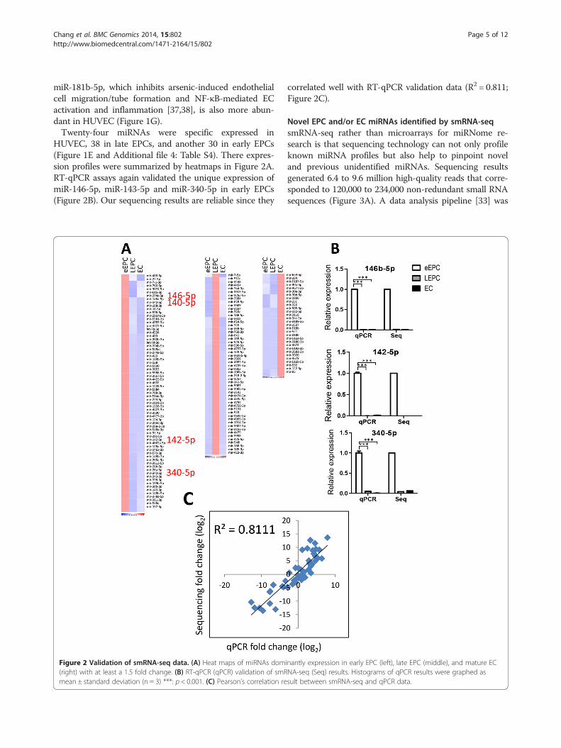
HUVEC, 38 in late EPCs, and another 30 in early EPCs(Figure 1E and Additional file 4: Table S4). There expres-sion profiles were summarized by heatmaps in Figure 2A.RT-qPCR assays again validated the unique expression ofmiR-146-5p, miR-143-5p and miR-340-5p in early EPCs(Figure 2B). Our sequencing results are reliable since they
Figure 2 Validation of smRNA-seq data. (A) Heat maps of miRNAs dom(right) with at least a 1.5 fold change. (B) RT-qPCR (qPCR) validation of smRmean ± standard deviation (n = 3) ***: p < 0.001. (C) Pearson’s correlation re
correlated well with RT-qPCR validation data (R2 = 0.811;Figure 2C).
Novel EPC and/or EC miRNAs identified by smRNA-seqsmRNA-seq rather than microarrays for miRNome re-search is that sequencing technology can not only profileknown miRNA profiles but also help to pinpoint noveland previous unidentified miRNAs. Sequencing resultsgenerated 6.4 to 9.6 million high-quality reads that corre-sponded to 120,000 to 234,000 non-redundant small RNAsequences (Figure 3A). A data analysis pipeline [33] was
inantly expression in early EPC (left), late EPC (middle), and mature ECNA-seq (Seq) results. Histograms of qPCR results were graphed assult between smRNA-seq and qPCR data.

Figure 3 smRNA-seq reveals novel miRNAs in endothelial lineage cells. (A) A flowchart describes the data analysis pipeline. The numbers ofsequences and reads remaining at each step of the data analysis are indicated. Reads numbers include sequences map to more than one locusin the genome. (B) Deduced RNA secondary structures of a set of newly discovered miRNAs. (C) A Venn diagram showing unique and commonnovel miRNAs in each cell type. (D) The top ten differentially expressed novel miRNAs between late EPC and other cell types. Areas and numbersabove the circles indicate miRNA RPM values in late EPCs. (E) Validation of smRNA-seq data by RT-qPCR. All histograms were graphed as mean ±standard deviation (n = 3) **: p < 0.01, ***: p < 0.001.
Chang et al. BMC Genomics 2014, 15:802 Page 6 of 12http://www.biomedcentral.com/1471-2164/15/802
applied to remove mRNA contamination and collect thereads not mapped to any known miRNA loci. Novel miR-NAs predicted by both of the two independent bioinfor-matics algorithms (miRDeep2 and MIREAP, Figure 3A)were recognized as candidate novel miRNAs. These dataprocessing steps yielded 14 ~ 16 unique genomic locithat potentially encoded novel miRNAs in either celltype (Figure 3A, indicated by an arrow).All these putative novel miRNAs had the potential to
fold into a hairpin secondary structure, which is one ofthe criteria necessary for miRNAs. Figure 3B illustratessome examples. Among these candidates, eleven novelmiRNAs were uniquely expressed in early EPCs, eight inlate EPCs, while another nine in matured ECs (Figure 3C).Additional file 5: Table S5 and Figure 3D illustrates theabundance, sequences and chromosomal coordinates ofthese novel miRNAs (temporally designated as miR-N), aswell as cell type distributions. The existence of four novelmiRNAs (N5-3p/-5p and N12-3p/-5p) in EPCs and ECswas verified by RT-qPCR (Figure 3E).
miR-221/222 cluster miRNAs contribute to EPC motilityand blood vessel formationConsistent with the less active nature of HUVEC(Figure 1C), two anti-angiogenic miRNAs, miR-221and miR-222, were more abundant in matured ECs(Figure 1G). It has been reported that levels of miR-221 and miR-222 are higher in EPCs from patientswith coronary artery disease (CAD) [19,20], but theanti-angiogenic role of miR-221/222 and the down-stream mechanism in late EPCs is unclear. We verifiedthat late EPCs isolated from the peripheral blood ofCAD patients expressed more miR-221/222, especiallymiR-221, than those from healthy controls (Figure 4A).More significantly, levels of circulating miR-221/222 in theplasma of CAD patients were also higher than those inhealthy controls (Figure 4B, p < 0.0001). We also tested theeffect of VEGF on the expression of miR-221/222 to exam-ine whether they are downregulated in an angiogenesis-promoting environment. VEGF repressed the levels ofmatured miR-222 and pri-miR-221/222 in diseased late

Figure 4 Decreased miR-221/222 levels in CAD patients and the contribution of miR-221/222 in late EPC functions. (A-B) RT-qPCR resultsshow miR-221/222 expression levels in late EPCs (A) and plasma (B) from health controls and CAD patients. ***: p < 0.0001 by Mann–Whitney test.(C) RT-qPCR results of miR-221 (left), miR-222 (middle) and pri-miR-221/222 (right) levels in late EPCs treated with recombinant VEGFA (n = 3). *: p< 0.05.(D) Tube formation (upper) and Transwell migration (lower) assays were performed in late EPCs transfected with indicated miRNAs for evaluation ofendothelial functions. Left histograms: RT-qPCR of miRNA expression levels; Right histograms: quantitative results of tube formation and migrationassays (n = 3). **: p< 0.01, ***: p< 0.001. (E-F) Overexpression of miR-221 or miR-222 in zebrafish embryos causes abnormal blood vascular development.Embryos in the Tg(kdrl:EGFP)s843 background were injected with 460 pg of negative control RNA (n = 100), 560 pg RNA of miR-221 (n = 106), or 560 pgRNA of miR-222 (n = 62). Blood vessel patterns were observed (E) and quantified (F) 48 hours postfertilization (hpf). DLAV: dorsal longitudinal anastomoticvessel; ISV: intersegmental vessels; DA: dorsal aorta; PCV: posterior cardinal vein.
Chang et al. BMC Genomics 2014, 15:802 Page 7 of 12http://www.biomedcentral.com/1471-2164/15/802
EPCs (Figure 4C). However, miR-221 levels were not sig-nificantly affected by VEGF (Figure 4C), indicating a post-transcriptional event may also be involved in the regulationof miR-221 levels in CAD late EPCs.Overexpressing miR-221/222 reduced the in vitro cel-
lular motility and microvasculature formation ability oflate EPCs (Figure 4D), which partly explained why these
2 miRs were more abundant in disease EPCs and in ma-ture ECs. Since EPCs are capable of forming new bloodvessels even in the absence of a pre-existing vessel net-work [6] and knockdown experiments showed that miR-221 is required for endothelial tip cell behaviors duringvascular development [39], we examined the role of miR-221/222 in the formation of the blood vessels in vivo.

Chang et al. BMC Genomics 2014, 15:802 Page 8 of 12http://www.biomedcentral.com/1471-2164/15/802
Over-expression of either miR-221 or miR-222 in zebra-fish embryos resulted in the deregulation of blood vesselpattern during development (Figure 4E, abnormal bloodvessels indicated by arrows). miR-222, but not miR-221,led to a significant reduction in blood vessel densityin vivo (Figure 4F).
miR-221/222 affect genes involved in hypoxia response,cell migration, energy supply and so onTo explore the underlying mechanisms, we alignedgenes down-regulated by miR-221 and miR-222. A totalof 845 genes were commonly repressed by both miRNAs(Figure 5A). Gene ontology analysis showed that genesinvolved in hypoxia response, cell migration, energy
Figure 5 miR-221 and miR-222 regulate PIK3R1 and ETS1, respectively, incommonly repressed by miR-221 and miR-222 (left). (right) A heat map showingprocesses among these genes according to the Gene Ontology database weremiR-221. (C) RT-qPCR results show ETS1 (left) and ETS2 (right) expression levels iMann–Whitney test. (D) Reporter activity of the ETS1 on two 3′UTR reporter conbinding site (lower) after cotransfection with the miR-221, miR-222 or empty vecshow the levels of PIK3R1 in health and CAD late EPCs (upper), or in late EPCs omiR-221 and miR-222 regulate EPC motility and tube formation functions throu
supply from glucose or fatty acids metabolism, and TGF-beta receptor signaling pathway were selectively repressedby both miRs (Figure 5A, underlined), consistent with theanti-angiogenic role of these two functional-related miR-NAs. Twelve cell migration genes (AGT, CITED2, CXCR4,F2RL1, F3, HDAC6, IRS2, IGFBP3, PTEN, PIK3R1,PTPRM, & TGFBR3) were reduced in the presence ofmiR-221/222, consistent with the reduced cell motilityphenotype in Figure 4D. Repression of CXCR4 levels byboth miR-221 and miR-222 was confirmed by RT-qPCR(Additional file 6: Figure S1).Nevertheless, no miR-221- or miR-222-binding site
could be identified on CXCR4 mRNA (not shown), wesearched for direct targets for either miRNAs. We found
late EPCs. (A) A Venn diagram illustrates there were 845 probe setsthe relative expression levels of these genes. Significant enriched biologicalalso indicated. (B) Predicted duplex formed between ETS1 and miR-222/n late EPCs from health controls (PB) and CAD patients. **: p< 0.01 bystructs containing the conserved binding site (upper) and poorly conservedtor into 293 T cells (n = 3). (E) miR-221 targets PIK3R1 in late EPCs. RT-qPCRverexpressed with miR-221. miR-222, or both (lower) (n = 3). (F) A model ofgh PIK3R1 and ETS1, respectively.

Chang et al. BMC Genomics 2014, 15:802 Page 9 of 12http://www.biomedcentral.com/1471-2164/15/802
ETS1 is among miR-222-suppressed genes in late EPCsand possesses miR-221/-222 binding sites on its 3′UTRaccording to the TargetScan bioinformatics prediction(Figure 5B). Since miR-221 and miR-222 are known totarget ETS2 and ETS1, respectively, in matured bloodvessel endothelial cells [31], we tested whether ETS1 andETS2 levels in late EPCs from CAD patients. OnlyETS1, but not ETS2, were differentially expressed be-tween diseased and healthy late EPCs (Figure 5C). Thedirect repression of ETS1 translation by miR-221 andmiR-222 was examined by 3′UTR reporter assays. Thereare two miR-221/222 binding sites on 3′UTR of ETS1(NM_005238) predicted by TargetScanHuman (http://www.targetscan.org/vert_61/), one is conserved (3403–3409 of ETS1 3′UTR); another is poorly conserved(1053–1059 of ETS1 3′UTR.) Only miR-222, but notmiR-221, inhibited ETS1 translation through both puta-tive binding sites (Figure 5D). Reporter assays on mu-tated ETS1 3′-UTR to show that the miR-222 bindingsite is functional have been done [31].We also searched target(s) for miR-221 in CAD late
EPCs. It is known that miR-221 promotes tip cell behaviorduring vascular development through the repression ofphosphoinositide-3-kinase regulatory subunit 1 (PIK3R1)[39]. PIK3R1 levels were reduced in diseased late EPCs(Figure 5E, upper panel). When miR-221 and miR-222were overexpressed in late EPCs, only miR-221, but notmiR-222, repressed PIK3R1 (Figure 5E, lower panel).
DiscussionCirculating miRNAs have attracted major interest asbiomarkers for cardiovascular diseases [40]. Besides theircell intrinsic function, recent studies reported that miR-NAs are released by cells and can be detected in theblood [41]. Here we identified the miRNome profiles ofearly EPC, late EPC and HUVEC of the same donor.Late EPC was found to possess the best angiogenic ac-tivities and less miR-221/222 expression. Furthermore,circulating miR-221 and miR-222 levels in the plasma ofCAD patients were found to be higher. Accordingly,other EPC and EC miRNAs revealed in this study mayalso hold the potential of being new biomarkers inplasma for monitoring CAD or CAD-like cases amonghigh risk population (such as the metabolic syndromepopulation) (Additional file 3: Table S3).Human EPCs research these years is quite active, partly
due to the clinical application potentials of these cells.The angiogenic abilities as well as homing of transplantedEPCs to injured sites are critical parameters for therapy tobe successful. The number of circulating EPCs in a pa-tient’s peripheral blood correlates with the prognosis ofmany diseases, such as cancer, diabetes, stroke, and CVD[42,43]. These cells are also potential sources for cell ther-apy that aims to enhance the neovascularization of tissue
engineered constructs or ischemic tissues [44]. Manystudies evaluated the possibility of transplantation of lateEPC for improving disease recovery, for example, thelong-term stroke outcome in a mouse model of transientmiddle cerebral artery occlusion (MCAO) [44,45]. Angio-genic activities as well as homing of transplanted EPCs toinjured sites are therefore critical parameters for therapyto be successful. Genomic tools, including transcriptomicand proteomic ones, have been applied to decipher thegene expression profiles of early and late EPCs for under-standing and manipulating genes in EPCs for therapeuticpurposes [45,46]. We recently pinpointed novel surfacebiomarkers and stemness genes in different EPCs, as wellas between EPCs and matured ECs [11]. With this infor-mation, it is possible to sort EPCs from circulation as wellas to identify molecular targets crucial for EPC function-ing during wound healing and tumor angiogenesis. Herewe further weighted in this field by disclosing miRNA pro-files in different EPCs and mature ECs. More in-depthwetlab work is still necessary to explore genes and miRNAresponsible for EPC or EC activities.RNA-seq, an application of the emerging ultrahigh
throughput next generation sequencing (NGS) technolo-gies, has become an attractive alternative to microarrays.Not only does it allow researchers to identify differentiallyexpressed genes, RNA-seq can further identify novel tran-scripts that have not yet been recorded in any literaturesor databases [47]. The novelty of this study is also that wepinpointed a significant number of novel miRNAs thatvary across EPCs and ECs (Figure 3 and Additional file 5:Table S5). These findings show that the full repertoire ofmiRNAs in endothelial lineage cells is larger and morecomplicated than previously appreciated. Unmasking thevarious roles of newly identified miRNAs in EPC/EC ac-tivity and endothelial differentiation will provide new in-sights for regenerative medicine. We envision our reportwill serve as a resource and road map for future studiesthat are aimed at improving our understanding of the vari-ous regulatory pathways that ultimately modulate EPC/ECactivities and angiogenesis.Clinical trials using bone marrow-derived CD34+ or
CD133+ stem cells have reported only marginal benefitsfor treating CAD patients [39,48,49]. Goretti et al. demon-strated that miR-16 family (miR-15a/-15b/-16/-103/-107)which overexpressed in early EPCs may restrict the vesselrepair function of EPCs through inhibition of cell cycleprogression associated gene CCND1, CCNE1 and CDK6[36]. Compared with their results, which were performedby microarray screening, we also identified similar miR-NAs up- or down-regulated in early EPC (up: let-7 g-5p,miR-16-5p, miR-26b-5p, miR-30b-3p, miR-140-5p, miR-146a-5p, miR-146a-3p and miR-338-3p) or in late EPC(miR-27a-3p, miR-27b-5p, miR-27b-3p, miR-151a-5p andmiR-193a-5p). Nevertheless, Goretti et al. did not include

Chang et al. BMC Genomics 2014, 15:802 Page 10 of 12http://www.biomedcentral.com/1471-2164/15/802
mature ECs in their comparison. Here we used 3 endothe-lial lineage cell types so provide another level of informa-tion. Cell type-specific miRs will be changed accordingly.Furthermore, owing to the advanced smRNA-seq tech-nique, we identified more miRNAs than the traditionalmicroarray strategy did.miR-221 and miR-222 are two highly homologous
miRNAs encoded in tandem from human chromosomeXp11.3 and are highly conserved in vertebrates. We ob-served defective vascular growth when miR-221 or miR-222, especially miR-222, was overexpressed in zebrafish.Overexpressing miR-221 in zebrafish caused deregulatedblood vessel pattern but not the reduction of vesseldensity, which may partly due to the fact that miR-221 isrequired for vascular development by promoting tip cellmigration and proliferation in zebrafish during vasculardevelopment [39]. miR-221 and miR-222 also inhibiterythropoiesis and erythroleukemic cell growth viadown-modulating cKit [50]. miR-221/222 inhibit EC[31] and EPC motility and microtubule formation. Con-sistent with these observations, in late EPCs from CADpatients the expression levels of miR-221 and miR-222are increased (Figure 4A) [20]. Reduced levels of circu-lating EPCs independently predict atherosclerotic dis-ease progression and development of cardiovascularevents [7]. Clinical studies demonstrated that levels ofcirculating EPCs are associated with vascular endothe-lial function and cardiovascular risk factors, and help toidentify patients at increased cardiovascular risk [8].Circulating EPCs in peripheral blood are mobilizedfrom bone marrow by chemokine stimulation such aschemokine (C-X-C motif ) ligand 12 (CXCL12; alsoknown as stromal derived factor-1, SDF-1) [51]. Wefound both miR-221 and miR-222 repressed in lateEPCs the levels of chemokine (C-X-C motif ) receptor 4(CXCR4), the receptor for CXCL12 (Figure 4), partlyexplains why miR-221/222 inhibit late EPC motility(Figure 4C), and EPC levels were reduced in the periph-eral blood of CAD patients. Since neither miR-221 normiR-222 targets CXCR4 directly according to bioinfor-matics prediction, how CXCR4 levels are regulated isstill an open question. We demonstrated another candi-date miR-221 target gene – PIK3R1, which is also import-ant in regulating endothelial tip cell migration andproliferation in EPC [39]. PIK3R1 is the p85α regulatorysubunit of phosphoinositide-3-kinase (PI3K). Previousstudy indicated miR-221 may regulate the delicate stoichi-ometry of PIK3R1 subunit to maintain the correct subcel-lular localization and regulate downstream flt4 expression[39]. ETS1 is a known target of miR-222 in mature endo-thelial cells [31] and contributes to embryonic angiogen-esis regulation and may play a role in the modulation andmaturation of EPC. miR-221 and miR-222 each hasunique targets and different mechanisms to repress the
activities of diseased late EPCs (Figure 5F). Other bio-marker candidates among our mined miRNAs with sup-porting references are indicated in Additional file 3:Tables S3.
ConclusionsIn summary, our results demonstrate miR-221 and miR-222 repress the levels of PIK3R1 and ETS1, respectively toregulate angiogenic features in EPCs and ECs (Figure 5F)and suggest mechanisms of why late EPC levels and activ-ities are reduced in CAD patients. Overall, our data provideevidences that miRNA research will not only help us tounderstand more about EPC biology, but will also lead tothe development of novel biomarkers and therapeutic mo-dalities for the prevention and treatment of CAD and otherischemia-related diseases such as stroke and diabetes.
Additional files
Additional file 1: Table S1. Oligo sequences.
Additional file 2: Table S2. Baseline characteristic of 55 studiedsubjects in healthy and CAD patients.
Additional file 3: Table S3. miRNA expression profile in Figures 1Fand 2A.
Additional file 4: Table S4. Stage specific miRNA expression profile.
Additional file 5: Table S5. Predicted novel miRNAs.
Additional file 6: Figure S1. RT-qPCR validation showing therepression of CXCR4 levels in late EPCs overexpressing with miR-221 ormiR-222 (n=3).
Competing interestsThe authors declare that they have no competing interests.
Authors’ contributionsTC, HW and C-C C conceived the whole study design; TC and TH performed thebioinformatics data analysis; HL, YC, YW and CH performed wetlab experiments;S-J C, CT and C-H C provided clinical samples; RY, CW, TT and S-M C providedclinical consultations. The manuscript was written by TC, TH and HW. All authorsread and approved the final manuscript.
AcknowledgementsThis work is supported by National Science Council (NSC; NSC101-2320-B-010-059-MY3, NSC101-2627-B-010-003, NSC102-2622-B-010-002, and NSC101-2321-B-010-011), Tri-Service General Hospital (TSGH-C102-027), Veterans General HospitalsUniversity System of Taiwan (VGHUST) Joint Research Program, Tsou’s Foundation(VGHUST102-G7-3-2), National Health Research Institutes (NHRI-EX102-10254SI),UST-UCSD International Center for Excellence in Advanced Bioengineeringsponsored by the Taiwan NSC I-RiCE Program (NSC101-2911-I-009-101), and inpart a grant from National Yang-Ming University (Ministry of Education, Aim forthe Top University Plan).
Author details1Institute of Microbiology and Immunology, National Yang-Ming University,Taipei, Taiwan. 2Institute of Engineering in Medicine, UC San Diego, La Jolla,USA. 3Genome Research Center, National Yang-Ming University, Taipei,Taiwan. 4Department of Education and Research, Taipei City Hospital, Taipei,Taiwan. 5Department of Obstetrics and Gynecology, Hsin-Chu MackayMemorial Hospital, Hsinchu, Taiwan. 6Department of Bioscience Technology,Chung Yuan Christian University, Chung-Li, Taiwan. 7Department ofLaboratory Medicine and Biotechnology, College of Medicine, Tzu-ChiUniversity, Hualien, Taiwan. 8Division of Cardiology, Department of InternalMedicine, Tri-Service General Hospital, National Defense Medical Center,Taipei, Taiwan.

Chang et al. BMC Genomics 2014, 15:802 Page 11 of 12http://www.biomedcentral.com/1471-2164/15/802
Received: 8 April 2014 Accepted: 26 August 2014Published: 18 September 2014
References1. Werner N, Kosiol S, Schiegl T, Ahlers P, Walenta K, Link A, Bohm M, Nickenig
G: Circulating endothelial progenitor cells and cardiovascular outcomes.N Engl J Med 2005, 353(10):999–1007.
2. Grisar JC, Haddad F, Gomari FA, Wu JC: Endothelial progenitor cells incardiovascular disease and chronic inflammation: from biomarker totherapeutic agent. Biomark Med 2011, 5(6):731–744.
3. Asahara T, Murohara T, Sullivan A, Silver M, van der Zee R, Li T,Witzenbichler B, Schatteman G, Isner JM: Isolation of putative progenitorendothelial cells for angiogenesis. Science 1997, 275(5302):964–967.
4. Reyes M, Dudek A, Jahagirdar B, Koodie L, Marker PH, Verfaillie CM: Originof endothelial progenitors in human postnatal bone marrow. J Clin Invest2002, 109(3):337–346.
5. Real C, Caiado F, Dias S: Endothelial progenitors in vascular repair andangiogenesis: how many are needed and what to do? CardiovascHematol Disord Drug Targets 2008, 8(3):185–193.
6. Hristov M, Erl W, Weber PC: Endothelial progenitor cells: isolation andcharacterization. Trends Cardiovasc Med 2003, 13(5):201–206.
7. Briasoulis A, Tousoulis D, Antoniades C, Stefanadis C, Papageorgiou N: The roleof endothelial progenitor cells in vascular repair after arterial injury andatherosclerotic plaque development. Cardiovasc Ther 2010, 29(2):125–139.
8. Jialal I, Devaraj S, Singh U, Huet BA: Decreased number and impairedfunctionality of endothelial progenitor cells in subjects with metabolicsyndrome: implications for increased cardiovascular risk. Atherosclerosis2010, 211(1):297–302.
9. Critser PJ, Voytik-Harbin SL, Yoder MC: Isolating and defining cells toengineer human blood vessels. Cell Prolif 2011, 44(Suppl 1):15–21.
10. Yoder MC: Endothelial progenitor cell: a blood cell by many other namesmay serve similar functions. J Mol Med (Berl) 2013, 91(3):285–295.
11. Cheng CC, Chang SJ, Chueh YN, Huang TS, Huang PH, Cheng SM, Tsai TN,Chen JW, Wang HW: Distinct angiogenesis roles and surface markers ofearly and late endothelial progenitor cells revealed by functional groupanalyses. BMC Genomics 2013, 14(1):182.
12. Croce CM, Calin GA: miRNAs, cancer, and stem cell division. Cell 2005,122(1):6–7.
13. Lim LP, Lau NC, Garrett-Engele P, Grimson A, Schelter JM, Castle J, Bartel DP,Linsley PS, Johnson JM: Microarray analysis shows that some microRNAsdownregulate large numbers of target mRNAs. Nature 2005,433(7027):769–773.
14. Wang QZ, Xu W, Habib N, Xu R: Potential uses of microRNA in lungcancer diagnosis, prognosis, and therapy. Curr Cancer Drug Targets 2009,9(4):572–594.
15. Brase JC, Wuttig D, Kuner R, Sultmann H: Serum microRNAs asnon-invasive biomarkers for cancer. Mol Cancer 2010, 9:306.
16. Iguchi H, Kosaka N, Ochiya T: Versatile applications of microRNA inanti-cancer drug discovery: from therapeutics to biomarkers. Curr DrugDiscov Technol 2010, 7(2):95–105.
17. Poliseno L, Tuccoli A, Mariani L, Evangelista M, Citti L, Woods K, MercatantiA, Hammond S, Rainaldi G: MicroRNAs modulate the angiogenicproperties of HUVECs. Blood 2006, 108(9):3068–3071.
18. Kuehbacher A, Urbich C, Dimmeler S: Targeting microRNA expression toregulate angiogenesis. Trends Pharmacol Sci 2008, 29(1):12–15.
19. Minami Y, Satoh M, Maesawa C, Takahashi Y, Tabuchi T, Itoh T, Nakamura M:Effect of atorvastatin on microRNA 221 / 222 expression in endothelialprogenitor cells obtained from patients with coronary artery disease.Eur J Clin Invest 2009, 39(5):359–367.
20. Zhang Q, Kandic I, Kutryk MJ: Dysregulation of angiogenesis-relatedmicroRNAs in endothelial progenitor cells from patients with coronaryartery disease. Biochem Biophys Res Commun 2011, 405(1):42–46.
21. Cheng CC, Lo HH, Huang TS, Cheng YC, Chang ST, Chang SJ, Wang HW:Genetic module and miRNome trait analyses reflect the distinctbiological features of endothelial progenitor cells from differentanatomic locations. BMC Genomics 2012, 13(1):447.
22. Wang HW, Huang TS, Lo HH, Huang PH, Lin CC, Chang SJ, Liao KH, Tsai CH,Chan CH, Tsai CF, Cheng YC, Chiu YL, Tsai TN, Cheng CC, Cheng SM:Deficiency of the microRNA-31-microRNA-720 pathway in the plasmaand endothelial progenitor cells from patients with coronary arterydisease. Arterioscler Thromb Vasc Biol 2014, 34(4):857–869.
23. Plummer PN, Freeman R, Taft RJ, Vider J, Sax M, Umer BA, Gao D, Johns C,Mattick JS, Wilton SD, Ferro V, McMillan NA, Swarbrick A, Mittal V, Mellick AS:MicroRNAs regulate tumor angiogenesis modulated by endothelialprogenitor cells. Cancer Res 2013, 73(1):341–352.
24. Goff LA, Davila J, Swerdel MR, Moore JC, Cohen RI, Wu H, Sun YE, Hart RP:Ago2 immunoprecipitation identifies predicted microRNAs in humanembryonic stem cells and neural precursors. PLoS One 2009, 4(9):e7192.
25. Bar M, Wyman SK, Fritz BR, Qi J, Garg KS, Parkin RK, Kroh EM, Bendoraite A,Mitchell PS, Nelson AM, Ruzzo WL, Ware C, Radich JP, Gentleman R,Ruohola-Baker H, Tewari M: MicroRNA discovery and profiling in humanembryonic stem cells by deep sequencing of small RNA libraries.Stem Cells 2008, 26(10):2496–2505.
26. Morin RD, O’Connor MD, Griffith M, Kuchenbauer F, Delaney A, Prabhu AL,Zhao Y, McDonald H, Zeng T, Hirst M, Eaves CJ, Marra MA: Application ofmassively parallel sequencing to microRNA profiling and discovery inhuman embryonic stem cells. Genome Res 2008, 18(4):610–621.
27. Skreka K, Schafferer S, Nat IR, Zywicki M, Salti A, Apostolova G, Griehl M,Rederstorff M, Dechant G, Huttenhofer A: Identification of differentiallyexpressed non-coding RNAs in embryonic stem cell neural differentiation.Nucleic Acids Res 2012, 40(13):6001–6015.
28. Bissels U, Wild S, Tomiuk S, Hafner M, Scheel H, Mihailovic A, Choi YH,Tuschl T, Bosio A: Combined characterization of microRNA and mRNAprofiles delineates early differentiation pathways of CD133+ and CD34+hematopoietic stem and progenitor cells. Stem Cells 2011,29(5):847–857.
29. Yoo JK, Kim J, Choi SJ, Noh HM, Kwon YD, Yoo H, Yi HS, Chung HM, Kim JK:Discovery and characterization of novel microRNAs during endothelialdifferentiation of human embryonic stem cells. Stem Cells Dev 2012,21(11):2049–2057.
30. Chen YH, Lin SJ, Lin FY, Wu TC, Tsao CR, Huang PH, Liu PL, Chen YL, ChenJW: High glucose impairs early and late endothelial progenitor cells bymodifying nitric oxide-related but not oxidative stress-mediated mechanisms.Diabetes 2007, 56(6):1559–1568.
31. Wu YH, Hu TF, Chen YC, Tsai YN, Tsai YH, Cheng CC, Wang HW: Themanipulation of miRNA-gene regulatory networks by KSHV inducesendothelial cell motility. Blood 2011, 118(10):2896–2905.
32. Chen C, Ridzon DA, Broomer AJ, Zhou Z, Lee DH, Nguyen JT, Barbisin M,Xu NL, Mahuvakar VR, Andersen MR, Lao KQ, Livak KJ, Guegler KJ: Real-timequantification of microRNAs by stem-loop RT-PCR. Nucleic Acids Res 2005,33(20):e179.
33. Cheng WC, Chung IF, Huang TS, Chang ST, Sun HJ, Tsai CF, Liang ML, WongTT, Wang HW: YM500: a small RNA sequencing (smRNA-seq) database formicroRNA research. Nucleic Acids Res 2013, 41(D1):D285–D294.
34. Mortazavi A, Williams BA, McCue K, Schaeffer L, Wold B: Mapping andquantifying mammalian transcriptomes by RNA-Seq. Nat Methods 2008,5(7):621–628.
35. Hsieh JY, Huang TS, Cheng SM, Lin WS, Tsai TN, Lee OK, Wang HW:miR-146a-5p circuitry uncouples cell proliferation and migration, but notdifferentiation, in human mesenchymal stem cells. Nucleic Acids Res 2013,41(21):9753–9763.
36. Goretti E, Rolland-Turner M, Leonard F, Zhang L, Wagner DR, Devaux Y:MicroRNA-16 affects key functions of human endothelial progenitorcells. J Leukoc Biol 2013, 93(5):645–655.
37. Cui Y, Han Z, Hu Y, Song G, Hao C, Xia H, Ma X: MicroRNA-181b andmicroRNA-9 mediate arsenic-induced angiogenesis via NRP1.J Cell Physiol 2012, 227(2):772–783.
38. Sun X, He S, Wara AK, Icli B, Shvartz E, Tesmenitsky Y, Belkin N, Li D,Blackwell TS, Sukhova GK, Croce K, Feinberg MW: Systemic delivery ofmicroRNA-181b inhibits nuclear factor-kappaB activation, vascularinflammation, and atherosclerosis in apolipoprotein E-deficient mice.Circ Res 2014, 114(1):32–40.
39. Nicoli S, Knyphausen CP, Zhu LJ, Lakshmanan A, Lawson ND: miR-221 isrequired for endothelial tip cell behaviors during vascular development.Dev Cell 2012, 22(2):418–429.
40. Fichtlscherer S, De Rosa S, Fox H, Schwietz T, Fischer A, Liebetrau C, WeberM, Hamm CW, Roxe T, Muller-Ardogan M, Bonauer A, Zeiher AM, DimmelerS: Circulating microRNAs in patients with coronary artery disease.Circ Res 2010, 107(5):677–684.
41. Diehl P, Fricke A, Sander L, Stamm J, Bassler N, Htun N, Ziemann M, HelbingT, El-Osta A, Jowett JB, Peter K: Microparticles: major transport vehicles fordistinct microRNAs in circulation. Cardiovasc Res 2012, 93(4):633–644.

Chang et al. BMC Genomics 2014, 15:802 Page 12 of 12http://www.biomedcentral.com/1471-2164/15/802
42. Vasa M, Fichtlscherer S, Aicher A, Adler K, Urbich C, Martin H, Zeiher AM,Dimmeler S: Number and migratory activity of circulating endothelialprogenitor cells inversely correlate with risk factors for coronary arterydisease. Circ Res 2001, 89(1):E1–E7.
43. Briguori C, Testa U, Riccioni R, Colombo A, Petrucci E, Condorelli G, MarianiG, D’Andrea D, De Micco F, Rivera NV, Puca AA, Peschle C: Correlationsbetween progression of coronary artery disease and circulatingendothelial progenitor cells. Faseb J 2010, 24(6):1981–1988.
44. Fuchs S, Dohle E, Kolbe M, Kirkpatrick CJ: Outgrowth endothelial cells:sources, characteristics and potential applications in tissue engineeringand regenerative medicine. Adv Biochem Eng Biotechnol 2010,123:201–217.
45. Yang WH, Lan HY, Huang CH, Tai SK, Tzeng CH, Kao SY, Wu KJ, Hung MC,Yang MH: RAC1 activation mediates Twist1-induced cancer cell migration.Nat Cell Biol 2012, 14(4):366–374.
46. Maeng YS, Choi HJ, Kwon JY, Park YW, Choi KS, Min JK, Kim YH, Suh PG,Kang KS, Won MH, Kim YM, Kwon YG: Endothelial progenitor cell homing:prominent role of the IGF2-IGF2R-PLCbeta2 axis. Blood 2009,113(1):233–243.
47. Wang Z, Gerstein M, Snyder M: RNA-Seq: a revolutionary tool fortranscriptomics. Nat Rev Genet 2009, 10(1):57–63.
48. Lipinski MJ, Biondi-Zoccai GG, Abbate A, Khianey R, Sheiban I, Bartunek J,Vanderheyden M, Kim HS, Kang HJ, Strauer BE, Vetrovec GW: Impact ofintracoronary cell therapy on left ventricular function in the setting ofacute myocardial infarction: a collaborative systematic review andmeta-analysis of controlled clinical trials. J Am Coll Cardiol 2007,50(18):1761–1767.
49. Losordo DW, Schatz RA, White CJ, Udelson JE, Veereshwarayya V, Durgin M,Poh KK, Weinstein R, Kearney M, Chaudhry M, Burg A, Eaton L, Heyd L,Thorne T, Shturman L, Hoffmeister P, Story K, Zak V, Dowling D, Traverse JH,Olson RE, Flanagan J, Sodano D, Murayama T, Kawamoto A, Kusano KF,Wollins J, Welt F, Shah P, Soukas P: Intramyocardial transplantation ofautologous CD34+ stem cells for intractable angina: a phase I/IIadouble-blind, randomized controlled trial. Circulation 2007,115(25):3165–3172.
50. Felli N, Fontana L, Pelosi E, Botta R, Bonci D, Facchiano F, Liuzzi F, Lulli V,Morsilli O, Santoro S, Valtieri M, Calin GA, Liu CG, Sorrentino A, Croce CM,Peschle C: MicroRNAs 221 and 222 inhibit normal erythropoiesis anderythroleukemic cell growth via kit receptor down-modulation.Proc Natl Acad Sci U S A 2005, 102(50):18081–18086.
51. Yamaguchi J, Kusano KF, Masuo O, Kawamoto A, Silver M, Murasawa S,Bosch-Marce M, Masuda H, Losordo DW, Isner JM, Asahara T: Stromalcell-derived factor-1 effects on ex vivo expanded endothelial progenitorcell recruitment for ischemic neovascularization. Circulation 2003,107(9):1322–1328.
doi:10.1186/1471-2164-15-802Cite this article as: Chang et al.: miRNome traits analysis on endotheliallineage cells discloses biomarker potential circulating microRNAs whichaffect progenitor activities. BMC Genomics 2014 15:802.
Submit your next manuscript to BioMed Centraland take full advantage of:
• Convenient online submission
• Thorough peer review
• No space constraints or color figure charges
• Immediate publication on acceptance
• Inclusion in PubMed, CAS, Scopus and Google Scholar
• Research which is freely available for redistribution
Submit your manuscript at www.biomedcentral.com/submit