Vascular Endothelial Growth Factor Gene Polymorphisms and Risk of Primary Lung Cancer
Upload
khangminh22Category
view
4download
0
Citation: Requena-Ocaña, N.;
Flores-Lopez, M.; Papaseit, E.;
García-Marchena, N.; Ruiz, J.J.;
Ortega-Pinazo, J.; Serrano, A.;
Pavón-Morón, F.J.; Farré, M.; Suarez,
J.; et al. Vascular Endothelial Growth
Factor as a Potential Biomarker of
Neuroinflammation and Frontal
Cognitive Impairment in Patients with
Alcohol Use Disorder. Biomedicines
2022, 10, 947. https://doi.org/
10.3390/biomedicines10050947
Academic Editor: Masaru Tanaka
Received: 24 February 2022
Accepted: 16 April 2022
Published: 20 April 2022
Publisher’s Note: MDPI stays neutral
with regard to jurisdictional claims in
published maps and institutional affil-
iations.
Copyright: © 2022 by the authors.
Licensee MDPI, Basel, Switzerland.
This article is an open access article
distributed under the terms and
conditions of the Creative Commons
Attribution (CC BY) license (https://
creativecommons.org/licenses/by/
4.0/).
biomedicines
Article
Vascular Endothelial Growth Factor as a Potential Biomarker ofNeuroinflammation and Frontal Cognitive Impairment inPatients with Alcohol Use DisorderNerea Requena-Ocaña 1,2, María Flores-Lopez 1 , Esther Papaseit 3,4, Nuria García-Marchena 1,5, Juan Jesús Ruiz 6,Jesús Ortega-Pinazo 1, Antonia Serrano 1, Francisco Javier Pavón-Morón 1,7,8 , Magí Farré 3,4 , Juan Suarez 1,9,* ,Fernando Rodríguez de Fonseca 1,* and Pedro Araos 1,10,*
1 Laboratorio de Medicina Regenerativa (LMR), Unidad de Gestión Clínica de Salud Mental,Instituto de Investigación Biomédica de Málaga (IBIMA), Hospital Regional Universitario de Málaga,Avda. Carlos Haya 82, Sótano, 29010 Malaga, Spain; [email protected] (N.R.-O.);[email protected] (M.F.-L.); [email protected] (N.G.-M.); [email protected] (J.O.-P.);[email protected] (A.S.); [email protected] (F.J.P.-M.)
2 Departamento de Psicobiología, Facultad de Psicología, Universidad Complutense de Madrid,Campus de Somosaguas, 28040 Madrid, Spain
3 Department of Clinical Pharmacology, Hospital Universitari Trias I Pujol and Institut de Recerca GermansTrias I Pujol (HUGTiP-IGTP), 08916 Badalona, Spain; [email protected] (E.P.);[email protected] (M.F.)
4 Department of Pharmacology, Therapeutics and Toxicology, Universitat Autònoma de Barcelona,08913 Cerdanyola del Vallès, Spain
5 Institut D, Investigació en Ciències de la Salut Germans Trias i Pujol (IGTP), Unidad de Adicciones-Serviciode Medicina Interna, Campus Can Ruti, Carrer del Canyet s/n, 08916 Badalona, Spain
6 Centro Provincial de Drogodependencias (CPD) de Málaga, Diputación de Málaga, C/Ana Solo de Zaldívar, n◦ 3,29010 Malaga, Spain; [email protected]
7 Instituto de Investigación Biomédica de Málaga (IBIMA), Unidad de Gestión Clínica del Corazón,Hospital Universitario Virgen de la Victoria de Málaga, Planta 5a-Sección Central, 29010 Malaga, Spain
8 Centro de Investigación Biomédica en Red Enfermedades Cardiovasculares (CIBERCV),Instituto de Salud Carlos III, Calle de Melchor Fernández Almagro, 3, 28029 Madrid, Spain
9 Department of Anatomy, Legal Medicine and History of Science, School of Medicine, University of Malaga,Boulevard Louis Pasteur 32, 29071 Malaga, Spain
10 Departamento de Psicobiología y Metodología de las CC del Comportamiento, Facultad de Psicología,Universidad de Málaga, 29016 Malaga, Spain
* Correspondence: [email protected] (J.S.); [email protected] (F.R.d.F.); [email protected] (P.A.)
Abstract: (1) Background: Alcohol Use Disorder (AUD) is associated with functional disruptionof several brain structures that may trigger cognitive dysfunction. One of the mechanisms ofalcohol-associated cognitive impairment has been proposed to arise from its direct impact on theimmune system, which culminates in the release of cytokines and chemokines which can eventu-ally reach the brain. Alcohol can also disrupt the blood–brain barrier, facilitating the penetrationof pro-inflammatory molecules throughout vascular endothelial growth factor A (VEGFA). Thus,alcohol-induced alterations in chemokines and VEGFA might contribute to the neuroinflammationand cognitive impairment associated with AUD. (2) Methods: The present cross-sectional studyinvestigates whether patients with AUD (n = 86) present cognitive disability associated to alterationsin plasma concentration of SDF-1, fractalkine, eotaxin, MCP-1, MIP-1α and VEGFA when comparedto control subjects (n = 51). (3) Results: The analysis indicated that SDF-1 and MCP-1 concentrationswere higher in AUD patients than in controls. Concentrations of VEGFA were higher in AUD pa-tients with severe frontal deficits, and the score of frontal lobe functions was negatively correlatedwith VEGFA and fractalkine. Acute alcohol effects on VEGFA plasma levels in healthy volunteersdemonstrated the induction of VEGFA release by heavy alcohol drinking. VEGFA was positivelycorrelated with pro-inflammatory chemokines in AUD patients with frontal cognitive impairment.(4) Conclusions: we propose VEGFA/chemokine monitoring as biomarkers of potential cognitiveimpairment in AUD patients.
Biomedicines 2022, 10, 947. https://doi.org/10.3390/biomedicines10050947 https://www.mdpi.com/journal/biomedicines

Biomedicines 2022, 10, 947 2 of 19
Keywords: alcohol use disorders; addiction; VEGFA; blood–brain barrier; chemokines; fractalkine;neuroinflammation; cognitive dysfunction; neurodegeneration; dementia
1. Introduction
Alcohol use disorder (AUD) is one of the main global health problems, carryinga significant social and economic burden. Alcohol abuse is responsible for more than3 million deaths annually in the world, with the highest rates of alcohol consumption beingin the European Union [1]. In Spain, alcohol is the most consumed drug among the generalpopulation (15–64 years): 91.2% at some time in their life, 75.2% in the last year, 62.7% inthe last 30 days and 7.4% daily during the last month [2].
Among medical consequences related to AUD, we can highlight the induction of liverand pancreatic disease [3,4], psychiatric comorbidities, and other substance use disordersthroughout life [5]. Major depressive disorders and anxiety disorders are the most prevalentcomorbid psychiatric disorders, whereas cocaine and cannabis misuse are the most frequentcomorbid substance use disorders associated with AUD [6,7]. Furthermore, a growing num-ber of studies indicate that alcohol abuse is a major contributor to the development of anytype of dementia, especially when there is an early onset of the cognitive impairment [8,9].In addition, lifetime presence of chronic alcohol dependence has been suggested as an inde-pendent risk factor for the development of dementia. Thus, alcohol-related dementia (ARD)has been reported to be one the most prevalent, especially in young men (from 8.27 per100,000 to 25.6% in several study populations) [10]. Moreover, heavy alcohol consumptionhas been related with a rapid progression of cognitive decline in aging [11]. According tothese data, it is widely known that chronic alcohol consumption has a profound impact onbrain structures that support higher cognitive functions [12–14].
Regarding molecular mechanisms mediating alcohol abuse-associated cognitive im-pairment, it is thought that it is derived from alcohol-induced neuroinflammation medi-ated by oxidative stress and by the release of proinflammatory signaling molecules (i.e.,chemokines/cytokines), ultimately leading to neuronal apoptosis and even necrosis. Thisprocess may eventually lead to a permanent derangement of cognition, resulting in de-mentia. Increasing evidence supports an essential role for Toll-like receptors (TLRs) inalcohol-induced neurodegenerative disease [15]. Recent studies have observed that alcoholstimulates brain immune cells (microglia and astrocytes) by activating TLR (mainly TLR4)and NOD-like receptors. This activation culminates in the production of proinflammatorycytokines and chemokines, leading to neuroinflammation and neuronal damage in thecortex and the hippocampus. Hence, activation of TLR4 by ethanol triggers fast down-stream signaling pathways such as mitogen-activated protein kinases (MAPKs) and nuclearfactor-kappa B (NF-kB), eventually promoting the expression and release of cytokines,chemokines, and inflammatory mediators [16,17]. These events can be potentiated byalcohol-induced neuroinflammation inflammatory signals at peripheral tissues (i.e., gut,liver or pancreas) reaching the brain through ethanol’s disruption of the blood–brain bar-rier [18]. Thus, substantial evidence suggests that alcohol impacts the immune systemand induces an up-regulation of cytokines and chemokines which are associated withbehavioral changes and cognitive impairment [19,20].
Chemokines (chemotactic cytokines) are immune signals involved in cellular migra-tion and intercellular communication. These proteins also act as modulators in neuronaltransmission and contribute to communication between glia and neuronal cells [21,22].In addition, cytokines are involved in the regulation of cell development, survival, andregeneration of the central nervous system [23,24]. These signals are important componentsof the neuroimmune system that contribute to neuronal activity, neuroendocrine function,brain development, synaptic plasticity, and circuity of mood in drug addiction [25,26]. Fur-thermore, chemokine decompensation described in plasma, serum and cerebrospinal fluidhas been associated with several psychiatric and neurodegenerative diseases such as mild

Biomedicines 2022, 10, 947 3 of 19
cognitive impairment, Alzheimer’s disease, Parkinson’s disease, schizophrenia, bipolardisorder and major depression [27–30]. However, despite the interaction between alcoholand immunological mediators having been well investigated [31,32], little is known aboutwhether these immunoinflammatory signals impact the development of AUD-associatedcognitive impairment. Long-lasting brain induction of the proinflammatory cytokinestumor necrosis factor alpha (TNFα), interleukin (IL)-1β and monocyte chemoattractantprotein-1 (MCP-1) and the anti-inflammatory cytokine IL-10 have been related to microglialactivation and reduced neurogenesis in mice exposed to LPS endotoxin after ethanol treat-ment [33]. However, plasma chemokines have been evaluated almost exclusively in thecontext of liver disease in alcohol-dependent patients [34,35].
On the other hand, Vascular Endothelial Growth Factor A (VEGFA) could be a poten-tial candidate for explaining how alcohol facilitates both infiltration and inflammation inthe brain. VEGFA is a protein that belongs to the family of growth factors and is commonlyknown for its role in angiogenesis and vascular permeability. In addition, VEGFA plays afundamental role in the development and adult nervous system since it is involved in theextension and complexity of the microvasculature that supplies the necessary nutrients andoxygen in the brain. In this way, the effects of VEGFA on the nervous system have beenrelated to neuroprotection, neurogenesis, and synaptic plasticity through the stimulationof neural stem cells and safeguarding the integrity of the blood–brain barrier [36]. There-fore, changes in VEGFA concentrations could affect the function and survival of neuronsby not providing enough nutrients or producing hypoxia, which has been related to thedeterioration of cognitive function [37]. Furthermore, VEGFA is a potent vasodilator andangiogenic factor released under hypoxic and stressful conditions via endothelial nitricoxide synthase [38,39]. Altered levels of VEGFA have been related to several neurodegen-erative and neurological disorders, such as Alzheimer’s disease, vascular dementia andstroke [36]. Nevertheless, VEGFA is still poorly understood in the field of substance usedisorders. Heberlein (2010) observed that VEGFA serum levels increase during alcoholwithdrawal, and it might be intimately associated with alcohol intoxication and the severityof the addiction reflected by recurrent episodes of alcohol intoxication [40]. In addition,augmented VEGFA levels have been found in alcoholic liver disease patients compared tocontrols, showing a positive association with cholestatic enzymes [41].
Considering the previous antecedents, in the present study, we investigated the poten-tial association of plasma concentrations of the chemokines stromal cell-derived factor 1(SDF-1), fractalkine, eotaxin, MCP-1, macrophage inflammatory protein 1 alpha (MIP-1α)and the trophic factor VEGFA to frontal cognitive impairment in AUD patients. In addition,to fully understand the effects of alcohol on VEGFA, we studied the plasma concentrationof this trophic factor after acute alcohol intake. The ultimate goal was to identify a potentiallink between AUD-associated cognitive impairment and plasma levels of VEGFA andchemokines that might be eventually useful for clinical purposes.
2. Materials and Methods2.1. Recruitment and Screening of Participants
The cross-sectional study included 137 Caucasian volunteers divided into two groups:86 abstinent AUD patients (alcohol group) in outpatient treatment and 51 control subjects(control group) matched by age, body mass index (BMI) and proportion of sex. Patientswere recruited at the Psychiatry Service of the Hospital Universitario 12 de Octubre (Madrid,Spain) and Centro Provincial de Drogodependencias (Málaga, Spain). Control participantswere included from databases of healthy subjects (without presence of cognitive impair-ment, medical diseases and substance use disorders) of the Biobanco Nacional de DNA.In addition, we performed a brief frontal neuropsychological evaluation in 59% of AUDpatients (Figure 1).

Biomedicines 2022, 10, 947 4 of 19Biomedicines 2022, 10, x FOR PEER REVIEW 4 of 20
Figure 1. Diagram showing the design of the cross-sectional study.
AUD Patients and Control Volunteers
To be included in the present study, participants had to meet the following inclusion
criteria: people aged 18 to 65 years in the abstinence phase, being in outpatient treatment
and willingness to participate by signing the informed consent. As we wanted to control
for potential interferences in plasma concentrations of chemokines and VEGFA, the ex-
clusion criteria included: use of anti-inflammatory drugs or MAOI’s, personal history of
long-term inflammatory disease or cancer, pregnant or breast-feeding women and infec-
tious diseases such as Hepatitis C, Hepatitis B and HIV.
An additional group of healthy subjects was recruited at Hospital German Trias I
Pujol from Badalona, Spain, to investigate the acute actions of alcohol on VEGFA plasma
concentrations. The study design was simple blind, non-randomized, non-controlled
study of the experimental administration of alcohol simulating a “binge drinking” epi-
sode. Ten healthy male subjects were recruited and administrated an alcoholic beverage
containing 100 g of alcohol (312 mL vodka Absolut® , Ahus, Sweden) were mixed with 588
mL of orange soda [Trina® Orange No gas. Suntury Limited, Dōjimahama, Japan], total
volume 900 mL. The alcoholic beverage was distributed in 6 identical glasses (volume 150
mL) and consumed continuously over a 2 h period (15 min per glass). Participants were
selected after a general medical examination to exclude any psychopathological condition.
Subjects signed an informed consent prior to participation and were economically com-
pensated for any inconvenience caused during the trial. The participants had a mean age
of 22 ± 2 years, mean weight 73.0 ± 9.2 kg, mean height 180 ± 6.5 cm and index body mass
(IBM) 22.5 ± 1.9 kg/m2. They drank an average of 13.7 ± 8.3 g of alcohol per day and re-
ported a mean 1.3 ± 1.7 alcohol binge episodes per month.
2.2. Ethical Statement
Written informed consent was obtained from each participant after a complete de-
scription of the study. All participants had the opportunity to discuss any questions or
problems. For the cross-sectional study, the design and the recruitment protocols were
approved by the Ethics Committee of the Hospital Regional Universitario de Málaga
(PND 2019/040). The acute alcohol administration experiment protocol was approved by
the local Human Research Ethics Committee (CEI Hospital Universitari Germans Trias i
Pujol, Badalona, Spain) and registered at ClinicalTrials.gov (NCT02232789). All proce-
dures were in strict accordance with the Ethical Principles for Medical Research with Hu-
man Subjects adopted in the Declaration of Helsinki by the World Medical Association
(64th General Assembly of the WMA, Fortaleza, Brazil, October 2013) and Recommenda-
tion No. R (97) 5 of the Committee of Ministers to the Member States on the protection of
medical data (1997), and the Spanish law on data protection [Regulation (EU) 2016/679 of
the European Parliament and of the Council of 27 April 2016 on the protection of natural
Figure 1. Diagram showing the design of the cross-sectional study.
AUD Patients and Control Volunteers
To be included in the present study, participants had to meet the following inclusioncriteria: people aged 18 to 65 years in the abstinence phase, being in outpatient treatmentand willingness to participate by signing the informed consent. As we wanted to control forpotential interferences in plasma concentrations of chemokines and VEGFA, the exclusioncriteria included: use of anti-inflammatory drugs or MAOI’s, personal history of long-terminflammatory disease or cancer, pregnant or breast-feeding women and infectious diseasessuch as Hepatitis C, Hepatitis B and HIV.
An additional group of healthy subjects was recruited at Hospital German Trias IPujol from Badalona, Spain, to investigate the acute actions of alcohol on VEGFA plasmaconcentrations. The study design was simple blind, non-randomized, non-controlled studyof the experimental administration of alcohol simulating a “binge drinking” episode. Tenhealthy male subjects were recruited and administrated an alcoholic beverage containing100 g of alcohol (312 mL vodka Absolut®, Ahus, Sweden) were mixed with 588 mL oforange soda [Trina® Orange No gas. Suntury Limited, Dojimahama, Japan], total volume900 mL. The alcoholic beverage was distributed in 6 identical glasses (volume 150 mL)and consumed continuously over a 2 h period (15 min per glass). Participants were se-lected after a general medical examination to exclude any psychopathological condition.Subjects signed an informed consent prior to participation and were economically compen-sated for any inconvenience caused during the trial. The participants had a mean age of22 ± 2 years, mean weight 73.0 ± 9.2 kg, mean height 180 ± 6.5 cm and index body mass(IBM) 22.5 ± 1.9 kg/m2. They drank an average of 13.7 ± 8.3 g of alcohol per day andreported a mean 1.3 ± 1.7 alcohol binge episodes per month.
2.2. Ethical Statement
Written informed consent was obtained from each participant after a complete descrip-tion of the study. All participants had the opportunity to discuss any questions or problems.For the cross-sectional study, the design and the recruitment protocols were approved bythe Ethics Committee of the Hospital Regional Universitario de Málaga (PND 2019/040).The acute alcohol administration experiment protocol was approved by the local HumanResearch Ethics Committee (CEI Hospital Universitari Germans Trias i Pujol, Badalona,Spain) and registered at ClinicalTrials.gov (NCT02232789). All procedures were in strictaccordance with the Ethical Principles for Medical Research with Human Subjects adoptedin the Declaration of Helsinki by the World Medical Association (64th General Assemblyof the WMA, Fortaleza, Brazil, October 2013) and Recommendation No. R (97) 5 of theCommittee of Ministers to the Member States on the protection of medical data (1997), andthe Spanish law on data protection [Regulation (EU) 2016/679 of the European Parliamentand of the Council of 27 April 2016 on the protection of natural persons with regard to theprocessing of personal data and the free circulation of such data, and repealing Directive

Biomedicines 2022, 10, 947 5 of 19
95/46/EC (General Data Protection Regulation)]. All collected data received code numbersto maintain privacy and confidentiality.
2.3. Psychiatric and Neuropsychological Evaluation
The Spanish version of the PRISM (Psychiatric Research Interview for Substanceand Mental Diseases) diagnostic interview was used for the evaluation of substance usedisorders and other psychiatric disorders according to the criteria of the DSM-IV-TR(Diagnostic and Statistical Manual of Mental Disorders, 4th edition). The PRISM is a semi-structured interview with good psychometric properties in the evaluation of substanceuse disorders and in the main comorbid psychiatric disorders related to the substance usepopulation [42,43].
The neuropsychological evaluation was performed using the Spanish version of theFrontal Assessment Battery (FAB) for the diagnosis related to frontal lobe dysfunctions [44]that have demonstrated reliability and good psychometric properties. The total FABscore was obtained from 0 to 18 evaluating the subdomains respectively: grasp, go-no-go,conflictive, lexical fluency, and motor skills. A cut-off point lower than 16 separates normalfrontal deficits from mild ones, and a cut-off point lower than 13 separates mild and severefrontal syndrome.
2.4. Obtaining Plasma Samples
Blood samples were obtained in the morning after fasting for 8–12 h (before psychiatricinterviews). Venous blood was extracted into 10 mL K2 EDTA tubes (BD, Franklin Lakes, NJ,USA) and immediately processed to obtain plasma. Blood samples are centrifuged at 2200 gfor 15 min (4 ◦C) and individually tested for infectious diseases using 3 commercial rapidtests for HIV, Hepatitis B and Hepatitis C (Strasbourg, Cedex, France). Finally, the plasmasamples were individually aliquoted, recorded and stored at −80 ◦C until further analysis.
2.5. Multiplexed Bead Immunoassay
Plasma concentrations of SDF-1, eotaxin, MIP-1, MCP-1, fractalkine and VEGFAwere measured by using a human custom 7-ProcartaPlex bead immunoassay kit (In-vitrogen, cat. no. PPX-07-MXH6ANW, Waltham, MA, USA) in a Luminex xMAP®®
technology—MAGPIG system (ThermoFisher, Waltham, MA, USA). Sensitivity was ap-proximately 13, 33, 12, 51, 39 and 78 pg/mL for SDF-1, eotaxin, MIP-1α, MCP-1, fractalkineand VEGFA, respectively. Mean intra-assay variation (%CV replicates) was 5.3, 9.3, 10.3,7.1, 11.1 and 12.1%, respectively, and mean inter-assay variation (%CV) was 29.7, 30.1, 44.6,48.5, 36.7 and 19.9%, respectively, for all analyses. The minimum detectable concentrationvalues were attributed to missing values that were under the standard curve.
2.6. Statistical Analysis
All data in tables are expressed as numbers and percentage of subjects [n (%)] or meansand standard deviations (SD). The significance of the differences in the qualitative variableswas determined through Fisher’s exact test (Chi-square). The normal distribution of thevariables was assessed using Lilliefors corrected Kolmogorov-Smirnov test. For continuousvariables that did not meet the assumption of normality, statistical analyses were performedusing non-parametric Mann–Whitney U-test for comparisons between two groups andSpearman for correlations. For continuous variables that met the assumption of normality,we used an ANOVA with repeated measures. Lastly, a principal components analysis withvarimax rotation and bivariate relationships (correlation) was performed to determine thedifferent profiles of alcohol-abstinent patients with cognitive decline. Only variables with afactor load of at least 0.3 (i.e., those that share at least 10% of the variance with a factor) wereused for the interpretation. A p-value less than 0.05 was considered statistically significant.Statistical analyses were carried out using GraphPad Prism version 5.04 and IBM SPSSStatistical version 22 (IBM, Armonk, NY, USA). For the time-course analysis of the acuteeffects of alcohol on VEGFA, ANOVA with repeated measures design was selected. In

Biomedicines 2022, 10, 947 6 of 19
the case of plasma concentrations of VEGFA a non-parametric Friedman test for repeatedmeasures was selected. A value of p < 0.05 was considered statistically significant.
3. Results3.1. Sociodemographic Characteristics
Table 1 shows a socio-demographic description of the total sample. We selected86 abstinent patients with AUD diagnosis and 51 healthy control subjects matched for sex,age and BMI. The mean age of the AUD group was 44 years and the 81% were men with aBMI index of 26. A significant difference was observed between the two sample groupswhen educational level and occupation was analyzed (p < 0.022, p < 0.001).
Table 1. Socio-demographic characteristics of the total sample.
Total Sample (n = 141)
Variables Control Group(n = 52)
Alcohol Group(n = 89) Statistic p-Value
Age (Mean ± SD) Years 47.14 ± 5.29 44.16 ± 11.88 1867.50 1 0.056
Body Mass Index(Mean ± SD) Kg/m2 27.15 ± 3.59 26.36 ± 4.84 1907.50 1 0.117
Sex [n (%)] WomenMen
17 (32.70)35 (67.30)
17 (19.10)72 (80.90) 3.313 2 0.069
Education Degree[n (%)]
ElementarySecondaryUniversity
13 (25)20 (38.50)19 (36.50)
36 (40.40)38 (42.70)15 (16.90)
7.672 2 0.022
Occupation[n (%)]
EmployedUnemployed
RetiredOther
45 (86.50)0
3 (5.80)4 (7.70)
19 (21.30)39 (43.80)13 (14.60)18 (20.20)
59.081 2 <0.001
1 Value was calculated with Mann–Whitney U-test. 2 Value was calculated with Fischer’s exact test. Bold valuesare statistically significant for p < 0.05.
3.2. Alcohol-Related Variables in AUD Group
The variables related to the AUD group were evaluated and are described in Table 2.The mean age at first drink of alcohol was 15 years, while the average age of the AUD onsetwas 26 years with 15 years of problematic alcohol use. The mean of severity criteria ofaddiction was 8 (based on DSM-5) and they had a length of 322 days of abstinence at themoment of the evaluation.
Table 2. Clinical characteristics of AUD patients with and without frontal cognitive impairment.
VariablesAUD Group
Total AUD(n = 89)
AUD with FCI(n = 28)
AUD without FCI(n = 23) Statistic p-Value
Age at first alcohol use(Mean ± SD) Years 14.69 ± 4.027 14.42 ± 3.62 15.62 ± 3.71 271 1 0.330
Age at onset of AUD(Mean ± SD) Years 25.99 ± 9.591 28.38 ± 12.31 26.20 ± 9.58 266.50 1 0.417
Length of AUD diagnosis(Mean ± SD) Years 15.06 ± 11.314 11.46 ± 8.96 15.19 ± 11.40 228 1 0.334
Severity criteria(Mean ± SD) Criteria [1–11] 8.09 ± 2.114 7.96 ± 2.20 8.52 ± 2.32 299.50 1 0.666
Length of abstinence(Mean ± SD) Days 322.12 ± 908.545 63.46 ± 60.69 432.95 ± 1069.93 305.50 1 0.961

Biomedicines 2022, 10, 947 7 of 19
Table 2. Cont.
VariablesAUD Group
Total AUD(n = 89)
AUD with FCI(n = 28)
AUD without FCI(n = 23) Statistic p-Value
Comorbid substanceuse disorders
[n (%)]
TobaccoCocaine
CannabisSedatives
69 (77.50)43 (48.30)19 (21.30)7 (7.90)
21 (75)12 (42.90)4 (14.30)
-
21 (91.30)11 (47.80)4 (17.40)4 (17.40)
2.264 2
0.1260.0905.180
0.1320.7230.7640.023
Comorbid psychiatricdisorders
[n (%)]
MoodAnxietyADHD
PersonalityPsychotic
44 (49.4)24 (27)
19 (21.30)14 (15.70)
8 (9)
14 (50)6 (21.40)3 (10.70)5 (17.90)3 (10.70)
8 (34.80)5 (21.70)2 (8.70)4 (17.40)1 (4.30)
1.192 2
0.0010.0570.0020.694
0.2750.9790.8110.9660.405
Psychiatric medication[n (%)]
AntidepressantsAnxiolytics
AntipsychoticsDisulfiram
Anticraving
46 (51.70)56 (62.90)10 (11.20)35 (39.30)9 (10.10)
17 (60.70)15 (53.60)
2 (7.10)14 (50)
5 (17.90)
12 (52.20)18 (78.30)
1 (4.30)10 (43.50)1 (4.30)
0.375 2
3.3700.1750.2162.117
0.5400.0660.6760.6420.204
Abbreviations: FCI = Frontal Cognitive Impairment, ADHD = attention deficit hyperactivity disorder (childhood).1 Value was calculated with Mann–Whitney U-test. 2 Value was calculated with Fischer’s exact test. Bold valuesare statistically significant for p < 0.05.
Regarding other substance use disorders, tobacco (77%) and cocaine (48%) were themost prevalent drugs among AUD patients. In addition, an elevated prevalence of othercomorbid psychiatric disorders was observed, with lifetime mood and anxiety disordersbeing the most frequently diagnosed, in 49% and 27%, respectively. Furthermore, 87% of theabstinent alcohol patients received psychiatric medication during the last year: anxiolytics(63%), antidepressants (52%), antipsychotics (11%) and anticraving (10%). Finally, 39% ofthe AUD group was treated with disulfiram.
The neuropsychological evaluation revealed that 55% of the AUD group showed somedeficits related to frontal cognition (assessed by FAB): 45% did not have frontal deficits, 31%had mild cognitive deficits, and 23% showed severe cognitive impairment. We observed ahigh prevalence of sedative use disorders in patients without frontal cognitive impairedcompared to patients with cognitive impairment (U = 5.180, p = 0.023). However, we didnot find significant differences in other alcohol-related variables between patients with andwithout frontal cognitive impairment.
3.3. Plasma Concentrations of VEGFA and Chemokines in Abstinent AUD Patients
The impact of alcohol dependence on plasma concentrations of VEGFA and chemokineswas studied in the total sample using Mann–Whitney U-test. Plasma concentrations ofSDF-1 (U = 1615, p = 0.010) and MCP-1 (U = 1354, p < 0.001) were significantly higher inthe alcohol group compared to the control group (Figure 2). However, we did not observemajor differences in MIP-1α, eotaxine, fractalkine and VEGFA between the alcohol groupand the control group (Table S1).
Moreover, correlation analysis between plasma concentrations of VEGFA and chemokinesand age at first alcohol use, age at onset of AUD, length of AUD diagnosis, severity criteria,and length of abstinence were conducted in these AUD patients. Plasma levels of SDF-1,eotaxin and VEGFA were found to be significantly correlated with alcohol addiction severitybased on alcohol criteria (rho = 0.211, p < 0.048; rho = 0.250, p < 0.018; rho = 0.234, p < 0.027,respectively) (Table S2).

Biomedicines 2022, 10, 947 8 of 19Biomedicines 2022, 10, x FOR PEER REVIEW 8 of 20
Control Alcohol
0.0
0.5
1.0
1.5
2.05
10
15
20
MIP
-1α
(p
g/m
L)
n = 52 n = 89
Control Alcohol
0
100
200
300
400500
1000
1500
2000
2500
SD
F-1
(p
g/m
L)
*
n = 52 n = 89
Control Alcohol
0
5
10
15
20
25
30
Eo
tax
in (
pg
/mL
)
n = 52 n = 89
Control Alcohol
0
1
2
3
4
5102030408090
Fra
cta
lkin
e (
pg
/mL
)
n = 52 n = 89
Control Alcohol
0
50
100
150
MC
P-1
(p
g/m
L)
***
n = 52 n = 89
Control Alcohol
0
50
100
150
200400
500
600
VE
GF
A (
pg
/mL
)
n = 52 n = 89
A B
E
C D
F
Figure 2. Plasma concentrations of VEGFA and chemokines in the alcohol group vs the control
group (n = 141). (A) MIP-1α (pg/mL), (B) SDF-1 (pg/mL), (C) Eotaxin (pg/mL), (D) Fractalkine
(pg/mL), (E) MCP-1 (pg/mL), and (F) VEGFA (pg/mL). Box and whiskers plotted at the 5–95 per-
centile. Dots are individual values. Data were analyzed by Mann–Whitney U-test. (*) p < 0.05 and
(***) p < 0.001 denote significant differences compared with the control group.
3.4. Plasma Concentrations of VEGFA and Chemokines in Abstinent AUD Patients with Frontal
Cognitive Impairment
To explore the influence of frontal cognition integrity on plasma concentrations of
VEGFA and chemokines, we performed a Kruskal Wallis test with “frontal cognitive im-
pairment” (no, mild, and severe) as a factor. We did not find a significant effect of “frontal
cognitive impairment” on plasma concentrations of VEGFA and chemokines (Table S3).
However, as shown in Figure 3, circulant levels of VEGFA almost reached the significance
(K = 5.404, p = 0.067), having higher plasma concentrations of VEGFA the AUD patients
with severe frontal cognitive impairment than those without frontal deficits (U = 72.50, p
= 0.021).
Figure 2. Plasma concentrations of VEGFA and chemokines in the alcohol group vs the control group(n = 141). (A) MIP-1α (pg/mL), (B) SDF-1 (pg/mL), (C) Eotaxin (pg/mL), (D) Fractalkine (pg/mL),(E) MCP-1 (pg/mL), and (F) VEGFA (pg/mL). Box and whiskers plotted at the 5–95 percentile. Dotsare individual values. Data were analyzed by Mann–Whitney U-test. (*) p < 0.05 and (***) p < 0.001denote significant differences compared with the control group.
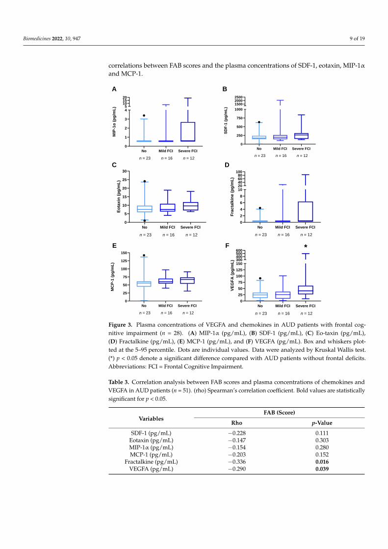
3.4. Plasma Concentrations of VEGFA and Chemokines in Abstinent AUD Patients with FrontalCognitive Impairment
To explore the influence of frontal cognition integrity on plasma concentrations ofVEGFA and chemokines, we performed a Kruskal Wallis test with “frontal cognitiveimpairment” (no, mild, and severe) as a factor. We did not find a significant effect of “frontalcognitive impairment” on plasma concentrations of VEGFA and chemokines (Table S3).However, as shown in Figure 3, circulant levels of VEGFA almost reached the significance(K = 5.404, p = 0.067), having higher plasma concentrations of VEGFA the AUD patientswith severe frontal cognitive impairment than those without frontal deficits (U = 72.50,p = 0.021).
3.5. Correlation Analyses between Frontal Cognition and Plasma Concentrations of VEGFA andChemokines in AUD Patients
For deeper analysis, we explored the relationship between frontal lobe functions (eval-uated by FAB) and plasma concentrations of VEGFA and chemokines using Spearmancorrelations (rho). As shown in Table 3, there was a significant and negative correlationbetween FAB score and plasma concentrations of VEGFA (rho = −0.290, p = 0.039). Wealso observed a significant and negative correlation between FAB score and plasma con-centrations of fractalkine (rho= −0.336, p = 0.016). However, we did not find significant
Biomedicines 2022, 10, 947 9 of 19
correlations between FAB scores and the plasma concentrations of SDF-1, eotaxin, MIP-1αand MCP-1.
Biomedicines 2022, 10, x FOR PEER REVIEW 9 of 20
No Mild FCI Severe FCI
0
1
2
3
45
101520
MIP
-1α
(p
g/m
L)
n = 23 n = 16 n = 12
No Mild FCI Severe FCI
0
250
500
750
1000
150020002500
SD
F-1
(p
g/m
L)
n = 23 n = 16 n = 12
No Mild FCI Severe FCI
0
5
10
15
20
25
30
Eo
tax
in (
pg
/mL
)
n = 23 n = 16 n = 12
No Mild FCI Severe FCI
0
2
4
6
8
1020406080
100
Fra
cta
lkin
e (
pg
/mL
)n = 23 n = 16 n = 12
No Mild FCI Severe FCI
0
25
50
75
100
125
150
MC
P-1
(p
g/m
L)
n = 23 n = 16 n = 12
No Mild FCI Severe FCI
0
25
50
75
100
125
150300400500600
VE
GF
A (
pg
/mL
)
n = 23 n = 16 n = 12
A B
C D
E F *
Figure 3. Plasma concentrations of VEGFA and chemokines in AUD patients with frontal cognitive
impairment (n = 28). (A) MIP-1α (pg/mL), (B) SDF-1 (pg/mL), (C) Eo-taxin (pg/mL), (D) Fractalkine
(pg/mL), (E) MCP-1 (pg/mL), and (F) VEGFA (pg/mL). Box and whiskers plotted at the 5–95 per-
centile. Dots are individual values. Data were analyzed by Kruskal Wallis test. (*) p < 0.05 denote a
significant difference compared with AUD patients without frontal deficits. Abbreviations: FCI =
Frontal Cognitive Impairment.
3.5. Correlation Analyses between Frontal Cognition and Plasma Concentrations of VEGFA and
Chemokines in AUD Patients
For deeper analysis, we explored the relationship between frontal lobe functions
(evaluated by FAB) and plasma concentrations of VEGFA and chemokines using Spear-
man correlations (rho). As shown in Table 3, there was a significant and negative correla-
tion between FAB score and plasma concentrations of VEGFA (rho = −0.290, p = 0.039). We
also observed a significant and negative correlation between FAB score and plasma con-
centrations of fractalkine (rho= −0.336, p = 0.016). However, we did not find significant
correlations between FAB scores and the plasma concentrations of SDF-1, eotaxin, MIP-
1α and MCP-1.
Figure 3. Plasma concentrations of VEGFA and chemokines in AUD patients with frontal cog-nitive impairment (n = 28). (A) MIP-1α (pg/mL), (B) SDF-1 (pg/mL), (C) Eo-taxin (pg/mL),(D) Fractalkine (pg/mL), (E) MCP-1 (pg/mL), and (F) VEGFA (pg/mL). Box and whiskers plot-ted at the 5–95 percentile. Dots are individual values. Data were analyzed by Kruskal Wallis test.(*) p < 0.05 denote a significant difference compared with AUD patients without frontal deficits.Abbreviations: FCI = Frontal Cognitive Impairment.
Table 3. Correlation analysis between FAB scores and plasma concentrations of chemokines andVEGFA in AUD patients (n = 51). (rho) Spearman’s correlation coefficient. Bold values are statisticallysignificant for p < 0.05.
VariablesFAB (Score)
Rho p-Value
SDF-1 (pg/mL) −0.228 0.111Eotaxin (pg/mL) −0.147 0.303MIP-1α (pg/mL) −0.154 0.280MCP-1 (pg/mL) −0.203 0.152
Fractalkine (pg/mL) −0.336 0.016VEGFA (pg/mL) −0.290 0.039

Biomedicines 2022, 10, 947 10 of 19
3.6. Correlation Analyses between Plasma Concentrations of VEGFA and Chemokines in AUDPatients with and without Mild Cognitive Impairment
Moreover, we wanted to explore the relationship between plasma concentrations ofchemokines and VEGFA depending on the integrity of frontal lobe function (evaluated byFAB) using Spearman correlations (rho). As shown in Figure 4, AUD patients with cognitiveimpairment displayed strong positive and significant correlations between VEGFA withall chemokines [SDF-1 (rho = 0.787, p < 0.001), eotaxin (rho = 0.678, p < 0.001), MIP-1α(rho = 0.592, p = 0.001), MCP-1 (rho = 0.601, p = 0.001), fractalkine (rho = 0.706, p < 0.001)],while we only observed a positive and significant correlation between VEGFA and SFD-1(rho = 0.532, p = 0.009) for AUD patients without cognitive impairment (Table S4).
Biomedicines 2022, 10, x FOR PEER REVIEW 10 of 20
Table 3. Correlation analysis between FAB scores and plasma concentrations of chemokines and
VEGFA in AUD patients (n = 51). (rho) Spearman’s correlation coefficient. Bold values are statisti-
cally significant for p < 0.05.
Variables FAB (Score)
Rho p-Value
SDF-1 (pg/mL) −0.228 0.111
Eotaxin (pg/mL) −0.147 0.303
MIP-1α (pg/mL) −0.154 0.280
MCP-1 (pg/mL) −0.203 0.152
Fractalkine (pg/mL) −0.336 0.016
VEGFA (pg/mL) −0.290 0.039
3.6. Correlation Analyses between Plasma Concentrations of VEGFA and Chemokines in AUD
Patients with and without Mild Cognitive Impairment
Moreover, we wanted to explore the relationship between plasma concentrations of
chemokines and VEGFA depending on the integrity of frontal lobe function (evaluated by
FAB) using Spearman correlations (rho). As shown in Figure 4, AUD patients with cogni-
tive impairment displayed strong positive and significant correlations between VEGFA
with all chemokines [SDF-1 (rho = 0.787, p < 0.001), eotaxin (rho = 0.678, p < 0.001), MIP-1α
(rho = 0.592, p = 0.001), MCP-1 (rho = 0.601, p = 0.001), fractalkine (rho = 0.706, p < 0.001)],
while we only observed a positive and significant correlation between VEGFA and SFD-
1 (rho = 0.532, p = 0.009) for AUD patients without cognitive impairment (Table S4).
Figure 4. Correlations analysis between VEGFA and chemokines in AUD patients with and without
frontal cognitive impairment (n = 51). Colors show Spearman’s rho correlation coefficient.
3.7. Differential Profiles Associated with VEGFA and Chemokines in AUD Patients with Frontal
Cognitive Impairment
To understand the contribution of VEGFA and chemokines in the frontal cognitive
decline of AUD patients, a principal component analysis was performed. Two compo-
nents together explained 80.67% of the variance associated with cognitive impairment in
AUD patients (Figure 5). Component 1 explained 56.61% of the total variance and was
composed of SDF-1, fractalkine and VEGFA, which had high factor loads (0.984, 0.983,
0.757, respectively). Component 2 explained 24.06% of the total variance and was com-
posed of MIP-1α, eotaxin, MCP-1 and VEGFA, which had high factor loads (0.669, 0.904,
0.807, 0.582, respectively).
Figure 4. Correlations analysis between VEGFA and chemokines in AUD patients with and withoutfrontal cognitive impairment (n = 51). Colors show Spearman’s rho correlation coefficient.
3.7. Differential Profiles Associated with VEGFA and Chemokines in AUD Patients with FrontalCognitive Impairment
To understand the contribution of VEGFA and chemokines in the frontal cognitivedecline of AUD patients, a principal component analysis was performed. Two componentstogether explained 80.67% of the variance associated with cognitive impairment in AUDpatients (Figure 5). Component 1 explained 56.61% of the total variance and was com-posed of SDF-1, fractalkine and VEGFA, which had high factor loads (0.984, 0.983, 0.757,respectively). Component 2 explained 24.06% of the total variance and was composedof MIP-1α, eotaxin, MCP-1 and VEGFA, which had high factor loads (0.669, 0.904, 0.807,0.582, respectively).
Biomedicines 2022, 10, x FOR PEER REVIEW 11 of 20
Figure 5. Exploratory principal component analysis in AUD patients with frontal deficits (n = 51).
Two components together explained 80.67% of the variance associated with frontal cognitive im-
pairment in AUD patients.
3.8. Plasma Concentrations of VEGFA and Chemokines in Comorbid Medical, Psychiatric
Medication and Substance Use Problems in AUD Patients
We wanted to investigate whether medical and psychiatric comorbidity, other con-
comitant substance use disorder or using psychotropic medication could affect plasma
concentrations of VEGFA and chemokines in the alcohol group. Plasma concentrations of
MIP-1α (U = 567, p = 0.002) and VEGFA (U = 552, p = 0.005) were significantly higher in
AUD patients with comorbid psychiatric disorder compared to those without psychiatric
comorbidity (Table 4). Moreover, plasma concentrations of MIP-1α (U = 633, p = 0.005)
were significantly higher in AUD patients with comorbid substance use disorder com-
pared to those without comorbid substance use disorder (Table S5). Regarding comorbid
medical problems and the use of psychotropic medication, we did not observe major dif-
ferences in plasma levels of chemokines and VEGFA (Tables S6 and S7).
Table 4. Plasma concentrations of chemokines and VEGFA grouped according to comorbid psychi-
atric disorder. Bold values are statistically significant for p < 0.05.
AUD Group (n = 89)
Variables
Comorbid Psychiatric Disorder
(n = 60)
No Comorbid Psychiatric Disorder
(n = 29)
Statistics
U-value p-Value Mean [95% CI] Mean [95% CI]
SDF-1 (pg/mL) 271.9046
[165.9141–377.8952]
189.3975
[143.9530–234.8421] 831 0.828
Eotaxin (pg/mL) 7.77278
[6.57250–8.97306]
7.47531
[5.57394–9.37668] 806 0.575
MIP-1α (pg/mL) 1.8189
[1.0330–2.6048]
0.6189
[0.5586–0.6793] 567 0.002
MCP-1 (pg/mL) 48.9765
[42.4613–55.4917]
48.4279
[36.5812–60.2747] 813 0.618
Fractalkine (pg/mL) 2.0546
[0.6521–3.4570]
0.7399
[0.1778–1.3020] 741 0.160
VEGFA (pg/mL) 36.4530
[28.3181–44.5879]
20.1710
[14.3319–26.0100] 552 0.005
3.9. Time Course of Plasma Concentrations of VEGFA after an Acute Administration of Alcohol
(100 g) in Healthy Male Volunteers
To clarify how an acute intoxicating dose of alcohol affects circulating levels of
VEFGA, we administered 100 g of alcohol to male healthy volunteers whose daily average
alcohol intake was of 13.7 ± 8.3. Plasma samples were taken previous to (time 0) and 2, 8,
and 24 h after oral ingestion of alcohol. As expected, plasma ethanol level peaked at 2 h
Figure 5. Exploratory principal component analysis in AUD patients with frontal deficits (n = 51).Two components together explained 80.67% of the variance associated with frontal cognitive impair-ment in AUD patients.

Biomedicines 2022, 10, 947 11 of 19
3.8. Plasma Concentrations of VEGFA and Chemokines in Comorbid Medical, PsychiatricMedication and Substance Use Problems in AUD Patients
We wanted to investigate whether medical and psychiatric comorbidity, other con-comitant substance use disorder or using psychotropic medication could affect plasmaconcentrations of VEGFA and chemokines in the alcohol group. Plasma concentrations ofMIP-1α (U = 567, p = 0.002) and VEGFA (U = 552, p = 0.005) were significantly higher inAUD patients with comorbid psychiatric disorder compared to those without psychiatriccomorbidity (Table 4). Moreover, plasma concentrations of MIP-1α (U = 633, p = 0.005)were significantly higher in AUD patients with comorbid substance use disorder comparedto those without comorbid substance use disorder (Table S5). Regarding comorbid medicalproblems and the use of psychotropic medication, we did not observe major differences inplasma levels of chemokines and VEGFA (Tables S6 and S7).
Table 4. Plasma concentrations of chemokines and VEGFA grouped according to comorbid psychiatricdisorder. Bold values are statistically significant for p < 0.05.
AUD Group (n = 89)
Variables
Comorbid Psychiatric Disorder(n = 60)
No Comorbid Psychiatric Disorder(n = 29)
Statistics
U-Value p-ValueMean [95% CI] Mean [95% CI]
SDF-1 (pg/mL) 271.9046[165.9141–377.8952]
189.3975[143.9530–234.8421] 831 0.828
Eotaxin (pg/mL) 7.77278[6.57250–8.97306]
7.47531[5.57394–9.37668] 806 0.575
MIP-1α (pg/mL) 1.8189[1.0330–2.6048]
0.6189[0.5586–0.6793] 567 0.002
MCP-1 (pg/mL) 48.9765[42.4613–55.4917]
48.4279[36.5812–60.2747] 813 0.618
Fractalkine (pg/mL) 2.0546[0.6521–3.4570]
0.7399[0.1778–1.3020] 741 0.160
VEGFA (pg/mL) 36.4530[28.3181–44.5879]
20.1710[14.3319–26.0100] 552 0.005
3.9. Time Course of Plasma Concentrations of VEGFA after an Acute Administration of Alcohol(100 g) in Healthy Male Volunteers
To clarify how an acute intoxicating dose of alcohol affects circulating levels of VEFGA,we administered 100 g of alcohol to male healthy volunteers whose daily average alcoholintake was of 13.7 ± 8.3. Plasma samples were taken previous to (time 0) and 2, 8, and24 h after oral ingestion of alcohol. As expected, plasma ethanol level peaked at 2 h afteringestion and decreased to a third of the 2 h concentration 8 h after the ingestion, beingundetectable 24 h after oral intake (Figure 6A). Levels of VEGFA were very variable inbetween these subjects so a non-parametric statistics with repeated measures approach wastaken. Data indicated that alcohol modified plasma VEGFA, being elevated 8 h (Figure 6B)after the ingestion of alcohol (Friedman’s statistic for n = 9, four groups, was 9.26, p < 0.03).Analysis of the percentage of change from basal values indicated the alcohol induceda 2-fold change on VEGFA circulating concentrations with respect to the basal values(Figure 6C, ANOVA repeated measures F (8,24) = 5.14, p < 0.001). Twenty-four hours afterthe intake of alcohol, the % of change had a non-significative average fold change of 1.7,suggesting that alcohol induced sustained increases of VEGFA. However, this assumptionneeds to be conclusively determined.

Biomedicines 2022, 10, 947 12 of 19
Biomedicines 2022, 10, x FOR PEER REVIEW 12 of 20
after ingestion and decreased to a third of the 2 h concentration 8 h after the ingestion,
being undetecTable 24 h after oral intake (Figure 6A). Levels of VEGFA were very variable
in between these subjects so a non-parametric statistics with repeated measures approach
was taken. Data indicated that alcohol modified plasma VEGFA, being elevated 8 h (Fig-
ure 6B) after the ingestion of alcohol (Friedman’s statistic for n = 9, four groups, was 9.26,
p < 0.03). Analysis of the percentage of change from basal values indicated the alcohol
induced a 2-fold change on VEGFA circulating concentrations with respect to the basal
values (Figure 6C, ANOVA repeated measures F (8,24) = 5.14, p < 0.001). Twenty-four
hours after the intake of alcohol, the % of change had a non-significative average fold
change of 1.7, suggesting that alcohol induced sustained increases of VEGFA. However,
this assumption needs to be conclusively determined.
Figure 6. Plasma concentrations of alcohol (A) and VEGFA (B) at 2, 8 and 24 h after alcohol (100 g)
ingestion in male (n = 9–10) healthy volunteers. 0 represents the time of ingestion. Alcohol resulted
in increase in plasma concentrations of VEGF 8 h after its ingestion (Friedman’s non-parametric test
for repeated measures, * p < 0.05 versus 0 time). (C) Percentage of change of VGEFA calculated over
basal concentrations (ANOVA with repeated measures, *** p < 0.001 versus 0 time). ns= non-signif-
icant.
4. Discussion
While the association between immune signaling and neuropsychiatric disorders has
been widely investigated, its link with AUD-related cognitive impairment remains poorly
investigated. We lack relevant information concerning how immune mediator-induced
proinflammatory states in the brain influence the neuroadaptations derived from chronic
alcohol consumption, and how they impact executive functions. In the present study, we
unveil a link between plasma concentrations of VEGFA and chemokines with frontal lobe
dysfunction in abstinent AUD patients treated in an outpatient setting. The relevance of
the present study rests on the confirmation of the link between alcohol-induced dysfunc-
tions in modulators of the blood–brain barrier (i.e., VEGFA) and neuroinflammation (i.e.,
chemokines) with the presence of cognitive impairment. The main findings are as follows:
(i) AUD patients had increased plasma concentrations of SDF-1 and MCP-1 compared to
control subjects; (ii) there were higher circulant levels of VEGFA in AUD patients with
severe frontal deficits than in those without cognitive impairment; (iii) acute administra-
tion of a heavy dose of VEGFA resulted in a delayed increase (8 h after alcohol ingestion)
of plasma VEGFA concentration; (iv) the integrity of frontal lobe functions was negatively
correlated with VEGFA and fractalkine; (v) plasma concentrations of VEGFA were
strongly and positively correlated with all chemokines in AUD patients with frontal defi-
cits but not in those without frontal impairment; (vi) two components together explained
80.67% of the variance associated with frontal deficits in AUD patients, with VEGFA act-
ing as a link between all chemokines; and (vii) plasma concentrations of some chemokines
Figure 6. Plasma concentrations of alcohol (A) and VEGFA (B) at 2, 8 and 24 h after alcohol (100 g)ingestion in male (n = 9–10) healthy volunteers. 0 represents the time of ingestion. Alcohol resulted inincrease in plasma concentrations of VEGF 8 h after its ingestion (Friedman’s non-parametric test forrepeated measures, * p < 0.05 versus 0 time). (C) Percentage of change of VGEFA calculated over basalconcentrations (ANOVA with repeated measures, *** p < 0.001 versus 0 time). ns = non-significant.
4. Discussion
While the association between immune signaling and neuropsychiatric disorders hasbeen widely investigated, its link with AUD-related cognitive impairment remains poorlyinvestigated. We lack relevant information concerning how immune mediator-inducedproinflammatory states in the brain influence the neuroadaptations derived from chronicalcohol consumption, and how they impact executive functions. In the present study, weunveil a link between plasma concentrations of VEGFA and chemokines with frontal lobedysfunction in abstinent AUD patients treated in an outpatient setting. The relevance ofthe present study rests on the confirmation of the link between alcohol-induced dysfunc-tions in modulators of the blood–brain barrier (i.e., VEGFA) and neuroinflammation (i.e.,chemokines) with the presence of cognitive impairment. The main findings are as follows:(i) AUD patients had increased plasma concentrations of SDF-1 and MCP-1 compared tocontrol subjects; (ii) there were higher circulant levels of VEGFA in AUD patients with se-vere frontal deficits than in those without cognitive impairment; (iii) acute administration ofa heavy dose of VEGFA resulted in a delayed increase (8 h after alcohol ingestion) of plasmaVEGFA concentration; (iv) the integrity of frontal lobe functions was negatively correlatedwith VEGFA and fractalkine; (v) plasma concentrations of VEGFA were strongly and posi-tively correlated with all chemokines in AUD patients with frontal deficits but not in thosewithout frontal impairment; (vi) two components together explained 80.67% of the varianceassociated with frontal deficits in AUD patients, with VEGFA acting as a link between allchemokines; and (vii) plasma concentrations of some chemokines changed in AUD patientswith comorbid psychiatric (MIP-1α, VEGFA) and substance use (MIP-1 α) disorders.
A growing literature indicates that the pharmacodynamic action of several drugs involveschanges in the neuroimmune signal [45]. Some studies have reported that alcohol-relatedbehaviors are interrupted when the innate immune network is disturbed [46,47]. Thus,Blednov et al. (2005) showed that gene deletion of CCR2, MCP-1 or MIP-1α reduced mo-tivational effects of alcohol consumption in mice [48]. Moreover, Steinar et al. (2019)found an increase in the plasma concentrations of IL-6, IFNγ and MCP-1 in patients witha history of chronic alcohol overconsumption [49]. In agreement with these reports, theAUD patients of the present study displayed higher plasma concentrations of SDF-1 andMCP-1 than control subjects. SDF-1 was also associated (along with eotaxin) with worseseverity of alcohol addiction. Even post mortem analysis reported increased expressionof MCP-1 in multiple limbic brain regions in alcoholic subjects [50]. Taking into accountthe high prevalence of cocaine use disorder in AUD patients, it is important to note thatextensive preclinical research has reported how the chemokines SDF-1 and MCP-1 pro-

Biomedicines 2022, 10, 947 13 of 19
mote cocaine-related behaviors in a brain region specific manner [51–53]. Our group hasdescribed that severity of cocaine consumption is related to IL-1β, SDF-1 and fractalkine incocaine use disorder patients [54]. Furthermore, we found higher plasma concentrations offractalkine in abstinent cocaine patients with comorbid major depressive disorder than inthose without this psychiatric condition [55]. In addition, we demonstrated the induction ofa potent fractalkine signaling associated with cocaine-induced sensitization and extinctionin mice [56].
Regarding cognitive function, 55% of the AUD patients recruited in the present studydisplayed some kind of frontal cognitive impairment. Accordingly, executive functions areparticularly affected in AUD patients who show deficits in domains related to cognitivecontrol, flexibility, inhibition, planning and working memory [57,58]. However, there areother neuropsychological processes that could be disrupted, including memory, emotion,and social cognition [59]. It is important to note that despite there being evidence of partialrecovery of certain cognitive functions after cessation of alcohol intake [13,60], deficits inother domains may remain stable during sobriety [57,61]. Additionally, cognitive impair-ment can compromise efforts to initiate and maintain abstinence by impacting treatmenteffectiveness [62]. It is thought that increased vulnerability to alcohol-induced workingmemory impairment may impact in the ability to moderate alcohol consumption [63]and cognitive training could reduce the number of beverages in these patients [64]. Fur-thermore, our results indicate that AUD patients have low educational and occupationalattainment, which has been related to worse neuropsychological performance, more indi-cators of neurocognitive disorders, early drug use onset and development of addiction,high-severity substance-related problems, and worse treatment outcomes [65–67]. Thus,excessive alcohol consumption has been linked to worse cognitive functioning in patientswith low socioeconomic status operated by educational and occupational achievements [68].Moreover, a longitudinal study has suggested that an educational level lower than highschool and a low job occupation is associated with an increased risk of dementia in alcoholpatients [69]. Similarly, our group has recently found that a high educational level couldplay a protective role in the onset, development, and progression of cocaine use disor-ders and could also protect against cognitive impairment caused by alcohol consumptionthroughout life [70,71]. Interestingly, in this study we found a negative association betweenthe state of frontal lobe functions with two signaling molecules, VEGFA and fractalkine,involved in vascular function and neural plasticity.
VEGFA has been related to neuroprotection, neurogenesis, and synaptic plasticitymechanisms in the central nervous system through stimulation of neuronal stem cellsand safeguarding the integrity of the blood–brain barrier [36]. Despite VEGFA levelshaving been linked to several neurodegenerative and neurological disorders [36], theireffects could be time dependent. Augmented VEGFA levels in cerebrospinal fluid andplasma have been reported in Alzheimer’s disease and vascular dementia patients [72,73]probably as a consequence of hypoperfusion and hypoxia. Nevertheless, improvement inlearning and memory after a bilateral carotid artery occlusion has been associated with anincrease in VEGFA expression in the hippocampus, which suggests that VEGFA signalingcould compensate for cognitive impairment [74,75]. Similarly, partial increases in VEGFAstimulate vasodilation, angiogenesis and neuroprotection mechanisms, which are beneficialfor the brain in later stages after cerebral ischemia [76]. However, early VEGFA increasesmay lead to undesirable effects in cerebral ischemia, such as an increase in blood–brainbarrier permeability and infiltration of immune cells inducing neuroinflammation andedema [77–79].
With reference to the latter, in the present study we observed that increases in VEGFAwere associated with worse severity of alcohol addiction, severe frontal deficits and theelevation of all chemokines in frontal cognitive impaired AUD patients. Interestingly, in apilot study with healthy male volunteers with a history of moderate alcohol consumption,we found that acute alcohol administration resulted in a delayed increase in plasma VEGFA,observed 8 h after alcohol intake. Because of the increased vascular permeability induced

Biomedicines 2022, 10, 947 14 of 19
by VEGFA, it is reasonable to think that this action of alcohol might facilitate neuroin-flammation by opening the blood–brain barrier to pro-inflammatory signals originating inperipheral tissues, especially in the intestine. Supporting this hypothesis, in our principalcomponent analysis, we found that the interaction of chemokines and VEGFA explainedthe 80.67% of the variance associated with frontal deficits in AUD patients, observing thatVEGFA has an essential role as a factor interacting with the pro-inflammatory immuneresponse associated with alcohol consumption. Consistent with our results, using cellularand animal models, Muneer et al. (2012) found that chronic alcohol exposure disrupts theblood–brain barrier across degradation of endothelial VEGF receptor 2. This also increasescirculating levels of VEGFA leading to neuronal death and inflammation in the brain [80].Moreover, Louboutin et al. (2012) reported increases in VEGFA levels and blood vesseldensity in cerebral tissue within two weeks of the onset of ethanol consumption in rats [81].Moreover, despite VEGFA not being an inflammatory cytokine, it can activate nuclearfactor-enhancer of activated B-cell kappa light chains (NF-KB) and nuclear factor of acti-vated T-cells (NFAT) signaling cascades that could promote a chemotaxis response involvedin the angiogenic process (although this role remains unknown) [82]. Thus, our resultsin AUD patients with frontal deficits might suggest two things: (1) chronic alcohol abusemight lead to alterations in the concentrations of VEGFA that increase the permeability ofthe blood–brain barrier, leading to infiltration of immune cells and inflammation in thebrain [80,81], and/or (2) under the presence of hypoperfusion and hypoxia as a result ofalcohol-derived brain damage, concentrations of VEGFA might ultimately increase as acompensatory signal in order to form new blood vessels and recruit chemokines to theaffected brain area. Lastly, it has been found that higher circulant levels of VEGFA in majordepression and its alterations are related to impaired cognitive function in schizophre-nia [83,84]. This might explain why psychiatric comorbidity affected plasma concentrationsof VEGFA in our study. It is important to note that cognitive deficits found in patients withaddictions can often be exacerbated by comorbid psychiatric disorders [85].
Lastly, previous studies have reported that fractalkine (CX3CL1) develops an essentialrole in the neuronal–microglial intercommunication [86]. This chemokine is expressed frombrain neurons that control activation of microglia through its binding receptor CX3CR1. Inharmful conditions, neurons release fractalkine in order to stimulate proliferation, activa-tion and migration at the site of the brain injury [87,88]. CX3CL1-KO mice showed alteredmicroglial function and neurotoxicity following LPS injection as well as more neuronaldamage in Parkinson’s disease and amyotrophic lateral sclerosis [88]. In accordance withthis, Sokolowki et al. (2014) found that CX3CL1-KO mice revealed signals of neuronalapoptosis after ethanol treatment, suggesting a role in the clearance of those apoptoticcells [86]. Moreover, additional studies have reported that mild–moderate Alzheimer’sdisease patients had higher plasma levels of CX3CL1 than those with severe Alzheimer’sdisease [89,90]. These results suggest that fractalkine and CX3CR1 signaling might actas a neuroprotective mechanism through the microglial activity modulation in the earlystage of brain injury while this signal seems to disappear when neuronal damage is es-tablished [91]. This may indicate that the AUD patients in this study are actively fightingagainst cognitive impairment.
5. Conclusions, Limitations, and Future Perspectives
In conclusion, a lifetime of chronic alcohol consumption leads to a proinflammatorysystemic condition revealed by enhanced circulating chemokines, and to frontal cognitive im-pairment. The trophic factor VEGFA appears to be a relevant contributor to alcohol-associatedneuroinflammation, probably through its role on controlling blood–brain barrier permeabil-ity, ultimately leading to impaired cognition. In addition, fractalkine could act as a signalof brain damage in early stages of cognitive impairment. Potential biomarkers could beuseful and reliable tools in patients with AUD for confirming the diagnosis, defining thecurrent stage of the AUD, and diagnosing these patients early.

Biomedicines 2022, 10, 947 15 of 19
Nevertheless, this study has several limitations that should be taken into account infuture research. First, we do not know the time course of the effects of alcohol on thesechemokines, either after acute or chronic alcohol consumption, nor its alterations in early orextended abstinence. We must also investigate whether sociodemographic variables, espe-cially educational level, time of alcohol consumption, age of alcohol drinking initiation, etc.,might contribute to the cognitive performance in both control and AUD patients. Finally,we lack significant representation of the female population, which precludes investigationof sex differences in chemokines and VEGFA. However, the data obtained clearly point tothe need of considering these immunoinflammatory signals and trophic factors as relevantbiomarkers of AUD-associated complications. Moreover, this concept should be extendedto the analysis of immunomodulators capable of activating chronic inflammation. There areseveral biochemical pathways affected by alcohol consumption that need to be consideredunder the light of the present discoveries. For example, the tryptophan/kynurenine path-way is a potent immunomodulatory system that can modify inflammation, learning andmemory [92,93]. The intestinal microbiota (which is determinant for AUD) [94] participatesin these pathways, modifying the presence of pro- and anti-inflammatory mediators andeventually growth factors.
As a future perspective, we need to integrate all the information related to this multi-plicity of inflammatory signals in a single model of alcohol addiction. Although certain fac-tors such as VEGFA or fractalkine may contribute to important aspects of alcohol addiction,the complexity of the interactions of these inflammatory signaling proteins goes beyond ourcurrent technique and knowledge. Further clinical and technological research is necessaryto elucidate the role of these factors in the etiology of AUD and associated comorbidities.
Supplementary Materials: The following supporting information can be downloaded at: https://www.mdpi.com/article/10.3390/biomedicines10050947/s1, Table S1: Plasma concentrations ofchemokines and VEGFA in AUD group versus control subjects; Table S2: Correlation analysis be-tween FAB score and chemokines in AUD patients; Table S3: Correlation analysis between plasmaconcentrations of chemokines and VEGFA in AUD patients with and without cognitive impairment;Table S4: Plasma concentrations of chemokines and VEGFA grouped according to comorbid psy-chiatric disorder; Table S5: Plasma concentrations of chemokines and VEGFA grouped accordingto comorbid substance use disorder; Table S6: Plasma concentrations of chemokines and VEGFAgrouped according to comorbid medical problem; Table S7: Plasma concentrations of chemokinesand VEGFA grouped according to the use of psychotropic medication last year.
Author Contributions: Conception and design: J.S., F.R.d.F. and P.A. Data acquisition: N.R.-O., P.A.,M.F.-L., N.G.-M., E.P. and J.J.R. Data analysis and interpretation: N.R.-O., P.A., N.G.-M., F.J.P.-M., E.P.and M.F. Draft writing: N.R.-O., J.S., F.R.d.F. and A.S. Review and editing: P.A., J.J.R., J.O.-P., A.S.,F.J.P.-M. and M.F. Final approval: All authors. All authors have read and agreed to the publishedversion of the manuscript.
Funding: RETICS Red de Trastornos Adictivos, Instituto de Salud Carlos III (ISCIII), Ministerio deCiencia e Innovación and European Regional Development Funds—European Union (ERDF-EU)(RD16/0017/0001 and RD16/0017/003); ISCIII, ERDF-EU (PI20/01399, PI19/01577, PI19/00886);Ministerio de Sanidad, Delegación de Gobierno para el Plan Nacional sobre Drogas (PND 2020I048,PND 2019I040, PND 2018I044, PND 2018I033, PND 2016I024 and PND 2018I037) and Consejería deSalud y Familia, Junta de Andalucía (Neuro-RECA, RIC-0111-2019). F.J.P.-M. (CPII19/00022) andA.S. (CPII19/00031) hold “Miguel Servet II” research contracts from the National System of Health,ISCIII, ERDF-EU. F.J.P.-M. also holds a “Nicolas Monardes” contract from Servicio Andaluz de Salud,Consejería de Salud y Familia, Junta de Andalucía (C1-0049-2019). P.A. has a research contract(UMA-FEDERJA-076) funded by the Ministry of Economy and Knowledge—Regional Governmentof Andalucía and ERDF-EU. “PFIS” Predoctoral research contract (FI18/00249) financed by ISCIIIERDF/ESF and NGM has a ‘Sara Borrell’ Research Contract (CD19/00019) financed by ISCIII andERDF-EU. The funding sources had no further role in study design; in the collection, analysis, andinterpretation of data; in writing of the report; and in the decision to submit the paper for publication.

Biomedicines 2022, 10, 947 16 of 19
Institutional Review Board Statement: The study was conducted in accordance with the Declarationof Helsinki and approved by the Ethics Committee of the Portal de Ética de la Investigación Biomédicade Andalucía-PEIBA (Consejería de Salud y Familias, Junta de Andalucía) and Hospital RegionalUniversitario de Málaga (PND2018I033). The acute alcohol administration experiment protocol wasapproved by the local Human Research Ethics Committee (CEI Hospital Universitari Germans Triasi Pujol, Badalona, Spain) and registered at ClinicalTrials.gov (NCT02232789). The clinical trial wasconducted in accordance with the Declaration of Helsinki and Spanish laws concerning clinical research.
Informed Consent Statement: Informed consent was obtained from all subjects involved in the study.
Data Availability Statement: Not applicable.
Conflicts of Interest: The authors declare no conflict of interest. The funders had no role in the designof the study; in the collection, analyses, or interpretation of data; in the writing of the manu-script, orin the decision to publish the results.
References1. World Health Organization. Global Status Report on Alcohol and Health 2018; World Health Organization: Geneva, Switzerland, 2019.2. OEDA. Observatorio Español de las Drogas y las Adicciones (OEDA) [2017]. Informe Anual 2017. Informe EDADES 2017. Plan
Nacional Sobre Drogas. 2019. Available online: https://pnsd.sanidad.gob.es/profesionales/sistemasInformacion/home.htm(accessed on 23 February 2022).
3. Louvet, A.; Mathurin, P. Alcoholic liver disease: Mechanisms of injury and targeted treatment. Nat. Rev. Gastroenterol. Hepatol.2015, 12, 231–242. [CrossRef] [PubMed]
4. Setiawan, V.W.; Monroe, K.; Lugea, A.; Yadav, D.; Pandol, S. Uniting Epidemiology and Experimental Disease Models forAlcohol-Related Pancreatic Disease. Alcohol Res. Curr. Rev. 2017, 38, 173–182.
5. Marchena, N.G.; Araos, P.; Pavón, F.J.; Ponce, G.; Pedraz, M.; Serrano, A.; Arias, F.; Romero-Sanchiz, P.; Suárez, J.; Pastor, A.; et al.Psychiatric comorbidity and plasma levels of 2-acyl-glycerols in outpatient treatment alcohol users. Analysis of gender differences.Adicciones 2016, 29, 83. [CrossRef]
6. Boden, J.; Fergusson, D.M. Alcohol and depression. Addiction 2011, 106, 906–914. [CrossRef]7. Samet, S.; Fenton, M.C.; Nunes, E.; Greenstein, E.; Aharonovich, E.; Hasin, D. Effects of independent and substance-induced
major depressive disorder on remission and relapse of alcohol, cocaine and heroin dependence. Addiction 2013, 108, 115–123.[CrossRef]
8. Schwarzinger, M.; Pollock, B.G.; Hasan, O.S.M.; Dufouil, C.; Rehm, J.; Baillot, S.; Guibert, Q.; Planchet, F.; Luchini, S. Contributionof alcohol use disorders to the burden of dementia in France 2008-13: A nationwide retrospective cohort study. Lancet PublicHealth 2018, 3, e124–e132. [CrossRef]
9. Withall, A.; Draper, B.; Seeher, K.; Brodaty, H. The prevalence and causes of younger onset dementia in Eastern Sydney, Australia.Int. Psychogeriatr. 2014, 26, 1955–1965. [CrossRef]
10. Cheng, C.; Huang, C.-L.; Tsai, C.-J.; Chou, P.-H.; Lin, C.-C.; Chang, C.-K. Alcohol-Related Dementia: A Systemic Review ofEpidemiological Studies. J. Psychosom. Res. 2017, 58, 331–342. [CrossRef]
11. Woods, A.J.; Porges, E.C.; Bryant, V.E.; Seider, T.; Gongvatana, A.; Kahler, C.W.; De La Monte, S.; Monti, P.M.; Cohen, R.A. CurrentHeavy Alcohol Consumption is Associated with Greater Cognitive Impairment in Older Adults. Alcohol. Clin. Exp. Res. 2016, 40,2435–2444. [CrossRef]
12. Hayes, V.; Demirkol, A.; Ridley, N.; Withall, A.; Draper, B. Alcohol-related cognitive impairment: Current trends and futureperspectives. Neurodegener. Dis. Manag. 2016, 6, 509–523. [CrossRef]
13. Ros-Cucurull, E.; Palma-Álvarez, R.F.; Cardona-Rubira, C.; García-Raboso, E.; Jacas, C.; Grau-López, L.; Abad, A.C.; Rodríguez-Cintas, L.;Ros-Montalbán, S.; Casas, M.; et al. Alcohol use disorder and cognitive impairment in old age patients: A 6 months follow-upstudy in an outpatient unit in Barcelona. Psychiatry Res. 2018, 261, 361–366. [CrossRef]
14. Horton, L.; Duffy, T.; Martin, C. Neurocognitive, psychosocial and functional status of individuals with alco-hol-related braindamage (ARBD) on admission to specialist residential care. Drugs Educ. Prev. Policy 2015, 22, 416–427. [CrossRef]
15. Montesinos, J.; Pascual, M.; Pla, A.; Maldonado, C.; Rodríguez-Arias, M.; Miñarro, J.; Guerri, C. TLR4 elimination preventssynaptic and myelin alterations and long-term cognitive dysfunctions in adolescent mice with intermittent ethanol treatment.Brain Behav. Immun. 2015, 45, 233–244. [CrossRef]
16. Dwivedi, D.K.; Kumar, D.; Kwatra, M.; Pandey, S.N.; Choubey, P.; Lahkar, M.; Jangra, A. Voluntary alcohol consumptionexacerbated high fat diet-induced cognitive deficits by NF-κB-calpain dependent apoptotic cell death in rat hippocampus:Ameliorative effect of melatonin. Biomed. Pharmacother. 2018, 108, 1393–1403. [CrossRef] [PubMed]
17. Yang, J.-Y.; Xue, X.; Tian, H.; Wang, X.-X.; Dong, Y.-X.; Wang, F.; Zhao, Y.-N.; Yao, X.-C.; Cui, W.; Wu, C.-F. Role of microgliain ethanol-induced neurodegenerative disease: Pathological and behavioral dysfunction at different developmental stages.Pharmacol. Ther. 2014, 144, 321–337. [CrossRef] [PubMed]
18. Banks, W.A. Peptides and the blood-brain barrier. Peptides 2015, 72, 16–19. [CrossRef]

Biomedicines 2022, 10, 947 17 of 19
19. Pascual, M.; Baliño, P.; Aragón, C.M.; Guerri, C. Cytokines and chemokines as biomarkers of ethanol-induced neuroinflammationand anxiety-related behavior: Role of TLR4 and TLR2. Neuropharmacology 2015, 89, 352–359. [CrossRef] [PubMed]
20. Yen, C.-H.; Ho, P.-S.; Yeh, Y.-W.; Liang, C.-S.; Kuo, S.-C.; Huang, C.-C.; Chen, C.-Y.; Shih, M.-C.; Ma, K.-H.; Sung, Y.-F.; et al.Differential cytokine levels between early withdrawal and remission states in patients with alcohol dependence. Psychoneuroen-docrinology 2017, 76, 183–191. [CrossRef] [PubMed]
21. Rostène, W.; Kitabgi, P.; Parsadaniantz, S.M. Chemokines: A new class of neuromodulator? Nat. Rev. Neurosci. 2007, 8, 895–903.[CrossRef]
22. Watson, A.E.S.; Goodkey, K.; Footz, T.; Voronova, A. Regulation of CNS precursor function by neuronal chemokines. Neurosci.Lett. 2020, 715, 134533. [CrossRef]
23. Comerford, I.; McColl, S.R. Mini-review series: Focus on chemokines. Immunol. Cell Biol. 2011, 89, 183–184. [CrossRef] [PubMed]24. Palomino, D.C.T.; Marti, L.C. Chemokines and immunity. Einstein (São Paulo) 2015, 13, 469–473. [CrossRef] [PubMed]25. Cui, C.; Shurtleff, D.; Harris, R.A. Neuroimmune Mechanisms of Alcohol and Drug Addiction. Int. Rev. Neurobiol. 2014, 118, 1–12.
[CrossRef] [PubMed]26. Lacagnina, M.; Rivera, P.D.; Bilbo, S.D. Glial and Neuroimmune Mechanisms as Critical Modulators of Drug Use and Abuse.
Neuropsychopharmacology 2017, 42, 156–177. [CrossRef] [PubMed]27. Ivanovska, M.; Abdi, Z.; Murdjeva, M.; Macedo, D.; Maes, A.; Maes, M. CCL-11 or Eotaxin-1: An Immune Marker for Ageing and
Accelerated Ageing in Neuro-Psychiatric Disorders. Pharmaceuticals 2020, 13, 230. [CrossRef]28. Wang, R.; Li, H. A focus on CXCR4 in Alzheimer’s disease. Brain Circ. 2017, 3, 199–203. [CrossRef]29. Stuart, M.; Baune, B. Chemokines and chemokine receptors in mood disorders, schizophrenia, and cognitive impairment: A
systematic review of biomarker studies. Neurosci. Biobehav. Rev. 2014, 42, 93–115. [CrossRef]30. Teixeira, A.L.; Gama, C.S.; Rocha, N.P.; Teixeira, M.M. Revisiting the role of eotaxin-1/CCL11 in psychiatric disorders. Front.
Psychiatry 2018, 9, 1–6. [CrossRef]31. Wei, Z.; Chen, L.; Zhang, J.; Cheng, Y. Aberrations in peripheral inflammatory cytokine levels in substance use disorders: A
meta-analysis of 74 studies. Addiction 2020, 115, 2257–2267. [CrossRef]32. Achur, R.N.; Freeman, W.M.; Vrana, K.E. Circulating Cytokines as Biomarkers of Alcohol Abuse and Alcoholism. J. Neuroimmune
Pharmacol. 2010, 5, 83–91. [CrossRef]33. Qin, L.; He, J.; Hanes, R.N.; Pluzarev, O.; Hong, J.-S.; Crews, F.T. Increased systemic and brain cytokine production and
neuroinflammation by endotoxin following ethanol treatment. J. Neuroinflammation 2008, 5, 10. [CrossRef] [PubMed]34. Gao, B.; Xu, M. Chemokines and alcoholic hepatitis: Are chemokines good therapeutic targets? Gut 2014, 63, 1683–1684. [CrossRef]
[PubMed]35. McClain, C.J.; Shedlofsky, S.; Barve, S.; Hill, D.B. Cytokines and alcoholic liver disease. Alcohol Health Res. World 1997, 21, 317–320.
[PubMed]36. Lange, C.; Storkebaum, E.; de Almodovar, C.R.; Dewerchin, M.; Carmeliet, P. Vascular endothelial growth factor: A neurovascular
target in neurological diseases. Nat. Rev. Neurol. 2016, 12, 439–454. [CrossRef] [PubMed]37. Klintsova, A.Y.; Hamilton, G.F.; Boschen, K.E. Long-Term Consequences of Developmental Alcohol Exposure on Brain Structure
and Function: Therapeutic Benefits of Physical Activity. Brain Sci. 2012, 3, 1–38. [CrossRef]38. Bates, D.O. Vascular endothelial growth factors and vascular permeability. Cardiovasc. Res. 2010, 87, 262–271. [CrossRef]39. Tammela, T.; Enholm, B.; Alitalo, K.; Paavonen, K. The biology of vascular endothelial growth factors. Cardiovasc. Res. 2005, 65,
550–563. [CrossRef]40. Heberlein, A.; Muschler, M.; Lenz, B.; Frieling, H.; Büchl, C.; Gröschl, M.; Riera, R.; Kornhuber, J.; Bleich, S.; Hillemacher, T. Serum
levels of vascular endothelial growth factor a increase during alcohol withdrawal. Addict. Biol. 2010, 15, 362–364. [CrossRef]41. Kasztelan-Szczerbinska, B.; Surdacka, A.; Slomka, M.; Rolinski, J.M.; Celinski, K.; Cichoz-Lach, H.; Madro, A.; Szczerbinski, M.
Angiogenesis-Related Biomarkers in Patients with Alcoholic Liver Disease: Their Association with Liver Disease Complicationsand Outcome. Mediat. Inflamm. 2014, 2014, 673032. [CrossRef]
42. Torrens, M.; Serrano, D.; Astals, M.; Pérez-Domínguez, G.; Martín-Santos, R. Diagnosing Comorbid Psychiatric Disorders inSubstance Abusers: Validity of the Spanish Versions of the Psychiatric Research Interview for Substance and Mental Disordersand the Structured Clinical Interview for DSM-IV. Am. J. Psychiatry 2004, 161, 1231–1237. [CrossRef]
43. Hasin, D.S.; O’Brien, C.P.; Auriacombe, M.; Borges, G.; Bucholz, K.; Budney, A.; Compton, W.M.; Crowley, T.; Ling, W.;Petry, N.M.; et al. DSM-5 Criteria for Substance Use Disorders: Recommendations and Rationale. Am. J. Psychiatry 2013, 170,834–851. [CrossRef] [PubMed]
44. Del Alamo, A.R.; Alonso, M.J.C.; Marín, L.C. FAB: A preliminar Spanish application of the frontal assessment battery to 11 groupsof patients. Revista de Neurología 2003, 36, 605–608.
45. Ahearn, O.C.; Watson, M.N.; Rawls, S.M. Chemokines, cytokines and substance use disorders. Drug Alcohol Depend. 2021,220, 108511. [CrossRef] [PubMed]
46. Blednov, Y.; Benavidez, J.; Geil, C.; Perra, S.; Morikawa, H.; Harris, R. Activation of inflammatory signaling by lipopolysaccharideproduces a prolonged increase of voluntary alcohol intake in mice. Brain Behav. Immun. 2011, 25, S92–S105. [CrossRef] [PubMed]
47. Blednov, Y.A.; Ponomarev, I.; Geil, C.; Bergeson, S.; Koob, G.F.; Harris, R.A. Neuroimmune regulation of alcohol consumption:Behavioral validation of genes obtained from genomic studies. Addict. Biol. 2012, 17, 108–120. [CrossRef]
Biomedicines 2022, 10, 947 18 of 19
48. Blednov, Y.A.; Bergeson, S.E.; Walker, D.; Ferreira, V.M.; Kuziel, W.A.; Harris, R.A. Perturbation of chemokine networks by genedeletion alters the reinforcing actions of ethanol. Behav. Brain Res. 2005, 165, 110–125. [CrossRef]
49. Bjørkhaug, S.T.; Neupane, S.P.; Bramness, J.G.; Aanes, H.; Skar, V.; Medhus, A.W.; Valeur, J. Plasma cytokine levels in patients withchronic alcohol overconsumption: Relations to gut microbiota markers and clinical correlates. Alcohol 2020, 85, 35–40. [CrossRef]
50. He, J.; Crews, F.T. Increased MCP-1 and microglia in various regions of the human alcoholic brain. Exp. Neurol. 2008, 210, 349–358.[CrossRef]
51. Kim, J.; Connelly, K.L.; Unterwald, E.M.; Rawls, S.M. Chemokines and cocaine: CXCR4 receptor antagonist AMD3100 attenuatescocaine place preference and locomotor stimulation in rats. Brain, Behav. Immun. 2017, 62, 30–34. [CrossRef]
52. Trecki, J.; Unterwald, E. Modulation of cocaine-induced activity by intracerebral administration of CXCL12. Neuroscience 2009,161, 13–22. [CrossRef]
53. Trocello, J.M.; Rostène, W.; Melik-Parsadaniantz, S.; Godefroy, D.; Roze, E.; Kitabgi, P.; Kuziel, W.A.; Chalon, S.; Caboche, J.;Apartis, E. Implication of CCR2 Chemokine Receptor in Cocaine-Induced Sensitization. J. Mol. Neurosci. 2011, 44, 147–151.[CrossRef] [PubMed]
54. Araos, P.; Pedraz, M.; Serrano, A.; Lucena, M.; Barrios, V.; García-Marchena, N.; Campos-Cloute, R.; Ruiz, J.J.; Romero, P.;Suárez, J.; et al. Plasma profile of pro-inflammatory cytokines and chemokines in cocaine users under outpatient treatment:Influence of cocaine symptom severity and psychiatric co-morbidity. Addict. Biol. 2014, 20, 756–772. [CrossRef] [PubMed]
55. García-Marchena, N.; Barrera, M.; Mestre-Pintó, J.I.; Araos, P.; Serrano, A.; Pérez-Mañá, C.; Papaseit, E.; Fonseca, F.; Ruiz, J.J.;De Fonseca, F.R.; et al. Inflammatory mediators and dual depression: Potential biomarkers in plasma of primary and substance-induced major depression in cocaine and alcohol use disorders. PLoS ONE 2019, 14, e0213791. [CrossRef] [PubMed]
56. Montesinos, J.; Castilla-Ortega, E.; Sánchez-Marín, L.; Montagud-Romero, S.; Araos, P.; Pedraz, M.; Porras-Perales, Ó.;García-Marchena, N.; Serrano, A.; Suárez, J.; et al. Cocaine-induced changes in CX3CL1 and inflammatory signaling path-ways in the hippocampus: Association with IL1β. Neuropharmacology 2020, 162, 107840. [CrossRef]
57. Romero-Martínez, Á.; Vitoria-Estruch, S.; Moya-Albiol, L. Perfil cognitivo de los alcohólicos abstinentes durante un periodo detiempo prolongado en comparación con un grupo de hombres que no consumen alcohol. Adicciones 2018, 32, 19–31. [CrossRef]
58. Joos, L.; Schmaal, L.; Goudriaan, A.E.; Fransen, E.; Brink, W.V.D.; Sabbe, B.G.C.; Dom, G. Age of Onset and NeuropsychologicalFunctioning in Alcohol Dependent Inpatients. Alcohol. Clin. Exp. Res. 2013, 37, 407–416. [CrossRef]
59. Freeman, C.R.; Wiers, C.E.; Sloan, M.E.; Zehra, A.; Ramirez, V.; Wang, G.-J.; Volkow, N.D. Emotion Recognition Biases in AlcoholUse Disorder. Alcohol. Clin. Exp. Res. 2018, 42, 1541–1547. [CrossRef]
60. Kopera, M.; Wojnar, M.; Brower, K.; Glass, J.; Nowosad, I.; Gmaj, B.; Szelenberger, W. Cognitive functions in abstinentalcohol-dependent patients. Alcohol 2012, 46, 665–671. [CrossRef]
61. Sachdeva, A.; Chandra, M.; Choudhary, M.; Dayal, P.; Anand, K.S. Alcohol-Related Dementia and Neurocognitive Impairment: AReview Study. Int. J. High Risk Behav. Addict. 2016, 5, e27976. [CrossRef]
62. Le Berre, A.-P.; Fama, R.; Sullivan, E.V. Executive Functions, Memory, and Social Cognitive Deficits and Recovery in ChronicAlcoholism: A Critical Review to Inform Future Research. Alcohol. Clin. Exp. Res. 2017, 41, 1432–1443. [CrossRef]
63. Lechner, W.V.; Day, A.M.; Metrik, J.; Leventhal, A.M.; Kahler, C.W. Effects of alcohol-induced working memory decline on alcoholconsumption and adverse consequences of use. Psychopharmacology 2015, 233, 83–88. [CrossRef] [PubMed]
64. Khemiri, L.; Brynte, C.; Stunkel, A.; Klingberg, T.; Jayaram-Lindstrom, N. Working Memory Training in Alcohol Use Disorder: ARandomized Controlled Trial. Alcohol. Clin. Exp. Res. 2019, 43, 135–146. [CrossRef] [PubMed]
65. Toledo-Fernández, A.; Marín-Navarrete, R.; Villalobos-Gallegos, L.; Salvador-Cruz, J.; Benjet, C.; Roncero, C. Testing whethercognitive reserve as measured by self-rating of stimulating activities moderates the association of polysubstance use andneurocognitive disorder. Cogn. Neuropsychiatry 2019, 24, 421–433. [CrossRef] [PubMed]
66. Toledo-Fernández, A.; Villalobos-Gallegos, L.; Salvador-Cruz, J.; Benjet, C.; Roncero, C.; Marín-Navarrete, R. Differential Effectsof Cognitive Reserve on the Neurocognitive Functioning of Polysubstance Users: An Exploratory Analysis Using MixtureRegression. Int. J. Ment. Health Addict. 2019, 18, 500–514. [CrossRef]
67. Cutuli, D.; De Guevara-Miranda, D.L.; Castilla-Ortega, E.; Santín, L.; Sampedro-Piquero, P. Highlighting the Role of Cognitiveand Brain Reserve in the Substance use Disorder Field. Curr. Neuropharmacol. 2019, 17, 1056–1070. [CrossRef]
68. Sabia, S.; Guéguen, A.; Berr, C.; Berkman, L.; Ankri, J.; Goldberg, M.; Zins, M.; Singh-Manoux, A. High alcohol consumption inmiddle-aged adults is associated with poorer cognitive performance only in the low socio-economic group. Results from theGAZEL cohort study. Addiction 2011, 106, 93–101. [CrossRef]
69. Sabia, S.; Fayosse, A.; Dumurgier, J.; Dugravot, A.; Akbaraly, T.; Britton, A.; Kivimaki, M.; Singh-Manoux, A. Alcohol consumptionand risk of dementia: 23 year follow-up of Whitehall II cohort study. BMJ 2018, 362, k2927. [CrossRef]
70. Requena-Ocaña, N.; Flores-Lopez, M.; Martín, A.S.; García-Marchena, N.; Pedraz, M.; Ruiz, J.J.; Serrano, A.; Suarez, J.; Pavón, F.J.;de Fonseca, F.R.; et al. Influence of gender and education on cocaine users in an outpatient cohort in Spain. Sci. Rep. 2021, 11, 20928.[CrossRef]
71. Requena-Ocaña, N.; Araos, P.; Flores, M.; García-Marchena, N.; Silva-Peña, D.; Aranda, J.; Rivera, P.; Ruiz, J.J.; Serrano, A.;Pavón, F.J.; et al. Evaluation of neurotrophic factors and education level as predictors of cognitive decline in alcohol use disorder.Sci. Rep. 2021, 11, 15583. [CrossRef]
72. Tarkowski, E.; Issa, R.; Sjögren, M.; Wallin, A.; Blennow, K.; Tarkowski, A.; Kumar, P. Increased intrathecal levels of the angiogenicfactors VEGF and TGF-β in Alzheimer’s disease and vascular dementia. Neurobiol. Aging 2002, 23, 237–243. [CrossRef]

Biomedicines 2022, 10, 947 19 of 19
73. Chiappelli, M.; Borroni, B.; Archetti, S.; Calabrese, E.; Corsi, M.M.; Franceschi, M.; Padovani, A.; Licastro, F. VEGF Gene andPhenotype Relation with Alzheimer’s Disease and Mild Cognitive Impairment. Rejuvenation Res. 2006, 9, 485–493. [CrossRef]
74. Zhang, N.; Xing, M.; Wang, Y.; Liang, H.; Yang, Z.; Shi, F.; Cheng, Y. Hydroxysafflor yellow A improves learning and memory in arat model of vascular dementia by increasing VEGF and NR1 in the hippocampus. Neurosci. Bull. 2013, 30, 417–424. [CrossRef][PubMed]
75. Zhang, N.; Xing, M.; Wang, Y.; Tao, H.; Cheng, Y. Repetitive transcranial magnetic stimulation enhances spatial learning andsynaptic plasticity via the VEGF and BDNF-NMDAR pathways in a rat model of vascular dementia. Neuroscience 2015, 311,284–291. [CrossRef] [PubMed]
76. de Almodovar, C.R.; Lambrechts, D.; Mazzone, M.; Carmeliet, P. Role and Therapeutic Potential of VEGF in the Nervous System.Physiol. Rev. 2009, 89, 607–648. [CrossRef] [PubMed]
77. Argaw, A.T.; Gurfein, B.T.; Zhang, Y.; Zameer, A.; John, G.R. VEGF-mediated disruption of endothelial CLN-5 promotesblood-brain barrier breakdown. Proc. Natl. Acad. Sci. USA 2009, 106, 1977–1982. [CrossRef] [PubMed]
78. Chi, O.Z.; Hunter, C.; Liu, X.; Tan, T.; Weiss, H.R. Effects of VEGF on the Blood-Brain Barrier Disruption Caused by Hyperosmo-larity. Pharmacology 2008, 82, 187–192. [CrossRef]
79. Argaw, A.T.; Asp, L.; Zhang, J.; Navrazhina, K.; Pham, T.; Mariani, J.N.; Mahase, S.; Dutta, D.J.; Seto, J.; Kramer, E.G.; et al.Astrocyte-derived VEGF-A drives blood-brain barrier disruption in CNS inflammatory disease. J. Clin. Investig. 2012, 122,2454–2468. [CrossRef]
80. Muneer, P.M.A.; Alikunju, S.; Szlachetka, A.M.; Haorah, J. The Mechanisms of Cerebral Vascular Dysfunction and Neuroin-flammation by MMP-Mediated Degradation of VEGFR-2 in Alcohol Ingestion. Arter. Thromb. Vasc. Biol. 2012, 32, 1167–1177.[CrossRef]
81. Louboutin, J.-P.; Marusich, E.; Gao, E.; Agrawal, L.; Koch, W.J.; Strayer, D.S. Ethanol protects from injury due to ischemia andreperfusion by increasing vascularity via vascular endothelial growth factor. Alcohol 2012, 46, 441–454. [CrossRef]
82. Claesson-Welsh, L.; Welsh, M. VEGFA and tumour angiogenesis. J. Intern. Med. 2013, 273, 114–127. [CrossRef]83. Rampino, A.; Annese, T.; Torretta, S.; Tamma, R.; Falcone, R.M.; Ribatti, D. Involvement of Vascular Endothelial Growth Factor in
schizophrenia. Neurosci. Lett. 2021, 760, 136093. [CrossRef] [PubMed]84. Shi, Y.; Luan, D.; Song, R.; Zhang, Z. Value of peripheral neurotrophin levels for the diagnosis of depression and response to
treatment: A systematic review and meta-analysis. Eur. Neuropsychopharmacol. 2020, 41, 40–51. [CrossRef] [PubMed]85. Yang, B.; Ren, Q.; Zhang, J.-C.; Chen, Q.-X.; Hashimoto, K. Altered expression of BDNF, BDNF pro-peptide and their precursor
proBDNF in brain and liver tissues from psychiatric disorders: Rethinking the brain-liver axis. Transl. Psychiatry 2017, 7, e1128.[CrossRef] [PubMed]
86. Sokolowski, J.D.; Chabanon-Hicks, C.N.; Han, C.Z.; Heffron, D.S.; Mandell, J.W. Fractalkine is a “find-me” signal released byneurons undergoing ethanol-induced apoptosis. Front. Cell. Neurosci. 2014, 8, 360. [CrossRef]
87. Hatori, K.; Nagai, A.; Heisel, R.; Ryu, J.K.; Kim, S.U. Fractalkine and fractalkine receptors in human neurons and glial cells. J.Neurosci. Res. 2002, 69, 418–426. [CrossRef]
88. Cardona, A.E.; Pioro, E.P.; Sasse, M.E.; Kostenko, V.; Cardona, S.M.; Dijkstra, I.M.; Huang, D.; Kidd, G.; Dombrowski, S.; Dutta, R.; et al.Control of microglial neurotoxicity by the fractalkine receptor. Nat. Neurosci. 2006, 9, 917–924. [CrossRef]
89. Perea, J.R.; Lleó, A.; Alcolea, D.; Fortea, J.; Avila, J.; Bolós, M. Decreased CX3CL1 Levels in the Cerebrospinal Fluid of Patientswith Alzheimer’s Disease. Front. Neurosci. 2018, 12, 609. [CrossRef]
90. Strobel, S.; Grünblatt, E.; Riederer, P.; Heinsen, H.; Arzberger, T.; Al-Sarraj, S.; Troakes, C.; Ferrer, I.; Monoranu, C.M. Changes inthe expression of genes related to neuroinflammation over the course of sporadic Alzheimer’s disease progression: CX3CL1,TREM2, and PPARγ. J. Neural Transm. 2015, 122, 1069–1076. [CrossRef]
91. Suresh, P.; Phasuk, S.; Liu, I. Modulation of microglia activation and Alzheimer’s disease: CX3 chemokine ligand 1/CX3CR andP2X7R signaling. Tzu-Chi Med. J. 2021, 33, 1–6.
92. Tanaka, M.; Tóth, F.; Polyák, H.; Szabó, Á.; Mándi, Y.; Vécsei, L. Immune Influencers in Action: Metabolites and Enzymes of theTryptophan-Kynurenine Metabolic Pathway. Biomedicines 2021, 9, 734. [CrossRef]
93. Battaglia, S. Neurobiological advances of learned fear in humans. Adv. Clin. Exp. Med. 2022, 31, 217–221. [CrossRef] [PubMed]94. Segovia-Rodríguez, L.; Echeverry-Alzate, V.; Rincón-Pérez, I.; Calleja-Conde, J.; Bühler, K.M.; Giné, E.; Albert, J.; Hinojosa, J.A.;
Huertas, E.; Gómez-Gallego, F.; et al. Gut microbiota and voluntary alcohol consumption. Transl. Psychiatry 2022, 12, 1–10.[CrossRef] [PubMed]
Copyright © 2022 FDOKUMEN