Gene delivery nanoparticles specific for human microvasculature and macrovasculature
Upload
independentCategory
view
0download
0
Vascular Endothelial Growth Factor Induces AbnormalMicrovasculature in the Endoglin Heterozygous Mouse Brain
*§Bin Xu, *Yong Qin Wu, *Madeleine Huey, �Helen M. Arthur, ¶Douglas A. Marchuk,*Tomoki Hashimoto, *†‡William L. Young, and *†Guo-Yuan Yang
From the Departments of *Anesthesia and Perioperative Care, †Neurological Surgery, and ‡Neurology, Center forCerebrovascular Research, UC San Francisco, California, U.S.A.; the §Department of Neurosurgery, Hua-Shan Hospital,Fu-Dan University, Shanghai, China; the �Institute of Human Genetics, University of Newcastle, United Kingdom; and the¶Department of Molecular Genetics and Microbiology, Duke University Medical Center, Durham, North Carolina, U.S.A.
Summary: Hereditary hemorrhagic telangiectasia (HHT), as-sociated with brain arteriovenous malformations, is caused bya loss of function mutation in either the endoglin (HHT1) oractivin receptor-like kinase 1 gene (ALK-1, HHT2). Endoglinheterozygous (Eng+/−)mice have been proposed as a diseasemodel. To better understand the role of endoglin in vascularmalformation development, we examined the effect of vascularendothelial growth factor (VEGF) hyperstimulation on mi-crovessels in adult endoglin heterozygous (Eng+/−) mice usingan adenoviral vector to deliver recombinant human VEGF165
cDNA (AdhVEGF) into basal ganglia. VEGF expression wasincreased in AdhVEGF mice compared with the AdlacZ andsaline group (P < 0.05) and localized to multiple cell types(neurons, astrocytes, endothelial cells, and smooth musclecells) by double-labeled immunostaining. VEGF overexpres-
sion increased microvessel count for up to 4 weeks in both theEng+/+ and Eng+/− groups (Eng+/+ 185 ± 14 vs. Eng+/− 201 ± 10microvessels/mm2). Confocal microscopic examination re-vealed grossly abnormal microvessels in eight of nine Eng+/−
mouse brains compared with zero of nine in Eng+/+ mice (P <0.05). Abnormal microvessels featured enlargement, clustering,twist, or spirals. VEGF receptor Flk-1 and TGF-� receptor 1(T�R1) expression were reduced in the Eng+/− mouse braincompared with control.
Excessive VEGF stimulation may play a pivotal role in theinitiation and development of brain vessel malformations instates of relative endoglin insufficiency in adulthood. Theseobservations are relevant to our general understanding of themaintenance of vascular integrity. Key Words: Abnormal—Angiogenesis—Endoglin—Microvasculature—VEGF.
Hereditary hemorrhagic telangiectasia (HHT), an au-tosomal dominant vascular malformation, is a heteroge-neous disorder in terms of its clinical manifestations.HHT is caused by a loss of function mutation in eitherthe endoglin (HHT 1) or activin receptor-like kinase 1genes (HHT 2). Of patients with HHT, 10% to 25%develop life-threatening complications, such as pulmo-nary or brain arteriovenous malformations (BAVM) (vanden Driesche et al., 2003). Endoglin (CD105), a TGF-�1receptor-associated glycoprotein, is expressed on the sur-face of endothelial cells of capillaries, veins, and arteriesin mammals (Gougos and Letarte, 1990). Endoglin bindsTGF-�1 and TGF-�3 by associating with the TGF-� IIreceptor (Cheifetz et al., 1992) and is a regulator of
TGF-� action in endothelial cells. Endoglin-deficientmice died at embryonic day 10.5 to 11.5 because ofvascular developmental anomalies demonstrating the re-quirement of endoglin for angiogenesis (Li et al., 1999).Cerebral lesions in patients with HHT are detected onlyafter they are symptomatic, whereas in the mouse modelwe are able to investigate the development of the smallabnormal vessels seen in this study. The phenotype of theendoglin heterozygous mice mimics human HHT disor-der, and some of them develop abnormal vascular lesionswith increasing age (Bourdeau et al., 2001; Torsney etal., 2003). The endoglin heterozygous mice provide aunique tool to study the genetic role of microvasculardisorders, including BAVM (Satomi et al., 2003).
Vascular endothelial growth factor (VEGF) is a se-creted endothelial cell mitogen. VEGF binds its receptorson endothelial cells, activating endothelial cells to re-lease proteases, proliferate, and migrate to form new vas-cular structures. Using adenoviral vector delivery VEGF(AdhVEGF) in the mouse brain, we have successfully
Supported by a grant from the NIH R01 NS27713 (WLY) and R21NS45123 (GYY).
Address correspondence and reprint requests to Guo-Yuan Yang,UCSF, Center for Cerebrovascular Research, Departments of Anesthe-sia, 1001 Potrero Ave, 3C-38, San Francisco, CA 94110; e-mail:[email protected].
Journal of Cerebral Blood Flow & Metabolism24:237–244 © 2004 The International Society for Cerebral Blood Flow and MetabolismPublished by Lippincott Williams & Wilkins, Inc., Baltimore
237 DOI: 10.1097/01.WCB.0000107730.66603.51
developed a focal brain angiogenesis model (Xu et al.,2003). Clinical studies demonstrate that VEGF is greatlyincreased in hemorrhagic stroke, cerebral ischemia, Alz-heimer disease, and human BAVM (Hayashi et al., 2003;Sonstein et al., 1996; Tarkowski et al., 2002; Zhang etal., 2000). However, the nature of VEGF-induced angio-genesis in the normal adult brain is unknown. A funda-mental question that remains to be resolved in under-standing the pathobiology of BAVM is whether theregional nature of vascular malformations is germline,such as HHT or venous malformations (Vikkula et al.,1996), or sporadic in nature, such as sporadic BAVM(Arteriovenous Malformation Study Group, 1999; Fleet-wood and Steinberg, 2002). It is possible that some sub-clinical event might locally alter the balance of vascularsignaling, such as minor trauma or ischemia in a subjectwith a genetic predispostion to have altered angiogenicresponses.
Based upon clinical observations and an experimentalbrain angiogenesis model, we hypothesized that abnor-mal brain microvessel malformation may be initiated,developed, or enhanced in adulthood by focal hyper-stimulation of angiogenesis, such as ischemia, trauma,inflammation, and tumors. These disorders inducegrowth factors and cytokine production and release. Totest this hypothesis, we induced focal hyperstimulationexperimentally by localized delivery of AdhVEGF intothe brain of Eng+/− mice. With this approach, we expectto answer the following questions:
1. Does VEGF overexpression induce focal angiogene-sis in the adult Eng+/− mouse brain?
2. If so, what is the difference of microvascular mor-phology between Eng+/− and Eng +/+ mice?
3. What are the possible roles of VEGF and endoglin inthe development of angiogenic responses in adulthood?
METHODS
Animal preparation and genotypingProcedures for the use of laboratory animals were approved
by the institutional animal care and use committee. The Eng+/−
mice have been previously described (Arthur et al., 2000;
Torsney et al., 2003). Wild type (Eng+/+) mice were generatedfrom the same chimeric founder and interbred among them-selves. Both wild type and mutant strains are generation N3 inthe C57BL/6 background. For routine genotyping, polymerasechain reaction (PCR) contained two primer pairs simulta-neously. For the LacZ gene, primers were AGGTGCGGATT-GAAAATGG and CGGTCAGACGATTCATTGG; to detectwild type endoglin gene, primers from the region deleted in themutant allele were used, that is, ACCATCTTGTCCTGAG-TAGCG and TGAGCCTGACGGGAAACTG. Annealing tem-perature was 58°C, and 35 cycles were used for tail DNA. PCRproducts were resolved on 1.5% agarose gels.
Induction of angiogenesis in the mouse brainAll designed protocols and procedures were reviewed and
approved by the University of California Animal Care and UseCommittee. Eng+/− or Eng+/+ littermates at 8 to 10 weeks wereused for the studies. Mice were anesthetized using ketamine (80mg/kg) and Xylazine (0.5 g/kg) intraperitoneally. After place-ment in a stereotaxic apparatus (David Kopf Instruments, Tu-junga, CA, U.S.A.), a midline incision was made, and a holewas drilled in the skull leaving the dura intact (0.6 mm anteriorto the coronal suture and 2 mm lateral to the sagittal suture). A33-gauge pipet needle with 10 �L Hamilton syringe was ste-reotaxically inserted into the striatum to a final depth of 3 mm.A single injection of 2 �L virus suspension (AdhVEGF orAdlacZ) containing 1 × 10⁸ plaque forming units (pfu) wasinjected at a rate of 0.2 �L/minute. The needle was left in placefor 5 minutes and then gradually withdrawn over the course of15 minutes. The bone window was sealed, and the skin wasclosed. Animals were killed at 7, 14, 21, and 28 days afterAdhVEGF injection.
ImmunohistochemistrySections were dried for 30 minutes and prefixed in acetone
for 15 minutes at −20°C. The sections were incubated withLaszlo’s blocking solution (10 mM Trizma, 500 mM NaCl, and0.05% Tween-20, pH7.6) for 30 minutes in a moisture chamberto block nonspecific binding and then incubated with rabbitanti-VEGF antibody (1:75 dilution, Neomarker, Fremont, CA,U.S.A.) overnight at room temperature plus 2 hours at 37°C.Sequential double fluorescence staining for NeuN, glial fibril-lary acidic protein (GFAP), mouse endothelial cell (MEC), andmyosin were incubated at 4°C overnight. Biotinylated second-ary antibodies were incubated for 1 hour. Finally, sections wereincubated with Rhodamine (Texas Red) and fluoresceinisothiocyanate (FITC) conjugated Streptavidin for 60 minutes(see Table 1 for detailed antibodies).
Lectin staining. Five minutes before the animal was killed,100 �L of fluorescein-lycopersicon esculentum lectin (100 �g,
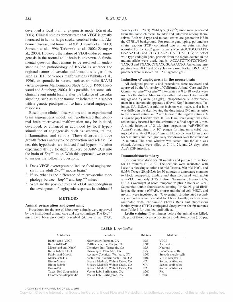
TABLE 1. Antibodies
Antibodies Vendors Dilution Markers
Rabbit aanti-VEGF NeoMarker; Fremont, CA 1:75 VEGFRat anti-GFAP CalBiochem; San Diego, CA 1:500 AstrocytesMouse and anti-NeuN Chemicon Int.: Temecula, CA 1:75 NeuronsRat anti-MEC 13.3 Pharmingen; Palo Alto, CA 1:75 Endothelial cellsRabbit anti-Myosin Accurate Chemical; Westbury, NY 1:500 Smooth muscle cellsMouse anti-Flk-1 Santa Cruz Biotech; Santa Cruz, CA 1:100 VEGF receptor IIBiotin-Mouse Biocare Medical; Walnut Creek, CA N/A Second antibodiesBiotin-Rabbit Biocare Medical; Walnut Creek, CA N/A Second antibodiesBiotin-Rat Biocare Medical; Walnut Creek, CA N/A Second antibodiesTaxes, Red-Streptavidin Vector Lab; Burlingame, CA 1:200 RedFluorescein-Streptavidin Vector Lab; Burlingame, CA 1:200 Green
B. XU ET AL.238
J Cereb Blood Flow Metab, Vol. 24, No. 2, 2004

Vector Labs, CA, U.S.A.) was injected intravenously. Micewere exsanguinated while perfused intracardially with a solu-tion of 4% paraformaldehyde in 10 mM PBS (pH 7.4). Thebrain was removed and postfixed in the same solution for 4hours then immersed in 30% sucrose solution until sedimented.The brain was then embedded in Tissue-Tek O.C.T. (SakuraFinetek, CA, U.S.A.). Coronal sections of 20-�m thicknesswere cut for lectin staining. Lectin staining is a simple andreproducible method for microvessel staining. However, it canonly stain perfused microvessels. At the early stage of angio-genesis, some microvessels may be formed, but they may be inthe “no perfusion” stage. Therefore, other evidence such as cellproliferation assay is necessary.
Microvessel counting. Two brain coronal sections from thelectin fluorescein staining brain, 1 mm frontier and 1 mm pos-terior from the needle track, were chosen for microvesselcounting. Three areas (just left, right, and bottom of the needletrack) were chosen with a 10× objective lens, and microvesselcounting was carried out on these pictures. Only vessels with aclearly defined lumen or a well-defined linear vessel shapewere taken into account. The numbers of microvessels werecalculated as the mean of the blood vessel counts obtained fromthe three images.
Microvascular morphology. A series of 40 brain coronalsections were cut around the needle track in each animal 3weeks after AdhVEGF injection. Each section (20 �m) wasexamined using confocal microscopy (Bio-Rad, Hercules, CA,U.S.A.). A section image of 1 �m was captured and recon-structed for three-dimensional observation if abnormal mi-crovessels were detected. The results were analyzed to detectthe occurrence of abnormal vessels in the Eng+/− and Eng+/+
mice.Western blot analysis. The brain tissues were homogenized
in a lysis buffer. The protein concentrations were determinedusing the BCA protein assay kits (Pierce, IL, U.S.A.). An equalamount of protein (30 �g) was loaded on 4% to 12% gradientgel (Invitrogen, CA, U.S.A.) for electrophoresis of protein.Subsequently, proteins were electroblotted onto a polyvinyl-idene difluoride membrane (PVDF) (Bio-Rad, CA, U.S.A.) ina transfer buffer. The membrane was placed in 5% nonfat milkTBS with 0.1% Tween 20 (TBST) pH 7.6 for 1 hour to blocknonspecific binding. The membrane was immunoprobed withanti-VEGF antibody (1:400 dilution, CalBiochem, San Diego,CA, U.S.A.) for 2 hours. After washing, the membrane wasincubated with horseradish-peroxidase-conjugated anti-rabbitantibody (1:20,000 dilution, Amersham, Buckinghamshire, En-gland) and then reacted with an enhanced ECL (Amersham).The membrane was wrapped in plastic and exposed to Kodakfilm. The film was developed according to the manufacturer’sinstructions. Detection of TGF-�R1 was performed in similarmanner with the appropriate antibodies.
Statistical analysisParametric data between Eng+/− and Eng+/+ mice was com-
pared with Student’s t-test. Nonparametric data were comparedusing Mann-Whiney U test. All data were presented as mean ±SD. A probability value of less than 5% was considered statis-tically significant.
RESULTS
Increased expression of VEGF and angiogenesis inAdhVEGF-transduced mice
We examined the VEGF expression in the adult mousebrain after AdhVEGF gene transfer. Dose response stud-
ies showed 2 �L of 1 × 10⁸ particles induce highestVEGF levels without visible side effect. Mortality wouldoccur if the dose was greater than 1 × 10⁹ particle levels(data not shown here). VEGF immunostaining showedthat little immunoreactivity could be detected in theAdlacZ-transduced and the saline-treated mice. How-ever, extensive VEGF immunoreactivity could be de-tected in the AdhVEGF-transduced mouse brain, and thispositive staining was localized in the AdhVEGF-injectedhemisphere (Fig. 1, top panel). Double-labeled fluores-cence studies revealed that neurons, astrocytes, endothe-lial cells, and smooth muscle cells were colocalized withVEGF in cell bodies after AdhVEGF gene transfer (Fig.1, middle panel). At day 7, VEGF-positive staining wasmainly detected in the neurons. At day 14, VEGF posi-tive staining was detected mostly in the astrocytes, and insome endothelial and smooth muscle cells, but not inneurons. At days 21 and 28, many endothelial cells andsmooth muscle cells expressed VEGF, whereas astro-cytes and neurons exhibited much less positive staining.These results demonstrated that neurons, astrocytes, en-dothelial cells, and smooth muscle cells are the mainsources of VEGF after AdhVEGF transduction. Next, wecounted the number of microvessels in the AdhVEGF-,AdlacZ-, and saline-treated mice using fluorescent lectinstaining. Compared with normal brain microvasculature,the numbers of microvessels were not significantly dif-ferent in either the AdlacZ-transduced or saline-treatedmice. However, the number of microvessels in the Adh-VEGF-transduced mice was significantly increased after2 to 4 weeks of AdhVEGF gene transfer (Fig. 1, bottompanel, P < 0.05).
VEGF increased brain microvessels in both Eng+/−
and Eng+/+ miceEng+/− and Eng+/+ mice were identified by a standard
PCR-based genotyping assay. No visible abnormal be-havior or external vascular lesions were detected in theEng+/− and Eng+/+ mice before experimental interven-tion at 2 months of age. Neither mortality nor evidenceof hemorrhage was observed during the 3 weeks ofAdhVEGF gene transfer in either the Eng+/− or Eng+/+ mice.
The first experiment demonstrated that AdhVEGF-induced brain angiogenesis occurred between 2 to 4weeks. Therefore, we counted the number of microves-sels in the Eng+/− and Eng+/+ mice after 3 weeks ofAdhVEGF transduction. The number of microvessels inthe AdhVEGF injection site was significantly increasedin both the Eng+/− and Eng+/+ mice compared with thesaline-treated mice (Fig. 2, left panel). However, the in-creased numbers of microvessels were not significantlydifferent between the Eng+/− and Eng+/+ mice (Fig. 2,right panel, P > 0.05). This shows that AdhVEGF trans-duction can induce focal angiogenesis in adult mousebrain tissue in both Eng+/− and Eng+/+ mice.
VEGF IN ENG+/− 239
J Cereb Blood Flow Metab, Vol. 24, No. 2, 2004

Abnormal microvessel developed in the Eng+/− miceWe next examined the microvascular morphologic
changes. Forty coronal sections in each animal were ex-amined under confocal microscopy. In the Eng+/+ group,we observed angiogenic changes, such as dilated“mother” vessels and newly formed sprouts, but no ab-normal microvasculature was detected in any of the ninemice. However, we observed abnormal microvasculaturein eight of the nine Eng+/− mice (Eng+/− vs. Eng+/+; 8/9vs. 0/9, P < 0.05). These abnormal microvessels usuallydeveloped in the ipsilateral cortex or the abundant mi-crovessel areas adjacent to the needle track. These mi-crovessels showed abnormal patterns such as a massiveor single enlargement, clustered, twisted, or spiral mi-crovessels (Table 2 and Fig. 3a). In three-dimensionalreconstruction sections, these microvessels clearlyshowed abnormal vascular structure (Fig. 3b). Thisshows that VEGF hyperstimulation can drive abnormalmicrovessel formation in the adult Eng+/− mice.
Flk-1 expressionTo determine the success of AdhVEGF gene transfer,
we also measured expression of VEGF and Flk-1 in theEng+/− mice after AdhVEGF gene transduction. VEGFexpression was greatly increased both in the Eng+/− andEng+/+ mice, and this increase persisted at least 3 weeksafter AdhVEGF transduction (Fig. 1, top panel). Flk-1positive staining was mainly colocalized on the mi-crovessels. Of interest is the fact that the induced level ofFlk-1 expression on microvessels in the Eng+/− mice wasgreatly reduced compared with the Eng+/+ mice (Fig. 4,P < 0.05).
T�R-1 expressionTo identify the role of endoglin during angiogenesis,
we examined brain T�R1 expression in the Eng+/− mice.Western blot analysis demonstrated that T�R-1 expres-sion was comparable in the Eng+/− and Eng+/+ mice after3 weeks of saline injection. Expression of T�R-1 in-
FIG. 1. (Upper left) One experiment of VEGF expression inWestern blot analysis. VEGF shows a major band at 45 kDa innonreducing gels. VEGF expression in injection region is greatlyincreased after 3 weeks of AdhVEGF transduction. VEGF ex-pression is not significantly different between the Eng+/+ and theEng+/− mice. (Bottom left) VEGF immunostaining was performedafter 7 days of AdhVEGF transduction. There is no VEGF positivestaining in the saline-injected mice. However, VEGF positivestaining was detected in the basal ganglia of AdhVEGF-injectedmice (arrows). LV, lateral ventricle; BG, basal ganglia. Bar = 500µm. (Middle) Photomicrographs show double-labeled immuno-staining in the mouse brain after AdhVEGF gene transfer. Greenfluorescence staining with anti NeuN, GFAP, MEC, and Myosinantibodies, which identify these positive cells as neurons (A),astrocytes (D), endothelial cells (G), and smooth muscle cells (J).VEGF positive staining (B, E, H, and K, red color) is detected inthe ipsilateral hemisphere adjacent to the AdhVEGF-injected re-gion, especially near the needle track. C, F, I, and L show colo-calization of green and red color, indicating neurons, astrocytes,endothelial cell, and smooth muscle cells are the source of brainVEGF after AdhVEGF gene transfer. Bar = 50 µm. (Upper right)Bar graph shows the number of lectin-positive staining microves-sels in the AdhVEGF-, AdlacZ-transduced, and saline-treatedmouse brain after 1 to 4 weeks of AdhVEGF injection. Lectin-positive microvessels are counted at the sections +0.5 and −0.5mm related to needle track. Values are mean ± SD, N = 6 pergroup. * P < 0.05, AdhVEGF group vs. AdlacZ or saline treatedgroups. VEGF, vascular endothelial growth factor.
B. XU ET AL.240
J Cereb Blood Flow Metab, Vol. 24, No. 2, 2004

creased after AdhVEGF transduction in both the Eng+/−
and Eng+/+ mice. However, the increase of T�R-1 in theEng+/− mice was greatly attenuated compared with theEng+/+ mice (Fig. 5, P < 0.05). This result suggests thatT�R-1 expression is closely related to endoglin levels.
TABLE 2. Abnormal microvasculature in Eng+/− mice
Abnormal type Eng+/− (N � 9) Eng+/+ (N � 9)
Mass enlargement 1 0Single enlargement 2 0Vessel clusters 2 0Twisted 2 0Spirals microvessels 2 0
FIG. 2. (Upper panel) Lectin-stained microvessels in the Eng+/+
(A and B) and the Eng+/− (C and D) mouse brain after 3 weeks ofsaline or AdhVEGF gene transduction. The number of microves-sels appears comparable in the Eng+/+ and the Eng+/− mousebrain after saline- (A and C) or AdhVEGF-treated (B and D) mice.Bar = 25 µm. (Bottom Bar Graph) Number of lectin staining mi-crovessels in the Eng+/+ and the Eng+/− mouse brain after 3weeks of saline or AdhVEGF transduction. Values are mean ±SD, N = 9 per group. The number of microvessels was signifi-cantly increased after 3 weeks of AdhVEGF transduction in boththe Eng+/+ and the Eng+/− mice. However, there was no statisticaldifference between the Eng+/+ and the Eng+/− mice (P > 0.05).
FIG. 3A. Changes of microvasculature morphology in the Eng+/+
and Eng+/− mouse brain after VEGF overexpression. No abnor-mal microvasculature after saline injection was detected in theEng+/− mice (A). Typical angiogenic changes are detected in theEng+/+ mice after AdhVEGF transduction but no abnormal micro-vasculature (B). Abnormal microvasculature morphology is de-tected in the Eng+/− mouse brain. These abnormal microvesselscould display a mass (C), single enlargement (D and E), vesselclusters (F), twisted (G and H), or spirals microvessels (I). Arrowsindicated the localized abnormal microvasculature. The abnormalmicrovasculature development is mainly observed in the ipsilat-eral cortex near the needle track. Bar = 50 µm. VEGF, vascularendothelial growth factor.
FIG. 3B. Example of three-dimensional imaging of cerebral mi-crovessels in an Eng+/− mouse using a confocal microscope. Aseries of 1-µm thick sections are reconstructed, and 20 differentangle pictures were taken. The arrows indicate the morphology ofabnormal microvessels from different angles. Bar = 50 µm.
VEGF IN ENG+/− 241
J Cereb Blood Flow Metab, Vol. 24, No. 2, 2004

DISCUSSION
The most important finding in this study is that VEGFhyperstimulation promotes angiogenesis to a similar de-gree in the adult endoglin haploinsufficient and wild typemice but that the incidence of abnormal microvessel for-mation in endoglin haploinsufficient mice is dramaticallyhigher. Furthermore, we demonstrated that Flk-1 andT�R-1 expression was reduced in the Eng+/− mice, sug-gesting that haploinsufficiency of endoglin may down-regulate Flk-1 and T�R-1 activation. These changes mayresult in the development of abnormal microvessels inthe Eng+/− mice.
We demonstrated that AdhVEGF transduction notonly induces focal angiogenesis in the adult mouse brainbut also promotes abnormal microvessel formation in theEng+/− mice. This result provides a possible mechanismfor the genesis of focal vascular malformations on themicroscopic level. It may be consistent with the hypoth-esis that both mucocutaneous fragility and eventual for-mation of solid organ AVM in adult patients with HHTmay be linked to a local increase in VEGF and associatedsignaling pathways (Guttmacher et al., 1995; Plauchu etal., 1989; Shovlin and Letarte, 1999). The variable HHTphenotype in the mouse models may reflect the widerange of disease severity in patients with HHT1, and, inalmost all cases, only very localized sites are affected. Ithas therefore been proposed that other factors can locallyprecipitate the disease. This study shows that localVEGF release may be such a factor.
Mice carrying two independently derived targeted mu-tations in the endoglin gene have been extensively stud-ied as a model of HHT1 (Bourdeau et al., 2001; Torsneyet al., 2003). In both cases, the frequency of developmentof HHT-like bleeding vessels in Eng+/− mice was foundto depend upon the background strain. The lesions de-veloped at a much lower frequency and at a later age inthe C57BL/6 background compared with 129/Ola. Fur-thermore, abnormal cerebral vessels have only been re-ported in C57BL/6mice aged 25 to 40 weeks (Satomi etal., 2003). We therefore used young (8 to 10 weeks old)Eng+/− mice in C57BL/6 background for our study be-cause HHT had not been seen in young mice in thisbackground. We also found that Eng+/− mice in theC57BL/6 background show no visible abnormal vascularstructure or hemorrhage up to 12 weeks of age.
We demonstrated that a high incidence of abnormalmicrovessels occurred in eight of nine Eng+/− mice afterAdhVEGF gene transduction but did not occur in the
FIG. 5. (Upper) One experiment of TGF-�R1 expression inWestern blot analysis. TGF-�R1 presents in the major band at 55kDa. TGF-�R1 is greatly increased both in the Eng+/+ and Eng+/−
mice after 3 weeks of AdhVEGF transduction. (Lower) Bar graphshows that VEGF induced TGF-�R1 expression in the Eng+/+ andEng+/− mice after 3 weeks of saline or AdhVEGF transduction.Values are mean ± SD, N = 3 per group. *P < 0.05, TGF-�R1expression is attenuated in the Eng+/− mice compared with theEng+/+ mice after AdhVEGF injection.-
FIG. 4. Bar graph shows the Flk-1 positive cell index examina-tion in the Eng+/− and Eng+/+ mice after 3 weeks of saline orAdhVEGF transduction. Values are mean ± SD, N=9 per group.*P < 0.05, Flk-1 positive index is much lower in the Eng+/− micecompared with Eng+/+ mice.
B. XU ET AL.242
J Cereb Blood Flow Metab, Vol. 24, No. 2, 2004

nine Eng+/+ mice, suggesting that the blood vessels inEng+/− mice are abnormal in their response to VEGF,which indicates that during physiologic events that causeVEGF overexpression such as ischemia, inflammation,or wound repair, abnormal microvessels could develop.
Human HHT lesions are extremely variable and in-clude dilated vessels, arteriovenous malformations, andcapillary telangiectasias. However, it is not known howthese abnormalities develop and what the starting pointis. Cerebral lesions in HHT patients are detected onlyafter they are symptomatic, whereas in the mouse model,we are able to investigate the development of the smallabnormal vessels seen in this study. We have shown thatAdhVEGF-induced angiogenic morphological changesoccurred in the ipsilateral hemisphere in the Eng+/+ mice.These changes included angiogenic “sprouts,” tightlypacked “glomerulold” structures, bridging, and dilated“mother vessels.” We also identified similar brain angio-genic changes in other species such as CD-1 mice afterAdhVEGF transduction (Xu et al., 2003). However,VEGF overexpression induced different angiogenicchanges in the Eng+/− mouse brain. These newly formedmicrovessels showed abnormal morphology such as amass or single enlargement, clustered, twisted, or spiralmicrovessels. Confocal microscopy and three-dimen-sional reconstruction techniques enable clear identifica-tion of the abnormal microvasculature without having toresort to scanning electron microscopy (Satomi et al.,2003). These changes are similar to small cerebralAVMs in the adult HHT patient brain and may predis-pose the mice to brain hemorrhage.
There are two main features in patients with HHT:abnormal vessels and bleeding. Not all abnormal vesselsbleed, but they can cause severe effects with relation tothe affected organ. For example, dilated vessels in thebrain may cause blood brain barrier leakage and edema.On the other hand, bleeding generally occurs from theabnormal vessels. Thus these two features of HHT arerelated but not the same. We have no evidence that thesevessels in the mice are those that will bleed. We can onlysurmise that these small areas of abnormal morphologyare likely to represent the beginning of a more maturenidus seen in patients.
The mechanism of abnormal microvessel formation inEng+/− mouse brain is unclear. Flk-1 levels are reducedin Eng+/− mice, which is an important finding. Duringstable homeostasis, Flk-1 expression usually falls withinwith the normal range in the Eng+/+ mice (Shalaby et al.,1995). However, during VEGF hyperstimulation, endo-thelial cells and smooth muscle cells may be unable toproduce sufficient Flk-1 for a normal response to theangiogenesis in the Eng+/− mice. As a result, cell prolif-eration and migration would be affected. In support ofthis hypothesis, the Eng+/− mice exhibit abnormal vesselswith irregular layers of collagen and elastin, adding to
the fragility of these vessels (Torsney et al., 2003). Un-balanced proliferation and migration of different celltype could cause newly formed microvessel instabilityand subsequent abnormal microvessel formation duringangiogenesis.
Endoglin has been shown to be a negative modulatorof TGF-� signaling (Lastres et al., 1996). Local endoglinlevels may play a crucial role in the control of the TGF-�1 autoregulatory pathway. We found that T�R-1 ex-pression was significantly reduced in the Eng+/− mice,suggesting that endoglin can control the TGF-�1 auto-regulatory pathway. TGF-�1 is a critical cytokine thathelps to stabilize and mature the newly formed microves-sels in the adult mice. A decrease in either TGF-�1 orTGF-�1 receptors can lead to unstable cellular interac-tions in the vessel wall, dilated vessels, and vascularabnormalities. Such alterations could damage other an-giogenic regulatory mechanisms and lead to deteriora-tion of the vascular network associated with the progres-sion of HHT (Bourdeau et al., 2000).
Despite these observations, it is still unclear how en-doglin affects TGF-� signaling during angiogenesis andhow this signaling pathway regulates newly formed mi-crovessel maturation. Our results are consistent with thehypothesis that VEGF may play a role in the develop-ment of vascular malformations, although the exactmechanism by which this occurs remains to be deter-mined. It is likely that factors other than the simple ad-dition of VEGF contribute to the development of abnor-mal vessels because otherwise one might expect to seelesions in tissues where VEGF was normally upregulatedduring normal physiologic states, such as the endometri-um during menses, or routine scar formation. We suggestinstead that there are local tissue considerations that alsoinfluence the development of AVM.
CONCLUSION
We have demonstrated that VEGF hyperstimulationinduces abnormal microvessels in the Eng+/− mousebrain but not in the brains of wild type animals. TheEng+/− mice appeared otherwise normal during the cur-rent observation period in the early stage of life. Whenchallenged by environmental changes that upregulate an-giogenic processes, endoglin haploinsufficiency appearsto cause downregulation of the TGF-�1 pathway, result-ing in abnormal microvessel formation. These resultssuggest that VEGF plays a role in the development ofarteriovenous malformation in adult patients with HHT.
Acknowledgments. The authors thank Carroll Schreibmanand Broderick Belenson for editorial assistance and the col-laborative support staff of the CCR http://avm.ucsf.edu.
REFERENCESArteriovenous Malformation Study Group (1999) Current concepts:
arteriovenous malformations of the brain in adults. N Engl J Med340:1812–1818
VEGF IN ENG+/− 243
J Cereb Blood Flow Metab, Vol. 24, No. 2, 2004

Arthur HM, Ure J, Smith AJ, Renforth G, Wilson DI, Torsney E,Charlton R, Parums DV, Jowett T, Marchuk DA, Burn J, DiamondAG (2000) Endoglin, an ancillary TGFbeta receptor, is required forextraembryonic angiogenesis and plays a key role in heart devel-opment. Dev Biol 217:42–53
Bourdeau A, Cymerman U, Paquet ME, Meschino W, McKinnon WC,Guttmacher AE, Becker L, Letarte M (2000) Endoglin expressionis reduced in normal vessels but still detectable in arteriovenousmalformations of patients with hereditary hemorrhagic telangiec-tasia type 1. Am J Pathol 156:911–923
Bourdeau A, Faughnan ME, McDonald ML, Paterson AD, Wanless IR,Letarte M (2001) Potential role of modifier genes influencingtransforming growth factor- beta1 levels in the development ofvascular defects in endoglin heterozygous mice with hereditaryhemorrhagic telangiectasia. Am J Pathol 158:2011–2020
Cheifetz S, Bellon T, Cales C, Vera S, Bernabeu C, Massague J, LetarteM (1992) Endoglin is a component of the transforming growthfactor-beta receptor system in human endothelial cells. J BiolChem 267:19027–19030
Fleetwood IG, Steinberg GK (2002) Arteriovenous malformations.Lancet 359:863–873
Gougos A, Letarte M (1990) Primary structure of endoglin, an RGD-containing glycoprotein of human endothelial cells. J Biol Chem265:8361–8364
Guttmacher AE, Marchuk DA, White RI (1995) Hereditary hemor-rhagic telangiectasia. N Engl J Med 333:918–924
Hayashi T, Noshita N, Sugawara T, Chan PH (2003) Temporal profileof angiogenesis and expression of related genes in the brain afterischemia. J Cereb Blood Flow Metab 23:166–180
Lastres P, Letamendia A, Zhang H, Rius C, Almendro N, Raab U,Lopez LA, Langa C, Fabra A, Letarte M, Bernabeu C (1996)Endoglin modulates cellular responses to TGF-beta 1. J Cell Biol133:1109–1121
Li DY, Sorensen LK, Brooke BS, Urness LD, Davis EC, Taylor DG,Boak BB, Wendel DP (1999) Defective angiogenesis in mice lack-ing endoglin. Science 284:1534–1537
Plauchu H, de Chadarevian JP, Bideau A, Robert JM (1989) Age-related clinical profile of hereditary hemorrhagic telangiectasia inan epidemiologically recruited population. Am J Med Genet32:291–297
Satomi J, Mount RJ, Toporsian M, Paterson AD, Wallace MC, Harri-son RV, Letarte M (2003) Cerebral vascular abnormalities in amurine model of hereditary hemorrhagic telangiectasia. Stroke34:783–789
Shalaby F, Rossant J, Yamaguchi TP, Gertsenstein M, Wu XF, Breit-man ML, Schuh AC (1995) Failure of blood-island formation andvasculogenesis in Flk-1-deficient mice. Nature 376:62–66
Shovlin CL, Letarte M (1999) Hereditary hemorrhagic telangiectasiaand pulmonary arteriovenous malformations: issues in clinicalmanagement and review of pathogenic mechanisms. Thorax54:714–729
Sonstein WJ, Kader A, Michelsen WJ, Llena JF, Hirano A, Casper D(1996) Expression of vascular endothelial growth factor in pediat-ric and adult cerebral arteriovenous malformations: an immunocy-tochemical study. J Neurosurg 85:838–845
Tarkowski E, Issa R, Sjogren M, Wallin A, Blennow K, Tarkowski A,Kumar P (2002) Increased intrathecal levels of the angiogenicfactors VEGF and TGF-beta in Alzheimer’s disease and vasculardementia. Neurobiol Aging 23:237–243
Torsney E, Charlton R, Diamond AG, Burn J, Soames JV, Arthur HM(2003) Mouse model for hereditary hemorrhagic telangiectasia hasa generalized vascular abnormality. Circulation 107:1653–1657
van den Driesche S, Mummery CL, Westermann CJ (2003) Hereditaryhemorrhagic telangiectasia: an update on transforming growth fac-tor beta signaling in vasculogenesis and angiogenesis. CardiovascRes 58:20–31
Vikkula M, Boon LM, Carraway KL, Calvert JT, Diamonti AJ,Goumnerov B, Pasyk KA, Marchuk DA, Warman ML, CantleyLC, Mulliken JB, Olsen BR (1996) Vascular dysmorphogenesiscaused by an activating mutation in the receptor tyrosine kinasetie2. Cell 87:1181–1190
Xu B, Yang GY, Hashimoto T, Huey M, Young WL (2003) Stereotaticinjection of vascular endothelial growth factor adenovirus inducedfocal angiogenesis in mature mouse brain. J Cereb Blood FlowMetab 23:202
Zhang ZG, Zhang L, Jiang Q, Zhang R, Davies K, Powers C, BruggenN, Chopp M (2000) VEGF enhances angiogenesis and promotesblood-brain barrier leakage in the ischemic brain. J Clin Invest106:829–838
B. XU ET AL.244
J Cereb Blood Flow Metab, Vol. 24, No. 2, 2004
Copyright © 2022 FDOKUMEN