Gene delivery nanoparticles specific for human microvasculature and macrovasculature
8
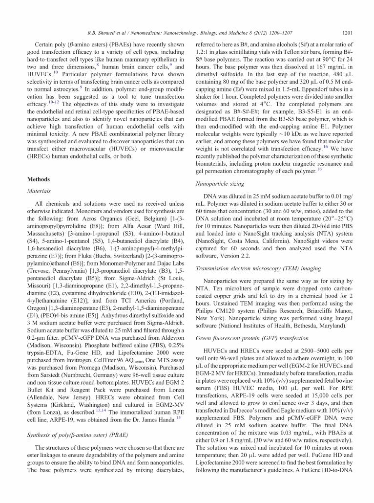
Research Article Gene delivery nanoparticles specific for human microvasculature and macrovasculature Ron B. Shmueli, BS a,1 , Joel C. Sunshine, MS a,1 , Zhenhua Xu, PhD b , Elia J. Duh, MD b , Jordan J. Green, PhD a,b, ⁎ a Department of Biomedical Engineering, the Johns Hopkins University School of Medicine, Baltimore, Maryland, USA b The Wilmer Eye Institute, the Johns Hopkins University School of Medicine, Baltimore, Maryland, USA Received 3 September 2011; accepted 16 January 2012 Abstract Endothelial cell dysfunction is a critical component of ocular diseases such as age-related macular degeneration and diabetic retinopathy. An important limitation in endothelial cell research is the difficulty in achieving efficient transfection of these cells. A new polymer library was here synthesized and utilized to find polymeric nanoparticles that can transfect macrovascular (human umbilical vein, HUVECs) and microvascular (human retinal, HRECs) endothelial cells. Nanoparticles were synthesized that can achieve transfection efficiency of up to 85% for HRECs and 65% for HUVECs. These nanoparticle systems enable high levels of expression while avoiding problems associated with viral gene delivery. The polymeric nanoparticles also show cell-specific behavior, with a high correlation between microvascular and macrovascular transfection (R 2 = 0.81) but low correlation between retinal endothelial and retinal epithelial transfection (R 2 = 0.21). These polymeric nanoparticles can be used in vitro as experimental tools and potentially in vivo to target and treat vascular-specific diseases. From the Clinical Editor: Polymeric nanoparticles were synthesized with the goal of transfecting endothelial cells, which are commonly considered difficult targets. The authors report excellent transfection efficiency of up to 85% for human retinal and 65% for human umbilical vein endothelial cells. These NPs can be used in vitro as experimental tools and potentially in vivo to target and treat vascular-specific diseases. © 2012 Elsevier Inc. All rights reserved. Key words: Nonviral gene delivery; Nanoparticles; Ocular diseases; Human retinal endothelial cells Endothelial cells play important roles in various ocular diseases, such as age-related macular degeneration, diabetic retinopathy, and retinoblastoma. 1 The dysregulation and subse- quent angiogenic proliferation of these ocular microvascular endothelial cells represents the key step in most retinal causes of blindness. Regulation of this angiogenesis by antiangiogenic drugs such as ranibizumab is now the first-line therapy, and there is continued interest in more effective antiangiogenic therapies. 2 Gene delivery is one such alternative method for delivery of antiangiogenic factors to endothelial cells, such as endostatin, angiostatin, and vascular endothelial growth factor (VEGF)– binding protein. 3,4 In addition, gene therapy can be used to correct specific genetic deficiencies within the endothelial cell population. In this strategy, therapeutic genes that can either add or block a function are delivered to a targeted cell population. Additionally, such gene delivery methods can also be very useful in the study of cellular biology and disease. Nonviral transfection of human retinal endothelial cells (HRECs) remains a challenge, as does transfection of many other cell types. For example, one recent report using lipid- coated magnetic nanoparticles achieved only ∼5% transfection efficacy. 5 Leading commercial reagents, such as Lipofectamine 2000 (Invitrogen, Carlsbad, California), can improve the transfection of HRECs. One study found that this approach could lead to 42–67% knockdown of a target receptor's surface expression following plasmid transfection. 6 Macrovascular (human umbilical vein, HUVECs) endothelial cells are also generally difficult to transfect, as a lead polymer (polyethyleni- mine) plus magnetofection yielded transfection efficacy of only 39% positive cells. 7 New nanomedicines are needed to further increase the effectiveness of nonviral gene delivery. BASIC SCIENCE Nanomedicine: Nanotechnology, Biology, and Medicine 8 (2012) 1200 – 1207 nanomedjournal.com The work was supported in part by the Maryland Technology Development Corporation–Maryland Stem Cell Research Fund (2009- MSCRFE-0098-00), the National Institutes of Health (R21CA152473), and the Medical Scientist Training Program (to J.C.S.). E.J.D. is supported by a Career Development Award from Research to Prevent Blindness. ⁎ Corresponding author: Department of Biomedical Engineering and the Wilmer Eye Institute, The Johns Hopkins University School of Medicine, Baltimore, Maryland 21231, USA. E-mail address: [email protected] (J.J. Green). 1 These authors contributed equally to this manuscript. 1549-9634/$ – see front matter © 2012 Elsevier Inc. All rights reserved. doi:10.1016/j.nano.2012.01.006 Please cite this article as: R.B. Shmueli, J.C. Sunshine, Z. Xu, E.J. Duh, J.J. Green. Gene delivery nanoparticles specific for human microvasculature and macrovasculature. Nanomedicine: NBM 2012;8:1200-1207, doi:10.1016/j.nano.2012.01.006
-
Upload
johnshopkins -
Category
Documents
-
view
2 -
download
0
Transcript of Gene delivery nanoparticles specific for human microvasculature and macrovasculature

BASIC SCIENCE
Nanomedicine: Nanotechnology, Biology, and Medicine8 (2012) 1200–1207
Research Article
Gene delivery nanoparticles specific for human microvasculatureand macrovasculature
Ron B. Shmueli, BSa,1, Joel C. Sunshine, MSa,1, Zhenhua Xu, PhDb,Elia J. Duh, MDb, Jordan J. Green, PhDa,b,⁎
aDepartment of Biomedical Engineering, the Johns Hopkins University School of Medicine, Baltimore, Maryland, USAbThe Wilmer Eye Institute, the Johns Hopkins University School of Medicine, Baltimore, Maryland, USA
Received 3 September 2011; accepted 16 January 2012
nanomedjournal.com
Abstract
Endothelial cell dysfunction is a critical component of ocular diseases such as age-related macular degeneration and diabetic retinopathy.An important limitation in endothelial cell research is the difficulty in achieving efficient transfection of these cells. A new polymer librarywas here synthesized and utilized to find polymeric nanoparticles that can transfect macrovascular (human umbilical vein, HUVECs) andmicrovascular (human retinal, HRECs) endothelial cells. Nanoparticles were synthesized that can achieve transfection efficiency of up to85% for HRECs and 65% for HUVECs. These nanoparticle systems enable high levels of expression while avoiding problems associatedwith viral gene delivery. The polymeric nanoparticles also show cell-specific behavior, with a high correlation between microvascular andmacrovascular transfection (R2 = 0.81) but low correlation between retinal endothelial and retinal epithelial transfection (R2 = 0.21). Thesepolymeric nanoparticles can be used in vitro as experimental tools and potentially in vivo to target and treat vascular-specific diseases.
From the Clinical Editor: Polymeric nanoparticles were synthesized with the goal of transfecting endothelial cells, which are commonlyconsidered difficult targets. The authors report excellent transfection efficiency of up to 85% for human retinal and 65% for human umbilicalvein endothelial cells. These NPs can be used in vitro as experimental tools and potentially in vivo to target and treat vascular-specific diseases.© 2012 Elsevier Inc. All rights reserved.
Key words: Nonviral gene delivery; Nanoparticles; Ocular diseases; Human retinal endothelial cells
Endothelial cells play important roles in various oculardiseases, such as age-related macular degeneration, diabeticretinopathy, and retinoblastoma.1 The dysregulation and subse-quent angiogenic proliferation of these ocular microvascularendothelial cells represents the key step in most retinal causes ofblindness. Regulation of this angiogenesis by antiangiogenicdrugs such as ranibizumab is now the first-line therapy, and thereis continued interest in more effective antiangiogenic therapies.2
Gene delivery is one such alternative method for delivery ofantiangiogenic factors to endothelial cells, such as endostatin,
The work was supported in part by the Maryland TechnologyDevelopment Corporation–Maryland Stem Cell Research Fund (2009-MSCRFE-0098-00), the National Institutes of Health (R21CA152473), andthe Medical Scientist Training Program (to J.C.S.). E.J.D. is supported by aCareer Development Award from Research to Prevent Blindness.
⁎Corresponding author: Department of Biomedical Engineering and theWilmer Eye Institute, The Johns Hopkins University School of Medicine,Baltimore, Maryland 21231, USA.
E-mail address: [email protected] (J.J. Green).1 These authors contributed equally to this manuscript.
1549-9634/$ – see front matter © 2012 Elsevier Inc. All rights reserved.doi:10.1016/j.nano.2012.01.006
Please cite this article as: R.B. Shmueli, J.C. Sunshine, Z. Xu, E.J. Duh, J.J. Grmacrovasculature. Nanomedicine: NBM 2012;8:1200-1207, doi:10.1016/j.nano
angiostatin, and vascular endothelial growth factor (VEGF)–binding protein.3,4 In addition, gene therapy can be used tocorrect specific genetic deficiencies within the endothelial cellpopulation. In this strategy, therapeutic genes that can either addor block a function are delivered to a targeted cell population.Additionally, such gene delivery methods can also be very usefulin the study of cellular biology and disease.
Nonviral transfection of human retinal endothelial cells(HRECs) remains a challenge, as does transfection of manyother cell types. For example, one recent report using lipid-coated magnetic nanoparticles achieved only ∼5% transfectionefficacy.5 Leading commercial reagents, such as Lipofectamine2000 (Invitrogen, Carlsbad, California), can improve thetransfection of HRECs. One study found that this approachcould lead to 42–67% knockdown of a target receptor's surfaceexpression following plasmid transfection.6 Macrovascular(human umbilical vein, HUVECs) endothelial cells are alsogenerally difficult to transfect, as a lead polymer (polyethyleni-mine) plus magnetofection yielded transfection efficacy of only39% positive cells.7 New nanomedicines are needed to furtherincrease the effectiveness of nonviral gene delivery.
een. Gene delivery nanoparticles specific for human microvasculature and.2012.01.006
1201R.B. Shmueli et al / Nanomedicine: Nanotechnology, Biology, and Medicine 8 (2012) 1200–1207
Certain poly (β-amino esters) (PBAEs) have recently showngood transfection efficacy to a variety of cell types, includinghard-to-transfect cell types like human mammary epithelium intwo and three dimensions,8 human brain cancer cells,9 andHUVECs.10 Particular polymer formulations have shownselectivity in terms of transfecting brain cancer cells as comparedto normal astrocytes.9 In addition, polymer end-group modifi-cation has been suggested as a tool to tune transfectionefficacy.10-12 The objectives of this study were to investigatethe endothelial and retinal cell-type specificities of PBAE-basednanoparticles and also to identify novel nanoparticles that canachieve high transfection of human endothelial cells withminimal toxicity. A new PBAE combinatorial polymer librarywas synthesized and evaluated to discover nanoparticles that cantransfect either macrovascular (HUVECs) or microvascular(HRECs) human endothelial cells, or both.
Methods
Materials
All chemicals and solutions were used as received unlessotherwise indicated.Monomers and vendors used for synthesis arethe following: from Acros Organics (Geel, Belgium) [1-(3-aminopropyl)pyrrolidine (E8)]; from Alfa Aesar (Ward Hill,Massachusetts) [3-amino-1-propanol (S3), 4-amino-1-butanol(S4), 5-amino-1-pentanol (S5), 1,4-butanediol diacrylate (B4),1,6-hexanediol diacrylate (B6), 1-(3-aminopropyl)-4-methylpi-perazine (E7)]; from Fluka (Buchs, Switzerland) [2-(3-aminopro-pylamino)ethanol (E6)]; from Monomer-Polymer and Dajac Labs(Trevose, Pennsylvania) [1,3-propanediol diacrylate (B3), 1,5-pentanediol diacrylate (B5)]; from Sigma-Aldrich (St Louis,Missouri) [1,3-diaminopropane (E1), 2,2-dimethyl-1,3-propane-diamine (E2), cystamine dihydrochloride (E10), 2-(1H-imidazol-4-yl)ethanamine (E12)]; and from TCI America (Portland,Oregon) [1,3-diaminopentane (E3), 2-methyl-1,5-diaminopentane(E4), (PEO)4-bis-amine (E5)]. Anhydrous dimethyl sulfoxide and3 M sodium acetate buffer were purchased from Sigma-Aldrich.Sodium acetate buffer was diluted to 25mM and filtered through a0.2-μm filter. pCMV-eGFP DNA was purchased from Aldevron(Madison, Wisconsin). Phosphate buffered saline (PBS), 0.25%trypsin-EDTA, Fu-Gene HD, and Lipofectamine 2000 werepurchased from Invitrogen. CellTiter 96 AQueous One MTS assaywas purchased from Promega (Madison, Wisconsin). Purchasedfrom Sarstedt (Numbrecht, Germany) were 96-well tissue cultureand non-tissue culture round-bottom plates. HUVECs and EGM-2Bullet Kit and Reagent Pack were purchased from Lonza(Allendale, New Jersey). HRECs were obtained from CellSystems (Kirkland, Washington) and cultured in EGM2-MV(from Lonza), as described.13,14 The immortalized human RPEcell line, ARPE-19, was obtained from the Dr. James Handa.15
Synthesis of poly(β-amino ester) (PBAE)
The structures of these polymers were chosen so that there areester linkages to ensure degradability of the polymers and aminegroups to ensure the ability to bind DNA and form nanoparticles.The base polymers were synthesized by mixing diacrylates,
referred to here as B#, and amino alcohols (S#) at a molar ratio of1.2:1 in glass scintillating vials with Teflon stir bars, forming B#-S# base polymers. The reaction was carried out at 90oC for 24hours. The base polymer was then dissolved at 167 mg/mL indimethyl sulfoxide. In the last step of the reaction, 480 μLcontaining 80 mg of the base polymer and 320 μL of 0.5 M end-capping amine (E#) were mixed in 1.5-mL Eppendorf tubes in ashaker for 1 hour. Completed polymers were divided into smallervolumes and stored at 4oC. The completed polymers aredesignated as B#-S#-E#; for example, B3-S5-E1 is an end-modified PBAE formed from the B3-S5 base polymer, which isthen end-modified with the end-capping amine E1. Polymermolecular weights were typically ∼10 kDa as we have reportedearlier, and among these polymers we have found that molecularweight is not correlated with transfection efficacy.16 We haverecently published the polymer characterization of these syntheticbiomaterials, including proton nuclear magnetic resonance andgel permeation chromatography of each polymer.16
Nanoparticle sizing
DNAwas diluted in 25 mM sodium acetate buffer to 0.01 mg/mL. Polymer was diluted in sodium acetate buffer to either 30 or60 times that concentration (30 and 60 w/w, ratios), added to theDNA solution and incubated at room temperature (20o–25oC)for 10 minutes. Nanoparticles were then diluted 20-fold into PBSand loaded into a NanoSight tracking analysis (NTA) system(NanoSight, Costa Mesa, California). NanoSight videos werecaptured for 60 seconds and then analyzed used the NTAsoftware, Version 2.2.
Transmission electron microscopy (TEM) imaging
Nanoparticles were prepared the same way as for sizing byNTA. Ten microliters of sample were dropped onto carbon-coated copper grids and left to dry in a chemical hood for 2hours. Unstained TEM imaging was then performed using thePhilips CM120 system (Philips Research, Briarcliffs Manor,New York). Nanoparticle sizing was performed using ImageJsoftware (National Institutes of Health, Bethesda, Maryland).
Green fluorescent protein (GFP) transfection
HUVECs and HRECs were seeded at 2500–5000 cells perwell onto 96-well plates and allowed to adhere overnight, in 100μL of the appropriate medium per well (EGM-2 for HUVECs andEGM-2MV for HRECs). Immediately before transfection, mediain plates were replaced with 10% (v/v) supplemented fetal bovineserum (FBS) HUVEC media, 100 μL per well. For RPEtransfections, ARPE-19 cells were seeded at 15,000 cells perwell and allowed to grow to confluence over 3 days, and thentransfected inDulbecco's modified Eaglemediumwith 10% (v/v)supplemented FBS. Polymers and pCMV-eGFP DNA werediluted in 25 mM sodium acetate buffer. The final DNAconcentration of the mixture was 0.03 mg/mL, with PBAEs ateither 0.9 or 1.8 mg/mL (30 w/w and 60 w/w ratios, respectively).The solution was mixed and incubated for 10 minutes at roomtemperature; then 20 μL were added per well. FuGene HD andLipofectamine 2000were screened to find the best formulation byfollowing the manufacturer's guidelines. A FuGene HD-to-DNA
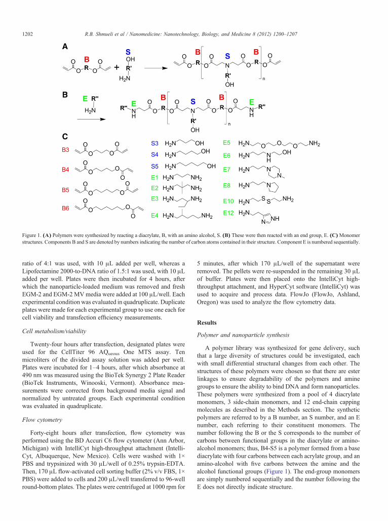
Figure 1. (A) Polymers were synthesized by reacting a diacrylate, B, with an amino alcohol, S. (B) These were then reacted with an end group, E. (C)Monomerstructures. Components B and S are denoted by numbers indicating the number of carbon atoms contained in their structure. Component E is numbered sequentially.
1202 R.B. Shmueli et al / Nanomedicine: Nanotechnology, Biology, and Medicine 8 (2012) 1200–1207
ratio of 4:1 was used, with 10 μL added per well, whereas aLipofectamine 2000-to-DNA ratio of 1.5:1 was used, with 10 μLadded per well. Plates were then incubated for 4 hours, afterwhich the nanoparticle-loaded medium was removed and freshEGM-2 and EGM-2 MVmedia were added at 100 μL/well. Eachexperimental condition was evaluated in quadruplicate. Duplicateplates were made for each experimental group to use one each forcell viability and transfection efficiency measurements.
Cell metabolism/viability
Twenty-four hours after transfection, designated plates wereused for the CellTiter 96 AQueous One MTS assay. Tenmicroliters of the divided assay solution was added per well.Plates were incubated for 1–4 hours, after which absorbance at490 nm was measured using the BioTek Synergy 2 Plate Reader(BioTek Instruments, Winooski, Vermont). Absorbance mea-surements were corrected from background media signal andnormalized by untreated groups. Each experimental conditionwas evaluated in quadruplicate.
Flow cytometry
Forty-eight hours after transfection, flow cytometry wasperformed using the BD Accuri C6 flow cytometer (Ann Arbor,Michigan) with IntelliCyt high-throughput attachment (Intelli-Cyt, Albuquerque, New Mexico). Cells were washed with 1×PBS and trypsinized with 30 μL/well of 0.25% trypsin-EDTA.Then, 170 μL flow-activated cell sorting buffer (2% v/v FBS, 1×PBS) were added to cells and 200 μL/well transferred to 96-wellround-bottom plates. The plates were centrifuged at 1000 rpm for
5 minutes, after which 170 μL/well of the supernatant wereremoved. The pellets were re-suspended in the remaining 30 μLof buffer. Plates were then placed onto the IntelliCyt high-throughput attachment, and HyperCyt software (IntelliCyt) wasused to acquire and process data. FlowJo (FlowJo, Ashland,Oregon) was used to analyze the flow cytometry data.
Results
Polymer and nanoparticle synthesis
A polymer library was synthesized for gene delivery, suchthat a large diversity of structures could be investigated, eachwith small differential structural changes from each other. Thestructures of these polymers were chosen so that there are esterlinkages to ensure degradability of the polymers and aminegroups to ensure the ability to bind DNA and form nanoparticles.These polymers were synthesized from a pool of 4 diacrylatemonomers, 3 side-chain monomers, and 12 end-chain cappingmolecules as described in the Methods section. The syntheticpolymers are referred to by a B number, an S number, and an Enumber, each referring to their constituent monomers. Thenumber following the B or the S corresponds to the number ofcarbons between functional groups in the diacrylate or amino-alcohol monomers; thus, B4-S5 is a polymer formed from a basediacrylate with four carbons between each acrylate group, and anamino-alcohol with five carbons between the amine and thealcohol functional groups (Figure 1). The end-group monomersare simply numbered sequentially and the number following theE does not directly indicate structure.
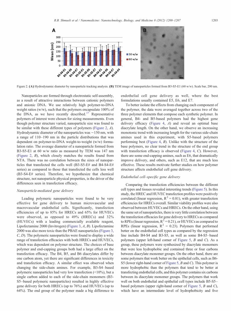
Figure 2. (A) Hydrodynamic diameter by nanoparticle tracking analysis. (B) TEM image of nanoparticles formed from B3-S5-E1 (60 w/w). Scale bar, 200 nm.
1203R.B. Shmueli et al / Nanomedicine: Nanotechnology, Biology, and Medicine 8 (2012) 1200–1207
Nanoparticles are formed through electrostatic self-assembly,as a result of attractive interactions between cationic polymersand anionic DNA. We use relatively high polymer-to-DNAweight ratios (w/w), such that the polymers encapsulate 100% ofthe DNA, as we have recently described.17 Representativepolymers of interest were chosen for sizing measurements. Eventhough polymer structure varied, nanoparticle size was found tobe similar with these different types of polymers (Figure 2, A).Hydrodynamic diameter of the nanoparticles was ∼150 nm, witha range of 110–190 nm in the particle distributions that wasdependent on polymer-to-DNA weight-to-weight (w/w) formu-lation ratio. The average diameter of a nanoparticle formed fromB3-S5-E1 at 60 w/w ratio as measured by TEM was 147 nm(Figure 2, B), which closely matches the results found fromNTA. There was no correlation between the sizes of nanopar-ticles that transfected the cells well (B3-S5-E# and B4-S4-E#series) as compared to those that transfected the cells less well(B3-S4-E# series). Therefore, we hypothesize that chemicalstructure, not nanoparticle physical properties, is the driver of thedifferences seen in transfection efficacy.
Nanoparticle-mediated gene delivery
Leading polymeric nanoparticles were found to be veryeffective for gene delivery to human microvascular andmacrovascular endothelial cells. Significantly, transfectionefficiencies of up to 85% for HRECs and 65% for HUVECswere observed, as opposed to 49% (HRECs) and 32%(HUVECs) with a leading commercially available reagent,Lipofectamine 2000 (Invitrogen) (Figure 3, A, B). Lipofectamine2000 was also more toxic than the PBAE nanoparticles (Figure 3,C, D). The polymeric nanoparticles were found to display a widerange of transfection efficacies with both HRECs and HUVECs,which was dependent on polymer structure. The choices of basepolymer and end-capping groups both had a large effect on thetransfection efficacy. The B4, B5, and B6 diacrylates differ byone carbon atom, yet there are significant differences in toxicityand transfection efficacy. A similar effect was observed whenchanging the side-chain amines. For example, B3-S4–basedpolymeric nanoparticles had very low transfection (b10%), but asingle carbon added to each of the side-chain monomers (B3-S5–based polymeric nanoparticles) resulted in highly effectivegene delivery for both HRECs (up to 76%) and HUVECs (up to64%). The end group of the polymer made a big difference to
endothelial cell gene delivery as well, where the bestformulations usually contained E5, E6, and E7.
To better isolate the effects from changing each component ofthe polymer, the data were averaged together across two of thethree polymer elements that compose each synthetic polymer. Ingeneral, B4- and B5-based polymers had the highest genedelivery efficacy (Figure 4, A) and reveal an optimal basediacrylate length. On the other hand, we observe an increasingmonotonic trend with increasing length for the various side-chainamines used in this experiment, with S5-based polymersperforming best (Figure 4, B). Unlike with the structure of thebase polymers, no clear trend in the structure of the end groupwith transfection efficacy is observed (Figure 4, C). However,there are some end-capping amines, such as E6, that dramaticallyimprove delivery, and others, such as E12, that are much lesseffective. These results motivate further studies on how polymerstructure affects endothelial cell gene delivery.
Endothelial cell–specific gene delivery
Comparing the transfection efficiencies between the differentcell types and tissues revealed interesting trends (Figure 5). In thisstudy, theHREC andHUVEC transfection profiles were positivelycorrelated (linear regression, R2 = 0.81), with greater transfectionefficiencies for HRECs overall. Similar viability profiles were alsoobserved between HUVECs and HRECs. On the other hand, usingthe same set of nanoparticles, there is very little correlation betweenthe transfection efficacies for gene delivery toHRECs as comparedto RPEs (linear regression, R2 = 0.21), or HUVECs as compared toRPEs (linear regression, R2 = 0.23). Polymers that performedbetter on the endothelial cell types as compared by the regressionline include B4-S4 and B3-S5, as well as some B4-S5–basedpolymers (upper left-hand corner of Figure 5, B and C). As agroup, these polymers were synthesized by diacrylate monomersthat were less hydrophobic and contained three or four carbonsbetween diacrylate monomer groups. On the other hand, there aresome polymers that work better on the epithelial cells, such as B6-S5 (lower right-hand corner of Figure 5, B and C). This polymer ismore hydrophobic than the polymers that tend to be better attransfecting endothelial cells, and this polymer contains six carbonsbetween its diacrylate monomer groups. The polymers that workwell on both endothelial and epithelial cell types include B5-S5–based polymers (upper right-hand corner of Figure 5, B and C),which have an intermediate level of hydrophobicity and five
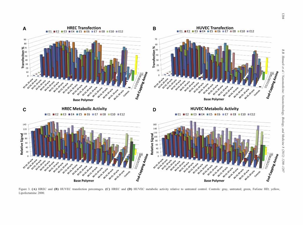
Figure 3. (A) HREC and (B) HUVEC transfection percentages. (C) HREC and (D) HUVEC metabolic activity relative to untreated control. Controls: gray, untreated; green, FuGene HD; yellow,Lipofectamine 2000.
1204R.B.Shm
ueliet
al/Nanom
edicine:Nanotechnology,
Biology,
andMedicine
8(2012)
1200–1207
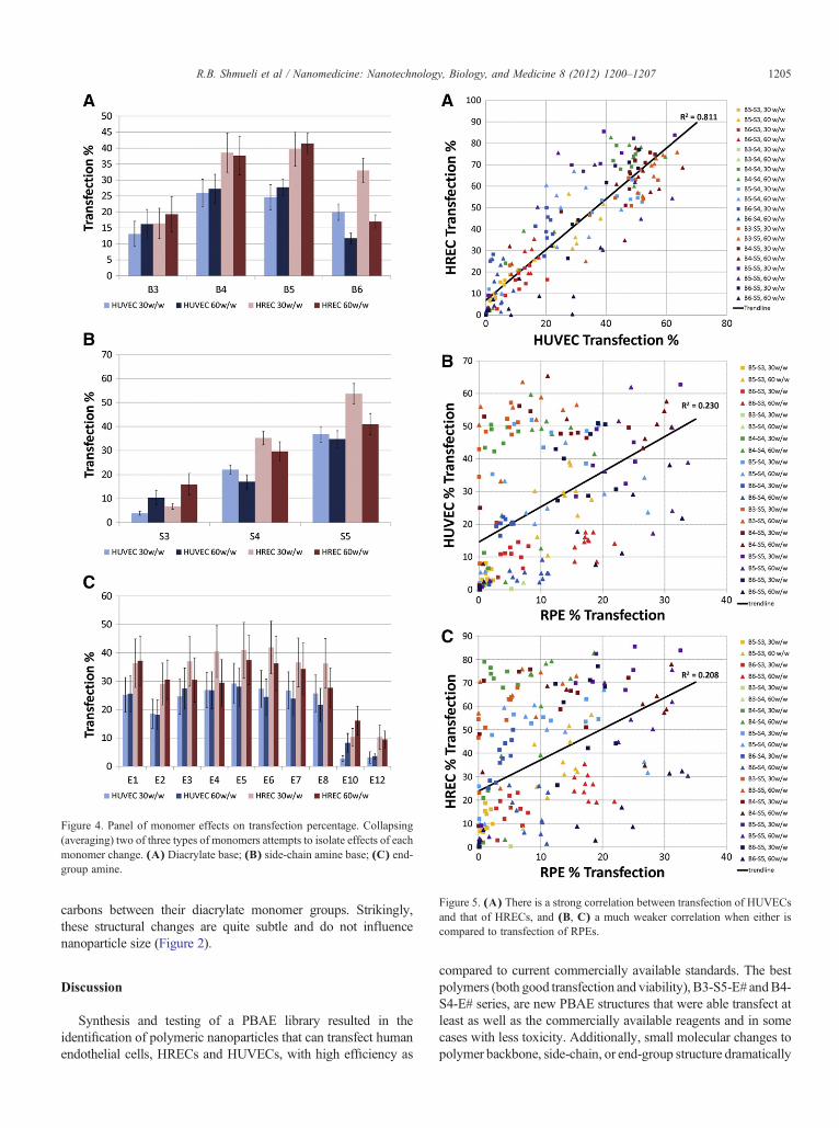
Figure 4. Panel of monomer effects on transfection percentage. Collapsing(averaging) two of three types of monomers attempts to isolate effects of eachmonomer change. (A) Diacrylate base; (B) side-chain amine base; (C) end-group amine.
Figure 5. (A) There is a strong correlation between transfection of HUVECsand that of HRECs, and (B, C) a much weaker correlation when either iscompared to transfection of RPEs.
1205R.B. Shmueli et al / Nanomedicine: Nanotechnology, Biology, and Medicine 8 (2012) 1200–1207
carbons between their diacrylate monomer groups. Strikingly,these structural changes are quite subtle and do not influencenanoparticle size (Figure 2).
Discussion
Synthesis and testing of a PBAE library resulted in theidentification of polymeric nanoparticles that can transfect humanendothelial cells, HRECs and HUVECs, with high efficiency as
compared to current commercially available standards. The bestpolymers (both good transfection and viability), B3-S5-E# andB4-S4-E# series, are new PBAE structures that were able transfect atleast as well as the commercially available reagents and in somecases with less toxicity. Additionally, small molecular changes topolymer backbone, side-chain, or end-group structure dramatically
1206 R.B. Shmueli et al / Nanomedicine: Nanotechnology, Biology, and Medicine 8 (2012) 1200–1207
changed transfection efficiency. The best polymers were ones thatcontained B4 and B5, and S4 and S5, with a few different E#groups. The most hydrophilic polymers, such as B3-S4, were notgood at transfecting the cells, and their toxicity was also minimal.On the other hand, themost hydrophobic polymers, such as theB6-S5 series, were usually relatively toxic to the endothelial cells,subsequently also reducing the transfection efficiency. This trendis mirrored when looking at the effects of the different diacrylatemonomers used (Figure 4, A), where B4 and S5 performed best.Whereas among certain structures, high hydrophobicity correlatedwith increased cytotoxicity and reduced transfection, overall, whenall the data are evaluated in combination, there is not a strongcorrelation between transfection efficiency and viability (R2
=0.06, data not shown).Interestingly, the gene delivery efficacy across the entire
polymer library was highly correlated between transfection ofHREC and HUVEC cells: nanoparticle formulations thatperformed well for HRECs also performed well for HUVECs(R2 = 0.81). This is dramatically different when compared to theperformance of the same nanoparticles with RPEs, indicating thatformulations could potentially be found that selectively transfectone ophthalmic cell type and not the other, as can be observed inFigure 5, B, C. To our knowledge this phenomenon—thatspecific ocular cell types can be targeted directly by fine-tuningof the polymer structure that composes nanoparticles—is strikingand surprising. This finding also suggests that it may be possibleto combine biomaterial-mediated targeting from this class ofmaterials with other methods of gene delivery targeting, such asligand- or coating-mediated cell uptake18-20 and transcriptionaltargeting,21,22 to further improve specificity and efficacy to onlyone specific type of cell.
In particular, we show that the polymers in the middle of thehydrophobicity scale for this PBAE library worked very well forthe endothelial cells, but much less so for the epithelial cells. Onthe other hand, those that were more hydrophobic, such as theB6-S5, worked well for the epithelial cells. Taken together, thesecorrelations indicate that there may be natural cell-typespecificity for polymeric nanoparticles of this class.
Safe and effective gene delivery vehicles can have a largeimpact on biological studies and for the treatment of diseases.For example, researchers have developed a system for genedelivery of sFLT using adeno-associated virus to bind VEGF inmonkeys as a potential treatment for age-related maculardegeneration.23 However, viral gene therapy has limitationsthat may preclude many clinical applications because ofcarrying-capacity constraints and safety concerns.24 In contrast,our study reveals premiere nonviral nanoparticles that areenabling technologies for transfection of endothelial cells invitro and are promising for use in vivo as delivery vehicles forgenetic nanomedicines. We also show that polymer structureitself may enable cell-specific nonviral gene delivery.
Acknowledgments
The authors thank Corey Bishop for help with TEM imagingand Dr. James Handa's lab (JHMI–Wilmer Eye Institute) forARPE-19 cells.
References
1. Jo DH, Kim JH, Kim JH. How to overcome retinal neuropathy: the fightagainst angiogenesis-related blindness. Arch Pharm Res 2010;33:1557-65.
2. Bhise NS, Shmueli RB, Sunshine JC, Tzeng SY, Green JJ. Drug deliverystrategies for therapeutic angiogenesis and antiangiogenesis. ExpertOpin Drug Deliv 2011;8:485-504.
3. Adhim Z, Lin X, Huang W, Morishita N, Nakamura T, Yasui H, et al.E10A, an adenovirus-carrying endostatin gene, dramatically increasedthe tumor drug concentration of metronomic chemotherapy with low-dose cisplatin in a xenograft mouse model for head and neck squamous-cell carcinoma. Cancer Gene Ther 2012;19:144-52.
4. Campochiaro PA. Gene transfer for ocular neovascularization andmacular edema. Gene Ther 2012;19:121-6.
5. Prow T, Smith JN, Grebe R, Salazar JH, Wang N, Kotov N, et al.Construction, gene delivery, and expression of DNA tethered nanopar-ticles. Mol Vis 2006;12:606-15.
6. Spoerri PE, Afzal A, Li Calzi S, Shaw LC, Cai J, Pan H, et al. Effects ofVEGFR-1, VEGFR-2, and IGF-IR hammerhead ribozymes on glucose-mediated tight junction expression in cultured human retinal endothelialcells. Mol Vis 2006;12:32-42.
7. Krotz F, Sohn HY, Gloe T, Plank C, Pohl U. Magnetofectionpotentiates gene delivery to cultured endothelial cells. J Vasc Res2003;40:425-34.
8. Bhise NS, Gray RS, Sunshine JC, Htet S, Ewald AJ, Green JJ. Therelationship between terminal functionalization and molecular weight ofa gene delivery polymer and transfection efficacy in mammary epithelial2-D cultures and 3-D organotypic cultures. Biomaterials 2010;31:8088-96.
9. Tzeng SY, Guerrero-Cazares H, Martinez EE, Sunshine JC, Quinones-Hinojosa A, Green JJ. Non-viral gene delivery nanoparticles based onPoly(beta-amino esters) for treatment of glioblastoma. Biomaterials2011;32:5402-10.
10. Sunshine J, Green JJ, Mahon KP, Yang F, Eltoukhy AA, Nguyen DN,et al. Small-molecule end-groups of linear polymer determine cell-typegene-delivery efficacy. Adv Mater 2009;21:4947-51.
11. Green JJ, Shi J, Chiu E, Leshchiner ES, Langer R, Anderson DG.Biodegradable polymeric vectors for gene delivery to human endothelialcells. Bioconjug Chem 2006;17:1162-9.
12. Green JJ, Langer R, Anderson DG. A combinatorial polymer libraryapproach yields insight into nonviral gene delivery. Accounts Chem Res2008;41:749-59.
13. Maiti D, Xu Z, Duh EJ. Vascular endothelial growth factor inducesMEF2C and MEF2-dependent activity in endothelial cells. InvestOphthalmol Vis Sci 2008;49:3640-8.
14. Xu Z, Yu Y, Duh EJ. Vascular endothelial growth factor upregulatesexpression of ADAMTS1 in endothelial cells through protein kinase Csignaling. Invest Ophthalmol Vis Sci 2006;47:4059-66.
15. Handa JT, Reiser KM, Matsunaga H, Hjelmeland LM. The advancedglycation endproduct pentosidine induces the expression of PDGF-B inhuman retinal pigment epithelial cells. Exp Eye Res 1998;66:411-9.
16. Sunshine JC, Akanda MI, Li D, Kozielski KL, Green JJ. Effects of basepolymer hydrophobicity and end-group modification on polymeric genedelivery. Biomacromolecules 2011;12:3592-600.
17. Bhise NS, Shmueli RB, Gonzalez J, Green JJ. A novel assay forquantifying the number of plasmids encapsulated by polymer nanopar-ticles. Small 2012;8:367-73.
18. Green JJ, Chiu E, Leshchiner ES, Shi J, Langer R, Anderson DG.Electrostatic ligand coatings of nanoparticles enable ligand-specific genedelivery to human primary cells. Nano Lett 2007;7:874-9.
19. Harris TJ, Green JJ, Fung PW, Langer R, Anderson DG, Bhatia SN.Tissue-specific gene delivery via nanoparticle coating. Biomaterials2010;31:998-1006.
20. Shmueli RB, Anderson DG, Green JJ. Electrostatic surface modifica-tions to improve gene delivery. Expert Opin Drug Deliv 2010;7:535-50.

1207R.B. Shmueli et al / Nanomedicine: Nanotechnology, Biology, and Medicine 8 (2012) 1200–1207
21. Huang YH, Zugates GT, Peng W, Holtz D, Dunton C, Green JJ, et al.Nanoparticle-delivered suicide gene therapy effectively reduces ovariantumor burden in mice. Cancer Res 2009;69:6184-91.
22. Showalter SL, Huang YH, Witkiewicz A, Costantino CL, Yeo CJ, GreenJJ, et al. Nanoparticulate delivery of diphtheria toxin DNA effectivelykills mesothelin expressing pancreatic cancer cells. Cancer Biol Ther2008;7:1584-90.
23. MacLachlan TK, Lukason M, Collins M, Munger R, Isenberger E,Rogers C, et al. Preclinical safety evaluation of AAV2-sFLT01-A genetherapy for age-related macular degeneration. Mol Ther 2011;19:326-34.
24. Thomas CE, Ehrhardt A, Kay MA. Progress and problems withthe use of viral vectors for gene therapy. Nat Rev Genet 2003;4:346-58.