Synthesis, Characterization, and in Vivo Delivery of siRNA-Squalene Nanoparticles Targeting Fusion...
10
Published: May 11, 2011 r2011 American Chemical Society 4067 dx.doi.org/10.1021/jm2000272 | J. Med. Chem. 2011, 54, 4067–4076 ARTICLE pubs.acs.org/jmc Synthesis, Characterization, and in Vivo Delivery of siRNA-Squalene Nanoparticles Targeting Fusion Oncogene in Papillary Thyroid Carcinoma Mouna Raouane, †,‡,§ Didier Desmaele, † Marie Gilbert-Sirieix, ‡ Claire Gueutin, † Fatima Zouhiri, † Claudie Bourgaux, † Elise Lepeltier, † Ruxandra Gref, † Ridha Ben Salah, § Gary Clayman, || Liliane Massaad-Massade,* ,‡ and Patrick Couvreur* ,† † Laboratoire de Physicochimie, Pharmacotechnie et Biopharmacie, Facult e de Pharmacie, UMR CNRS 8612, Universit e Paris Sud 11, 5 Rue J. B. Cl ement, 92296 Ch ^ atenay-Malabry, France ‡ Laboratoire de Vectorologie et Transfert de G enes, UMR CNRS 8203, Institut Gustave Roussy, 114 Rue Edouard Vaillant, 94805 Villejuif, France § Unit e de Recherche de Biophysique, 00/UR/08-02, Facult e de M edecine Ibn El Jazzar de Sousse, Universit e de Sousse, Avenue Mohamed Karoui, 4002 Sousse, Tunisia ) Department of Head and Neck Surgery, The University of Texas MD Anderson Cancer Center, Houston, Texas 77030, United States b S Supporting Information ’ INTRODUCTION Among all thyroid cancers, the papillary thyroid carcinoma (PTC) 1 is the most frequent. 2 It is associated with somatic mutations of the RET proto-oncogene encoding for a membrane tyrosine kinase (TK) receptor that is normally not expressed in the thyroid follicular cells. The most common variant is RET/PTC1 which is formed by an intrachromosomic rearrange- ment, leading to the juxtaposition of the RET TK domain with the genes H4. 35 RET/PTC1 fusion oncogene occurred in children after the nuclear fallout in the Chernobyl area 6 and after external irradiation to medical treatment. 7,8 The prognosis of PTC is generally good, but a considerable number of patients, approximately 30% as shown after 30 years of follow-up, have recurrent disease. This constitutes, therefore, an area of important research on emerging therapies such as using small interfering RNA (siRNA) for targeting the RET/PTC1 fusion oncogene and inducing the silencing of the corresponding protein. Such therapeutic approach is highly specific because RET/PTC1 is present only in the tumor cells and not in the surrounding normal cells. Recently, a siRNA RET/PTC1 efficient and specific to RET/PTC1 oncogene has been discovered; this opens new prospects in the treatment by siRNAs of PTC with RET/PTC1 junction. 9 However, in vivo delivery of siRNA is a key challenge because the biological efficacy of the siRNAs is ham- pered by their poor stability in biological fluids and low intracel- lular penetration due to their highly hydrophilic and anionic character. 10,11 So far, a wide variety of approaches including viral vectors and nonviral delivery systems, such as liposomes, nanoparticles, micelles, and polyplexes, 1214 have been investigated to enhance target cells uptake and silence potency in vivo. However, the Received: January 17, 2011 ABSTRACT: We report the conjugation of the natural lipid squalene (SQ) with a small interfering RNA (siRNA), against the junction oncogene RET/PTC1, usually found in papillary thyroid carcinoma (PTC). The acyclic isoprenoid chain of squalene has been covalently coupled with siRNA RET/ PTC1 at the 3 0 -terminus of the sense strand via maleimide sulfhydryl chemistry. Remarkably, the linkage of siRNA RET/ PTC1 to squalene led to an amphiphilic molecule that self- organized in H 2 O as siRNA-SQ RET/PTC1 nanoparticles (NPs). The siRNA-SQ RET/PTC1 NPs, stable in H 2 O, were used for biological studies. In vitro, they did not show any cytotoxicity. Interestingly, in vivo, on a mice xenografted RET/ PTC1 experimental model, RET/PTC1-SQ NPs were found to inhibit tumor growth and RET/PTC1 oncogene and oncopro- tein expression after 2.5 mg/kg cumulative dose intravenous injections. In conclusion, these results showed that the “squalenoylation” offers a new noncationic plate-form for the siRNA delivery.
-
Upload
independent -
Category
Documents
-
view
2 -
download
0
Transcript of Synthesis, Characterization, and in Vivo Delivery of siRNA-Squalene Nanoparticles Targeting Fusion...
Published: May 11, 2011
r 2011 American Chemical Society 4067 dx.doi.org/10.1021/jm2000272 | J. Med. Chem. 2011, 54, 4067–4076
ARTICLE
pubs.acs.org/jmc
Synthesis, Characterization, and in Vivo Delivery of siRNA-SqualeneNanoparticles Targeting Fusion Oncogene in Papillary ThyroidCarcinomaMouna Raouane,†,‡,§ Didier Desmaele,† Marie Gilbert-Sirieix,‡ Claire Gueutin,† Fatima Zouhiri,†
Claudie Bourgaux,† Elise Lepeltier,† Ruxandra Gref,† Ridha Ben Salah,§ Gary Clayman,||
Liliane Massaad-Massade,*,‡ and Patrick Couvreur*,†
†Laboratoire de Physicochimie, Pharmacotechnie et Biopharmacie, Facult�e de Pharmacie, UMR CNRS 8612, Universit�e Paris Sud 11,5 Rue J. B. Cl�ement, 92296 Chatenay-Malabry, France‡Laboratoire de Vectorologie et Transfert de G�enes, UMR CNRS 8203, Institut Gustave Roussy, 114 Rue Edouard Vaillant,94805 Villejuif, France§Unit�e de Recherche de Biophysique, 00/UR/08-02, Facult�e de M�edecine Ibn El Jazzar de Sousse, Universit�e de Sousse,Avenue Mohamed Karoui, 4002 Sousse, Tunisia
)Department of Head and Neck Surgery, The University of Texas MD Anderson Cancer Center, Houston, Texas 77030, United States
bS Supporting Information
’ INTRODUCTION
Among all thyroid cancers, the papillary thyroid carcinoma(PTC)1 is the most frequent.2 It is associated with somaticmutations of the RET proto-oncogene encoding for a membranetyrosine kinase (TK) receptor that is normally not expressedin the thyroid follicular cells. The most common variant isRET/PTC1 which is formed by an intrachromosomic rearrange-ment, leading to the juxtaposition of the RET TK domain withthe genes H4.3�5 RET/PTC1 fusion oncogene occurred inchildren after the nuclear fallout in the Chernobyl area6 andafter external irradiation to medical treatment.7,8
The prognosis of PTC is generally good, but a considerablenumber of patients, approximately 30% as shown after 30 years offollow-up, have recurrent disease. This constitutes, therefore, anarea of important research on emerging therapies such as usingsmall interfering RNA (siRNA) for targeting the RET/PTC1fusion oncogene and inducing the silencing of the corresponding
protein. Such therapeutic approach is highly specific becauseRET/PTC1 is present only in the tumor cells and not in thesurrounding normal cells. Recently, a siRNA RET/PTC1 efficientand specific to RET/PTC1 oncogene has been discovered; thisopens new prospects in the treatment by siRNAs of PTC withRET/PTC1 junction.9 However, in vivo delivery of siRNA is a keychallenge because the biological efficacy of the siRNAs is ham-pered by their poor stability in biological fluids and low intracel-lular penetration due to their highly hydrophilic and anioniccharacter.10,11
So far, a wide variety of approaches including viral vectors andnonviral delivery systems, such as liposomes, nanoparticles,micelles, and polyplexes,12�14 have been investigated to enhancetarget cells uptake and silence potency in vivo. However, the
Received: January 17, 2011
ABSTRACT: We report the conjugation of the natural lipidsqualene (SQ) with a small interfering RNA (siRNA), againstthe junction oncogene RET/PTC1, usually found in papillarythyroid carcinoma (PTC). The acyclic isoprenoid chain ofsqualene has been covalently coupled with siRNA RET/PTC1 at the 30-terminus of the sense strand via maleimide�sulfhydryl chemistry. Remarkably, the linkage of siRNA RET/PTC1 to squalene led to an amphiphilic molecule that self-organized in H2O as siRNA-SQ RET/PTC1 nanoparticles(NPs). The siRNA-SQ RET/PTC1 NPs, stable in H2O, wereused for biological studies. In vitro, they did not show anycytotoxicity. Interestingly, in vivo, on a mice xenografted RET/PTC1 experimental model, RET/PTC1-SQNPs were found toinhibit tumor growth and RET/PTC1 oncogene and oncopro-tein expression after 2.5 mg/kg cumulative dose intravenousinjections. In conclusion, these results showed that the “squalenoylation” offers a new noncationic plate-form for the siRNA delivery.

4068 dx.doi.org/10.1021/jm2000272 |J. Med. Chem. 2011, 54, 4067–4076
Journal of Medicinal Chemistry ARTICLE
safety of viral vectors is questionable because of immunogenicityand possible recombination of oncogenes. On the other hand,most of the nonviral vectors are designed using polycations,either polymers or lipids, whose (cyto)toxicity is now welldocumented.15,16 This is the reason why the conjugation ofnucleic acids with neutral lipids offers an interesting alternative.Thus, DNA-programmed lipids, consisting of two 18-carbonalkyl hydrophobic tails linked covalently to the 50-termini of a9-mer single stranded DNA, have been proposed as responsiveliposomes.17 In another interesting approach, a simple lipid tailphosphoramidite with diacyl chains has been attached onto theend of an aptamer via a polyethylene glycol linker to obtain self-assembledmicelles able to target cancer cells.18 Different types ofcholesterol-conjugated siRNA were also synthesized to elucidatethe requirements for siRNA delivery in vivo. However,the pharmacological efficacy of these approaches remains to bedemonstrated in experimental pathologies.19 Noteworthy, the invitro demonstration that alkyne-modified lipids derived fromcholesterol and covalently conjugated to azido oligonucleotidescould efficiently inhibit hepatitis C virus translation in humanhepatic cells has been done by the group of Barthelemy.20 Thus,although further in vivo efficacy is needed, amphiphilic oligonu-cleotides may represent an interesting marriage of nucleic acidswith lipids.21 In this context, we introduce here the idea tocovalently link siRNA with the squalene, a natural and nonioniclipid. This triterpene is known for its dietary benefits,22 biocom-patibility, and inertness so that it is already extensively used asexcipient in numerous pharmaceutical formulations for oral andparenteral administration.23 From a safety point of view, this lipid
is therefore more desirable than cholesterol and fatty acid, ofwhich intravenous administration may be linked with cardiovas-cular events. Thus, in the present study, we propose to conjugateRET/PTC1 siRNA to squalene in order to improve siRNAhydrophobicity and stability and to demonstrate the efficacy ofthis treatment on a mice xenografted RET/PTC1-expressingtumor. The concept to chemically link the squalene to a biologi-cally active drug molecule in order to create a bioconjugate, whichcan self-aggregate as nanoassemblies in water, has already beendemonstrated using (mono)nucleosides analogues with antic-ancer (e.g., gemcitabine in leukemic cancer) or antiviral activity(ddC).24 Interestingly, the gemcitabine�squalene nanoassemblieswere able to deliver this drug efficiently into the cell cytoplasmthrough a nonendocytotic pathway, thus bypassing lysosomaldegradation.25 This is another reason why we have extended thisstrategy here to siRNA RET/PTC1 oligonucleotides (ONs).
’CHEMISTRY
One of the critical problems in RNA-based therapeutic applica-tions is the delivery of intact siRNA across plasma membranes ofcells. In the present study, we have designed a new squalene�siRNAbioconjugate to protect siRNA fromdegradation and to improve thecell delivery by increasing the overall lipophilicity of the nucleic acidderivative. In order to protect the siRNA RET/PTC1 against 30-exonucleases, simultaneously preventing steric hindrance towardrecognition of the targeted mRNA by the antisense strand and alsoallowing fixation of proteic RISC complex at the 50-end (thatrequires phosphorylation), we directly linked squalene at the 30-end of the 21-mer sense strand siRNA 1 (Figure 1). This couplingstrategy was based on previous studies showing that the modifica-tion of the 50-hydroxyl groups of the guide siRNA strand bymethoxy groups completely hampered the siRNAs function in
Scheme 1. Synthesis of Squalenoyl Maleimide 5 and Con-jugate Addition of the Sense Strand siRNA 1 with 5 To GiveRET/PTC1 siRNA-SQ (9)
Figure 1. Chemical structure of the siRNA sense strand 1 and of thesqualenoyl activated moieties 2�5.

4069 dx.doi.org/10.1021/jm2000272 |J. Med. Chem. 2011, 54, 4067–4076
Journal of Medicinal Chemistry ARTICLE
Drosophila embryo lysate and HeLa cell extract.29,30 At the outset ofthis study, we chose to link covalently the siRNA sense strand 1 tosqualene through a disulfide linkage, taking advantage of the3-mercaptopropyl phosphate group introduced at the 30-terminalof the oligonucleotide during its manufacturing on solid support.However, attempts to condense 1with either the squalene derivative2 or its more electrophilic C-5 nitro analogue 3met with repetitive
failure (data not shown). We thus envisioned that the challengingsqualenoylationmight bemore efficiently achieved by the use of themaleimide group as thiol acceptor as previously described byWeberet al.27 Toward this goal, the maleimide squalene 4 was preparedfrom the corresponding aminosqualene by condensation withN-(ethoxycarbonyl)maleimide 6 (Scheme 1). However, attemptsat coupling siRNA 1 with 4 in a PBS (phosphate buffered saline)solution/DMFmixturewere similarly found fruitless, providing onlyaminute amount of the desired conjugate (5%) based on theHPLCtrace and MALDI-TOF MS analysis.
In order to increase the maleimide functional group accessi-bility to the thiol-modifiedON, we have intercalated a small etherlinker between the maleimide group and the SQ moiety. Thesynthesis of the requisite squalenoyl maleimide 5 is outlined in
Figure 2. Characterization of RET/PTC1 siRNA-SQ sense strand bioconjugate. (a) HPLC profile of reaction mixture. Arrows show the elution peal ofRET/PTC1 siRNA bioconjugate. Significant increase in the reaction yield (from 7% to 55%) was observed when 30% methanol was added to thereaction mixture as a cosolvent and when reaction activation was performed by microwave irradiation. Chromatogram 1 corresponds to the reactionmixture obtained in conventional heating conditions. Chromatogram 2 corresponds to the reaction mixture obtained in the presence of 30% methanoland reaction activation by microwave irradiation. (b) MALDI-TOF mass spectrometry analysis of the RP-HPLC purified RET/PTC1 siRNA-SQ sensestrand bioconjugate. The MALDI-TOF mass spectrum shows a bioconjugate molecular weight of 7365.14 Da in agreement with the calculatedmolecular weight.
Table 1. RP-HPLC Elution Times of RET/PTC1 siRNA-SQversus RET/PTC1 siRNA
compd elution time (min)
CGU UAC CAU CGA GGA UCC AAA 10.5
CGU UAC CAU CGA GGA UCC AAA SQ 17.5
4070 dx.doi.org/10.1021/jm2000272 |J. Med. Chem. 2011, 54, 4067–4076
Journal of Medicinal Chemistry ARTICLE
the Scheme 1. Briefly, the maleimide moiety was first tetheredwith an ethoxyethanol linker according to the method of Kellerand Rudinger28 starting from N-(ethoxycarbonyl)maleimide 6.Condensation of the resulting maleimide alcohol 7 with themixed anhydride derived from 1,10,2-tris-norsqualenic acid 823
and ethyl chloroformate provided adduct 5 in 35% yield.(Scheme 1). Nevertheless, we observed no significant increasein the reaction yield (7%) with the siRNA (Figure 2a, chromato-gram 1), although fewer byproducts were formed than withmaleimide 4. By contrast, when 50% of methanol was added ascosolvent, the adduct 9 was obtained in 45% yield. A furtherimprovement was gained when microwave irradiation (10 min,70 �C)was substituted to the conventional heating, providing thedesired squalenoyl oligonucleotide 9 in 55% yield (Figure 2a,chromatogram 2). Calculated RET/PTC1 siRNA conjugateweight was 7365 Da, in agreement with the MALDI-TOF massspectrometry determined weight 7365.14 Da (Figure 2b). Puri-fication of the crude product on polymeric RP-HPLC columnand lyophilization then afforded the pure squalenoyl-modifiedoligonucleotide 9 which was characterized by MALDI mass
spectrometry. Althoughwemay expect that the conjugate additionof the thiol group on themaleimide double bond gave a mixture oftwo diastereomers, only one peakwas observed in theHPLC trace.Given the sizes of both the oligonucleotide and the squalene withrespect to the maleimide ring and the conformational flexibilityaround the linker, we hypothesize that this supplementary asym-metric center will be without repercussion.
The modification of the siRNA thiol backbone with squalenicacid at the 30-end of the sense strand was found to dramaticallyenhance the lipophilicity as represented by the delayed elutiontimes (Table 1).
’RESULTS AND DISCUSSION
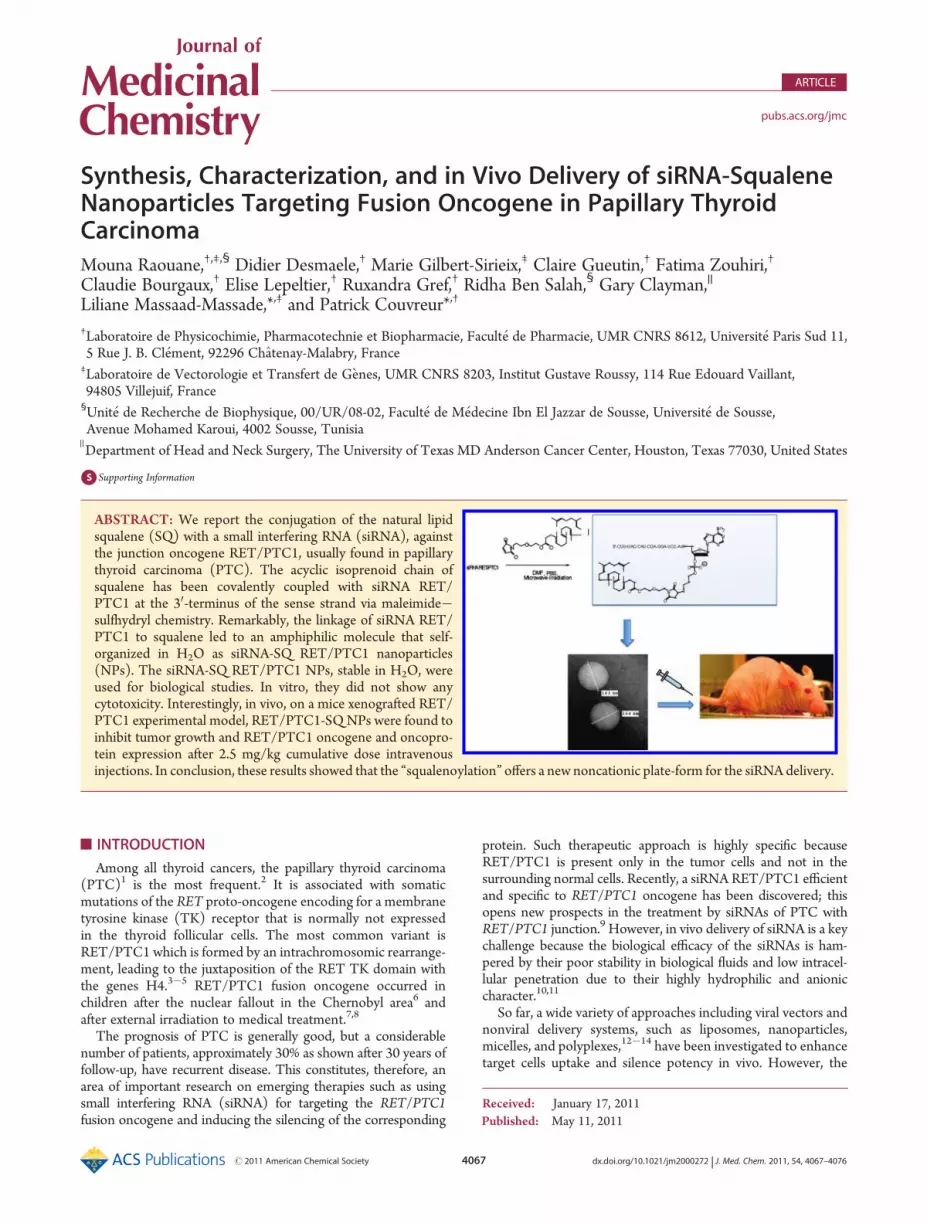
RET/PTC1 siRNA-SQ Nanoparticles Characterization. Pur-ified RET/PTC1 siRNA-SQ sense strand 9 and RET/PTC1siRNA antisense strand were hybridized, and duplex formationwas assessed by 4% agarose gel electrophoresis (data not shown).The attachment of a squalene moiety with the siRNA duplexinduced the spontaneous formation of siRNA-SQ nanoparticles inH2O (control siRNA-SQ and RET/PTC1 siRNA-SQNPs) with asize of 165( 10 nm and a polydispersity index of 0.2 as measuredby laser light scattering (number of measurements n = 10). Thisemphasizes the important contribution of the squalene to theoverall physicochemical behavior of the conjugate, as also illu-strated by the impressive increase in the lipophilic character ofRET/PTC1 siRNA-SQ shown by RP-HPLC analysis. RET/PTC1siRNA-SQ NPs dimension was confirmed by electronic micro-scopy and cryotransmission electronmicroscopy showing sphericalregular particles without visible supramolecular organization, asillustrated in Figure 3a and Figure 3b. The nanoparticles werefound to be negatively charged, with amean ζ potential of�26mV,which is in favor of the establishment of repulsive forces allowingthe stabilization of the NPs suspension; both size and ζ potential ofRET/PTC1 siRNA-SQ NPs remained nearly constant after in-cubation for 48 h at room temperature and at 37 �C (Figure 3c).Then the stability of the RET/PTC1 siRNA-SQ duplex in nano-particle formulation was tested in vitro for 24 h in 10% serum-containing media and in serum free media, relative to the nakedRET/PTC1 siRNA. In both conditions, the stability of RET/PTC1
Figure 3. Characterization of RET/PTC1 siRNA-SQ NPs. Represen-tative images of the RET/PTC1 siRNA-SQ NPs obtained by (a)cryoelectron microscopy and by (b) transmission electron microscopy.RET/PTC1 siRNA-SQ NPs were found to be spherical in shape. Thenanoparticle size, as observed by TEM, correlated well with the sizemeasured by laser light scattering (Nanosizer). (c) Mean diameter(black curves) and ζ potential (gray curves) evolution of RET/PTC1siRNA-SQ NPs upon incubation in water at room temperature and at37 �C. Both size and ζ potential of RET/PTC1 siRNA-SQNPs remainednearly constant after incubation for 48 h at room temperature and at37 �C (n = 3 experiments).
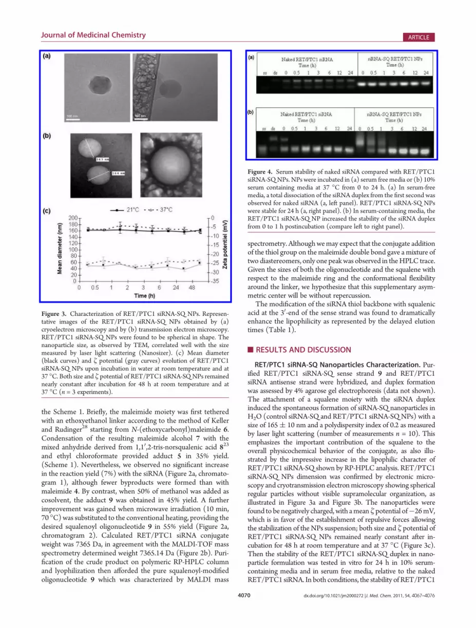
Figure 4. Serum stability of naked siRNA compared with RET/PTC1siRNA-SQNPs. NPs were incubated in (a) serum freemedia or (b) 10%serum containing media at 37 �C from 0 to 24 h. (a) In serum-freemedia, a total dissociation of the siRNA duplex from the first second wasobserved for naked siRNA (a, left panel). RET/PTC1 siRNA-SQ NPswere stable for 24 h (a, right panel). (b) In serum-containing media, theRET/PTC1 siRNA-SQ NP increased the stability of the siRNA duplexfrom 0 to 1 h postincubation (compare left to right panel).

4071 dx.doi.org/10.1021/jm2000272 |J. Med. Chem. 2011, 54, 4067–4076
Journal of Medicinal Chemistry ARTICLE
siRNA-SQ NPs was improved relative to the naked RET/PTC1siRNA in solution. In serum free media, naked siRNA displayedimmediate and complete dissociation of the duplex (Figure 4a, leftpanel), whereas RET/PTC1 siRNA-SQNPs were stable until 24 h(Figure 4a, right panel). In serum-containing media, the RET/PTC1 siRNA-SQ NP also increased the stability of the siRNAduplex from 0 to 1 h postincubation (Figure 4b, compare left toright panel). Thus, nanoparticles have clearly improved the nucleicacid stability in both serum and serum freemedia. This should allowan improved cell/tissue delivery of RET/PTC1, since its stabilityduring 1 h in serum conditionsmay be sufficient to reach the tumoras an intact molecule.In Vitro Evaluation. MTT assay did not show any significant
inhibition of growth rate for RET/PTC1 siRNA-SQ NPs treatedcells when compared to untreated cells after 24, 48, and 72 hincubations. This experiment was done for TPC-1 cells (data notshown) and for BHP 10-3 cells (Figure 5). It was observed that∼95% of the cells were still metabolically active after RET/PTC1siRNA-SQ NPs treatment. By qRT-PCR we found that the RET/PTC1 siRNA significantly reduced RET/PTC1 mRNA levels by∼80%, whereas the control siRNA did not show any statisticaldifference when compared with untreated cells (Figure 6a). Thedown-regulation of RET/PTC1 mRNA levels by siRNA wasparalleled by a decrease in RET/PTC1 protein content (Figure 6b).Thus, the cytotoxicity of RET/PTC1 siRNA-SQ NPs wasevaluated in this model, but no cell growth inhibition was notedand the absence of RET/PTC1 gene silencing was observed too(data not shown), possibly because in cell culture conditions, thelimited enzymatic content did not allow the cleavage of thesqualene needed for nanoparticle disaggregation before furthermRNA sequence recognition. From the other side, this experi-ment has also highlighted the absence of any cell toxicity usingsqualene nanoparticles as noncationic nanocarriers of siRNA.In Vivo Evaluation. In a preliminary experiment, we have
tested the tumorigenicity of the TPC-1 and BHP 10-3 cells innude mice. It was observed that TPC-1 cells were not able toform tumors, whereas BHP 10-3 were tumorgenic after sub-cutaneous injection of 106, 2.5 � 106, and 5 � 106 cells. Thenthe efficiency of RET/PTC1 siRNA-SQ NPs was tested on BHP10-3, 2.5 � 106 cells subcutaneously inoculated into irradiatednude mice. When tumors reached around 50 mm3, mice were
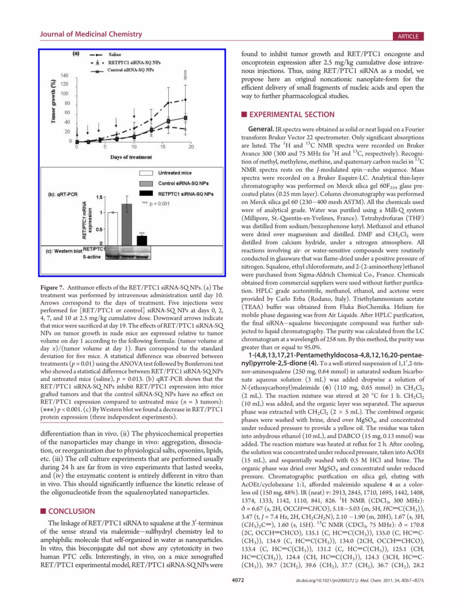
treated by 2.5 mg/kg in five intravenous injections as described inExperimental Section. The ANOVA test followed by Bonferroni’stest showed a statistical difference between treatments (p = 0.01).RET/PTC1 siRNA-SQ NP significantly reduced the tumor sizewith a tumor growth inhibition rate of 70% (p < 0.05), whereas thecontrol siRNA-SQNPdid not significantly inhibit the tumor growth(p > 0.05) compared to the control saline (Figure 7a). Then thesilencing ability of RET/PTC1 siRNA-SQ NPs was assessed inthree tumor biopsies by qRT-PCR and the protein content byWestern blot. By qRT-PCR it was found that RET/PTC1 siRNA-SQNPs significantly reduced RET/PTC1mRNA levels by approxi-mately 80%, whereas the control siRNA-SQ NPs did not showany statistical difference when compared to the control saline(Figure 7b). The down-regulation of RET/PTC1 mRNA levelsby RET/PTC1 siRNA-SQ NPs was paralleled by a decrease inRET/PTC1 protein content (Figure 7c).Thus, whereas RET/PTC1 siRNA-SQ NPs did not display
any cytotoxicity in vitro, significant regression of tumor growth(i.e., 70%) was observed in vivo with the RET/PTC1 siRNA-SQNPs, whereas the control siRNA-SQ NPs injected under thesame conditions failed to do so. This in vivo anticancer activitywas even paralleled by RET/PTC1 oncogene and oncoproteinexpressions inhibition, revealing that at least a fraction of theintravenously injected siRNA was delivered in an active forminside the tumor cells. These data demonstrate once again thatprediction of in vivo pharmacological efficacy through cellculture experiments is uncertain, even when experiments areperformed on exactly the same tumor cell line. We have alreadyobserved such a phenomenon with siRNA loaded onto chitosan-coated nanospheres.26 The reasons are numerous: (i) Cells inculture may exhibit different pattern of gene expression and
Figure 5. Cell viability at different concentrations of RET/PTC1siRNA-SQ NPs. MTT assay was performed on BHP 10-3 cells asdescribed in Experimental Section. Cell viability was calculated as100� [(A570 of NP treated cells� A570 of blank)]/[(A570 of controlcells� A570 of blank)]. No statistical difference was observed betweenuntreated cells and RET/PTC1 siRNA-SQ NP treated cells at 24, 48,and 72 h.
Figure 6. Gene silencing efficiency of RET/PTC1 siRNA in the BHP10�3 cell line. (a) The RET/PTC1 mRNA expression was analyzed byreal time quantitative reverse transcription PCR (qRT-PCR) for treatedcells by siRNA RET/PTC1 (black) or siRNA CT (gray) and comparedto untreated cells (white). Experiments were performed 24 h aftertransfection with 50 nM RET/PTC1 siRNA or control siRNA usingLipofectamine 2000. Measurement shows a reduction of RET/PTC1mRNA levels after transfection of RET/PTC1 siRNA. The barsrepresent the mean ( SD of three independent experiments: (///)Significant change compared to untreated cells using ANOVA followedby Bonferroni’s test (p < 0.001). (b) RET/PTC1 protein level inuntransfected and transfected cells with RET/PTC1 siRNA or CTsiRNA at 50 nM. Proteins were extracted 24 h after transfection andanalyzed by Western blot using Ret antibody. β-Actin was used asloading control for Western blot. Experiments were performed intriplicate.
4072 dx.doi.org/10.1021/jm2000272 |J. Med. Chem. 2011, 54, 4067–4076
Journal of Medicinal Chemistry ARTICLE
differentiation than in vivo. (ii) The physicochemical propertiesof the nanoparticles may change in vivo: aggregation, dissocia-tion, or reorganization due to physiological salts, opsonins, lipids,etc. (iii) The cell culture experiments that are performed usuallyduring 24 h are far from in vivo experiments that lasted weeks,and (iv) the enzymatic content is entirely different in vitro thanin vivo. This should significantly influence the kinetic release ofthe oligonucleotide from the squalenoylated nanoparticles.
’CONCLUSION
The linkage of RET/PTC1 siRNA to squalene at the 30-terminusof the sense strand via maleimide�sulfhydryl chemistry led toamphiphilic molecule that self-organized in water as nanoparticles.In vitro, this bioconjugate did not show any cytotoxicity in twohuman PTC cells. Interestingly, in vivo, on a mice xenograftedRET/PTC1experimentalmodel, RET/PTC1 siRNA-SQNPswere
found to inhibit tumor growth and RET/PTC1 oncogene andoncoprotein expression after 2.5 mg/kg cumulative dose intrave-nous injections. Thus, using RET/PTC1 siRNA as a model, wepropose here an original noncationic nanoplate-form for theefficient delivery of small fragments of nucleic acids and open theway to further pharmacological studies.
’EXPERIMENTAL SECTION
General. IR spectra were obtained as solid or neat liquid on a Fouriertransform Bruker Vector 22 spectrometer. Only significant absorptionsare listed. The 1H and 13C NMR spectra were recorded on BrukerAvance 300 (300 and 75 MHz for 1H and 13C, respectively). Recogni-tion of methyl, methylene, methine, and quaternary carbon nuclei in 13CNMR spectra rests on the J-modulated spin�echo sequence. Massspectra were recorded on a Bruker Esquire-LC. Analytical thin-layerchromatography was performed on Merck silica gel 60F254 glass pre-coated plates (0.25 mm layer). Column chromatography was performedon Merck silica gel 60 (230�400 mesh ASTM). All the chemicals usedwere of analytical grade. Water was purified using a Milli-Q system(Millipore, St.-Quentin-en-Yvelines, France). Tetrahydrofuran (THF)was distilled from sodium/benzophenone ketyl. Methanol and ethanolwere dried over magnesium and distilled. DMF and CH2Cl2 weredistilled from calcium hydride, under a nitrogen atmosphere. Allreactions involving air- or water-sensitive compounds were routinelyconducted in glassware that was flame-dried under a positive pressure ofnitrogen. Squalene, ethyl chloroformate, and 2-(2-aminoethoxy)ethanolwere purchased from Sigma-Aldrich Chemical Co., France. Chemicalsobtained from commercial suppliers were used without further purifica-tion. HPLC grade acetonitrile, methanol, ethanol, and acetone wereprovided by Carlo Erba (Rodano, Italy). Triethylammonium acetate(TEAA) buffer was obtained from Fluka BioChemika. Helium formobile phase degassing was from Air Liquide. After HPLC purification,the final siRNA�squalene bioconjugate compound was further sub-jected to liquid chromatography. The purity was calculated from the LCchromatogram at a wavelength of 258 nm. By thismethod, the purity wasgreater than or equal to 95.0%.1-(4,8,13,17,21-Pentamethyldocosa-4,8,12,16,20-pentae-
nyl)pyrrole-2,5-dione (4).To a well-stirred suspension of 1,10,2-tris-nor-aminosqualene (250 mg, 0.64 mmol) in saturated sodium bicarbo-nate aqueous solution (3 mL) was added dropwise a solution ofN-(ethoxycarbonyl)maleimide (6) (110 mg, 0.65 mmol) in CH2Cl2(2 mL). The reaction mixture was stirred at 20 �C for 1 h. CH2Cl2(10 mL) was added, and the organic layer was separated. The aqueousphase was extracted with CH2Cl2 (2 � 5 mL). The combined organicphases were washed with brine, dried over MgSO4, and concentratedunder reduced pressure to provide a yellow oil. The residue was takeninto anhydrous ethanol (10 mL), and DABCO (15 mg, 0.13 mmol) wasadded. The reaction mixture was heated at reflux for 2 h. After cooling,the solution was concentrated under reduced pressure, taken into AcOEt(15 mL), and sequentially washed with 0.5 M HCl and brine. Theorganic phase was dried over MgSO4 and concentrated under reducedpressure. Chromatographic purification on silica gel, eluting withAcOEt/cyclohexane 1:1, afforded maleimido squalene 4 as a color-less oil (150 mg, 48%). IR (neat) ν: 2913, 2845, 1710, 1695, 1442, 1408,1374, 1333, 1142, 1110, 841, 826. 1H NMR (CDCl3, 300 MHz):δ = 6.67 (s, 2H, OCCHdCHCO), 5.18�5.03 (m, 5H,HCdC(CH3)),3.47 (t, J = 7.4 Hz, 2H, CH2CH2N), 2.10�1.90 (m, 20H), 1.67 (s, 3H,(CH3)2Cd), 1.60 (s, 15H). 13C NMR (CDCl3, 75 MHz): δ = 170.8(2C, OCCHdCHCO), 135.1 (C, HCdC(CH3)), 135.0 (C, HCdC-(CH3)), 134.9 (C, HCdC(CH3)), 134.0 (2CH, OCCHdCHCO),133.4 (C, HCdC(CH3)), 131.2 (C, HCdC(CH3)), 125.1 (CH,HCdC(CH3)), 124.4 (CH, HCdC(CH3)), 124.3 (3CH, HCdC-(CH3)), 39.7 (2CH2), 39.6 (CH2), 37.7 (CH2), 36.7 (CH2), 28.2
Figure 7. Antitumor effects of the RET/PTC1 siRNA-SQNPs. (a) Thetreatment was performed by intravenous administration until day 10.Arrows correspond to the days of treatment. Five injections wereperformed for [RET/PTC1 or control] siRNA-SQ NPs at days 0, 2,4, 7, and 10 at 2.5 mg/kg cumulative dose. Downward arrows indicatethat mice were sacrificed at day 19. The effects of RET/PTC1 siRNA-SQNPs on tumor growth in nude mice are expressed relative to tumorvolume on day 1 according to the following formula: (tumor volume atday x)/(tumor volume at day 1). Bars correspond to the standarddeviation for five mice. A statistical difference was observed betweentreatments (p = 0.01) using the ANOVA test followed by Bonferroni testwho showed a statistical difference between RET/PTC1 siRNA-SQNPsand untreated mice (saline), p = 0.013. (b) qRT-PCR shows that theRET/PTC1 siRNA-SQ NPs inhibit RET/PTC1 expression into micegrafted tumors and that the control siRNA-SQ NPs have no effect onRET/PTC1 expression compared to untreated mice (n = 3 tumors):(///) p< 0.001. (c) ByWestern blot we found a decrease in RET/PTC1protein expression (three independent experiments).

4073 dx.doi.org/10.1021/jm2000272 |J. Med. Chem. 2011, 54, 4067–4076
Journal of Medicinal Chemistry ARTICLE
(2CH2), 26.8 (CH2), 26.6 (2CH2), 26.5 (CH2), 25.6 (CH3,(CH3)2Cd), 17.6 (CH3), 16.0 (3CH3), 15.7 (CH3). HRMS (þESI):m/z 466.3686 ([M þ H]þ calcd for C31H48NO2 466.3685).2-[2-(2,5-Dioxo-2,5-dihydro-1H-pyrrol-1-yl)ethoxy]ethyl
(4E,8E,12E,16E)-4,8,13,17,21-Pentamethyl-docosa-4,8,12,16,20-pentaenoate (5).To an ice-cooled solution of squalenic acid 8(400 mg, 1.0 mmol) in THF (4 mL) was added triethylamine (121 mg,1.2 mmol) followed by ethyl chloroformate (114 mg, 1.05 mmol). Theresulting mixture was stirred at 0 �C for 30 min. A solution of alcohol 7(203 mg, 1.1 mmol) in THF (2 mL) was added dropwise, and themixture was stirred at 20 �C for 24 h. The mixture was concentratedunder reduced pressure, and the residue was treated with 1 M HCl(2 mL). The mixture was extracted with CH2Cl2 (3 � 20 mL). Thecombined organic layers were washedwith brine, dried overMgSO4, andconcentrated under reduced pressure to provide a reddish oil. Chroma-tographic purification on silica gel, eluting with pentane/ether 3:1, gave198 mg (35% yield) of maleimide 5 as a colorless oil. IR (neat) ν:3000�2800, 1737, 1710, 1438, 1406, 1386, 1321, 1295, 1135, 1110,1043, 976, 828. 1H NMR (CDCl3, 300 MHz): δ = 6.70 (s, 2H,OCCHdCHCO), 5.15�5.02 (m, 5H, HCdC(CH3)), 4.16 (t, J = 4.5Hz, 2H,) 3.74�3.71 (m, 2H), 3.70�3.60 (t, J = 4.8 Hz, 4H), 2.40�2.35(m, 2H), 2.30�2.20 (m, 2H), 2.10�1.90 (m, 16H), 1.66 (s, 3H,(CH3)2Cd), 1.60 (s, 15H). 13C NMR (acetone-d6, 75 MHz): δ =172.8 (2C, OCCHdCHCO), 171.1 (C, CO2), 135.2 (C, HCdC-(CH3)), 135.1 (C, HCdC(CH3)), 135.0 (C, HCdC(CH3)), 134.8(2CH, OCCH=CHCO), 133.9 (C, HCdC(CH3)), 131.2 (C, HCdC-(CH3)), 125.3 (CH, HCdC(CH3)), 124.8 (3CH, HCdC(CH3)),124.7 (CH, HCdC(CH3)), 68.7 (CH2), 67.7 (CH2), 63.4 (CH2),40.1 (2CH2), 39.9 (CH2), 37.3 (CH2), 35.0 (CH2), 33.2 (CH2), 28.5(2CH2), 27.1 (CH2), 26.9 (CH2), 26.8 (CH2), 25.5 (CH3, (CH3)2Cd),17.4 (CH3), 15.8 (2CH3), 15.7(CH3), 15.6 (CH3). MS (þESI): m/z(%) = 590.6 (100) [M þ Na]þ. HRMS (þESI): m/z 590.3826 ([M þNa]þ calcd for C35H53NO5Na 590.3826).siRNAs Used and Chemical Modifications. The RET/PTC1
siRNA and the irrelevant siRNA scrambled sequence used as a controlin the experiments were previously described.9 The siRNA sequenceswere as follows: RET/PTC1 siRNA sense strand, 50-r(CGU UAC CAUCGA GGA UCC A)d(AA)-30; RET/PTC1 siRNA antisense strand,50-r(U GGA UCC UCG AUG GUA ACG)d(CT)-30; control siRNAsense strand, 50-r(GCCAGUGUCACCGUCAAGG)d(AG)-30; controlsiRNA antisense strand, 50-r(CCU UGA CGG UGA CAC UGG C)d(TT)-30.
All single-stranded RNAs were synthesized by Eurogentec (Belgium)and were characterized by matrix assisted laser desorption/ionizationtime-of-flight mass spectrometry (MALDI-TOF MS) and purifiedby RP-HPLC. Single-stranded RNAs were synthesized as 19-mers withtwo -30 overhanging 20-deoxynucleotides residues to provide stabilizationagainst nucleases as described by Tuschl et al.27 To allow functionalization,a 3-mercaptopropyl phosphate group was introduced at the 30-end of thesense strand of each siRNA sequence (1, Figure 1).
To generate siRNA from RNA single strands, equimolar amounts ofboth forward and reverse strands were annealed in annealing buffer[30 mMHEPES-KOH (pH 7.4), 2 mMMg acetate, 100 mMK acetate]for 1 min at 95 �C and then kept 1 h at room temperature before storingat �20 �C.Synthesis and Characterization of RET/PTC1 siRNA�
Squalene Bioconjugate. Thiol modified RET/PTC1 siRNA sensestrand 1 (0.33 mg, 0.05 nmoles) in 667 μL of PBS (0.1 M, pH 7) wasmixed with squalenoyl maleimide 5 (3.33 mg, 5.86 nmoles) in 33 μL ofDMF and 30% methanol with a molecular ratio of RNA/SQ (1:100).The reaction mixture was activated in microwave (10 min, 70 �C) undermagnetic stirring. The solution was lyophilized, and the residue waswashed with ethanol and acetone (3 � 1 mL) to eliminate the excesssqualenoyl maleimide, air-dried, and then dissolved in 1 mL of RNase
free water. The progress of the reaction and purification was monitoredby RP-HPLC on a polymeric column as described below. The cou-pling yield was determined based on the UV absorbance of the species.The identity of the conjugate was confirmed by MALDI-TOF massspectrometry.HPLC Equipment and Conditions. The HPLC separation was
performed on a Waters high-performance liquid chromatography sys-tem equipped with a photodiode array detector (Waters 2996) whosewavelength range was between 190 and 800 nm, a pump (model Waters600 controller), and an automatic injector (Waters 717 autosampler).The stationary phase consists of a nonporous, alkylated poly(styrenedivinylbenzene) column (Hamilton 10 μm, 4.6 mm� 250 mm, PEEK)protected by precolumn (Hamilton). The data acquisition software isEmpower. Flow rate was 1.2 mL/min. Injection volumes were 100 μL.The column temperature was maintained at 20 �C throughout theseparation. Chromatography was performed using two mobile phasesA and B. Mobile phase A consisted of an aqueous solution of 0.2 MTEAA (5%), pH 7.0, with 5% acetonitrile (v/v), and mobile phase Bconsisted of 95% acetonitrile with 5% TEAA (v/v). All solvents weredegassed ultrasonically. Chromatograms were recorded at a wavelengthof 258 nm. Quantification of peaks was performed by integration of peakareas. RP HPLC analyses were performed using the following gradientcondition: 0�8 min linear gradient from 0% to 24% of B; 8�16 minlinear gradient from 24% to 90% of B; 16�18 min back to 0% B; and18�28min re-equilibration with 100% of A. siRNA for downstream useswere purified by manual peak collection. Fractions were collected for 1min, corresponding to a fraction volume of 1.2 mL, and then werelyophilized. All lyophilized siRNA fractions were reconstituted inDEPC-treated water.MALDI-TOF Mass Spectrometry. Matrix-assisted laser desorp-
tion ionization (MALDI) tandem mass spectrometry was performedwith a Perseptive Voyager DE STR MALDI time-of-flight (TOF) massspectrometer (Perseptive Biosystems) equipped with a Tektronix TDS540C digital oscilloscope (500MHz, digitization rate 2 gigasample 3 s
�1)and with a N2 laser at the IMAGIF/ICSN, Gif-sur-Yvette, France.Cells and Cell Culture. Two human papillary thyroid carcinoma cell
lines, TPC-1, kindly provided by Dr. C. Dupuy (UMR 8200 CNRS, IGR,Villejuif, France), and BHP 10-3,28 kindly provided by Dr. Clayman Gary(MD Anderson Cancer Center, Houston), harboring the RET/PTC1rearrangement were used. Cells were maintained at 37 �C, 5% CO2 inDulbecco’s modified Eagle medium (DMEM) (Invitrogen, France). Med-ium was supplemented with 10% fetal calf serum (FCS), 100 U/mLpenicillin, and 100mg/mL streptomycin (Invitrogen, France). The volumeof cell culture medium used during experiments was 2 mL/well for cellscultured in six-well plates.Transfection Experiments. Transfections of RET/PTC1 siRNA
or of control siRNA were carried out using Lipofectamine 2000transfection reagent (Invitrogen, France) on TPC-1 and BHP 10-3 cellsaccording to the manufacturer’s instructions. Transfections were per-formed in serum-free OPTI-MEM using a siRNA concentration of50 nMand 6μLof Lipofectamine 2000 as described byGilbert-Sirieix et al.9
RNA Extraction and Real Time Quantitative Reverse Tran-scription PCR (qRT-PCR) Analysis. For the determination of theRET/PTC1 expression, total RNAs of untreated cells, RET/PTC1siRNA, and control siRNA transfected cells were extracted using RNeasymini-kit (Qiagen Ltd., West Sussex, U.K.). Total RNA quantity andquality were assessed using the Nanodrop ND-1000 spectrophoto-meter (Wilmington, U.S.). M-MLV reverse transcriptase buffer pack(Promega, France) was used for reverse transcription. Briefly, 1 μg oftotal RNAwas incubated in a 20μL final volume ofM-MLVRT for 1 h at42 �C. Then RET/PTC1 mRNA expression was determined by qRT-PCR on 1 μL of the reverse transcription product in a 25 μL finalvolume. The following primers were used for RET/PTC1 amplification(290 bp): forward 50-AGATAGAGCTGGAGACCTAC-30 and reverse

4074 dx.doi.org/10.1021/jm2000272 |J. Med. Chem. 2011, 54, 4067–4076
Journal of Medicinal Chemistry ARTICLE
50-CTGCTTCAGGACGTTGAA-30. RPL 13A forward 50-AGATAGA-GCTGGAGACCTAC-30 and reverse 50-CTGCTTCAGGACGTTGAA-30 primers were used as an internal gene control. The amplification wasmonitored on ABI PRISM 7000 Taqman (Applied Biosystems, Perkin-Elmer) using SYBR GreenER qPCR Supermix for ABI PRISM(Invitrogen, France) according to the manufacturer's instructions. Sam-ples were run in duplicate with primer sets of the gene of interest and theRPL 13A control gene. Gene regulation was determined by the 2�ΔΔCt
method29,30 and normalized to RPL13A levels for human cell lines.31 Theresults are given as relative fold compared with untreated cells.Protein Extraction and Western Blot Analysis. For Western
blot analysis, total protein extracts were obtained using M-PER reagent(Thermo Scientific, Pierce) with protease inhibitors cocktail (Roche).Proteins were titrated by the BCA method using the BCA protein assay(Thermo Scientific, Pierce). An amount of 30 μg of cell extracts wasboiled in Laemmli loading buffer and separated on 10% sodium dodecylsulfate�polyacrylamide electrophoresis gel. Proteins were transferredusing the iBlot dry blotting system (Invitrogen, France) on precutnitrocellulose membranes and then blocked with 0.2% casein (I-Blockreagent, Tropix) in PBS with 0.1% Tween-20. The membranes wereincubated overnight at 4 �Cwith the primary antibodies specific for RET(C-19: sc-167; Abgene 1:200) or β-actin (rabbit polyclonal, 1:1000;Sigma-Aldrich Chimie S.a.r.l.) used as an internal control. Blots werewashed and incubated with anti-rabbit antibody conjugated to alkalinephosphatase (1:20,000, Tropix) for 1 h at room temperature andsubsequently washed and revealed using CDP Star chemoluminescencereagent (Perkin-Elmer).Preparation and Characterization of RET/PTC1 siRNA-SQ
Nanoparticles. RET/PTC1 siRNA-SQ NPs were prepared by nano-precipitation as follows. Purified squalene modified RET/PTC1 siRNAsense strand and RET/PTC1 siRNA antisense strand were hybridized,and duplex formation was assessed by 4% agarose gel electrophoresis.Then 20 μM RET/PTC1 siRNA-SQ bioconjugate was first lyophilizedand then dissolved in 0.5 mL of ethanol and added dropwise undermagnetic stirring (500 rpm) into 1 mL of a 5% aqueous dextrosesolution. Then ethanol was completely evaporated using a Rotavapor toobtain an aqueous suspension of pure RET/PTC1 siRNA-SQ nano-assemblies. Control siRNA-SQ NPs were prepared using the sameprotocol as described above.Particle Size Measurement. The hydrodynamic diameter of the
nanoparticles was measured at 20 �C by quasi-elastic light scatteringusing a Nanosizer 4 (Malvern Instrument Ltd., France), operating at90 �C. Practically, an amount of 100 μL of the suspension was diluted in0.9 mL of Milli-Q water. The temperature was allowed to equilibrate 5min before measurement. The results gave the mean hydrodynamicdiameter of the dispersed particles obtained from three independentdeterminations. The standard deviation of the size distribution and thepolydispersity index were also given.ζ Potential Determination. The ζ potential of the RET/PTC1
siRNA-SQ NP was determined as follows. Dilution of the nanoparticlessuspension [1:10 (v/v)] was performed in 1 mM NaCl and analyzedwith a Zetasizer 4 (Malvern Instrument Ltd., France).
In order to assess the stability of the nanoparticles suspension, 0.2 μMRET/PTC1 siRNA-SQNPs were incubated at room temperature and at37 �C for 0.5, 1, 2, 3, 6, 24, and 48 h. Size and ζ potential were measuredat different time points.Transmission Electron Microscopy and Cryotransmission
Electron Microscopy. Transmission electron microscopy (TEM)was performed using a Philips EM208 with a large format CCD cameraAMT at the CCME Orsay. The samples were deposited on a carbon-coated form war film on copper grids. After 5 min, the excess wasremoved and stained with neutral 1% aqueous phosphotungstic acidduring 30 s. For cryoelectron microscopy, samples were pipetted on toholey grids in a humid chamber, blotted with filter paper for 15 s, and
quick-frozen by plunging them in liquid ethane cooled with liquid N2.Images were taken at 200 kVat a magnification of 3000�.Stability of RET/PTC1 siRNA-SQ NPs in Serum and in Serum-
FreeMedium. Serumstability of an aqueous solutionof nakedRET/PTC1siRNA vs RET/PTC1 siRNA-SQ NP was assessed using a 4% agarosegel electrophoresis stained with ethidium bromide. Then 5 μM nakedRET/PTC1 siRNA and RET/PTC1 siRNA-SQ NPs were incubated inserum freemedium and inmedium containing 10% fetal calf serum at 37 �C.At each predetermined time point (0, 0.5, 1, 3, 6, 12, and 24 h), aliquots ofeach sample (10 μL) were collected and stored at �20 �C. Then 1 μL of100 bp DNA ladder (Interchim, Montluc-on, France) was run alongside theproducts.Cytotoxicity Assay. Cytotoxicity of nanoparticles was evaluated on
BHP 10-3 cells by the MTT (3-(4,5-dimethylthiazol-2-yl)-2,5-diphenylte-trazolium bromide) method.32 The cells were seeded in 12-well plates(1.5� 105 cells per well) and incubated with 1mL of Dulbecco’s modifiedeagle medium (DMEM) containing 10% fetal calf serum and antibiotics(100U/mL penicillin, 100 μg/mL streptomycin) at 37 �C, 5%CO2,. After24 h, the cell culture medium was replaced by 1 mL of fresh culturemedium and treated with RET/PTC1 siRNA-SQ NP at different con-centrations (i.e., 6.25, 12.5, 25, and 50 nM). After 24, 48, and 72 hpostincubation at 37 �C, 100 μL of MTT reagent (5 mg/mL in PBS)(Sigma-Aldrich, Saint Quentin Fallavier, France) were added in each well.After 2 h of incubation at 37 �C, the cells were lysed by 1 mL of a 10%sodium dodecyl sulfate (SDS) in 10 mMHCl solution overnight at 37 �C.An amount of 200 μL of the medium contained in each well was thentransferred to a 96-well plate, and the optical density was measured at570 nmwavelength using a microplate reader (MRXII photometer, DynexTechnology). Untreated cells were used as a control. Each nanoparticleconcentration condition was tested twice, and each measurement wasmade in duplicate. Results are expressed as a percentage of untreated cells.siRNA Administration to Nude Mice. All animal experiments
were carried out according to the French laws of animal welfare and wereapproved by the Ethics Commission of the official veterinary authorities.First, we performed tumorigenicity tests of TPC-1 and BHP 10-3 cellson nude mice produced by the laboratory. One day before tumorimplantation mice were preirradiated by 5 Gy, then subcutaneous PTCtumors were induced by inoculation of 2.5� 106 human BHP 10-3 cellsin the right flank. When the tumor volume reached 40�50 mm3, 3groups of 5 mice were intravenously treated with either saline or with acumulative dose of 2.5 mg/kg (RET/PTC1 or control) siRNA-SQ NPsdispersed in a 9 g/L NaCl solution given in 5 intravenous injections of100 μL each at day 0, 2, 4, 7, and 10. After the first injection, the size andthe volume of the tumors were evaluated over 19 days. Tumour size wasmeasured in two dimensions with a calliper-like instrument. Individualtumor volumes (V) were calculated by the formula V = (length �[width]2)/2. At the end of the experiment, the animals were sacrificedand tumors were excised and snap frozen immediately thereafter forqRT-PCR analysis and Western blotting.RNA and Protein Extractions from Tumors. Three tumor bi-
opsies of each group of animals were taken at the end of the experiments.Total RNA was extracted using RNeasy mini-kit (Qiagen Ltd., WestSussex, U.K.) according to the manufacturer's instructions. Then reversetranscription and qRT-PCR were performed as previously described.
The same tumors were used for total proteins extraction usingM-PER reagent (Thermo Scientific, Pierce) with protease inhibitorscocktail (Roche) and analyzed by Western blot as described above.Statistical Analysis. For in vitro and in vivo studies, data are
presented as the mean( SD. Themean values of treatment groups werecompared with one-way ANOVA. When ANOVA showed that therewere significant differences between the groups, Dunnett’s test orBonferroni’s test was used to identify the sources of these differences.p e 0.05 was considered statistically significant.
4075 dx.doi.org/10.1021/jm2000272 |J. Med. Chem. 2011, 54, 4067–4076
Journal of Medicinal Chemistry ARTICLE
’ASSOCIATED CONTENT
bS Supporting Information. Reverse phase HPLC chroma-togram of the purified RET/PTC1 siRNA-SQ and spectroscopicdata for compounds 4 and 5. This material is available free ofcharge via the Internet at http://pubs.acs.org.
’AUTHOR INFORMATION
Corresponding Author*For L.M.-M.: phone, 33 1 42 11 62 42; fax, 33 1 42 11 53 14;e-mail, [email protected]. For P.C.: phone, 33 1 46 83 53 96;fax, 33 1 46 61 93 34; e-mail, [email protected].
’ACKNOWLEDGMENT
The research leading to these results has received fundingfrom the European Research Council under the EuropeanCommunity’s Seventh Framework Programme FP7/2007-2013Grant Agreement No. 249835.
’ABBREVIATIONS USED
siRNA, small interfering RNA;PTC, papillary thyroid carcinoma;RET, rearranged during transfection;ON, oligonucleotide;RISC,RNA-induced silencing complex;TK, tyrosine kinase;RPL13A, ribo-somal protein 13A; siRNA CT, siRNA control; qRT-PCR, real timequantitative reverse transcription PCR;Ct, cycle threshold; FCS,fetal calf serum;DMEM, Dulbecco’s modified Eagle medium; SQ,squalene;NP, nanoparticle;TEAA, triethylammonium acetate; ddC,dideoxycytosine;MTT, 3-(4,5-dimethylthiazol-2-yl)-2,5-diphenyl-tetrazolium bromide; SDS, sodium dodecyl sulfate;THF, tetrahydro-furan; PBS, phosphate buffered saline;DMF, dimethylformamide;DABCO, 1,4-diazabicyclo[2.2.2]octane;DEPC, diethyl pyrocarbo-nate;RP-HPLC, reverse phase high-performance liquid chromatog-raphy;MALDI-TOF MS, matrix-assisted laser desorption ionizat-ion time-of-flight mass spectrometer;TEM, transmission electronmicroscopy
’REFERENCES
(1) Gimm, O. Thyroid cancer. Cancer Lett. 2001, 163, 143–156.(2) Hundahl, S. A.; Cady, B.; Cunningham, M. P.; Mazzaferri, E.;
McKee, R. F.; Rosai, J.; Shah, J. P.; Fremgen, A. M.; Stewart, A. K.;Holzer, S. Initial results from a prospective cohort study of 5583 cases ofthyroid carcinoma treated in the United States during 1996. U.S. andGerman Thyroid Cancer Study Group. An American College ofSurgeons Commission on Cancer Patient Care Evaluation study. Cancer2000, 89, 202–217.(3) Smanik, P. A.; Furminger, T. L.; Mazzaferri, E. L.; Jhiang, S. M.
Breakpoint characterization of the ret/PTC oncogene in human papil-lary thyroid carcinoma. Hum. Mol. Genet. 1995, 4, 2313–2318.(4) Santoro, M.; Melillo, R. M.; Fusco, A. RET/PTC activation in
papillary thyroid carcinoma: European Journal of Endocrinology PrizeLecture. Eur. J. Endocrinol. 2006, 155, 645–653.(5) Santoro, M.; Melillo, R. M.; Carlomagno, F.; Fusco, A.; Vecchio,
G. Molecular mechanisms of RET activation in human cancer. Ann. N.Y.Acad. Sci. 2002, 963, 116–121.(6) DeLellis, R. A. Pathology and genetics of thyroid carcinoma.
J. Surg. Oncol. 2006, 94, 662–669.(7) Jhiang, S. M. The RET proto-oncogene in human cancers.
Oncogene 2000, 19, 5590–5597.(8) Mizuno, T.; Iwamoto, K. S.; Kyoizumi, S.; Nagamura, H.;
Shinohara, T.; Koyama, K.; Seyama, T.; Hamatani, K. Preferentialinduction of RET/PTC1 rearrangement by X-ray irradiation. Oncogene2000, 19, 438–443.
(9) Gilbert-Sirieix, M.; Ripoche, H.; Malvy, C.; Massaad-Massade, L.Effects of silencing RET/PTC1 junction oncogene in human papillarythyroid carcinoma cells. Thyroid 2010, 20, 1053–1065.
(10) Brummelkamp, T. R.; Bernards, R.; Agami, R. A system forstable expression of short interfering RNAs in mammalian cells. Science2002, 296, 550–553.
(11) Bertling, W. M.; Gareis, M.; Paspaleeva, V.; Zimmer, A.;Kreuter, J.; Nurnberg, E.; Harrer, P. Use of liposomes, viral capsids,and nanoparticles as DNA carriers. Biotechnol. Appl. Biochem. 1991,13, 390–405.
(12) Halder, J.; Kamat, A. A.; Landen, C. N., Jr.; Han, L. Y.;Lutgendorf, S. K.; Lin, Y. G.; Merritt, W. M.; Jennings, N. B.; Chavez-Reyes, A.; Coleman, R. L.; Gershenson, D. M.; Schmandt, R.; Cole,S. W.; Lopez-Berestein, G.; Sood, A. K. Focal adhesion kinase targetingusing in vivo short interfering RNA delivery in neutral liposomes forovarian carcinoma therapy. Clin. Cancer Res. 2006, 12, 4916–4924.
(13) Zimmermann, T. S.; Lee, A. C.; Akinc, A.; Bramlage, B.;Bumcrot, D.; Fedoruk, M. N.; Harborth, J.; Heyes, J. A.; Jeffs, L. B.;John, M.; Judge, A. D.; Lam, K.; McClintock, K.; Nechev, L. V.; Palmer,L. R.; Racie, T.; Rohl, I.; Seiffert, S.; Shanmugam, S.; Sood, V.;Soutschek, J.; Toudjarska, I.; Wheat, A. J.; Yaworski, E.; Zedalis, W.;Koteliansky, V.; Manoharan, M.; Vornlocher, H. P.; MacLachlan, I.RNAi-mediated gene silencing in non-human primates. Nature 2006,441, 111–114.
(14) Soutschek, J.; Akinc, A.; Bramlage, B.; Charisse, K.; Constien,R.; Donoghue, M.; Elbashir, S.; Geick, A.; Hadwiger, P.; Harborth, J.;John, M.; Kesavan, V.; Lavine, G.; Pandey, R. K.; Racie, T.; Rajeev, K. G.;Rohl, I.; Toudjarska, I.; Wang, G.; Wuschko, S.; Bumcrot, D.;Koteliansky, V.; Limmer, S.; Manoharan, M.; Vornlocher, H. P.Therapeutic silencing of an endogenous gene by systemic administrationof modified siRNAs. Nature 2004, 432, 173–178.
(15) Lv, H.; Zhang, S.; Wang, B.; Cui, S.; Yan, J. Toxicity of cationiclipids and cationic polymers in gene delivery. J. Controlled Release 2006,114, 100–119.
(16) Kuo, W. T.; Huang, H. Y.; Huang, Y. Y. Intracellular trafficking,metabolism and toxicity of current gene carriers. Curr. Drug Metab.2009, 10, 885–894.
(17) Thompson, M. P.; Chien, M. P.; Ku, T. H.; Rush, A. M.;Gianneschi, N. C. Smart lipids for programmable nanomaterials. NanoLett. 2010, 10, 2690–2693.
(18) Wu, Y.; Sefah, K.; Liu, H.; Wang, R.; Tan, W. DNA aptamer-micelle as an efficient detection/delivery vehicle toward cancer cells.Proc. Natl. Acad. Sci. U.S.A. 2010, 107, 5–10.
(19) Wolfrum, C.; Shi, S.; Jayaprakash, K. N.; Jayaraman, M.; Wang,G.; Pandey, R. K.; Rajeev, K. G.; Nakayama, T.; Charrise, K.; Ndungo,E. M.; Zimmermann, T.; Koteliansky, V.; Manoharan, M.; Stoffel, M.Mechanisms and optimization of in vivo delivery of lipophilic siRNAs.Nat. Biotechnol. 2007, 25, 1149–1157.
(20) Godeau, G.; Staedel, C.; Barthelemy, P. Lipid-conjugatedoligonucleotides via “click chemistry” efficiently inhibit hepatitis C virustranslation. J. Med. Chem. 2008, 51, 4374–4376.
(21) Gissot, A.; Camplo, M.; Grinstaff, M. W.; Barthelemy, P.Nucleoside, nucleotide and oligonucleotide based amphiphiles: a suc-cessful marriage of nucleic acids with lipids. Org. Biomol. Chem. 2008,6, 1324–1333.
(22) Miettinen, T. A.; Vanhanen, H. Serum concentration andmetabolism of cholesterol during rapeseed oil and squalene feeding.Am. J. Clin. Nutr. 1994, 59, 356–363.
(23) Reddy, L. H.; Couvreur, P. Squalene: a natural triterpene for usein disease management and therapy. Adv. Drug Delivery Rev. 2009,61, 1412–1426.
(24) Reddy, L. H.; Dubernet, C.; Mouelhi, S. L.; Marque, P. E.;Desmaele, D.; Couvreur, P. A new nanomedicine of gemcitabinedisplays enhanced anticancer activity in sensitive and resistant leukemiatypes. J. Controlled Release 2007, 124, 20–27.
(25) Bildstein, L.; Dubernet, C.; Marsaud, V.; Chacun, H.;Nicolas, V.; Gueutin, C.; Sarasin, A.; Benech, H.; Lepetre-Mouelhi, S.;Desmaele, D.; Couvreur, P. Transmembrane diffusion of gemcitabine

4076 dx.doi.org/10.1021/jm2000272 |J. Med. Chem. 2011, 54, 4067–4076
Journal of Medicinal Chemistry ARTICLE
by a nanoparticulate squalenoyl prodrug: an original drug deliverypathway. J. Controlled Release 2010, 147, 163–170.(26) Toub, N.; Bertrand, J. R.; Tamaddon, A.; Elhamess, H.;
Hillaireau, H.; Maksimenko, A.; Maccario, J.; Malvy, C.; Fattal, E.;Couvreur, P. Efficacy of siRNA nanocapsules targeted against the EWS-Fli1 oncogene in Ewing sarcoma. Pharm. Res. 2006, 23, 892–900.(27) Tuschl, T. Expanding small RNA interference. Nat. Biotechnol.
2002, 20, 446–448.(28) Ahn, S. H.; Henderson, Y.; Kang, Y.; Chattopadhyay, C.;
Holton, P.; Wang, M.; Briggs, K.; Clayman, G. L. An orthotopic modelof papillary thyroid carcinoma in athymic nude mice. Arch. Otolaryngol.Head Neck Surg. 2008, 134, 190–197.(29) Livak, K. J.; Schmittgen, T. D. Analysis of relative gene
expression data using real-time quantitative PCR and the 2(-delta deltaC(T)) method. Methods 2001, 25, 402–408.(30) Seifeddine, R.; Dreiem, A.; Blanc, E.; Fulchignoni-Lataud,
M. C.; Le Frere Belda, M. A.; Lecuru, F.; Mayi, T. H.; Mazure, N.;Favaudon, V.; Massaad, C.; Barouki, R.; Massaad-Massade, L. Hypoxiadown-regulates CCAAT/enhancer binding protein-alpha expression inbreast cancer cells. Cancer Res. 2008, 68, 2158–2165.(31) Seifeddine, R.; Dreiem, A.; Tomkiewicz, C.; Fulchignoni-
Lataud, M. C.; Brito, I.; Danan, J. L.; Favaudon, V.; Barouki, R.;Massaad-Massade, L. Hypoxia and estrogen co-operate to regulate geneexpression in T-47D human breast cancer cells. J. Steroid Biochem. Mol.Biol. 2007, 104, 169–179.(32) Mosmann, T. Rapid colorimetric assay for cellular growth and
survival: application to proliferation and cytotoxicity assays. J. Immunol.Methods 1983, 65, 55–63.