RET Information Bulletin 2021 CONTENT SS . NO.. 12-13 13 ...
Upload
independentCategory
view
1download
0
CLINICAL STUDY
RET protein expression has no prognostic impact on thelong-term outcome of papillary thyroid carcinoma
Fulvio Basolo, Eleonora Molinaro1, Laura Agate1, Aldo Pinchera1, Luca Pollina, Gennaro Chiappetta3,Carmen Monaco4, Massimo Santoro4, Alfredo Fusco5, Paolo Miccoli2, Rossella Elisei1, Marco Capezzone1 andFurio Pacini1
Dipartimento di Oncologia, 1Dipartimento di Endocrinologia e Metabolismo, 2Dipartimento di Chirurgia, UniversitaÁ di Pisa, Pisa, Italy,3Istituto dei Tumori di Napoli, Fondazione Senatore Pascale, Naples, Italy, 4Endocrinologia ed Oncologia Sperimentale,CNR c/o Dipartimento di Biologia e Patologia Cellulare e Molecolare, UniversitaÁ di Napoli, Naples, Italy and5Dipartimento di Medicina Sperimentale e Clinica, UniversitaÁ di Catanzaro, Italy
(Correspondence should be addressed to F Pacini, Department of Endocrinology, University of Pisa, Via Paradisa, 2, 56124 Pisa, Italy;Email: [email protected])
Abstract
Background: RET proto-oncogene rearrangements (RET/PTC) are causative events in the pathogenesisof a subset of papillary thyroid cancer (PTC). The prevalence of RET/PTC varies in different countriesand according to speci®c clinical features: it is higher after radiation exposure and it is claimed to behigher in young patients. Con¯icting results are reported regarding the prognostic role of RET/PTCactivation.Objective: To investigate the prognostic meaning of RET/PTC rearrangement on the long termoutcome of PTC.Methods: We have studied the expression of the RET encoded protein in 127 papillary thyroidcarcinomas by immunohistochemistry using a polyclonal antibody against the tyrosine-kinasedomain of the RET protein. These cases have been collected during 1970±1985, and have a mean(^s.d.) period of follow-up of 18:6^3:7 years (range 12±27 years). The results have been comparedwith the patients' outcome.Results: The tyrosine-kinase domain of RET was expressed in 82 (64.6%) papillary carcinomas.Among them, RET was highly expressed in 65 (51.2%) cases and moderately expressed in 17(13.4%). RET expression was absent in 45 (35.4%) cases. No correlation was found between RETexpression and other parameters such as sex, age at diagnosis, tumor class and histological variant.Follow-up analysis showed no in¯uence of RET expression on patients' outcome. By multivariateanalysis, age (.45 years) and tumor class IV, but not sex and RET expression were adverse prognosticindicators of death.Conclusion: In conclusion, our analysis indicates that RET expression is frequently found in PTC, andhas no in¯uence on tumor outcome.
European Journal of Endocrinology 145 599±604
Introduction
Papillary thyroid carcinoma (PTC) represents the mostcommon malignant endocrine tumor. In its classicalform it is a well differentiated neoplasm that meta-stasizes mainly to the regional lymph nodes and hasfavorable prognosis. Besides the classical form, thereare several morphological variants that have moreaggressive clinical features (1, 2).
RET/PTC oncogene activation has been detected inabout 50% of cases of human thyroid papillarycarcinomas (3). Three main isoforms (RET/PTC-1, -2and -3) have been described so far. The RET/PTC-1oncogene derives from the fusion of the tyrosine-kinase
domain of the RET proto-oncogene, which encodes areceptor-type tyrosine-kinase, with the 5 0-terminalregion of another gene, named `H4' or `D10S170'.In RET/PTC-2 and RET/PTC-3 the 5 0 portion isrepresented by the regulatory subunit `RI' of thecAMP dependent protein kinase A and by apreviously unknown gene named `RFG' respectively.RET/PTC-1 and -3, the most frequently involved,derive from a paracentric inversion of chromosome10 (4, 5) while RET/PTC-2 originates from atranslocation between chromosomes 10 and 17(6). In addition, several variants of RET/PTC-1 and-3 and other unrelated new rearrangements havebeen described (7±10).
ISSN 0804-4643European Journal of Endocrinology (2001) 145 599±604
q 2001 Society of the European Journal of Endocrinology Online version via http://www.eje.org
RET/PTC rearrangements are more frequent inyoung patients (11) and in patients with a previoushistory of exposure to ionizing radiation, both external(12) and internal (13±16). The activation of RETproto-oncogene has consistently been found to berestricted to papillary thyroid cancer (17), suggestingthat RET oncogene expression is an important andspeci®c event in this particular tumor histotype.
A high rate of RET/PTC activation was reported inpapillary microcarcinomas (18, 19) suggesting that itis an early event in thyroid tumorigenesis. Sugg et al.(20) found a high rate of RET/PTC rearrangements in39 microcarcinomas from 21 patients and concludedthat these genetic lesions are early events but are not amarker of progression to clinically relevant tumors. Incontrast, two other reports (21, 22) have shown thatRET/PTC activation is associated with the ability of thetumor to metastasize.
Immunohistochemistry has recently been set up toanalyze RET/PTC activation on paraf®n-embeddedtumoral samples. The rationale for using immuno-histochemistry is based on the observation that the RETproto-oncogene is normally expressed in neural crestderived tissues but not in thyroid follicular cells;conversely, its rearranged forms are expressed inthyroid neoplastic cells, as a consequence of a changeof the promoter. Therefore, RET expression in follicularneoplastic thyroid cells re¯ects RET activation. A goodcorrelation between positive immunohistochemistrywith anti-RET polyclonal antibodies (the same asused in our study) and RET/PTC rearrangementsdetected by RT-PCR, has been reported in severalstudies (3, 15, 20).
In the present study, we have analyzed RETexpression by immunohistochemistry in a largegroup of patients with PTC, followed for at least 12years. The correlation of RET expression with clinicaland histological parameters has been studied todetermine the prognostic role of RET proto-oncogeneactivation.
Patients and methods
Patients
The study group included 127 patients with PTC,treated with total thyroidectomy (plus lympha-denectomy in 52) from 1970 to 1985 and followed atregular intervals up to the present time. After surgery,all patients underwent thyroid residue ablation with 131Itherapy (mean^s.d.: 81^22 mCi; range: 30±140 mCi).The subsequent follow-up strategy was based onperiodic measurements of serum thyroglobulin (Tg)whilst on and off l-thyroxine suppressive therapy, anddiagnostic 131I whole body scan (WBS). In the case ofelevated serum Tg and/or abnormal uptake in theWBS, patients were treated with therapeutic doses of
131I up to normalization of serum Tg and dis-appearance of abnormal areas of uptake in the WBS.This procedure was necessary in 70 patients (55.1%)with regional or distant metastases. The cumulativedose of radioiodine received by these patients was387^350 mCi (range 60±1585 mCi). Surgical reinter-vention on the neck was performed in 15 (11.8%)patients. Two patients were treated by surgery forisolated bone metastasis. Five (3.9%) patients weretreated with external radiotherapy for persistent tumorin the neck deprived of 131I uptake, 2 (1.6%) patientsreceived chemotherapy and 2 (1.6%) chemotherapyplus external radiotherapy for diffuse metastatic andlocal disease.
As reported in Table 1, 88 patients were females(69.3%) and 39 were males (30.7%), with a female tomale (F:M) ratio of 2.2:1. Mean (^s.d.) age at diagnosiswas 38:3^17 years (range: 8±81 years). According tothe tumor stage classi®cation proposed by DeGroot et al.(23), 42 patients (33.1%) had class I tumors (tumorslimited to the thyroid gland), 51 (40.2%) had class IItumors (lymph node metastases), 12 (9.4%) had classIII tumors (tumor extending outside the thyroid gland)and 13 (10.2%) had class IV tumors (distant meta-stases). In 9 (7.1%) patients it was not possible toascertain the class of tumor.
At the end of follow-up, 116 (91.3%) patients werealive, 95 (74.8%) were free of disease and 21 (16.5%)had persistent disease. Eleven patients (8.7%) had diedfrom thyroid cancer.
Table 1 Epidemiological and clinical data of 127 patients (88females, 39 males; F:M, 2.2:1; age at diagnosis 38:3^17 years(range 8±81)) with papillary thyroid cancer.
n %
Tumor classI Intra-thyroidal tumor 42 33.1II Lymph node metastases 51 40.2III Extra-thyroidal 12 9.4IV Distant metastases 13 10.2Unknown 9 7.1
OutcomeRemission 95 74.8Persistence 21 16.5Death 11 8.7
Post-surgical treatmentsResidue ablation 127 100Additional 131I 70 55.1Neck reintervention 15 11.8Bone surgery 2 1.6External radiotherapy 5 3.9Chemotherapy 2 1.6Chemotherapy 1 external radiotherapy 2 1.6
Histological variantsClassical 63 49.6Follicular 37 29.1Columnar 3 2.4Tall cells 8 6.3Solid 16 12.6
600 F Basolo and others EUROPEAN JOURNAL OF ENDOCRINOLOGY (2001) 145
www.eje.org
Mean (^s.d.) follow-up was 18:5^3:8 years (range12±27 years) in alive patients, and 4:1^3:7 years(range 1±12 years) in dead patients.
Thyroid tissue
Immunohistochemical studies were performed onarchival material stored in the local Division ofPathology. Histology (reviewed in all cases) wasclassical variant in 63 (49.6%), follicular variant in37 (29.1%), solid variant in 16 (12.6%), tall cellvariant in 8 (6.3%) and columnar variant in 3 (2.4%)
(Table 1). Normal peritumoral tissues were used ascontrols.
Immunohistochemistry
RET expression was evaluated using a polyclonalantibody raised against the tyrosine-kinase (TK)domain of RET (anti-RETTK), as previously character-ized (3, 15, 24). After formalin ®xation, processing, andparaf®n embedding, 3±5-mm-thick sections were pre-pared. Paraf®n sections were dewaxed in xylene andrehydrated through graded alcohol. The sections werethen placed in a solution of methanol and 2% hydrogenperoxide for 30 min, washed in distilled H2O and PBSand incubated overnight at 4 8C in a humidi®edchamber with primary antibody diluted 1:100 in PBS.After the sections were stained with the avidin-biotinimmunoperoxidase complex technique, the immuno-reactivity was visualized by incubating the slides indiaminobenzidine. Then the slides were washed,counterstained with hematoxylin, dehydrated withalcohol and xylene and mounted with a permanentmounting medium. Negative controls were performed byomitting the primary antibody. The immunoreactivitywas cytoplasmic.
Sections were scored independently by two pathol-ogists (F B and L P) using a method describedpreviously (25): the intensity of staining was scoredfrom 0 to 3 (0=absent; 1=weak; 2=moderate and3=strong); the proportion of malignant cells positivelystained was scored from 0 to 4 (0=no positive cells;1 �# 10% positive cells; 2 � 11±50%; 3 � 51±75%and 4 � 76±100%). The two scores were added to yieldthe total score (0±7). Immunoreactivity was de®ned as
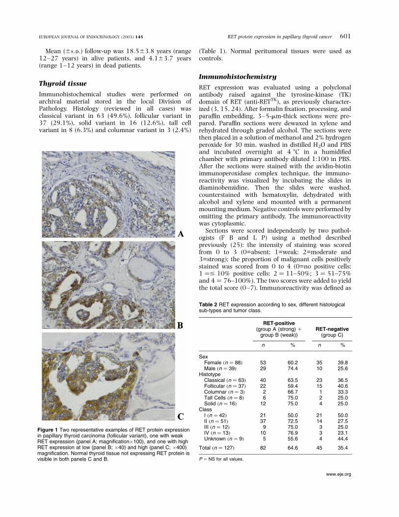
Figure 1 Two representative examples of RET protein expressionin papillary thyroid carcinoma (follicular variant), one with weakRET expression (panel A; magni®cation�100), and one with highRET expression at low (panel B; �40) and high (panel C; �400)magni®cation. Normal thyroid tissue not expressing RET protein isvisible in both panels C and B.
Table 2 RET expression according to sex, different histologicalsub-types and tumor class.
RET-positive(group A (strong) 1
group B (weak))RET-negative
(group C)
n % n %
SexFemale �n � 88� 53 60.2 35 39.8Male �n � 39� 29 74.4 10 25.6
HistotypeClassical �n � 63� 40 63.5 23 36.5Follicular �n � 37� 22 59.4 15 40.6Columnar �n � 3� 2 66.7 1 33.3Tall Cells �n � 8� 6 75.0 2 25.0Solid �n � 16� 12 75.0 4 25.0
ClassI �n � 42� 21 50.0 21 50.0II �n � 51� 37 72.5 14 27.5III �n � 12� 9 75.0 3 25.0IV �n � 13� 10 76.9 3 23.1Unknown �n � 9� 5 55.6 4 44.4
Total �n � 127� 82 64.6 45 35.4
P � NS for all values.
RET protein expression in papillary thyroid cancer 601EUROPEAN JOURNAL OF ENDOCRINOLOGY (2001) 145
www.eje.org

strong when the total score was $4 (group A), weakwhen it was between 1 and 3 (group B) and negativewhen it was 0 (group C).
Statistical analysis
Survival curves were plotted by the Kaplan-Meiermethod. Statistical analysis of survival was performedby the Log Rank test and Cox proportional hazardmodel as speci®ed. Chi square analysis was used for allthe other statistics.
Results
RET protein expression byimmunohistochemistry
A total of 82 (64.6%) PTC expressed RET protein withdifferent patterns of intensity and numbers of cells.Using the score derived from the two parameters, 65tumors (51.2%) had strong RET expression (group A),17 (13.4%) had positive but weak expression (group B),and 45 (35.4%) had no expression (group C). Asigni®cant positive correlation (r � 0:87; P , 0:0001;by simple regression analysis) was found between thedegree of cell staining and the proportion of cellsexpressing the RET protein.
Immunohistochemical detection of RET protein wasconsistently observed in the cytoplasm of the neoplasticcells (Fig. 1). RET protein expression was absent in thenormal peritumoral tissues.
Correlation with clinical data
As shown in Table 2, sex, histological sub-types andtumor class did not in¯uence the expression of RET.Similarly, RET expression was not different in thevarious age groups: in particular, younger patientswere not associated with a higher frequency of RETexpression (Table 3). Expression of RET according to the®nal outcome is reported in Table 4: positive RET
Table 3 RET expression according to age at diagnosis.
RET-positive(group A (strong) 1
group B (weak))RET-negative
(group C)
Age n % n %
5±29 years �n � 44� 29 65.9 15 34.130±44 years �n � 43� 27 62.8 16 37.245±90 years �n � 40� 26 65.0 14 35.0
P � NS for all values.
Table 4 RET expression according to outcome.
RET-positive(group A (strong) 1
group B (weak))RET-negative
(group c)
Outcome n % n %
Remission �n � 95� 60 63.2 35 36.8Persistence �n � 21� 16 76.2 5 23.8Death �n � 11� 6 54.5 5 45.5
Total 82 64.6 45 35.4
P � NS for all values.
Table 5 Risk factors of death from cancer.
Risk factor P-value Relative risk
Age (years) 0.072931±45 vs 0±30 N.S..45 vs 0±30 0.0028 2.25
RET protein expressionGroup B vs C N.S.Group A vs C N.S.
Sex N.S.Tumor class 0.003
II vs I 0.0549 0.57III vs I N.S.IV vs I 0.002 2.8
N.S., not signi®cant.
Figure 2 Survival probability (cancer related deaths only), by Kaplan-Meier method, in relation to RET protein expression. Theprobability of survival is similar for patients of groups A, B or C (P � 0:79; by Long Rank test).
602 F Basolo and others EUROPEAN JOURNAL OF ENDOCRINOLOGY (2001) 145
www.eje.org
expression (group A 1 group B) was not associatedwith a speci®c outcome. This was true even whentumors with weak RET expression (group B) wereexcluded from the statistical comparison or weregrouped together with tumors with no RET expression(group B 1 group C).
The cumulative survival rate at 20 years was 91.3%.As shown in Fig. 2, the survival probability did notdiffer according to the pattern of RET expression�P � 0:799�:
By multivariate analysis (Cox proportional hazardmodel; Table 5), signi®cant independent predictors ofdeath were: age .45 years (relative risk: 2.25) andtumor class IV (relative risk: 2.8). Sex and RETexpression had no in¯uence on survival.
DiscussionThe relationship between RET/PTC and clinical±histological features has been studied by severalauthors with con¯icting results. In some studies,RET/PTC rearrangements have been found morefrequently in microcarcinomas and in the classicalvariant of PTC (17±19). No rearrangements werefound in poorly differentiated or in undifferentiatedtumors by Tallini et al. (3). The same authors found nocorrelation between RET/PTC activation and tumorsize, clinical stage of the disease at presentation andpresence of metastases (3). RET/PTC has been asso-ciated with tumors of patients of younger age, thosewith the best prognosis, and in addition has beencorrelated with markers of low proliferative activity,such as MIB-1 (26, 27). Altogether, these studies leadto the tentative conclusion that RET/PTC is predomi-nant in well differentiated papillary thyroid carcinomaswhich do not progress to less differentiated phenotype.
On the other hand, other authors have found asigni®cant association between RET activation andpapillary tumors exhibiting a more advanced andaggressive phenotype at presentation, including theassociation with distant metastases (21, 22, 28). Theseauthors speculated on the possibility that RET expres-sion might be associated with an adverse ®nal outcome.
Apart from these data, the role of RET activation ontumor outcome has never been addressed in a largeseries of patients with long term follow-up. Our studyhas been designed to investigate this aspect, usingimmunohistochemistry for RET protein on archivalmaterial from a retrospective series of papillary thyroidcancer patients followed for a mean time of 18:5^3:8years.
The anti-RET antibody used in this study reactsagainst the tyrosine-kinase domain of RET which ispresent in any form of the rearranged RET protein, aswell as in the normal RET protein. Since RET proto-oncogene is not expressed in normal follicular cells (3,29), we can assume that the detection of the TKdomain with our antibody represents evidence that a
RET rearrangement has taken place. This statement isvalidated by previous studies, showing that our anti-body correlates well with RET oncogene activationdetected by RT-PCR (3, 15, 20), and by the constantabsence of RET expression in the normal peritumoraltissues of our patients.
In our study, no difference was found in the survivalof RET-positive or RET-negative cases. The signi®cantadverse prognostic variables found in this study werethose usually reported in several large series of PTCs:older age and distant metastases at diagnosis. We canspeculate that, while RET is an important initiatingevent, the activation of other oncogenes or theinactivation of tumor suppressor genes are responsiblefor the progression to more aggressive and treatment-resistant tumors. The behavior of a human RET-positive papillary thyroid cancer resembles thatexperimentally found in transgenic mice expressingRET/PTC-1, which develop slowly progressive thyroidtumors not causing the premature death of the animals(30). The higher prevalence of RET rearrangementfound in young patients by previous studies (11, 19)was not con®rmed in our series.
In conclusion, our study shows that RET-positivepapillary thyroid carcinomas are not associated with aprognosis different from RET-negative cases. RETexpression gives no prognostic information in theclinical management of papillary thyroid carcinoma.
AcknowledgementsThis work has been supported in part by grants fromthe Associazione Italiana Ricerca sul Cancro (AIRC);the European Union, INCO-COPERNICUS: project IC15-CT980314; and Ministero dell'UniversitaÁ della RicercaScienti®ca e Tecnologica (MURST) 1998.
L Agate is the recipient of a grant from FondazioneItaliana per la Ricerca sul Cancro (FIRC).
References1 Rosai J, Carcangiu ML & DeLellis RA. Tumor of the thyroid gland.
In Atlas of Tumor Pathology, 3rd series, fascicle 5. Washington DC:Armed Forces Institute of Pathology, 1992.
2 Carcangiu ML, Zampi G & Rosai J. Poorly differentiated (`insular')thyroid carcinoma. A reinterpretation of Langhans' `wucherndeStruma'. American Journal of Surgical Pathology 1984 8 655±668.
3 Tallini G, Santoro M, Pelie M et al. RET/PTC oncogene activationde®nes a subset of papillary thyroid carcinomas lacking evidenceof progression to poorly differentiated or undifferentiated tumorphenotypes. Clinical Cancer Research 1998 4 287±295.
4 Grieco M, Santoro M, Berlingeri MT et al. PTC is a novelrearranged form of the ret proto-oncogene and is frequentlydetected in vivo in human thyroid papillary carcinomas. Cell1990 60 557±563.
5 Jhiang SM, Smanik P & Mazzaferri EL. Development of a singlestep duplex RT-PCR detecting different forms of ret activation andidenti®cation of the third form of in vivo ret activation in humanpapillary thyroid carcinoma. Cancer Letters 1994 78 69±76.
6 Bongarzone I, Monzini N, Borrello MG et al. Molecularcharacterization of thyroid tumor-speci®c transforming sequenceformed by the fusion of RET tyrosine kinase and the regulatory
RET protein expression in papillary thyroid cancer 603EUROPEAN JOURNAL OF ENDOCRINOLOGY (2001) 145
www.eje.org
subunit RI alpha of cyclic AMP-dependent protein kinase.Molecular and Cellular Biology 1993 13 358±366.
7 Klugbauer S, Demidchik EP, Lengfelder E & Rabes H. Detection ofa novel type of RET rearrangement (PTC5) in thyroid carcinomasafter Chernobyl and analysis of the involved RET-fused geneRFG51. Cancer Research 1998 58 198±203.
8 Klugbauer S, Jauch A, Lengfelder E, Demidchik E & Rabes HM. Anovel type of RET rearrangement (PTC8) in childhood papillarythyroid carcinomas and characterization of the involved gene(RFG81). Cancer Research 2000 60 7028±7032.
9 Rabes HM & Klugbauer S. Molecular genetics of childhoodpapillary thyroid carcinomas after irradiation: high prevalence ofRET rearrangement. Recent results. Cancer Research 1998 154149±165.
10 Elisei R, Romei C, Vorontsova T et al. RET/PTC rearrangements inthyroid nodules: studies in irradiated and not irradiated,malignant and benign thyroid lesions in children and adults.Journal of Clinical Endocrinology and Metabolism 2001 86 3211±3216.
11 Bongarzone I, Fugazzola L, Vigneri P et al. Age-related activationof the tyrosine kinase receptor protooncogenes ret and NTRK1 inpapillary thyroid carcinoma. Journal of Clinical Endocrinology andMetabolism 1996 81 2006±2009.
12 Bounacer A, Wicker R, Caillou B et al. High prevalence ofactivating ret proto-oncogene rearrangements in thyroid tumorsfrom patients who had received external radiation. Oncogene1997 15 1263±1273.
13 Fugazzola L, Pilotti S, Pinchera A et al. Oncogenic rearrange-ments of the Ret proto-oncogene in papillary thyroid carcinomasfrom children exposed to the Chernobyl nuclear accident. CancerResearch 1995 55 5617±5620.
14 Klugbauer S, Lengfelder E, Demidchick EP & Rabes HM. Highprevalence of Ret rearrangement in thyroid tumors of childrenfrom Belarus after the Chernobyl reactor accident. Oncogene1995 11 2459±2467.
15 Thomas GA, Bunnel H, Cook HA et al. High prevalence ofRET/PTC rearrangements in Ukrainian and Belorussianpost-Chernobyl thyroid papillary carcinomas: a strong corre-lation between RET-PTC3 and the solid-follicular variant.Journal of Clinical Endocrinology and Metabolism 1999 844232±4238.
16 Pacini F, Elisei R, Romei C & Pinchera A. RET proto-oncogenemutations in thyroid carcinoma: clinical relevance. Journal ofEndocrinological Investigation 2000 23 328±338.
17 Santoro M, Carlomagno F, Hay ID et al. Ret oncogeneactivation in human thyroid neoplasm is restricted topapillary cancer subtype. Journal of Clinical Investigation1992 89 1517±1522.
18 Viglietto G, Chiappetta G, Martinez-Tello FJ et al. RET/PTConcogene activation is an early event in thyroid carcinogenesis.Oncogene 1995 11 1207±1210.
19 Soares P, Fonseca E, Wynford-Thomas D & Sobrinho-Simoes M.Sporadic ret-rearranged papillary carcinoma of the thyroid: asubset of slow growing, less aggressive thyroid neoplasms?Journal of Pathology 1998 185 71±78.
20 Sugg SL, Ezzat S, Rosen IB, Freeman JL & Asa SL. Distinctmultiple RET/PTC gene rearrangements in multifocal papillarythyroid neoplasia. Journal of Clinical Endocrinology and Metabolism1998 83 4116±4122.
21 Jhiang SM, Caruso DR, Gilmore E et al. Detection of the PTC/RETTPC oncogene in human thyroid carcinomas. Oncogene 19927 1331±1337.
22 Di Renzo MF, Olivero M, Ferro S et al. Overexpression of thec-MET/HGF receptor gene in human thyroid carcinomas.Oncogene 1992 7 2549±2553.
23 DeGroot LJ, Kaplan EL, McConik M & Straus FH. Natural history,treatment and course of papillary thyroid carcinoma. Journal ofClinical Endocrinology and Metabolism 1990 71 414±424.
24 Santoro M, Wong WT, Aroca P et al. An epidermal growth factorreceptor/RET chimera generates mitogenic and transformingsignals: evidence for RET-speci®c signaling pathway. Molecularand Cellular Biology 1994 14 663±675.
25 Fontanini G, Campani D, Roncella M et al. Expression ofinterleukin 6 (IL-6) correlates with oestrogen receptor inhuman breast carcinoma. British Journal of Cancer 1999 80579±584.
26 Sugg SL, Zheng L, Rosen IB, Freeman JL, Ezzat S & Asa SL. RET/PTC-1, -2, and -3 oncogene rearrangements in human thyroidcarcinomas: implications for metastatic potential? Journal ofClinical Endocrinology and Metabolism 1996 81 3360±3365.
27 Katoh R, Bray CE, Suzuki K et al. Growth activity in hyperplasticand neoplastic human thyroid determined by an immunohisto-chemical staining procedure using monoclonal antibody MIB-1.Human Pathology 1995 26 139±146.
28 Bongarzone I, Vigneri P, Mariani L, Collini P, Pilotti S &Pierotti MA. RET/NTRK1 rearrangements in thyroid glandtumors of the papillary carcinoma family: correlation withclinicopathological features. Clinical Cancer Research 1998 4223±228.
29 Schuchardt A, D'Agati V, Larsson-Blomberg L, Costantini F &Pachnis V. Defects in the kidney and enteric nervous system ofmice lacking the tyrosine kinase receptor RET. Nature 1994 367380±383.
30 Santoro M, Chiappetta G, Cerrato A et al. Development of thyroidpapillary carcinomas secondary to tissue-speci®c expression ofthe RET/PTC1 oncogene on transgenic mice. Oncogene 1996 121821±1826.
Received 2 March 2001
Accepted 5 July 2001
604 F Basolo and others EUROPEAN JOURNAL OF ENDOCRINOLOGY (2001) 145
www.eje.org
Copyright © 2022 FDOKUMEN