Endostatin induces autophagy in endothelial cells by modulating Beclin 1 and β-catenin levels
Upload
independentCategory
view
0download
0
CDK8 is a colorectal cancer oncogene that regulates β-cateninactivity
Ron Firestein1,3,6,7, Adam J. Bass1,4,6,7, So Young Kim1,4,6,7, Ian F. Dunn1,2,6,7, SerenaJ. Silver7, Isil Guney1,6,7, Ellen Freed1, Azra H. Ligon3, Natalie Vena1, Shuji Ogino1,3, MilanG. Chheda1,5,7, Pablo Tamayo7, Stephen Finn3, Yashaswi Shrestha1,6,7, Jesse S.Boehm7, Supriya Jain1, Emeric Bojarski1, Craig Mermel1,6,7, Jordi Barretina1,6,7, JenniferA. Chan3,7, Jose Baselga8, Josep Tabernero8, David E. Root8, Charles S. Fuchs1, MassimoLoda1,3, Ramesh A. Shivdasani1,4, Matthew Meyerson1,3,6,7, and William C. Hahn1,4,6,7
1Department of Medical Oncology, Dana-Farber Cancer Institute, 44 Binney Street, Boston, MA 02115U.S.A.
2Department of Neurosurgery, Brigham and Women's Hospital and Harvard Medical School, Boston, MA02115 U.S.A.
3Department of Pathology, Brigham and Women's Hospital and Harvard Medical School, Boston, MA 02115U.S.A.
4Department of Medicine, Brigham and Women's Hospital and Harvard Medical School, Boston, MA 02115U.S.A.
5Department of Neuro-oncology, Brigham and Women's Hospital and Harvard Medical School, Boston, MA02115 U.S.A.
6Center for Cancer Genome Discovery, Dana-Farber Cancer Institute, 44 Binney Street, Boston, MA 02115U.S.A.
7Broad Institute of Harvard and M.I.T., 7 Cambridge Center, Cambridge, MA 02142 U.S.A.
8Department of Medical Oncology, Hospital Vall d'Hebron, Passeig Vall d'Hebron, 119-129, 08035Barcelona, Spain.
AbstractAberrant activation of the canonical Wnt/β-catenin pathway occurs in almost all colorectal cancersand contributes to their growth, invasion and survival. Although dysregulated β-catenin activitydrives colon tumorigenesis, additional genetic perturbations are required to elaborate fully malignantdisease. To identify genes that both modulate β-catenin activity and are essential for colon cancercell proliferation, we conducted two loss-of-function screens in human colon cancer cells andcompared genes identified in these screens with an analysis of copy-number alterations in coloncancer specimens. One of these genes, CDK8, which encodes a member of the mediator complex, islocated at 13q12.13, a region of recurrent copy number gain in a substantial fraction of colon cancers.Suppression of CDK8 expression inhibited proliferation in colon cancer cells characterized by highlevels of CDK8 and β-catenin hyperactivity. CDK8 kinase activity was necessary for β-catenin driventransformation and expression of several β-catenin transcriptional targets. Together theseobservations suggest that therapeutic interventions targeting CDK8 may confer clinical benefit inβ-catenin-driven malignancies.
Correspondence and Requests for materials should be addressed to W.C.H. (Email: [email protected])..
NIH Public AccessAuthor ManuscriptNature. Author manuscript; available in PMC 2009 September 25.
Published in final edited form as:Nature. 2008 September 25; 455(7212): 547–551. doi:10.1038/nature07179.
NIH
-PA Author Manuscript
NIH
-PA Author Manuscript
NIH
-PA Author Manuscript
The Wnt/β-catenin pathway is implicated in over 90% of colon cancers and in a fraction ofother human malignancies. Loss of the tumor suppressor APC or activating CTNNB1 (β-catenin) mutations results in constitutive activity of the β-catenin-T cell factor (TCF)transcriptional complex, which drives adenoma formation1,2. Although mutations in TP53 orK-RAS cooperate with dysregulated β-catenin signaling to program a fully malignantphenotype3, these mutations are found in less than half of β-catenin-driven colon cancers4.
To identify oncogenes that modulate β-catenin-dependent transcription and regulate coloncancer cell proliferation, we conducted two RNAi-based loss-of-function screens. Weengineered DLD1 colon cancer cells, which harbor APC deletions and depend on β-catenin forproliferation5, to stably express “TOPFLASH” β-catenin-luciferase and “FOPFLASH”mutant-Renilla reporter constructs6,7 (DLD1Rep). Suppression of β-catenin expression inDLD1Rep cells by three β-catenin-specific short hairpin RNAs (shRNA) markedly reduced theTOPFLASH/FOPFLASH ratio (Fig. 1a), confirming that reporter activity requires β-cateninexpression. We then screened DLD1Rep cells with a shRNA library containing 4849 shRNAsthat target 1000 genes, including 95% of the human kinome6. We found 34 genes whoseexpression was necessary for β-catenin activity, including two known β-catenin regulators,CSNK1G38 and CSNK1E9 (Fig. 1b and Supplementary Table 1).
In parallel, we performed an arrayed, kinase-enriched shRNA screen in another β-catenin-dependent colon cancer cell line, HCT116, to identify genes essential for cancer cellproliferation. We identified 166 candidate genes necessary for proliferation using the criteriathat at least two shRNAs targeting the same gene induced a decrease in proliferation. Amongthe genes identified in this screen were the oncogenes KRAS and MYC (Fig. 1c andSupplementary Table 2). Compilation of genes from the two screens revealed nine whosesuppression affected both β-catenin transcriptional activity and colon cancer cell proliferation(Fig. 1d).
To determine whether any of these genes are amplified in colon cancers, we used single-nucleotide polymorphism (SNP) arrays and the GISTIC10,11 statistical method to conduct agenome-wide analysis of autosomal copy number (CN) alterations in primary resectionspecimens from 123 human colorectal adenocarcinomas (Fig. 2a). Among the nine genesidentified by our RNAi screens, only CDK8 resides within a particularly significant ampliconat 13q12.13-13q12.2 (false discovery rate (FDR) =1×10⁻²⁹) (Fig. 2a). These findings confirmrecent reports that a large portion of chromosome 13 is amplified in colon cancers12,13. Fifty-eight of 123 (47%) samples harbored this region of CN gain (Fig. 2b and Supplementary Table3).
To confirm these findings, we performed fluorescence in situ hybridization (FISH), usingprobes specific for CDK8 and RB1 (chromosome 13 control probe), on a tissue microarray(TMA) carrying 50 evaluable colon cancer specimens. We detected CDK8 CN gain in 31 of50 (62%) cases. 20 out of these 50 cases exhibited gains in both CDK8 and RB1 indicative ofpolysomy and consistent with recent observations linking RB/E2F1, β-catenin and CDK814.We also found CDK8 amplifications in 7 of these tumors (defined as CDK8:Control ratio ≥2)and low to moderate level CN gain (CDK8:Control ratio >1 and <2) in an additional 4specimens (Fig. 2c and Supplementary Table 4). Immunohistochemical analysis of CDK8expression in the same 50 specimens revealed elevated protein levels in 13 of 50 (26%) coloncancer samples, including those that showed CDK8 CN gain (Supplementary Fig. 1 andSupplementary Table 4). These observations indicate that CDK8 is amplified andoverexpressed in a substantial fraction of colon cancers.
The minimal region shared by these tumors encompasses 16 annotated genes (SupplementaryFig. 2a). To determine if CDK8 is the primary target of this amplicon, we first assessed
Firestein et al. Page 2
Nature. Author manuscript; available in PMC 2009 September 25.
NIH
-PA Author Manuscript
NIH
-PA Author Manuscript
NIH
-PA Author Manuscript
expression of these genes in colon cancer cells harboring chromosome 13q CN gain and foundthat 4 of the genes were not expressed (Supplementary Figure 2b). We suppressed theexpression of the remaining 12 genes in four cell lines, two (HT29, COLO-205) that harbor13q CN gain and two (SW837, T84) that show 13q loss (Supplementary Fig. 3a, b). To analyzethe screen results on a per-gene basis in cell lines with either 13q12 CN gain or deletion, weused an adaptation of the GSEA15 method and found that CDK8 was the only gene in thisregion required for proliferation of cell lines harboring 13q gain (FDR=0.24) (Fig. 2d andSupplementary Table 5). These observations suggested that colon cancer cells harboring13q12.2 amplification are particularly dependent on CDK8 expression for proliferation.
We then analyzed CDK8 CN gain and protein expression in a panel of 12 colon cancer lines.Four (COLO-205, HT29, SW1463, and SW480)16 of these 12 lines were found to harborCDK8 gain (Supplementary Fig. 3a, b), and these cell lines exhibited the highest levels ofCDK8 protein (Fig. 2e). Two additional colon cancer cell lines disomic for CDK8 (DLD1 andHCT116) also exhibited elevated CDK8 protein levels (Fig. 2d and Supplementary Fig.3a, b).Suppression of CDK8 expression induced substantially decreased proliferation in all six celllines with elevated CDK8 protein levels (Fig. 2f,g) but inhibited proliferation rates in the celllines with lower CDK8 protein levels to a lesser degree (Fig. 2h,i). Suppressing CDK8 in coloncancer cells reduced the fraction of cells in G1 and S phase, increased the number of aneuploidcells, and dramatically slowed cell proliferation without inducing apoptosis (SupplementaryFig. 4), similar to what was observed upon suppression of β-catenin. These observationsdemonstrate that colon cancer cells that express elevated CDK8 levels are highly dependenton its expression for proliferation.
To determine if CDK8 induces cell transformation, we overexpressed wild type CDK8 or apreviously reported kinase inactive, substitution mutant (D173A; CDK8-KD)17 in immortalmurine fibroblasts (NIH 3T3) (Fig. 3a). CDK8 expression induced focus formation, anchorage-independent colony growth, and tumor formation in immunodeficient animals (Fig. 3b-e),whereas the CDK8-KD mutant failed to transform the cells. These observations confirm thatCDK8 is a bona fide oncogene, whose kinase activity is necessary for oncogenic activity.
To dissect the relationship between CDK8 and β-catenin activity, we measured endogenousβ-catenin activity in the 12 cell lines used above. The RKO, COLO-741, HCA-7 and FHC celllines do not harbor known APC or β-catenin mutations18,19,20 and, as predicted, exhibitedlow levels of β-catenin activity. Of these four cell lines, suppression of CDK8 induced asubstantial proliferation effect only in COLO-741 (Fig. 2h and Supplementary Fig. 5a).Similarly, of the 12 cell lines tested, the six cell lines with highest CDK8 elevated levels showeda greater dependence on β-catenin for proliferation (Supplementary Fig. 5b).
CDK8 is a cyclin-dependent kinase member of the mediator complex, which couplestranscriptional regulators to the basal transcriptional machinery21. To explore the role ofCDK8 in modulating β-catenin transcriptional activity, we confirmed that suppressing CDK8with two independent, CDK8-specific shRNAs (shCDK8-1, shCDK8-2) in an additional cellline, SW480, also reduced β-catenin-dependent transcriptional activity (Fig. 4a). CDK8 kinaseactivity depends on the co-factor Cyclin C22, and we found that Cyclin C knockdownpreferentially affected colon cancer cell lines with chromosome 13q gain (Supplementary Fig.6a, b). To test whether CDK8 kinase activity is required to regulate β-catenin activity, weexpressed wild-type CDK8 or CDK8-KD in DLD1Rep cells carrying a shRNA targeting the3'-untranslated region, shCDK8-1 and found that only wild-type CDK8 partially rescued theeffects of suppressing endogenous CDK8(Supplementary Fig. 6c). These observationsdemonstrate that the kinase activity of CDK8 is necessary for both CDK8-inducedtransformation and β-catenin driven transcription.
Firestein et al. Page 3
Nature. Author manuscript; available in PMC 2009 September 25.
NIH
-PA Author Manuscript
NIH
-PA Author Manuscript
NIH
-PA Author Manuscript
The TCF-β-catenin complex regulates expression of several genes implicated in colon cancer,including MYC23, Axin226, and LEF127. Suppression of CDK8 in DLD1 and COLO-205cells reduced expression of each of genes (Fig. 4b). In contrast, we failed to observe changesin the expression of Notch or HES-1 (Fig. 4c), previously reported targets of CDK824. Thus,CDK8 modulates a subset of β-catenin driven genes previously implicated in cancer23,25,26.
We then performed chromatin immunoprecipitation (ChIP) near two verified β-catenin/TCFbinding elements (TBE)27 in the MYC promoter, as an example of a β-catenin regulated gene,to test whether CDK8 directly modulates MYC expression at the promoter level. We foundCDK8 associated with the MYC promoter (Fig. 4d). We therefore asked if loss of CDK8 bindingat the MYC promoter affects the ability of β-catenin to bind at the proximal and distal TBEs.Suppression of CDK8 expression reduced the amount of β-catenin bound to the proximalelement within the MYC promoter but had little effect on the amount associated with the distalelement (Fig. 4e). These observations implicate CDK8 and the mediator complex21 as a directregulator of β-catenin-driven transcription.
To test whether CDK8 activity is required for β-catenin driven transformation, we expressedthe dominantly interfering CDK8-KD mutant28 in transformed NIH-3T3 cells expressing aconstitutively active β-catenin mutant (Fig. 3c). Disruption of CDK8 activity inhibited β-catenin driven transformation, whereas a dominantly interfering TCF construct, previouslyshown to inhibit β-catenin-induced cellular transformation29, only partially abrogated CDK8-mediated transformation (Fig. 3d). These observations suggest that while CDK8 is requiredfor β-catenin mediated transformation, the full capacity of CDK8 to transform cells may extendbeyond its ability to activate β-catenin.
In summary, we have used an integrated approach to identify CDK8 as an oncogene in asubstantial fraction of colorectal cancers and demonstrate that its kinase activity is essentialfor its ability to regulate β-catenin dependent transcription and transformation. Theseobservations indicate that CDK8 acts in part by co-activating β-catenin driven transcription incolon cancers characterized by both high CDK8 expression and β-catenin activity.Accordingly, therapeutic interventions that target the CDK8 kinase activity in such cancersmay be of clinical value.
Methods SummaryLentiviral infections were performed using pLKO.1 lentiviral shRNA constructs generated bythe RNAi Consortium (TRC)6 and are listed in Supplementary Table 6. For high throughputscreening, cells infected with lentiviruses were allowed to grow for 4 d (DLD1Rep) or 5 d(HCT116). Suppression of β-catenin activity was defined as the ability of at least one shRNAto decrease activity more than 2 standard deviations (SD) below the Z score. Suppression ofproliferation was defined as the capacity for at least one shRNA to decrease proliferation morethan 1.5 SD below the mean Z score and at least one additional shRNA targeting the samekinase to decrease proliferation more that 1 SD below the mean Z score. For tissue analyses,a Tissue Microarray (TMA) composed of human colon cancer tissue and matching patientnormal colon was subjected to immunohistochemical staining. For FISH, BAC clones werehybridized in dual colors to four-micron TMA sections as follows: the RB1 probe labeled inSpectrumGreen was used as a chromosome 13 control probe and the RP11-726I20 BAC,spanning CDK8, was labeled with SpectrumOrange dUTP (both from Abbott Molecular/Vysis,Inc.).
Firestein et al. Page 4
Nature. Author manuscript; available in PMC 2009 September 25.
NIH
-PA Author Manuscript
NIH
-PA Author Manuscript
NIH
-PA Author Manuscript
Supplementary MaterialRefer to Web version on PubMed Central for supplementary material.
AcknowledgementsWe thank E. Shin for expert technical assistance in immunohistochemistry, M. Miri for assistance with samplecollection, and G. Getz for assistance with SNP array analysis. This work was supported in part by a T32 NIH grant(R.F.) and a GI SPORE Career Development Grant #P50CA127003 (R.F.), a Harvard-MIT Clinical InvestigatorTraining Program Fellowship (A.B), Department of Defense Prostate Cancer Postdoctoral Fellowships (I.G, S.Y),Warren-Whitman-Richardson, Hagerty Foundation Research Fellowships (I.D.) and K12 award (M.G.C.). J.B. is aBeatriu de Pinos Fellow of the Departament d'Educació i Universitats de la Generalitat de Catalunya. In accordancewith Harvard Medical School guidelines, we disclose that W.C.H., M.M., M.L. and R.A.S. are consultants for Novartis.
The SNP data can be found at: http://research3.dfci.harvard.edu/cdk8colon/index.php
References1. Bienz M, Clevers H. Linking colorectal cancer to Wnt signaling. Cell 2000;103:311–20. [PubMed:
11057903]2. Camp RL, Chung GG, Rimm DL. Automated subcellular localization and quantification of protein
expression in tissue microarrays. Nat Med 2002;8:1323–7. [PubMed: 12389040]3. Vogelstein B, et al. Genetic alterations during colorectal-tumor development. N Engl J Med
1988;319:525–32. [PubMed: 2841597]4. Smith G, et al. Mutations in APC, Kirsten-ras, and p53--alternative genetic pathways to colorectal
cancer. Proc Natl Acad Sci U S A 2002;99:9433–8. [PubMed: 12093899]5. van de Wetering M, et al. The beta-catenin/TCF-4 complex imposes a crypt progenitor phenotype on
colorectal cancer cells. Cell 2002;111:241–50. [PubMed: 12408868]6. Moffat J, et al. A lentiviral RNAi library for human and mouse genes applied to an arrayed viral high-
content screen. Cell 2006;124:1283–98. [PubMed: 16564017]7. Korinek V, et al. Constitutive transcriptional activation by a beta-catenin-Tcf complex in APC−/−
colon carcinoma. Science 1997;275:1784–7. [PubMed: 9065401]8. Davidson G, et al. Casein kinase 1 gamma couples Wnt receptor activation to cytoplasmic signal
transduction. Nature 2005;438:867–72. [PubMed: 16341016]9. Hino S, Michiue T, Asashima M, Kikuchi A. Casein kinase I epsilon enhances the binding of Dvl-1
to Frat-1 and is essential for Wnt-3a-induced accumulation of beta-catenin. J Biol Chem2003;278:14066–73. [PubMed: 12556519]
10. Beroukhim R, et al. Assessing the significance of chromosomal aberrations in cancer: methodologyand application to glioma. Proc Natl Acad Sci U S A 2007;104:20007–12. [PubMed: 18077431]
11. Weir BA, et al. Characterizing the cancer genome in lung adenocarcinoma. Nature 2007;450:893–8.[PubMed: 17982442]
12. Martin ES, et al. Common and distinct genomic events in sporadic colorectal cancer and diversecancer types. Cancer Res 2007;67:10736–43. [PubMed: 18006816]
13. Tsafrir D, et al. Relationship of gene expression and chromosomal abnormalities in colorectal cancer.Cancer Res 2006;66:2129–37. [PubMed: 16489013]
14. Morris EJ,JJ, Y F, Di Stefano L, Herr A, Moon NS, Kwon EJ, Haigis KM, Naar AM, Dyson NJ. E2F1represses β-catenin transcription and is antagonized by both pRB and CDK8. Submitted to Nature.2008
15. Subramanian A, et al. Gene set enrichment analysis: a knowledge-based approach for interpretinggenome-wide expression profiles. Proc Natl Acad Sci U S A 2005;102:15545–50. [PubMed:16199517]
16. Garraway LA, et al. Integrative genomic analyses identify MITF as a lineage survival oncogeneamplified in malignant melanoma. Nature 2005;436:117–22. [PubMed: 16001072]
17. Gold MO, Rice AP. Targeting of CDK8 to a promoter-proximal RNA element demonstrates catalysis-dependent activation of gene expression. Nucleic Acids Res 1998;26:3784–8. [PubMed: 9685496]
Firestein et al. Page 5
Nature. Author manuscript; available in PMC 2009 September 25.
NIH
-PA Author Manuscript
NIH
-PA Author Manuscript
NIH
-PA Author Manuscript
18. Ilyas M, Tomlinson IP, Rowan A, Pignatelli M, Bodmer WF. Beta-catenin mutations in cell linesestablished from human colorectal cancers. Proc Natl Acad Sci U S A 1997;94:10330–4. [PubMed:9294210]
19. Sparks AB, Morin PJ, Vogelstein B, Kinzler KW. Mutational analysis of the APC/beta-catenin/Tcfpathway in colorectal cancer. Cancer Res 1998;58:1130–4. [PubMed: 9515795]
20. Rowan AJ, et al. APC mutations in sporadic colorectal tumors: A mutational “hotspot” andinterdependence of the “two hits”. Proc Natl Acad Sci U S A 2000;97:3352–7. [PubMed: 10737795]
21. Conaway RC, Sato S, Tomomori-Sato C, Yao T, Conaway JW. The mammalian Mediator complexand its role in transcriptional regulation. Trends Biochem Sci 2005;30:250–5. [PubMed: 15896743]
22. Tassan JP, Jaquenoud M, Leopold P, Schultz SJ, Nigg EA. Identification of human cyclin-dependentkinase 8, a putative protein kinase partner for cyclin C. Proc Natl Acad Sci U S A 1995;92:8871–5.[PubMed: 7568034]
23. Sansom OJ, et al. Myc deletion rescues Apc deficiency in the small intestine. Nature 2007;446:676–9. [PubMed: 17377531]
24. Fryer CJ, White JB, Jones KA. Mastermind recruits CycC:CDK8 to phosphorylate the Notch ICDand coordinate activation with turnover. Mol Cell 2004;16:509–20. [PubMed: 15546612]
25. Yook JI, et al. A Wnt-Axin2-GSK3beta cascade regulates Snail1 activity in breast cancer cells. NatCell Biol 2006;8:1398–406. [PubMed: 17072303]
26. Murakami T, et al. Constitutive activation of Wnt/beta-catenin signaling pathway in migration-activemelanoma cells: role of LEF-1 in melanoma with increased metastatic potential. Biochem BiophysRes Commun 2001;288:8–15. [PubMed: 11594745]
27. He TC, et al. Identification of c-MYC as a target of the APC pathway. Science 1998;281:1509–12.[PubMed: 9727977]
28. Chiang MY, et al. Identification of a conserved negative regulatory sequence that influences theleukemogenic activity of NOTCH1. Mol Cell Biol 2006;26:6261–71. [PubMed: 16880534]
29. Kolligs FT, Hu G, Dang CV, Fearon ER. Neoplastic transformation of RK3E by mutant beta-cateninrequires deregulation of Tcf/Lef transcription but not activation of c-myc expression. Mol Cell Biol1999;19:5696–706. [PubMed: 10409758]
Firestein et al. Page 6
Nature. Author manuscript; available in PMC 2009 September 25.
NIH
-PA Author Manuscript
NIH
-PA Author Manuscript
NIH
-PA Author Manuscript
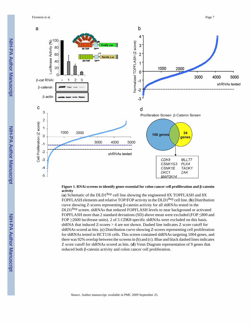
Figure 1. RNAi screens to identify genes essential for colon cancer cell proliferation and β-cateninactivity(a) Schematic of the DLD1Rep cell line showing the engineered 8X TOPFLASH and 8XFOPFLASH elements and relative TOP/FOP activity in the DLD1Rep cell line. (b) Distributioncurve showing Z-scores representing β-catenin activity for all shRNAs tested in theDLD1Rep screen. shRNAs that reduced FOPFLASH levels to near background or activatedFOPFLASH more than 2 standard deviations (SD) above mean were excluded (FOP ≤800 andFOP ≥2600 luciferase units). 2 of 5 CDK8-specific shRNAs were excluded on this basis.shRNA that induced Z-scores > 4 are not shown. Dashed line indicates Z score cutoff forshRNAs scored as hits. (c) Distribution curve showing Z-scores representing cell proliferationfor shRNAs tested in HCT116 cells. This screen contained shRNAs targeting 1004 genes, andthere was 92% overlap between the screens in (b) and (c). Blue and black dashed lines indicatesZ score cutoff for shRNAs scored as hits. (d) Venn Diagram representation of 9 genes thatreduced both β-catenin activity and colon cancer cell proliferation.
Firestein et al. Page 7
Nature. Author manuscript; available in PMC 2009 September 25.
NIH
-PA Author Manuscript
NIH
-PA Author Manuscript
NIH
-PA Author Manuscript
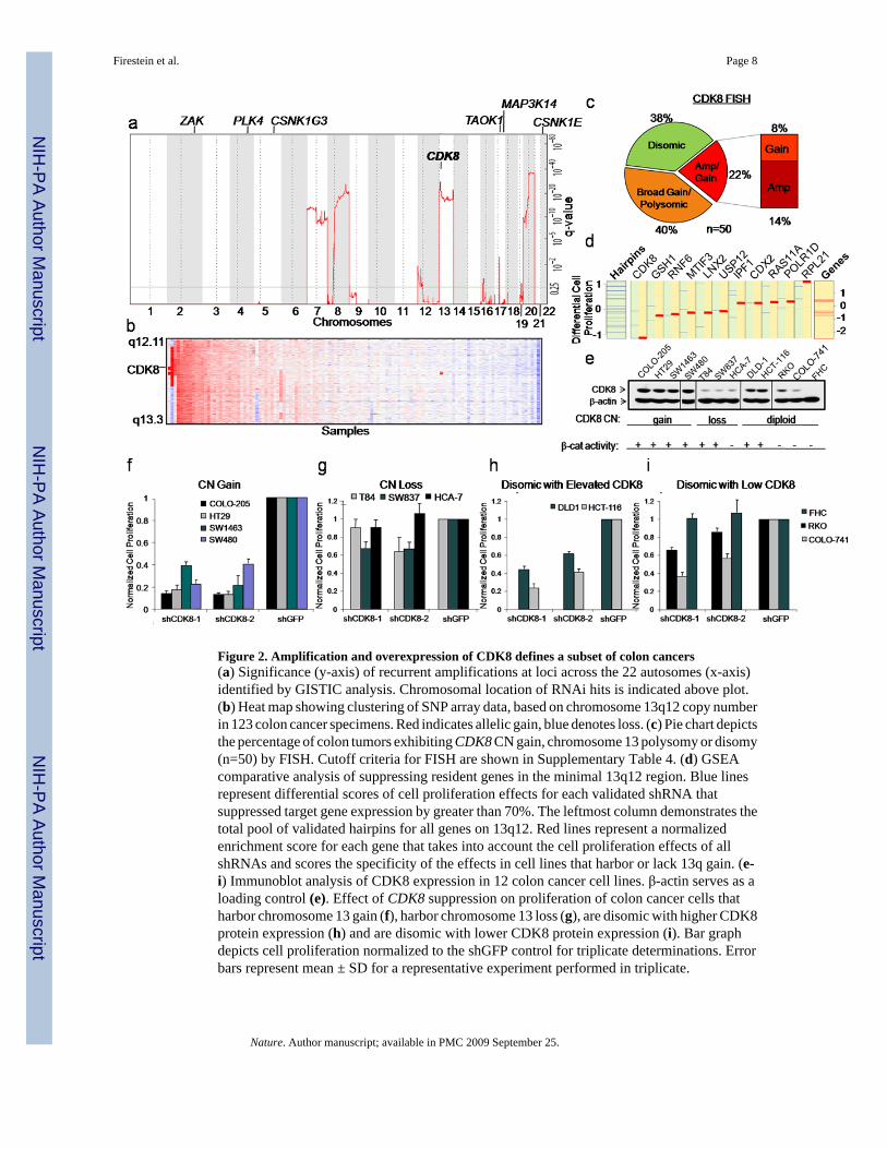
Figure 2. Amplification and overexpression of CDK8 defines a subset of colon cancers(a) Significance (y-axis) of recurrent amplifications at loci across the 22 autosomes (x-axis)identified by GISTIC analysis. Chromosomal location of RNAi hits is indicated above plot.(b) Heat map showing clustering of SNP array data, based on chromosome 13q12 copy numberin 123 colon cancer specimens. Red indicates allelic gain, blue denotes loss. (c) Pie chart depictsthe percentage of colon tumors exhibiting CDK8 CN gain, chromosome 13 polysomy or disomy(n=50) by FISH. Cutoff criteria for FISH are shown in Supplementary Table 4. (d) GSEAcomparative analysis of suppressing resident genes in the minimal 13q12 region. Blue linesrepresent differential scores of cell proliferation effects for each validated shRNA thatsuppressed target gene expression by greater than 70%. The leftmost column demonstrates thetotal pool of validated hairpins for all genes on 13q12. Red lines represent a normalizedenrichment score for each gene that takes into account the cell proliferation effects of allshRNAs and scores the specificity of the effects in cell lines that harbor or lack 13q gain. (e-i) Immunoblot analysis of CDK8 expression in 12 colon cancer cell lines. β-actin serves as aloading control (e). Effect of CDK8 suppression on proliferation of colon cancer cells thatharbor chromosome 13 gain (f), harbor chromosome 13 loss (g), are disomic with higher CDK8protein expression (h) and are disomic with lower CDK8 protein expression (i). Bar graphdepicts cell proliferation normalized to the shGFP control for triplicate determinations. Errorbars represent mean ± SD for a representative experiment performed in triplicate.
Firestein et al. Page 8
Nature. Author manuscript; available in PMC 2009 September 25.
NIH
-PA Author Manuscript
NIH
-PA Author Manuscript
NIH
-PA Author Manuscript
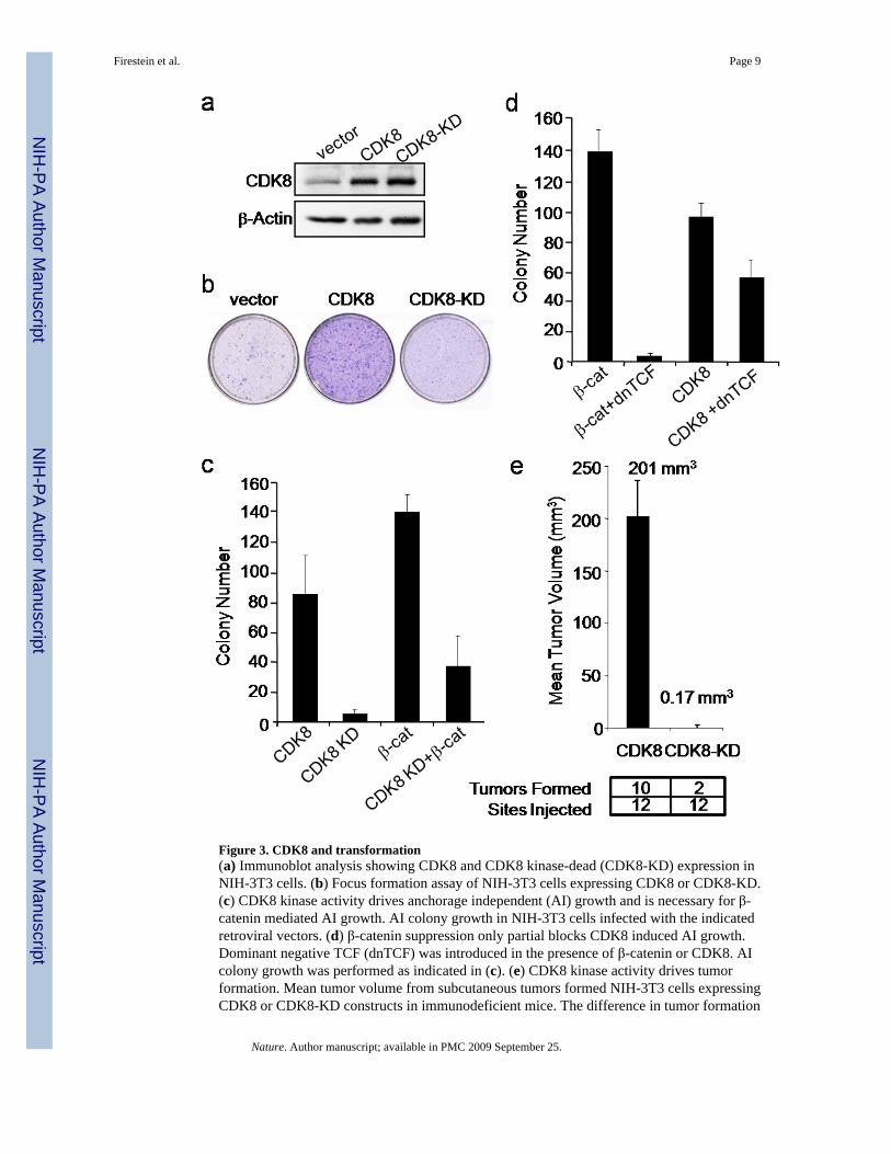
Figure 3. CDK8 and transformation(a) Immunoblot analysis showing CDK8 and CDK8 kinase-dead (CDK8-KD) expression inNIH-3T3 cells. (b) Focus formation assay of NIH-3T3 cells expressing CDK8 or CDK8-KD.(c) CDK8 kinase activity drives anchorage independent (AI) growth and is necessary for β-catenin mediated AI growth. AI colony growth in NIH-3T3 cells infected with the indicatedretroviral vectors. (d) β-catenin suppression only partial blocks CDK8 induced AI growth.Dominant negative TCF (dnTCF) was introduced in the presence of β-catenin or CDK8. AIcolony growth was performed as indicated in (c). (e) CDK8 kinase activity drives tumorformation. Mean tumor volume from subcutaneous tumors formed NIH-3T3 cells expressingCDK8 or CDK8-KD constructs in immunodeficient mice. The difference in tumor formation
Firestein et al. Page 9
Nature. Author manuscript; available in PMC 2009 September 25.
NIH
-PA Author Manuscript
NIH
-PA Author Manuscript
NIH
-PA Author Manuscript

between CDK8 and CDK8-KD was statistically significant, as assessed by unpaired t-test (pvalue = 0.0001). All experiments were performed in triplicate, and mean ± SD is shown.
Firestein et al. Page 10
Nature. Author manuscript; available in PMC 2009 September 25.
NIH
-PA Author Manuscript
NIH
-PA Author Manuscript
NIH
-PA Author Manuscript
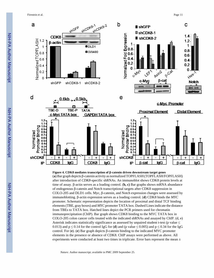
Figure 4. CDK8 mediates transcription of β-catenin driven downstream target genes(a) Bar graph depicts β-catenin activity as normalized TOPFLASH (TOPFLASH/FOPFLASH)after introduction of CDK8-specific shRNAs. An immunoblot shows CDK8 protein levels attime of assay. β-actin serves as a loading control. (b, c) Bar graphs shows mRNA abundanceof endogenous β-catenin and Notch transcriptional targets after CDK8 suppression inCOLO-205 and DLD1 cells. Myc, β-catenin, and Notch expression changes were assessed byimmunoblotting. β-actin expression serves as a loading control. (d) CDK8 binds the MYCpromoter. Schematic representation depicts the location of proximal and distal TCF bindingelements (TBE, gray boxes) and MYC promoter TATA box. Dashed Lines indicate the distancefrom TBEs to TATA box. Hatched lines depict the PCR primers used for chromatinimmunoprecipitation (ChIP). Bar graph shows CDK8 binding to the MYC TATA box inCOLO-205 colon cancer cells treated with the indicated shRNAs and assayed by ChIP. (d, e)Asterisk indicates statistically significance as assessed by unpaired student t-test (p value ≤0.013) and p ≤ 0.14 for the control IgG for (d) and (p value ≤ 0.005) and p ≤ 0.34 for the IgGcontrol. For (e). (e) Bar graph depicts β-catenin binding to the indicated MYC promoterelements in the presence or absence of CDK8. ChIP assays were performed as above. Allexperiments were conducted at least two times in triplicate. Error bars represent the mean ±
Firestein et al. Page 11
Nature. Author manuscript; available in PMC 2009 September 25.
NIH
-PA Author Manuscript
NIH
-PA Author Manuscript
NIH
-PA Author Manuscript

SD for a representative experiment performed in triplicate. Asterisk indicates a statisticallysignificance as assessed by unpaired student t-test
Firestein et al. Page 12
Nature. Author manuscript; available in PMC 2009 September 25.
NIH
-PA Author Manuscript
NIH
-PA Author Manuscript
NIH
-PA Author Manuscript
Copyright © 2022 FDOKUMEN