
ERG oncogene modulates prostaglandin signaling in prostate cancer cells
Upload
independentCategory
view
3download
0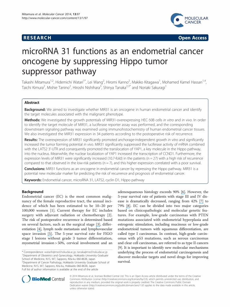
Mitamura et al. Molecular Cancer 2014, 13:97http://www.molecular-cancer.com/content/13/1/97
RESEARCH Open Access
microRNA 31 functions as an endometrial canceroncogene by suppressing Hippo tumorsuppressor pathwayTakashi Mitamura1,2, Hidemichi Watari1*, Lei Wang2, Hiromi Kanno2, Makiko Kitagawa1, Mohamed Kamel Hassan1,4,Taichi Kimura2, Mishie Tanino2, Hiroshi Nishihara3, Shinya Tanaka2,3* and Noriaki Sakuragi1
Abstract
Background: We aimed to investigate whether MIR31 is an oncogene in human endometrial cancer and identifythe target molecules associated with the malignant phenotype.
Methods: We investigated the growth potentials of MIR31-overexpressing HEC-50B cells in vitro and in vivo. In orderto identify the target molecule of MIR31, a luciferase reporter assay was performed, and the correspondingdownstream signaling pathway was examined using immunohistochemistry of human endometrial cancer tissues.We also investigated the MIR31 expression in 34 patients according to the postoperative risk of recurrence.
Results: The overexpression of MIR31 significantly promoted anchorage-independent growth in vitro and significantlyincreased the tumor forming potential in vivo. MIR31 significantly suppressed the luciferase activity of mRNA combinedwith the LATS2 3’-UTR and consequently promoted the translocation of YAP1, a key molecule in the Hippo pathway,into the nucleus. Meanwhile, the nuclear localization of YAP1 increased the transcription of CCND1. Furthermore, theexpression levels of MIR31 were significantly increased (10.7-fold) in the patients (n = 27) with a high risk of recurrencecompared to that observed in the low-risk patients (n = 7), and this higher expression correlated with a poor survival.
Conclusions: MIR31 functions as an oncogene in endometrial cancer by repressing the Hippo pathway. MIR31 is apotential new molecular marker for predicting the risk of recurrence and prognosis of endometrial cancer.
Keywords: Endometrial cancer, microRNA 31, LATS2, cyclin D1, Hippo pathway
BackgroundEndometrial cancer (EC) is the most common malig-nancy of the female reproductive tract, the annual inci-dence of which has been estimated to be 10–20 per100,000 women [1]. Current therapy for EC includessurgery with adjuvant radiation or chemotherapy [2].The risk of postoperative recurrence is determined basedon several factors, such as the surgical stage [3], differ-entiation [4], lymph node metastasis and lymphovascularspace invasion [5]. The 5-year survival rate for FIGOstage I lesions without grade 3 tumor differentiation,myometrial invasion > 50%, cervical involvement and an
* Correspondence: [email protected]; [email protected] of Obstetrics and Gynecology, Hokkaido University GraduateSchool of Medicine, N15, W7, Sapporo, Kita-ku 060-8638, Japan2Department of Cancer Pathology, Hokkaido University Graduate School ofMedicine, N15, W7, Sapporo, Kita-ku, Hokkaido 060-8638, JapanFull list of author information is available at the end of the article
© 2014 Mitamura et al.; licensee BioMed CentCommons Attribution License (http://creativecreproduction in any medium, provided the orDedication waiver (http://creativecommons.orunless otherwise stated.
adenosquamous histology exceeds 90% [6]. However, the5-year survival rate of patients with stage III and IV dis-ease is dramatically decreased, ranging from 42% [7] to79% [8]. EC can be divided into two major categoriesbased on clinicopathologic and molecular genetic fea-tures. For example, low-grade carcinomas with PTENmutations associated with endometrial hyperplasia andestrogenic stimulation, including mucinous or low-gradeendometrioid tumors with squamous differentiation, arecalled type I carcinomas. In contrast, high-grade carcin-omas with p53 mutations, such as serous carcinomasand clear cell carcinomas, are referred to as type II cancers[9]. It is important to identify new molecular mechanismsunderlying the process of endometrial carcinogenesis anddiscover molecular targets and novel drugs for improvingsurvival.
ral Ltd. This is an Open Access article distributed under the terms of the Creativeommons.org/licenses/by/2.0), which permits unrestricted use, distribution, andiginal work is properly credited. The Creative Commons Public Domaing/publicdomain/zero/1.0/) applies to the data made available in this article,

Mitamura et al. Molecular Cancer 2014, 13:97 Page 2 of 11http://www.molecular-cancer.com/content/13/1/97
microRNAs (MIRs) are endogenous non-coding RNAsof 18 to 25 nucleotides in length that play importantroles in regulating the gene expression. The matureforms of MIRs silence the gene expression by binding tothe 3’-untranslated region (UTR) of target mRNAs andinitiate the translational repression and/or cleavage ofcognate mRNAs [10]. MIRs have frequently been impli-cated in carcinogenesis [11-14]. In the setting of EC,MIR152 [15], MIR194 [16], MIR34b [17], MIR204 [18],MIR145 [19] and MIR129-2 [20] have been reported tobe tumor suppressor genes, and MIR125b [21] has beenreported to be an oncogene (oncomir). Furthermore,MIR31 has been reported to be an oncomir in varioushuman cancers, including colorectal [22], esophageal[23], lung [24], oral [25] and head and neck [26] cancer,and a tumor suppressor gene in breast [27] and gastric[28] cancers and malignant mesothelioma [29]. How-ever, little is known about the biological functions ofMIR31 in EC.The Hippo pathway is crucial in regulating the size of
organs, and its dysregulation contributes to tumorigen-esis [30]. Recently, it was reported that deregulation ofthe Hippo pathway occurs at a high frequency in abroad range of human cancers, including lung [24],hepatocellular [31], colon [32] and prostate cancer [33],and is often correlated with a poor patient prognosis.LATS2 represents a core component in the kinase cas-cade of the mammalian Hippo pathway. Interestingly, ithas been reported that the Hippo pathway is requiredfor anoikis and that the LATS2 expression levels aresignificantly downregulated in patients with metastaticprostate cancer [33].In this study, we aimed to investigate whether MIR31
is an oncomir in human EC and identify the direct targetassociated with the malignant phenotype of EC.
ResultsMIR31 is correlated with enhanced colony formation ofEC cell linesIn order to investigate whether the MIR31 expression iscorrelated with the tumorigenesis of EC, we performedcolony formation assays. We confirmed the MIR31 ex-pression in three EC cell-lines, HEC-50B, HEC-1A andHEC-108, using qRT-PCR and found that the MIR31levels were lowest in the HEC-108 cells, followed byHEC-1A and HEC-50B cells (Additional file 1: Figure S1,Lanes 1, 2 and 3). The colony number was increased inthe same order as the MIR31 expression under two differ-ent serum concentrations (Additional file 2: Figure S2).
The overexpression of MIR31 enhances tumorigenesisin vitro and in vivoWe established HEC-50B cells overexpressing MIR31by introducing precursor-MIR31 using lentivirus vectors
because the MIR31 expression level of HEC-50B wasmodest among the several adenocarcinoma cell lines ana-lyzed (Additional file 1: Figure S1) and lentivirus vectorscan be efficiently transfected into this cell line (HEC-50Bmock and MIR31). The presence of a mature-MIR31expression was confirmed using qRT-PCR (Figure 1a).Although MIR31 overexpression did not affect in vitro
cell proliferation under the standard culture conditions(data not shown), it significantly promoted colony forma-tion under serum starvation (Figure 1b). Additionally,an MIR31-specific inhibitor significantly restrained colonyformation (Additional file 3: Figure S3). The MIR31-mediated tumorigenic effects were confirmed in an in vivomodel. A significant increase in tumor weight wasobserved in the HEC-50B cells with MIR31 overexpres-sion compared with that noted in the controls in thenude mice subcutaneous tumor model (Figure 1c).These findings demonstrate that MIR31 induces a moreaggressive phenotype of EC.
MIR31 reduces the protein levels of LATS2 by inhibitingtranslationIn order to elucidate the mechanisms by which MIR31promotes tumorigenesis, in silico prediction models wereemployed to identify the target mRNAs of MIR31 [10].Among several candidates, we focused on LATS2 be-cause it is a known tumor suppressor gene that hasbeen previously reported to be a direct target of MIR31[24,34]. One potential binding site for MIR31 was foundin the 3’-UTR region of LATS2 mRNA (Figure 2a).To confirm that LATS2 is a target of MIR31 in HEC-
50B cells, the protein levels of LATS2 were analyzed inHEC-50B cells overexpressing MIR31. We found thatLATS2 was downregulated in the MIR31-overexpressingcells, whereas LATS2 was increased by the MIR31-specific inhibitor compared with that observed in thecontrol cells in a Western blot analysis (Figure 2b). Wenext performed a luciferase reporter assay to assesswhether MIR31 inhibits the translation of LATS2. Thedetection of a normalized luciferase activity revealed thatMIR31 significantly suppressed the activity of luciferasecombined with wild-type LATS2 3’-UTR in the HEC-50B MIR31 cells, whereas no differences were observedfollowing treatment with the control luciferase andLATS2 3’-UTR possessing a mutation in the putativeMIR31-binding site (Figure 2c). As no significant differ-ences in the LATS2 mRNA levels were observed be-tween the HEC-50B control and MIR31-overexpressingcells (Additional file 4: Figure S4), MIR31 does not ap-pear to degrade LATS2 mRNA. These results suggestthat MIR31 directly binds to LATS2 mRNA and regu-lates the LATS2 protein expression via translationalinhibition.
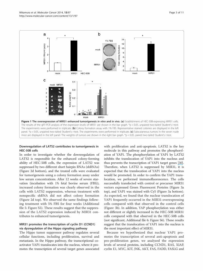
Figure 1 The overexpression of MIR31 enhanced tumorigenesis in vitro and in vivo. (a) Establishment of HEC-50B-expressing MIR31 cells.The results of the qRT-PCR analysis of the expression levels of MIR31 are shown in the bar graph. *p < 0.05, unpaired two-tailed Student’s t-test.The experiments were performed in triplicate. (b) Colony formation assay with 1% FBS. Representative stained colonies are displayed in the leftpanel. *p < 0.05, unpaired two-tailed Student’s t-test. The experiments were performed in triplicate. (c) Subcutaneous tumors in the seven nudemice are displayed in the left panel. The weights of tumors are shown in the right bar graph. *p < 0.05, paired two-tailed Student’s t-test.
Mitamura et al. Molecular Cancer 2014, 13:97 Page 3 of 11http://www.molecular-cancer.com/content/13/1/97
Downregulation of LATS2 contributes to tumorigenesis inHEC-50B cellsIn order to investigate whether the downregulation ofLATS2 is responsible for the enhanced colony-formingability of HEC-50B cells, the expression of LATS2 wassuppressed by two different short hairpin RNAs (shRNAs)(Figure 2d bottom), and the treated cells were evaluatedfor tumorigenesis using a colony formation assay underlow serum concentrations. After 12 weeks of severe star-vation (incubation with 1% fetal bovine serum (FBS)),increased colony formation was clearly observed in thecells with LATS2 suppression, whereas treatment withnonspecific shRNA did not affect colony formation(Figure 2d top). We observed the same findings follow-ing treatment with 5% FBS for four weeks (Additionalfile 5: Figure S5). These results suggest that the suppres-sion of the LATS2 expression induced by MIR31 con-tributes to enhanced tumorigenesis.
MIR31 promotes the transcription of cyclin D1 (CCND1)via dysregulation of the Hippo signaling pathwayThe Hippo tumor suppressor pathway regulates severalcellular functions, including proliferation, survival andmetastasis. In the Hippo pathway, the transcriptional co-activator YAP1 translocates into the nucleus, where it pro-motes the transcription of several target genes associated
with proliferation and anti-apoptosis. LATS2 is the keymolecule in this pathway and promotes the phosphoryl-ation of YAP1. The phosphorylation of YAP1 by LATS2inhibits the translocation of YAP1 into the nucleus andthus prevents the transcription of YAP1 target genes [30].Therefore, when LATS2 is suppressed by MIR31, it isexpected that the translocation of YAP1 into the nucleuswould be promoted. In order to confirm the YAP1 trans-location, we performed immunofluorescence. The cellssuccessfully transfected with control or precursor MIR31vectors expressed Green Fluorescent Proteins (Figure 3atop), and YAP1 was stained with Cy5 (Figure 3a bottom).As expected, we found that the nuclear translocation ofYAP1 frequently occurred in the MIR31-overexpressingcells compared with that observed in the control cells(Figure 3b). In addition, YAP phosphorylation was eithernot different or slightly increased in the HEC-50B MIR31cells compared with that observed in the HEC-50B cells(not significant, Additional file 6: Figure S6). These resultssuggest that the translocation of YAP1 into the nucleus isthe most important effect of MIR31.Because we hypothesized that nuclear YAP1 pro-
motes the transcription of several anti-apoptosis andpro-proliferation genes, we analyzed the expressionlevels of several proteins, including CCND1, RAS, XIAP,cyclin E1, MYC, KIT, JNK, AKT, FAS, FADD, FASLG and

Figure 2 MIR31 regulates the LATS2 expression by inhibiting translation. (a) The potential binding site for MIR31 in 3’UTR of LATS2 mRNA.(b) The expression level of LATS2 in the mock and MIR31-overexpressing cells (top). The LATS2 expression was increased by the MIR31-specificinhibitor (bottom). The results of immunoblotting for LATS2 and α-tubulin are shown. (c) The luciferase activity after transfection of the indicated3’-UTR-driven reporter constructs. Reporter plasmids containing no oligonucleotides as a Control, the wild-type 3’UTR region of LATS2 as a Wildtype and the mutant 3’UTR region as a Mutant. *p < 0.05, unpaired two-tailed Student’s t-test. (d) Colony formation assay (bar graph) andimmunoblotting for LATS2 and α-tubulin following shRNA transfection (bottom panel). *p < 0.05, unpaired two-tailed Student’s t-test. All experimentswere performed in triplicate.
Mitamura et al. Molecular Cancer 2014, 13:97 Page 4 of 11http://www.molecular-cancer.com/content/13/1/97
BCL2, in the HEC-50B MIR31-overexpressing and controlcells using immunoblotting (Additional file 7: Figure S7).We found the MIR31 overexpression to be associatedwith increased CCND1, RAS and XIAP expressionlevels (Figure 3c, Additional file 8: Figure S8a top) andthey also decreased in the anti-MIR31-oligonucleotide-induced cells (Figure 3c, Additional file 8: Figure S8abottom). Because these results strongly suggest thatnuclear YAP1, which is increased by the MIR31 expres-sion, promotes the transcription of these targets, weperformed luciferase reporter assays in order to investigatethe influence of MIR31 on the transcription of CCND1,RAS and XIAP. The detection of a normalized luciferaseactivity revealed that the MIR31 expression significantlyincreased the activity of luciferase driven by the CCND1,RAS and XIAP promoters compared with that observedin the control cells (Figure 3d Line 1–2, Additional file 8:
Figure S8b). In addition, we focused on the CCND1 ex-pression and investigated the influence of nuclear YAP1overexpression on the CCND1 expression. HEC-50B cellswere transfected with a YAP1 expression vector, the resultsof which confirmed that nuclear YAP1 was overexpressedon immunofluorescence (Additional file 9: Figure S9).As expected, YAP suppression by anti-YAP siRNA sig-nificantly decreased the activity of luciferase driven bythe CCND1 promotor, and the expression of CCND1was increased by YAP1 overexpression (Figure 3d, Lines2–3, Figure 3e).We also found a correlation between the MIR31, LATS2
and CCND1 expression in vivo (mouse 7 in Figure 1c). TheLATS2 expression was increased in the control cell tumorscompared with that observed in the MIR31-expressingtumor cells, and the CCND1 levels were increased in thetumors formed from MIR31-expressing cells (Figure 3f).
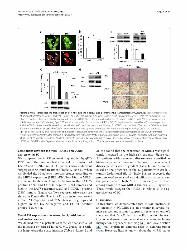
Figure 3 MIR31 promotes the translocation of YAP1 into the nucleus and promotes the transcription of CCND1. (a) Representative cellsof immunofluorescence for GFP and YAP1, x600. The nuclei are indicated by white arrows. *The translocation of YAP1 into the nucleus was notobserved in the cells unsuccessfully transfected with pre-MIR31. The cells were cultured under standard conditions with 5% fetal bovine serum.(b) Ratio of nuclear YAP1 staining. *p < 0.05, unpaired two-tailed Student’s t-test. (c) The CCND1 levels were increased by MIR31 overexpressionand the CCND1 levels were decreased by the MIR31-specific inhibitor on immunoblotting for CCND1 and α-tubulin. The ratio of CCND1/α-tubulinis shown in the bar graph. (d) The CCND1 levels were increased by YAP1 overexpression. Immunoblotting for YAP1, CCND1 and α-tubulin.(e) The luciferase activity after transfection of the reporter constructs containing the LATS2 promotor region normalized to the GAPDH promotorregion (top). Immunoblotting for YAP and α-tubulin following siRNA transfection (bottom). Mock and MIR31 cells were transfected with non-targetingsiRNA. *p < 0.05, unpaired two-tailed Student’s t-test. (f) Correlation between the MIR31 expression and results of the immunohistochemical analysis ofLATS2 and CCND1 in vivo. Representative results are shown in micrographs, x100. All experiments were performed in triplicate.
Mitamura et al. Molecular Cancer 2014, 13:97 Page 5 of 11http://www.molecular-cancer.com/content/13/1/97
Correlations between the MIR31, LATS2 and CCND1expression in ECWe compared the MIR31 expression quantified by qRT-PCR and the immunohistochemical expression ofLATS2 and CCND1 in 34 EC patients who underwentsurgery as their initial treatment (Table 1, Lane 1). Whenwe divided the 34 patients into two groups according tothe MIR31 expression (MIR31/RNU44 = 15), the MIR31expression levels were found to be low in the LATS2-positive (73%) and CCND1-negative (27%) tumors andhigh in the LATS2-negative (25%) and CCND1-positive(75%) tumors. (Figure 4a; Two representative cases areshown in Figure 4b). The MIR31 expression was lowestin the LATS2-positive and CCND1-negative groups andhighest in the LATS2-negative and CCND1-positivegroups (Figure 4c).
The MIR31 expression is increased in high-risk humanendometrial cancersWe defined low-risk patients as those who satisfied all ofthe following criteria: pT1a, pN0, M0, grade1 or 2 with-out lymphovascular space invasion (Table 1, Lanes 2 and
3). We found that the expression of MIR31 was signifi-cantly increased in the high-risk patients (Figure 4d).All patients with recurrent disease were classified ashigh-risk patients. Since most tumors in the recurrentdisease patients were of grade 2 (Table 1, Lane 4), we fo-cused on the prognosis of the 13 patients with grade 2tumors (Additional file 10: Table S1). As expected, theprogression-free survival was significantly worse amongthe patients with high MIR31 tumors (> = 0.8) thanamong those with low MIR31 tumors (<0.8) (Figure 5).These results suggest that MIR31 is related to the ag-gressiveness of EC.
DiscussionIn this study, we demonstrated that MIR31 functions asan oncomir in EC. MIR31 is an oncomir in several hu-man cancers and a tumor suppressor gene in others. Wespeculate that MIR31 has a specific function in eachtype of malignancy, and several mechanisms, includingmethylation-dependent silencing [35] and local deletion[29], may explain its different roles in different tumortypes. However, little is known about the MIR31 status

Table 1 Clinical features of human endometrial cancer
Total Low-risk High-risk Recurrence
n (%) 34 (100) 7 (21) 27 (79) 7 (21)
Age, mean(range)
59 (38–78) 58 (38–75) 60 (45–78) 56 (45–67)
FIGO stage,n (%)
I 15 (44) 7 (100) 8 (30) 1 (14)
II 3 (9) 0 (0) 3 (11) 1 (14)
III 14 (41) 0 (0) 14 (52) 3 (43)
IV 2 (6) 0 (0) 2 (7) 2 (29)
Grade, n (%)
1 15 (44) 5 (71) 10 (37) 0 (0)
2 13 (38) 2 (29) 11 (41) 6 (86)
3 6 (18) 0 (0) 6 (22) 1 (14)
Lymphovascularspace invasion,n (%)
+ 10 (29) 0 (0) 10 (37) 2 (29)
- 24 (71) 7 (100) 17 (63) 5 (71)
MIR31, mean 18.40 2.05 21.90 9.15
(range) (0.15 - 284.06) (0.33 - 5.98) (0.15 - 284.06) (0.70 - 27.77)
Mitamura et al. Molecular Cancer 2014, 13:97 Page 6 of 11http://www.molecular-cancer.com/content/13/1/97
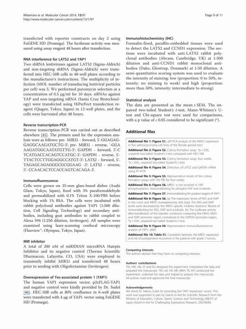
in patients with EC. In previous studies that reportedMIR31 to be an oncomir, MIR31 was found to regulateRAS p21 GTPase Activating Protein 1 (RASA1) [22] andRhoBTB1 [36] in colorectal cancer, LATS2 and PP2Aregulatory subunit B alpha isoform (PPP2R2A) [24] inlung cancer and factor-inhibiting hypoxia-induciblefactor (FIH) [26] in head and neck carcinomas. It isplausible that MIR31 represses the expression of tumorsuppressor genes, such as LATS2, to act as an oncomirand indirectly promotes the transcription of genes re-lated to cell cycle control and tumorigenesis (Figure 6).We herein demonstrated that the Hippo pathway is in-
volved in EC tumorigenesis and correlates with a poorpatient prognosis. Although no significant relationshipswere observed between the immunohistochemical ex-pression of LATS2 and clinical risk factors, includinglymph node metastasis, cervical invasion or lymphovas-cular space invasion, such relationships deserve furtherinvestigation in a larger patient cohort. It is reasonable topostulate that the Hippo pathway regulates the CCND1expression via YAP1 translocation into the nucleus in EC,as the cyclin family is known to be a major target of theHippo pathway [37-39]. The overexpression of CCND1may not be an independent factor causing tissue over-growth, since it is suggested that the overexpression of allknown yorkie targets fails to mimic the effect of yorkieitself in driving tissue growth in drosophila [40]. AsMIR31 tends to block the cell apoptosis induced by
ultraviolet treatment in HEC-50B cells (data not shown),we speculate that other transcriptional targets of YAP1associated with apoptosis, such as XIAP, may be coor-dinately regulated with CCND1. The Hippo pathway isknown to be related to the p53 activity, for example,LATS2 tumor suppressor augments p53-mediated apop-tosis by promoting the nuclear proapoptotic function ofASPP1 [41,42]. On the other hand, a p53 mutation isfound in some patients with aggressive histologic subtypesof endometrial cancer [43]. Our findings suggested theexistence of a possible connection between MIR31 andp53 mutation in EC which thus induces the aggressivenessof EC. Additionally, the connection between MIR31 andthe p53 mutation could therefore explain the reason whyMIR31 either promotes or suppresses different cancers.As mentioned above, recommendations for postope-
rative adjuvant therapy for EC are based on the riskassessment of recurrence for each individual patient. Inthis study, we divided 34 EC patients into two groupsaccording to the criteria generally used to determinewhether postoperative adjuvant therapy is required andfound a strong correlation between the MIR31 expres-sion and these clinical risk factors. These results suggestthat MIR31 is potentially a new molecular marker fordistinguishing the risk of recurrence combined withhistological findings. However, the small sample size ofthe present study limits the robustness of our findings,and further investigation in a larger patient cohort isnecessary.
ConclusionsIn conclusion, we herein demonstrated that MIR31 pro-motes EC tumorigenesis. MIR31 indirectly promotes thetranslocation of YAP1 into the nucleus by repressing thetumor suppressor gene LATS2.
MethodsHuman endometrial tumor tissuesTumor specimens were obtained from patients with ECtreated at Hokkaido University Hospital under institu-tional review board approval (registration ID: 011–0157).Informed consent was obtained from each subject.Patients treated at Hokkaido University Hospital between2006 and 2012 were eligible for inclusion. All sampleswere obtained at the initial surgery. RNA was extractedusing the RecoverAll™ Total nucleic Acid Isolation Kit(Ambion, Austin, TX, USA) from formalin-fixed, paraffin-embedded tissues. We set the samples on the slide glassand microscopically recognized the malignantly trans-formed epithelial lesion, then cored out the epitheliallesion. MIR31 was detected using quantitative real-timePCR (qRT-PCR). All experiments were performed threetimes, and ratio of the mean MIR31 level relative to theendogenous control RNU44 level was calculated.
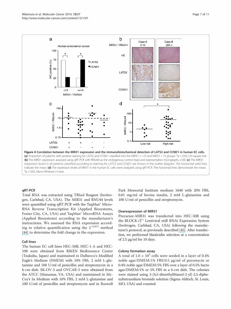
Figure 4 Correlation between the MIR31 expression and the immunohistochemical detection of LATS2 and CCND1 in human EC cells.(a) Proportion of patients with positive staining for LATS2 and CCND1 classified into the MIR31 > =15 and MIR31 < 15 groups. *p < 0.05, Chi-square test.(b) The MIR31 expression assessed using qRT-PCR with RNU44 as the endogenous control (top) and representative micrographs, x100. (c) The MIR31expression levels in all patients classified according to staining for LATS2 and CCND1 are shown in the scatter diagram. The horizontal solid linesindicate the mean. (d) The expression levels of MIR31 in the human EC cells were analyzed using qRT-PCR. The horizontal lines demonstrate the mean.*p < 0.05, Mann-Whitney's U-test.
Mitamura et al. Molecular Cancer 2014, 13:97 Page 7 of 11http://www.molecular-cancer.com/content/13/1/97
qRT-PCRTotal RNA was extracted using TRIzol Reagent (Invitro-gen, Carlsbad, CA, USA). The MIR31 and RNU44 levelswere quantified using qRT-PCR with the TaqMan® Micro-RNA Reverse Transcription Kit (Applied Biosystems,Foster City, CA, USA) and TaqMan® MicroRNA Assays(Applied Biosystems) according to the manufacturer'sinstructions. We assessed the RNA expression accord-ing to relative quantification using the 2-ΔΔCt method[44] to determine the fold change in the expression.
Cell linesThe human EC cell lines HEC-50B, HEC-1-A and HEC-108 were obtained from RIKEN BioResource Center(Tsukuba, Japan) and maintained in Dulbecco's ModifiedEagle’s Medium (DMEM) with 10% FBS, 2 mM L-glu-tamine and 100 U/ml of penicillin and streptomycin in a6-cm dish. SK-OV-3 and OVCAR-3 were obtained fromthe ATCC (Manassas, VA, USA) and maintained in Mc-Coy’s 5a Medium with 10% FBS, 2 mM L-glutamine and100 U/ml of penicillin and streptomycin and in Roswell
Park Memorial Institute medium 1640 with 20% FBS,0.01 mg/ml of bovine insulin, 2 mM L-glutamine and100 U/ml of penicillin and streptomycin.
Overexpression of MIR31Precursor-MIR31 was transfected into HEC-50B usingthe BLOCK-iT™ Lentiviral miR RNAi Expression System(Invitrogen, Carlsbad, CA, USA) following the manufac-turer’s protocol, as previously described [45]. After transfec-tion, we performed blasticidin selection at a concentrationof 2.5 μg/ml for 10 days.
Colony formation assayA total of 1.0 × 10⁵ cells were seeded in a layer of 0.4%noble agar/DMEM/1% FBS/0.5 μg/ml of puromycin or0.4% noble agar/DMEM/5% FBS over a layer of 0.5% bactoagar/DMEM/1% or 5% FBS in a 6-cm dish. The colonieswere stained using 3-[4,5-dimethylthiazol-2-yl]-2,5-diphe-nyltetrazolium bromide solution (Sigma-Aldrich, St. Louis,MO, USA) and counted.
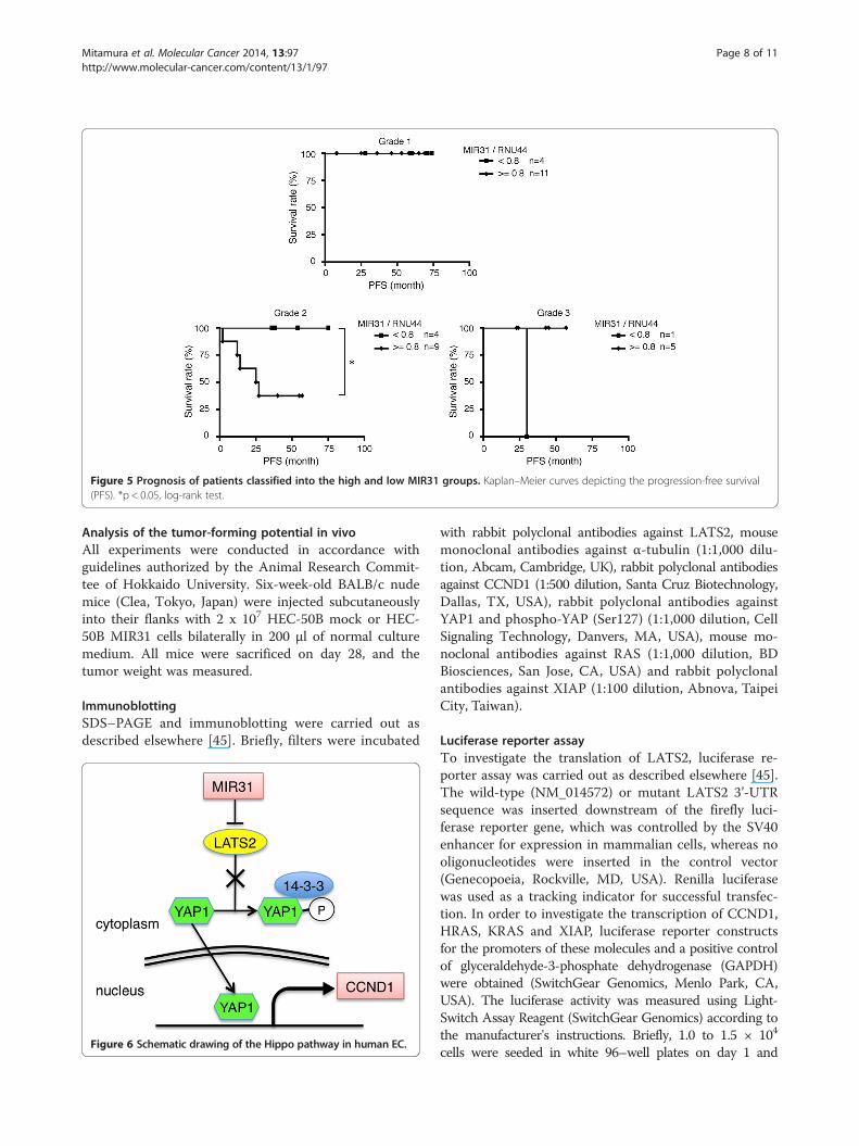
Figure 5 Prognosis of patients classified into the high and low MIR31 groups. Kaplan–Meier curves depicting the progression-free survival(PFS). *p < 0.05, log-rank test.
Mitamura et al. Molecular Cancer 2014, 13:97 Page 8 of 11http://www.molecular-cancer.com/content/13/1/97
Analysis of the tumor-forming potential in vivoAll experiments were conducted in accordance withguidelines authorized by the Animal Research Commit-tee of Hokkaido University. Six-week-old BALB/c nudemice (Clea, Tokyo, Japan) were injected subcutaneouslyinto their flanks with 2 x 10⁷ HEC-50B mock or HEC-50B MIR31 cells bilaterally in 200 μl of normal culturemedium. All mice were sacrificed on day 28, and thetumor weight was measured.
ImmunoblottingSDS–PAGE and immunoblotting were carried out asdescribed elsewhere [45]. Briefly, filters were incubated
Figure 6 Schematic drawing of the Hippo pathway in human EC.
with rabbit polyclonal antibodies against LATS2, mousemonoclonal antibodies against α-tubulin (1:1,000 dilu-tion, Abcam, Cambridge, UK), rabbit polyclonal antibodiesagainst CCND1 (1:500 dilution, Santa Cruz Biotechnology,Dallas, TX, USA), rabbit polyclonal antibodies againstYAP1 and phospho-YAP (Ser127) (1:1,000 dilution, CellSignaling Technology, Danvers, MA, USA), mouse mo-noclonal antibodies against RAS (1:1,000 dilution, BDBiosciences, San Jose, CA, USA) and rabbit polyclonalantibodies against XIAP (1:100 dilution, Abnova, TaipeiCity, Taiwan).
Luciferase reporter assayTo investigate the translation of LATS2, luciferase re-porter assay was carried out as described elsewhere [45].The wild-type (NM_014572) or mutant LATS2 3’-UTRsequence was inserted downstream of the firefly luci-ferase reporter gene, which was controlled by the SV40enhancer for expression in mammalian cells, whereas nooligonucleotides were inserted in the control vector(Genecopoeia, Rockville, MD, USA). Renilla luciferasewas used as a tracking indicator for successful transfec-tion. In order to investigate the transcription of CCND1,HRAS, KRAS and XIAP, luciferase reporter constructsfor the promoters of these molecules and a positive controlof glyceraldehyde-3-phosphate dehydrogenase (GAPDH)were obtained (SwitchGear Genomics, Menlo Park, CA,USA). The luciferase activity was measured using Light-Switch Assay Reagent (SwitchGear Genomics) according tothe manufacturer's instructions. Briefly, 1.0 to 1.5 × 10⁴
cells were seeded in white 96–well plates on day 1 and
Mitamura et al. Molecular Cancer 2014, 13:97 Page 9 of 11http://www.molecular-cancer.com/content/13/1/97
transfected with reporter constructs on day 2 usingFuGENE HD (Promega). The luciferase activity was mea-sured using assay reagent 48 hours after transfection.
RNA interference for LATS2 and YAP1Two shRNA lentiviruses against LATS2 (Sigma-Aldrich)and non-targeting shRNA (Sigma-Aldrich) were trans-fected into HEC-50B cells in 48-well plates according tothe manufacturer's instructions. The multiplicity of in-fection (MOI, number of transducing lentiviral particlesper cell) was 5. We performed puromycin selection at aconcentration of 0.5 μg/ml for 10 days. siRNAs againstYAP and non-targeting siRNA (Santa Cruz Biotechnol-ogy) were transfected using HiPerFect transfection re-agent (Qiagen, Tokyo, Japan) in 12-well plates, and thecells were harvested after 48 hours.
Reverse transcription-PCRReverse transcription-PCR was carried out as describedelsewhere [45]. The primers used for the expression ana-lysis were as follows: pre- MIR31 - forward, 5'-GGAGAG-GAGGCAAGATGCTG-3'; pre- MIR31 - reverse, '-GGAAAGATGGCAATATGTTG-3': GAPDH - forward, 5'-CTCATGACCACAGTCCATGC-3': GAPDH - reverse, 5'-TTACTCCTTGGAGGCCATGT-3': LATS2 - forward, 5'-TAGAGCAGAGGGCGCGGAAG -3': LATS2 - reverse,5'- CCAACACTCCACCAGTCACAGA-3'.
ImmunofluorescenceCells were grown on 35-mm glass-based dishes (AsahiGlass, Tokyo, Japan), fixed with 3% paraformaldehydeand permeabilized with 0.1% Triton X-100/PBS beforeblocking with 1% BSA. The cells were incubated withrabbit polyclonal antibodies against YAP1 (1:500 dilu-tion, Cell Signaling Technology) and secondary anti-bodies, including goat antibodies to rabbit coupled toAlexa 594 (1:250 dilution, Invitrogen). All samples wereexamined using laser-scanning confocal microscopy(Fluoview™, Olympus, Tokyo, Japan).
MIR inhibitorA total of 200 nM of miRIDIAN microRNA HairpinInhibitor and its negative control (Thermo ScientificDharmacon, Lafayette, CO, USA) were employed totransiently inhibit MIR31 and transfected 48 hoursprior to seeding with Oligofectamine (Invitrogen).
Overexpression of Yes-associated protein 1 (YAP1)The human YAP1 expression vector, p2xFLAG-YAP1and negative control were kindly provided by Dr. Sudol[46]. HEC-50B cells at 80% confluence in 6-well plateswere transfected with 4 μg of YAP1 vector using FuGENEHD (Promega).
Immunohistochemistry (IHC)Formalin-fixed, paraffin-embedded tissues were usedto detect the LATS2 and CCND1 expression. The sec-tions were incubated with anti-LATS2 rabbit poly-clonal antibodies (Abcam, Cambridge, UK) at 1:300dilution and anti-CCND1 rabbit monoclonal anti-bodies (Dako, Glostrup, Denmark) at 1:50 dilution. Asemi-quantitative scoring system was used to evaluatethe intensity of staining: low (proportion: 0 to 50%, in-tensity: no staining to weak) and high (proportion:more than 50%, intensity: intermediate to strong).
Statistical analysisThe data are presented as the mean ± SEM. The un-paired two-tailed Student’s t-test, Mann-Whitney's U-test and Chi-square test were used for comparisons,with a p value of < 0.05 considered to be significant (*).
Additional files
Additional file 1: Figure S1. qRT-PCR analysis of the MIR31 expressionin five adenocarcinoma cell lines of the female genital tract.
Additional file 2: Figure S2. Colony formation assay. *p < 0.05,unpaired two-tailed Student’s t-test compared with HEC-108.
Additional file 3: Figure S3. Colony formation assay, four weeks.*p < 0.05, unpaired two-tailed Student’s t-test.
Additional file 4: Figure S4. Detection of LATS2 and GAPDH mRNAusing RT–PCR.
Additional file 5: Figure S5. Representative results of the colonyformation assays with 5% FBS for four weeks.
Additional file 6: Figure S6. MIR31 is not involved in YAPphosphorylation. Immunoblotting for phospho-YAP and α-tubulin.
Additional file 7: Figure S7. Immunoblotting for putative targets of YAP1.
Additional file 8: Figure S8. (a) The expression levels of RAS and XIAPin the mock and MIR31-overexpressing cells (top). The RAS and XIAPlevels were decreased by the MIR31-specific inhibitor (bottom). Results ofimmunoblotting for RAS, XIAP and α-tubulin. (b) The luciferase activityafter transfection of the reporter constructs containing the HRAS, KRASand XIAP promotor region normalized to the GAPDH promotor region.*p < 0.05, unpaired two-tailed Student’s t-test.
Additional file 9: Figure S9. Representative immunofluorescenceanalysis of YAP1, x600.
Additional file 10: Table S1. Correlation between the MIR31 expressionand risk of postoperative recurrence in the patients with grade 2 tumors.
Competing interestsThe authors declare that they have no competing interests.
Authors’ contributionsTM, HW, HN, ST and NS designed the experiment, interpreted the data andprepared the manuscript. TM, LW, HK, MK, MKH, TK, MT conducted theexperiment, collected the data and helped to prepare the manuscript.All authors read and approved the final manuscript.
AcknowledgementsWe thank Dr. Marius Sudol for providing the YAP1 expression vector. Thiswork was supported in part by Grants-in-Aid for Scientific Research from theMinistry of Education, Culture, Sports, Science and Technology (MEXT) ofJapan (Grant-in-Aid for Challenging Exploratory Research, 25670690).
Mitamura et al. Molecular Cancer 2014, 13:97 Page 10 of 11http://www.molecular-cancer.com/content/13/1/97
Author details1Department of Obstetrics and Gynecology, Hokkaido University GraduateSchool of Medicine, N15, W7, Sapporo, Kita-ku 060-8638, Japan. 2Departmentof Cancer Pathology, Hokkaido University Graduate School of Medicine, N15,W7, Sapporo, Kita-ku, Hokkaido 060-8638, Japan. 3Department ofTranslational Pathology, Hokkaido University Graduate School of Medicine,Sapporo, Japan. 4Biotechnology Department, Faculty of Science, Port SaidUniversity, Port Fouad, Port Said, Egypt.
Received: 16 October 2013 Accepted: 21 April 2014Published: 29 April 2014
References1. Jemal A, Bray F, Center MM, Ferlay J, Ward E, Forman D: Global cancer
statistics. CA Cancer J Clin 2011, 61:69–90.2. Amant F, Moerman P, Neven P, Timmerman D, Van Limbergen E, Vergote I:
Endometrial cancer. Lancet 2005, 366:491–505.3. Pecorelli S: Revised FIGO staging for carcinoma of the vulva, cervix, and
endometrium. Int J Gynaecol Obstet 2009, 105:103–104.4. Creasman WT, Morrow CP, Bundy BN, Homesley HD, Graham JE, Heller PB:
Surgical pathologic spread patterns of endometrial cancer. AGynecologic Oncology Group Study. Cancer 1987, 60:2035–2041.
5. Sakuragi N, Hareyama H, Todo Y, Yamada H, Yamamoto R, Fujino T,Sagawa T, Fujimoto S: Prognostic significance of serous and clear celladenocarcinoma in surgically staged endometrial carcinoma. Acta ObstetGynecol Scand 2000, 79:311–316.
6. Carey MS, O'Connell GJ, Johanson CR, Goodyear MD, Murphy KJ, Daya DM,Schepansky A, Peloquin A, Lumsden BJ: Good outcome associated with astandardized treatment protocol using selective postoperative radiationin patients with clinical stage I adenocarcinoma of the endometrium.Gynecol Oncol 1995, 57:138–144.
7. Randall ME, Filiaci VL, Muss H, Spirtos NM, Mannel RS, Fowler J, Thigpen JT,Benda JA: Randomized phase III trial of whole-abdominal irradiationversus doxorubicin and cisplatin chemotherapy in advanced endometrialcarcinoma: a Gynecologic Oncology Group Study. J Clin Oncol 2006,24:36–44.
8. Aoki Y, Kase H, Watanabe M, Sato T, Kurata H, Tanaka K: Stage IIIendometrial cancer: analysis of prognostic factors and failure patternsafter adjuvant chemotherapy. Gynecol Oncol 2001, 83:1–5.
9. Kurman RJ, Ellenson L, Ronnett B: Blaustein's Pathology of the Female GenitalTract. 6th edition. New York: Springer; 2011.
10. Bartel DP: MicroRNAs: target recognition and regulatory functions. Cell2009, 136:215–233.
11. Chou YT, Lin HH, Lien YC, Wang YH, Hong CF, Kao YR, Lin SC, Chang YC, LinSY, Chen SJ, Chen HC, Yeh SD, Wu CW: EGFR promotes lungtumorigenesis by activating miR-7 through a Ras/ERK/Myc pathway thattargets the Ets2 transcriptional repressor ERF. Cancer Res 2010,70:8822–8831.
12. Kong W, He L, Coppola M, Guo J, Esposito NN, Coppola D, Cheng JQ:MicroRNA-155 regulates cell survival, growth, and chemosensitivity bytargeting FOXO3a in breast cancer. J Biol Chem 2010, 285:17869–17879.
13. Li N, Kaur S, Greshock J, Lassus H, Zhong X, Wang Y, Leminen A, Shao Z,Hu X, Liang S, Katsaros D, Huang Q, Butzow R, Weber BL, Coukos G, ZhangL: A combined array-based comparative genomic hybridization andfunctional library screening approach identifies mir-30d as an oncomirin cancer. Cancer Res 2012, 72:154–164.
14. Schetter AJ, Leung SY, Sohn JJ, Zanetti KA, Bowman ED, Yanaihara N,Yuen ST, Chan TL, Kwong DL, Au GK, Liu CG, Calin GA, Croce CM, Harris CC:MicroRNA expression profiles associated with prognosis and therapeuticoutcome in colon adenocarcinoma. JAMA 2008, 299:425–436.
15. Tsuruta T, Kozaki K, Uesugi A, Furuta M, Hirasawa A, Imoto I, Susumu N, AokiD, Inazawa J: miR-152 is a tumor suppressor microRNA that is silenced byDNA hypermethylation in endometrial cancer. Cancer Res 2011,71:6450–6462.
16. Dong P, Kaneuchi M, Watari H, Hamada J, Sudo S, Ju J, Sakuragi N:MicroRNA-194 inhibits epithelial to mesenchymal transition ofendometrial cancer cells by targeting oncogene BMI-1. Mol Cancer 2011,10:99.
17. Hiroki E, Suzuki F, Akahira J, Nagase S, Ito K, Sugawara J, Miki Y, Suzuki T,Sasano H, Yaegashi N: MicroRNA-34b functions as a potential tumor
suppressor in endometrial serous adenocarcinoma. Int J Cancer 2012,131:E395–404.
18. Chung TK, Lau TS, Cheung TH, Yim SF, Lo KW, Siu NS, Chan LK, Yu MY,Kwong J, Doran G, Barroilhet LM, Ng AS, Wong RR, Wang VW, Mok SC,Smith DI, Berkowitz RS, Wong YF: Dysregulation of microRNA-204 mediatesmigration and invasion of endometrial cancer by regulating FOXC1. Int JCancer 2012, 130:1036–1045.
19. Wu Y, Liu S, Xin H, Jiang J, Younglai E, Sun S, Wang H: Up-regulation ofmicroRNA-145 promotes differentiation by repressing OCT4 in humanendometrial adenocarcinoma cells. Cancer 2011, 117:3989–3998.
20. Huang YW, Liu JC, Deatherage DE, Luo J, Mutch DG, Goodfellow PJ, MillerDS, Huang TH: Epigenetic repression of microRNA-129-2 leads to overex-pression of SOX4 oncogene in endometrial cancer. Cancer Res 2009,69:9038–9046.
21. Jiang F, Liu T, He Y, Yan Q, Chen X, Wang H, Wan X: MiR-125b promotesproliferation and migration of type II endometrial carcinoma cellsthrough targeting TP53INP1 tumor suppressor in vitro and in vivo. BMCCancer 2011, 11:425.
22. Sun D, Yu F, Ma Y, Zhao R, Chen X, Zhu J, Zhang CY, Chen J, Zhang J:MicroRNA-31 Activates the RAS Pathway and Functions as an OncogenicMicroRNA in Human Colorectal Cancer by Repressing RAS p21 GTPaseActivating Protein 1 (RASA1). J Biol Chem 2013, 288:9508–9518.
23. Alder H, Taccioli C, Chen H, Jiang Y, Smalley KJ, Fadda P, Ozer HG, HuebnerK, Farber JL, Croce CM, Fong LY: Dysregulation of miR-31 and miR-21induced by zinc deficiency promotes esophageal cancer. Carcinogenesis2012, 33:1736–1744.
24. Liu X, Sempere LF, Ouyang H, Memoli VA, Andrew AS, Luo Y, Demidenko E,Korc M, Shi W, Preis M, Dragnev KH, Li H, Direnzo J, Bak M, Freemantle SJ,Kauppinen S, Dmitrovsky E: MicroRNA-31 functions as an oncogenicmicroRNA in mouse and human lung cancer cells by repressing specifictumor suppressors. J Clin Invest 2010, 120:1298–1309.
25. Liu CJ, Lin SC, Yang CC, Cheng HW, Chang KW: Exploiting salivary miR-31as a clinical biomarker of oral squamous cell carcinoma. Head Neck 2012,34:219–224.
26. Liu CJ, Tsai MM, Hung PS, Kao SY, Liu TY, Wu KJ, Chiou SH, Lin SC, Chang KW:miR-31 ablates expression of the HIF regulatory factor FIH to activate theHIF pathway in head and neck carcinoma. Cancer Res 2010, 70:1635–1644.
27. Valastyan S, Reinhardt F, Benaich N, Calogrias D, Szasz AM, Wang ZC, BrockJE, Richardson AL, Weinberg RA: A pleiotropically acting microRNA, miR-31,inhibits breast cancer metastasis. Cell 2009, 137:1032–1046.
28. Zhang Y, Guo J, Li D, Xiao B, Miao Y, Jiang Z, Zhuo H: Down-regulation ofmiR-31 expression in gastric cancer tissues and its clinical significance.Med Oncol 2010, 27:685–689.
29. Ivanov SV, Goparaju CM, Lopez P, Zavadil J, Toren-Haritan G, Rosenwald S,Hoshen M, Chajut A, Cohen D, Pass HI: Pro-tumorigenic effects of miR-31loss in mesothelioma. J Biol Chem 2010, 285:22809–22817.
30. Zhao B, Li L, Lei Q, Guan KL: The Hippo-YAP pathway in organ size controland tumorigenesis: an updated version. Genes Dev 2010, 24:862–874.
31. Zhao B, Wei X, Li W, Udan RS, Yang Q, Kim J, Xie J, Ikenoue T, Yu J, Li L,Zheng P, Ye K, Chinnaiyan A, Halder G, Lai ZC, Guan KL: Inactivation of YAPoncoprotein by the Hippo pathway is involved in cell contact inhibitionand tissue growth control. Genes Dev 2007, 21:2747–2761.
32. Steinhardt AA, Gayyed MF, Klein AP, Dong J, Maitra A, Pan D, MontgomeryEA, Anders RA: Expression of Yes-associated protein in common solidtumors. Hum Pathol 2008, 39:1582–1589.
33. Zhao B, Li L, Wang L, Wang CY, Yu J, Guan KL: Cell detachment activatesthe Hippo pathway via cytoskeleton reorganization to induce anoikis.Genes Dev 2012, 26:54–68.
34. Liu X, Cheng Y, Chen X, Yang J, Xu L, Zhang C: MicroRNA-31 regulated bythe extracellular regulated kinase is involved in vascular smooth musclecell growth via large tumor suppressor homolog 2. J Biol Chem 2011,286:42371–42380.
35. Augoff K, McCue B, Plow EF, Sossey-Alaoui K: miR-31 and its host genelncRNA LOC554202 are regulated by promoter hypermethylation intriple-negative breast cancer. Mol Cancer 2012, 11:5.
36. Xu RS, Wu XD, Zhang SQ, Li CF, Yang L, Li DD, Zhang BG, Zhang Y, Jin JP,Zhang B: The tumor suppressor gene RhoBTB1 is a novel target ofmiR-31 in human colon cancer. Int J Oncol 2013, 42:676–682.
37. Goulev Y, Fauny JD, Gonzalez-Marti B, Flagiello D, Silber J, Zider A:SCALLOPED interacts with YORKIE, the nuclear effector of the hippotumor-suppressor pathway in Drosophila. Curr Biol 2008, 18:435–441.

Mitamura et al. Molecular Cancer 2014, 13:97 Page 11 of 11http://www.molecular-cancer.com/content/13/1/97
38. Hamaratoglu F, Willecke M, Kango-Singh M, Nolo R, Hyun E, Tao C,Jafar-Nejad H, Halder G: The tumour-suppressor genes NF2/Merlin andExpanded act through Hippo signalling to regulate cell proliferation andapoptosis. Nat Cell Biol 2006, 8:27–36.
39. Wang Y, Xie C, Li Q, Xu K, Wang E: Clinical and prognostic significance ofYes-associated protein in colorectal cancer. Tumour Biol 2013,34:2169–2174.
40. Thompson BJ, Cohen SM: The Hippo pathway regulates the bantammicroRNA to control cell proliferation and apoptosis in Drosophila.Cell 2006, 126:767–774.
41. Aylon Y, Ofir-Rosenfeld Y, Yabuta N, Lapi E, Nojima H, Lu X, Oren M: TheLats2 tumor suppressor augments p53-mediated apoptosis by promotingthe nuclear proapoptotic function of ASPP1. Genes Dev 2010, 24:2420–2429.
42. Vigneron AM, Ludwig RL, Vousden KH: Cytoplasmic ASPP1 inhibitsapoptosis through the control of YAP. Genes Dev 2010, 24:2430–2439.
43. Sakuragi N, Watari H, Ebina Y, Yamamoto R, Steiner E, Koelbl H, Yano M,Tada M, Moriuchi T: Functional analysis of p53 gene and the prognosticimpact of dominant-negative p53 mutation in endometrial cancer. Int JCancer 2005, 116:514–519.
44. Livak KJ, Schmittgen TD: Analysis of relative gene expression data usingreal-time quantitative PCR and the 2(−Delta Delta C(T)) Method. Methods2001, 25:402–408.
45. Mitamura T, Watari H, Wang L, Kanno H, Hassan MK, Miyazaki M, Katoh Y,Kimura T, Tanino M, Nishihara H, Tanaka S, Sakuragi N: Downregulation ofmiRNA-31 induces taxane resistance in ovarian cancer cells throughincrease of receptor tyrosine kinase MET. Oncogenesis 2013, 2:e40.
46. Komuro A, Nagai M, Navin NE, Sudol M: WW domain-containing proteinYAP associates with ErbB-4 and acts as a co-transcriptional activator forthe carboxyl-terminal fragment of ErbB-4 that translocates to the nucleus.J Biol Chem 2003, 278:33334–33341.
doi:10.1186/1476-4598-13-97Cite this article as: Mitamura et al.: microRNA 31 functions as anendometrial cancer oncogene by suppressing Hippo tumor suppressorpathway. Molecular Cancer 2014 13:97.
Submit your next manuscript to BioMed Centraland take full advantage of:
• Convenient online submission
• Thorough peer review
• No space constraints or color figure charges
• Immediate publication on acceptance
• Inclusion in PubMed, CAS, Scopus and Google Scholar
• Research which is freely available for redistribution
Submit your manuscript at www.biomedcentral.com/submit
Copyright © 2022 FDOKUMEN