Large-scale analysis by SAGE reveals new mechanisms of v-erbA oncogene action
17
BioMed Central Page 1 of 17 (page number not for citation purposes) BMC Genomics Open Access Research article Large-scale analysis by SAGE reveals new mechanisms of v-erbA oncogene action Corinne Bresson* 1,2 , Celine Keime 1,2 , Claudine Faure 1,2 , Yann Letrillard 1,2 , Maud Barbado 1,2 , Sandra Sanfilippo 1,2 , Najate Benhra 1,2 , Olivier Gandrillon 1,2 and Sandrine Gonin-Giraud 1,2 Address: 1 Université de Lyon, Lyon, F-69003, France and 2 CNRS, UMR5534, Centre de génétique moléculaire et cellulaire, Villeurbanne, F-69622, France Email: Corinne Bresson* - [email protected]; Celine Keime - [email protected]; Claudine Faure - [email protected]; Yann Letrillard - [email protected]; Maud Barbado - [email protected]; Sandra Sanfilippo - [email protected]; Najate Benhra - [email protected]; Olivier Gandrillon - [email protected] lyon1.fr; Sandrine Gonin-Giraud - [email protected] * Corresponding author Abstract Background: The v-erbA oncogene, carried by the Avian Erythroblastosis Virus, derives from the c-erbAα proto-oncogene that encodes the nuclear receptor for triiodothyronine (T3R). v-ErbA transforms erythroid progenitors in vitro by blocking their differentiation, supposedly by interference with T3R and RAR (Retinoic Acid Receptor). However, v-ErbA target genes involved in its transforming activity still remain to be identified. Results: By using Serial Analysis of Gene Expression (SAGE), we identified 110 genes deregulated by v-ErbA and potentially implicated in the transformation process. Bioinformatic analysis of promoter sequence and transcriptional assays point out a potential role of c-Myb in the v-ErbA effect. Furthermore, grouping of newly identified target genes by function revealed both expected (chromatin/transcription) and unexpected (protein metabolism) functions potentially deregulated by v-ErbA. We then focused our study on 15 of the new v-ErbA target genes and demonstrated by real time PCR that in majority their expression was activated neither by T3, nor RA, nor during differentiation. This was unexpected based upon the previously known role of v-ErbA. Conclusion: This paper suggests the involvement of a wealth of new unanticipated mechanisms of v-ErbA action. Background The Avian Erythroblastosis Virus (AEV) induces erythro- leukemia and sarcomas in chickens [1]. This retrovirus also transforms erythroid progenitors and chicken embryo fibroblasts in vitro. The AEV carries two viral onco- genes in its genome, v-erbA and v-erbB. The expression of v-ErbA alone is sufficient to block differentiation of eryth- rocytic progenitors in vitro and in vivo [2,3]. v-erbA is derived from the c-erbA proto-oncogene, which encodes the alpha form of the nuclear receptor for the thyroid hor- mone triiodothyronine (T3Rα) [4]. This oncogene is Published: 26 October 2007 BMC Genomics 2007, 8:390 doi:10.1186/1471-2164-8-390 Received: 21 May 2007 Accepted: 26 October 2007 This article is available from: http://www.biomedcentral.com/1471-2164/8/390 © 2007 Bresson et al; licensee BioMed Central Ltd. This is an Open Access article distributed under the terms of the Creative Commons Attribution License (http://creativecommons.org/licenses/by/2.0 ), which permits unrestricted use, distribution, and reproduction in any medium, provided the original work is properly cited.
-
Upload
independent -
Category
Documents
-
view
2 -
download
0
Transcript of Large-scale analysis by SAGE reveals new mechanisms of v-erbA oncogene action
BioMed CentralBMC Genomics
ss
Open AcceResearch articleLarge-scale analysis by SAGE reveals new mechanisms of v-erbA oncogene actionCorinne Bresson*1,2, Celine Keime1,2, Claudine Faure1,2, Yann Letrillard1,2, Maud Barbado1,2, Sandra Sanfilippo1,2, Najate Benhra1,2, Olivier Gandrillon1,2 and Sandrine Gonin-Giraud1,2Address: 1Université de Lyon, Lyon, F-69003, France and 2CNRS, UMR5534, Centre de génétique moléculaire et cellulaire, Villeurbanne, F-69622, France
Email: Corinne Bresson* - [email protected]; Celine Keime - [email protected]; Claudine Faure - [email protected]; Yann Letrillard - [email protected]; Maud Barbado - [email protected]; Sandra Sanfilippo - [email protected]; Najate Benhra - [email protected]; Olivier Gandrillon - [email protected]; Sandrine Gonin-Giraud - [email protected]
* Corresponding author
AbstractBackground: The v-erbA oncogene, carried by the Avian Erythroblastosis Virus, derives from thec-erbAα proto-oncogene that encodes the nuclear receptor for triiodothyronine (T3R). v-ErbAtransforms erythroid progenitors in vitro by blocking their differentiation, supposedly byinterference with T3R and RAR (Retinoic Acid Receptor). However, v-ErbA target genes involvedin its transforming activity still remain to be identified.
Results: By using Serial Analysis of Gene Expression (SAGE), we identified 110 genes deregulatedby v-ErbA and potentially implicated in the transformation process. Bioinformatic analysis ofpromoter sequence and transcriptional assays point out a potential role of c-Myb in the v-ErbAeffect. Furthermore, grouping of newly identified target genes by function revealed both expected(chromatin/transcription) and unexpected (protein metabolism) functions potentially deregulatedby v-ErbA. We then focused our study on 15 of the new v-ErbA target genes and demonstrated byreal time PCR that in majority their expression was activated neither by T3, nor RA, nor duringdifferentiation. This was unexpected based upon the previously known role of v-ErbA.
Conclusion: This paper suggests the involvement of a wealth of new unanticipated mechanisms ofv-ErbA action.
BackgroundThe Avian Erythroblastosis Virus (AEV) induces erythro-leukemia and sarcomas in chickens [1]. This retrovirusalso transforms erythroid progenitors and chickenembryo fibroblasts in vitro. The AEV carries two viral onco-genes in its genome, v-erbA and v-erbB. The expression of
v-ErbA alone is sufficient to block differentiation of eryth-rocytic progenitors in vitro and in vivo [2,3]. v-erbA isderived from the c-erbA proto-oncogene, which encodesthe alpha form of the nuclear receptor for the thyroid hor-mone triiodothyronine (T3Rα) [4]. This oncogene is
Published: 26 October 2007
BMC Genomics 2007, 8:390 doi:10.1186/1471-2164-8-390
Received: 21 May 2007Accepted: 26 October 2007
This article is available from: http://www.biomedcentral.com/1471-2164/8/390
© 2007 Bresson et al; licensee BioMed Central Ltd. This is an Open Access article distributed under the terms of the Creative Commons Attribution License (http://creativecommons.org/licenses/by/2.0), which permits unrestricted use, distribution, and reproduction in any medium, provided the original work is properly cited.
Page 1 of 17(page number not for citation purposes)

BMC Genomics 2007, 8:390 http://www.biomedcentral.com/1471-2164/8/390
expressed as a gag-v-ErbA fusion protein that binds to theDNA but does not bind the hormone T3 [4].
The most widely accepted hypothesis is that v-ErbA antag-onizes ligand-dependent activation by T3R, to silence T3responsive genes even in the presence of T3 [5-7]. Thefinding that overexpression of T3R and addition of ligandcan overcome the blockade of differentiation induced byv-ErbA supports this assumption [8]. Moreover, it wasshown that v-ErbA also inhibits transcription mediated byligand-activated Retinoic Acid Receptors (RAR) and thatthis inhibition is also of importance since a non-trans-forming point mutant for v-ErbA, S61G, is unable to blocktranscription mediated by RAR [9].
It has therefore been proposed that the v-ErbA oncopro-tein represses genes that are normally upregulated by T3and Retinoic Acid (RA) and that are upregulated duringthe differentiation process [5,9]. Nevertheless, thishypothesis remains speculative as long as v-ErbA targetgenes harboring those properties are not found. So far,four v-ErbA target genes have been described: Carbonicanhydrase II (CAII), erythrocyte anion transporter (Band3),integrin α2 subunit (VLA2) and δ-aminolevulinate synthase(ALA-S) [10-12]. It has been demonstrated only in thecase of the CAII gene that v-ErbA acts as a dominant neg-ative of T3R by occluding the DNA response element andablating the T3 mediated activation [13]. In contrast, theexpression of Band3, ALA-S and VLA2 genes is not control-led by T3 suggesting that v-ErbA may act independently ofT3R [10,11]. This is consistent with the observation that v-ErbA also interferes with pathways controlled by othertranscription factors [9,14]. Furthermore, functional stud-ies have clearly demonstrated that the repression of Band3and CAII by v-ErbA is not sufficient to account for the dif-ferentiation blocking ability of this oncoprotein [15].
Therefore, the precise mechanisms of v-ErbA actionremain unclear and the identification of relevant v-ErbAtarget genes represents a mandatory step in its under-standing. We thus decided to use a large-scale transcrip-tomic approach by SAGE (Serial Analysis of GeneExpression) to identify v-ErbA target genes involved in itstransforming activity. One of the most important advan-tages of this technique is the possibility to obtain a com-prehensive view of all the transcripts present in a cellpopulation at a given time point, allowing the discoveryof new genes [16]. We have used a cellular model of nor-mal immature erythroid progenitors called T2ECs (TGF-αTGF-β induced erythrocytic cells) [17] which are the natu-ral target cells of v-ErbA. In order to identify v-ErbA targetgenes responsible for the transformation process inducedby v-ErbA, we have compared the transcriptome of T2ECsexpressing an oncogenic form of v-ErbA with the tran-scriptome of T2ECs expressing the S61G mutant of v-ErbA
[9,18]. This mutant, which carries a glycine in the DNAbinding domain, is equivalent to the c-erbA sequence atthis position, and is defective in its ability to inhibit differ-entiation and to induce erythroid transformation [18].
Thus, the comparison between the transcriptome of cellsexpressing either the transforming form of v-ErbA or theS61G mutant of v-ErbA allowed us to generate a list of 110differentially expressed genes between these two condi-tions, 15 of which were observed to be repressed by v-ErbA by real-time PCR on different cell populations. Abioinformatic approach revealed the presence of c-Mybbinding sites in the promoters of v-ErbA target genes. Theidentification of these sites coupled with a functional c-Myb transcriptional assay point out a putative role of c-Myb in the transformation process. Unexpectedly, the glo-bal analysis of gene functions reveals that two main cellu-lar functions, protein metabolism and chromatinstructure/transcriptional regulation, could be deregulatedby v-ErbA. We further show here that the expression ofmost newly identified v-ErbA target genes is neither underthe control of T3 nor RA, nor upregulated during differen-tiation. In conclusion, we have identified several new v-ErbA target genes unknown up to now and we present evi-dence that v-erbA oncogene action involves new unantici-pated mechanisms of action.
ResultsPotential v-ErbA target genes identified by SAGE analysisWe constructed two SAGE libraries of T2ECs infectedeither with the XJ12 retrovirus which expresses the onco-genic form of v-erbA (VA) or with the S61G retroviruswhich expresses a non-transforming form of v-erbA(NTVA) in order to identify v-ErbA target genes responsi-ble for transformation. 59391 tags were obtained from theVA library and 37347 tags from the NTVA library. Weobtained 5764 different tags appearing more than once inthe VA library and 4153 different tags in the NTVA library.
The comparison of these two SAGE libraries revealed 110differentially expressed genes between these two condi-tions with a p-value adjusted for multiple testing < 0.1(Figure 1). 44 of them were up-regulated and 66 down-regulated in T2ECs expressing the transforming form of v-ErbA (VA) as compared to T2ECs expressing the non-transforming form of v-ErbA (NTVA). In view of theseresults, v-ErbA seems to have mostly a repressive action ongene transcription because it represses more genes than itactivates. This essentially repressive action of v-ErbA ongene expression was further confirmed by real-time PCR(see below). However, the vast majority of tags wereequally abundant in these two libraries, suggesting thatthe switch from a normal state toward a pathological stateinvolves a very limited number of genes.
Page 2 of 17(page number not for citation purposes)

BMC Genomics 2007, 8:390 http://www.biomedcentral.com/1471-2164/8/390
Presence of c-Myb binding sites in the promoters of v-ErbA repressed target genesIn order to investigate the molecular mechanisms bywhich v-ErbA regulates gene expression, we next searchedfor common motifs in the promoters of the v-ErbA targetgenes identified by SAGE. For that purpose, we first iden-tified SAGE tags by using Identitag [19] and then alignedthe corresponding transcripts on the chicken genome. Ithas been demonstrated for CAII that v-ErbA binds to sev-eral domains in the proximal promoter sequence, up to600 bp of the transcription start site (TSS) [20]. Moreover,it has been demonstrated that the position of transcrip-tion factor binding sites often lie within a few kb 5' of thebasal promoter [21]. We therefore decided to consider as
promoter regions 3 kbp 5' of the TSS and 1 kbp 3' of theTSS.
We obtained 31 promoter sequences from genes thatappeared as being repressed by the transforming form ofv-ErbA by SAGE and 21 sequences from genes thatappeared as being activated by the same form of v-ErbAcompared to the non transforming form of v-ErbA. Wefirst searched for known v-ErbA response elements [22-25]. All the nuclear receptors of the c-ErbA family recog-nize derivatives of the same hexameric DNA core motif 5'-AGGTCA-3' [25]. We found response elements basedupon this motif only in 3 promoters. The response ele-ment PAL-0 (AGGTCATGACCT) is present in RPL11 and
Comparative gene expression levels between VA and NTVA librariesFigure 1Comparative gene expression levels between VA and NTVA libraries. The occurrence number of each tag (normal-ized in tag per million) in VA (X axis) and NTVA (Y axis) libraries is represented on a logarithmic scale. Each point can repre-sent one or more tags. Colour-coding is based on a statistical analysis (Z test [47]), adjusted for multiple testing according to the method proposed by Benjamini and Hochberg [48]. Black dots represent the 110 tags having an adjusted p-value < 0.1. These tags correspond to genes that are significantly differentially expressed between these 2 conditions. Among these genes, 44 are up-regulated (the corresponding points are located below the first bisecting line) and 66 are down-regulated (the corre-sponding points are located above the first bisecting line) in T2ECs expressing the transforming form of v-ErbA as compared to T2ECs expressing the non-transforming form of v-ErbA. The gray dots represent all the other tags.
Page 3 of 17(page number not for citation purposes)

BMC Genomics 2007, 8:390 http://www.biomedcentral.com/1471-2164/8/390
Succinate promoters while the response element DR-4(AGGTCAnnnnAGGTCA) is present in RPS3 and Succinatepromoters (data not shown). It has been demonstratedthat T3R binds preferentially and rather specifically to T3response element (TRE) of the DR-4 structure [25,26]. Thepalindromic element without a spacer (PAL-0) is not spe-cific to T3R since it works efficiently as a retinoic acidresponsive element (RARE) [25,26]. However, despite thepresence of the T3 response element in RPS3 promoter,the expression of this gene did not vary in response to T3(see below). To conclude, out of 52 promoters analyzed,we have only identified 3 promoters with known responseelement for T3R, RAR and v-ErbA. It is conceivable thatthe 4 kb region considered is not sufficient since bindingsites that would be more distal to the TSS will be missedby this approach. Furthermore, it is also obvious that thesimple presence of a transcription factor binding site(TFBS) does not imply an activity of that transcription fac-tor. Finally, even if the protein binds the TFBS, we cannotbe informed about the direction of the regulation (i.e.activation or inhibition). Nevertheless, we have found fewv-ErbA/T3R/RAR response elements in this region that isin accordance with the experimental assessment of theexpression pattern of newly identified v-ErbA target genesunder the influence of T3R and RAR (see below).
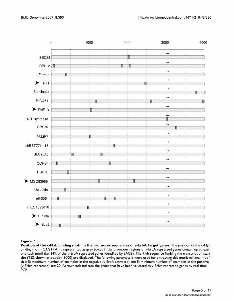
Since very few v-ErbA target gene promoters contain aknown response element for T3R, RAR and v-ErbA, wesearched for motifs that could be enriched in these pro-moters, without any a priori on these motifs except itsminimal length. For this purpose, we used an implemen-tation of an extended FAVST (Finite Automata-based VSTconstruction) algorithm [27]. This allowed us to identifya c-Myb binding site (CAGTTA) [28] as a signature motifof many newly identified v-ErbA repressed target genescompared with v-ErbA activated target genes. Indeed, 64%of v-ErbA repressed genes have the c-Myb binding motif intheir promoter sequences (Figure 2). Among the v-ErbAtarget genes that contain at least one c-Myb binding site intheir promoter region, several have been validated as v-ErbA repressed target genes by real-time RT-PCR (seebelow). This suggests a potential role for c-Myb in the v-ErbA induced transformation.
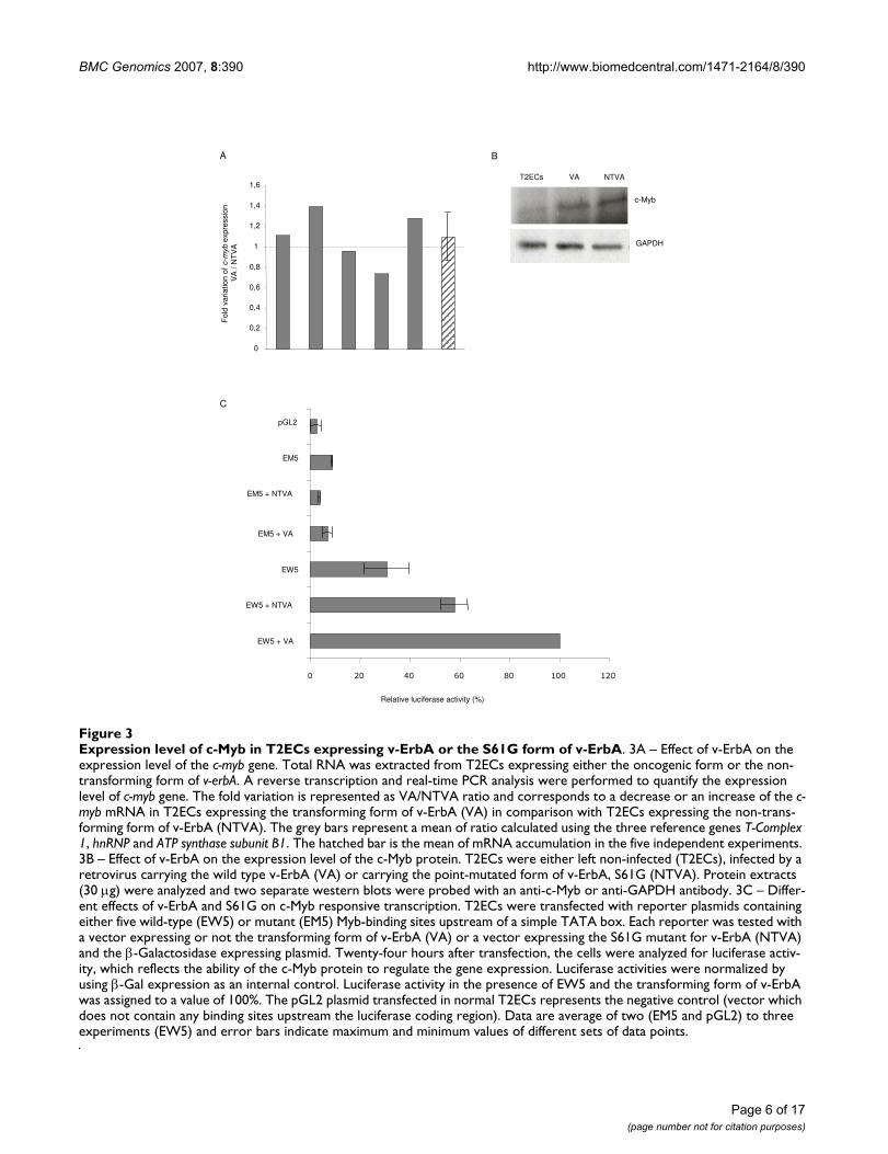
To determine the role of c-Myb in this transformationprocess, the expression level of this gene was quantified byreal-time PCR in T2ECs expressing the transforming formof v-ErbA (VA) compared to T2ECs expressing the non-transforming form of v-ErbA (NTVA) in five independentexperiments (Figure 3A). The expression of c-myb did notvary between these two conditions. Similarly, we observedthat the expression level of the c-Myb protein was also notmodified in cells expressing v-ErbA compared with cellsexpressing the non-transforming S61G mutant (Figure3B). Although the level of c-Myb protein is increased by
both forms of v-ErbA compared with non-infected T2ECs,these results suggest that this upregulation of c-Myb by v-ErbA is not sufficient to induce a transformation process.
Finally, we used a gene reporter assay to test the ability ofv-ErbA to transactivate c-Myb. We transfected T2ECs witha reporter plasmid in which five strong c-Myb bindingsites were placed upstream of a simple TATA box and aluciferase cDNA [29]. This reporter plasmid (EW5) wasclearly active in T2ECs (Figure 3C), which express c-Myb(see above). Moreover, a 1.7-fold increase of luciferaseactivity was seen with the transforming form of v-ErbA(VA) compared to the S61G mutant for v-ErbA (NTVA). Acontrol reporter gene containing five mutant c-Myb bind-ing sites (EM5) was much less active in these cells, and itsactivity was unaffected by the different forms of v-ErbA. Itis thus conceivable that the c-Myb binding to a real pro-moter, presenting a higher complexity with several tran-scription factor binding sites, silencers and enhancers,could result in an inhibition of gene expression controlledby such a promoter. Therefore, although the direction ofthe functional interaction was unexpected (activation ver-sus repression), these data nevertheless demonstrate thatv-ErbA can indeed functionally interact directly or indi-rectly with the transcriptional activity of endogenous c-Myb in T2ECs.
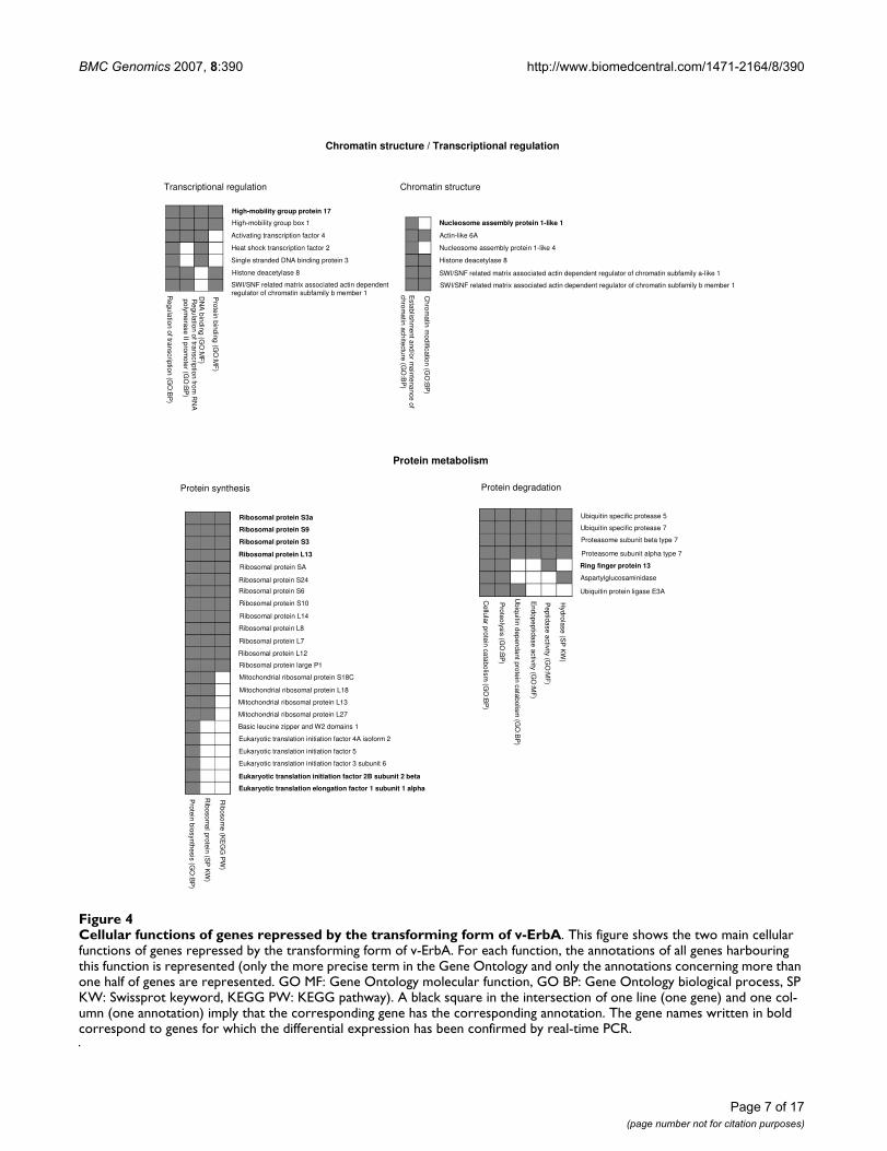
v-ErbA mostly affects chromatin/transcription and protein metabolism genesWe clustered the newly identified v-ErbA target genesaccording to the cellular function encoded by their corre-sponding proteins (Figure 4). Each protein may be impli-cated in different functions i.e. a protein may belong todifferent groups.
The use of functional annotations (mainly Gene Ontol-ogy (GO) annotations) allowed us to find that many v-ErbA repressed target genes are involved in two main cel-lular functions: chromatin structure/transcriptional regu-lation and protein metabolism.
The remodelling of chromatin and transcription regula-tion constituted a cellular function that was expected to bederegulated by v-ErbA. Indeed, v-ErbA represses transcrip-tion via association with corepressors such as NCoR andSMRT [30]. The corepressors, in turn, recruit histonedeacetylase (HDAC) containing complexes that stabilizerepressive chromatin structure to assure efficient silencingof downstream genes [31,32].
Protein metabolism is the most represented and the mostunanticipated category. It suggests the importance of thetranslation and the protein degradation processes duringtransformation. Among the genes that are implicated inprotein synthesis, we have observed an important number
Page 4 of 17(page number not for citation purposes)
BMC Genomics 2007, 8:390 http://www.biomedcentral.com/1471-2164/8/390
Page 5 of 17(page number not for citation purposes)
Position of the c-Myb binding motif in the promoter sequences of v-ErbA target genesFigure 2Position of the c-Myb binding motif in the promoter sequences of v-ErbA target genes. The position of the c-Myb binding motif (CAGTTA) is represented as grey boxes in the promoter regions of v-ErbA repressed genes containing at least one such motif (i.e. 64% of the v-ErbA repressed genes identified by SAGE). The 4 kb sequence flanking the transcription start site (TSS, shown at position 3000) are displayed. The following parameters were used for extracting this motif: minimal motif size: 5; maximum number of examples in the negative (v-ErbA activated) set: 5; minimum number of examples in the positive (v-ErbA repressed) set: 20. Arrowheads indicate the genes that have been validated as v-ErbA repressed genes by real time PCR.
SEC23
RPL12
Ferritin
TPT1
Succinate
RPL37a
RNF13
ATP synthase
RPS10
PSMB7
chEST771m18
SLC25A6
COPS4
HSC70
MGC83969
Ubiquitin
eIF356
chEST593n16
RPS3a
Sca2
0 1000 2000 3000 4000
BMC Genomics 2007, 8:390 http://www.biomedcentral.com/1471-2164/8/390
Page 6 of 17(page number not for citation purposes)
Expression level of c-Myb in T2ECs expressing v-ErbA or the S61G form of v-ErbAFigure 3Expression level of c-Myb in T2ECs expressing v-ErbA or the S61G form of v-ErbA. 3A – Effect of v-ErbA on the expression level of the c-myb gene. Total RNA was extracted from T2ECs expressing either the oncogenic form or the non-transforming form of v-erbA. A reverse transcription and real-time PCR analysis were performed to quantify the expression level of c-myb gene. The fold variation is represented as VA/NTVA ratio and corresponds to a decrease or an increase of the c-myb mRNA in T2ECs expressing the transforming form of v-ErbA (VA) in comparison with T2ECs expressing the non-trans-forming form of v-ErbA (NTVA). The grey bars represent a mean of ratio calculated using the three reference genes T-Complex 1, hnRNP and ATP synthase subunit B1. The hatched bar is the mean of mRNA accumulation in the five independent experiments. 3B – Effect of v-ErbA on the expression level of the c-Myb protein. T2ECs were either left non-infected (T2ECs), infected by a retrovirus carrying the wild type v-ErbA (VA) or carrying the point-mutated form of v-ErbA, S61G (NTVA). Protein extracts (30 µg) were analyzed and two separate western blots were probed with an anti-c-Myb or anti-GAPDH antibody. 3C – Differ-ent effects of v-ErbA and S61G on c-Myb responsive transcription. T2ECs were transfected with reporter plasmids containing either five wild-type (EW5) or mutant (EM5) Myb-binding sites upstream of a simple TATA box. Each reporter was tested with a vector expressing or not the transforming form of v-ErbA (VA) or a vector expressing the S61G mutant for v-ErbA (NTVA) and the β-Galactosidase expressing plasmid. Twenty-four hours after transfection, the cells were analyzed for luciferase activ-ity, which reflects the ability of the c-Myb protein to regulate the gene expression. Luciferase activities were normalized by using β-Gal expression as an internal control. Luciferase activity in the presence of EW5 and the transforming form of v-ErbA was assigned to a value of 100%. The pGL2 plasmid transfected in normal T2ECs represents the negative control (vector which does not contain any binding sites upstream the luciferase coding region). Data are average of two (EM5 and pGL2) to three experiments (EW5) and error bars indicate maximum and minimum values of different sets of data points.
0 20 40 60 80 100 120
EW5 + VA
EW5 + NTVA
EW5
EM5 + VA
EM5 + NTVA
EM5
pGL2
Relative luciferase activity (%)
C
A
0
0,2
0,4
0,6
0,8
1
1,2
1,4
1,6
Fo
ld v
aria
tio
n o
f c-m
yb
exp
ressio
n
VA
/ N
TV
A
B
VA NTVA
c-Myb
T2ECs
GAPDH
BMC Genomics 2007, 8:390 http://www.biomedcentral.com/1471-2164/8/390
Page 7 of 17(page number not for citation purposes)
Cellular functions of genes repressed by the transforming form of v-ErbAFigure 4Cellular functions of genes repressed by the transforming form of v-ErbA. This figure shows the two main cellular functions of genes repressed by the transforming form of v-ErbA. For each function, the annotations of all genes harbouring this function is represented (only the more precise term in the Gene Ontology and only the annotations concerning more than one half of genes are represented. GO MF: Gene Ontology molecular function, GO BP: Gene Ontology biological process, SP KW: Swissprot keyword, KEGG PW: KEGG pathway). A black square in the intersection of one line (one gene) and one col-umn (one annotation) imply that the corresponding gene has the corresponding annotation. The gene names written in bold correspond to genes for which the differential expression has been confirmed by real-time PCR.
Chromatin structure / Transcriptional regulation
Transcriptional regulation
High-mobility group box 1
High-mobility group protein 17
Activating transcription factor 4
Heat shock transcription factor 2
Single stranded DNA binding protein 3
Histone deacetylase 8
SWI/SNF related matrix associated actin dependent
regulator of chromatin subfamily b member 1Pro
tein
bin
din
g (G
O:M
F)
DN
A b
ind
ing
(GO
:MF
)R
eg
ula
tion
of tra
nscrip
tion
from
RN
A
po
lym
era
se
II pro
mo
ter (G
O:B
P)
Re
gu
latio
n o
f tran
scrip
tion
(GO
:BP
)
Chromatin structure
Nucleosome assembly protein 1-like 1
Actin-like 6A
Nucleosome assembly protein 1-like 4
Histone deacetylase 8
SWI/SNF related matrix associated actin dependent regulator of chromatin subfamily a-like 1
SWI/SNF related matrix associated actin dependent regulator of chromatin subfamily b member 1
Ch
rom
atin
mo
dific
atio
n (G
O:B
P)
Esta
blis
hm
en
t an
d/o
r ma
inte
na
nce
of
ch
rom
atin
ach
itectu
re (G
O:B
P)
Protein metabolism
Protein synthesis Protein degradation
Ribosomal protein S3a
Ribosomal protein S9
Ribosomal protein S3
Ribosomal protein L13
Ribosomal protein SA
Ribosomal protein S24
Eukaryotic translation initiation factor 3 subunit 6
Ribosomal protein S6
Ribosomal protein S10
Ribosomal protein L14
Ribosomal protein L8
Ribosomal protein L7
Ribosomal protein L12
Ribosomal protein large P1
Mitochondrial ribosomal protein S18C
Mitochondrial ribosomal protein L18
Mitochondrial ribosomal protein L13
Mitochondrial ribosomal protein L27
Basic leucine zipper and W2 domains 1
Eukaryotic translation initiation factor 4A isoform 2
Eukaryotic translation initiation factor 5
Eukaryotic translation initiation factor 2B subunit 2 beta
Eukaryotic translation elongation factor 1 subunit 1 alpha
Rib
oso
me
(KE
GG
PW
)
Rib
oso
ma
l pro
tein
(SP
KW
)
Pro
tein
bio
syn
the
sis
(GO
:BP
)
Ubiquitin specific protease 5
Ubiquitin specific protease 7
Proteasome subunit beta type 7
Proteasome subunit alpha type 7
Ring finger protein 13
Aspartylglucosaminidase
Ubiquitin protein ligase E3A
Hyd
rola
se
(SP
KW
)
Pe
ptid
ase
activity (G
O:M
F)
En
do
pe
ptid
ase
activ
ity (G
O:M
F)
Ub
iqu
itin d
ep
en
da
nt p
rote
in c
ata
bo
lism
(GO
:BP
)
Pro
teo
lysis (G
O:B
P)
Ce
llula
r pro
tein
ca
tab
olis
m (G
O:B
P)
BMC Genomics 2007, 8:390 http://www.biomedcentral.com/1471-2164/8/390
of ribosomal proteins. However, the expression of allribosomal proteins is not affected by v-ErbA since a vastnumber of genes encoding ribosomal proteins (RPL3,RPL7, RPL12, RPL24, RPL29, RPS4, RPS6, RPS10 andRPS13) was shown not to vary by real time PCR (data notshown). This suggests that only a specific subset of someribosomal proteins is affected by v-ErbA.
The other genes down-regulated by v-ErbA, which are notimplicated in these two cellular functions, are eitherimplicated in different functions or their functions areunknown. Among these genes, stem cell antigen 2 (sca2)and transforming growth factor-β1 (TGF-β1) encode pro-teins that are involved in the regulation of intracellularsignalling pathways. Indeed, we have observed that v-ErbA is able to modulate the TGF-β signalling pathway inT2ECs (Gonin-Giraud et al., submitted). These data sug-gest a role for v-ErbA in intracellular signalling pathwaysin agreement with its cytoplasmic localisation [33].
In contrast, the candidate genes whose expression is up-regulated by v-ErbA are fewer and are involved in a greatervariety of functions, such as sodium-calcium exchange,chaperone activity, nuclear transport and others.
15 v-ErbA target genes validated by real time-PCR on multiple experimentsThe relative expression levels of v-ErbA target genes iden-tified by SAGE were quantified by real-time PCR in T2ECsexpressing the transforming form of v-ErbA (VA) com-pared to T2ECs expressing the non-transforming form ofv-ErbA (NTVA) in five independent experiments. Weselected 40 candidates and used the sequence of the corre-sponding transcripts in order to design PCR primers. Thedifferential expression between VA and NTVA conditionshas been observed for 15 of them (Figure 5). All thesegenes are repressed by the transforming form of v-ErbAcompared to the non-transforming one. The majority ofthese genes are involved in protein synthesis, like ribos-omal proteins (RPS3, RPS3a, RPS9, RPL13) and transla-tion initiation and elongation factors (eIF2B2, eEF1α1).
We observed that genes were not repressed at the samefold variation in the five different experiments. This can bedue to a biological variability between different cultures,as we showed in our previous analysis of the transcrip-tome of T2ECs in a self-renewal or in a differentiationstate [34]. By contrast, the expression of the 25 candidates,which have not been confirmed by real-time PCR, couldbe explained by different hypothesis (multiple or errone-ous identification, variability in gene expression, specificgene variations during the culture that was used for pre-paring SAGE mRNAs, etc...) as developed in Damiola etal., 2004.
To conclude, out of the 40 candidates tested, we haveidentified 15 v-ErbA repressed target genes. None of thesegenes were previously known to be a v-ErbA target gene.
The expression of the majority of newly identified v-ErbA target genes is not activated nor repressed during differentiationIn order to test the initial hypothesis that the erythroidprogenitors transformation by v-ErbA involves a blockadeof the differentiation process, we compared the sets ofgenes obtained by SAGE analysis in this and in our previ-ous studies [34]. We have identified 2 genes whose expres-sion was found to be increased in T2ECs in adifferentiation state versus self-renewal state [34] anddecreased in T2ECs expressing v-ErbA versus T2ECsexpressing S61G (Figure 6A, left part). On the other hand,we have identified 3 genes whose expression was found tobe decreased in T2ECs in a differentiation state versus self-renewal state [34] and increased in T2ECs expressing v-ErbA versus T2ECs expressing S61G (Figure 6A, rightpart). These 5 genes may be involved both in the T2ECsself-renewal and in the transforming process induced byv-ErbA. Altogether, there is a very modest overlap betweenthese gene lists, suggesting that in majority the v-ErbA tar-get genes might not be deregulated during differentiation,in contrast with our expectations.
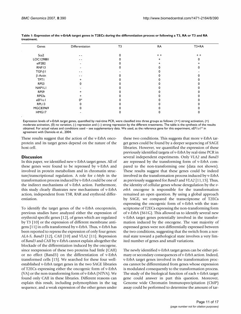
We therefore studied by real time PCR the expression ofthe 15 confirmed v-ErbA target genes during the differen-tiation process (Table 1 and see Additional File 1). Weobserved that, among these v-ErbA target genes, some areactivated such as TPT1, RPS9 and RPS3a. This confirmsthat differentiation related target genes can indeed beinhibited by v-ErbA. However, we observed that theexpression of most v-ErbA target genes is either repressedor does not vary during differentiation. These results sug-gest other mechanisms of v-ErbA action, independent of amere blockade of differentiation.
T3 and RA do not activate the majority of newly identified v-ErbA target genesThe v-ErbA oncoprotein was initially described to be atranscriptional repressor for genes normally activated byT3 Receptor (T3R) and RA Receptor (RAR) [5,9]. There-fore, we have analyzed the expression of v-ErbA targetgenes by real-time PCR following a treatment by T3 andRA (Table 1 and see Additional File 2). We observed that,among these genes, some are activated by RAR becausetheir expression is induced by RA treatment in normalcells, such as sca2, LOC129881 and eIF2B2. This confirmsthat activation by RAR is indeed inhibited by v-ErbA.However, the expression of a vast majority of newly iden-tified v-ErbA target genes is either repressed by T3 or doesnot vary in response to T3 and RA. These results suggest
Page 8 of 17(page number not for citation purposes)

BMC Genomics 2007, 8:390 http://www.biomedcentral.com/1471-2164/8/390
Page 9 of 17(page number not for citation purposes)
Real time PCR validation on multiple experimentsFigure 5Real time PCR validation on multiple experiments. The results of the real-time PCR quantification of genes repressed by the transforming form of v-ErbA and identified by SAGE are presented. Total RNA was extracted from the T2ECs express-ing either the oncogenic form or the non-transforming form of v-erbA. A reverse transcription and real-time PCR analysis were performed to quantify the expression level of different genes. The fold repression is represented, corresponding to a decrease of mRNA accumulation in T2ECs expressing the transforming form of v-ErbA (VA) in comparison with T2ECs expressing the non-transforming form of v-ErbA (NTVA). The grey bars represent a mean of ratio calculated using three reference genes T-Complex 1, hnRNP and ATP synthase subunit B1. The hatched bar is the mean of mRNA accumulation in the five independent experiments.
Sca2
1
1,5
2
2,5
3
3,5
4
4,5
Fold
re
pre
ssio
n b
y V
A/N
TV
A
LOC129881
1
1,2
1,4
1,6
1,8
2
2,2
eIF2B2
1
1,1
1,2
1,3
1,4
RNF13
1
1,4
1,8
2,2
2,6
TGF-ββββ1
1
1,5
2
2,5
3
3,5
ββββ-Actin
1
1,5
2
2,5
3
3,5
TPT1
0,8
1
1,2
1,4
1,6
RPS3
1
1,1
1,2
1,3
1,4
1,5
1,6
1,7
NAP1L1
1
1,1
1,2
1,3
1,4
1,5
1,6
RPS9
1
1,1
1,2
1,3
1,4
RPS3a
1
1,2
1,4
1,6
1,8
eEF1αααα1
1
1,1
1,2
1,3
1,4
1,5
1,6
RPL13
1
1,4
1,8
2,2
2,6
2,8
MGC83969
1
1,2
1,4
1,6
HMG17
1
1,2
1,4
1,6
1,8
2
Fold
re
pre
ssio
n b
y V
A/N
TV
AF
old
re
pre
ssio
n b
y V
A/N
TV
AF
old
re
pre
ssio
n b
y V
A/N
TV
AF
old
re
pre
ssio
n b
y V
A/N
TV
A
Fold
re
pre
ssio
n b
y V
A/N
TV
AF
old
re
pre
ssio
n b
y V
A/N
TV
AF
old
re
pre
ssio
n b
y V
A/N
TV
AF
old
re
pre
ssio
n b
y V
A/N
TV
AF
old
re
pre
ssio
n b
y V
A/N
TV
A
Fold
re
pre
ssio
n b
y V
A/N
TV
AF
old
re
pre
ssio
n b
y V
A/N
TV
AF
old
re
pre
ssio
n b
y V
A/N
TV
AF
old
re
pre
ssio
n b
y V
A/N
TV
AF
old
re
pre
ssio
n b
y V
A/N
TV
A

BMC Genomics 2007, 8:390 http://www.biomedcentral.com/1471-2164/8/390
that v-ErbA must also act by T3R and RAR independentmechanisms in the transformation process.
Moreover, among the 11 differentiation target genes iden-tified, the expression of only 3 genes is activated duringthe differentiation process and repressed by v-ErbA (Table1 Figure 6B). To conclude, most of the newly identified v-ErbA target genes are not activated by T3, RA and differen-tiation.
The newly identified v-ErbA target genes are cell type specificThe v-ErbA oncoprotein was initially demonstrated to bea constitutive repressor of genes activated by T3 in CV-1
cells [5,6] and by T3 and RA in MCF-7 cells [9]. This sug-gests that the new mechanisms of v-ErbA action high-lighted in this paper could be due to the cell type in whichits action was studied. In order to determine if the newlyidentified v-ErbA target genes identified in T2ECs are thesame target genes of v-ErbA as in other cells, we have com-pared them with Chicken Embryo Fibroblasts (CEFs). v-ErbA has been described to alter the growth control ofthese cells [35]. We observed that the expression of mostnewly identified v-ErbA target genes was not modulatedby v-ErbA in CEFs other than the sca2 gene (Figure 7). v-ErbA does not have the same biological effect on differentcells (CEFs and T2ECs) and the genes deregulated underits influence are not the same in these two cellular models.
Assessment of the v-ErbA target gene expression in response to different factorsFigure 6Assessment of the v-ErbA target gene expression in response to different factors. 6A – Comparison between the v-ErbA and the differentiation target genes. The Venn Diagrams represent a comparison between the list of v-ErbA repressed target genes (this study) and the genes upregulated during T2ECs differentiation [34] (Left part of the figure) or the list of v-ErbA activated target genes and the genes repressed during T2ECs differentiation (Right part of the figure). 6B – Comparison of v-ErbA, differentiation, T3 and RA target genes. The Venn diagram summarizes the overlap in the sets of genes that were activated or repressed in response to v-ErbA or/and T3 or/and RA or/and during the differentiation process in T2ECs.
7
4
1T3
(1 gene)
RA
(3 genes)
v-ErbA
(15 genes)
Differentiation
(11 genes)
B
3
A
DifferentiationDifferentiationv-ErbA
264 52
(66
repressed genes)
(54
up-regulated genes )
v-ErbA
341 66
(44
up-regulated genes)
(69
repressed genes )
Page 10 of 17(page number not for citation purposes)
BMC Genomics 2007, 8:390 http://www.biomedcentral.com/1471-2164/8/390
These results suggest that the action of the v-ErbA onco-protein and its target genes depend on the nature of thehost cell.
DiscussionIn this paper, we identified new v-ErbA target genes. All ofthese genes were found to be repressed by v-ErbA andinvolved in protein metabolism and in chromatin struc-ture/transcriptional regulation. A role for c-Myb in thetransformation process induced by v-ErbA could be one ofthe indirect mechanisms of v-ErbA action. Furthermore,this study clearly illustrates new mechanisms of v-ErbAaction, independent from T3R, RAR and erythroid differ-entiation.
To identify the target genes of the v-ErbA oncoprotein,previous studies have analyzed either the expression oferythroid specific genes [12], of genes which are regulatedby T3 [10] or the expression of different membrane anti-gens [11] in cells transformed by v-ErbA. Thus, v-ErbA hasbeen reported to repress the expression of only four genes:ALA-S, Band3 [12], CAII [10] and VLA2 [11]. Repressionof Band3 and CAII by v-ErbA cannot explain altogether theblockade of the differentiation induced by the oncogene,since reexpression of these two proteins had little (CAII)or no effect (Band3) on the differentiation of v-ErbAtransformed cells [15]. We searched for these four well-established v-ErbA target genes in the two SAGE librariesof T2ECs expressing either the oncogenic form of v-ErbA(VA) or the non-transforming form of v-ErbA (NTVA). Wefound only CAII in these libraries. Different reasons canexplain this result, including polymorphism in the tagsequence, and a weak expression of the other genes under
these two conditions. This suggests that more v-ErbA tar-get genes could be found by a deeper sequencing of SAGElibraries. However, we quantified the expression of thesepreviously identified targets of v-ErbA by real-time PCR inseveral independent experiments. Only VLA2 and Band3are repressed by the transforming form of v-ErbA com-pared to the non-transforming one (data not shown).These results suggest that these genes could be indeedinvolved in the transformation process induced by v-ErbAas previously suggested for Band3 and VLA2 [11,15]. Thus,the identity of cellular genes whose deregulation by the v-erbA oncogene is responsible for the transformationremained an open question. By using a global approachby SAGE, we compared the transcriptome of T2ECsexpressing the oncogenic form of v-ErbA with the tran-scriptome of T2ECs expressing the non-transforming formof v-ErbA (S61G). This allowed us to identify several newv-ErbA target genes potentially involved in the transfor-mation induced by the oncogene. The vast majority ofexpressed genes were not differentially expressed betweenthe two conditions, suggesting that the switch from a nor-mal state toward a pathological state involves a very lim-ited number of genes and small variations.
The newly identified v-ErbA target genes can be either pri-mary or secondary consequences of v-ErbA action. Indeed,v-ErbA target genes involved in the transformation proc-ess cannot be differentiated from genes whose expressionis modulated consequently to the transformation process.The study of the biological function of each v-ErbA targetgene could answer in part this question. Moreover,Genome wide Chromatin Immunoprecipitation (ChIP)assay could be performed to determine the amount of tar-
Table 1: Expression of the v-ErbA target genes in T2ECs during the differentiation process or following a T3, RA or T3 and RA treatment.
Genes Differentiation T3 RA T3+RA
Sca2 - - 0 + + + +LOC129881 - - 0 + 0
eIF2B2 - 0 + +RNF13 - 0 0 0TGF-β1 - - - 0 -β-Actin - - 0 0 0TPT1 + 0 0 0RPS3 0 0 0 0
NAP1L1 - 0 0 0RPS9 + 0 0 0RPS3a + 0 0 0
eEF1α1 0* 0 0 0RPL13 0 0 0 0
MGC83969 0 0 0 0HMG17 - 0 0 0
Expression levels of v-ErbA target genes, quantified by real-time PCR, were classified into three groups as follows: (++) strong activation, (+) moderate activation, (0) no variation, (-) repression and (--) strong repression by the different treatments. The table is the synthesis of the results obtained. For actual values and conditions used – see supplementary data. We used, as the reference gene for this experiment, eEF1α1* in agreement with Damiola et al., 2004.
Page 11 of 17(page number not for citation purposes)

BMC Genomics 2007, 8:390 http://www.biomedcentral.com/1471-2164/8/390
get genes that are susceptible to be under the direct influ-ence of v-ErbA. Among the four previously establishedtarget genes of v-ErbA, only CAII was demonstrated to bea primary target of v-ErbA [13,20]. The molecular mecha-nisms through which ALA-S, Band3 and VLA2 arerepressed by v-ErbA remain elusive.
The bioinformatic analysis of the v-ErbA target gene pro-moter sequences revealed the presence of c-Myb bindingsites as a motif that appears more frequently in the pro-moters of v-ErbA repressed target genes. We observed thatv-ErbA upregulates c-Myb expression and that the level ofc-myb expression was equivalent in cells expressing either
wild-type or S61G forms of v-ErbA, as previously demon-strated by Bauer et al. [14]. Since the non-transformingform of v-ErbA also induces c-Myb upregulation, this doesnot seem to be sufficient per se to induce the transforma-tion process. In contrast, reporter gene experiments with aplasmid containing c-Myb binding sites upstream theluciferase gene suggest that v-ErbA, in contrast to S61G,could activate c-Myb by either direct or indirect interac-tions which will lead to the repression of a subset of keygenes. These data propose that a functional interactionwith c-Myb could play a role in the transforming processinduced by v-ErbA. The c-myb proto-oncogene encodes atranscription factor that functions as an activator as well as
Expression of the v-ErbA target genes identified in T2ECs, in CEFs expressing v-ErbA or the S61G form of v-ErbAFigure 7Expression of the v-ErbA target genes identified in T2ECs, in CEFs expressing v-ErbA or the S61G form of v-ErbA. Total RNA was extracted from CEFs expressing either the oncogenic form or the non-transforming form of v-erbA. A reverse transcription and real-time PCR analysis were performed to quantify the expression level of genes, which are repressed by v-ErbA in T2ECs. The fold variation is represented as VA/NTVA ratio and corresponds to a decrease or an increase of the different mRNA in CEFs expressing the transforming form of v-ErbA (VA) in comparison with CEFs expressing the non-transforming form of v-ErbA (NTVA). The results were normalized using four reference genes T-Complex 1, hnRNP, ATP synthase subunit B1 and GAPDH, and correspond to the mean with standard deviation of four to five independent experi-ments. This figure presents the results of the expression level quantification of most v-ErbA target genes, except for MGC83969 gene that could not be amplified.
0
0,5
1
1,5
2
2,5
RPS3a
Sca2
RPL1
3
TGF-β
1
TPT1
LOC12
9881
RPS3
eEF1α
1
eIF2B
2
β-Actin
RNF13
RPS9
NAP1L
1
HMG17
Fold
vari
ation o
f m
RN
A a
ccum
ula
tion
VA
/NT
VA
Page 12 of 17(page number not for citation purposes)
BMC Genomics 2007, 8:390 http://www.biomedcentral.com/1471-2164/8/390
a repressor of transcription and appears to play a criticalrole in the regulation of hematopoietic cell differentiation[36]. Its transcription is typically down-regulated duringthe erythroid cell maturation process [37] and constitutiveexpression of c-myb blocks erythroid differentiation [38].
It has been proposed that v-ErbA represses at least a subsetof genes that are normally upregulated by T3 and/or RAand that are possibly linked to the differentiation programof the cell [5,9]. However, our results suggest that v-ErbAcould modulate the transcription of a third group of genesthat are not under the control of RAR or T3R, or notaffected by the differentiation process. The evidence thatall TR target genes are not necessary v-ErbA target genes isalso supported by DNA binding specificity studiesstrengthened by functional assays [23,24]. The evidencethat some RAR target genes are not v-ErbA target genes isalso reinforced by experiments using different retinoicacid response elements and seems to depend upon the celltype tested [9,39-41]. The v-erbA oncogene has accumu-lated mutations that could have created a v-ErbA specificsubset of target genes with some TR target genes, someRAR target genes and possibly some target genes for othertranscription factors. This could lead to the specific repres-sion of a subset of relevant key genes.
The protein synthesis function is one of the most promi-nent category of v-ErbA deregulated genes, and one of themost unexpected. We observed a reduction in the expres-sion of several genes encoding proteins involved in pro-tein synthesis, suggesting an important role of variationsin translation during transformation. However, theexpression of all proteins involved in translation, in par-ticularly ribosomal proteins, is not affected by v-ErbA. It istherefore unlikely that a global change in the amount ofribosomes might result from v-ErbA action. In support ofthis, several studies have suggested an extraribosomalfunction of at least some ribosomal proteins [42]. Inter-estingly, ribosomal proteins, translation initiation andelongation factors have been found to play roles in regu-lating cell growth and transformation due to such extrari-bosomal activities [42]. One should also note thatvariation in the amount of mRNA encoding ribosomalproteins has already been observed to be a common fea-ture in large scale transcriptomic analysis ([43] and refer-ences therein).
Altogether, this paper suggests the involvement of awealth of new unanticipated and possibly indirect mech-anisms of v-ErbA action, independently of T3R, RAR anddifferentiation. The function of the deregulated genes iscurrently under study in our laboratory.
MethodsCell cultureTGF-β1 and TGF-α were purchased from PeproTech as alyophilized human recombinant protein and resus-pended as previously described [17].
All-trans Retinoic acid was purchased from SIGMA, pre-pared as a 10-3 M solution in ethanol and subsequentlydiluted in α-MEM.
3,3',5-Triiodo-L-thyronine (Triiodothyronine) was pur-chased from SIGMA, prepared as a 10-3 M solution in 0,1NNaOH and subsequently diluted in α-MEM.
T2ECs were generated from SPAFAS white leghorn chick-ens (PA12 line from INRA, Tours, France) as previouslydescribed [17]. These cells were expanded in LMI medium(α-MEM medium, 10% FBS (Fetal Bovine Serum), 10-3 MHepes, 10-4 M β-mercaptoethanol, 10-6 M dexamethasone,5 ng/ml TGF-α, 1 ng/ml TGF-β1, penicillin and strepto-mycin (100 U/ml)).
Differentiating cells were obtained by changing themedium of exponentially growing cells into DM17medium (α-MEM medium, 10% FBS (Fetal BovineSerum), 10-3 M Hepes, 10-4 M β-mercaptoethanol, penicil-lin and streptomycin (100 U/ml), 15% ACS (anemicchicken serum), 10 ng/ml of insulin) [17]. ACS was pre-pared as previously described [41].
Chicken Embryo Fibroblasts (CEFs) were generated fromSPAFAS white leghorn chickens (LD1 line from INRA,Tours, France) as previously described [44]. These cellswere expanded in growth medium (Ham's F10 medium,TPB, 5% FBS, 1% NCS (Normal Chicken Serum), 0,1%Sodium Bicarbonate, penicillin and streptomycin (100 U/ml) and 2,5 µg/ml Amphotericin B).
Viral infectionsViruses were produced by cotransfection of pXJ12 [35] orpS61G [9] with pRAV-1 plasmid on CEFs. pXJ12 coex-presses the Neor gene and the v-erbA oncogene [45] andpS61G coexpresses the Neor gene and a mutant version ofv-erbA oncogene [9]. CEFs were infected with viral super-natant XJ12 and S61G previously obtained from CEFstransfected, selected in growth medium containing 0,4mg/ml G418 (Gibco) for 4 days and then expanded ingrowth medium.
T2ECs were infected with viral supernatant obtained fromCEFs infected and selected for 5 days in the presence of 3mg/ml G418. The infected cells were purified by centrifu-gation through a density gradient on Lymphocytes Sepa-ration Medium (Eurobio) to get rid of dead cells anddebris, and seeded in LMI medium.
Page 13 of 17(page number not for citation purposes)
BMC Genomics 2007, 8:390 http://www.biomedcentral.com/1471-2164/8/390
SAGE libraries generation and data analysisRNA extraction and reverse transcription assay were per-formed as previously described [41].
We prepared RNA samples from T2ECs infected with XJ12(VA) or infected with S61G (NTVA). Both cultures wereoriginated from the same cell preparation and divided justbefore the infection of cells. The infection was validatedby PCR verification of v-erbA mRNA, and verification ofthe presence of the mutation S61G before proceedingwith the construction of the libraries (data not shown).
SAGE library construction was performed with 10 µg oftotal RNA, using the I-SAGE™ kit (Invitrogen, Carlsbad,CA, USA) according to the manufacturer's protocol. NewPCR primers were redesigned for verification steps onchicken mRNAs. cDNA synthesis verification was per-formed using chicken Cyclin D1 primers (5'-ACGCTCAG-GGACTATACAGG-3' and 5'-GTCTGATGGAGTTGTCGG-3'; AT = 62°C) and chicken Cyclin D2 primers (5'-GTGCTAGTAAGCAACCTTAGGC-3' and 5'-GAACAAGCCTGACTTTCTGTGC-3'; AT = 55°C). NlaIIIdigestion verification was performed using chicken CyclinD1 primers and chicken c-rel primers (5'-CTGCTCGAAT-TCAAGCTTCT-3' and 5'-CCAGTTCTTTAATTACCAAACC-3'; AT = 55°C). Linker ligation and BsmFI digestion verifi-cation was performed using the DTP-1 and DTP-2 primersprovided with the I-SAGE™ kit together with one CyclinD2 primer. Concatemer clone sequencing was performedby Genome Express (Meylan, France) and by the Geno-scope (Evry, France). Tags were extracted from concatemersequences by using the R package Sagenhaft [46]. Theoccurrence number of each tag was computed by takinginto account the sequencing errors in concatemersequences (estimated by the Phred quality score for eachbase), by using the modelling described in Beissbarth etal., 2004.
Tag identification was performed using Identitag [19] andchicken transcript sequences from the TIGR Gene Indexrelease 10 and from the Refseq databank.
In order to assess the significance of the differentialexpression of tags between the two libraries, we imple-mented the Z-test [47] using the R language. We adjustedthe resulting p-values for multiple testing by using themethod proposed by Benjamini and Hochberg [48] andimplemented in the Bioconductor R package multtest.
Because the completion of annotation is higher forhuman genes than for chicken ones, the classification intofunctional groups is based on the annotation of humangenes orthologous to chicken differentially expressedgenes. The classification was made with the DAVID soft-ware [49].
Real-time PCRReal-Time PCR was performed with a LightCycler system(Roche) or MX3000P Stratagene using the LightCyclerFastStart DNA Master SYBR Green I (Roche) according tothe manufacturer's instructions. Primer design was carriedout using the Primer3 software system [50]. In order toassess the quality and the efficiency of the RT reactions, weanalysed the expression of different invariant genes,namely T-Complex1 (Sigenae accession number:BBSRC60295473F1.1.3.1), hnRNP (Sigenae accessionnumber: AB038230.1.3.153), ATP Synthase subunit B1(GenBank accession number: XM_417993.1), GAPDH(GenBank accession number: K01458) and eEF1α1 (Gen-Bank accession number: NM_204157.2) (only in differen-tiation experiments) as internal standards. In order toquantify gene expression, standard curves were generated,for each pair of primers, by dilution series of a sample. Therelative expression ratio (R) for each target gene was per-formed using a previously published mathematicalmethod [51]:
ratio = (Etarget) ∆CP target(control - sample)/(Eref) ∆CP ref(control - sam-
ple)
The reference gene is a house-keeping gene transcript. Forthe target gene and the reference gene, E is the real timePCR efficiency and ∆CP is the crossing point deviation ofcontrol versus sample. Real time PCR efficiencies were cal-culated according to E = 10(-1/slope). For information aboutthe specific PCR conditions and primers sequences used,please contact the authors.
Western Blot AnalysisCells were harvested and washed in cold PBS supple-mented with 1% phosphatase inhibitor cocktail 2 (Sigma)and complete mini EDTA-free protease inhibitor cocktail(Roche). The protein concentration was determined byusing the Dc protein Assay (BioRad). Cells were then lysedin Laemmli buffer (0,125 M Tris pH6,8, 2% SDS, 10%glycerol, 5% β-mercaptoethanol and 0,1% bromophenolblue) following 5 min at 100°C. 30 µg of proteins wereanalyzed in a sodium dodecyl sulfate-polyacrylamide gelelectrophoresis (SDS-PAGE) (12,5% acrylamide) andblotted on Protean BA85 nitrocellulose membrane. Afterblockage in 0,1% tween-20 tris-buffered saline (TBS) 5%nonfat milk during 1 hour at room temperature, themembrane was probed with primary antibodies (dilution1/200 in TBS-0,1%Tween-5% nonfat milk for c-Myb anti-body (M-19, Santa Cruz) with an incubation of 4 hours atroom temperature; dilution 1/30000 in TBS-0,1%Tween-5% BSA for GAPDH antibody (14C10, Cell signaling)with an incubation of 1 hour at room temperature). Themembranes were washed twice in TBS-0,1%Tween beforeincubation during 1 hour at room temperature with ahorseradish-peroxidase-coupled secondary anti-rabbit
Page 14 of 17(page number not for citation purposes)

BMC Genomics 2007, 8:390 http://www.biomedcentral.com/1471-2164/8/390
IgG antibody (dilution 1/7000 in TBS-0,1%Tween-5%nonfat milk for c-Myb analysis or in TBS-0,1%Tween-5%BSA for GAPDH analysis). Proteins were detected usingthe ECL plus western blotting detection system (Amer-sham).
Promoter sequence analysisThe WASHUC1 chicken genome assembly was down-loaded from the Ensembl ftp site [52] as of September2006. Transcripts were aligned on the chicken genomeusing the BLAT algorithm [53]. The most 5' position ofthis alignment was used as a putative transcription startsite (TSS). 3 kbp 5' of the TSS and 1 kbp 3' of the TSS wereextracted and stored as promoter regions. This gave us twosets of promoters: 31 sequences from genes that appearedas being repressed by v-ErbA by SAGE and 21 sequencesfrom genes that appeared as being activated by v-ErbA. Wethen applied on those two sets of sequences an originalalgorithm called FAVST (Finite Automata-based VST con-struction) [27]. Marguerite, an implementation of anextended FAVST algorithm will be described elsewhere(Mitasiunaité, I. et al., in preparation; see also [54]). Incontrast with other motif-searching algorithm, Margueritedoes not try to find statistically overrepresented motifs, ascompared to a random distribution. Marguerite is an exactalgorithm that searches exhaustively for regular expres-sion-type patterns that are over-represented in a given setof sequences (positive set) and under-represented in a dif-ferent but related set of sequences (negative set) withoutany a priori about these patterns excepted their minimallength. Three parameters are required for running Margue-rite: the minimum length of the pattern, the maximumnumber of examples in the negative set, and the mini-mum number of examples in the positive set (see legendto figure 2). The identification of the putative transcrip-tion factor binding motifs found was done using thePATCH function of the commercial version of TRANSFAC[55].
Transcriptional activation assayTransient transfections were performed by nucleofectionmethod (Amaxa Technology). For each nucleofectionassay, 10 × 106 cells were resuspended in 100 µl of nucle-ofector buffer (Cell line Nucleofector kit V, reference VCA-1003; Amaxa Biosciences) and nucleofected with the T-16program. The β-galactosidase expressing plasmid (pCMV-β-Gal; 0,5 µg), luc reporter plasmids containing either fivewild-type (polyA-EW5-LUC; 5 µg) or mutant (poly-A-EM5-LUC; 5 µg) Myb binding sites [29], basic pGL2 (5 µg;Promega), pXJ12 expressing the v-erbA oncogene (VA; 5µg) [35,45] and pS61G expressing a mutant version of v-erbA (NTVA; 5 µg) [9,18] were transfected into T2ECs.Twenty-four hours after transfection, the cells wereassayed for luciferase activity (Bright-Glo™ LuciferaseAssay system; Promega) and β-Gal activity (Gal-screen®
System; Applied Biosystems). All transfections were per-formed two to three times and the pCMV-β-Gal controlwas used to normalize the transfection experiments. Rela-tive luciferase activities were obtained.
Authors' contributionsCB performed most of the experiments and drafted themanuscript. CK carried out most of the bioinformaticalanalysis. CF and SG constructed the libraries. MB per-formed the transcriptional activation assays. SS and NBparticipated to the real-time PCR analysis. OG and YL per-formed promoter sequence analysis. OG and SG con-ceived the study and participated in its design andcoordination. All authors read and approved the finalmanuscript.
Additional material
AcknowledgementsWe thank the Centre National de Séquençage (Génoscope), and especially Patrick Wincker and Carolle Dossat for their help in sequencing of one SAGE library. Sequencing at Genoscope was supported by CNRG. We are indebted toward Ieva Mitasiunaité, Christophe Rigotti and Jean-François Boulicaut for their invaluable help in using Marguerite, and for stimulating discussions. We are grateful to Amel Bendjeladi for technical help with the real time PCR experiments. We would like to warmly thank Hong Wen and Joseph Lipsick for their generous gift of c-Myb-responsive reporter plas-mids, François Morle's and Guy Mouchiroud's groups for their gift of respectively pGL2 and pCMV-β-Gal. We thank also Edmund Derrington for manuscript corrections. This work was supported by grants from the Ligue contre le cancer, the CNRS, the UCBL, the Region Rhône Alpes, the Fon-dation de France and the Association pour la recherche contre le cancer (ARC).
References1. Graf T, Beug H: Avian leukemia viruses: interaction with their
target cells in vivo and in vitro. Biochim Biophys Acta 1978,516(3):269-299.
2. Casini T, Graf T: Bicistronic retroviral vector reveals capacityof v-erbA to induce erythroleukemia and to co-operate withv-myb. Oncogene 1995, 11(6):1019-1026.
Additional file 1Gene expression quantification in T2ECs during the differentiation proc-ess. The data provided represent the results of the real time PCR quantifi-cation of v-ErbA target genes during the T2ECs differentiation.Click here for file[http://www.biomedcentral.com/content/supplementary/1471-2164-8-390-S1.ppt]
Additional file 2Gene expression quantification in T2ECs grown in the presence of T3, RA or T3 and RA. The data provided represent the results of the real time PCR quantification of v-ErbA target genes in T2ECs grown in the presence of T3, RA, T3 and RA.Click here for file[http://www.biomedcentral.com/content/supplementary/1471-2164-8-390-S2.ppt]
Page 15 of 17(page number not for citation purposes)
http://www.ncbi.nlm.nih.gov/entrez/query.fcgi?cmd=Retrieve&db=PubMed&dopt=Abstract&list_uids=7566959

BMC Genomics 2007, 8:390 http://www.biomedcentral.com/1471-2164/8/390
3. Gandrillon O, Jurdic P, Pain B, Desbois C, Madjar JJ, Moscovici MG,Moscovici C, Samarut J: Expression of the v-erbA product, analtered nuclear hormone receptor, is sufficient to transformerythrocytic cells in vitro. Cell 1989, 58(1):115-121.
4. Sap J, Munoz A, Damm K, Goldberg Y, Ghysdael J, Leutz A, Beug H,Vennstrom B: The c-erb-A protein is a high-affinity receptorfor thyroid hormone. Nature 1986, 324(6098):635-640.
5. Damm K, Thompson CC, Evans RM: Protein encoded by v-erbAfunctions as a thyroid-hormone receptor antagonist. Nature1989, 339(6226):593-597.
6. Sap J, Munoz A, Schmitt J, Stunnenberg H, Vennstrom B: Repressionof transcription mediated at a thyroid hormone responseelement by the v-erb-A oncogene product. Nature 1989,340(6230):242-244.
7. Thormeyer D, Baniahmad A: The v-erbA oncogene (review). IntJ Mol Med 1999, 4(4):351-358.
8. Disela C, Glineur C, Bugge T, Sap J, Stengl G, Dodgson J, StunnenbergH, Beug H, Zenke M: v-erbA overexpression is required toextinguish c-erbA function in erythroid cell differentiationand regulation of the erbA target gene CAII. Genes Dev 1991,5(11):2033-2047.
9. Sharif M, Privalsky ML: v-erbA oncogene function in neoplasiacorrelates with its ability to repress retinoic acid receptoraction. Cell 1991, 66(5):885-893.
10. Pain B, Melet F, Jurdic P, Samarut J: The carbonic anhydrase IIgene, a gene regulated by thyroid hormone and erythropoi-etin, is repressed by the v-erbA oncogene in erythrocyticcells. New Biol 1990, 2(3):284-294.
11. Mey A, Gandrillon O, McNagny KM, Clegg DO, Samarut J: The v-erbA oncogene blocks expression of alpha2/beta1 integrin anormal inhibitor of erythroid progenitor proliferation. Onco-gene 2002, 21(18):2864-2872.
12. Zenke M, Kahn P, Disela C, Vennstrom B, Leutz A, Keegan K, HaymanMJ, Choi HR, Yew N, Engel JD, et al.: v-erbA specifically sup-presses transcription of the avian erythrocyte anion trans-porter (band 3) gene. Cell 1988, 52(1):107-119.
13. Braliou GG, Ciana P, Klaassen W, Gandrillon O, Stunnenberg HG:The v-ErbA oncoprotein quenches the activity of an eryth-roid-specific enhancer. Oncogene 2001, 20(7):775-787.
14. Bauer A, Ulrich E, Andersson M, Beug H, von Lindern M: Mecha-nism of transformation by v-ErbA: substitution for steroidhormone receptor function in self renewal induction. Onco-gene 1997, 15(6):701-715.
15. Fuerstenberg S, Leitner I, Schroeder C, Schwarz H, Vennstrom B,Beug H: Transcriptional repression of band 3 and CAII in v-erbA transformed erythroblasts accounts for an importantpart of the leukaemic phenotype. Embo J 1992,11(9):3355-3365.
16. Velculescu VE, Zhang L, Vogelstein B, Kinzler KW: Serial analysisof gene expression. Science 1995, 270(5235):484-487.
17. Gandrillon O, Schmidt U, Beug H, Samarut J: TGF-beta cooperateswith TGF-alpha to induce the self-renewal of normal eryth-rocytic progenitors: evidence for an autocrine mechanism.Embo J 1999, 18(10):2764-2781.
18. Bonde BG, Sharif M, Privalsky ML: Ontogeny of the v-erbA onco-protein from the thyroid hormone receptor: an alteration inthe DNA binding domain plays a role crucial for v-erbA func-tion. J Virol 1991, 65(4):2037-2046.
19. Keime C, Damiola F, Mouchiroud D, Duret L, Gandrillon O: Identi-tag, a relational database for SAGE tag identification andinterspecies comparison of SAGE libraries. BMC Bioinformatics2004, 5:143.
20. Rascle A, Ghysdael J, Samarut J: c-ErbA, but not v-ErbA, com-petes with a putative erythroid repressor for binding to thecarbonic anhydrase II promoter. Oncogene 1994,9(10):2853-2867.
21. Wray GA, Hahn MW, Abouheif E, Balhoff JP, Pizer M, Rockman MV,Romano LA: The evolution of transcriptional regulation ineukaryotes. Mol Biol Evol 2003, 20(9):1377-1419.
22. Bonde BG, Privalsky ML: Sequence-specific DNA binding by thev-erbA oncogene protein of avian erythroblastosis virus. JVirol 1990, 64(3):1314-1320.
23. Judelson C, Privalsky ML: DNA recognition by normal and onco-genic thyroid hormone receptors. Unexpected diversity inhalf-site specificity controlled by non-zinc-finger determi-nants. J Biol Chem 1996, 271(18):10800-10805.
24. Subauste JS, Koenig RJ: Comparison of the DNA binding specif-icity and function of v-ErbA and thyroid hormone receptoralpha 1. J Biol Chem 1995, 270(14):7957-7962.
25. Wahlstrom GM, Sjoberg M, Andersson M, Nordstrom K, VennstromB: Binding characteristics of the thyroid hormone receptorhomo- and heterodimers to consensus AGGTCA repeatmotifs. Mol Endocrinol 1992, 6(7):1013-1022.
26. Rascle A, Gandrillon O, Cabello G, Samarut J: The v-erbA onco-gene. In Oncogenes as transcriptional regulators Volume Vol. 1: Retroviraloncogenes. Edited by: Ghysdael J, Yaniv M. Basel , Birkhäuser PublishingLtd; 1997:119-165.
27. Lee SD, De Raedt L: An efficient algorithm for mining stringdatabases under constraints. Proceedings KDID’04 2004,Springer-Verlag:108-129.
28. Howe KM, Watson RJ: Nucleotide preferences in sequence-specific recognition of DNA by c-myb protein. Nucleic Acids Res1991, 19(14):3913-3919.
29. Smarda J, Sugarman J, Glass C, Lipsick J: Retinoic acid receptoralpha suppresses transformation by v-myb. Mol Cell Biol 1995,15(5):2474-2481.
30. Chen JD, Evans RM: A transcriptional co-repressor that inter-acts with nuclear hormone receptors. Nature 1995,377(6548):454-457.
31. Heinzel T, Lavinsky RM, Mullen TM, Soderstrom M, Laherty CD,Torchia J, Yang WM, Brard G, Ngo SD, Davie JR, Seto E, EisenmanRN, Rose DW, Glass CK, Rosenfeld MG: A complex containingN-CoR, mSin3 and histone deacetylase mediates transcrip-tional repression. Nature 1997, 387(6628):43-48.
32. Wong J, Patterton D, Imhof A, Guschin D, Shi YB, Wolffe AP: Dis-tinct requirements for chromatin assembly in transcrip-tional repression by thyroid hormone receptor and histonedeacetylase. Embo J 1998, 17(2):520-534.
33. Bonamy GM, Guiochon-Mantel A, Allison LA: Cancer promotedby the oncoprotein v-ErbA may be due to subcellular mislo-calization of nuclear receptors. Mol Endocrinol 2005,19(5):1213-1230.
34. Damiola F, Keime C, Gonin-Giraud S, Dazy S, Gandrillon O: Globaltranscription analysis of immature avian erythrocytic pro-genitors: from self-renewal to differentiation. Oncogene 2004,23(46):7628-7643.
35. Gandrillon O, Jurdic P, Benchaibi M, Xiao JH, Ghysdael J, Samarut J:Expression of the v-erbA oncogene in chicken embryofibroblasts stimulates their proliferation in vitro andenhances tumor growth in vivo. Cell 1987, 49(5):687-697.
36. Graf T: Myb: a transcriptional activator linking proliferationand differentiation in hematopoietic cells. Curr Opin Genet Dev1992, 2(2):249-255.
37. Kuehl WM, Bender TP, Stafford J, McClinton D, Segal S, DmitrovskyE: Expression and function of the c-myb oncogene duringhematopoietic differentiation. Curr Top Microbiol Immunol 1988,141:318-323.
38. Clarke MF, Kukowska-Latallo JF, Westin E, Smith M, Prochownik EV:Constitutive expression of a c-myb cDNA blocks Friendmurine erythroleukemia cell differentiation. Mol Cell Biol 1988,8(2):884-892.
39. Hermann T, Hoffmann B, Piedrafita FJ, Zhang XK, Pfahl M: V-erbArequires auxiliary proteins for dominant negative activity.Oncogene 1993, 8(1):55-65.
40. Rascle A, Ferrand N, Gandrillon O, Samarut J: Myb-Ets fusiononcoprotein inhibits thyroid hormone receptor/c-ErbA andretinoic acid receptor functions: a novel mechanism ofaction for leukemogenic transformation by E26 avian retro-virus. Mol Cell Biol 1996, 16(11):6338-6351.
41. Gandrillon O, Samarut J: Role of the different RAR isoforms incontrolling the erythrocytic differentiation sequence. Inter-ference with the v-erbA and p135gag-myb-ets nuclear onco-genes. Oncogene 1998, 16(5):563-574.
42. Naora H: Involvement of ribosomal proteins in regulating cellgrowth and apoptosis: translational modulation or recruit-ment for extraribosomal activity? Immunol Cell Biol 1999,77(3):197-205.
43. Becquet C, Blachon S, Jeudy B, Boulicaut JF, Gandrillon O: Strong-association-rule mining for large-scale gene-expression dataanalysis: a case study on human SAGE data. Genome Biol 2002,3(12):RESEARCH0067.
Page 16 of 17(page number not for citation purposes)
http://www.ncbi.nlm.nih.gov/entrez/query.fcgi?cmd=Retrieve&db=PubMed&dopt=Abstract&list_uids=2568887
http://www.ncbi.nlm.nih.gov/entrez/query.fcgi?cmd=Retrieve&db=PubMed&dopt=Abstract&list_uids=2568887
http://www.ncbi.nlm.nih.gov/entrez/query.fcgi?cmd=Retrieve&db=PubMed&dopt=Abstract&list_uids=2568887
http://www.ncbi.nlm.nih.gov/entrez/query.fcgi?cmd=Retrieve&db=PubMed&dopt=Abstract&list_uids=2879242
http://www.ncbi.nlm.nih.gov/entrez/query.fcgi?cmd=Retrieve&db=PubMed&dopt=Abstract&list_uids=2879242
http://www.ncbi.nlm.nih.gov/entrez/query.fcgi?cmd=Retrieve&db=PubMed&dopt=Abstract&list_uids=2733791
http://www.ncbi.nlm.nih.gov/entrez/query.fcgi?cmd=Retrieve&db=PubMed&dopt=Abstract&list_uids=2733791
http://www.ncbi.nlm.nih.gov/entrez/query.fcgi?cmd=Retrieve&db=PubMed&dopt=Abstract&list_uids=2569164
http://www.ncbi.nlm.nih.gov/entrez/query.fcgi?cmd=Retrieve&db=PubMed&dopt=Abstract&list_uids=2569164
http://www.ncbi.nlm.nih.gov/entrez/query.fcgi?cmd=Retrieve&db=PubMed&dopt=Abstract&list_uids=2569164
http://www.ncbi.nlm.nih.gov/entrez/query.fcgi?cmd=Retrieve&db=PubMed&dopt=Abstract&list_uids=1682217
http://www.ncbi.nlm.nih.gov/entrez/query.fcgi?cmd=Retrieve&db=PubMed&dopt=Abstract&list_uids=1682217
http://www.ncbi.nlm.nih.gov/entrez/query.fcgi?cmd=Retrieve&db=PubMed&dopt=Abstract&list_uids=1682217
http://www.ncbi.nlm.nih.gov/entrez/query.fcgi?cmd=Retrieve&db=PubMed&dopt=Abstract&list_uids=1679679
http://www.ncbi.nlm.nih.gov/entrez/query.fcgi?cmd=Retrieve&db=PubMed&dopt=Abstract&list_uids=1679679
http://www.ncbi.nlm.nih.gov/entrez/query.fcgi?cmd=Retrieve&db=PubMed&dopt=Abstract&list_uids=1679679
http://www.ncbi.nlm.nih.gov/entrez/query.fcgi?cmd=Retrieve&db=PubMed&dopt=Abstract&list_uids=2126201
http://www.ncbi.nlm.nih.gov/entrez/query.fcgi?cmd=Retrieve&db=PubMed&dopt=Abstract&list_uids=2126201
http://www.ncbi.nlm.nih.gov/entrez/query.fcgi?cmd=Retrieve&db=PubMed&dopt=Abstract&list_uids=2126201
http://www.ncbi.nlm.nih.gov/entrez/query.fcgi?cmd=Retrieve&db=PubMed&dopt=Abstract&list_uids=2830979
http://www.ncbi.nlm.nih.gov/entrez/query.fcgi?cmd=Retrieve&db=PubMed&dopt=Abstract&list_uids=2830979
http://www.ncbi.nlm.nih.gov/entrez/query.fcgi?cmd=Retrieve&db=PubMed&dopt=Abstract&list_uids=2830979
http://www.ncbi.nlm.nih.gov/entrez/query.fcgi?cmd=Retrieve&db=PubMed&dopt=Abstract&list_uids=9264411
http://www.ncbi.nlm.nih.gov/entrez/query.fcgi?cmd=Retrieve&db=PubMed&dopt=Abstract&list_uids=9264411
http://www.ncbi.nlm.nih.gov/entrez/query.fcgi?cmd=Retrieve&db=PubMed&dopt=Abstract&list_uids=9264411
http://www.ncbi.nlm.nih.gov/entrez/query.fcgi?cmd=Retrieve&db=PubMed&dopt=Abstract&list_uids=1354613
http://www.ncbi.nlm.nih.gov/entrez/query.fcgi?cmd=Retrieve&db=PubMed&dopt=Abstract&list_uids=1354613
http://www.ncbi.nlm.nih.gov/entrez/query.fcgi?cmd=Retrieve&db=PubMed&dopt=Abstract&list_uids=1354613
http://www.ncbi.nlm.nih.gov/entrez/query.fcgi?cmd=Retrieve&db=PubMed&dopt=Abstract&list_uids=7570003
http://www.ncbi.nlm.nih.gov/entrez/query.fcgi?cmd=Retrieve&db=PubMed&dopt=Abstract&list_uids=7570003
http://www.ncbi.nlm.nih.gov/entrez/query.fcgi?cmd=Retrieve&db=PubMed&dopt=Abstract&list_uids=1672166
http://www.ncbi.nlm.nih.gov/entrez/query.fcgi?cmd=Retrieve&db=PubMed&dopt=Abstract&list_uids=1672166
http://www.ncbi.nlm.nih.gov/entrez/query.fcgi?cmd=Retrieve&db=PubMed&dopt=Abstract&list_uids=1672166
http://www.ncbi.nlm.nih.gov/entrez/query.fcgi?cmd=Retrieve&db=PubMed&dopt=Abstract&list_uids=7916146
http://www.ncbi.nlm.nih.gov/entrez/query.fcgi?cmd=Retrieve&db=PubMed&dopt=Abstract&list_uids=7916146
http://www.ncbi.nlm.nih.gov/entrez/query.fcgi?cmd=Retrieve&db=PubMed&dopt=Abstract&list_uids=7916146
http://www.ncbi.nlm.nih.gov/entrez/query.fcgi?cmd=Retrieve&db=PubMed&dopt=Abstract&list_uids=1968105
http://www.ncbi.nlm.nih.gov/entrez/query.fcgi?cmd=Retrieve&db=PubMed&dopt=Abstract&list_uids=1968105
http://www.ncbi.nlm.nih.gov/entrez/query.fcgi?cmd=Retrieve&db=PubMed&dopt=Abstract&list_uids=8631892
http://www.ncbi.nlm.nih.gov/entrez/query.fcgi?cmd=Retrieve&db=PubMed&dopt=Abstract&list_uids=8631892
http://www.ncbi.nlm.nih.gov/entrez/query.fcgi?cmd=Retrieve&db=PubMed&dopt=Abstract&list_uids=8631892
http://www.ncbi.nlm.nih.gov/entrez/query.fcgi?cmd=Retrieve&db=PubMed&dopt=Abstract&list_uids=7713893
http://www.ncbi.nlm.nih.gov/entrez/query.fcgi?cmd=Retrieve&db=PubMed&dopt=Abstract&list_uids=7713893
http://www.ncbi.nlm.nih.gov/entrez/query.fcgi?cmd=Retrieve&db=PubMed&dopt=Abstract&list_uids=7713893
http://www.ncbi.nlm.nih.gov/entrez/query.fcgi?cmd=Retrieve&db=PubMed&dopt=Abstract&list_uids=1324417
http://www.ncbi.nlm.nih.gov/entrez/query.fcgi?cmd=Retrieve&db=PubMed&dopt=Abstract&list_uids=1324417
http://www.ncbi.nlm.nih.gov/entrez/query.fcgi?cmd=Retrieve&db=PubMed&dopt=Abstract&list_uids=1324417
http://www.ncbi.nlm.nih.gov/entrez/query.fcgi?cmd=Retrieve&db=PubMed&dopt=Abstract&list_uids=1861984
http://www.ncbi.nlm.nih.gov/entrez/query.fcgi?cmd=Retrieve&db=PubMed&dopt=Abstract&list_uids=1861984
http://www.ncbi.nlm.nih.gov/entrez/query.fcgi?cmd=Retrieve&db=PubMed&dopt=Abstract&list_uids=7739532
http://www.ncbi.nlm.nih.gov/entrez/query.fcgi?cmd=Retrieve&db=PubMed&dopt=Abstract&list_uids=7739532
http://www.ncbi.nlm.nih.gov/entrez/query.fcgi?cmd=Retrieve&db=PubMed&dopt=Abstract&list_uids=7566127
http://www.ncbi.nlm.nih.gov/entrez/query.fcgi?cmd=Retrieve&db=PubMed&dopt=Abstract&list_uids=7566127
http://www.ncbi.nlm.nih.gov/entrez/query.fcgi?cmd=Retrieve&db=PubMed&dopt=Abstract&list_uids=9139820
http://www.ncbi.nlm.nih.gov/entrez/query.fcgi?cmd=Retrieve&db=PubMed&dopt=Abstract&list_uids=9139820
http://www.ncbi.nlm.nih.gov/entrez/query.fcgi?cmd=Retrieve&db=PubMed&dopt=Abstract&list_uids=9139820
http://www.ncbi.nlm.nih.gov/entrez/query.fcgi?cmd=Retrieve&db=PubMed&dopt=Abstract&list_uids=9430643
http://www.ncbi.nlm.nih.gov/entrez/query.fcgi?cmd=Retrieve&db=PubMed&dopt=Abstract&list_uids=9430643
http://www.ncbi.nlm.nih.gov/entrez/query.fcgi?cmd=Retrieve&db=PubMed&dopt=Abstract&list_uids=9430643
http://www.ncbi.nlm.nih.gov/entrez/query.fcgi?cmd=Retrieve&db=PubMed&dopt=Abstract&list_uids=2884040
http://www.ncbi.nlm.nih.gov/entrez/query.fcgi?cmd=Retrieve&db=PubMed&dopt=Abstract&list_uids=2884040
http://www.ncbi.nlm.nih.gov/entrez/query.fcgi?cmd=Retrieve&db=PubMed&dopt=Abstract&list_uids=2884040
http://www.ncbi.nlm.nih.gov/entrez/query.fcgi?cmd=Retrieve&db=PubMed&dopt=Abstract&list_uids=1638119
http://www.ncbi.nlm.nih.gov/entrez/query.fcgi?cmd=Retrieve&db=PubMed&dopt=Abstract&list_uids=1638119
http://www.ncbi.nlm.nih.gov/entrez/query.fcgi?cmd=Retrieve&db=PubMed&dopt=Abstract&list_uids=3063446
http://www.ncbi.nlm.nih.gov/entrez/query.fcgi?cmd=Retrieve&db=PubMed&dopt=Abstract&list_uids=3063446
http://www.ncbi.nlm.nih.gov/entrez/query.fcgi?cmd=Retrieve&db=PubMed&dopt=Abstract&list_uids=2832742
http://www.ncbi.nlm.nih.gov/entrez/query.fcgi?cmd=Retrieve&db=PubMed&dopt=Abstract&list_uids=2832742
http://www.ncbi.nlm.nih.gov/entrez/query.fcgi?cmd=Retrieve&db=PubMed&dopt=Abstract&list_uids=2832742
http://www.ncbi.nlm.nih.gov/entrez/query.fcgi?cmd=Retrieve&db=PubMed&dopt=Abstract&list_uids=8093812
http://www.ncbi.nlm.nih.gov/entrez/query.fcgi?cmd=Retrieve&db=PubMed&dopt=Abstract&list_uids=8093812
http://www.ncbi.nlm.nih.gov/entrez/query.fcgi?cmd=Retrieve&db=PubMed&dopt=Abstract&list_uids=8887663
http://www.ncbi.nlm.nih.gov/entrez/query.fcgi?cmd=Retrieve&db=PubMed&dopt=Abstract&list_uids=8887663
http://www.ncbi.nlm.nih.gov/entrez/query.fcgi?cmd=Retrieve&db=PubMed&dopt=Abstract&list_uids=8887663
http://www.ncbi.nlm.nih.gov/entrez/query.fcgi?cmd=Retrieve&db=PubMed&dopt=Abstract&list_uids=9482102
http://www.ncbi.nlm.nih.gov/entrez/query.fcgi?cmd=Retrieve&db=PubMed&dopt=Abstract&list_uids=9482102
BMC Genomics 2007, 8:390 http://www.biomedcentral.com/1471-2164/8/390
Publish with BioMed Central and every scientist can read your work free of charge
"BioMed Central will be the most significant development for disseminating the results of biomedical research in our lifetime."
Sir Paul Nurse, Cancer Research UK
Your research papers will be:
available free of charge to the entire biomedical community
peer reviewed and published immediately upon acceptance
cited in PubMed and archived on PubMed Central
yours — you keep the copyright
Submit your manuscript here:http://www.biomedcentral.com/info/publishing_adv.asp
BioMedcentral
44. Hanafusa H: Rapid transformation of cells by Rous sarcomavirus. Proc Natl Acad Sci U S A 1969, 63(2):318-325.
45. Benchaibi M, Mallet F, Thoraval P, Savatier P, Xiao JH, Verdier G,Samarut J, Nigon V: Avian retroviral vectors derived from aviandefective leukemia virus: role of the translational context ofthe inserted gene on efficiency of the vectors. Virology 1989,169(1):15-26.
46. Beissbarth T, Hyde L, Smyth GK, Job C, Boon WM, Tan SS, Scott HS,Speed TP: Statistical modeling of sequencing errors in SAGElibraries. Bioinformatics 2004, 20 Suppl 1:I31-I39.
47. Kal AJ, van Zonneveld AJ, Benes V, van den Berg M, Koerkamp MG,Albermann K, Strack N, Ruijter JM, Richter A, Dujon B, Ansorge W,Tabak HF: Dynamics of gene expression revealed by compar-ison of serial analysis of gene expression transcript profilesfrom yeast grown on two different carbon sources. Mol BiolCell 1999, 10(6):1859-1872.
48. Benjamini Y, Hochberg Y: Controlling the false discovery rate :a practical and powerful approach to multiple testing. J RoyStat Soc 1995, 57(B):289-300.
49. Dennis G Jr., Sherman BT, Hosack DA, Yang J, Gao W, Lane HC,Lempicki RA: DAVID: Database for Annotation, Visualization,and Integrated Discovery. Genome Biol 2003, 4(5):P3.
50. Primer3 Software system [http://frodo.wi.mit.edu/cgi-bin/primer3/primer3_www.cgi]
51. Pfaffl MW: A new mathematical model for relative quantifica-tion in real-time RT-PCR. Nucleic Acids Res 2001, 29(9):e45.
52. Ensembl ftp site [ftp://ftp.ensembl.org/pub/]53. Kent WJ: BLAT--the BLAST-like alignment tool. Genome Res
2002, 12(4):656-664.54. Mitasiunaité I, Boulicaut JF: Looking for monotonicity properties
of a similarity constraint on sequences. In Proceedings of 2006ACM SAC'2006, Data Mining track ACM Press; 2006:546-552.
55. TRANSFAC [http://www.biobase-international.com/pages/index.php]
Page 17 of 17(page number not for citation purposes)
http://www.ncbi.nlm.nih.gov/entrez/query.fcgi?cmd=Retrieve&db=PubMed&dopt=Abstract&list_uids=4308272
http://www.ncbi.nlm.nih.gov/entrez/query.fcgi?cmd=Retrieve&db=PubMed&dopt=Abstract&list_uids=4308272
http://www.ncbi.nlm.nih.gov/entrez/query.fcgi?cmd=Retrieve&db=PubMed&dopt=Abstract&list_uids=2564222
http://www.ncbi.nlm.nih.gov/entrez/query.fcgi?cmd=Retrieve&db=PubMed&dopt=Abstract&list_uids=2564222