
Silence of Hippo Pathway Associates with Pro-Tumoral ... - MDPI
Upload
thompsonlabCategory
view
0download
0
Article
The Spectrin cytoskeleton regulates the Hipposignalling pathwayGeorgina C Fletcher1,†, Ahmed Elbediwy1,†, Ichha Khanal1,†, Paulo S Ribeiro2,3,†, Nic Tapon2,* &
Barry J Thompson1,**
Abstract
The Spectrin cytoskeleton is known to be polarised in epithelialcells, yet its role remains poorly understood. Here, we show thatthe Spectrin cytoskeleton controls Hippo signalling. In the develop-ing Drosophila wing and eye, loss of apical Spectrins (alpha/beta-heavy dimers) produces tissue overgrowth and mis-regulation ofHippo target genes, similar to loss of Crumbs (Crb) or the FERM-domain protein Expanded (Ex). Apical beta-heavy Spectrin binds toEx and co-localises with it at the apical membrane to antagoniseYki activity. Interestingly, in both the ovarian follicular epitheliumand intestinal epithelium of Drosophila, apical Spectrins and Crbare dispensable for repression of Yki, while basolateral Spectrins(alpha/beta dimers) are essential. Finally, the Spectrin cytoskeletonis required to regulate the localisation of the Hippo pathway effec-tor YAP in response to cell density human epithelial cells. Our find-ings identify both apical and basolateral Spectrins as regulators ofHippo signalling and suggest Spectrins as potential mechano-sensors.
Keywords cytoskeleton; Hippo signalling; Spectrin; YAP
Subject Categories Cell Adhesion, Polarity & Cytoskeleton; Development &
Differentiation
DOI 10.15252/embj.201489642 | Received 29 July 2014 | Revised 15 January
2015 | Accepted 21 January 2015
Introduction
The Hippo pathway transduces signals from the cell surface to the
nucleus to control tissue growth and regeneration in animals (Pan,
2010; Halder & Johnson, 2011; Tapon & Harvey, 2012). Recent work
implicates the Hippo pathway as a key regulator of stem cell prolif-
eration (Camargo et al, 2007; Cai et al, 2010; Karpowicz et al, 2010;
Shaw et al, 2010; Staley & Irvine, 2010; Zhang et al, 2010, 2011;
Cordenonsi et al, 2011). The core of the pathway was discovered in
Drosophila and includes the upstream kinase Hippo (MST1/2 in
mammals) and the downstream kinase Warts (LATS1/2 in
mammals), which acts to phosphorylate and inhibit the transcrip-
tional activator Yorkie (Yki; YAP/TAZ in mammals) (Harvey et al,
2003; Udan et al, 2003; Wu et al, 2003; Huang et al, 2005). Yki then
acts with the Mask (MASK1/2 in mammals) co-factor to switch the
nuclear DNA-binding protein Scalloped (TEAD1-4 in mammals)
from a transcriptional repressor to an activator (Wu et al, 2008;
Koontz et al, 2013; Sansores-Garcia et al, 2013; Sidor et al, 2013).
Several proteins can act upstream of the core kinase cascade,
including the apical FERM-domain proteins Expanded (Ex; similar
to both FRMD6 and AMOT proteins in humans) and Merlin (Mer;
the NF2 tumour suppressor in humans), which act in parallel with
activate Hippo signalling (Hamaratoglu et al, 2006; Irvine, 2012). In
Drosophila wing or eye epithelia, mutation of ex is sufficient to
cause mild tissue overgrowth, but ex, mer double mutants cause a
much stronger overgrowth phenotype, similar to hpo or wts mutants
(Hamaratoglu et al, 2006). Ex is recruited to the membrane by the
transmembrane protein Crumbs (Crb), an apical polarity determi-
nant that can form apical cell–cell junctions in epithelia (Chen et al,
2010; Ling et al, 2010; Robinson et al, 2010). Mutants in crb there-
fore cause a mild overgrowth phenotype in wing and eye epithelia
(Chen et al, 2010; Ling et al, 2010). Mer is also recruited to
apical cell–cell junctions, where it binds to the Kibra (Kib) protein
(Baumgartner et al, 2010; Genevet et al, 2010; Yu et al, 2010). ex,
kib or kib, mer double mutants cause a strong hpo-like overgrowth
phenotype, suggesting that the three proteins act together upstream
of the core kinase cascade (Baumgartner et al, 2010; Genevet et al,
2010; Yu et al, 2010). In addition, ex, kib double mutants strongly
affect polarisation of Crb in the ovarian follicular epithelium and
polarisation of the actin cytoskeleton for border cell migration, func-
tions that are independent of nuclear signalling via Yki (Fletcher
et al, 2012; Lucas et al, 2013).
In mammalian cells in culture, the Hippo pathway responds to
mechanical stimulation, being activated in densely confluent epithe-
lial cells (such that YAP is cytoplasmic) and becoming inactivated
when cells are sparse and stretched out across their substrate (such
that YAP becomes nuclear) (Dupont et al, 2011; Aragona et al,
2013). A functioning F-actin cytoskeleton is required for this
1 Epithelial Biology Laboratory, Cancer Research UK – London Research Institute, London, UK2 Apoptosis and Cell Proliferation Laboratory, Cancer Research UK – London Research Institute, London, UK3 Barts Cancer Institute, Queen Mary University of London, London, UK
*Corresponding author. E-mail: [email protected]**Corresponding author. Tel: +44 207 269 3353; E-mail: [email protected]†These authors contributed equally to this work
ª 2015 The Authors. Published under the terms of the CC BY 4.0 license The EMBO Journal 1
Published online: February 23, 2015

response to mechanical stimulation, but whether F-actin itself is
the molecular mechanosensor or whether other molecules might
mediate this function remains unclear (Dupont et al, 2011;
Aragona et al, 2013). Interestingly, regulation of YAP by mechani-
cal stretching and the F-actin cytoskeleton appears to be partly
independent of LATS phosphorylation of YAP and likely involves
a yet unidentified mechanism (Dupont et al, 2011; Aragona et al,
2013; Gaspar & Tapon, 2014).
Here, we identify the Spectrin cytoskeleton as crucial upstream
regulator of Yki in the developing wing, eye, follicular epithelium
and border cells. We further show that human Spectrins are essen-
tial for regulation of YAP in response to cell density in human cells.
Results
To identify novel regulators of Hippo signalling, we performed an in
vivo RNAi screen in the Drosophila wing for novel genes controlling
tissue growth (M. Campos & B. J. Thompson, manuscript in
preparation). In this screen, we identified the apical Spectrin cyto-
skeleton components a-Spectrin (a-Spec) and b-heavy Spectrin
(bHSpec)—also known as Karst (Kst)—as producing moderate wing
and eye overgrowth phenotypes, similar to RNAi knock-down of
Crb (Fig 1A–F and Supplementary Figs S1 and S2). Spectrins are
large cytoskeletal proteins that form hexagonal networks at the
intracellular surface of the plasma membrane in all animal cells and
have been reported to have mechanosensory properties (Bennett &
Baines, 2001; Johnson et al, 2007; Bennett & Healy, 2009; Stabach
et al, 2009; Meng & Sachs, 2012; Krieg et al, 2014). The Spectrin
cytoskeleton is polarised in Drosophila epithelia, with dimers of
a- and bH-Spec/Kst localising to the apical domain and dimers of
a- and b-Spec localising to the basolateral domain (Thomas &
Kiehart, 1994; Lee et al, 1997; Thomas et al, 1998; Thomas &
Williams, 1999; Zarnescu & Thomas, 1999; Medina et al, 2002).
Notably, RNAi knock-down of the basolateral b-Spec did not have a
consistent effect on eye or wing size (Fig 1G and H and Supplemen-
tary Figs S3 and S4). Furthermore, just as crb and ex mutants are
known to genetically interact with kib, knock-down of a-Spec or
bH-Spec/Kst strongly enhanced the overgrowth caused by a kib null
mutant in the eye (Fig 1I–R).
Despite previous reports that apical bH-Spec/Kst interacts physi-
cally with Crb, genetic analysis of bH-spec/kst mutants indicated that
it is dispensable for polarisation of Crb and for epithelial polarity in
general (Thomas et al, 1998; Zarnescu & Thomas, 1999; Medina
et al, 2002; Pellikka et al, 2002). Since Crb is known to regulate
Hippo signalling (Chen et al, 2010; Ling et al, 2010; Robinson et al,
2010), we tested whether loss of apical Spectrins also affects Hippo
signalling outputs. We examined interommatidial cells in the pupal
retina and found that loss of a-Spec or bH-Spec/Kst increases cell
number and this effect is magnified by concominant mutation of kib
(Fig 2A–F). We also examined the expression of the key Hippo
reporter gene, ex.lacZ, in wing discs expressing a-spec RNAi in the
posterior compartment with hh.Gal4. We found that, compared to
controls, wing discs expressing a-spec RNAi exhibit a slightly
elevated level of ex.lacZ expression in the posterior compartment
(Fig 2G and H). This elevation of ex.lacZ expression is similar in
magnitude to that caused by kib RNAi and becomes stronger in
a-spec, kib double RNAi wing discs, similar to hpo RNAi (Fig 2I–K).
These results show that apical Spectrins regulate Yki activity in the
Drosophila wing and eye. They also show that Spectrins act in paral-
lel with Kibra, in the same manner as Ex (Baumgartner et al, 2010),
as the double-mutant spectrin kibra or expanded kibra each cause a
stronger phenotype than the single mutants alone (Baumgartner
et al, 2010).
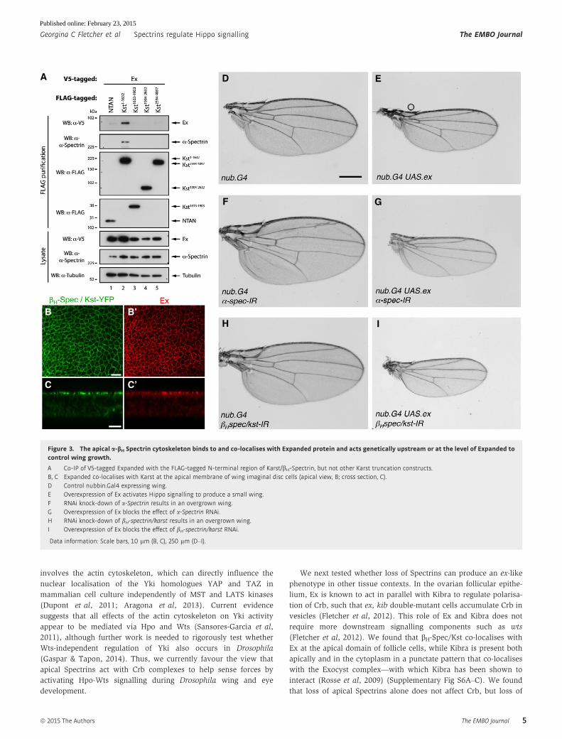
We next investigated the mechanism by which apical Spectrins
regulate Yki activity. Since Spectrins act in parallel with Kibra—
similar to Ex (Baumgartner et al, 2010)—and have been shown to
physically associate with Crb (Medina et al, 2002; Pellikka et al,
2002)—again similar to Ex (Chen et al, 2010; Ling et al, 2010;
Robinson et al, 2010)—we tested whether Ex might interact with
Spectrins. We performed co-immunoprecipitation experiments
from Drosophila S2 cells expressing V5-tagged Ex and a series of
constructs expressing portions of the very large bH-Spec/Kst protein.We found that Ex interacts strongly with the N-terminal region of
bH-Spec/Kst (Fig 3A). Pulling down the N-terminal region of
bH-Spec/Kst with Ex also co-immunoprecipitated endogenous
a-Spec, which is known to form dimers with bH-Spec/Kst (Fig 3A).
In vivo, we found that bH-Spec/Kst co-localises with Ex at the apical
domain of wing disc epithelial cells (Fig 3B and C). a-Spec or
bH-Spec/Kst is not required to localise Ex or Crb apically (Supple-
mentary Fig S5 and data not shown). Instead, they appear to be
required for normal activation of Ex signalling to Hpo and Wts, as
the tissue overgrowth phenotype of a-Spec or bH-Spec/Kst knock-down is completely suppressed by overexpression of Ex (Fig 3D–I).
These results show that apical Spectrins bind to and co-localise with
Ex and act genetically upstream or at the level of Ex to regulate
signalling to Yki.
Hippo signalling has been proposed to have a possible mechano-
sensory role in the Drosophila wing disc, where a pattern of stretch-
ing and compression of cells at their apical surfaces correlates with
the pattern of Yki activity as measured with ex.lacZ (Fig 4A and B;
Aegerter-Wilmsen et al, 2007; Legoff et al, 2013; Mao et al, 2013;
Schluck et al, 2013). We found that this pattern of stretching and
compression influences the intensity of Crb and apical Spectrin
staining in cells (Fig 4A–F). Thus, Crb and bH-Spec/Kst are concen-
trated at the junctions of small compressed cells and diluted at the
junctions of stretched cells in a manner that inversely correlates
with ex.lacZ expression. This correlation suggests a potential model
of mechanosensory regulation of Yki activity via Spectrin-dependent
clustering of Crb complexes (Fig 4G). According to this model,
stretching of cells would exert force upon the apical Spectrin cyto-
skeleton that would de-cluster Crb complexes and therefore reduce
Hpo and Wts activation and increase Yki activity (Fig 4G; see also
Discussion).
To test this model, we aimed to induce clustering of Crb
complexes by overexpression of a form of Crb whose intracellular
domain was replaced with GFP (CrbExTM-GFP; Fig 5A) (Pellikka
et al, 2002; Thompson et al, 2013). Overexpression of CrbExTM-GFP
during wing development with nub.Gal4 resulted in a small wing
phenotype, highly similar to overexpression of Wts (Fig 5B–D).
Furthermore, the overgrowth phenotype caused by RNAi knock-
down of apical Spectrins was completely suppressed by co-
expression of either CrbExTM-GFP or Wts (Fig 5E–J). CrbExTM-GFP
appears to act upstream of Wts, because the tissue undergrowth
phenotype induced by CrbExTM-GFP is suppressed by RNAi knock-
down of Wts (Fig 5K and L). These results are consistent with the
The EMBO Journal ª 2015 The Authors
The EMBO Journal Spectrins regulate Hippo signalling Georgina C Fletcher et al
2
Published online: February 23, 2015

notion that clustering of Crb complexes induces Hippo signalling to
inhibit tissue growth, lending support to a model of mechanosensa-
tion involving clustering of Crb complexes. However, other interpre-
tations and models of mechanosensation are also possible.
One alternative model of mechanosensation involves recruit-
ment of Warts to E-cadherin via the Ajuba protein (Rauskolb
et al, 2014). This mechanism appears to be distinct from and to
act in parallel with the Spectrin/Crumbs-mediated version we
A B I J
K L
M
O
N
C D
E F
G
P Q R
H
Figure 1. The Spectrin cytoskeleton restricts tissue growth in the Drosophila eye and wing.
A–O UAS.RNAi lines were driven with eyeless.Gal4 gmr.Gal4 for expression during eye development or nubbin-Gal4 for expression during wing development. (A, B) Controladult Drosophila eye (A) and wing (B). (C, D) a-spectrin RNAi results in overgrowth of the eye (C) and wing (D). (E, F) bH-spectrin/karst RNAi results in overgrowth ofthe eye (E) and wing (F). (G, H) b-spectrin RNAi does not affect eye size (G) or wing size (H). (I, J) kibra RNAi results in overgrowth of the eye (I) and wing (J). (K, L)a-spectrin, kibra double RNAi results in stronger overgrowth of the eye (K) and wing (L). (M, N) bH-spectrin/karst, kibra double RNAi results in stronger overgrowthof the eye (M) and wing (N). (O) Quantification of female wing sizes by pixel area, 5 wings per genotype were measured. Error bars show standard deviation.
P–R The eyeless FLP MARCM system was used to generate clonally mutant fly eyes. kibra mutant eyes (Q) overgrow slightly compared to controls (P), while kibramutant eyes expressing a-spectrin RNAi (R) overgrow strongly compared to controls (P).
Data information: Scale bars, 250 lm.
ª 2015 The Authors The EMBO Journal
Georgina C Fletcher et al Spectrins regulate Hippo signalling The EMBO Journal
3
Published online: February 23, 2015

propose, because loss of Ajuba and overexpression of CrbExTM-
GFP have an additive effect in suppressing tissue growth
(Fig 5M–O). Furthermore, we do not observe increased recruit-
ment of Warts-GFP to E-cadherin in response to tissue stretching
in the wing imaginal disc (Fig 5P–R), suggesting that this alterna-
tive model does not explain the physiological control of Hippo
signalling in this context. A second alternative model of mechano-
sensation involves activation of the JNK pathway by forces
(Codelia et al, 2014). However, blocking JNK signalling in
Drosophila does not affect tissue growth, and there is no evidence
for physiological regulation of JNK activation by forces in the
wing imaginal disc. A third alternative model of mechanosensation
A B
C D
E E’
F F’
G G’ G’’
H H’ H’’
I I’ I’’
J J’ J’’
K’K K’’
Figure 2. The Spectrin cytoskeleton represses interommatidial cell number and Hippo target gene expression in parallel with Kibra.
A–F The eyeless FLP MARCM system was used to generate mutant eyes that also express UAS.RNAi targeting the Spectrin cytoskeleton. Pupal retinas were examined at42–46 h after puparium formation (APF). Cell membranes were marked with Dlg staining. (A) Control pupal retina showing cone cells surrounded byinterommatidial cells. (B) kibra mutant pupal retina displaying additional interommatidial cells. (C) Pupal retina expressing a-spectrin RNAi showing additionalinterommatidial cells. (D) kibra mutant pupal retinas expressing a-spectrin RNAi showing many additional interommatidial cells. (E, F) Clones of a-spectrin mutantcells (GFP negative) (E) and bH-spectrin/karst mutant cells (GFP negative) (F) show extra interommatidial cells. Scale bars, 20 lm.
G–K UAS.RNAi lines were driven with hh.Gal4 UAS.GFP for expression in the posterior compartment and contained the ex.lacZ reporter transgene. (G) Control wingshowing a normal ex.lacZ expression pattern, which is low at the dorsal–ventral boundary but high in the proximal regions of the wing disc. (H) RNAi knock-downof a-Spectrin results in a mild up-regulation of ex.lacZ in the posterior compartment. (I) RNAi knock-down of kibra results in a mild up-regulation of ex.lacZ in theposterior compartment. (J) Dual RNAi knock-down of both a-spectrin and kibra results in enhanced up-regulation of ex.lacZ in the posterior compartment. (K) RNAiknock-down of hippo results in an up-regulation of ex.lacZ in the posterior compartment. Scale bars, 50 lm.
The EMBO Journal ª 2015 The Authors
The EMBO Journal Spectrins regulate Hippo signalling Georgina C Fletcher et al
4
Published online: February 23, 2015
involves the actin cytoskeleton, which can directly influence the
nuclear localisation of the Yki homologues YAP and TAZ in
mammalian cell culture independently of MST and LATS kinases
(Dupont et al, 2011; Aragona et al, 2013). Current evidence
suggests that all effects of the actin cytoskeleton on Yki activity
appear to be mediated via Hpo and Wts (Sansores-Garcia et al,
2011), although further work is needed to rigorously test whether
Wts-independent regulation of Yki also occurs in Drosophila
(Gaspar & Tapon, 2014). Thus, we currently favour the view that
apical Spectrins act with Crb complexes to help sense forces by
activating Hpo-Wts signalling during Drosophila wing and eye
development.
We next tested whether loss of Spectrins can produce an ex-like
phenotype in other tissue contexts. In the ovarian follicular epithe-
lium, Ex is known to act in parallel with Kibra to regulate polarisa-
tion of Crb, such that ex, kib double-mutant cells accumulate Crb in
vesicles (Fletcher et al, 2012). This role of Ex and Kibra does not
require more downstream signalling components such as wts
(Fletcher et al, 2012). We found that bH-Spec/Kst co-localises with
Ex at the apical domain of follicle cells, while Kibra is present both
apically and in the cytoplasm in a punctate pattern that co-localises
with the Exocyst complex—with which Kibra has been shown to
interact (Rosse et al, 2009) (Supplementary Fig S6A–C). We found
that loss of apical Spectrins alone does not affect Crb, but loss of
A
B’B
C’C
D E
F G
H I
Figure 3. The apical a-bH Spectrin cytoskeleton binds to and co-localises with Expanded protein and acts genetically upstream or at the level of Expanded tocontrol wing growth.
A Co-IP of V5-tagged Expanded with the FLAG-tagged N-terminal region of Karst/bH-Spectrin, but not other Karst truncation constructs.B, C Expanded co-localises with Karst at the apical membrane of wing imaginal disc cells (apical view, B; cross section, C).D Control nubbin.Gal4 expressing wing.E Overexpression of Ex activates Hippo signalling to produce a small wing.F RNAi knock-down of a-Spectrin results in an overgrown wing.G Overexpression of Ex blocks the effect of a-Spectrin RNAi.H RNAi knock-down of bH-spectrin/karst results in an overgrown wing.I Overexpression of Ex blocks the effect of bH-spectrin/karst RNAi.
Data information: Scale bars, 10 lm (B, C), 250 lm (D–I).
ª 2015 The Authors The EMBO Journal
Georgina C Fletcher et al Spectrins regulate Hippo signalling The EMBO Journal
5
Published online: February 23, 2015

apical Spectrins in combination with mutation of kib or sec15
(encoding an Exocyst component) causes a strong accumulation of
Crb in vesicles (Supplementary Fig S6D–M). Thus, apical Spectrins
are involved in Ex-mediated regulation of Crb polarisation in follicle
cells.
We noted that a-spec RNAi in kib mutant clones caused a strong
overproliferation and multilayering phenotype in follicle cells
(Supplementary Fig S6M). We therefore tested whether Spectrins
are required for regulation of Yki activity in the ovarian follicular
epithelium. Yki is known to promote early proliferation and
suppress apoptosis in follicle cells until stage 6, during which time it
expresses the transcription factor Cut (Huang & Kalderon, 2014).
After stage 6, Cut expression is silenced and cells arrest proliferation
and switch to an endocycle. When Hippo signalling is disrupted
after stage 6, Cut is known to be re-expressed in a group of posterior
follicle cells that continue to proliferate (Meignin et al, 2007;
Polesello & Tapon, 2007; Genevet et al, 2010; Yu et al, 2010).
Surprisingly, we found that mutation of either bH-spec/kst or crb is
not sufficient to drive early overproliferation (Fig 6A–D) or to
induce Cut in late-stage posterior follicle cells (Fig 6G–I). Multilayer-
ing only occurs in around 30% of crb mutant clones, while mutation
of a-spec or b-spec leads to early overproliferation and multilayering
in 95% of clones (Fig 6E and F) and also leads to re-expression of
Cut in the posterior follicle cells (Fig 6J and K). Furthermore,
another Hippo target gene, ex.lacZ, is also ectopically expressed in
posterior follicle cells mutant for a-spec or b-spec but not in bH-spec/kst or crb mutant clones (Fig 6L–P). Notably, mutation of b-speccauses a disruption of membrane tension such that the normally
regular hexagonal shape of follicle cells becomes disorganised
(Fig 6Q–S). These findings indicate that the basolateral a-b Spectrin
cytoskeleton controls membrane tension and Hippo signalling to the
nucleus in the follicular epithelium.
A
G
B C D
E F
Figure 4. The apical a-bH Spectrin cytoskeleton may be mechanosensory in the wing imaginal disc.
A Schematic diagram of central compression and circumferencial stretching in the third instar wing pouch.B–D Third instar wing imaginal disc stained for beta-galactosidase expressed from the expanded.lacZ reporter gene (B), Crb (C) and Kst-YFP (D). Quantification of line
intensity shown below. Note inverse correlation of Crb and Kst-YFP with ex.lacZ. Scale bars, 50 lm.E, F Zoom of Kst-YFP cells under compression (E) and under stretch (F). Scale bars, 10 lm.G Schematic diagram of the effect of force on the apical spectrin cytoskeleton leading to declustering of Crumbs complexes and reduced Hippo signalling.
The EMBO Journal ª 2015 The Authors
The EMBO Journal Spectrins regulate Hippo signalling Georgina C Fletcher et al
6
Published online: February 23, 2015

A small group of anterior follicle cells, called border cells, are
known to delaminate from the epithelium at stage 8 of develop-
ment and migrate across the egg chamber to reach the oocyte at
the posterior by stage 10. We previously showed that upstream
Hippo pathway components have a key role in promoting border
cell migration by regulating the actin cytoskeleton (Lucas et al,
2013). We found that a-spec RNAi knock-down in kib mutant
border cell clusters causes a strong delay in reaching the oocyte
by stage 10 of oogenesis (Supplementary Fig S7A–E). a-spec RNAi
knock-down alone, or mutation of kib alone, causes a milder
phenotype (Supplementary Fig S7E). Interestingly, RNAi knock-
down of bH-spec/kst did not cause a phenotype, alone or in
combination with kib (Supplementary Fig S7E). Accordingly, we
found that bH-Spec/Kst is not actually expressed in border cells,
while b-Spec localises around the entire plasma membrane with
a-Spec (Supplementary Fig S7F–I). These results indicate that a-bSpectrin dimers regulate Hippo signalling during border cell
migration.
A B C D
E F G
H I J
K L
M N
O
RP Q
Figure 5. CrbExTM-GFP restricts tissue growth and acts upstream of Wts and in parallel with Ajuba.
A Schematic diagram of clustering of Crb complexes by expression of CrbExTM-GFP.B Control nub.Gal4 wing.C, D Expression of UAS.CrbExTM-GFP with nub.Gal4 (C) or UAS.Wts with nub.Gal4 (D) results in a small wing.E RNAi knock-down of a-Spectrin results in an overgrown wing.F, G Expression of UAS.CrbExTM-GFP (F) or UAS.Wts (G) blocks the effect of a-Spectrin RNAi.H RNAi knock-down of bH-spectrin/karst results in an overgrown wing.I, J Expression of UAS.CrbExTM-GFP (I) or UAS.Wts (J) blocks the effect of a-Spectrin RNAi.K, L RNAi knock-down of Wts results in a strongly overgrown wing (K) and expression of UAS.CrbExTM-GFP does not block the effect of wts RNAi (L).M, N RNAi knock-down of Ajuba results in a small wing (M), and expression of UAS.CrbExTM-GFP enhances the effect of ajuba RNAi (N).O Quantification of various wing sizes. Error bars show standard deviation.P, Q Localisation of Wts-GFP (P) and E-cadherin (Q) in the third instar wing imaginal disc.R Quantification of line intensity in (P) and (Q). No increased Wts-GFP intensity is observed in proximal regions where cells are subject to stretching.
Data information: Scale bars, 250 lm (B–N), 40 lm (P, Q).
ª 2015 The Authors The EMBO Journal
Georgina C Fletcher et al Spectrins regulate Hippo signalling The EMBO Journal
7
Published online: February 23, 2015

A A’
B B’
C C’
D D’
E E’
F
Q
F’
G G’
H H’
I I’
J J’
K K’
M M’
N N’
O O’
P P’
R S
L L’
Figure 6. Basolateral a-b Spectrins are required for Hippo signalling and membrane tension in the Drosophila follicular epithelium.
A Control egg chamber stained for DAPI to mark nuclei.B Overexpression of Yki3SA stimulates follicle cell proliferation (MARCM clone, GFP positive).C–F Mutation of bH-spectrin/karst (C) or crb (D) does not increase follicle cell proliferation (GFP negative clone), while mutation of a-spectrin (E) or b-spectrin (F)
stimulates follicle cell proliferation (GFP negative clone).G Control egg chamber stained for Cut, a marker of proliferating cells that is normally down-regulated after stage 6 of oogenesis.H–K Mutation of bH-spectrin/karst (H) or crb (I) does not increase Cut expression in follicle cells (GFP negative clone), while mutation of a-spectrin (J) or b-spectrin (K)
stimulates Cut expression in posterior follicle cells (GFP negative clone).L Control egg chamber showing ex.lacZ reporter gene expression.M–P Mutation of bH-spectrin/karst (M) or crb (N) does not increase ex.lacZ expression in follicle cells (GFP negative clone), while mutation of a-spectrin (O) or b-spectrin
(P) stimulates ex.lacZ expression in posterior follicle cells (GFP negative clone).Q Wild-type follicle cells showing normal hexagonal packing of epithelial membranes.R Mutation of b-spectrin disrupts the normal hexagonal packing, suggesting a role in maintaining membrane tension.S Clone of b-spectrin mutant cells (GFP negative) showing differential membrane tension at the interface with wild-type cells (GFP positive).
Data information: Scale bars, 25 lm (A–R), 10 lm (S).
The EMBO Journal ª 2015 The Authors
The EMBO Journal Spectrins regulate Hippo signalling Georgina C Fletcher et al
8
Published online: February 23, 2015

The Hippo pathway was recently shown to have a key role in
regulating stem cell proliferation in the Drosophila intestine
(Karpowicz et al, 2010; Shaw et al, 2010; Staley & Irvine, 2010).
RNAi knock-down of wts or overexpression of yki in enterocytes
with the myo1A.G4 driver leads to induction of stem cell prolifera-
tion (Karpowicz et al, 2010; Shaw et al, 2010; Staley & Irvine,
2010). However, the upstream regulators that control Hippo signal-
ling in the intestine have not been identified. We therefore exam-
ined the requirement for Crb and Spectrins in the adult fly intestine.
We found that RNAi of crb or apical bH-spec/kst did not lead to stem
cell overproliferation (Fig 7A–C). However, RNAi silencing of
a-spec or b-spec does produce a strong stem cell proliferation
response, similar to overexpression of Yki (Fig 7D–G). Furthermore,
a Hippo pathway reporter gene DIAP1-HRE-GFP is activated in the
overproliferating stem cells (Fig 7H–K). These results indicate that
the basolateral a-b Spectrin cytoskeleton, rather than the apical
a-bH-Spectrin cytoskeleton, is crucial to promote Yki activity in
enterocytes.
The Spectrin cytoskeleton is conserved between Drosophila and
humans, but is more complex in humans as it features many variant
b-Spectrins. To test whether the Spectrin cytoskeleton also regulates
Hippo signalling in human cells, we silenced expression of the sole
human non-erythrocyte alpha-Spectrin protein, SPTAN1, and a
major non-erythrocyte b-Spectrin, SPTBN1, by siRNA transfection of
Caco-2 cells. The human homologue of Yki, called YAP, is normally
nuclear in sparsely plated cells, but becomes cytoplasmic in densely
confluent epithelial monolayers due to mechanical cues (Dupont
et al, 2011) (Fig 8A and B). We found that siRNA knock-down of
A B C G
D E F H
J K
I
D’ E’ F’
A’ B’ C’
Figure 7. Basolateral a-b Spectrins are required to restrict cell proliferation in the Drosophila intestinal epithelium.
A Control myo1A.Gal4 UAS.GFP adult midgut stained for phospho-histone H3 to mark mitotic stem cells.B–E RNAi knock-down of bH-spectrin/karst (B) or crumbs (C) does not increase stem cell proliferation, while RNAi knock-down of b-spectrin (D) or a-spectrin (E) strongly
stimulates stem cell proliferation.F Overexpression of Yki strongly stimulates stem cell proliferation.G Quantification of the number of PH3-positive mitotic cells. N > 13 samples. Statistical analysis performed using the two-tailed Student’s t-test. Error bars show
standard deviation. ***P-value ≤ 0.001.H–K A Hippo reporter DIAP1-HRE-GFP is normally expressed in sporadic stem cells. Its expression is not affected by RNAi knock-down of bH-spectrin/karst (I), but is
up-regulated in many stem cells by RNAi knock-down of a-spectrin (J) or by overexpression of Yki (K).
Data information: Scale bars, 100 lm.
ª 2015 The Authors The EMBO Journal
Georgina C Fletcher et al Spectrins regulate Hippo signalling The EMBO Journal
9
Published online: February 23, 2015

SPTAN1 or SPTBN1 in densely confluent epithelia is sufficient to send
YAP to the nucleus and to reduce YAP phosphorylation and LATS1
phosphorylation (Fig 8C–G). These results show that the Spectrin
cytoskeleton is crucial for regulation of YAP in response to density in
human cell culture and that disruption of Spectrins has similar effects
to mechanical stretching of cells by plating at low density.
Discussion
Our results identify the Spectrin cytoskeleton as an important
upstream regulator of the Hippo signalling pathway in both
Drosophila and human cells. In all fly tissues we have examined,
Spectrins are essential to maintain full repression of Yki activity.
However, different tissues exhibit different dependence on apical
a-bH versus basolateral a-b Spectrins. The localisation of Spectrin
proteins in each model tissue we have examined is summarised in
Supplementary Fig S8.
In the developing wing and eye, apical a-bH-Spectrins act with
Crb and Ex to activate Hippo signalling and thereby restrict tissue
growth (Figs 1 and 2). Since Crb binds to Ex (Chen et al, 2010; Ling
et al, 2010), and Ex can bind to both Kibra (Genevet et al, 2010)
and Spectrins (Fig 2), and Crb can also associate with Spectrins
(Medina et al, 2002; Pellikka et al, 2002), these five proteins are
A A’ E
F
G
A’’
B
C C’
B’ B’’
C’’
D D’ D’’
Figure 8. Human Spectrin SPTAN1 and SPTBN1 are required for the YAP localisation response to cell density.
A Control Caco-2 epithelial cells plated a low density show strong nuclear YAP localisation.B Control Caco-2 epithelial cells plated at high density show relocalisation of YAP to the cytoplasm.C, D siRNA knock-down of SPTAN1 (C) or SPTBN1 (D) prevents relocalisation of YAP to the cytoplasm.E Quantification of YAP localisation in (B–D).F Western blotting reveals that SPTAN1 or SPTBN1 are required for normal levels of YAP phosphorylation, and of LATS1 phosphorylation.G siRNAs against SPTAN1 or SPTBN1 efficiently deplete their target proteins.
Data information: Scale bars, 20 lm.
The EMBO Journal ª 2015 The Authors
The EMBO Journal Spectrins regulate Hippo signalling Georgina C Fletcher et al
10
Published online: February 23, 2015

likely to form a complex at the apical domain of Drosophila epithe-
lial cells. Kibra is also able to associate with the FERM-domain
protein Merlin (Mer) at the apical domain (Genevet et al, 2010; Yu
et al, 2010), and we are able to co-immunoprecipitate both Kibra
and Mer with apical a-bH Spectrins (Supplementary Fig S9). In
contrast, Hpo and Wts localise primarily to the cytoplasm, but must
transiently associate with upstream pathway components at the
plasma membrane in order to be activated (Lucas et al, 2013; Yin
et al, 2013). Direct binding of Wts to Mer and indirect binding of
Hpo via Sav to a network of WW domain–PPXY motif interactions
appear to facilitate activation of Hpo and Wts at the plasma
membrane, where molecular clustering may induce Hpo auto-
phosphorylation and phosphorylation of Wts to drive signalling. In
the wing and eye epithelium, apical a-bH Spectrins may assist this
signal-activating process by forming a dense meshwork that
promotes clustering of Crb-Ex-Kib-Mer complexes to induce Hpo
and Wts kinase activity to restrict growth (Fig 4). In support of this
model, inducing clustering of endogenous Crb by overexpressing a
form of Crb lacking the intracellular domain is sufficient to over-
come the loss of Spectrins and to suppress wing growth (Fig 5).
In the developing follicular epithelium and adult intestinal epithe-
lium, basolaterally localised a-b Spectrins, rather than apical a-bHSpectrins, are crucial for repression of Yki activity (Figs 6 and 7).
This finding may explain why apical Crb is surprisingly not required
for Yki repression in these tissues (Figs 6 and 7). This finding is all
the more remarkable because apical a-bH Spectrins do act in parallel
with Kibra to polarise Crb in follicle cells (Supplementary Fig S6).
Thus, the mere presence of Crb, Ex, Kib and a-bH Spectrins in a cell
does not necessitate that these proteins will form the primary
control mechanism for the regulation of Yki. How basolateral a-bSpectrins connect to Hippo signalling and/or regulation of Yki in
these tissues remains to be explored. Nevertheless, in all Drosophila
tissues we have examined, either the a-b or a-bH Spectrins are
necessary for normal repression of Yki.
In human epithelial cells, polarisation of a (SPTAN1) and b(SPTBN1) Spectrins is much less apparent than in Drosophila
(Supplementary Fig S8). Nevertheless, our data show that these
Spectrins are essential for regulating the classic YAP localisation
response to cell density in culture (Fig 8), as are homologues of Crb
(CRB3), Ex (AMOT) and Mer (NF2) (Zhao et al, 2007, 2011; Varelas
et al, 2010). This response to cell density was shown to be a
mechanosensory response that requires an intact F-actin cytoskele-
ton (Dupont et al, 2011). However, the identity of the mechanosen-
sory molecule(s) that mediate this response is not clear. Our
findings suggest that the Spectrin cytoskeleton is a candidate for this
Hippo pathway mechanosensor.
Several lines of evidence support a mechanosensory function for
Spectrins. Firstly, electron microscopy studies show that Spectrins
form a dense meshwork at the plasma membrane that can stretch
out under force, separating the N-terminal nodes at which FERM-
domain proteins bind (Hirokawa et al, 1983; Byers & Branton, 1985;
Ursitti et al, 1991). Secondly, individual Spectrin molecules can
deform under force via conformational change (Johnson et al, 2007;
Stabach et al, 2009). Thirdly, construction of a FRET-based Spectrin
sensor reveals that Spectrins are under tension and can sense force
in human cells in culture (Meng & Sachs, 2012) and in C. elegans
neurons (Krieg et al, 2014). Finally, loss of Spectrins causes a loss
of membrane tension in C. elegans neurons (Krieg et al, 2014) as
well as red blood cells (Stokke et al, 1986), and our results
are consistent with a similar role in Drosophila epithelial cells
(Figs 2A–F and 6Q–S). Thus, it is likely that force may cause altera-
tions in the Spectrin cytoskeleton that modulate Hippo signalling,
possibly by spatially separating the N-terminal nodes of the network
that bind to FERM-domain proteins, thereby inducing de-clustering
of upstream pathway components and signal inhibition (Fig 4G).
Since Spectrins can bind to F-actin, it is also possible that force
upon Spectrins may influence the actin cytoskeleton and therefore
potentially influence Yki activation independently of Hpo and Wts
activation, as has been shown for the mammalian homologues
YAP/TAZ (Dupont et al, 2011; Aragona et al, 2013). Furthermore,
Wts has also been shown to influence F-actin polymerisation via
regulation of Ena/VASP proteins (Lucas et al, 2013), raising the
possibility that canonical Hpo-Wts signalling may also act via the
regulation of F-actin dynamics to control Yki, in addition to direct
phosphorylation of the Yki protein by Wts. Further work is neces-
sary to test whether direct F-actin regulation of Yki localisation
occurs in Drosophila as it does in mammalian cells (Gaspar &
Tapon, 2014).
Regardless of which signalling mechanisms Spectrins use to
control Yki activity, their molecular nature makes them excellent
candidate mechanosensors and Spectrins are clearly able to influ-
ence Yki-dependent tissue growth. Furthermore, there is good
evidence that forces can and do contribute to promoting cell prolif-
eration in Drosophila wing epithelia (Aegerter-Wilmsen et al, 2007;
Legoff et al, 2013; Mao et al, 2013; Schluck et al, 2013), and it has
been proposed that forces may also drive proliferation in the follicu-
lar epithelium (Wang & Riechmann, 2007). In addition, input from
the Dachsous-Fat pathway to Hippo signalling is mediated via the
atypical myosin Dachs, which has been shown to promote tension
at apical cell–cell junctions (Mao et al, 2011). Thus, it is not
implausible that forces could be sensed by Drosophila Spectrins as
part of the physiological control of tissue growth during develop-
ment and that this function could help explain how disparate
upstream regulators of Hippo signalling, such as Crb-Crb junctions
and the Dachsous-Fat cadherin system, converge to control Yki
activity.
A physiological role for Spectrins as sensors of tension that regu-
late Hippo signalling may well be conserved across metazoa. For
example, the dramatic effect of forces on human skin growth is well
known, and the Hippo pathway has been shown to regulate skin
proliferation in mice (Schlegelmilch et al, 2011; Silvis et al, 2011;
Zhang et al, 2011). Notably, a recently proposed alternative mecha-
nism for mechanosensation involving recruitment of Wts to a-cate-nin via the Ajuba protein is less likely to be conserved in mammals
(Das Thakur et al, 2010; Rauskolb et al, 2014). In ajuba mutants,
Wts recruitment to a-catenin is disrupted, leading to stronger Hippo
signalling and tissue undergrowth in Drosophila (Rauskolb
et al, 2014). However, in mammals, loss of a-catenin leads to
weaker Hippo signalling and tissue overgrowth in mouse skin
(Schlegelmilch et al, 2011; Silvis et al, 2011) and loss of adherens
junctions also weakens Hippo signalling to activate YAP in cultured
human cells (Kim et al, 2011). Thus, our identification of Spectrins
as potential mechanosensors in the Hippo pathway provides an
important novel mechanism that is more likely to be conserved in
all metazoans and may be a critical control mechanism in both
normal development and cancer.
ª 2015 The Authors The EMBO Journal
Georgina C Fletcher et al Spectrins regulate Hippo signalling The EMBO Journal
11
Published online: February 23, 2015

Materials and Methods
Mitotic clones in follicle cells were generated using the FLP/FRT
system and were marked either negatively (absence of GFP) or posi-
tively (presence of GFP; MARCM). Third instar larvae were heat-
shocked once at 37°C for 1 h and dissected 4 days after eclosion.
Expression of UAS-driven transgenes in follicle cells was achieved
with the MARCM system and in wing imaginal disc cells with the
posterior compartment driver hh.Gal4 and in enterocytes with
Myo1A.G4.
Drosophila genotypes
See Supplementary Materials and Methods for all Drosophila geno-
types used in this study.
Immunostaining of ovaries, imaginal discs and pupal retinas
Ovaries and imaginal discs were dissected in PBS, fixed for 20 min in
4% paraformaldehyde in PBS, washed for 30 min in PBS/0.1%
Triton X-100 (PBST) and blocked for 15 min in 5% normal goat
serum/PBST (PBST/NGS). Primary antibodies were diluted in PBST/
NGS, and samples were incubated overnight at 4°C. Optical cross
sections through the middle of egg chambers are shown in all figures.
Primary antibodies used were as follows: mouse anti-Cut (1:100
DSHB), mouse anti-bgal (Promega, 1:500), rabbit anti-expanded
(1:200, gift from A. Laughon, University of Wisconsin-Madison,
Madison, WI, USA), rabbit anti-PKCf (C-20) (1:250; Santa Cruz), rat
anti-Crumbs (1:300; U. Tepass), mouse anti-Dlg (1:250, DSHB),
rabbit anti-Kibra (1:200; Genevet et al, 2010) and guinea pig anti-
Sec 15 (1:1,000, gift from H. Bellen, Howard Hughes Medical Insti-
tute, Baylor College of Medicine).
Secondary antibodies (all from Molecular Probes; Invitrogen)
were used at 1:500 for 2–4 h prior to multiple washes in PBST and
staining with DAPI at 1 lg/ml for 10–30 min prior to mounting on
slides in Vectashield (Vector labs). Images were taken with a Leica
SP5 confocal microscope and processed with Adobe Photoshop.
Immunostaining of intestinal epithelia
Flies were maintained on standard media, which was changed every
3 days. Crosses were set up and maintained at 18°C until adulthood.
Three- to four-day-old adults were shifted to 29°C for 10 days for
induction of transgene expression. Female adult flies were dissected
in 1× PBS. The gastrointestinal tract was removed and fixed in 0.5×
PBS with 4% paraformaldehyde for 30 min. Samples were washed
in 0.1% Triton X-100 (PBST), permeabilised for 30 min in 0.3%
PBST and pre-blocked for 1 h in 10% NGS before incubation with
primary antibody overnight at 4°C in 5% NGS. Samples were
washed in PBST and pre-blocked for 1 h in 10% NGS before incuba-
tion with secondary antibody in 5% NGS for 2 h at room tempera-
ture. Samples were stained with DAPI and washed in PBST,
followed by PBS and mounted in Vectashield.
The following primary antibody was used: rabbit anti-phospho-
histone H3 (Millipore) 1/1,000. Secondary antibodies (all from
Molecular Probes; Invitrogen) were used at 1:500. Images were
acquired on a Zeiss LSM710 confocal microscope and were
processed using Adobe Photoshop.
Molecular biology
The Expanded expression plasmid was generated previously
(Genevet et al, 2010). b-heavy Spectrin expression plasmids were
generated using Gateway technology. Constructs detailed below
were PCR-amplified and cloned into the pDONR Zeo Entry vector.
For Destination vectors, the pAWF and pAWH plasmids from the
Drosophila Gateway Vector Collection were used.
The constructs (and primers) used were as follows:
Karst isoform A; aa 1–1632
Fwd primer—50 atgacccagcgggacggcatcatcaag 30
Rev primer—50 ggactttgctgccggcaggtgttc 30
Karst isoform A; aa 1633–1903
Fwd primer—50 atgaatgaattgggtcagaatctgcaccaagccc 30
Rev primer—50 gagttgttcctgtcgcttctcaacctcatt 30
Karst isoform A; aa 1904–2632
Fwd primer—50 atgagaacttcctgggagaaccttctccagc 30
Rev primer—50 ctgctcgccctgttgtttcagtccagc 30
Karst isoform A; aa 2594–4097
Fwd primer—50 atgcagtcaccatccactatcaacgattgcgag 30
Rev primer—50 ctgtggcgggacttgactcgagcg 30
Co-immunoprecipitation
Drosophila S2 cell extracts were prepared as previously described
(Genevet et al, 2010), and FLAG-tagged proteins were purified using
anti-FLAG M2 Affinity Agarose Gel (Sigma) before elution with lysis
buffer [50 mM Tris pH 7.5, 150 mM NaCl, 1% Triton X-100, 10%
glycerol and 1 mM EDTA, plus phosphatase inhibitor cocktails 1
and 2 (Sigma), protease inhibitor cocktail (Roche) and 0.1 M NaF]
supplemented with FLAG peptide. Detection of purified proteins and
associated complexes was performed using chemiluminescence (GE
Healthcare). Western blots were probed with anti-FLAG (mouse M2;
Sigma), anti-V5 (mouse Novex, Life Technologies), anti-a-Spectrin(mouse 3A9, Developmental Studies Hybridoma Bank—DHSB) and
anti-Tubulin (mouse E7, DSHB) antibodies.
For Co-IP from embryos, Drosophila Karst YFP knock-in embryos
(DGRC 115285) or Wiso embryos were collected over 24 h at 22°C
before being lysed in buffer containing 10 mM Tris pH 7.5, 150 mM
NaCl, 0.5% NP-40 and 0.5 mM EDTA (Chromotek), plus PhosSTOP
Phosphatase Inhibitor Cocktail Tablets (Roche), protease inhibitor
cocktail (Roche), 0.1 M NaF and 1 mM PMSF. Samples were left on
ice to solubilise for 20 min, before being centrifuged at high speed
(14,000 rpm for 30 min at 4°C) and the supernatant collected, pre-
cleared and incubated with GFP Trap-M beads (Chromotek).
Western blots were probed with anti-GFP (mouse; Roche), anti-
Merlin (guinea pig, R. Fehon), anti-a-Spectrin (mouse 3A9, Develop-
mental Studies Hybridoma Bank—DHSB) and anti-Kibra (rabbit,
Genevet et al, 2010) antibodies, before being detected with chemilu-
minescence (GE Healthcare).
Cell culture and transfection
Human Caco-2 adenocarcinoma colon cells were grown in DMEM
(Gibco: 41966) containing L-glutamine and sodium pyruvate
The EMBO Journal ª 2015 The Authors
The EMBO Journal Spectrins regulate Hippo signalling Georgina C Fletcher et al
12
Published online: February 23, 2015

supplemented with 10% heat-inactivated FCS and 100 lg/ml
streptomycin and 100 lg/ml penicillin. Cells were maintained in a
37°C incubator at 5% atmospheric CO2. All siRNA transfections
were performed with interferin transfection reagent (Polyplus) using
antibiotic-free media. Caco-2 cells were seeded upon 10 mm cover-
slips coated with 20 lg/ml fibronectin in a 48-well plate at either
low density or high density, and 2 h after seeding treated with the
siRNA/transfection mix. A final concentration of 100 nM siRNA was
used. The following day, the media was changed and another round
of siRNA transfection was performed. Cells were left for a total of
72 h before being processed for either immunofluorescence or
immunoblotting. siRNA oligonucleotides used for RNAi of SPTAN1
and SPTBN1 were as a siGENOME SMARTpool (Thermo scientific).
For fixation, cells were washed with 1× PBS and either fixed in
4% paraformaldehyde in PBS for 15 min, before permeabilising
with 0.3% Triton X-100 in blocking buffer (PBS containing 0.5%
BSA, 10 mM glycine, and 0.1% sodium azide), or �20°C methanol
for 5 min, before rehydrating with PBS. Samples were then washed
and incubated in blocking buffer for 1 h before staining. Primary
and secondary antibodies were incubated in blocking buffer. Cover-
slips were mounted using prolong anti-fade reagent (Invitrogen).
Antibodies, image acquisition and quantification for cell culture
Antibodies used in the paper were as follows: mouse and rabbit anti-
SPTAN1 (Santa Cruz and CST); mouse anti-SPTBN1 (Santa Cruz);
rabbit anti-YAP (Santa Cruz), rabbit anti-pYAP (CST); rabbit anti-
pLATS1 and anti-LATS1 (CST); mouse a-tubulin (Sigma). Secondary
antibodies used were either from Jackson immunoresearch or from
Invitrogen. DNA was stained with DAPI 1:1,000 and samples imaged
with a Zeiss 710 or a Leica SP5 confocal microscope. Quantification of
YAP localisation was scored in three categories: N = nuclear; N/C =
nuclear and cytoplasmic and C = cytoplasmic. Cells were assessed over
three independent experiments counting 350–500 cells per condition.
Supplementary information for this article is available online:
http://emboj.embopress.org
AcknowledgementsWe thank Graham Thomas for the spectrinmutant stocks and the cDNAs used to
generate the constructs for biochemical experiments. We also thank Karl Matter
for providing Caco-2 cells and Hugo Stocker for discussing unpublished data.
Author contributionsGCF and IK performed the experiments in Drosophila; AE performed the experi-
ments in mammalian cell culture; PSR performed the biochemical experi-
ments. BJT and NT supervised the experiments; BJT performed the initial
genetic screen, conceived the experiments and wrote the manuscript with
input from the other authors.
Conflict of interestThe authors declare that they have no conflict of interest.
References
Aegerter-Wilmsen T, Aegerter CM, Hafen E, Basler K (2007) Model for the regulation
of size in the wing imaginal disc of Drosophila.Mech Dev 124: 318– 326
Aragona M, Panciera T, Manfrin A, Giulitti S, Michielin F, Elvassore N, Dupont S,
Piccolo S (2013) A mechanical checkpoint controls multicellular growth
through YAP/TAZ regulation by actin-processing factors. Cell 154: 1047 – 1059
Baumgartner R, Poernbacher I, Buser N, Hafen E, Stocker H (2010) The WW
domain protein Kibra acts upstream of Hippo in Drosophila. Dev Cell 18:
309 – 316
Bennett V, Baines AJ (2001) Spectrin and ankyrin-based pathways: metazoan
inventions for integrating cells into tissues. Physiol Rev 81: 1353 – 1392
Bennett V, Healy J (2009) Membrane domains based on ankyrin and spectrin
associated with cell-cell interactions. Cold Spring Harb Perspect Biol 1: a003012
Byers TJ, Branton D (1985) Visualization of the protein associations in the
erythrocyte membrane skeleton. Proc Natl Acad Sci USA 82: 6153 – 6157
Cai J, Zhang N, Zheng Y, de Wilde RF, Maitra A, Pan D (2010) The Hippo
signaling pathway restricts the oncogenic potential of an intestinal
regeneration program. Genes Dev 24: 2383 – 2388
Camargo FD, Gokhale S, Johnnidis JB, Fu D, Bell GW, Jaenisch R,
Brummelkamp TR (2007) YAP1 increases organ size and expands
undifferentiated progenitor cells. Curr Biol 17: 2054 – 2060
Chen CL, Gajewski KM, Hamaratoglu F, Bossuyt W, Sansores-Garcia L, Tao C,
Halder G (2010) The apical-basal cell polarity determinant Crumbs regulates
Hippo signaling in Drosophila. Proc Natl Acad Sci USA 107: 15810 – 15815
Codelia VA, Sun G, Irvine KD (2014) Regulation of YAP by mechanical strain
through Jnk and Hippo signaling. Curr Biol 24: 2012 – 2017
Cordenonsi M, Zanconato F, Azzolin L, Forcato M, Rosato A, Frasson C, Inui
M, Montagner M, Parenti AR, Poletti A, Daidone MG, Dupont S, Basso G,
Bicciato S, Piccolo S (2011) The Hippo transducer TAZ confers cancer stem
cell-related traits on breast cancer cells. Cell 147: 759 – 772
Das Thakur M, Feng Y, Jagannathan R, Seppa MJ, Skeath JB, Longmore GD
(2010) Ajuba LIM proteins are negative regulators of the Hippo signaling
pathway. Curr Biol 20: 657 – 662
Dupont S, Morsut L, Aragona M, Enzo E, Giulitti S, Cordenonsi M, Zanconato
F, Le Digabel J, Forcato M, Bicciato S, Elvassore N, Piccolo S (2011) Role of
YAP/TAZ in mechanotransduction. Nature 474: 179 – 183
Fletcher GC, Lucas EP, Brain R, Tournier A, Thompson BJ (2012) Positive
feedback and mutual antagonism combine to polarize Crumbs in the
Drosophila follicle cell epithelium. Curr Biol 22: 1116 – 1122
Gaspar P, Tapon N (2014) Sensing the local environment: actin architecture
and Hippo signalling. Curr Opin Cell Biol 31C: 74 – 83
Genevet A, Wehr MC, Brain R, Thompson BJ, Tapon N (2010) Kibra is a regulator
of the Salvador/Warts/Hippo signaling network. Dev Cell 18: 300 – 308
Halder G, Johnson RL (2011) Hippo signaling: growth control and beyond.
Development 138: 9 – 22
Hamaratoglu F, Willecke M, Kango-Singh M, Nolo R, Hyun E, Tao C, Jafar-
Nejad H, Halder G (2006) The tumour-suppressor genes NF2/Merlin and
Expanded act through Hippo signalling to regulate cell proliferation and
apoptosis. Nat Cell Biol 8: 27 – 36
Harvey KF, Pfleger CM, Hariharan IK (2003) The Drosophila Mst ortholog, hippo,
restricts growth and cell proliferation and promotes apoptosis. Cell 114:
457 – 467
Hirokawa N, Cheney RE, Willard M (1983) Location of a protein of the fodrin-
spectrin-TW260/240 family in the mouse intestinal brush border. Cell 32:
953 – 965
Huang J, Wu S, Barrera J, Matthews K, Pan D (2005) The Hippo signaling
pathway coordinately regulates cell proliferation and apoptosis by
inactivating Yorkie, the Drosophila Homolog of YAP. Cell 122: 421 –434
Huang J, Kalderon D (2014) Coupling of Hedgehog and Hippo pathways
promotes stem cell maintenance by stimulating proliferation. J Cell Biol
205: 325 – 338
ª 2015 The Authors The EMBO Journal
Georgina C Fletcher et al Spectrins regulate Hippo signalling The EMBO Journal
13
Published online: February 23, 2015

Irvine KD (2012) Integration of intercellular signaling through the Hippo
pathway. Semin Cell Dev Biol 23: 812 – 817
Johnson CP, Tang HY, Carag C, Speicher DW, Discher DE (2007) Forced
unfolding of proteins within cells. Science 317: 663 – 666
Karpowicz P, Perez J, Perrimon N (2010) The Hippo tumor suppressor
pathway regulates intestinal stem cell regeneration. Development 137:
4135 – 4145
Kim NG, Koh E, Chen X, Gumbiner BM (2011) E-cadherin mediates contact
inhibition of proliferation through Hippo signaling-pathway components.
Proc Natl Acad Sci USA 108: 11930 – 11935
Koontz LM, Liu-Chittenden Y, Yin F, Zheng Y, Yu J, Huang B, Chen Q, Wu S,
Pan D (2013) The Hippo effector Yorkie controls normal tissue growth by
antagonizing scalloped-mediated default repression. Dev Cell 25: 388 – 401
Krieg M, Dunn AR, Goodman MB (2014) Mechanical control of the sense of
touch by beta-spectrin. Nat Cell Biol 16: 224 – 233
Lee JK, Brandin E, Branton D, Goldstein LS (1997) alpha-Spectrin is required
for ovarian follicle monolayer integrity in Drosophila melanogaster.
Development 124: 353 – 362
Legoff L, Rouault H, Lecuit T (2013) A global pattern of mechanical stress
polarizes cell divisions and cell shape in the growing Drosophila wing disc.
Development 140: 4051 – 4059
Ling C, Zheng Y, Yin F, Yu J, Huang J, Hong Y, Wu S, Pan D (2010) The apical
transmembrane protein Crumbs functions as a tumor suppressor that
regulates Hippo signaling by binding to Expanded. Proc Natl Acad Sci USA
107: 10532 – 10537
Lucas EP, Khanal I, Gaspar P, Fletcher GC, Polesello C, Tapon N, Thompson BJ
(2013) The Hippo pathway polarizes the actin cytoskeleton during
collective migration of Drosophila border cells. J Cell Biol 201: 875 – 885
Mao Y, Tournier AL, Bates PA, Gale JE, Tapon N, Thompson BJ (2011) Planar
polarization of the atypical myosin Dachs orients cell divisions in
Drosophila. Genes Dev 25: 131 – 136
Mao Y, Tournier AL, Hoppe A, Kester L, Thompson BJ, Tapon N (2013)
Differential proliferation rates generate patterns of mechanical tension
that orient tissue growth. EMBO J 32: 2790 – 2803
Medina E, Williams J, Klipfell E, Zarnescu D, Thomas G, Le Bivic A (2002)
Crumbs interacts with moesin and beta(Heavy)-spectrin in the apical
membrane skeleton of Drosophila. J Cell Biol 158: 941 – 951
Meignin C, Alvarez-Garcia I, Davis I, Palacios IM (2007) The salvador-warts-
hippo pathway is required for epithelial proliferation and axis
specification in Drosophila. Curr Biol 17: 1871 – 1878
Meng F, Sachs F (2012) Orientation-based FRET sensor for real-time imaging
of cellular forces. J Cell Sci 125: 743 – 750
Pan D (2010) The hippo signaling pathway in development and cancer. Dev
Cell 19: 491 – 505
Pellikka M, Tanentzapf G, Pinto M, Smith C, McGlade CJ, Ready DF, Tepass U
(2002) Crumbs, the Drosophila homologue of human CRB1/RP12, is
essential for photoreceptor morphogenesis. Nature 416: 143 – 149
Polesello C, Tapon N (2007) Salvador-warts-hippo signaling promotes
Drosophila posterior follicle cell maturation downstream of notch. Curr
Biol 17: 1864 – 1870
Rauskolb C, Sun S, Sun G, Pan Y, Irvine KD (2014) Cytoskeletal tension
inhibits hippo signaling through an Ajuba-Warts complex. Cell 158:
143 – 156
Robinson BS, Huang J, Hong Y, Moberg KH (2010) Crumbs regulates Salvador/
Warts/Hippo signaling in Drosophila via the FERM-domain protein
Expanded. Curr Biol 20: 582 – 590
Rosse C, Formstecher E, Boeckeler K, Zhao Y, Kremerskothen J, White MD,
Camonis JH, Parker PJ (2009) An aPKC-exocyst complex controls paxillin
phosphorylation and migration through localised JNK1 activation. PLoS
Biol 7: e1000235
Sansores-Garcia L, Bossuyt W, Wada K, Yonemura S, Tao C, Sasaki H, Halder
G (2011) Modulating F-actin organization induces organ growth by
affecting the Hippo pathway. EMBO J 30: 2325 – 2335
Sansores-Garcia L, Atkins M, Moya IM, Shahmoradgoli M, Tao C, Mills GB,
Halder G (2013) Mask is required for the activity of the Hippo pathway
effector Yki/YAP. Curr Biol 23: 229 – 235
Schlegelmilch K, Mohseni M, Kirak O, Pruszak J, Rodriguez JR, Zhou D, Kreger
BT, Vasioukhin V, Avruch J, Brummelkamp TR, Camargo FD (2011) Yap1
acts downstream of alpha-catenin to control epidermal proliferation. Cell
144: 782 – 795
Schluck T, Nienhaus U, Aegerter-Wilmsen T, Aegerter CM (2013) Mechanical
control of organ size in the development of the Drosophila wing disc. PLoS
One 8: e76171
Shaw RL, Kohlmaier A, Polesello C, Veelken C, Edgar BA, Tapon N (2010) The
Hippo pathway regulates intestinal stem cell proliferation during
Drosophila adult midgut regeneration. Development 137: 4147 – 4158
Sidor CM, Brain R, Thompson BJ (2013) Mask proteins are cofactors of Yorkie/
YAP in the Hippo pathway. Curr Biol 23: 223 – 228
Silvis MR, Kreger BT, Lien WH, Klezovitch O, Rudakova GM, Camargo FD,
Lantz DM, Seykora JT, Vasioukhin V (2011) Alpha-catenin is a tumor
suppressor that controls cell accumulation by regulating the localization
and activity of the transcriptional coactivator Yap1. Sci Signal 4: ra33
Stabach PR, Simonovic I, Ranieri MA, Aboodi MS, Steitz TA, Simonovic M,
Morrow JS (2009) The structure of the ankyrin-binding site of beta-
spectrin reveals how tandem spectrin-repeats generate unique ligand-
binding properties. Blood 113: 5377 – 5384
Staley BK, Irvine KD (2010) Warts and Yorkie mediate intestinal regeneration
by influencing stem cell proliferation. Curr Biol 20: 1580 – 1587
Stokke BT, Mikkelsen A, Elgsaeter A (1986) Spectrin, human erythrocyte
shapes, and mechanochemical properties. Biophys J 49: 319 – 327
Tapon N, Harvey KF (2012) The Hippo pathway–from top to bottom and
everything in between. Semin Cell Dev Biol 23: 768 – 769
Thomas GH, Kiehart DP (1994) Beta heavy-spectrin has a restricted tissue
and subcellular distribution during Drosophila embryogenesis. Development
120: 2039 – 2050
Thomas GH, Zarnescu DC, Juedes AE, Bales MA, Londergan A, Korte CC, Kiehart
DP (1998) Drosophila betaHeavy-spectrin is essential for development and
contributes to specific cell fates in the eye. Development 125: 2125 – 2134
Thomas GH, Williams JA (1999) Dynamic rearrangement of the spectrin
membrane skeleton during the generation of epithelial polarity in
Drosophila. J Cell Sci 112(Pt 17): 2843 – 2852
Thompson BJ, Pichaud F, Roper K (2013) Sticking together the Crumbs – an
unexpected function for an old friend. Nat Rev Mol Cell Biol 14: 307 – 314
Udan RS, Kango-Singh M, Nolo R, Tao C, Halder G (2003) Hippo promotes
proliferation arrest and apoptosis in the Salvador/Warts pathway. Nat Cell
Biol 5: 914 – 920
Ursitti JA, Pumplin DW, Wade JB, Bloch RJ (1991) Ultrastructure of the human
erythrocyte cytoskeleton and its attachment to the membrane. Cell Motil
Cytoskelet 19: 227 – 243
Varelas X, Samavarchi-Tehrani P, Narimatsu M, Weiss A, Cockburn K, Larsen
BG, Rossant J, Wrana JL (2010) The Crumbs complex couples cell density
sensing to Hippo-dependent control of the TGF-beta-SMAD pathway. Dev
Cell 19: 831 – 844
Wang Y, Riechmann V (2007) The role of the actomyosin cytoskeleton in
coordination of tissue growth during Drosophila oogenesis. Curr Biol 17:
1349 – 1355
The EMBO Journal ª 2015 The Authors
The EMBO Journal Spectrins regulate Hippo signalling Georgina C Fletcher et al
14
Published online: February 23, 2015

Wu S, Huang J, Dong J, Pan D (2003) hippo encodes a Ste-20 family protein
kinase that restricts cell proliferation and promotes apoptosis in
conjunction with salvador and warts. Cell 114: 445 – 456
Wu S, Liu Y, Zheng Y, Dong J, Pan D (2008) The TEAD/TEF family protein
Scalloped mediates transcriptional output of the Hippo growth-regulatory
pathway. Dev Cell 14: 388 – 398
Yin F, Yu J, Zheng Y, Chen Q, Zhang N, Pan D (2013) Spatial organization of
Hippo signaling at the plasma membrane mediated by the tumor
suppressor Merlin/NF2. Cell 154: 1342 – 1355
Yu J, Zheng Y, Dong J, Klusza S, Deng WM, Pan D (2010) Kibra functions as a
tumor suppressor protein that regulates Hippo signaling in conjunction
with Merlin and Expanded. Dev Cell 18: 288 – 299
Zarnescu DC, Thomas GH (1999) Apical spectrin is essential for epithelial
morphogenesis but not apicobasal polarity in Drosophila. J Cell Biol 146:
1075 – 1086
Zhang N, Bai H, David KK, Dong J, Zheng Y, Cai J, Giovannini M, Liu P, Anders RA,
Pan D (2010) The Merlin/NF2 tumor suppressor functions through the YAP
oncoprotein to regulate tissue homeostasis in mammals. Dev Cell 19: 27 – 38
Zhang H, Pasolli HA, Fuchs E (2011) Yes-associated protein (YAP)
transcriptional coactivator functions in balancing growth
and differentiation in skin. Proc Natl Acad Sci USA 108: 2270 –
2275
Zhao B, Wei X, Li W, Udan RS, Yang Q, Kim J, Xie J, Ikenoue T, Yu J, Li L,
Zheng P, Ye K, Chinnaiyan A, Halder G, Lai ZC, Guan KL (2007)
Inactivation of YAP oncoprotein by the Hippo pathway is involved in
cell contact inhibition and tissue growth control. Genes Dev 21:
2747 – 2761
Zhao B, Li L, Lu Q, Wang LH, Liu CY, Lei Q, Guan KL (2011) Angiomotin is a
novel Hippo pathway component that inhibits YAP oncoprotein. Genes Dev
25: 51 – 63
License: This is an open access article under the
terms of the Creative Commons Attribution 4.0
License, which permits use, distribution and
reproduction in any medium, provided the original
work is properly cited.
ª 2015 The Authors The EMBO Journal
Georgina C Fletcher et al Spectrins regulate Hippo signalling The EMBO Journal
15
Published online: February 23, 2015

Published online: February 23, 2015
Copyright © 2022 FDOKUMEN