Cytoskeleton (ZOOA CC-2-4-TH) Introduction - Surendranath ...
15
Cytoskeleton (ZOOA CC-2-4-TH) Introduction - What would happen if someone snuck in during the night and stole your skeleton? Just to be clear, that’s not very likely to happen, biologically speaking. But if it did somehow happen, the loss of your skeleton would cause your body to lose much of its structure. Your external shape would change, some of your internal organs might start moving out of place, and you would probably find it very difficult to walk, talk, or move. Interestingly enough, the same is true for a cell. We often think about cells as soft, unstructured blobs. But in reality, they are highly structured in much the same way as our own bodies. They have a network of filaments known as the cytoskeleton (literally, “cell skeleton”), which not only supports the plasma membrane and gives the cell an overall shape, but also aids in the correct positioning of organelles, provides tracks for the transport of vesicles, and (in many cell types) allows the cell to move. The cytoskeleton is a structure that helps cells maintain their shape and internal organization, and it also provides mechanical support that enables cells to carry out essential functions like division and movement. There is no single cytoskeletal component. Rather, several different components work together to form the cytoskeleton. What is the Cytoskeleton? The eukaryotic cytoskeleton is a network of three long filament systems, made from the repetitive assembly and disassembly of dynamic protein components. The primary filament systems comprising the cytoskeleton are microtubules, actin filaments, and intermediate filaments. It creates an internal architecture (see figure below) to give a cell its shape through elaborate linkage(s) to itself, the plasma membrane, and internal organelles. The cytoskeleton structure is modified by adhesion to neighboring cells or to the extracellular matrix (ECM). The strength and the type of these adhesions are pivotal for regulating the assembly/disassembly of the cytoskeleton components. This dynamic property enables cellular movement, which is governed by forces (both internal and external). This information is sensed by mechanosensors and disseminated via the cytoskeleton leading to chemical signalling and response.
-
Upload
khangminh22 -
Category
Documents
-
view
2 -
download
0
Transcript of Cytoskeleton (ZOOA CC-2-4-TH) Introduction - Surendranath ...
Cytoskeleton
(ZOOA CC-2-4-TH)
Introduction - What would happen if someone snuck in during the night and
stole your skeleton? Just to be clear, that’s not very likely to happen,
biologically speaking. But if it did somehow happen, the loss of your skeleton
would cause your body to lose much of its structure. Your external shape would
change, some of your internal organs might start moving out of place, and you
would probably find it very difficult to walk, talk, or move.
Interestingly enough, the same is true for a cell. We often think about cells as
soft, unstructured blobs. But in reality, they are highly structured in much the
same way as our own bodies. They have a network of filaments known as the
cytoskeleton (literally, “cell skeleton”), which not only supports the plasma
membrane and gives the cell an overall shape, but also aids in the correct
positioning of organelles, provides tracks for the transport of vesicles, and (in
many cell types) allows the cell to move. The cytoskeleton is a structure that
helps cells maintain their shape and internal organization, and it also provides
mechanical support that enables cells to carry out essential functions like
division and movement. There is no single cytoskeletal component. Rather,
several different components work together to form the cytoskeleton.
What is the Cytoskeleton?
The eukaryotic cytoskeleton is a network of three long filament systems, made
from the repetitive assembly and disassembly of dynamic protein components. The
primary filament systems comprising the cytoskeleton are microtubules, actin
filaments, and intermediate filaments. It creates an internal architecture (see figure
below) to give a cell its shape through elaborate linkage(s) to itself, the plasma
membrane, and internal organelles.
The cytoskeleton structure is modified by adhesion to neighboring cells or to
the extracellular matrix (ECM). The strength and the type of these adhesions are
pivotal for regulating the assembly/disassembly of the cytoskeleton components.
This dynamic property enables cellular movement, which is governed by forces
(both internal and external). This information is sensed by mechanosensors and
disseminated via the cytoskeleton leading to chemical signalling and response.
Although subunits of all three filament systems are present throughout the cell,
differences in the subunit structures and the attractive forces between them impart
each system with variable stabilities and distinct mechanical properties. These
characteristics explain their distribution in particular structures and/or regions of
the cell. Numerous cytoskeletal-associated proteins also help to regulate the spatial
and temporal distribution of the cytoskeleton. The organization and assembly of
one filament system is influenced by the others in a coordinated fashion for most
cellular functions.
Cytoskeleton Function -
The cytoskeleton extends throughout the cell's cytoplasm and directs a number
of important functions.
• It helps the cell maintain its shape and gives support to the cell.
• A variety of cellular organelles are held in place by the cytoskeleton.
• It assists in the formation of vacuoles.
• The cytoskeleton is not a static structure but is able to disassemble and
reassemble its parts in order to enable internal and overall cell mobility.
Types of intracellular movement supported by the cytoskeleton include
transportation of vesicles into and out of a
cell, chromosome manipulation during mitosis and meiosis, and organelle
migration.
• The cytoskeleton makes cell migration possible as cell motility is needed
for tissue construction and repair, cytokinesis (the division of the
cytoplasm) in the formation of daughter cells, and in immune
cell responses to germs.
• The cytoskeleton assists in the transportation of communication signals
between cells.
• It forms cellular appendage-like protrusions, such as cilia and flagella, in
some cells.
How Do Cells Move?
Cytoskeletal filaments provide the basis for cell movement. For
instance, cilia and (eukaryotic) flagella move as a result of microtubules sliding
along each other. In fact, cross sections of these tail-like cellular
extensions show organized arrays of microtubules. Other cell movements, such
as the pinching off of the cell membrane in the final step of cell division (also
known as cytokinesis) are produced by the contractile capacity of actin filament
networks. Actin filaments are extremely dynamic and can rapidly form and
disassemble. In fact, this dynamic action underlies the crawling behavior of
cells such as amoebae. At the leading edge of a moving cell, actin filaments are
rapidly polymerizing; at its rear edge, they are quickly depolymerizing. A large
number of other proteins participate in actin assembly and disassembly as well.
Cytoplasmic Streaming -
The cytoskeleton helps to make cytoplasmic streaming possible. Also known
as cyclosis, this process involves the movement of the cytoplasm to circulate
nutrients, organelles, and other substances within a cell. Cyclosis also aids in
endocytosis and exocytosis, or the transport of substance into and out of a cell.
As cytoskeletal microfilaments contract, they help to direct the flow of
cytoplasmic particles. When microfilaments attached to organelles contract, the
organelles are pulled along and the cytoplasm flows in the same direction.
Cytoplasmic streaming occurs in both prokaryotic and eukaryotic cells.
In protists, like amoebae, this process produces extensions of the cytoplasm
known as pseudopodia. These structures are used for capturing food and for
locomotion.
Cytoskeleton Structure -
The cytoskeleton is composed of at least three different types of
fibers: microtubules, microfilaments, and intermediate filaments. These
fibers are distinguished by their size with microtubules being the thickest and
microfilaments being the thinnest.
Protein Fibers -
• Microtubules are hollow rods functioning primarily to help support and
shape the cell and as "routes" along which organelles can move.
Microtubules are typically found in all eukaryotic cells. They vary in
length and measure about 25 nm (nanometers) in diameter.
• Microfilaments or actin filaments are thin, solid rods that are active
in muscle contraction. Microfilaments are particularly prevalent in
muscle cells. Similar to microtubules, they are typically found in all
eukaryotic cells. Microfilaments are composed primarily of the
contractile protein actin and measure up to 8 nm in diameter. They also
participate in organelle movement.
• Intermediate filaments can be abundant in many cells and provide
support for microfilaments and microtubules by holding them in place.
These filaments form keratins found in epithelial cells and neurofilaments
in neurons. They measure 10 nm in diameter.
Motor Proteins -
A number of motor proteins are found in the cytoskeleton. As their name
suggests, these proteins actively move cytoskeleton fibers. As a result,
molecules and organelles are transported around the cell. Motor proteins are
powered by ATP, which is generated through cellular respiration. There are
three types of motor proteins involved in cell movement.
• Kinesins move along microtubules carrying cellular components along
the way. They are typically used to pull organelles toward the cell
membrane.
• Dyneins are similar to kinesins and are used to pull cellular components
inward toward the nucleus. Dyneins also work to slide microtubules
relative to one another as observed in the movement of cilia and flagella.
• Myosins interact with actin in order to perform muscle contractions.
They are also involved in cytokinesis, endocytosis (endo-cyt-osis), and
exocytosis (exo-cyt-osis).
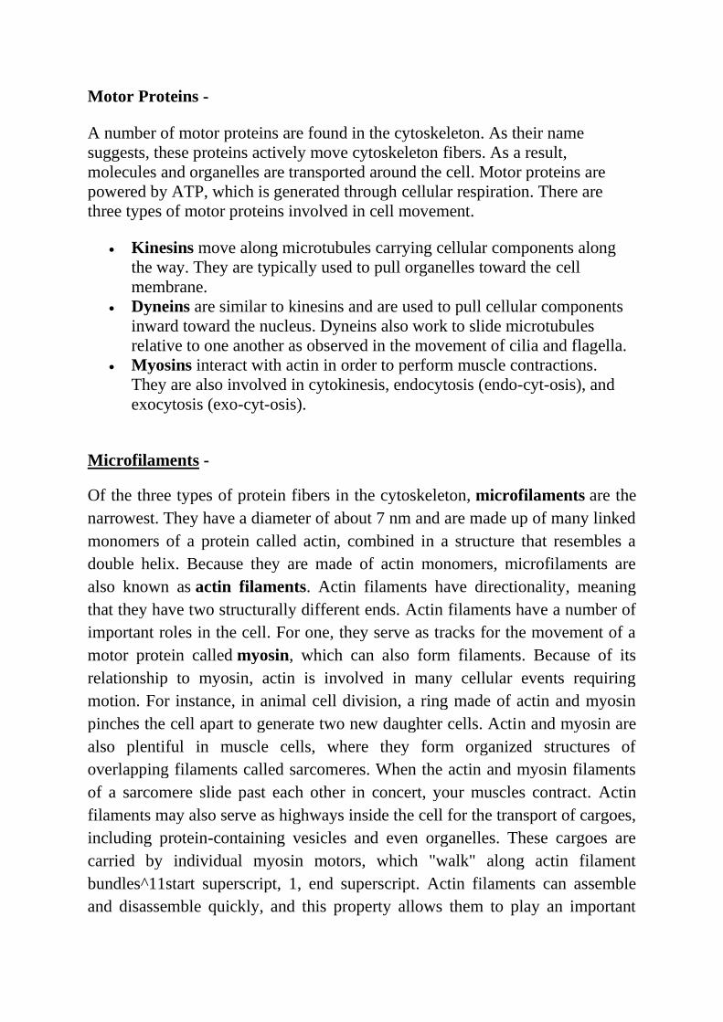
Microfilaments -
Of the three types of protein fibers in the cytoskeleton, microfilaments are the
narrowest. They have a diameter of about 7 nm and are made up of many linked
monomers of a protein called actin, combined in a structure that resembles a
double helix. Because they are made of actin monomers, microfilaments are
also known as actin filaments. Actin filaments have directionality, meaning
that they have two structurally different ends. Actin filaments have a number of
important roles in the cell. For one, they serve as tracks for the movement of a
motor protein called myosin, which can also form filaments. Because of its
relationship to myosin, actin is involved in many cellular events requiring
motion. For instance, in animal cell division, a ring made of actin and myosin
pinches the cell apart to generate two new daughter cells. Actin and myosin are
also plentiful in muscle cells, where they form organized structures of
overlapping filaments called sarcomeres. When the actin and myosin filaments
of a sarcomere slide past each other in concert, your muscles contract. Actin
filaments may also serve as highways inside the cell for the transport of cargoes,
including protein-containing vesicles and even organelles. These cargoes are
carried by individual myosin motors, which "walk" along actin filament
bundles^11start superscript, 1, end superscript. Actin filaments can assemble
and disassemble quickly, and this property allows them to play an important
role in cell motility (movement), such as the crawling of a white blood cell in
your immune system.
Finally, actin filaments play key structural roles in the cell. In most animal cells,
a network of actin filaments is found in the region of cytoplasm at the very edge
of the cell. This network, which is linked to the plasma membrane by special
connector proteins, gives the cell shape and structure.
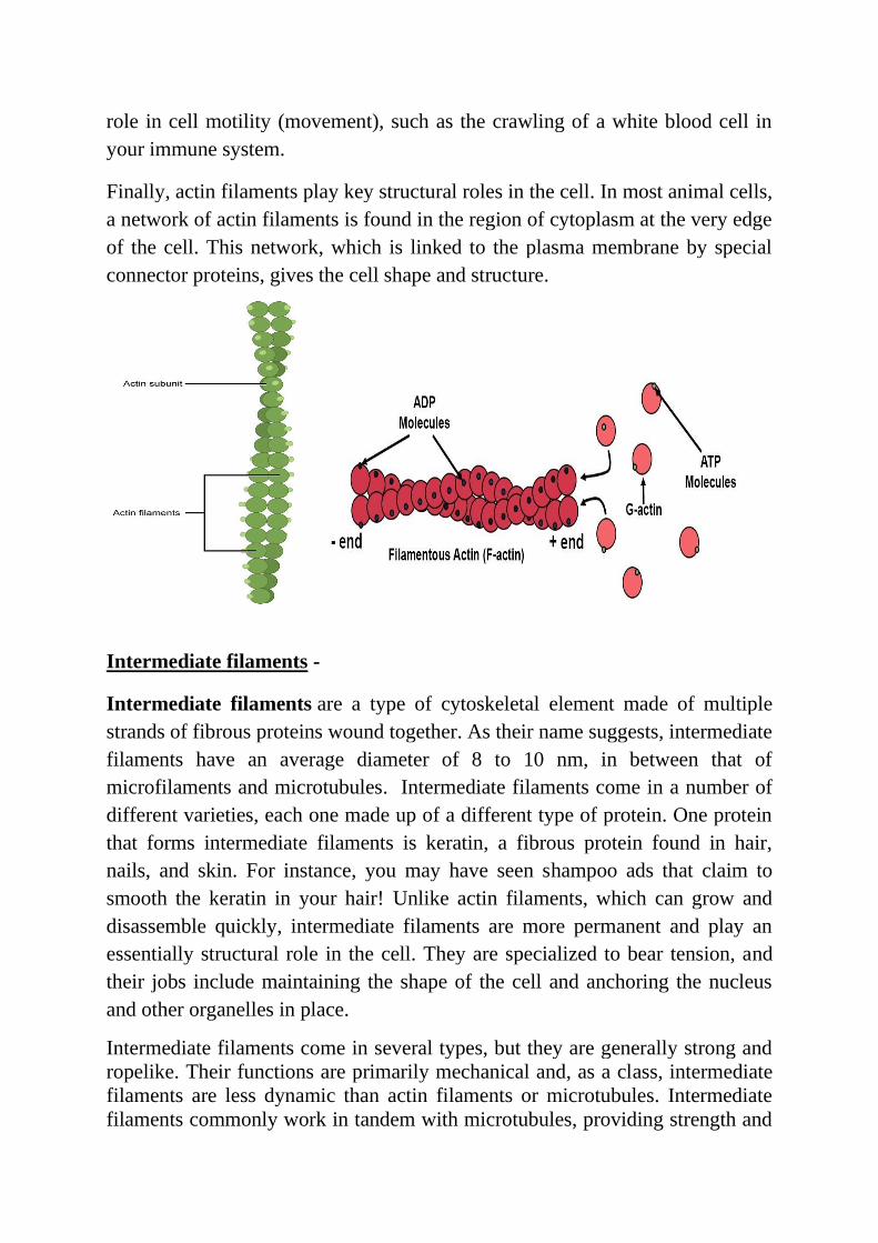
Intermediate filaments -
Intermediate filaments are a type of cytoskeletal element made of multiple
strands of fibrous proteins wound together. As their name suggests, intermediate
filaments have an average diameter of 8 to 10 nm, in between that of
microfilaments and microtubules. Intermediate filaments come in a number of
different varieties, each one made up of a different type of protein. One protein
that forms intermediate filaments is keratin, a fibrous protein found in hair,
nails, and skin. For instance, you may have seen shampoo ads that claim to
smooth the keratin in your hair! Unlike actin filaments, which can grow and
disassemble quickly, intermediate filaments are more permanent and play an
essentially structural role in the cell. They are specialized to bear tension, and
their jobs include maintaining the shape of the cell and anchoring the nucleus
and other organelles in place.
Intermediate filaments come in several types, but they are generally strong and
ropelike. Their functions are primarily mechanical and, as a class, intermediate
filaments are less dynamic than actin filaments or microtubules. Intermediate
filaments commonly work in tandem with microtubules, providing strength and
support for the fragile tubulin structures. All cells have intermediate filaments,
but the protein subunits of these structures vary. Some cells have multiple types
of intermediate filaments, and some intermediate filaments are associated with
specific cell types. For example, neurofilaments are found specifically in
neurons (most prominently in the long axons of these cells), desmin filaments
are found specifically in muscle cells, and keratins are found specifically in
epithelial cells. Other intermediate filaments are distributed more widely. For
example, vimentin filaments are found in a broad range of cell types and
frequently colocalize with microtubules. Similarly, lamins are found in all cell
types, where they form a meshwork that reinforces the inside of the nuclear
membrane. Note that intermediate filaments are not polar in the way that actin
or tubulin are.
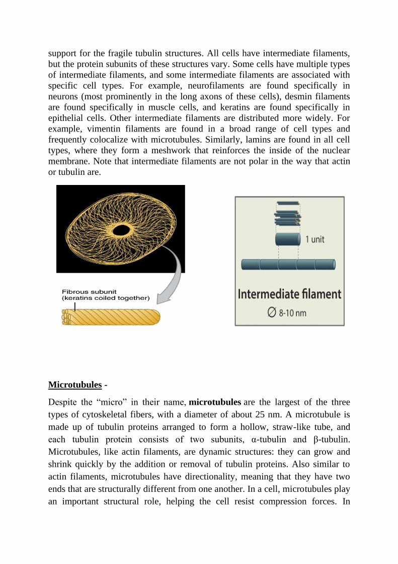
Microtubules -
Despite the “micro” in their name, microtubules are the largest of the three
types of cytoskeletal fibers, with a diameter of about 25 nm. A microtubule is
made up of tubulin proteins arranged to form a hollow, straw-like tube, and
each tubulin protein consists of two subunits, α-tubulin and β-tubulin.
Microtubules, like actin filaments, are dynamic structures: they can grow and
shrink quickly by the addition or removal of tubulin proteins. Also similar to
actin filaments, microtubules have directionality, meaning that they have two
ends that are structurally different from one another. In a cell, microtubules play
an important structural role, helping the cell resist compression forces. In

addition to providing structural support, microtubules play a variety of more
specialized roles in a cell. For instance, they provide tracks for motor proteins
called kinesins and dyneins, which transport vesicles and other cargoes around
the interior of the cell. During cell division, microtubules assemble into a
structure called the spindle, which pulls the chromosomes apart.
Tubulin contains two polypeptide subunits, and dimers of these subunits string
together to make long strands called protofilaments. Thirteen protofilaments
then come together to form the hollow, straw-shaped filaments of microtubules.
Microtubules are ever-changing, with reactions constantly adding and
subtracting tubulin dimers at both ends of the filament (Figure 1). The rates of
change at either end are not balanced — one end grows more rapidly and is
called the plus end, whereas the other end is known as the minus end. In cells,
the minus ends of microtubules are anchored in structures called microtubule
organizing centers (MTOCs). The primary MTOC in a cell is called
the centrosome, and it is usually located adjacent to the nucleus. Microtubules
tend to grow out from the centrosome to the plasma membrane. In nondividing
cells, microtubule networks radiate out from the centrosome to provide the basic
organization of the cytoplasm, including the positioning of organelles.
Flagella, Cilia, and Centrosomes -
Microtubules are also key components of three more specialized eukaryotic cell
structures: flagella, cilia and centrosomes. You may remember that our friends
the prokaryotes also have structures have flagella, which they use to move.
Don't get confused—the eukaryotic flagella we're about to discuss have pretty
much the same role, but a very different structure.
Flagella (singular, flagellum) are long, hair-like structures that extend from the
cell surface and are used to move an entire cell, such as a sperm. If a cell has
any flagella, it usually has one or just a few. Motile cilia (singular, cilium) are
similar, but are shorter and usually appear in large numbers on the cell surface.
When cells with motile cilia form tissues, the beating helps move materials
across the surface of the tissue. For example, the cilia of cells in your upper
respiratory system help move dust and particles out towards your nostrils.
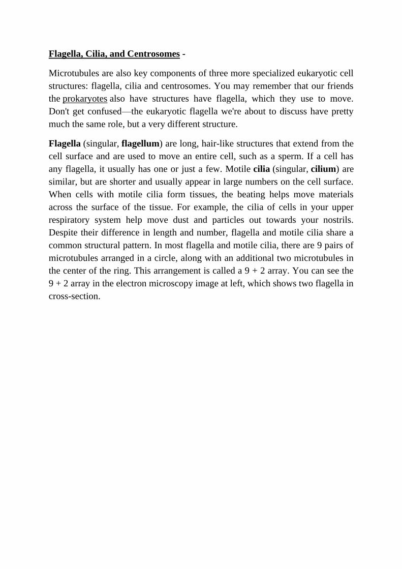
Despite their difference in length and number, flagella and motile cilia share a
common structural pattern. In most flagella and motile cilia, there are 9 pairs of
microtubules arranged in a circle, along with an additional two microtubules in
the center of the ring. This arrangement is called a 9 + 2 array. You can see the
9 + 2 array in the electron microscopy image at left, which shows two flagella in
cross-section.
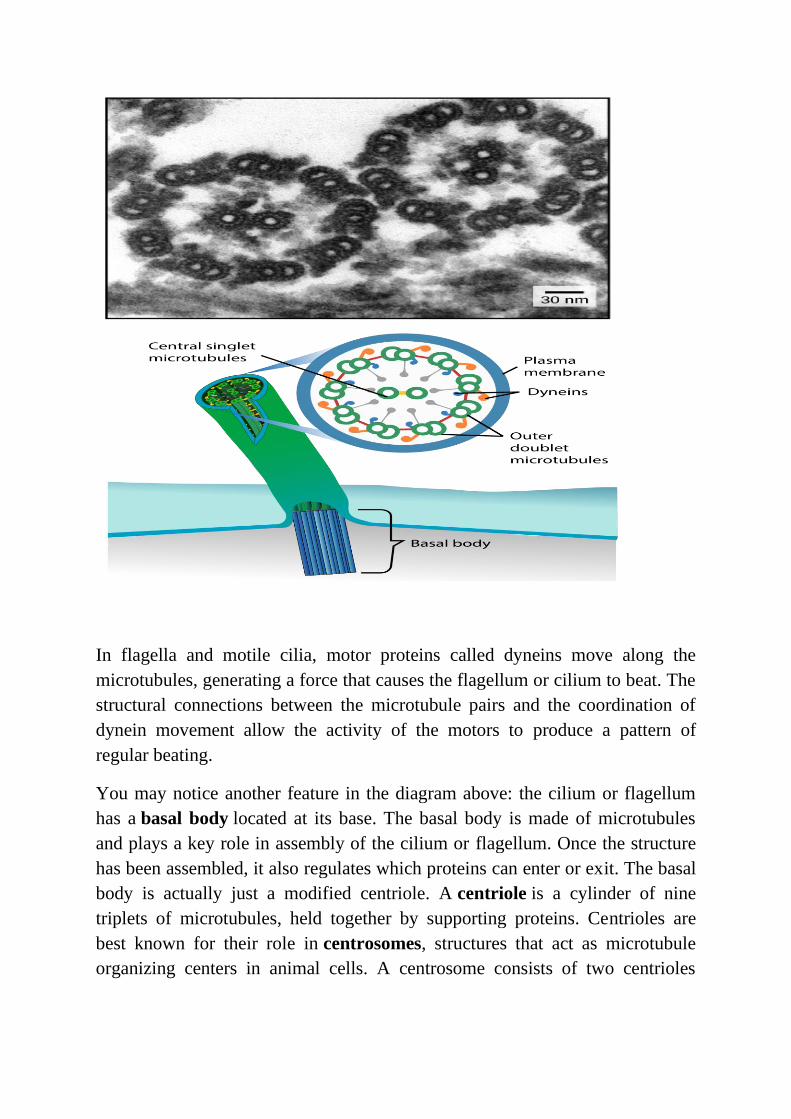
In flagella and motile cilia, motor proteins called dyneins move along the
microtubules, generating a force that causes the flagellum or cilium to beat. The
structural connections between the microtubule pairs and the coordination of
dynein movement allow the activity of the motors to produce a pattern of
regular beating.
You may notice another feature in the diagram above: the cilium or flagellum
has a basal body located at its base. The basal body is made of microtubules
and plays a key role in assembly of the cilium or flagellum. Once the structure
has been assembled, it also regulates which proteins can enter or exit. The basal
body is actually just a modified centriole. A centriole is a cylinder of nine
triplets of microtubules, held together by supporting proteins. Centrioles are
best known for their role in centrosomes, structures that act as microtubule
organizing centers in animal cells. A centrosome consists of two centrioles

oriented at right angles to each other, surrounded by a mass of "pericentriolar
material," which provides anchoring sites for microtubules.
The centrosome is duplicated before a cell divides, and the paired centrosomes
seem to play a role in organizing the microtubules that separate chromosomes
during cell division. However, the exact function of the centrioles in this
process still isn’t clear. Cells with their centrosome removed can still divide,
and plant cells, which lack centrosomes, divide just fine.
Accessory proteins organize filaments into higher-order structures -
Crosslinking of the filaments by specific motors or multivalent binding proteins
(accessory proteins) increases stability and forms higher-order structures. Such
organization facilitates generation of long-term contractile forces and occasionally
support compressive forces while being dynamic. These structures are connected
across cells through junctions and hence facilitate mechanotransduction and
cumulative response at a tissue- or organ-level (see the lower panel in the figure
below and “Mediators of mechanotransduction” for details on junctions).
Accessory proteins are a critical part of the signaling network that integrates extra-
and intracellular signals (e.g. force, ions etc.) with the cytoskeleton assembly
module(s). These can be specific for certain types of filaments. E.g., fimbrin binds
only actin filaments, while others like plectin are non-specific. Accessory factors
can also help regulate the stability, mechanical properties, and force production for
the individual filaments within the larger structure. For example, fascin crosslinks
actin filaments into rigid bundles that have mechanical strength for generating
protrusive force, while filamin cross links the actin filaments into gel-like networks
that are flexible and produce less force.
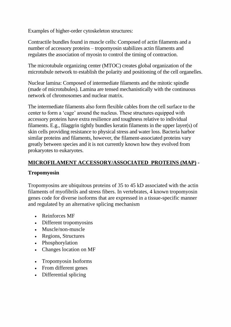
Examples of higher-order cytoskeleton structures:
Contractile bundles found in muscle cells: Composed of actin filaments and a
number of accessory proteins – tropomyosin stabilizes actin filaments and
regulates the association of myosin to control the timing of contraction.
The microtubule organizing center (MTOC) creates global organization of the
microtubule network to establish the polarity and positioning of the cell organelles.
Nuclear lamina: Composed of intermediate filaments and the mitotic spindle
(made of microtubules). Lamina are tensed mechanistically with the continuous
network of chromosomes and nuclear matrix.
The intermediate filaments also form flexible cables from the cell surface to the
center to form a ‘cage’ around the nucleus. These structures equipped with
accessory proteins have extra resilience and toughness relative to individual
filaments. E.g., filaggrin tightly bundles keratin filaments in the upper layer(s) of
skin cells providing resistance to physical stress and water loss. Bacteria harbor
similar proteins and filaments, however, the filament-associated proteins vary
greatly between species and it is not currently known how they evolved from
prokaryotes to eukaryotes.
MICROFILAMENT ACCESSORY/ASSOCIATED PROTEINS (MAP) -
Tropomyosin
Tropomyosins are ubiquitous proteins of 35 to 45 kD associated with the actin
filaments of myofibrils and stress fibers. In vertebrates, 4 known tropomyosin
genes code for diverse isoforms that are expressed in a tissue-specific manner
and regulated by an alternative splicing mechanism
• Reinforces MF
• Different tropomyosins
• Muscle/non-muscle
• Regions, Structures
• Phosphorylation
• Changes location on MF
• Tropomyosin Isoforms
• From different genes
• Differential splicing

Actin-related proteins (Arp2/3)
The Arp2/3 protein complex has been implicated in the control of actin
polymerization in cells.
• Arp2/3 protein complex
• control of polymerization
• lamellipodia localization
• human complex has 7 subunits
• ARP2, ARP3, ARC41, ARC34, ARC21, ARC20, and ARC16
• Listeria monocytogenes
• Induce actin polymerization by Arp2/3 protein complex at Listeria
surface
ACTIN MOTORS
Myosin I
• All cells
• One head domain
• Binds actin
Myosin II
• Muscle myosin
• Also other cells
• Dimer, 2 heads
• Bind to each other to form myosin filament
• Thick filament
MUSCLE -
• Striated
• Skeletal, cardiac
• sarcomeres
• Non-striated
• Smooth
Muscle Contraction -
• sliding of filaments actin against myosin
• troponin and tropomyosin
• contraction of skeletal and cardiac muscle regulated by Ca2+ flux
• smooth muscle cells and non-muscle cells
• contraction same mechanism
• contractile units smaller less highly ordered
• activity and state of assembly controlled by Ca2+ -regulated
phosphorylation of a myosin
MICROFILAMENT BINDING PROTEINS -
Cytochalasin D
• Fungal metabolite
• Binds barbed end
• inhibits polymerization and depolymerization
• Cell permeant
• Active in low micromolar
Phalloidin
• Fungal metabolite
• Binds and stabilizes F-actin
• Not cell permeant
• Fluorescent derivatives are used to stain F-actin in situ and in vitro
Jasplakinolide
• Sea sponge metabolite
• Binds and stabilizes F-actin competitively with phalloidin
• Causes nucleation
• Cell permeant
• Nanomolar Kd for F-actin
Latrunculin
• Sea sponge metabolite
• Binds monomeric actin
• inhibits polymerization
• Cell permeant
• Active at low nanomolar
MICROFILAMENT ACCESSORY/ASSOCIATED PROTEINS (MAP) –
In cell biology, microtubule-associated proteins (MAPs) are proteins that
interact with the microtubules of the cellular cytoskeleton.
The numerous identified MAPs have been largely divided into two categories:
Type I including MAP1 proteins and type II including MAP2, MAP4 and tau
proteins.
Type I: MAP1
MAP1a (MAP1A) and MAP1b (MAP1B) are the two major members of the
MAP1 family. They bind to microtubules through charge interactions, a
different mechanism to many other MAPs. While the C termini of these MAPs
bind the microtubules, the N termini bind other parts of the cytoskeleton or
the plasma membrane to control spacing of the microtubule within the cell.
Members of the MAP1 family are found in the axons and dendrites of nerve
cells.
Type II: MAP2, MAP4, and tau
Type II MAPs are found exclusively in nerve cells in mammals. These are the
most well studied MAPs—MAP2 and tau (MAPT)—which participate in
determining the structure of different parts of nerve cells, with MAP2 being
found mostly in dendrites and tau in the axon. These proteins have
a conserved C-terminal microtubule-binding domain and variable N-
terminal domains projecting outwards, probably interacting with other proteins.
MAP2 and tau stabilize microtubules, and thus shift the reaction kinetics in
favor of addition of new subunits, accelerating microtubule growth. Both MAP2
and tau have been shown to stabilize microtubules by binding to the outer
surface of the microtubule protofilaments. A single study has suggested that
MAP2 and tau bind on the inner microtubule surface on the same site in tubulin
monomers as the drug Taxol, which is used in treating cancer, but this study has
not been confirmed. MAP2 binds in a cooperative manner, with many MAP2
proteins binding a single microtubule to promote stabilization. Tau has the
additional function of facilitating bundling of microtubules within the nerve
cell.
The function of tau has been linked to the neurological condition Alzheimer's
disease. In the nervous tissue of Alzheimer's patients, tau forms abnormal
aggregates. This aggregated tau is often severely modified, most commonly
through hyperphosphorylation. As described above, phosphorylation of MAPs
causes them to detach from microtubules. Thus, the hyperphosphorylation of tau
leads to massive detachment, which in turn greatly reduces the stability of
microtubules in nerve cells. This increase in microtubule instability may be one
of the main causes of the symptoms of Alzheimer's disease.
In contrast to the MAPs described above, MAP4 (MAP4) is not confined to just
nerve cells, but rather can be found in nearly all types of cells. Like MAP2 and
tau, MAP4 is responsible for stabilization of microtubules. MAP4 has also been
linked to the process of cell division.
Other MAPs, and naming issues
Besides the classic MAP groups, novel MAPs have been identified that bind the
length of the microtubules. These include STOP (also known as MAP6),
and ensconsin (also known as MAP7).
In addition, plus end tracking proteins, which bind to the very tip of growing
microtubules, have also been identified. These
include EB1, EB2, EB3, p150Glued, Dynamitin, Lis1, CLIP170, CLIP115, CL
ASP1, and CLASP2.
Another MAP whose function has been investigated during cell division is
known as XMAP215 (the "X" stands for Xenopus). XMAP215 has generally
been linked to microtubule stabilization. During mitosis the dynamic
instability of microtubules has been observed to rise approximately tenfold.
This is partly due to phosphorylation of XMAP215, which makes catastrophes
(rapid depolymerization of microtubules) more likely. In this way the
phosphorylation of MAPs plays a role in mitosis.
There are many other proteins which affect microtubule behavior, such
as catastrophin, which destabilizes microtubules, katanin, which severs them,
and a number of motor proteins that transport vesicles along them. Certain
motor proteins were originally designated as MAPs before it was found that
they utilized ATP hydrolysis to transport cargo. In general, all these proteins are
not considered "MAPs" because they do not bind directly to tubulin monomers,
a defining characteristic of MAPs. MAPs bind directly to microtubules to
stabilize or destabilize them and link them to various cellular components
including other microtubules.