Cytoskeleton Dependent Mobility Dynamics of FcγRIIA ... - MDPI
20
Citation: Palankar, R.; Sachs, L.; Wesche, J.; Greinacher, A. Cytoskeleton Dependent Mobility Dynamics of FcγRIIA Facilitates Platelet Haptotaxis and Capture of Opsonized Bacteria. Cells 2022, 11, 1615. https://doi.org/10.3390/ cells11101615 Academic Editor: Francisco Rivero Received: 21 April 2022 Accepted: 10 May 2022 Published: 11 May 2022 Publisher’s Note: MDPI stays neutral with regard to jurisdictional claims in published maps and institutional affil- iations. Copyright: © 2022 by the authors. Licensee MDPI, Basel, Switzerland. This article is an open access article distributed under the terms and conditions of the Creative Commons Attribution (CC BY) license (https:// creativecommons.org/licenses/by/ 4.0/). cells Article Cytoskeleton Dependent Mobility Dynamics of FcγRIIA Facilitates Platelet Haptotaxis and Capture of Opsonized Bacteria Raghavendra Palankar *, Laura Sachs , Jan Wesche and Andreas Greinacher Institute for Transfusion Medicine, University Medicine Greifswald, 17489 Greifswald, Germany; [email protected] (L.S.); [email protected] (J.W.); [email protected] (A.G.) * Correspondence: [email protected] Abstract: Platelet adhesion and spreading at the sites of vascular injury is vital to hemostasis. As an integral part of the innate immune system, platelets interact with opsonized bacterial pathogens through FcγRIIA and contribute to host defense. As mechanoscavangers, platelets actively migrate and capture bacteria via cytoskeleton-rich, dynamic structures, such as filopodia and lamellipodia. However, the role of human platelet FcγRIIA in cytoskeleton-dependent interaction with opsonized bacteria is not well understood. To decipher this, we used a reductionist approach with well- defined micropatterns functionalized with immunoglobulins mimicking immune complexes at planar interfaces and bacteriamimetic microbeads. By specifically blocking of FcγRIIA and selective disruption of the platelet cytoskeleton, we show that both functional FcγRIIA and cytoskeleton are necessary for human platelet adhesion and haptotaxis. The direct link between FcγRIIA and the cytoskeleton is further explored by single-particle tracking. We then demonstrate the relevance of cytoskeleton-dependent differential mobilities of FcγRIIA on bacteria opsonized with the chemokine platelet factor 4 (PF4) and patient-derived anti-PF4/polyanion IgG. Our data suggest that efficient capture of opsonized bacteria during host-defense is governed by mobility dynamics of FcγRIIA on filopodia and lamellipodia, and the cytoskeleton plays an essential role in platelet morphodynamics at biological interfaces that display immune complexes. Keywords: platelets; FcγRIIA; immune complex; cytoskeleton; platelet factor 4; bacteria; micropatterns; quantum dots 1. Introduction Platelets are anucleate, discoidal multifunctional cellular fragments (2–5 μm in diam- eter) generated from the cytoplasm of nucleated, polypoid megakaryocytes in the bone marrow and released into the blood circulation [1]. In a healthy human, peripheral blood contains about 150–400 10 9 /L platelets. Platelets rapidly adhere and aggregate at the sites of vascular injury and seal the lesions by forming hemostatic plugs. In addition, under pathophysiological conditions, undesirable activation of platelets leads to thrombosis. Platelets thus play a fundamental role in both hemostasis and thrombosis. Furthermore, as an integral part of the innate immune system, platelets cross-talk with immune cells, such as neutrophils and macrophages, by secreting proinflammatory cytokines [2]. Be- yond these, platelets interact with diverse pathogens, such as viruses, bacteria, fungi, and protozoa, in a complex but delicately balanced manner, that may result in complications or in host defense [3–6]. For example, platelets interact directly and indirectly through their surface receptors with Gram-negative and Gram-positive bacteria and their secreted metabolites and proteins, leading to platelet activation [7–9]. However, human platelets also effectively bridge adaptive and innate immune systems to achieve host defense against bacterial pathogens [10–12]. These interactions and platelet responses to external biochemi- cal and biophysical stimuli depend on specific ligand-receptor recognition, sensing, and Cells 2022, 11, 1615. https://doi.org/10.3390/cells11101615 https://www.mdpi.com/journal/cells
-
Upload
khangminh22 -
Category
Documents
-
view
3 -
download
0
Transcript of Cytoskeleton Dependent Mobility Dynamics of FcγRIIA ... - MDPI
Citation: Palankar, R.; Sachs, L.;
Wesche, J.; Greinacher, A.
Cytoskeleton Dependent Mobility
Dynamics of FcγRIIA Facilitates
Platelet Haptotaxis and Capture of
Opsonized Bacteria. Cells 2022, 11,
1615. https://doi.org/10.3390/
cells11101615
Academic Editor: Francisco Rivero
Received: 21 April 2022
Accepted: 10 May 2022
Published: 11 May 2022
Publisher’s Note: MDPI stays neutral
with regard to jurisdictional claims in
published maps and institutional affil-
iations.
Copyright: © 2022 by the authors.
Licensee MDPI, Basel, Switzerland.
This article is an open access article
distributed under the terms and
conditions of the Creative Commons
Attribution (CC BY) license (https://
creativecommons.org/licenses/by/
4.0/).
cells
Article
Cytoskeleton Dependent Mobility Dynamics of FcγRIIAFacilitates Platelet Haptotaxis and Capture of Opsonized BacteriaRaghavendra Palankar *, Laura Sachs , Jan Wesche and Andreas Greinacher
Institute for Transfusion Medicine, University Medicine Greifswald, 17489 Greifswald, Germany;[email protected] (L.S.); [email protected] (J.W.);[email protected] (A.G.)* Correspondence: [email protected]
Abstract: Platelet adhesion and spreading at the sites of vascular injury is vital to hemostasis. Asan integral part of the innate immune system, platelets interact with opsonized bacterial pathogensthrough FcγRIIA and contribute to host defense. As mechanoscavangers, platelets actively migrateand capture bacteria via cytoskeleton-rich, dynamic structures, such as filopodia and lamellipodia.However, the role of human platelet FcγRIIA in cytoskeleton-dependent interaction with opsonizedbacteria is not well understood. To decipher this, we used a reductionist approach with well-defined micropatterns functionalized with immunoglobulins mimicking immune complexes atplanar interfaces and bacteriamimetic microbeads. By specifically blocking of FcγRIIA and selectivedisruption of the platelet cytoskeleton, we show that both functional FcγRIIA and cytoskeleton arenecessary for human platelet adhesion and haptotaxis. The direct link between FcγRIIA and thecytoskeleton is further explored by single-particle tracking. We then demonstrate the relevance ofcytoskeleton-dependent differential mobilities of FcγRIIA on bacteria opsonized with the chemokineplatelet factor 4 (PF4) and patient-derived anti-PF4/polyanion IgG. Our data suggest that efficientcapture of opsonized bacteria during host-defense is governed by mobility dynamics of FcγRIIA onfilopodia and lamellipodia, and the cytoskeleton plays an essential role in platelet morphodynamicsat biological interfaces that display immune complexes.
Keywords: platelets; FcγRIIA; immune complex; cytoskeleton; platelet factor 4; bacteria; micropatterns;quantum dots
1. Introduction
Platelets are anucleate, discoidal multifunctional cellular fragments (2–5 µm in diam-eter) generated from the cytoplasm of nucleated, polypoid megakaryocytes in the bonemarrow and released into the blood circulation [1]. In a healthy human, peripheral bloodcontains about 150–400 109/L platelets. Platelets rapidly adhere and aggregate at the sitesof vascular injury and seal the lesions by forming hemostatic plugs. In addition, underpathophysiological conditions, undesirable activation of platelets leads to thrombosis.Platelets thus play a fundamental role in both hemostasis and thrombosis. Furthermore,as an integral part of the innate immune system, platelets cross-talk with immune cells,such as neutrophils and macrophages, by secreting proinflammatory cytokines [2]. Be-yond these, platelets interact with diverse pathogens, such as viruses, bacteria, fungi, andprotozoa, in a complex but delicately balanced manner, that may result in complicationsor in host defense [3–6]. For example, platelets interact directly and indirectly throughtheir surface receptors with Gram-negative and Gram-positive bacteria and their secretedmetabolites and proteins, leading to platelet activation [7–9]. However, human plateletsalso effectively bridge adaptive and innate immune systems to achieve host defense againstbacterial pathogens [10–12]. These interactions and platelet responses to external biochemi-cal and biophysical stimuli depend on specific ligand-receptor recognition, sensing, and
Cells 2022, 11, 1615. https://doi.org/10.3390/cells11101615 https://www.mdpi.com/journal/cells

Cells 2022, 11, 1615 2 of 20
signaling processes that result in platelet tethering, activation, adhesion, and spreading.The human platelet receptor FcγRIIA has attracted significant attention in this regard dueto its relevance to fundamental platelet function in human health and disease and as atherapeutic target.
The FcγRIIA is a type 1 transmembrane glycoprotein of ≈40 kDa with a cytoplas-mic region containing an immunoreceptor tyrosine-based activation motif (ITAM) withtwo tyrosines that undergo phosphorylation. Present at numbers ranging from 900 up to5000 per platelet, FcγRIIA is the only FcγR expressed in human platelets, thereby makingplatelets the richest source of FcγRIIA in the human body [13,14]. While FcγRIIA has a lowaffinity for the constant region (Fc, fragment crystallizable) of monomeric IgG, immunecomplexes comprising IgG and IgG-opsonized targets such as bacteria are recognizedwith high avidity. Mechanistically, this is achieved by specific recognition of immunoglob-ulin (IgG) opsonized bacteria by the FcγRIIA (also called CD32a). During interactionwith IgG opsonized bacteria, crosslinking of FcγRIIA triggers platelet activation. Thisresults in extensive platelet spreading due to cytoskeletal rearrangement and subsequentdirected release of alpha-granules containing bactericidal substances [15]. Beyond hostdefense, platelet FcγRIIA mediated platelet activation by immune complexes betweenanti-platelet factor 4 (PF4) IgG, and PF4 is known to induce the release of neutrophil extra-cellular traps (NETs) from neutrophil granulocytes in heparin-induced thrombocytopenia(HIT) [16]. More recently, platelet FcγRIIA was shown to play a vital role in inducingvaccine-induced immune thrombotic thrombocytopenia (VITT) by anti-PF4 antibodies insome individuals after vaccination with the adenoviral vector vaccines ChAdOx1 nCoV-19(Oxford–AstraZeneca) and Ad26.COV2.S (Johnson & Johnson–Janssen) [17]. Similar toHIT, anti-PF4 antibodies have been implicated in releasing NETs from neutrophils by acti-vated platelets in VITT patients [18]. Thus, platelet FcγRIIA plays a central role in severalpathophysiological functions and interactions of platelets in the vascular system.
In platelet biology, one of the least understood phenomena is the ability of platelets toengage in the directional migration towards chemo- and hapotactic cues [19,20]. Recently,it has been shown that murine platelets exhibit cytoskeleton-dependent motility and act asmechanoscavangers of bacteria [21,22]. However, so far, the role of diffusion and mobilitydynamics of FcγRIIA on human platelets during ligand sensing and its dependency oncytoskeletal integrity impacting platelet haptotaxis is not entirely clear. To assess this,we developed a reductionist approach based on micropatterning of ligands, specificallyrecognized by platelet FcγRIIA. Using micropatterned arrays functionalized with IgG on2D planar surfaces and on microbeads that mimic immune complex presenting immunecomplexes, we demonstrate that platelet FcγRIIA mediated platelet haptotaxis is impactedby cytoskeletal integrity. In addition, taking advantage of single-particle tracking usingluminescent quantum dots labeling of FcγRIIA, we further show differential receptormobility dynamics are vital to the interaction and capture of bacteria opsonized withPF4 and anti-PF4/polyanion IgG (anti-PF4/P) from HIT patients. Our data suggest thatplatelets morphodynamic changes during platelet haptotaxis on immune complexes atdifferent bio-interfaces are strongly dependent on differential mobilities of FcγRIIA andcytoskeletal integrity.
2. Materials and Methods2.1. Reagents and Chemicals
Poly(ethylene glycol) methyl ether thiol (Mn = 800) (Cat.No. 729,108 Sigma-Aldrich(Seelze, Germany). 3-[Methoxy(polyethyleneoxy)propyl]trimethoxysilane; 90%, 6–9 PE-units (Cat.No. AB111226, ABCR GmbH & Co., Karlsruhe, Baden-Württemberg, Ger-many). Bovine serum albumin, Fraction V (Cat.No. BP1605-100, Fisher Scientific, Schwerte,North Rhine-Westphalia, Germany). BSA-fluorescein isothiocyanate conjugate (FITC)(Cat.No. A23015, ThermoFisher Scientific, Bremen, Germany). Water-soluble QdotTM
525 ITKTM Amino (PEG) Quantum Dots (Cat. No. Q21541MP, Thermo Fisher Scientific,Darmstadt, Hesse, Germany). Monoclonal anti-human FcγRIIA Fab (Clone 7.3) and isotype

Cells 2022, 11, 1615 3 of 20
control mouse IgG1 κ (MOPC31C)(Fab) from Ancell Corporation (Bayport, MN, USA).Bis[sulfosuccinimidyl] suberate (BS3), Dynabeads® MyOne™ carboxylic acid, 3,3′-Dithiobis(sulfosuccinimidylpropionate) (DTSSP), (1-ethyl-3-(3-dimethylaminopropyl)carbodiimidehydrochloride) (EDC), N-hydroxysuccinimide (NHS), Fluo-4 AM cell permeant calcium(Ca2+) indicator, goat anti-human IgG secondary antibody conjugated to Alexa Fluor 555,goat anti-human IgG secondary antibody conjugated to FITC were purchased from Invitro-gen GmbH (Darmstadt, Germany) and were used according to manufactures instructions.
2.2. Preparation and Functionalization of Micropatterned Arrays
Electron beam lithography (EBL) was used to prepare micropatterned arrays on glasscoverslips. Briefly, coverslips were cleaned using 1:1:5 solution of ammonium hydroxide:hydrogen peroxide: deionized water at 70 ◦C for 10 min and dried under nitrogen streamand treated for 20 min in UV/Ozone ProCleaner Plus (BioForce Nanosciences Inc., Ames,IA, USA). Cleaned coverslips were then sputter-coated with chromium (Q150T ES, QuorumTechnologies, Lewes, UK), followed by gold to create a conductive layer. To preventnonspecific interactions beyond functionalized micropatterns, gold-coated coverslips werefirst functionalized with poly(ethylene glycol) methyl ether thiol (Mn = 800) (Cat.No.729,108 Sigma-Aldrich) at a concentration of 5 mg/mL in deionized water for 24 h to forma self-assembled monolayer. To create micropatterned microarrays, 5% v/v bovine serumalbumin, Fraction V (Cat.No. BP1605-100, Fisher Scientific) in deionized water was spin-coated (DELTA6 RC TT, Süss Micro Tec) for 40 s at 2000 rpm on glass coverslips coated witha self-assembled monolayer of PEG-thiol. Electron beam lithography was performed in aZeiss Supra 40 VP scanning electron microscope (SEM) equipped with an ELPHY QuantumEBL system (Raith GmbH). Different electron beam dosages were tested starting from 10up to 100 µC/cm2 at increments of 10 µC/cm2. The final optimal electron beam dosage of80 µC/cm2 was chosen to produce high fidelity BSA micropatterns in a micropatterns ofdifferent shapes (square and circular shape with varying sizes and inter-pattern distances)were created to assess whether platelets are able to address morphodynamic changes duringadhesion and spreading process specifically. The micropatterns used for functionalizationwere patterned within a rectangular area of 1 mm× 5 mm in dimension to minimize samplevolume during adhesion assays. Electron beam exposed BSA-coated glass coverslips weredeveloped in deionized water for 10 min, dried under a stream of nitrogen, and stored inairtight containers at 4 ◦C until further use. BSA-based micropatterns were used within72 h of preparation.
2.3. Preparation of Heat-Aggregated Human IgG and Functionalization of Micropatternsand Microbeads
Heat-aggregated human IgG (Agg-IgG) was prepared by heating purified humanIgG (Jackson ImmunoResearch Laboratories, Inc. Baltimore Pike, West Grove, PA, USA)at 5 mg/mL in PBS for 25 min at 63 ◦C in a water bath, followed by centrifugation at10,000× g for 5 min. The supernatant was collected, stored at 4 ◦C, and used within24 h of preparation. Protein concentration was determined by bicinchoninic acid (BCA)protein assay kit using BSA as standard (Sigma-Aldrich Chemie GmbH, Munich, Germany).Agg-IgG was immobilized on BSA micropatterns and carboxylic microbeads using single-step conjugation chemistry through amine-reactive N-hydroxysulfosuccinimide (sulfo-NHS) ester using homobifunctional 3,3′-DTSSP and via EDC following manufacturersprotocols, respectively.
2.4. Preparation and Functionalization of Micropatterned Arrays with Live E. coli and PlateletAdhesion Assay
Live bacteria were immobilized on micropatterns using 1-Ethyl-3-(3-dimethylaminopropyl)-carbodiimide (EDC) and N-hydroxysuccinimide (NHS) chemistry, as previously described [23]Briefly, E. coli KPM121 (∆waaA) grown overnight in LB media were washed thrice in PBSand were suspended in ice-cold PBS (OD of 0.8). Bacteria immobilized on micropatternswere first incubated with 20 µg mL−1 PF4 for 30 min at 4 ◦C, and unbound excess was

Cells 2022, 11, 1615 4 of 20
gently rinsed in cold PBS, followed by incubation for 30 min with 10 µg mL−1 anti-PF4/PIgG purified from HIT patient sera. PF4 and anti-PF4/P IgG opsonized bacteria were thenpipetted onto BSA micropatterns pretreated and derivatized with EDC (1 mg mL−1) andNHS (0.1 mg mL−1), and incubated for 30 min at 4 ◦C on a horizontal shaker. Non-adherentbacteria were removed by gently rinsing the micropatterns in cold PBS. The binding ofanti-PF4/P IgG was verified by immunofluorescence using goat anti-human IgG secondaryantibodies conjugated to Alexa Fluor® 488. Platelet adhesion assay was performed at 37 ◦Cfor 30 min, and samples were fixed 2% v/v paraformaldehyde/PBS, pH 7.5 for 30 min,followed by rinsing quenching for 5 min in 30 mM glycine/PBS, pH 7.5.
2.5. Platelet Preparation, Adhesion Assays, Ca2+ Mobilization, and Fluorescence Microscopy
Platelets were isolated as described in [24]. All adhesion assays were performed withwashed platelets in modified Tyrodes buffer supplemented with albumin, glucose, MgCl2,and CaCl2. Glass coverslips with functionalized micropatterned arrays were mountedon the self-adhesive underside of bottomless chamber slides (sticky-Slide VI 0.4, Ibidi,Munich, Germany). For experiments evaluating Ca2+ transient imaging in platelets, washedplatelets were incubated with 5 µM Fluo-4 AM in the dark for 30 min at room temperature.Excess Fluo-4 AM was removed by centrifuging the platelets at 750 g for 5 min. Plateletadhesion assays and fluorescence imaging of Ca2+ mobilization transients during plateletadhesion assays were performed by exiting Fluo-4 AM with a 488 nm laser on a LeicaSP5 confocal laser-scanning microscope. For inhibition experiments, platelets (50 µL at10,000 platelets/µL) were pretreated for 30 min at 37 ◦C with the cytoskeletal inhibitorscytochalasin D at 25 µM, blebbistatin at 10 µM, and the receptor blockers anti-FcγRIIAmAb (clone IV.3, purified from hybridoma) at 10 µg/mL, and abciximab at 10 µg/mL. Forimmunofluorescence microscopy, platelets were fixed in 2% v/v paraformaldehyde/PBS,pH 7.5, for 30 min, then the excess was rinsed in PBS and its quenching for 5 min in 30 mMglycine/PBS, pH 7.5. Actin cytoskeleton was labeled with 10 pM Phalloidin ATTO 550.Confocal fluorescence images from six different regions of interest (ROIs) were acquiredfor each experimental condition, with experiments performed with platelets isolated fromsix healthy donors to quantify platelet adhesion on micropatterns. Platelet adhesion wascalculated as percent micropatterned area coverage.
2.6. Analysis of Platelet Filopodia Number and Length
The analysis of fixed platelets labeled for F-actin with fluorescent phalloidin wasperformed on ImageJ using the plugin Filopodyan [25]. Briefly, images of platelets labeledfor F-actin with fluorescent phalloidin were imported into ImageJ. Image segmentationwas performed using the Triangle algorithm to detect object pixels that produce a weakpeak in the histogram, followed by filtering false-positive events. The final output filescontain a descriptive matrix of platelet filopodia numbers and length analysis.
2.7. Quantification of Platelet Spread Area and Platelet Morphodynamics
Single platelet spread area on micropatterns and microbead arrays was quantifiedby Time-lapse. Images of platelets loaded with Fluo-4 AM spreading on micropatternswere acquired for 10–15 min at an interval of 30 s on a Leica SP5 confocal laser-scanningmicroscope. Image analysis was performed using image processing ADAPT -AutomatedDetection and Analysis of ProTrusions plugin for ImageJ [26]. Time-series images ofthe Fluo-4 AM channel were then imported into ImageJ running the ADAPT plugin. AGaussian filter was applied to minimize the noise, followed by grey-level thresholding tocreate a binary image. The platelet membrane boundary was taken as pixels borderingsegmented regions. The resulting segmentation map was used as the seed for the region-growing algorithm in the next time point of the time-series frame. A change in platelet areaover time was plotted from the final output data matrix.

Cells 2022, 11, 1615 5 of 20
2.8. SEM Analysis
Platelets were fixed with 2.5% glutaraldehyde in PBS for 20 min, followed by post-fixation with 1% osmium tetroxide (OsO4) in PBS for 20 min. Next, platelets were dehy-drated in graded aqueous ethanol solutions from 30% to 96% (each for 5 min), and thenin 100% ethanol (three steps of 5 min each). Samples were dried in Polaron Critical PointDryer (Quorum Technologies Ltd., Kent, UK) and coated with a thin layer of gold in sputtercoater images were acquired in Zeiss Supra 40 VP SEM.
2.9. Preparation of Monovalent QD and Monoclonal anti-Human FcγRIIA Fab Conjugate
Covalent conjugation of monoclonal mouse anti-human FcγRIIA Fab fragments toQD 525 PEG-amine using the homobifunctional amine to amine crosslinker bis [sulfosuc-cinimidyl] suberate (BS3) was performed according to manufactures protocol. Briefly, toprepare monovalent QDs conjugated to Fab fragment (QD to Fab ratio 1:1), 100 µL of 80 nMQD 525 PEG-amine in 50 mM borate buffer, pH 8.3 were washed and transferred to PBS,pH 7.4, followed by activation in the presence of 100 µM BS3. Excess BS3 was removedby passing the activated QD through the desalting column. For conjugation, 80 nM ofactivated QD in PBS, pH 7.4 were incubated with 80 nM monoclonal anti-human FcγRIIAFab fragments for 2 h at 4 ◦C. This was followed by incubation with 1 mg/mL BSA to blockall available surface reactive groups and reduce non-specific interactions. The remainingfree functional groups were quenched with 5 mM glycine, followed by centrifugal washingusing ultrafiltration (100 kD). The QD-Fab conjugate was suspended in sterile modified Ty-rodes buffer and stored at 4 ◦C until further use. As an isotype control, QD 525 PEG-aminewere conjugated to mouse IgG1κ following the same procedure as above. Conjugation ofQD to Fab was visualized and verified upon electrophoretic separation in 1.5% agarose gelfollowed by visualization in UV transilluminator (excitation at 405 nm). Characterizationof colloidal parameters such as size and zeta potential were performed by dynamic lightscattering (DLS) in a fixed scattering angle Zetasizer Nano-S system (Malvern InstrumentsLtd., Malvern, United Kingdom). The hydrodynamic diameter (nm) was measured at 25 ◦C,and light scattering was detected at 173◦. Surface zeta potential (ζ, mV) was performedin folded capillary zeta cells (DTS1070, Malvern Instruments Ltd., Malvern, UK). Dataanalysis was performed using Zetasizer software, Version 7.13 (Malvern Instruments Ltd.,Malvern, UK).
2.10. Labelling and Imaging of FcγRIIA on Platelets with Monovalent QD Conjugated toAnti-Human FcγRIIA Fab (QD-Fab)
50 µL of washed platelets (10 × 104/µL) were incubated with QD-Fab at a finalconcentration of 80 pM for 5 min at RT in modified Tyrodes buffer, followed by washingthrough centrifugation at 650× g for 10 min to remove unbound QD- Fab. Labeled plateletswere allowed to adhere and spread in the presence of thrombin (0.005 U/mL) on fibrinogen(10 µg/mL) and blocked with BSA (1% v/v) coated glass-bottomed dishes (Ibidi, Germany)for 15 min at 37 ◦C. Imaging of QD-Fab bound to FcγRIIA on platelet was performed ona Leica AM TIRF system (Leica, Wetzlar, Germany). The TIRF microscope was customequipped by the manufacturer with a 405-nm 50-mW diode laser, 100X HC PL APOCORR NA 1.47 oil immersion objective. TIRF angle of the evanescent wave was set to apenetration depth of 110 nm. Time-lapse image series were captured at a frequency of12.8 Hz (78 ms/frame), and 1000 individual frames were acquired using Leica DFC360FX monochrome digital charge-coupled device (CCD) camera with sensor cooling set to−20 ◦C. The dynamics of individual QD-Fab bound to FcγRIIA on platelets were calculatedfrom a series of 75 images spanning a total time of 5.8 s to avoid lateral drift from theimaging hardware and minimize artifacts due to rapid and continuous changes in plateletshape changes during adhesion and spreading on fibrinogen.

Cells 2022, 11, 1615 6 of 20
2.11. Single-Particle Tracking and Analysis of QD-Fab on Platelet Membrane
To assess the dynamics of a single QD-Fab labeled FcγRIIA on platelets, image se-quences acquired from TIRF microscopy were deconvoluted, and then corrected for drift inx-y axis using DeconvolutionLab2 and Manual Drift Correction plugins, respectively, onopen-source image-processing package Fiji [27,28]. Visualization and rendering of trajecto-ries from single QD-Fab bound to individual FcγRIIA were performed by single-particletracking (SPT) on deconvoluted image series using TrackMate plugin in Fiji [29]. To calcu-late diffusion coefficients (D) from mean squared displacement (MSD) from dynamics ofFcγRIIA receptor mobility, selected image series, lateral positions (x and y) from QD-Fabparticle trajectories, and corresponding intensities of individual QD-Fab from TrackMateanalysis were imported into SpatTrack, a MatLab based image analysis toolbox [30]. Thecalculated MSD was fitted using taking into consideration the combination of anomalousand directed diffusion of subpopulations FcγRIIA of moving by confined and directedtransport on platelet membrane due to contribution of subcortical cytoskeletal mesh toreceptor diffusion as follows:
MSD (t) = 4Dα tα + v2 t2
where D is the diffusion constant, v—velocity, t—time lag, and α—anomalous exponent.Track Length (L): QD-Fab bound to FcγRIIA on platelets was carried out using Imaris
Spot Tracker package (Imaris version 7.6.5, Bitplane, Switzerland). Track length, L, is thetotal length of displacements of QD-Fab bound to FcγRIIA within the track between themeasured time point indexes as follows:
L =tl
∑t−t f +1
√Dx(t, t− 1)2 + Dy(t, t− 1)2
where L is track length in µm, tl and tf are the first- and last-time index of track, and D istrack displacement length of QD-Fab bound to FcγRIIA in x and y position at time index t.
2.12. Statistical Analysis
Statistical analyses were performed with GraphPad Prism version 7.03 software. Dataare presented as mean ± SD and 10–90 percentile range.
3. Results3.1. Platelet Cytoskeletal Integrity Is Indispensable for FcγRIIa Mediated Adhesion and Spreadingon IgG Micropatterns
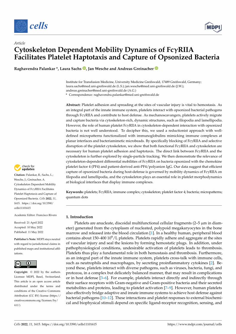
To assess the role of platelet FcγRIIA and cytoskeleton on adhesion, spreading, anddirectional guidance of platelets, we prepared high-fidelity micropatterned arrays differingin their geometry (squares and circular discs), size (2D adhesive area), and inter-pattern dis-tance (pitch) by electron beam lithography and functionalized them with heat aggregatedIgG (Agg-IgG) (Figure 1A, Supplementary Figures S1–S3). The non-patterned surface sur-rounding the micropatterns was passivated with a self-assembled monolayer of polyethy-lene glycol, offering a non-adhesive surface. In the absence of pharmacological blockers andinhibitors of FcγRIIA, αIIbβ3, actin, and myosin IIA, platelets adhered specifically (Control70.1% ± 7.9%), and showed extensive spreading and numerous filopodia (Figure 1B,C) onAgg-IgG micropatterns, irrespective of the micropattern shape and dimension. Blocking ofFcγRIIA interaction with IgG by pretreating platelets with mAb IV.3 revealed a significantreduction in platelet adhesion on Agg-IgG micropatterns (11.61% ± 4.42%; p < 0.0001) com-pared to untreated platelets (Figure 1B,C). Additionally, specific blocking of platelet αIIbβ3using abciximab decreased platelet adhesion on Agg.IgG micropatterns (35.08% ± 15.05%;p = 0.0012) (Figure 1B,C). This was expected, since integrin αIIbβ3 is known to facilitatefirm adhesion and spreading of platelets during FcγRIIA mediated platelet activation onIgG passivated surfaces [31]. Next, to assess the contribution of platelet cytoskeleton in

Cells 2022, 11, 1615 7 of 20
FcγRIIA mediated platelet adhesion on Agg-IgG micropatterns, platelets were pretreatedwith cytochalasin D and blebbistatin to inhibit F-actin assembly and interfere with acto-myosin complex formation by blocking myosin-ADP-Pi, respectively. Surprisingly, weobserved a reduction in platelet adhesion on Agg-IgG micropatterns upon cytochalasinD (14.91% ± 4.5%; p = 0.0005) and blebbistatin (22.54% ± 6.1%; p = 0.0019) pretreatmentof platelets. Subsequent analysis of single adherent platelets revealed reduced spread-ing area in the presence of cytochalasin D (16.6 µm2 ± 12.3; p < 0.0001) and blebbistatin(17.7 µm2 ± 11.9; p < 0.0001) in comparison to untreated platelets (Control: 83.7 µm2 ± 41.5 andCarrier (DMSO): 85.2 µm2 ± 44.9) (Figure 1D). Next, we analyzed the number and lengthof filopodia on single adherent platelets on Agg-IgG micropatterns. Untreated plateletsformed several (Control: 8.03 ± 4.15 and Carrier (DMSO): 7.77 ± 3.93 per single platelet)long filopodial extensions (Control: 3.78 µm ± 2.32 and Carrier (DMSO): 3.75 µm ± 2.21per single platelet). Conversely, fewer and shorter filopodia were visible in platelets pre-treated with cytochalasin D (0.38 µm± 0.095 per single platelet; p < 0.0001) and blebbistatin(0.38 µm ± 0.1 per single platelet; p < 0.0001) (Figure 1E,F). However, it is important to notethat the platelet adhesion on Agg-IgG was independent of the shape of the micropatterns.
Cells 2022, 11, x FOR PEER REVIEW 8 of 22
Figure 1. Specific recognition of Agg-IgG on micropatterns depends on platelet FcγRIIA mediated interactions, and cytoskeletal integrity is necessary for platelet adhesion and spreading. (A) CLSM images of micropatterned arrays of bovine serum albumin (BSA 4%) spiked with 1% BSA-FITC (in green) fabricated using electron beam lithography with different sizes inter-pattern pitch distances. Micropatterns were functionalized by covalent conjugation of heat aggregated human IgG (Agg-IgG detected using goat anti-human IgG Alexa 555 in magenta) to BSA in a one-step reaction using bifunctional linker 3,3’-Dithiobis(sulfosuccinimidylpropionate) (DTSSP); (B) representative confo-cal fluorescence microscopy images; and (C) analysis of the effect on platelet adhesion upon of pre-treatment of platelets with FcγRIIA blocking mAb IV.3 (4 μg/mL), integrin αIIbβ3 blocking anti-body, cytochalasin D (25 µM), and blebbistatin (10 µM) shows significant reduction in comparison with untreated platelets (control) and DMSO 0.05% (as carrier) on their spreading on Agg-IgG func-tionalized micropatterns (platelet F-actin in magenta, Agg-IgG detected using goat anti-human IgG Alexa 488 in green). Data represent mean ± SD from n = 6 donors, and statistical comparisons were performed using one-way ANOVA, followed by Tukey’s multiple comparisons for spreading area measurements. Inhibition of platelet F-actin polymerization by cytochalasin D (25 µM) and myosin II by ATPase inhibitor blebbistatin (10 µM) significantly reduces (D) platelet spreading area, (E) filopodia number, and (F) their length in single platelets in comparison with untreated platelets (control) and DMSO 0.05% (as carrier) on Agg-IgG functionalized micropatterns. Statistical compar-isons were performed by one-way ANOVA on ranks (non-parametric) followed by Dunn’s multiple comparisons test. Boxplots extend from 10 to 90 percentiles, and p < 0.05 is significant.
Figure 1. Specific recognition of Agg-IgG on micropatterns depends on platelet FcγRIIA mediatedinteractions, and cytoskeletal integrity is necessary for platelet adhesion and spreading. (A) CLSMimages of micropatterned arrays of bovine serum albumin (BSA 4%) spiked with 1% BSA-FITC (ingreen) fabricated using electron beam lithography with different sizes inter-pattern pitch distances.Micropatterns were functionalized by covalent conjugation of heat aggregated human IgG (Agg-IgG
Cells 2022, 11, 1615 8 of 20
detected using goat anti-human IgG Alexa 555 in magenta) to BSA in a one-step reaction usingbifunctional linker 3,3′-Dithiobis(sulfosuccinimidylpropionate) (DTSSP); (B) representative confocalfluorescence microscopy images; and (C) analysis of the effect on platelet adhesion upon of pretreat-ment of platelets with FcγRIIA blocking mAb IV.3 (4 µg/mL), integrin αIIbβ3 blocking antibody,cytochalasin D (25 µM), and blebbistatin (10 µM) shows significant reduction in comparison with un-treated platelets (control) and DMSO 0.05% (as carrier) on their spreading on Agg-IgG functionalizedmicropatterns (platelet F-actin in magenta, Agg-IgG detected using goat anti-human IgG Alexa 488in green). Data represent mean ± SD from n = 6 donors, and statistical comparisons were performedusing one-way ANOVA, followed by Tukey’s multiple comparisons for spreading area measurements.Inhibition of platelet F-actin polymerization by cytochalasin D (25 µM) and myosin II by ATPaseinhibitor blebbistatin (10 µM) significantly reduces (D) platelet spreading area, (E) filopodia number,and (F) their length in single platelets in comparison with untreated platelets (control) and DMSO0.05% (as carrier) on Agg-IgG functionalized micropatterns. Statistical comparisons were performedby one-way ANOVA on ranks (non-parametric) followed by Dunn’s multiple comparisons test.Boxplots extend from 10 to 90 percentiles, and p < 0.05 is significant.
3.2. Platelet Haptotaxis on IgG Planar Micropatterns Is Mediated by Dynamic Membrane Protrusions
Based on our observations of platelets during adhesion and spreading assays on2D planar micropatterns, we hypothesized (i) whether platelets display active morpho-dynamics coupled with haptotaxis on Agg-IgG micropatterns, and (ii) how filopodiaand cytoskeleton contribute to this phenomenon. Using time-lapse fluorescence videomicroscopy, we followed single platelet adhesion on Agg-IgG micropatterns with 1µminter-pattern pitch distance and assessed platelet shape change and calcium release insingle platelets (Figure 2A). In control platelets, soon after adhesion, we observed thatplatelets extend filopodia and adapt to the Agg-IgG micropattern shape while undergo-ing extensive membrane expansion through dynamic shape changes, and start spreadingbetween micropatterns (Figure 2B, Supplementary Videos S1 and S2). Additionally, weobserved an intermittent increase of calcium release bursts (Figure 2C) during plateletactivation and morphodynamic changes on untreated platelets on Agg-IgG micropatterns,whereas, upon cytochalasin D and blebbistatin treatment, filopodia formation and plateletmorphodynamics changes were abolished entirely, and platelets showed no visible increasein calcium release (Figure 2A,B, Supplementary Videos S3 and S4). Taken together, ourresults demonstrate that even though FcγRIIA is sufficient for the initial specific recognitionof Agg-IgG for firm platelet adhesion, spreading, and subsequent FcγRIIA-mediated acti-vation on Agg-IgG micropatterned planar surfaces, integrin αIIbβ3, and an intact plateletcytoskeleton are indispensable.
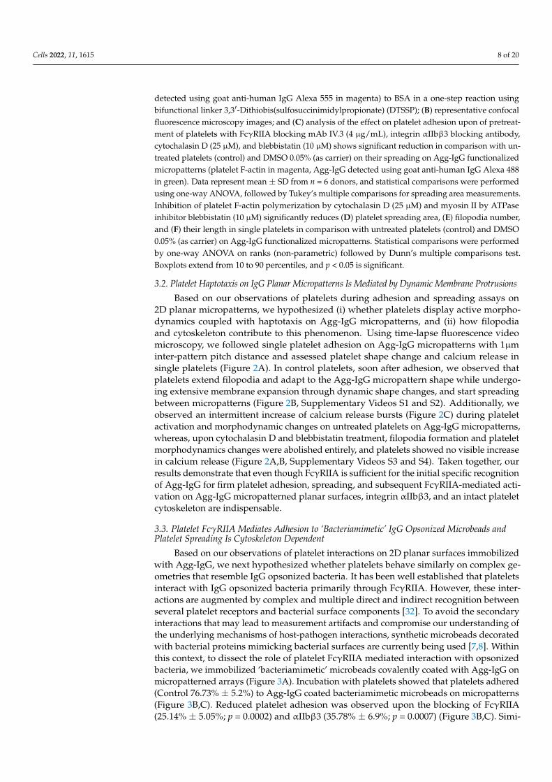
3.3. Platelet FcγRIIA Mediates Adhesion to ‘Bacteriamimetic’ IgG Opsonized Microbeads andPlatelet Spreading Is Cytoskeleton Dependent
Based on our observations of platelet interactions on 2D planar surfaces immobilizedwith Agg-IgG, we next hypothesized whether platelets behave similarly on complex ge-ometries that resemble IgG opsonized bacteria. It has been well established that plateletsinteract with IgG opsonized bacteria primarily through FcγRIIA. However, these inter-actions are augmented by complex and multiple direct and indirect recognition betweenseveral platelet receptors and bacterial surface components [32]. To avoid the secondaryinteractions that may lead to measurement artifacts and compromise our understanding ofthe underlying mechanisms of host-pathogen interactions, synthetic microbeads decoratedwith bacterial proteins mimicking bacterial surfaces are currently being used [7,8]. Withinthis context, to dissect the role of platelet FcγRIIA mediated interaction with opsonizedbacteria, we immobilized ‘bacteriamimetic’ microbeads covalently coated with Agg-IgG onmicropatterned arrays (Figure 3A). Incubation with platelets showed that platelets adhered(Control 76.73% ± 5.2%) to Agg-IgG coated bacteriamimetic microbeads on micropatterns(Figure 3B,C). Reduced platelet adhesion was observed upon the blocking of FcγRIIA(25.14% ± 5.05%; p = 0.0002) and αIIbβ3 (35.78% ± 6.9%; p = 0.0007) (Figure 3B,C). Simi-

Cells 2022, 11, 1615 9 of 20
larly, platelet adhesion to Agg-IgG bacteriamimetic microbeads was significantly reducedin platelets pretreated with cytochalasin D (25.3% ± 1.77%; p = 0.0003) and blebbistatin(27.6% ± 5.9%; p < 0.0001) (Figure 3B,C). Further assessment of morphological parametersof platelets pretreated with cytochalasin D and blebbistatin showed a significant reductionof single platelet spread area, number, and length of filopodia compared with untreatedand carrier control platelets (Figure 3D–F). Specifically, a decreased single platelet spread-ing area was observed in the presence of cytochalasin D (18.9 µm2 ± 8.9; p < 0.0001) andblebbistatin (21.7 µm2 ± 9; p < 0.0001) in comparison with untreated platelets (85.6 µm2 ± 46.9)and carrier (DMSO) (86.1 µm2 ± 51). An analysis of number and length of filopodia onsingle adherent platelets on Agg-IgG coated bacteriamimetic microbeads revealed un-treated platelets formed several (Control: 7.63 ± 4.22 and Carrier (DMSO): 7.61 ± 4.12per single platelet) and longer filopodial protrusions (Control: 3.85 µm ± 2.08 and Carrier(DMSO): 4.33 µm ±2.12 per single platelet). Conversely, fewer and shorter filopodia werevisible in platelets pretreated with cytochalasin D (2.19 ± 1.4; p < 0.0001 and 0.26 µm ± 0.1;p < 0.0001, respectively per single platelet) and blebbistatin (3.44 ± 2.1; p < 0.0001 and0.3 µm ± 0.08; p < 0.0001, respectively per single platelet).
Cells 2022, 11, x FOR PEER REVIEW 9 of 22
3.2. Platelet Haptotaxis on IgG Planar Micropatterns Is Mediated by Dynamic Membrane Protrusions
Based on our observations of platelets during adhesion and spreading assays on 2D planar micropatterns, we hypothesized (i) whether platelets display active morphody-namics coupled with haptotaxis on Agg-IgG micropatterns, and (ii) how filopodia and cytoskeleton contribute to this phenomenon. Using time-lapse fluorescence video micros-copy, we followed single platelet adhesion on Agg-IgG micropatterns with 1µm inter-pattern pitch distance and assessed platelet shape change and calcium release in single platelets (Figure 2A). In control platelets, soon after adhesion, we observed that platelets extend filopodia and adapt to the Agg-IgG micropattern shape while undergoing exten-sive membrane expansion through dynamic shape changes, and start spreading between micropatterns (Figure 2B, Supplementary Video S1 and S2). Additionally, we observed an intermittent increase of calcium release bursts (Figure 2C) during platelet activation and morphodynamic changes on untreated platelets on Agg-IgG micropatterns, whereas, upon cytochalasin D and blebbistatin treatment, filopodia formation and platelet morpho-dynamics changes were abolished entirely, and platelets showed no visible increase in calcium release (Figure 2A,B, Supplementary Video S3 and S4). Taken together, our re-sults demonstrate that even though FcγRIIA is sufficient for the initial specific recognition of Agg-IgG for firm platelet adhesion, spreading, and subsequent FcγRIIA-mediated ac-tivation on Agg-IgG micropatterned planar surfaces, integrin αIIbβ3, and an intact plate-let cytoskeleton are indispensable.
Figure 2. Platelets use filopodia as antennae to seek out and adhere to Agg-IgG on micropatterns through FcγRIIA and exhibit haptotaxis along laterally increasing Agg-IgG density on 2D planar micropatterns. (A) Platelets preloaded with Ca2+ indicator dye Fluo-AM were incubated on micro-patterns functionalized with Agg-IgG were imaged by time-lapse video microscopy. Representative time-lapse images of Ca2+ release and platelet morphodynamics during adhesion and spreading on Agg-IgG micropatterns with a pitch distance of 1µm show platelet filopodia actively scan their sur-rounding microenvironment as platelets adhere, spread in a polarized manner resembling hapto-taxis, and become activated on Agg-IgG micropatterns. This process is abrogated upon pretreatment of platelets with F-actin polymerization inhibitor cytochalasin D (25 µM) and myosin II ATPase
Figure 2. Platelets use filopodia as antennae to seek out and adhere to Agg-IgG on micropatternsthrough FcγRIIA and exhibit haptotaxis along laterally increasing Agg-IgG density on 2D planarmicropatterns. (A) Platelets preloaded with Ca2+ indicator dye Fluo-AM were incubated on micropat-terns functionalized with Agg-IgG were imaged by time-lapse video microscopy. Representativetime-lapse images of Ca2+ release and platelet morphodynamics during adhesion and spreading onAgg-IgG micropatterns with a pitch distance of 1µm show platelet filopodia actively scan their sur-rounding microenvironment as platelets adhere, spread in a polarized manner resembling haptotaxis,and become activated on Agg-IgG micropatterns. This process is abrogated upon pretreatment ofplatelets with F-actin polymerization inhibitor cytochalasin D (25 µM) and myosin II ATPase inhibitorblebbistatin (10 µM) (see Supplementary Videos S1–S4); (B) Analysis of platelet morphodynamics(shape changes over time) and (C) changes in calcium binding dye Fluo-4 AM fluorescence (meanfluorescence intensity- MFI) during intracellular calcium release from time-lapse video microscopyreveals platelets undergo minimal morphodynamics upon pretreatment with F-actin polymerizationinhibitor cytochalasin D (25 µM) and myosin II ATPase inhibitor blebbistatin (10 µM), indicatingAgg-IgG sensing by platelets through their FcγRIIA is dependent on the stability of the platelet

Cells 2022, 11, 1615 10 of 20
cytoskeleton, which in turn regulates filopodia formation. Data represents mean ± SD valuesfrom n = 6 (region of interests) from 6 donors. Fluo-4 AM calcium dynamics show mean ± SD fromn = 3 (region of interests). Statistical comparisons were performed by one-way ANOVA on ranks (non-parametric) followed by Dunn’s multiple comparisons test. Boxplots extend from 10 to 90 percentiles,and p < 0.05 is significant.
Cells 2022, 11, x FOR PEER REVIEW 11 of 22
Figure 3. Platelet cytoskeletal destabilization leads to a decrease in platelet spreading on Agg-IgG functionalized ‘bacteriamimetic’ microbeads arrays due to abrogation of filopodia formation (A) SEM and CLSM images (detected using goat anti-human IgG Alexa 488 in green) of Agg-IgG func-tionalized ‘bacteriamimetic’ microbead arrays; (B) Representative CLSM images of platelet adhe-sion and (C) analysis of spreading area of single platelets pretreated with FcγRIIA blocking mAb IV.3 (4 μg/mL), integrin αIIbβ3 blocking antibody, cytochalasin D (25 μM), and blebbistatin (10 µM) shows significant reduction in comparison with untreated platelets (control) and DMSO 0.05% (as a carrier) on their spreading on Agg-IgG functionalized ‘bacteriamimetic’ microbeads arrays (plate-let F-actin in magenta, Agg-IgG detected using goat anti-human IgG Alexa 488 in green). Data rep-resent mean ± SD from n = 6 donors, and statistical comparisons were performed using one-way ANOVA, followed by Tukey’s multiple comparisons test. Inhibition of platelet F-actin polymeriza-tion by cytochalasin D (25 µM) and myosin II ATPase inhibitor blebbistatin (10 µM) significantly reduces (D) spreading area, (E) number, and (F) length of filopodia in single platelets in comparison with untreated platelets (control) and DMSO 0.05% (as carrier) on Agg-IgG functionalized ‘bacteri-amimetic’ microbeads arrays. Statistical comparisons were performed by one-way ANOVA on ranks (non-parametric) followed by Dunn’s multiple comparisons test. Boxplots extend from 10 to 90 percentiles, and p < 0.05 is significant.
Figure 3. Platelet cytoskeletal destabilization leads to a decrease in platelet spreading on Agg-IgG functionalized ‘bacteriamimetic’ microbeads arrays due to abrogation of filopodia formation(A) SEM and CLSM images (detected using goat anti-human IgG Alexa 488 in green) of Agg-IgGfunctionalized ‘bacteriamimetic’ microbead arrays; (B) Representative CLSM images of plateletadhesion and (C) analysis of spreading area of single platelets pretreated with FcγRIIA blocking mAbIV.3 (4 µg/mL), integrin αIIbβ3 blocking antibody, cytochalasin D (25 µM), and blebbistatin (10 µM)shows significant reduction in comparison with untreated platelets (control) and DMSO 0.05% (as acarrier) on their spreading on Agg-IgG functionalized ‘bacteriamimetic’ microbeads arrays (plateletF-actin in magenta, Agg-IgG detected using goat anti-human IgG Alexa 488 in green). Data representmean ± SD from n = 6 donors, and statistical comparisons were performed using one-way ANOVA,followed by Tukey’s multiple comparisons test. Inhibition of platelet F-actin polymerization bycytochalasin D (25 µM) and myosin II ATPase inhibitor blebbistatin (10 µM) significantly reduces(D) spreading area, (E) number, and (F) length of filopodia in single platelets in comparison withuntreated platelets (control) and DMSO 0.05% (as carrier) on Agg-IgG functionalized ‘bacteriamimetic’microbeads arrays. Statistical comparisons were performed by one-way ANOVA on ranks (non-parametric) followed by Dunn’s multiple comparisons test. Boxplots extend from 10 to 90 percentiles,and p < 0.05 is significant.

Cells 2022, 11, 1615 11 of 20
Surprisingly, in contrast to the platelet haptotaxis observed on planar 2D ligandgradient micropatterns (Figure 2A), haptotactic migration was absent on Agg-IgG bacteri-amimetic microbeads. Instead, analysis of platelet adhesion phenotype by SEM showedfilopodial extensions tightly adhering and wrapping around the Agg-IgG coated mi-crobeads, but not in platelets treated with cytoskeletal inhibitors (Figure 4).
Figure 4. Scanning electron microscopy (SEM) of platelets interaction with Agg-IgG coated bacteri-amimetic microbeads on micropattern. In controls (A,B), platelets can be seen adhering and spreadingon the Agg-IgG beads. Extensive filopodia and membranous lamellipodia (indicated arrowheads)are visible in control platelets but not in platelets treated with cytoskeletal inhibitors Cytochalasin Dand Blebbistatin (C,D).
3.4. Lateral Mobility of FcγRIIA Is Dependent on Cytoskeletal Integrity
Since platelet cytoskeletal integrity plays a crucial role in filopodia and lamellipodiamediated adhesion, spreading and haptotaxis through FcγRIIA on 2D planar surfaces andmicrobead arrays functionalized with Agg-IgG, we next asked the question as to whether

Cells 2022, 11, 1615 12 of 20
platelet cytoskeleton also influences the underlying physical factor, such as mobility ofFcγRIIA. We used QDs for single-particle tracking (SPT) of FcγRIIA on the platelet plasmamembrane to assess this. Specific labeling, detection, and monitoring of single FcγRIIAin live platelets spread on fibrinogen surface were carried out using monovalent QDconjugated to a single monoclonal Fab fragment (QD-Fab) against the exofacial epitopeof FcγRIIA that recognizes the Fc portion of IgG (Figure 5A, Supplementary Figure S5).Assessment of single FcγRIIA track length based on SPT revealed FcγRIIA moved longerdistances on filopodia (1.13 µm ± 1.12) and lamellipodia (0.80 µm ± 1.09), comparedto those on the platelet body (0.48 µm ± 0.59) (Figure 5B,C, Supplementary Figure S6and Videos S5–S7). Disruption of F-actin assembly using cytochalasin D and blebbistatinmarkedly decreased the track length of single FcγRIIA mobilities on platelet filopodia,lamellipodia, and body (Figure 5C).
Cells 2022, 11, x FOR PEER REVIEW 13 of 22
Figure 5. Single-particle tracking of FcγRIIA on platelets using QD-Fab. (A) Fluorescence micro-graphs showing platelets labeled with QD + anti-FcγRIIA Fab bound to FcγRIIA followed by local-ization of single FcγRIIA by detection of single QD fluorescence emission (magenta) and overlay of tracks (yellow) identified from (B) time-lapse images of single QD-Fab bound to individual FcγRIIA along the filopodia (F), lamellipodia (L), and platelet body; Analysis of track length (C) from trajec-tories of single QD-Fab bound to FcγRIIA on filopodia, lamellipodia, and platelet body. Statistical comparisons were performed by one-way ANOVA on ranks (non-parametric), followed by Dunn’s multiple comparisons test. Boxplots extend from 10 to 90 percentiles, and p < 0.05 is significant and n ≥ 50 single QD-fab per dataset.
Next, we assessed the lateral mobility dynamics of single FcγRIIA on platelets. On platelet filopodia and lamellipodia, a single QD-Fab bound to FcγRIIA showed directional motion along the axis of the filopodia and towards the edge of the cytoplasmic rim in case of lamellipodia, while on the platelet body, the motion was largely confined (Figure 6A,B and Supplementary Figure S6). Diffusion constants (D, µm2/s) of single FcγRIIA were sig-nificantly higher on platelet filopodia (0.035 µm2/s ± 0.012; p < 0.0001) and lamellipodia
Figure 5. Single-particle tracking of FcγRIIA on platelets using QD-Fab. (A) Fluorescence mi-crographs showing platelets labeled with QD + anti-FcγRIIA Fab bound to FcγRIIA followed bylocalization of single FcγRIIA by detection of single QD fluorescence emission (magenta) and overlayof tracks (yellow) identified from (B) time-lapse images of single QD-Fab bound to individual FcγRIIAalong the filopodia (F), lamellipodia (L), and platelet body; Analysis of track length (C) from trajec-tories of single QD-Fab bound to FcγRIIA on filopodia, lamellipodia, and platelet body. Statisticalcomparisons were performed by one-way ANOVA on ranks (non-parametric), followed by Dunn’smultiple comparisons test. Boxplots extend from 10 to 90 percentiles, and p < 0.05 is significant andn ≥ 50 single QD-fab per dataset.

Cells 2022, 11, 1615 13 of 20
Next, we assessed the lateral mobility dynamics of single FcγRIIA on platelets. Onplatelet filopodia and lamellipodia, a single QD-Fab bound to FcγRIIA showed directionalmotion along the axis of the filopodia and towards the edge of the cytoplasmic rim in caseof lamellipodia, while on the platelet body, the motion was largely confined (Figure 6A,Band Supplementary Figure S6). Diffusion constants (D, µm2/s) of single FcγRIIA weresignificantly higher on platelet filopodia (0.035 µm2/s ± 0.012; p < 0.0001) and lamellipodia(0.034 µm2/s ± 0.011; p < 0.0001) compared to the platelet body (0.013 µm2/s ± 0.005)(Figure 6C). Disruption of platelet F-actin polymerization cytochalasin D and blebbistatin re-sulted in marked decrease in the FcγRIIA diffusion constant both on filopodia (0.016 µm2/s± 0.005 and 0.014 µm2/s ± 0.005, respectively) and lamellipodia (0.014 µm2/s ± 0.005and 0012 µm2/s ± 0.006), compared to the carrier (0.033 µm2/s ± 0.013 and 0.031 µm2/s± 0.012). On the contrary, the FcγRIIA diffusion constant on the platelet body remainedlargely unchanged in the presence of cytochalasin D (0.01 µm2/s ± 0.006) and blebbistatin(0.011 µm2/s ± 0.006) compared to the carrier (0.012 µm2/s ± 0.006).
Cells 2022, 11, x FOR PEER REVIEW 14 of 22
(0.034 µm2/s ± 0.011; p < 0.0001) compared to the platelet body (0.013 µm2/s ± 0.005) (Figure 6C). Disruption of platelet F-actin polymerization cytochalasin D and blebbistatin resulted in marked decrease in the FcγRIIA diffusion constant both on filopodia (0.016 µm2/s ± 0.005 and 0.014 µm2/s ± 0.005, respectively) and lamellipodia (0.014 µm2/s ± 0.005 and 0012 µm2/s ± 0.006), compared to the carrier (0.033 µm2/s ± 0.013 and 0.031 µm2/s ± 0.012). On the contrary, the FcγRIIA diffusion constant on the platelet body remained largely un-changed in the presence of cytochalasin D (0.01 µm2/s ± 0.006) and blebbistatin (0.011 µm2/s ± 0.006) compared to the carrier (0.012 µm2/s ± 0.006).
Figure 6. Differential lateral mobility dynamics of single FcγRIIA on platelets. (A) 2D localized tra-jectory and displacement (arrows with start and end positions) of single QD-Fab bound to FcγRIIA on platelet filopodia, lamellipodia, and platelet body as a function of lag time shown by color-coded time scale. (B) Mean squared displacement (MSD, µm2) as a function of the lag time of single QD-Fab trajectory shows the directional motion of FcγRIIA on platelet filopodia and lamellipodia and a confined motion on the platelet body (please note the different scales of the Y-axis in the different panels). (C) Comparison of the diffusion constant (D, µm2/s) of individual FcγRIIA calculated from single QD-Fab trajectory in untreated platelets (control) and DMSO 0.05% (as carrier) and after in-hibition of platelet F-actin polymerization by cytochalasin D (25 µM) and myosin II ATPase inhibitor blebbistatin (10 µM). Statistical comparisons were performed by one-way ANOVA on ranks (non-parametric), followed by Dunn’s multiple comparisons test. Boxplots extend from 10 to 90 percen-tiles, and p < 0.05 is significant and n ≥ 50 single QD-fab per dataset.
Figure 6. Differential lateral mobility dynamics of single FcγRIIA on platelets. (A) 2D localizedtrajectory and displacement (arrows with start and end positions) of single QD-Fab bound to FcγRIIAon platelet filopodia, lamellipodia, and platelet body as a function of lag time shown by color-codedtime scale. (B) Mean squared displacement (MSD, µm2) as a function of the lag time of single QD-Fabtrajectory shows the directional motion of FcγRIIA on platelet filopodia and lamellipodia and aconfined motion on the platelet body (please note the different scales of the Y-axis in the differentpanels). (C) Comparison of the diffusion constant (D, µm2/s) of individual FcγRIIA calculatedfrom single QD-Fab trajectory in untreated platelets (control) and DMSO 0.05% (as carrier) andafter inhibition of platelet F-actin polymerization by cytochalasin D (25 µM) and myosin II ATPaseinhibitor blebbistatin (10 µM). Statistical comparisons were performed by one-way ANOVA onranks (non-parametric), followed by Dunn’s multiple comparisons test. Boxplots extend from 10 to90 percentiles, and p < 0.05 is significant and n ≥ 50 single QD-fab per dataset.

Cells 2022, 11, 1615 14 of 20
3.5. Longer Track Lengths and Higher Lateral Mobility of FcγRIIA on Platelet Filopodia andLamellipodia Facilitate Sensing and Capture IgG Opsonized Bacterial Pathogens by Platelets
To assess the biological significance of FcγRIIA mobility on platelet-bacteria interac-tions, we incubated platelets on micropatterns functionalized with living E. coli opsonizedwith PF4 and anti-PF4/p IgG (Figure 7A). A higher percentage of untreated platelets specif-ically adhered to opsonized E. coli on micropatterns (Control 74.44% ± 8.8%), whereasplatelet adhesion was significantly inhibited upon blocking of FcγRIIA by antibody IV.3(19.57% ± 3.23%; p = 0.0002), as well as by blocking of αIIbβ3 by abciximab (41.53% ± 5.78%;p = 0.0057) (Figure 7B,C). Additionally, a markedly reduced platelet adhesion to PF4and anti-PF4/P IgG opsonized E. coli was observed in the presence of cytochalasin D(21.62% ± 4.11%; p = 0.0009) and blebbistatin (18.56% ± 2.97%; p = 0.0009) in comparison tothe carrier (DMSO) (72.45% ± 7.9%) (Figure 7B,C). Analysis of the platelet morphology ofadherent platelets on opsonized E. coli revealed decreased single platelet spreading areas inthe presence of cytochalasin D (7.72 µm2 ± 3.8) and blebbistatin (7.77 µm2 ± 4.6), in compar-ison with the untreated platelets (23.62 µm2 ± 13.4) and the carrier (DMSO) (20.1 µm2 ± 11.37)(Figure 7D). Furthermore, we found a reduction in the number and length of filopodialprotrusions in the presence of cytochalasin D and blebbistatin (Figure 7E,F). These resultsare consistent with our observations of platelet adhesion, spreading, and morphometricfeatures of filopodia observed on 2D planar and bacteriamimetic microbead micropatternsfunctionalized with the Agg-IgG that functions as a potent ligand for FcγRIIA.
Cells 2022, 11, x FOR PEER REVIEW 16 of 22
Figure 7. Platelets specifically recognize opsonized by PF4 + anti-PF4/P IgG via FcγRIIA using filo-podial protrusions, while their adhesion and spreading are regulated by cytoskeletal rearrange-ment. (A) Light transmission and CLSM immunofluorescence micrographs of live E. coli KPM121 (ΔwaaA) opsonized with PF4 + anti-PF4/P IgG immobilized on micropattern arrays (detected using goat anti-human IgG Alexa 488 in green); (B) Representative CLSM images of platelet adhesion and (C) analysis of spreading area of single platelets pretreated with FcγRIIA blocking mAb IV.3 (4 µg/mL), integrin αIIbβ3 blocking antibody, cytochalasin D (25 µM), and blebbistatin (10 µM) shows significant reduction comparison with untreated platelets (control) and DMSO 0.05% (as carrier) on their spreading on E. coli KPM121 (ΔwaaA) opsonized with PF4 and anti-PF4/P IgG (platelet F-actin in magenta, patient-derived anti-PF4/P IgG detected using goat anti-human IgG Alexa 488 in green). Data represent mean ± SD from n = 6 donors, and statistical comparisons were performed using one-way ANOVA, followed by Tukey’s multiple comparisons test. Inhibition of platelet F-actin polymerization by cytochalasin D (25 µM) and myosin II ATPase inhibitor blebbistatin (10 µM) sig-nificantly reduces (D) spreading area, (E) number and (F) length of filopodia in single platelets in comparison with untreated platelets (control) and DMSO 0.05% (as carrier) on opsonized E. coli. Statistical comparisons were performed by one-way ANOVA on ranks (non-parametric), followed by Dunn’s multiple comparisons test. Boxplots extend from 10 to 90 percentiles, and p < 0.05 is sig-nificant.
4. Discussion In human health and disease, platelet FcγRIIA plays a vital role in the pathophysiol-
ogy of diseases, such as HIT and VITT, where crosslinking of platelet FcγRIIA by anti-
Figure 7. Platelets specifically recognize opsonized by PF4 + anti-PF4/P IgG via FcγRIIA using filopodialprotrusions, while their adhesion and spreading are regulated by cytoskeletal rearrangement. (A) Light

Cells 2022, 11, 1615 15 of 20
transmission and CLSM immunofluorescence micrographs of live E. coli KPM121 (∆waaA) opsonizedwith PF4 + anti-PF4/P IgG immobilized on micropattern arrays (detected using goat anti-humanIgG Alexa 488 in green); (B) Representative CLSM images of platelet adhesion and (C) analysis ofspreading area of single platelets pretreated with FcγRIIA blocking mAb IV.3 (4 µg/mL), integrinαIIbβ3 blocking antibody, cytochalasin D (25 µM), and blebbistatin (10 µM) shows significant reduc-tion comparison with untreated platelets (control) and DMSO 0.05% (as carrier) on their spreadingon E. coli KPM121 (∆waaA) opsonized with PF4 and anti-PF4/P IgG (platelet F-actin in magenta,patient-derived anti-PF4/P IgG detected using goat anti-human IgG Alexa 488 in green). Datarepresent mean ± SD from n = 6 donors, and statistical comparisons were performed using one-wayANOVA, followed by Tukey’s multiple comparisons test. Inhibition of platelet F-actin polymerizationby cytochalasin D (25 µM) and myosin II ATPase inhibitor blebbistatin (10 µM) significantly reduces(D) spreading area, (E) number and (F) length of filopodia in single platelets in comparison withuntreated platelets (control) and DMSO 0.05% (as carrier) on opsonized E. coli. Statistical compar-isons were performed by one-way ANOVA on ranks (non-parametric), followed by Dunn’s multiplecomparisons test. Boxplots extend from 10 to 90 percentiles, and p < 0.05 is significant.
4. Discussion
In human health and disease, platelet FcγRIIA plays a vital role in the pathophysiologyof diseases, such as HIT and VITT, where crosslinking of platelet FcγRIIA by anti-plateletIgG antibodies occurs leading to platelet clearance, it may potentially also contribute tosome forms of immune thrombocytopenia (ITP) [18,33]. Additionally, platelet FcγRIIA isalso implicated in the clinical manifestation of the prothrombotic adverse drug effect HIT,leading to severe morbidity and mortality [24]. Briefly, platelet factor 4 (PF4) interacts withpolyanions (P), such as the anticoagulant heparin, and undergoes conformational changestriggering an immune response. Subsequently, immune complexes formed between PF4,P, and anti-PF4/P IgG produced by the human host induce crosslinking of the FcγRIIAon platelets leading to platelet activation and generation of excess thrombin leading tothrombocytopenic symptoms. By serendipity, anti-PF4/P IgG can effectively opsonizePF4 bound to both Gram-positive and Gram-negative bacteria, triggering platelet activa-tion and aggregation [34–36]. Similarly, PF4 has been shown to interact with adenoviralvector derived from chimpanzee adenovirus Y25 (ChAdOx1) and its components usedfor vaccination against COVID-19 [18,37,38]. Intriguingly, individuals who develop VITTpost-ChAdOx1 vaccination also develop anti-PF4 antibodies, which do not depend onheparin [39].
We have proposed that the anti-PF4 immune response is an evolutionary old immunereaction bridging innate and adapted immunity allowing granulocytes and platelets torecognize various pathogens coated with PF4 and opsonized by anti-PF4/P IgG via theFcγRIIa [15,35,40]. However, the role of platelet FcγRIIA dynamics and contribution ofthe platelet cytoskeleton during the processes leading to firm adhesion, spreading, andplatelet haptotaxis at interfaces, such as opsonized pathogens displaying the IgG as aprimary ligand, have not been explored. In particular, it is nearly impossible to dissect theeffects of specific pathogen-derived or platelet components in these complex processes ofpathogen-platelet interactions. We overcame this inherent limitation by using minimalisticmodels of biological interfaces mimicking inflammatory and pathogenic circumstancesusing lithography and biofunctionalization techniques.
Cell protrusions are essential for motility during chemotaxis and haptotaxis [41].During haptotaxis (directed movement), migrating cells rely on filopodial and lamellipodialprotrusions that act as sensors or antennae to probe the extracellular biochemical andmicro-and nano-environmental cues [42–44]. In platelets, upon stimulation by agonists,highly dynamic actin-rich filopodial protrusions and lamellipodia are formed [45]. Plateletfilopodia are involved in sensing topographic cues to initiate platelet spreading and playa key role in hemostasis [46,47]. Previously, geometrically defined micropatterns havebeen found to be helpful in investigating single platelet adhesion and platelet functionon various adhesive substrates [7,8,48–50]. We, therefore, used a systematic biomimetic

Cells 2022, 11, 1615 16 of 20
approach with stepwise increasing complexity starting from planar micropatterned arraysand bacteria-like microbead-based arrays, both functionalized with Agg-IgG as a specificligand for FcγRIIA ligand. On Agg-IgG micropatterns, we found platelet adhesion andspreading to be largely dependent on the specific interaction of FcγRIIA with its ligandand on cytoskeletal integrity.
Interestingly, we also found blocking of αIIbβ3 reduces platelet adhesion to Agg-IgG.These data agree with previous observations, which demonstrated not only FcγRIIA butalso fully functional αIIbβ3 is necessary for optimal platelet adhesion on interfaces exposingIgG [51]. In addition, our data of platelet morphodynamics showed directional haptotaxis ofplatelets mediated by highly dynamic filopodia and lamellipodia on Agg-IgG micropatternswhich was strongly abrogated upon cytoskeletal disruption. Similarly, platelets exhibited astrong dependency on FcγRIIA, and cytoskeletal integrity for adhesion on Agg-IgG coatedbacteriamimetic microbeads. Surprisingly, we did not observe the directional movement ofplatelets on Agg-IgG microbeads. However, extensive filopodia and lamellipodia could beseen tightly interacting with the microbeads. This was, again, abrogated upon cytoskeletaldisruption. One possible explanation could be that the surface topography and geometricconstraints and the spatial distribution of Agg-IgG on microbeads may limit efficientplatelet haptotaxis [52,53]. Another potential explanation is that platelet differentiallybehave depending on the three-dimensional structure of the ligand presented. On planar2D surfaces, they primarily spread, as this is probably most relevant for wound closure inhemostasis. 3D areas, however, signal pathogens, and this induces a function of plateletsrelated to host defense by immobilizing the pathogen and presenting it to phagocytosingcells [21]. Additionally, a predominantly localized adhesion with a lack of increasedspreading compared to 2D planar micropatterns can be attributed to the self-deposition ofreceptor-interacting ligands at the site of adhesion [54].
To further understand the interaction of platelet FcγRIIA and the cytoskeleton in lig-and recognition and haptotaxis, we investigated the lateral mobility dynamics of individualFcγRIIA. This was achieved by specific binding of an anti-FcγRIIA Fab conjugated to aquantum dot (QD). QDs are luminescent semiconductor nanoparticles with high, uniform,and photo-stable brightness, thus permitting detection to the single nanoparticle leveland reliable quantification of receptor mobilities [55,56]. Our data from single-particletracking of FcγRIIA on platelets show differential mobility of the receptor on differentparts of the platelets during their morphodynamic spreading. On filopodia and lamel-lipodia, FcγRIIA exhibits longer track lengths and a higher diffusion constant, but not onthe platelet body, which strongly depends on the cytoskeletal integrity. These data sug-gest a continuous actin-based “treadmilling” mechanism controls the dynamics of FcγRIIAlateral mobility on the platelets in regions where actin polymerization occurs at increasedrates [57–59]. This, in turn, may facilitate rapid ligand engagement by FcγRIIA on filopodiaand lamellipodia [60]. However, the confined mobility of FcγRIIA can potentially be at-tributed to diffusion barriers resulting from highly crosslinked actin and the abundance ofother surface receptors within nano- and microclusters [61–63]. Additionally, lipid raftsmay contribute to the localization and lateral mobility of FcγRIIA [64].
To explore the significance of our findings on differential mobility dynamics ofFcγRIIA, we recapitulated platelet-pathogen interactions on micropatterns. To demonstratethis, we used a well-understood opsonized bacterial system involving PF4 and HIT patient-derived anti-PF4/P IgG [15,35]. Similar to the interaction of platelets with Agg-IgG coatedopsonized bacteriamimetic microbeads, we observed a strong dependency on functionalFcγRIIA and cytoskeletal integrity for platelet adhesion and spreading. Platelets exhibitedextensive filopodia and lamellipodia, indicating that these dynamic structures are primarilyused by platelets to sense opsonized bacteria. We believe such interactions may have a widerange of implications for the response of platelets to opsonized bacteria during systemicinfections and during platelet-platelet interaction with IgG coated autologous cells, e.g., inautoimmune disorders such as anti-phospholipid syndrome.

Cells 2022, 11, 1615 17 of 20
Although our results show a strong dependency on human platelet FcγRIIA and cy-toskeleton as key partners during platelet adhesion, spreading, and haptotaxis on biologicalinterfaces with immune complexes, there are some limitations to our approach. In the cur-rent work, we have limited ourselves to a single ligand IgG (e.g., Agg-IgG and anti-PF4/PIgG). Thus, one cannot exclude the role of various biochemical and biophysical stimuli thatplatelets encounter during their interaction with IgG opsonized cells and pathogens in avascular system. Additionally, as mouse platelets do not express an Fc-receptor, our dataindicate that in vivo mouse experiments may not reflect the role of platelets in situationsin which antibodies are involved. However, the modular bottom-up system of micropat-terns provided here can be used to sequentially incorporate other variables to capture thecomplexity of platelet receptors and ligand interactions reflecting biological interfaces.
5. Conclusions
In summary, we demonstrate that platelet interaction via FcγRIIA at interfaces present-ing IgG in close proximities (e.g., aggregated IgG or anti-PF4 IgG on opsonized pathogens)primarily occurs through cytoskeleton rich dynamic filopodial and lamellipodial exten-sions. In addition, single-particle tracking revealed differential mobility dynamics ofhuman platelet FcγRIIA. Our data suggest both of these processes are mainly cytoskeletondependent. Furthermore, using facile nano/microfabricated biomimetic minimal interfacesin combination with specific blockers and inhibitors, we show that complex interactionsof platelets with molecular ligands on different geometries, including whole bacteria, canbe deciphered. Such an approach can be extended to interrogate a variety of plateletreceptor-mediated interactions with the potential for applications in basic biology, such asassessment of platelet function defects related to granule secretion, cytoskeleton, receptorsignaling, and for drug screening.
Supplementary Materials: The following supporting information can be downloaded at: https://www.mdpi.com/article/10.3390/cells11101615/s1, Figure S1: Schematics of micropatterned arrayfunctionalized with Agg-IgG used for platelet adhesion and spreading assays; Figure S2: Optimizationof electron beam dosage for preparation of high-fidelity micropatterns; Figure S3: Line profiles alongthe fluorescent micropatterns; Figure S4: Characterization of quantum dots (QD) by dynamic lightscattering (DLS); Figure S5: Confocal fluorescence microscopy of specific labeling of FcγRIIA onplatelets by QD-Fab; Figure S6: Representative images of individual tracks and trajectories of QD-Fab bound to platelet FcγRIIA; Video S1: Control platelet adhesion and haptotaxis on Agg.IgGmicropatterns; Video S2: Carrier Control (DMSO 0.05%) showing platelet adhesion and haptotaxison Agg.IgG micropatterns; Video S3: Cytochalasin D treated platelets fail to adhere firmly and donot spread on Agg.IgG micropatterns; Video S4: Blebbistatin treated platelets adhere weakly anddo not spread on Agg.IgG micropatterns; Video S5: Tracking dynamics of FcγRIIA via QD-Fab onplatelet filopodia. Legends-magenta circles indicate the localization of individual QD luminescence,and yellow lines represent their computed tracks. Image interval of 78 ms, and playback is at 5 fps;Video S6: Tracking dynamics of FcγRIIA via QD-Fab on platelet lamellipodia. Legends-magentacircles indicate the localization of individual QD luminescence, and yellow lines represent theircomputed tracks. Image interval of 78 ms, and playback is at 5 fps; Video S7: Tracking dynamics ofFcγRIIA via QD-Fab on platelet body. Legends-magenta circles indicate the localization of individualQD luminescence, and yellow lines represent their computed tracks. Image interval of 78 ms, andplayback is at 5 fps.
Author Contributions: Conceptualization, R.P. and A.G.; methodology, R.P., L.S. and J.W.; formalanalysis, R.P., J.W. and A.G.; investigation, R.P., L.S. and J.W.; data curation, R.P. and J.W.; writing—original draft preparation, R.P. and A.G.; writing—review and editing, R.P., L.S., J.W. and A.G.;visualization, R.P.; supervision, R.P. and A.G.; project administration, R.P. and A.G.; funding acquisi-tion, R.P. and A.G. All authors have read and agreed to the published version of the manuscript.
Funding: This work was supported by the Deutsche Forschungsgemeinschaft project number374031971–CRC/TR 240.

Cells 2022, 11, 1615 18 of 20
Institutional Review Board Statement: The study was conducted following the Declaration ofHelsinki and approved by the ethics Committee of the University Medicine Greifswald, Germany(Ethics approval BB012/18b).
Informed Consent Statement: The use of human platelets from healthy volunteers and sera frompatients with HIT was obtained with prior consent.
Data Availability Statement: The datasets generated and analyzed during the study are availablefrom the corresponding author.
Acknowledgments: The authors thank Jessica Fuhrmann and Mykola Medvidov for their excellenttechnical support. The authors thank Sven Hammerschmidt for providing E. coli KPM121 (∆waaA)strain and Oliver Otto for access to research instrumentation.
Conflicts of Interest: R.P., L.S. and J.W. declare no conflict of interest. AG reports personal fees andnon-financial support from Aspen, Boehringer Ingelheim, Instrumentation Laboratory, and Roche;grants from Ergomed, Rovi, Sagent, Portola, Fa. Blau Farmacêutica, Prosensa/Biomarin, DRK-BSDBaden- Würtemberg/Hessen, and Biokit; personal fees from Bayer Vital, Chromatec, Sanofi-Aventis,and GTH e.V; grants and personal fees from Macopharma; as well as grants and other from DRKBSDNSTOB. In addition, AG reports having a patent, application n. 2021032220550000DE, pending. Thefunders had no role in the study’s design, in the collection, analyses, or interpretation of data, in thewriting of the manuscript, or in the decision to publish the results.
References1. Machlus, K.R.; Italiano, J.E., Jr. The incredible journey: From megakaryocyte development to platelet formation. J. Cell Biol. 2013,
201, 785–796. [CrossRef] [PubMed]2. Deppermann, C.; Kubes, P. Start a fire, kill the bug: The role of platelets in inflammation and infection. Innate Immun. 2018, 24,
335–348. [CrossRef] [PubMed]3. Ali, R.A.; Wuescher, L.M.; Dona, K.R.; Worth, R.G. Platelets Mediate Host Defense against Staphylococcus aureus through Direct
Bactericidal Activity and by Enhancing Macrophage Activities. J. Immunol. 2017, 198, 344–351. [CrossRef] [PubMed]4. Kho, S.; Barber, B.E.; Johar, E.; Andries, B.; Poespoprodjo, J.R.; Kenangalem, E.; Piera, K.A.; Ehmann, A.; Price, R.N.; William, T.; et al.
Platelets kill circulating parasites of all major Plasmodium species in human malaria. Blood 2018, 132, 1332–1344. [CrossRef]5. Youssefian, T.; Drouin, A.; Masse, J.M.; Guichard, J.; Cramer, E.M. Host defense role of platelets: Engulfment of HIV and
Staphylococcus aureus occurs in a specific subcellular compartment and is enhanced by platelet activation. Blood 2002, 99,4021–4029. [CrossRef]
6. Beaulieu, L.M.; Clancy, L.; Tanriverdi, K.; Benjamin, E.J.; Kramer, C.D.; Weinberg, E.O.; He, X.; Mekasha, S.; Mick, E.;Ingalls, R.R.; et al. Specific Inflammatory Stimuli Lead to Distinct Platelet Responses in Mice and Humans. PLoS ONE 2015,10, e0131688. [CrossRef]
7. Palankar, R.; Binsker, U.; Haracska, B.; Wesche, J.; Greinacher, A.; Hammerschmidt, S. Interaction between the Staphylococcusaureus extracellular adherence protein Eap and its subdomains with platelets. Int. J. Med. Microbiol. 2018, 308, 683–691. [CrossRef]
8. Binsker, U.; Palankar, R.; Wesche, J.; Kohler, T.P.; Prucha, J.; Burchhardt, G.; Rohde, M.; Schmidt, F.; Broker, B.M.; Mamat, U.; et al.Secreted Immunomodulatory Proteins of Staphylococcus aureus Activate Platelets and Induce Platelet Aggregation. Thromb.Haemost. 2018, 118, 745–757. [CrossRef]
9. Zhu, W.; Gregory, J.C.; Org, E.; Buffa, J.A.; Gupta, N.; Wang, Z.; Li, L.; Fu, X.; Wu, Y.; Mehrabian, M.; et al. Gut MicrobialMetabolite TMAO Enhances Platelet Hyperreactivity and Thrombosis Risk. Cell 2016, 165, 111–124. [CrossRef]
10. Riaz, A.H.; Tasma, B.E.; Woodman, M.E.; Wooten, R.M.; Worth, R.G. Human platelets efficiently kill IgG-opsonized E. coli. FEMSImmunol. Med. Microbiol. 2012, 65, 78–83. [CrossRef]
11. Stocker, T.J.; Ishikawa-Ankerhold, H.; Massberg, S.; Schulz, C. Small but mighty: Platelets as central effectors of host defense.Thromb. Haemost. 2017, 117, 651–661. [CrossRef] [PubMed]
12. Nishat, S.; Wuescher, L.M.; Worth, R.G. Platelets enhance dendritic cell responses against S. aureus through CD40-CD40Linteractions. Infect. Immun. 2018, 86, e00186-18. [CrossRef] [PubMed]
13. Arman, M.; Krauel, K. Human platelet IgG Fc receptor FcgammaRIIA in immunity and thrombosis. J. Thromb. Haemost. 2015, 13,893–908. [CrossRef] [PubMed]
14. Qiao, J.; Al-Tamimi, M.; Baker, R.I.; Andrews, R.K.; Gardiner, E.E. The platelet Fc receptor, FcgammaRIIa. Immunol. Rev. 2015, 268,241–252. [CrossRef]
15. Palankar, R.; Kohler, T.P.; Krauel, K.; Wesche, J.; Hammerschmidt, S.; Greinacher, A. Platelets kill bacteria by bridging innate andadaptive immunity via platelet factor 4 and FcgammaRIIA. J. Thromb. Haemost. 2018, 16, 1187–1197. [CrossRef]
16. Perdomo, J.; Leung, H.H.L.; Ahmadi, Z.; Yan, F.; Chong, J.J.H.; Passam, F.H.; Chong, B.H. Neutrophil activation and NETosis arethe major drivers of thrombosis in heparin-induced thrombocytopenia. Nat. Commun. 2019, 10, 1322. [CrossRef]
Cells 2022, 11, 1615 19 of 20
17. Greinacher, A.; Thiele, T.; Warkentin, T.E.; Weisser, K.; Kyrle, P.A.; Eichinger, S. Thrombotic Thrombocytopenia after ChAdOx1nCov-19 Vaccination. N. Engl. J. Med. 2021, 384, 2092–2101. [CrossRef]
18. Greinacher, A.; Selleng, K.; Palankar, R.; Wesche, J.; Handtke, S.; Wolff, M.; Aurich, K.; Lalk, M.; Methling, K.; Volker, U.; et al.Insights in ChAdOx1 nCoV-19 vaccine-induced immune thrombotic thrombocytopenia. Blood 2021, 138, 2256–2268. [CrossRef]
19. Lowenhaupt, R.W.; Miller, M.A.; Glueck, H.I. Platelet migration and chemotaxis demonstrated in vitro. Thromb. Res. 1973, 3,477–487. [CrossRef]
20. Pitchford, S.C.; Momi, S.; Baglioni, S.; Casali, L.; Giannini, S.; Rossi, R.; Page, C.P.; Gresele, P. Allergen induces the migration ofplatelets to lung tissue in allergic asthma. Am. J. Respir. Crit. Care Med. 2008, 177, 604–612. [CrossRef]
21. Gaertner, F.; Ahmad, Z.; Rosenberger, G.; Fan, S.; Nicolai, L.; Busch, B.; Yavuz, G.; Luckner, M.; Ishikawa-Ankerhold, H.;Hennel, R.; et al. Migrating Platelets Are Mechano-scavengers that Collect and Bundle Bacteria. Cell 2017, 171, 1368–1382.e23.[CrossRef] [PubMed]
22. Nicolai, L.; Schiefelbein, K.; Lipsky, S.; Leunig, A.; Hoffknecht, M.; Pekayvaz, K.; Raude, B.; Marx, C.; Ehrlich, A.; Pircher, J.; et al.Vascular surveillance by haptotactic blood platelets in inflammation and infection. Nat. Commun. 2020, 11, 5778. [CrossRef]
23. Louise Meyer, R.; Zhou, X.; Tang, L.; Arpanaei, A.; Kingshott, P.; Besenbacher, F. Immobilisation of living bacteria for AFMimaging under physiological conditions. Ultramicroscopy 2010, 110, 1349–1357. [CrossRef] [PubMed]
24. Greinacher, A.; Warkentin, T.E. Heparin-Induced Thrombocytopenia. In Practical Transfusion Medicine; John Wiley & Sons, Ltd.:Hoboken, NJ, USA, 2013; pp. 322–334.
25. Urbancic, V.; Butler, R.; Richier, B.; Peter, M.; Mason, J.; Livesey, F.J.; Holt, C.E.; Gallop, J.L. Filopodyan: An open-source pipelinefor the analysis of filopodia. J. Cell Biol. 2017, 216, 3405–3422. [CrossRef]
26. Barry, D.J.; Durkin, C.H.; Abella, J.V.; Way, M. Open source software for quantification of cell migration, protrusions, andfluorescence intensities. J. Cell Biol. 2015, 209, 163–180. [CrossRef] [PubMed]
27. Sage, D.; Donati, L.; Soulez, F.; Fortun, D.; Schmit, G.; Seitz, A.; Guiet, R.; Vonesch, C.; Unser, M. DeconvolutionLab2: Anopen-source software for deconvolution microscopy. Methods 2017, 115, 28–41. [CrossRef] [PubMed]
28. Schindelin, J.; Arganda-Carreras, I.; Frise, E.; Kaynig, V.; Longair, M.; Pietzsch, T.; Preibisch, S.; Rueden, C.; Saalfeld, S.;Schmid, B.; et al. Fiji: An open-source platform for biological-image analysis. Nat. Methods 2012, 9, 676–682. [CrossRef] [PubMed]
29. Tinevez, J.Y.; Perry, N.; Schindelin, J.; Hoopes, G.M.; Reynolds, G.D.; Laplantine, E.; Bednarek, S.Y.; Shorte, S.L.; Eliceiri, K.W.TrackMate: An open and extensible platform for single-particle tracking. Methods 2017, 115, 80–90. [CrossRef] [PubMed]
30. Lund, F.W.; Jensen, M.L.; Christensen, T.; Nielsen, G.K.; Heegaard, C.W.; Wustner, D. SpatTrack: An imaging toolbox for analysisof vesicle motility and distribution in living cells. Traffic 2014, 15, 1406–1429. [CrossRef]
31. Zhi, H.; Dai, J.; Liu, J.; Zhu, J.; Newman, D.K.; Gao, C.; Newman, P.J. Platelet Activation and Thrombus Formation over IgGImmune Complexes Requires Integrin alphaIIbbeta3 and Lyn Kinase. PLoS ONE 2015, 10, e0135738. [CrossRef]
32. Hamzeh-Cognasse, H.; Damien, P.; Chabert, A.; Pozzetto, B.; Cognasse, F.; Garraud, O. Platelets and infections-complexinteractions with bacteria. Front. Immunol. 2015, 6, 82. [CrossRef] [PubMed]
33. Swinkels, M.; Rijkers, M.; Voorberg, J.; Vidarsson, G.; Leebeek, F.W.G.; Jansen, A.J.G. Emerging Concepts in Immune Thrombocy-topenia. Front. Immunol. 2018, 9, 880. [CrossRef] [PubMed]
34. Arman, M.; Krauel, K.; Tilley, D.O.; Weber, C.; Cox, D.; Greinacher, A.; Kerrigan, S.W.; Watson, S.P. Amplification of bacteria-induced platelet activation is triggered by FcgammaRIIA, integrin alphaIIbbeta3, and platelet factor 4. Blood 2014, 123, 3166–3174.[CrossRef] [PubMed]
35. Krauel, K.; Weber, C.; Brandt, S.; Zahringer, U.; Mamat, U.; Greinacher, A.; Hammerschmidt, S. Platelet factor 4 binding to lipid Aof Gram-negative bacteria exposes PF4/heparin-like epitopes. Blood 2012, 120, 3345–3352. [CrossRef]
36. Krauel, K.; Potschke, C.; Weber, C.; Kessler, W.; Furll, B.; Ittermann, T.; Maier, S.; Hammerschmidt, S.; Broker, B.M.; Greinacher, A.Platelet factor 4 binds to bacteria, [corrected] inducing antibodies cross-reacting with the major antigen in heparin-inducedthrombocytopenia. Blood 2011, 117, 1370–1378. [CrossRef] [PubMed]
37. Baker, A.T.; Boyd, R.J.; Sarkar, D.; Teijeira-Crespo, A.; Chan, C.K.; Bates, E.; Waraich, K.; Vant, J.; Wilson, E.; Truong, C.D.; et al.ChAdOx1 interacts with CAR and PF4 with implications for thrombosis with thrombocytopenia syndrome. Sci. Adv. 2021,7, eabl8213. [CrossRef]
38. Michalik, S.; Siegerist, F.; Palankar, R.; Franzke, K.; Schindler, M.; Reder, A.; Seifert, U.; Cammann, C.; Wesche, J.; Steil, L.; et al.Comparative analysis of ChAdOx1 nCoV-19 and Ad26.COV2.S SARS-CoV-2 vector vaccines. Haematologica 2022, 107, 947.[CrossRef]
39. Greinacher, A.; Selleng, K.; Mayerle, J.; Palankar, R.; Wesche, J.; Reiche, S.; Aebischer, A.; Warkentin, T.E.; Muenchhoff, M.;Hellmuth, J.C.; et al. Anti-platelet factor 4 antibodies causing VITT do not cross-react with SARS-CoV-2 spike protein. Blood 2021,138, 1269–1277. [CrossRef]
40. Greinacher, A. CLINICAL PRACTICE. Heparin-Induced Thrombocytopenia. N. Engl. J. Med. 2015, 373, 252–261. [CrossRef]41. Trepat, X.; Chen, Z.; Jacobson, K. Cell migration. Compr. Physiol. 2012, 2, 2369–2392. [CrossRef] [PubMed]42. Carter, S.B. Haptotaxis and the mechanism of cell motility. Nature 1967, 213, 256–260. [CrossRef] [PubMed]43. Heckman, C.A.; Plummer, H.K., 3rd. Filopodia as sensors. Cell Signal. 2013, 25, 2298–2311. [CrossRef] [PubMed]44. King, S.J.; Asokan, S.B.; Haynes, E.M.; Zimmerman, S.P.; Rotty, J.D.; Alb, J.G., Jr.; Tagliatela, A.; Blake, D.R.; Lebedeva, I.P.;
Marston, D.; et al. Lamellipodia are crucial for haptotactic sensing and response. J. Cell Sci. 2016, 129, 2329–2342. [CrossRef][PubMed]

Cells 2022, 11, 1615 20 of 20
45. Bender, M.; Palankar, R. Platelet Shape Changes during Thrombus Formation: Role of Actin-Based Protrusions. Hamostaseologie2021, 41, 14–21. [CrossRef]
46. Sandmann, R.; Koster, S. Topographic Cues Reveal Two Distinct Spreading Mechanisms in Blood Platelets. Sci. Rep. 2016, 6, 22357.[CrossRef]
47. Schurr, Y.; Sperr, A.; Volz, J.; Beck, S.; Reil, L.; Kusch, C.; Eiring, P.; Bryson, S.; Sauer, M.; Nieswandt, B.; et al. Plateletlamellipodium formation is not required for thrombus formation and stability. Blood 2019, 134, 2318–2329. [CrossRef]
48. Kita, A.; Sakurai, Y.; Myers, D.R.; Rounsevell, R.; Huang, J.N.; Seok, T.J.; Yu, K.; Wu, M.C.; Fletcher, D.A.; Lam, W.A. Microenvi-ronmental geometry guides platelet adhesion and spreading: A quantitative analysis at the single cell level. PLoS ONE 2011,6, e26437. [CrossRef]
49. Myers, D.R.; Qiu, Y.; Fay, M.E.; Tennenbaum, M.; Chester, D.; Cuadrado, J.; Sakurai, Y.; Baek, J.; Tran, R.; Ciciliano, J.C.; et al.Single-platelet nanomechanics measured by high-throughput cytometry. Nat. Mater. 2017, 16, 230–235. [CrossRef]
50. Medvedev, N.; Palankar, R.; Krauel, K.; Greinacher, A.; Delcea, M. Micropatterned array to assess the interaction of single plateletswith platelet factor 4-heparin-IgG complexes. Thromb. Haemost. 2014, 111, 862–872. [CrossRef]
51. Boylan, B.; Gao, C.; Rathore, V.; Gill, J.C.; Newman, D.K.; Newman, P.J. Identification of FcgammaRIIa as the ITAM-bearingreceptor mediating alphaIIbbeta3 outside-in integrin signaling in human platelets. Blood 2008, 112, 2780–2786. [CrossRef]
52. Ostrowski, P.P.; Grinstein, S.; Freeman, S.A. Diffusion Barriers, Mechanical Forces, and the Biophysics of Phagocytosis. Dev. Cell2016, 38, 135–146. [CrossRef] [PubMed]
53. Chen, T.; Callan-Jones, A.; Fedorov, E.; Ravasio, A.; Brugués, A.; Ong, H.T.; Toyama, Y.; Low, B.C.; Trepat, X.; Shemesh, T.; et al.Large-scale curvature sensing by directional actin flow drives cellular migration mode switching. Nat. Phys. 2019, 15, 393–402.[CrossRef] [PubMed]
54. Sakurai, Y.; Fitch-Tewfik, J.L.; Qiu, Y.; Ahn, B.; Myers, D.R.; Tran, R.; Fay, M.E.; Ding, L.; Spearman, P.W.; Michelson, A.D.; et al.Platelet geometry sensing spatially regulates alpha-granule secretion to enable matrix self-deposition. Blood 2015, 126, 531–538.[CrossRef] [PubMed]
55. Kairdolf, B.A.; Smith, A.M.; Stokes, T.H.; Wang, M.D.; Young, A.N.; Nie, S. Semiconductor quantum dots for bioimaging andbiodiagnostic applications. Annu. Rev. Anal. Chem. 2013, 6, 143–162. [CrossRef] [PubMed]
56. Lidke, D.S.; Nagy, P.; Heintzmann, R.; Arndt-Jovin, D.J.; Post, J.N.; Grecco, H.E.; Jares-Erijman, E.A.; Jovin, T.M. Quantum dotligands provide new insights into erbB/HER receptor-mediated signal transduction. Nat. Biotechnol. 2004, 22, 198–203. [CrossRef]
57. Paknikar, A.K.; Eltzner, B.; Koster, S. Direct characterization of cytoskeletal reorganization during blood platelet spreading. Prog.Biophys. Mol. Biol. 2018, 144, 166–176. [CrossRef]
58. Raz-Ben Aroush, D.; Ofer, N.; Abu-Shah, E.; Allard, J.; Krichevsky, O.; Mogilner, A.; Keren, K. Actin Turnover in LamellipodialFragments. Curr. Biol. 2017, 27, 2963–2973.e14. [CrossRef]
59. Finkenstaedt-Quinn, S.A.; Ge, S.; Haynes, C.L. Cytoskeleton dynamics in drug-treated platelets. Anal. Bioanal. Chem. 2015, 407,2803–2809. [CrossRef]
60. Jaumouille, V.; Farkash, Y.; Jaqaman, K.; Das, R.; Lowell, C.A.; Grinstein, S. Actin cytoskeleton reorganization by Syk regulatesFcgamma receptor responsiveness by increasing its lateral mobility and clustering. Dev. Cell 2014, 29, 534–546. [CrossRef]
61. Freeman, S.A.; Vega, A.; Riedl, M.; Collins, R.F.; Ostrowski, P.P.; Woods, E.C.; Bertozzi, C.R.; Tammi, M.I.; Lidke, D.S.; Johnson, P.; et al.Transmembrane Pickets Connect Cyto- and Pericellular Skeletons Forming Barriers to Receptor Engagement. Cell 2018, 172,305–317.e10. [CrossRef]
62. Freeman, S.A.; Goyette, J.; Furuya, W.; Woods, E.C.; Bertozzi, C.R.; Bergmeier, W.; Hinz, B.; van der Merwe, P.A.; Das, R.;Grinstein, S. Integrins Form an Expanding Diffusional Barrier that Coordinates Phagocytosis. Cell 2016, 164, 128–140. [CrossRef][PubMed]
63. Lin, J.; Kurilova, S.; Scott, B.L.; Bosworth, E.; Iverson, B.E.; Bailey, E.M.; Hoppe, A.D. TIRF imaging of Fc gamma receptormicroclusters dynamics and signaling on macrophages during frustrated phagocytosis. BMC Immunol. 2016, 17, 5. [CrossRef][PubMed]
64. Locke, D.; Chen, H.; Liu, Y.; Liu, C.; Kahn, M.L. Lipid rafts orchestrate signaling by the platelet receptor glycoprotein VI. J. Biol.Chem. 2002, 277, 18801–18809. [CrossRef] [PubMed]