The Cytoskeleton of the Living Cell as an Out-of-Equilibrium System
193
Phase Transitions in Cell Biology
-
Upload
independent -
Category
Documents
-
view
4 -
download
0
Transcript of The Cytoskeleton of the Living Cell as an Out-of-Equilibrium System

Phase Transitions in Cell Biology

Gerald H. Pollack · Wei-Chun ChinEditors
Phase Transitionsin Cell Biology
123

Editors
Gerald H. PollackDepartment of BioengineeringBox 355061University of WashingtonSeattle WA 98195USA
Wei-Chun ChinPO Box 2039Merced CA 95344USA
ISBN: 978-1-4020-8650-2 e-ISBN: 978-1-4020-8651-9
Library of Congress Control Number: 2008928026
c© 2008 Springer Science+Business Media B.V.No part of this work may be reproduced, stored in a retrieval system, or transmittedin any form or by any means, electronic, mechanical, photocopying, microfilming, recordingor otherwise, without written permission from the Publisher, with the exceptionof any material supplied specifically for the purpose of being enteredand executed on a computer system, for exclusive use by the purchaser of the work.
Printed on acid-free paper
9 8 7 6 5 4 3 2 1
springer.com

Preface
Why phase transitions?Phase transitions occur throughout nature. The most familiar example is the one
that occurs in water: the abrupt, discontinuous transition from a liquid to a gas or asolid, induced by a subtle environmental change. Practically magical, the ever-so-slight shift of temperature or pressure can induce an astonishing transition from oneentity to another that bears little resemblance to the first.
In a sense, this transition is a kind of amplification: a subtle change inducing aradical change. So “convenient” a feature is seen throughout the domains of physicsand chemistry, and one is therefore led to wonder whether it might also be commonto biology. Capitalizing on this kind of amplification would seem to be a sensibleapproach for Mother Nature to have taken as she built the domain of life. Smallchange yields big response. What could be more sensible?
The idea that such a phenomenon might be pervasive throughout biology wasfirst proposed in a book written by one of us (GP). Cells, Gels and the Engines ofLife proposed that the phase transition is indeed a central protagonist in the dramaof life. Many of the most fundamental cellular processes are arguably attributableto radical structural shifts triggered by subtle changes that pass above a criticalthreshold. These processes include transport, motion, signaling, division, and otherfundamental aspects of cellular function.
Largely on the basis of that book, a symposium was organized in Poitiers, France,to bring together people who have additional evidence for the role of phase transi-tions in biology, and this book is largely, albeit not completely, a compendium ofsome of the more far-reaching presentations. Contributions from several scientistswho were unable to attend are included as well.
The book should be suitable for anyone interested in the nature of biologicalfunction, particularly those who tire of lumbering along well trodden pathways ofpursuit, and are eager to hear something fresh. The book is replete with fresh in-terpretations of familiar phenomena, and should serve as an excellent gateway todeeper understanding.
v

vi Preface
It is our hope to awaken to the reader the idea that phase transitions play a roleas important in biology as they play in physics and chemistry. Biology, after all, islittle more than applied physics and chemistry. Is it not?
Seattle, WA, USA Gerald H. PollackMerced, CA, USA Wei-Chun Chin

Contents
On the Reversible Abrupt Structural Changes in Nerve FibersUnderlying Their Excitation and Conduction Processes . . . . . . . . . . . . . . . . . 1Ichiji Tasaki
Nonequilibrium Phase Transition in Scattered Cell CommunitiesCoupled by Auto/Paracrine-Like Signalling . . . . . . . . . . . . . . . . . . . . . . . . . . . 23H. Berry
Interfacial Water Compartments on Tendon/Collagen and in Cells . . . . . . . 43I.L. Cameron and G.D. Fullerton
The Role of Ion-Exchange on Trypsin Premature Activation in ZymogenGranules . . . . . . . . . . . . . . . . . . . . . . . . . . . . . . . . . . . . . . . . . . . . . . . . . . . . . . . . . . 51Y.X. Ding, E. Chen, K. Yang and W.-C. Chin
Whole-Cell Phase Transition in Neurons and its Possible Role inApoptotic Cell Death . . . . . . . . . . . . . . . . . . . . . . . . . . . . . . . . . . . . . . . . . . . . . . . 63F. Gallyas and J. Pal
Puzzles of Cell and Animal Physiology in View of the Chain-OrderingTransition in Lipid Membrane . . . . . . . . . . . . . . . . . . . . . . . . . . . . . . . . . . . . . . . 73D.P. Kharakoz
Ephemeral Gels: The Biological Example Applied to a New Type ofPolymers . . . . . . . . . . . . . . . . . . . . . . . . . . . . . . . . . . . . . . . . . . . . . . . . . . . . . . . . . . 95J. Picard, S. Giraudier and V. Larreta-Garde
The Cytoskeleton of the Living Cell as an Out-of-Equilibrium System . . . . 111Guillaume Lenormand, Adriano M. Alencar, Xavier Trepat, En-hua Zhou,Ben Fabry, James P. Butler and Jeffrey J. Fredberg
Unexpected Linkage Between Unstirred Layers, Exclusion Zones, andWater . . . . . . . . . . . . . . . . . . . . . . . . . . . . . . . . . . . . . . . . . . . . . . . . . . . . . . . . . . . . . 143Gerald H. Pollack and James Clegg
vii

viii Contents
“Autothixotropy” of Water – An Unknown Physical Phenomenon, andits Possible Importance for the Cytoskeleton . . . . . . . . . . . . . . . . . . . . . . . . . . . 153Bohumil Vybıral and Pavel Voracek
Propagation of Volume Phase Transitions as a Possible Mechanism forMovement in Biological Systems . . . . . . . . . . . . . . . . . . . . . . . . . . . . . . . . . . . . . 159L. Yeghiazarian and R. Lux
Cell Plasma Membranes and Phase Transitions . . . . . . . . . . . . . . . . . . . . . . . . 171Mark M. Banaszak Holl
Index . . . . . . . . . . . . . . . . . . . . . . . . . . . . . . . . . . . . . . . . . . . . . . . . . . . . . . . . . . . . . 183

Contributors
Adriano M. AlencarMolecular and Integrative Physiological Sciences, Department of EnvironmentalHealth, Harvard School of Public Health, Boston, MA, USA
Mark M. Banaszak HollDepartment of Chemistry and Program in Macromolecular Science andEngineering, Michigan Nanotechnology Institute for Medicine and BiologicalScience, 930 N. University Avenue, University of Michigan, Ann Arbor, MI48109-1055
H. BerryINRIA, Team Alchemy, Parc Club Orsay Universite, 3, rue J. Rostand, 91893Orsay Cedex France, [email protected]
James P. ButlerMolecular and Integrative Physiological Sciences, Department of EnvironmentalHealth, Harvard School of Public Health, Boston, MA, USA
I.L. CameronUniversity of Texas Health Science Center, San Antonio, USA,[email protected]
E. ChenSchool of Engineering, University of California, Merced, CA, 95344, USA
W.-C. ChinSchool of Engineering, University of California, UC Merced, P. O. Box 2039,Merced, CA, 95344, USA, [email protected]
James CleggSection of Molecular and Cellular Biology, and Bodega Marine Laboratory,University of California, Davis, 2099 Westside Road, Bodega Bay 94923, USA
ix

x Contributors
Y.X. DingDepartment of Bioengineering, University of Utah, Salt Lake City, UT 84112, USA
Ben FabryMolecular and Integrative Physiological Sciences, Department of EnvironmentalHealth, Harvard School of Public Health, Boston, MA, USA
Jeffrey J. FredbergMolecular and Integrative Physiological Sciences, Department of EnvironmentalHealth, Harvard School of Public Health, Boston, MA, USA
G.D. FullertonUniversity of Texas Health Science Center, San Antonio, USA
F. GallyasDepartment of Neurosurgery, University of Pecs, H-7623 Pecs, Ret utca 2, Hungary
S. GiraudierErrmece, University of Cergy Pontoise, BP 222, 95302 Pontoise, France
D.P. KharakozInstitute of Theoretical and Experimental Biophysics, Russian Academy ofSciences, Institutskaya street 3, 142290 Pushchino, Russia
V. Larreta-GardeErrmece, University of Cergy Pontoise, BP 222, 95302 Pontoise, France
Guillaume LenormandMolecular and Integrative Physiological Sciences, Department of Envi-ronmental Health, Harvard School of Public Health, Boston, MA, USA,[email protected]
R. LuxDepartment of Dentistry, UCLA, Los Angeles, CA, USA
J. PalDepartment of Neurosurgery, University of Pecs, H-7623 Pecs, Ret utca 2,Hungary

Contributors xi
J. PicardErrmece, University of Cergy Pontoise, BP 222, 95302 Pontoise, France
Gerald H. PollackDepartment of Bioengineering, Box 355061, University of Washington, SeattleWA, 98195, USA, [email protected]
Ichiji TasakiBldg. 13, Rm. 3E-25, LIMB, NICHD and LCMR, NIMH, National Institutesof Health, Bethesda, MD, 20892, USA, [email protected]
Xavier TrepatMolecular and Integrative Physiological Sciences, Department of EnvironmentalHealth, Harvard School of Public Health, Boston, MA, USA
Pavel VoraekLund Observatory, Box 43, SE-221 00 Lund, Sweden, [email protected]
Bohumil VybıralDepartment of Physics, Pedagogical Faculty, University of HradecKralove, Rokitanskeho 62, CZ-500 03 Hradec Kralove, Czech Republic,[email protected]
K. YangDepartment of Physiology, University of Toronto, Toronto, Ontario, M5S 1A8,Canada
L. YeghiazarianDepartment of Bioengineering, currently at the Department of Biostatistics, Schoolof Public Health, UCLA, Los Angeles, CA, USA
En-hua ZhouMolecular and Integrative Physiological Sciences, Department of EnvironmentalHealth, Harvard School of Public Health, Boston, MA, USA

On the Reversible Abrupt Structural Changesin Nerve Fibers Underlying Their Excitationand Conduction Processes
Ichiji Tasaki
Abstract The cortical gel layer of nerve fibers has the properties of a cation-exchanger. Hence, this layer can, and actually does, undergo a reversible abruptstructural change when monovalent cations (e.g. Na+) are substituted for the di-valent counter-ions (e.g. Ca2+). This structural change brings about a sudden risein the water content of the layer which in turn produces a large enhancement ofcation mobilities accompanied by a shift of ion-selectivity in favor of hydrophiliccations. Based on these grounds, it is argued that the electrophysiological processesknown as “nerve excitation and conduction” are, basically, manifestations of abruptstructural changes in the cortical gel layer. In recent studies, we have shown thatseveral aspects of the excitation phenomena can actually be reproduced by usingsynthetic polyanionic hydrogels in place of living nervous tissues. It is noted thatthese studies of synthetic model systems lead us to a better understanding of theprocess of divalent-monovalent cation-exchange in natural and artificial polyanionicgels.
Keywords Nerve excitation and conduction · structural phase transition in nervefiber · divalent-monovalent cation-exchange
1 Introduction
The polypeptide chains in solutions can be reversibly converted, as is well known,from the random coil to the helical form. Hydrogen bonds formed between differentgroups in one long polypeptide chain lead the whole chain into the helical form.This structural transformation is very sharp; that is, a change of a few degrees intemperature or a few percent of solvent composition is sufficient to complete thetransformation. Hence, the term “phase transition” has been employed to describethis reversible structural change (see Doty and Yang, 1956; Zimm and Bragg, 1959;Ptitsyn et al., 1968).
I. TasakiNational Institutes of Health, Bethesda, MD, 20892, USAe-mail: [email protected]
G.H. Pollack, W.-C. Chin (eds.), Phase Transitions in Cell Biology,C© Springer Science+Business Media B.V. 2008
1

2 I. Tasaki
Negatively charged polyelectrolyte gels in salt solutions can be converted fromthe swollen state to the compact state when the monovalent counter-ions are re-placed with divalent cations. This structural transformation is also sharp; that is,it can be initiated and completed by a small change in the salt composition of thesurrounding solution (see Katchalsky and Zwick, 1955; Kuhn, 1962; Tanaka, 1981;Tasaki and Byrne, 1992). There seems little doubt that such reversible, abruptchanges in the gel structure are a process of very common occurrence in variousbiological systems.
The objective of the present article is to demonstrate that the reversible abruptstructural changes occurring in the cortical gel layer of nerve fibers are at the baseof the process of excitation and conduction. There is abundant evidence to show thatthese structural changes are associated with divalent-monovalent cation-exchangestaking place in the negatively charged sites in the cortical gel layer.
2 Abrupt Structural Changes in Synthetic Polyanionic gels
In this section, attempts are made at elucidating the role of divalent-monovalentcation-exchange in the production of abrupt structural changes in polyanionic gels.To achieve this end, we describe, in some detail, the results of several observationswhich we have made on synthetic polyanionic gels during recent years. In the fol-lowing section, we treat the results obtained from living nerve fibers on the basis ofour knowledge about the abrupt structural changes in synthetic gels.
2.1 Discontinuous Volume Transition
We now know that the volume of a small piece of cross-linked Na-polyacrylate orNa-polymethacrylate gel can change discontinuously when Na-ions in the gel arereplaced, by gradual steps, with divalent cations (Tasaki and Byrne, 1992, 1994;Tasaki, 1999). In most of our studies, we have examined the effects of applicationof Na- and Ca-salts to the gel. However, we have observed on several occasionsthat similar results can be obtained by using other divalent cations, such as, Mg2+,Sr2+or Ba2+ in combination with other monovalent cations, such as, Li+, K+, Rb+,Cs+ or tetraalkylammonium ions.
An example of our results is shown in Fig. 1. Here, small spherical beads ofcross-linked polyacrylate gel of approximately the same diameter were placed ina series of petri dishes containing 40 mM NaCl solution (kept at pH 7.4). Smallaliquot quantities of a concentrated CaCl2 solution was added to the dishes, and thediameters of the gel beads were determined when equilibrium was reached betweenthe gel beads and the surrounding salt solution.
It is seen in the figure that the Ca-salt added to the solution produced a gradualdecrease in the gel diameter initially. However, at the point where the Ca2+ concen-tration in the dish rose to about 1.2 mM, there was a discontinuous decrease in the

Nerve Excitation and Conduction 3
Fig. 1 The diameter of a small spherical gel bead immersed in a 40 mM NaCl solution, plottedagainst the concentration of Ca2+ added to the solution (Tasaki and Byrne, 1992)
gel diameter. The volume of the gel bead fell at this point by a factor of roughly1/10. We now wish to know how this discontinuous fall in the gel volume is broughtabout.
As the first step toward achieving our goal, we made measurements of the quan-tities of Na+and Ca2+ inside individual gel beads, as a function of the concentrationof the Ca-salt in the dish. After some initial difficulties, we were able to obtain rea-sonably reproducible results, indicating that the quantity of Na+ inside the bead fallsand that of Ca2+ rises smoothly (but rather precipitously) as the Ca2+ concentrationoutside the beads is raised gradually from zero. As expected from the existence ofa high density of fixed negative charges in the gel beads, the sum of the quantitiesof Na+and Ca2+(expressed in unit of equivalents) was found to remain constantwithin the experimental uncertainty. We could not detect any sign of discontinuityin the quantity of Na+ or Ca2+ inside the gel beads in the entire range of Ca2+
concentration outside.It is known that the COO−-groups in macromolecules overwhelmingly prefer
Ca2+ to Na+ (Williams, 1970; Levine and Williams, 1982). In fact, we have seenthat, at the point of discontinuity of the gel volume, roughly 80% of the entire neg-ative charge inside the gel bead was neutralized by Ca2+and only about 20% byNa+. We have noted already that, at this point, the external concentration of Ca2+
was only about 1/33 (=1.2/40) of that of Na+. Such a remarkably high selectivityfor Ca2+ had never been considered in previous studies of living excitable tissues.
We expect that the external Ca2+-concentration required for inducing a discontin-uous volume transition varies according to the NaCl concentration in the dishes. Infact, when the NaCl concentration in the dish was raised from 25 to 150 mM, therewas a nearly proportionate rise in the required external Ca2+ concentration. Conse-quently, the concentration ratio [Ca2+]/[Na+] at the point of volume discontinuitywas found to be insensitive to the variation in the NaCl concentration in the dish.

4 I. Tasaki
Interestingly, this ratio [Ca2+]/[Na+] at the point of volume discontinuity appearsto vary inversely with the density (and probably with the regularity of distribution)of the fixed negative changes in the gel. We have seen that lowering the pH ofthe surrounding salt solution down to 5 or less brings about a noticeable increaseof this ratio. Furthermore, copolymerization of acrylic acid with acrylamide in gelsynthesis was found to bring about a marked rise of the ratio [Ca2+]/[Na+], evenwhen the uncharged component (acrylamide) was only 10% of that of the negativelycharged component (acrylic acid). No macroscopic discontinuity was observed inthe gel volume when the acrylamide content was 33% or more.
Finally, we note in Fig. 1 that, in the range of Ca2+ concentration higher thanabout 1.3 mM, the size of the gel bead was practically independent of the externalCa2+ concentration. It is known that Ca-ion is capable of forming a complex, bridg-ing C00−-group of one chain with another COO−-group in a neighboring chain (seeWilliams, 1970). The size of such a complex is probably determined roughly by theexclusion volumes of the hydrocarbon chains, the Ca-ions, the ligands and watermolecules involved. Thus, we visualize the compact, Ca2+-rich structure of the gelbead as stabilized by Ca-bridges between the COO−-groups in the gel bead (seeTasaki, 2005a).
2.2 Propagation of the Boundary Between Swollen and CompactRegions of a Gel Strand
We now describe a phenomenon that is directly related to the instability of the struc-ture which constitutes the boundary between the compact and swollen regions of agel strand. When one end of a strand of cross-linked polyacrylate gel in its compact(Ca2+-rich) state is immersed in a NaCl solution, swelling of the gel starts at thisend, thus creating a gel strand which consists of two structurally distinct regions,compact and swollen. The transitional region, or the boundary between the tworegions, of the gel strand was found to be remarkably short and sharp. By usingsuch a gel strand, it was found possible to induce continuous displacement of theboundary with the aid of an electric current applied to the strand.
The diagram at the top of Fig. 2 shows the arrangement employed. Here, a gelstrand in its compact state was placed across a 10 mm wide platform separating thesolution of NaCl from the CaCl2 solution in a plastic chamber. After covering thesurface of the gel strand with a thin layer of liquid paraffin, an electric current wasdelivered to the strand. The current was directed from the portion of the gel strandimmersed in the NaCl solution toward the other end immersed in the CaCl2 solution.As expected, the portion of the gel in the NaCl solution began to swell. And then,the boundary between the compact and swollen portions started to move towardsthe compact side.
With this experimental setup, there was a layer of salt solution on the surfaceof the gel strand and also on the platform. Consequently, the current employedwas considerably stronger than the intensity expected from a simple 2Na+→Ca2+

Nerve Excitation and Conduction 5
Fig. 2 Photomicrographs showing displacements of the boundary between the compact andswollen regions of a gel strand induced by electric current of 1 mA (Tasaki, 2002)
exchange at the sharp boundary. When the boundary was located on the platform,propagation of the boundary from the compact side of the strand to the swollen sidecould be induced by application of a current flowing in the reverse direction.
2.3 Formation of Bundles of Polyelectrolyte Chains
We now describe an interesting pattern of binding of divalent cations to the nega-tively charged chains in polyelectrolyte gels. We first demonstrate that exposure of

6 I. Tasaki
a swollen polyelectrolyte gel to a solution of divalent cation salt can produce highlyrefractile bundles of polymer chains in the gel (Tasaki, 2005b).
Strands of cross-linked Na-polyacrylate gel in their swollen state are whollytransparent and there is no structure recognizable in the strand under dark-fieldillumination. Individual polymer chains in the gel are highly hydrated and a con-siderable portion of the Na-ions in the gel are loosely associated with the chains(see Kern, 1939; Huizenga et al., 1950; Ikegami, 1964). When a swollen gel strandis exposed to a CaCl2 solution, the surface of the gel immediately becomes visible,and soon highly refractile bundles of polymer chains begin to appear on the surface.These bundles originate usually from the cut end or some irregular spots of the gelsurface and spread gradually into the interior of the gel stand.
The photomicrograph in Fig. 3, left, shows highly visible bundles of polymerchains stretching from the compact region to the swollen region of the gel strand.Because of a large change in the diameter of the strand at the boundary, the polymerchains near the surface are under tension which tends to align the chains in thelongitudinal direction. This parallel alignment of the polymer chains is consideredas the condition favorable for creating Ca2+-bridges and forming bundles of poly-electrolyte chains.
When Ca-ions are delivered directly into the interior of a swollen gel by theaid of a glass pipette, a quite different pattern of bundle formation is observed (seeFig. 3, right). Here, a pipette filled with a CaCl2 solution was pushed into the gel andan outwardly directed current was delivered to the gel by using an Ag-AgCl wireinserted in the pipette. The photomicrograph of the bundle formation in the figurewas taken shortly after delivering a 0.1 mA current for about 15 s. It is noted in thefigure that the pattern of bundles reflects the distribution of the stretched polymerchains created by insertion of the pipette. It is noted also that the process of bundleformation initiated by the Ca-ions in the vicinity of the orifice of the pipette spreadscooperatively along these chains.
Fig. 3 Left: Highly refractile bundles of polymer chains formed in the transitional zone betweenthe compact and swollen regions of a cross-linked polyacrylate gel strand. Right: Bundles of poly-electrolyte chains formed inside a swollen gel by application of an electric current by use of a glasspipette containing a CaCl2 solution (Tasaki, 2005b)

Nerve Excitation and Conduction 7
Since these bundles of polymer chains formed by the delivery of a current pulseare surrounded by many Na+-acrylate units of the chains in the gel, they tend to fadeaway little by little within a few minutes. When, however, a pulse of an inwardlydirected current is delivered to the gel, the portions of the bundles located in thevicinity of the orifice of the pipette promptly disappear as a consequence of therapidity of the Ca2+-Na+ exchange process.
The formation of bundles of biopolyelectrolyte chains with polyvalent cationsappears to be a quite general phenomenon in biology (see Tang et al., 1996).
2.4 Abrupt Changes in Electric Impedance of the Gel Associatedwith Ca2+-Na+ Exchange
When a compact gel layer undergoes a transition to a swollen state in associationwith a divalent-monovalent cation-exchange, there is a sudden change in the electricproperties of the layer. The ion mobilities in the gel layer are greatly enhanced bythe rise in the water content of the layer. It is therefore easy to demonstrate thatthe a.c impedance abruptly falls when a Ca2+-Na+ exchange induces a structuralchange in the superficial layers of a compact gel layer (see Fig. 4).
The diagram at the top of the figure schematically illustrates the setup employed.A 1 kHz a.c. was applied to a cylindrical gel rod by use of a pair of platinizedplatinum electrodes, placed one inside and the other outside the gel. The surface ofthe internal platinum electrode was completely insulated except for a short portionlocated inside the gel rod. The intensity of the a.c. was adjusted to give rise to analternating voltage of about 20 mV (rms) across the gel layer. This voltage wasamplified, half-wave rectified and was passed through a resistor-capacitor circuit.The resulting non-alternating (d.c.) voltage output was taken as a measure of theimpedance of the layer.
It is seen in the record shown in the figure that, following application of a 100 mMNaF solution to the surface of a compact gel rod, there were repetitive abrupt falls ofthe impedance. Note that fluoride (or phosphate) salts, instead of chloride salts, ofmonovalent cations had to be employed in these experiments. It is known that, in thelyotropic series of common anions, F− and HPO4
2− are the anions most effectivein precipitating various proteins (see Tasaki et al., 1965). We visualize the abruptstructural change observed in polyanionic gels as a transition from a compact state toa swollen (i.e. hydrated) state. Reflecting its position in the lyotropic series, chloridesalts tend to raise the water content of the gel; consequently, it is unfavorable toevoke abrupt changes in the a.c. impedance of the gel repetitively.
Abrupt changes in the a.c. impedance can be demonstrated in a compact gelsheet (about 1 mm in thickness) compressed between two thin plastic plates, eachof which has a small (about 1 mm in diameter) hole at a matching position. Whenone of the small surfaces of the layer is exposed to a 66 mM CaCl2 solution andthe other small surface to a 100 mM NaF solution, repetitive abrupt falls in theimpedance followed by fairly rapid recovery are observed. Under these conditions,

8 I. Tasaki
Fig. 4 Abrupt fall of the electric impedance across the superficial layers of a compact gel rodimmersed in a 100 mM NaF solution. The impedance at 1 kHz was 1.1 k�, and the change observedin the upper trace was about 1% initially. (Tasaki, 2005b)
the gel surfaces exposed to the salt solutions are subjected to a considerable me-chanical stress. This stress constrains the polymer chains near the gel surface andthis is considered as the predominant factor that brings about a rapid recovery of theimpedance loss.
2.5 Electric Potential Changes Associated with AbruptStructural Transitions
The variation of the potential difference across a compact gel layer can be inducedby application of a monovalent cation salt under a variety of experimental conditions(Tasaki, 2005b). An example of those observations is presented in Fig. 5. Here acompact, Ca-rich gel rod was compressed by means of two plastic plates separatinga NaF solution from a CaCl2 solution. The plastic plate facing the NaF solutionhad a small (0.5 mm diameter) hole, and the other plate had of a larger hole at thematching position.

Nerve Excitation and Conduction 9
Fig. 5 Variation of the potential difference across a compressed gel layer that were exposed to a100 mM NaF solution on one side and to a 66 mM CaCl2 solution on the other side. The amplitudesof the abrupt potential changes were between 1 and 24 mV (Tasaki, 2005b)
Many records of the potential variation taken under these experimental condi-tions were found to have time-courses that resemble those taken from living nervefibers (see the figure). A sharp rising phase, followed by a slower falling phase, ischaracteristic of these “responses” of the compact gels. The variability of the ampli-tude of these recorded “responses” is considered to arise from the non-uniformityof the size of the activated patches on the gel surface.
It is possible to evoke similar “responses” by application of an electric current toa thin layer of polyacrylate gel separating salt solutions containing monovalent anddivalent cations. To record such “responses”, however, A.C.-coupling of the outputsignals from the gel layer to the recording preamplifier input is required, becausethe applied current generates a large, slowly varying potential drop across the layer.
3 Excitation Processes in Nerve Fibers
This section is devoted to the description of several observations demonstrating thatthe electrophysiological phenomena known as “nerve excitation and conduction”are, basically, manifestations of reversible abrupt structural changes occurring inthe cortical gel layer of the nerve fiber. The process of divalent-monovalent cation-exchange in the layer assumes the principal part in the present discussion.

10 I. Tasaki
3.1 The Role of Ca2+ in Nerve Excitation: Jacques Loeb’s Theory
In 1883, Sidney Ringer published his recipe of the saline solution which is favorablefor maintaining the excitability of excised heart muscles and pointed out the impor-tance of Ca-salt in the solution. In the year 1900, Jacques Loeb published an articlesubtitled “The poisonous character of a pure NaCl solution” and showed that the“poisonous” effect of a pure NaCl solution on excitable tissues can be counteracted,or “antagonized”, by addition of the salt of Ca-ions.
Loeb formulated at that time a quite comprehensible theory of nerve excitation.It is highly instructive to see how Loeb visualized the process of nerve excitation acentury ago. The following sentences are taken from his paper and monograph: “Thesalts, or electrolytes in general, do not exist in living tissues as such exclusively, butare partly in combination with proteins or fatty acid. The salts or electrolytes donot enter into this combination as a whole, but through ions. The great importanceof these ion-proteid compounds (or soaps) lies in the fact that, by the substitutionof one ion for another, the physical properties of the proteid compound change(p. 327 in Loeb, 1900)”. “The normal irritability of animal tissues depends uponthe presence in these tissues of Na, K, Mg and Ca ions in the right proportion; – anysudden change in the relative proportions – gives rise to an activity or an inhibitionof activity (p. 95 in Loeb, 1906)”. “The quotient of the concentration of Na ion overthe Ca ions, CNa /CCa, becomes therefore of importance for phenomena of irritability(p. 79 in Loeb, 1906)”.
From these sentences, we can see that Loeb had a fairly good grasp of the processof exchange of Ca2+ for Na+ or K+ in living excitable tissues. It may appear verystrange that his theory did not gain much popularity among classical neurophysio-logists. The reason for this absence of wide acceptance of his theory might be, inpart at least, that the experimental evidence cited in support of his idea was veryindirect in nature and probably was not quite compelling. However, the main reasonappeared to be that most physiologists at that time were passionately preoccupiedwith W. Nernst’s mathematical theory of nerve excitation dealing with an abstractsemipermeable membrane (see Nernst, 1908). Consequently, Loeb’s theory of nerveexcitation remained dormant for a long time to come.
3.2 The Process of Ca2+-Na+ Exchange at the Baseof Action Potential Production
We have seen already that cross-linked polyanionic hydrogels can generate abruptpotential changes in response to a Ca2+-Na+ exchange. We now demonstrate thatsquid giant axons are capable of producing action potentials in association with asimple Ca2+-Na+ exchange occurring in the cortical gel layer of the axons (seeTasaki, 1982; 1999).
Here, we choose our observations made on squid giant axons under intracellularperfusion. The diagram at the top of Fig. 6 schematically illustrates the experimental

Nerve Excitation and Conduction 11
Fig. 6 Top: Schematic diagram illustrating the setup for intracellular perfusion of a squid giantaxon. Bottom: Action potentials recorded from an axon. The concentrations of the cations in theextracellular (OUT) and intracellular (IN) salt solutions are indicated (Tasaki, 1982)
arrangement employed. A giant axon (about 0.5 mm in diameter) is mounted ona plastic platform (30 mm wide). The major portion of the protoplasm inside theaxon between the tips of the two cannulae (“inlet” and “outlet” in the diagram) hasbeen removed beforehand (by suction). The inlet cannula is connected to a reservoirof the solution for perfusion (containing KF, NaF, glycerol, etc). The flow of thesolution inside the axon (roughly 25 μl/min) is maintained by adjusting the heightof the reservoir. The external surface of the axon is exposed to a Ca2+-containgsalt solution. Pulses of outwardly directed current are delivered across the corticallayer of the axon by use of an internal metal wire electrode (S in the diagram) and aground electrode (G). The responses of the layer to the current pulses are recordedwith non-polarizable electrodes (R and E).
The left-hand record in the figure was taken from an axon which was intracel-lularly perfused with a K+-salt solution and immersed in a chloride salt solutionof Ca2+ and Na+. We see that a brief pulse of outward current evoked an actionpotential which is very similar to those observed in intact axons (without intracel-lular perfusion). Under these conditions, we can maintain the ability of the axon togenerate full-sized action potentials for more than 10 h.
Shortly after the invention of the technique of intracellular perfusion, we foundthat the excitability can be maintained in axons intracellularly perfused with solu-tions of the fluoride or phosphate salts of Cs+, Na+, choline, tetraalkylammoniumions, etc, as long as the pH and the osmolarity of the intracellular solution are keptin a proper range. Furthermore, it was found possible to evoke action potentialsin axons immersed in the solution of the salt of Sr2+ or Ba2+ substituting Ca2+.[Note, however, that the salt of divalent cations introduced into the axon interior

12 I. Tasaki
irreversibly damages the axon.] No action potential can be elicited from axons whenthe Ca-ions (or their substitutes) in the external solution are completely replacedwith monovalent cations.
The record furnished in Fig. 6, right, is an example the action potentials takenfrom axons intracellularly perfused with a dilute NaF solution and immersed in aCaCl2 solution. Here, the cortical layer of the axon is sandwiched between a Na+-salt solution inside and a Ca2+-salt solutions outside. There is no Na+ outside theaxon. The concentration of Ca2+ in the external medium is high enough to maintainthe cortical layer in the Ca2+-rich, compact state. Hence, delivery of a pulse ofoutward current to the layer is expected to induce a 2Na+→Ca2+ exchange in thecortical layer.
From the finding that all-or-none action potentials can be observed under thesesimplified experimental conditions, we conclude that the process of Na+-Ca2+ ex-change occurring in its cortical layer of the axon is responsible for the generation ofthe observed action potentials.
3.3 A Wave of Reversible Abrupt Structural Change RunningAlong the Cortical Gel layer
In 1970, in his Tilden Lecture entitled “The biochemistry of sodium, potassium,magnesium, and calcium”, R. J. P. Williams predicted the existence of a mechanicalchange in the nerve fiber in association with a propagating nerve impulse. He said:“This (propagating nerve impulse) is often pictured as a physical event – electro-static field changes altering the membrane so that it changes from a potassium to asodium permeable condition. Could it not rather be that inward diffusion and bind-ing of calcium causing a running wave of structural change along the membrane?”(p. 362 in Williams, 1970). It was known at that time that, during repetitive stimula-tion of a squid giant axon, radioactively labeled Ca-ions in the surrounding mediumare rapidly incorporated into the axon (Hodgkin and Keynes, 1957). Later on, usingthe method of intracellular perfusion, it was shown that, at the peak of excitation,the influx of Ca2+across the cortical gel layer rises to an intensity 200–300 times ashigh as its influx at rest (see p. 224 in Tasaki, 1982).
Ten years after William’s prediction, we actually found an unmistakable sign ofa running wave of structural change—shrinkage of the squid giant axon followingthe production of an action potential (Iwasa and Tasaki, 1980). At the same time, wewere gratified to find, in addition, a definite sign of swelling of the axon which pre-ceded the shrinkage. This discovery of swelling had great impact on our discussionof the process of nerve excitation.
The schematic diagram at the top of Fig. 7 illustrates the design of the piezoelec-tric sensor device employed for detection of small and rapid mechanical changesin squid giant axons. As is well-known, these axons are capable of responding torepetitive brief shocks with production of full-sized action potentials for a longperiod of time. The record shown below was taken after averaging a large number

Nerve Excitation and Conduction 13
Fig. 7 Top: Schematic diagram illustrating the piezoelectric device for detecting small and rapidpressure changes associated with excitation of a squid giant axon. Bottom: Mechanical responseand action potential recorded simultaneously (Tasaki 1982)
of responses. It is seen that the swelling of the axon starts almost simultaneouslywith the onset of the action potential. Furthermore, the peak of the swelling roughlycoincides with the peak of the action potential.
This finding, obtained by use of a piezoelectric pressure sensor, was extendedby employing a Fotonic sensor for detection of a small displacement of the axonsurface (Tasaki and Iwasa, 1982). It was shown that, during the rising phase of theaction potential, the axon surface moves outwards and this surface displacement isgenerally in the range between 0.5 and 2 nm.
From these observations, we deduce the following conclusion: The propagatingnerve impulse is a running wave of reversible structural change, representing acontinuous displacement of the boundary between the site of swelling and the siteof shrinkage of the cortical gel layer.
The abrupt and drastic fall of the a.c impedance of the cortical layer associ-ated with action potential production (Cole and Curtis, 1939) can now be safelyattributed to the swelling (i.e. rise of water content) of the cortical layer runningalong the axon.
3.4 Spectral Analysis of Changes in Extrinsic FluorescenceAssociated with Nerve Excitation
In 1968 we made the discovery that nerve fibers stained with appropriate fluores-cent molecules can generate “optical responses” representing transient changes in

14 I. Tasaki
Fig. 8 Top: Samples of records of 2,6-TNS fluorescence changes associated with action potentialproduction. Bottom: the wavelength dependence of fluorescence of 2,6-TNS injected into a squidgiant axon (I) and that of fluorescence changes associated with action potential production (�I)(Tasaki et al., 1973)
intensity of the fluorescent light associated with action potential production (Tasakiet al., 1968). Our analyses of such optical responses have yielded much informationabout the structural changes which take place in the cortical layer of the nerve fiberduring excitation (see Tasaki, 1982). In this subsection, we describe one of thoseanalyses (see Fig. 8).
Anilinonaphthalene sulfonate (ANS) and toluidinylnaphthalene sulfonate (TNS)are known to emit intense fluorescent light when they are dissolved in organic sol-vent (with low solvent polarity), such as ethanol, but they do not fluoresce whendissolved in water. When water is added to an ethanol solution of these compounds,a fall in the fluorescence intensity is accompanied by a red-shift of the emissionspectrum. Taking advantage of this property, ANS and TNS have been used foroptically probing the hydrophobicity of the micro-environment of these moleculesinside proteins (see Weber and Laurence; 1954; McClure and Edelman, 1966).
Figure 8 shows the results obtained by using 2,6-TNS injected into a squid gi-ant axon. The broken line in the figure, marked (I ), represents the spectrum of thefluorescent light emitted by the 2,6-TNS probe under constant illumination withpolarized quasi-monochromatic light of 365 nm. The continuous line, marked (�I ),shows the transient fall in the fluorescent light intensity brought about by electricstimulation of the axon, plotted against the wavelength of the emitted light. Notethat the spectrum of �I is shifted to the shorter wavelength side of the spectrum I .
The significance of this finding can easily be understood on the basis of theCa2+-Na+ exchange process taking place in the cortical layer of the axon. In

Nerve Excitation and Conduction 15
the resting (Ca2+-rich) state of the axon, the micro-environment of the 2,6-TNSmolecules in the layer must be effectively hydrophobic, because the fluorescenceemission is intense. The transition of the cortical layer to its swollen state drasticallyenhances the hydrophilicity of the micro-environment of these probe molecules andbrings about an abrupt fall in light intensity accompanied by a red-shift of the emis-sion spectrum. We found that the observed spectrum of �Ican be reproduced bysubtracting the emission spectrum of this probe molecule dissolved in ethanol-watermixture from that observed in pure ethanol.
In this connection, it is to be remembered that the hydrophobic sites in the restingaxon are most likely to take up K-ions far more easily than Na-ions (see pp. 343,350 and 357 in Williams, 1970). In the squid giant axon at rest, addition of K-saltto the external medium readily causes a loss of excitability and a depression of theresting potential of the axon. We may now attribute this distinguishing property ofK-ions to the hydrophobicity of the cortical layer of an axon at rest revealed by theobservation described above.
In the swollen, hydrophilic state of the cortical layer, the cation mobilitiesare high and the selectivity for hydrophilic cations is enhanced. The behaviorof Na-ions – and other monovalent cations such as hydrazinium (see p. 211 inTasaki, 1982) – in the excited state of the axon may now be safely attributed tothe running wave of swelling of the cortical gel layer.
3.5 Production of All-or-none Action Potentials by ElectricStimulation of Myelinated Nerve Fibers
In myelinated nerve fibers, the axis-cylinder of the fiber is covered, as is well known,by a layer of myelin sheath except at the nodes of Ranvier. The occurrence of thetransient physiochemical events underlying the process of excitation and conductionis limited to the naked portion of the axis-cylinder at individual nodes of Ranvier(see Tasaki, 1982). The naked surface of the axis-cylinder at the node of a bullfrogmotor nerve fiber is 0.5–1 μm wide and roughly 10 μm in diameter. Here, we analyzehow action potentials are generated by electric stimulation of the node.
The records of action potentials developed by a single node of Ranvier furnishedin Fig. 9 were taken by use of the following arrangements: The node under study(N1in the diagram) was kept in physiological saline solution (containing Na+andCa2+) and was separated from the proximal portion of the fiber (including N0) bya narrow air-gap. The Ranvier nodes in this portion were rendered inexcitable byuse of a dilute anesthetizing solution. The distal portion of the fiber (between N1
and N2) was used as a part of the “pipette electrode” for recording action potentials.Brief voltage pulses were applied between node N1 and the proximal portion ofthe fiber, whereby generating pulses of outwardly directed current through the nodeunder study, N1.
It is seen in the figure that action potentials evoked were about 110 mV in ampli-tude. It is noteworthy that the amplitude was practically independent of the duration

16 I. Tasaki
Fig. 9 Action potentials of a single node of Ranvier evokeded by application of brief voltagepulses. The duration and voltage of the stimulating pulse employed are indicated in each of theframes of record. Several records of the responses to the same stimulus strength are superimposedin each frame (Tasaki, 1982)
of the stimulating pulses, as long as the evoked action potential started after the endof the pulse. That is, the response of a node of Ranvier to a brief stimulating pulseis all-or-none. The threshold potential was about 22 mV above the resting potentialand was also independent of the duration of the brief stimulating pulse employed.
It is known that the production of an action potential at the node is accompaniedby a drastic fall in the electric resistance of the cortical layer at the node (see p. 69in Tasaki, 1982). Hence, it is reasonable to assume that the cortical layer at the nodeis swollen and becomes hydrophilic in its excited state. In the resting state, the nodeis sensitive to “depolarization” by K-salt, implying that the layer is compact andeffectively hydrophobic. Thus, the mechanism of excitation of a node of Ranvier isconsidered to be basically the same as that of other excitable tissues.
The potential fall from the peak of the action potential is a reflection of the“relaxation process” which is considered to arise primarily from accumulation ofthe extracellular cations in the intracellular space (see p. 68 ibid.). The shoulder ofthe action potential is regarded as representing the onset of a transition of the corticallayer from its swollen state (modified by the relaxation process) to the compact state(see p. 274 ibid.).
We now analyze the potential changes brought about directly by a brief stim-ulating pulse. It is noted that, at the end of the stimulating pulse of the threshold

Nerve Excitation and Conduction 17
intensity, the observed potential change is close to the well-known value RT/F(≈ 25 mV), which is not large enough to appreciably alter the cation distributionin a cation exchanger membrane (see p. 264 ibid.). By such a stimulating pulse,only a small fraction of the entire cortical layer at the node is expected to be throwninto the excited (i.e. swollen) state. The state of the node containing swollen spots orpatches is unstable. As a consequence of the electric interaction of the swollen spotsand patches with the remaining surface area of the node, the cortical layer tendsto reach, eventually, either a uniformly swollen state or a uniformly compact state.Note the bifurcation of the potential trace in the records following the terminationof the applied brief stimulating pulse.
We represent the fraction of the cortical layer at the node excited directly bythe stimulating pulse by �. Then, the fraction of the remaining surface at the nodearea is (1 − � ). The intensity of the inwardly directed current through the swollenportion of the surface is given by �(Ee − V )/re , where Ee represents the e.m.f. ofthe layer in its excited state, re the resistance of the (entire) layer of the node in theexcited state and V is the intracellular potential in the “mixed” state. Analogously,the outward current flowing through the remaining (resting) area is given by (1− �)(Er − V )/rr, where Er and rr are the e.m.f. and the resistance of the layer in thecompact (i.e. resting) state.
Immediately after the ends of the stimulating pulse of the threshold intensity,namely, during the period in which the bifurcation of the potential trace is observed,the net current through the node should vanish. It follows from this that
α(Ee − V )
re+ (1 − α)
(Er − V )
rr= 0 (1)
By solving this equation for �, we obtain
α = 1(Ee − Er
V − Er− 1
)rr
re+ 1
(2)
In the present case, the action potential amplitude, (Ee − Er), is 110 mV, thepotential rise evoked by threshold stimulation, (V − Er), is about 22 mV, and theratio of the electric resistance of the layer at rest to that at the peak of excitation,rr/re, is about 10. From these values, we find that the fraction � is about 1/40. Theconclusion reached from this analysis is that activation of a very small portion ofthe nodal area by an applied stimulus does “trigger” transition of the entire nodalsurface to its excited state in an all-or-none manner.
We know that a decrease in the Ca2+-concentration in the external medium orsome other chemical treatment of nerve fibers can cause lowering of the thresholdstimulus strength. Hence, the fraction � can become even smaller than the valueestimated above. We now have good understanding of the origin of the all-or-nonebehavior in the process of action potential production.

18 I. Tasaki
3.6 Local Current, Cable Property and Conduction Velocityof Nerve Fibers
In the Handbook of Physiology published in the year 1879, Ludimar Hermann ad-vanced his local current (Stromchen) theory. He proposed in this book that nerveconduction is brought about by successive excitation of the neighboring resting areaof the nerve by the local electric current generated by the excited area.
In the following years, the importance of this concept of re-stimulation by virtueof the local current has been fully recognized by every investigator studying theprocess of nerve conduction. However, the identity of the structural elements inthe nerve (trunk) involved in the process of re-stimulation, “core conductor” and“sheath”, remained completely obscure for a long time.
Shortly before and after World War II, the cable properties of both myelinatedand non-myelinated nerve fibers were thoroughly clarified (see below), and the de-tails of the process of re-stimulation by the local current became abundantly clear.In this subsection, we briefly describe how the velocity of nerve conduction isdetermined by the cable properties of the nerve fiber involved in the process ofre-stimulation.
The electric resistance and capacitance of the myelin sheath were determinedby recording the action current (and the quantity of electricity) passing through ashort portion of an internodal (myelinated) segment under the condition that twonerve impulses, evoked near the two ends of the same fiber, collide and vanishat the site of the internode under study (see p. 72, Tasaki, 1982). The resistanceof the portion of the myelin sheath was evaluated from the quantity of electricitywhich traverses the sheath during one complete cycle of rise and fall of the po-tential associated with the action potentials simultaneously generated at the twonearest nodes. (Note that the capacitive contribution to the measured quantity van-ishes in this case.) The capacitance of the portion was determined from the po-tential and the quantity of electricity observed at the peak of the action potential,after subtraction of the resistive contribution at this moment. By applying the samemethod to a short portion of a nerve fiber including one node (rendered inexcitableby light anesthetization), the resistance and capacitance of the node at rest wereestimated.
These measurements have shown that the resistance and capacitance of themyelin sheath of the amphibian motor nerve fiber are, respectively, 2.9 × 10⁷ � cmand 1.6 ×10⁻¹¹ F/cm, the resistance-capacitance product being about 0.46 ms. Thisfinding indicated that the myelin sheath does not behave like a good insulator whenthe potential difference across the sheath is varying rapidly with time. Because ofthe capacitive flow of electric current through the myelin sheath, the local currentgenerated by abrupt excitation of one node does not reach the neighboring nodeinstantly. Evidently, the major portion of the internodal conduction time (about 0.1ms across a 2 ∼ 2.5 mm long internode) represents the time required for raising thepotential inside the neighboring node to the level high enough to trigger a transitionto its excited state (see p. 95 in Tasaki, 1982; Tasaki and Matsumoto, 2002).

Nerve Excitation and Conduction 19
We now turn to the cable property of squid giant axons. Owing to their great size(about 0.5 mm in diameter), the electric parameters of these axons can be deter-mined more-or-less directly. The capacitance of the cortical layer (C) is shown tobe about 1.0 μF/cm2, the ohmic resistance of the layer at the peak of excitation (R∗)is 25 ∼ 40 � cm2, and the resistivity of the axon interior (ρ) is 30 ∼ 70 � cm (seeCole and Hodgkin, 1939; Hodgkin and Huxley, 1952).
We have seen that, when a brief stimulating shock is delivered to a squid giantaxon, a traveling wave of rapid swelling is induced in the cortical layer of the axon.In the vicinity of the sharp boundary between the swollen (excited) and compact(resting) regions of the axon, the local currents are generated, spreading exponen-tially to the two sides of the boundary. The conduction velocity is determined by thespread of the local current in the vicinity of the boundary.
Some time after the inception of the technique of intracellular perfusion,Matsumoto and Tasaki (1977) examined the distribution of the local current in thesquid giant axon in great detail and found that there is a simple quantitative rela-tionship between the conduction velocity and the electric parameters. The equationdescribing the velocity, v, of an axon of diameter d is as follows:
v = 1
C
√d
8ρR∗ . (3)
By introducing the above-stated values of the electric parameters, the velocity,calculated for axons of 0.05 cm in diameter, is found to be between 15 and 28 m/s.with an average of 22 m/s. The agreement between the calculated and observedvalues is good. It is noted that, in non-myelinated nerve fibers, the conduction ve-locity is directly related to the symmetric spread of the local current to the compactand swollen sides of the boundary between the two structurally distinct regions (seeTasaki, 2006).
4 Conclusion
(1) COO− groups of polyelectrolyte chains in salt solutions bind Ca2+ preferen-tially and reversibly. The compact structure of a Ca2+-rich polyelectrolyte gelis stabilized by calcium-bridges formed between polyelectrolyte chains.
(2) The cortical gel layer of nerve fibers or cells can be converted from the compactstate to the swollen state by substituting monovalent cations (e.g. Na+) for thedivalent counter-cations (Ca2+). The cortical layer is effectively hydrophobic inits resting state, and hydrophilic in its excited state.
(3) The process of action potential production is nothing but an electrical manipu-lation of reversible abrupt structural changes occurring in the cortical gel layerof nerve fibers and cells.
Acknowledgments The author expresses his gratitude to Dr. Peter Basser and Dr. Ralph Nossalof the Laboratory of Integrative and Medical Biophysics, NICHD, for their continuous support.

20 I. Tasaki
References
Cole, K. S. and Curtis, H. J., 1939. Electric impedance of the squid giant axon during activity.J. Gen. Physiol. 22, 649–670
Cole, K. S. and Hodgkin, A. L, 1939. Membrane and protoplasm resistance in the squid giant axon.J. Gen. Physiol. 22, 671–687
Doty, P. and Yang, J. T., 1956. Polypeptides. VII. Poly-�-benzyl-L-glutamate: The helix-coil tran-sition in solution. J. Am. Chem. Soc. 78, 498–500
Hermann, L., 1879. Allgemeine Nervenphysiologie in Handbuch der Physiologie, 1ster Theil,1–196. F. C. W. Vogel, Leipzig
Hodgkin, A. L. and Huxley, A. F., 1952. A quantitative description of membrane current and itsapplication to conduction and excitation in nerve. J. Physiol. (London) 117, 500–544
Hodgkin, A. L. and Keynes, R. D., 1957. Movement of labelled calcium in squid giant axons.J. Physiol. (London) 138, 253–281
Huizenga, J. R., Grieger, P. F., and Wall, F. T., 1950. Electrolytic properties of aqueous solutions ofpolyacrylic acid and sodium hydroxide. I. Transference experiments using radioactive sodium.J. Am. Chem. Soc. 72, 2636–4232
Ikegami, A., 1964. Hydration and ion binding of polyelectrolytes. J. Polymer Sci. A. 2, 907–921Iwasa, K. and Tasaki, I., 1980. Mechanical changes in squid giant axons associated with production
of action potentials. Biochem.Biophys. Res. Commun. 95, 1328–1331Katchalsky, A. and Zwick, M., 1955. Mechanochemistry and ion exchange. J. Polymer Sci. 16,
221–234Kern, W., 1939. Der osmotische Druck wasseriger Losungen polyvalenter Sauren und ihrer Salze.
Z. phys. Chem. A 184, 197–210Kuhn, W., 1962. Andeung von chemischen Gleichgewichten und Losligkeitgleichgewichten bei
mechanischer Dehnung von Gelen. Koloid Z. u. Z. f. Polym. 182, 40–50Levine, B. A. and Williams, R. J. P., 1982. The chemistry of calcium ion and its biological rele-
vance. In: The role of calcium in biological systems (L. J. Anghileri and A. M. Tuffet-Anghilerieds), CRC Press, Inc. Florida. pp. 3–26
Loeb. J., 1900. On ion-proteid compounds and their role in the mechanics of the life phenomena.I. The poisonous character of a pure NaCl solution. Am. J. Physiol. 3, 327–338
Loeb, J., 1906. The Dynamics of the Living Matter, Columbia University Press., New YorkMatsumoto, G. and Tasaki, I. (1977) A study of conduction velocity in nonmyelinated nerve fiber.
Biophys. J. 20, 1–13McClure, W. O. and Edelman, G. M., 1966. Fluorescent probes for conformational states of pro-
teins. I. Mechanism of 2-p-toluidinylnaphthalene-6-sulfonate, a hydrophobic probe. Biochem-istry. 5, 1908–1918
Nernst, W., 1908. Zur Theorie des elektrischen Reizes. Pflugers Arch. f. d. ges. Physiol. 122,275–314
Ptitsyn, O. B., Kron, A. K., and Eizner, Yu. Ye. 1968. The models of the denaturation of glob-ular proteins. I. Theory of globula-coil transitions in macromolecules. J. Polymer Sci. C. 16,3509–3517
Ringer, S., 1883. A further contribution regarding the influence of the different constituents of theblood on the contraction of the heart. J. Physiol. (London) 4, 29–42
Tanaka, T., 1981. Gels. Sci. Am. 244, 110–123Tang, J. X., Wong, S., Tran, P. T., and Janmey, P. A., 1996. Counterion induced bundle formation
of rodlike polyelectrolytes. Ber. Bunsenges. Phys. Chem. 100, 796–806Tasaki, I., 1982. Physiology and Electrochemistry of Nerve Fibers. Academic Press New YorkTasaki, I., 1999. Rapid structural changes in nerve fibers and cells associated with their excitation
processes. Jpn. J. Physiol. 49, 125–136Tasaki, I. 2002. Spread of discrete structural changes in synthetic polyanionic gels: A model of
propagation of a nerve impulse. J. Theor. Biol. 218, 497–505

Nerve Excitation and Conduction 21
Tasaki, I., 2005a. Abrupt structural changes in polyanionic gels evoked by Na-Ca ion exchange:Their biological implications. Macromol. Symp. 227, 97–104
Tasaki, I., 2005b. Repetitive abrupt structural changes in polyanionic gels: A comparison withanalogous processes in nerve fibers. J. Theor. Biol. 236, 2–11
Tasaki, I., 2006. A note on the local current associated with the rising phase of a propagatingimpulse in nonmyelinated nerve fibers. Bull. Math. Biol. 68, 483–490
Tasaki, I. and Byrne P. M., 1992. Discontinuous volume transition in ionic gels and their possibleinvolvement in the nerve excitation process. Biopolymers. 32. 1019–1023
Tasaki, I. and Byrne, P. M., 1994. Discontinuous volume transition induced by calcium-sodium ionexchange in anionic gels and their neurobiological implications. Biopolymers. 34, 209–215
Tasaki, I., Carbone, E., Sisco, K., and Singer, I., 1973. Analyses of extrinsic fluorescence ofthe nerve membrane labeled with aminonaphthalene derivatives. Biochim. Biophys. Acta. 323,220–233
Tasaki, I. and Iwasa, K., 1982. Rapid pressure changes and surface displacements in the squid giantaxons associated with production of action potentials. Jpn. J. Physiol. 32, 69–81
Tasaki, I. and Matsumoto, G., 2002. On the cable theory of nerve conduction. Bull. Math, Biol. 64,1069–1082
Tasaki, I., Singer, I., and Takenaka, T., 1965. Effects of internal and external ionic environment onexcitability of squid giant axon. A macromolecular approach. J. Gen. Physiol. 48, 1095–1123
Tasaki, I., Watanabe, A., Sandlin, R., and Carnay, L, 1968. Changes in fluorescence, turbidity andbirefringence associated with nerve excitation. Proc. Nat. Acad. Sci. U.S.A. 61, 883–888
Weber, G. and Laurence, D. J. R., 1954. Fluorescent indicators of adsorption in aqueous solutionand on the solid phase. Biochem. J. 56, 31
Williams, R.J. P., 1970. Tilden Lecture. The biochemistry of sodium, potassium, magnesium andcalcium. Quart. Rev. Chem. Soc. 24, 331–365
Zimm, B. H. and Bragg, J. K., 1959. Theory of the phase transition between helix and random coilin polypeptide chains. J. Chem Phys. 31, 526–535


Nonequilibrium Phase Transitionin Scattered Cell Communities Coupledby Auto/Paracrine-Like Signalling
H. Berry
Abstract Auto/paracrine cell-to-cell communications via diffusive messengers canbe coupled to a positive feedback loop in which cell stimulation by a messengerresults in the production of new messengers. This yields a potential mechanismfor relay transmission of the emitted message. This paper investigates the influ-ence of noise on this mutual coupling of the cells with their environment, usingnumerical simulations of a stochastic minimal model. The results demonstrate thatthe deterministic (mean-field) approximation of this stochastic process fails shortof predicting its behaviour because of the presence of strong noise-induced fluc-tuations. Instead, the behaviour of the model can be explained by the occurrenceof a nonequilibrium phase transition, which is found to be in the universality classof directed percolation. This provides a theoretical framework to understand signaltransmission in these stochastic systems.
Keywords Signal transmission · autocrine relay · stochastic models · criticalphenomena · directed percolation
1 Introduction
Complex behaviours in cell communities such as self-organization and emergentphenomena may result from coupling of the cells with an environment they dynam-ically modify. For instance, cells often respond to molecules in their environmentvia intracellular signalling pathways that eventually result in altered concentrationof the very extracellular molecular species that triggered the pathway.
A well-known example is auto/paracrine signalling. In this paradigm, cells emita peptidic factor (e.g. EGF) that diffuses in the extracellular space until it reaches aneighbouring cell (paracrine signaling) or the source cell that emitted it (autocrinesignaling) (Wiley et al., 2003). Stimulation by the diffusive factor may in turn trigger
H. BerryINRIA, Team Alchemy, Parc Club Orsay Universite, 3, rue J. Rostand, 91893 Orsay Cedex Francee-mail: [email protected]
G.H. Pollack, W.-C. Chin (eds.), Phase Transitions in Cell Biology,C© Springer Science+Business Media B.V. 2008
23

24 H. Berry
intracellular signalling cascades (e.g. the MAPK pathway) that eventually lead tothe release of new diffusive factor molecules in the environment (positive feedbackloop) (Pribyl et al., 2003a; Freeman, 2000).
Strikingly, this process can be considered as a rather generic communicationpattern, that is not restricted to members of the EGF family or cytokines, but isalso encountered in cAMP-mediated communications between Dictyostelium dis-coideum cells; acylated homoserine lactone-mediated quorum sensing in Vibriofischeri (James et al., 2000) and other bacteria (de Kievit & Iglewski, 2000); oreven airborne virus spreading – which has been implied in the dissemination of thefoot-and-mouth disease (Gloster et al., 2005) or the influenza virus flu (Hammondet al., 1989) for instance.
Although dissimilar, these examples share common features. First, the messengertravels at random, through diffusional or nondirectional transport. Moreover, en-counter with the messenger molecule alters cell functioning in such a way that newmessengers are ultimately excreted. Finally, stimulation by the messenger moleculesis usually followed by a refractory or lag period during which the cell does notrespond to new encounters with messenger molecules.
Broadly speaking, this process can be thought of as implementing relay broad-casting: “if a message is received, relay it to one of your nearest neighbours”. Hence,though the spatial range of a single messenger molecule may be limited (Shvartsmanet al., 2001), the presence of the feedback loop may allow messages to be transmittedover long distances, very much like the spreading of epidemics. For instance, in adetailed deterministic continuous biophysical model, Pribyl et al. (2003a) showedthat this kind of relay autocrine signalling may give rise to messenger travellingwaves that spread over the entire cell population.
However, like many processes in cell biology, this mechanism comes with inher-ent noise or stochasticity at several levels. First, because of the diffusive nature ofthe messenger movements, the target cell of an emitted messenger is random, i.e.cannot be precisely specified. Secondly, because it relies on intrinsically stochasticbiochemical reactions, the triggering of an intracellular signalling pathway uponcell-messenger molecule interaction is probabilistic. Finally, at every moment, amessenger molecule can be removed from the system, either by extracellular prote-olysis, or by scavenging in the extracellular space.
In recent years, several papers have presented biophysical models for auto/paracrine signalling. However, to the best of our knowledge, these studies con-sisted either in deterministic models of auto/paracrine relay transmission (Pribylet al., 2003a,b) or in stochastic models for messenger-cell interactions, but withoutfeedback relay loops (Batsilas et al., 2003; Shvartsman et al., 2001).
The goal of the present work is thus to study the influence of noise in auto/paracrine-like relay broadcasting systems. In particular, it investigates the collectivebehaviour exhibited by the mutual coupling between cells and their environment,and how messages can be transmitted in stochastic conditions. In this framework,we focused on the basic mechanisms underlying these generic processes, and vol-untarily restricted our model to the elementary ingredients identified above. The re-sults demonstrate that classical deterministic modelling fails to predict the behaviour

Nonequilibrium Phase Transition in Cell Signalling 25
observed in stochastic simulations because of large fluctuations due to the intrinsicnoise. Moreover, we show that the behaviour of this stochastic system is due tothe occurrence of a nonequilibrium phase transition and present evidence that thisnonequilibrium phase transition is in the universality class of directed percolation.
2 The Model
As already mentioned above, we are interested here in the most fundamental pro-cesses implied in auto/paracrine-like relay signalling. Hence, we study a “min-imal” model that incorporates only the few basic ingredients enumerated above(messenger diffusion, feedback-positive loop for messenger relay, refractory phase).Furthermore, the model is intrinsically stochastic regarding cell stimulation by mes-senger molecules, as well as messenger molecule survival in the extracellular space.For the same reasons, we study a one-dimensional version of the model, i.e. mes-senger molecules and cells evolve in a one-dimensional lattice of linear size L withperiodic boundaries (i.e. a circle made of L equidistant lattice sites).
2.1 Messenger Molecules
Messenger molecules undergo isotropic diffusion on the lattice via non-interactingnearest-neighbour random walks without excluded volume conditions (i.e. severalmessengers can occupy a same site at a given time). Every messenger can also beremoved from the lattice with rate λ per time step.
2.2 Cells
Besides messenger molecules, the lattice also contains n immobile point cells thatare regularly scattered over the lattice. Each cell occupies a single lattice site, sothat, as n < L, the distance between two nearest-neighbour cells is several latticesites. Each cell i = 1 . . . n is associated with a dynamical variable φi(t) that takesinteger values between 0 and K − 1 and represents the state of cell i at simulationtime step t . Upon stimulation by a messenger molecule, a cell enters a refractoryphase which lasts K − 1 time steps and during which the cell is not responsiveto messenger molecules (see Fig. 1). The state φi = 0 is the quiescent state. Thetransition from φi = 0 to φi = 1 represents the stimulation step and occurs withprobability ω (per time step) if a messenger molecule is present on cell i lattice site.Transition from the state φi = j to φi = j + 1 then occurs with probability 1 (ateach time step) for j = 1 . . . K − 1. After K steps, the cell is back in the quiescentstate i.e. goes from φi = K − 1 to φi = 0 with probability 1. Finally, the transitionfrom φi = Kp to φi = Kp + 1 (Kp ∈ 1. . .K − 1) is accompanied by the release of anew messenger on cell i lattice site.

26 H. Berry
Fig. 1 Overview of the cell activation cycle. In the sequence, the cell state φi is indicated by abold number below the cell. Transition probabilities (at each time step) between two cell states areindicated by italicized numbers below corresponding arrows. Intervening messenger molecules aresymbolized by white triangles. During the refractory phase of the cycle (light grey background),the cell is not sensible to messenger molecules
2.3 Initialization
The initial state of the system corresponds to “full lattice” initial conditions: onemessenger molecule is initially positioned on each lattice site and the state of eachcell φi(0), ∀i = 1 . . . n, is randomly chosen from a uniform distribution between 0and K − 1.
2.4 Simulations
Numerical simulations basically implement the above listed rules (Fig. 1,Sections 2.1. and 2.2). For clarity, we present below the translation of these rulesin the point of view of the simulation algorithm.
After initialization, the system is updated as follows at each simulation step t > 0:
� The state φi of each cell i is first updated. If a messenger molecule is presenton cell i lattice site and if i is quiescent (φi(t − 1) = 0), its state is updated toφi(t) = 1 with probability ω. Note that, in the presence of m messengers on thesame cell site, the effective stimulation probability is P(φi = 0 → φi = 1) =1 − (1 −ω)m. Cells in the refractory phase (φi(t − 1) > 0) are updated accordingto φi(t) = φi(t −1)+1 or φi(t) = 0 if φi(t −1) = K −1. Finally, new messengersare created at each cell site for which the updated state variable φi(t) = Kp + 1.
� The messenger molecules are then updated according to two substeps. Eachmessenger is first independently and simultaneously moved to one of its twonearest-neighbour sites (chosen at random). Each molecule is then removed fromthe lattice with uniform probability λ.
2.5 Parameter Values
Unless otherwise stated, standard parameter values used in the present paper were:L = 500 × 103 sites, n = 50 × 103 cells, K = 20 and Kp = 10. For each parameter

Nonequilibrium Phase Transition in Cell Signalling 27
set, the results presented are averages over 10 different realizations of the initialconditions and of the simulation run. The system behaviour was mainly studiedthrough variations of the stimulation probability ω and the messenger removal prob-ability λ.
3 General Behaviour
3.1 Mean-Field Analysis
To predict the behaviour of the model, a first approach neglects the inherentlystochastic nature of the model and assumes that the fluctuations of the numberof messenger molecules on the lattice are not significant. This so-called “mean-field” approach fundamentally approximates the model as a deterministic process,using differential equations that are actually similar to the mass-action laws used inclassical (bio)chemical kinetics (see Berry (2003) for further details). Under theseassumptions, the system is predicted to asymptotically reach a steady state (i.e. astable fixed point). The messenger density ρ (= m/L where m is the total numberof messenger molecules on the lattice) in the steady state is given by:
ρss =⎧⎨⎩
0 i f ω < ωc
1
K − 1
(1
ωc− 1
ω
)i f ω ≥ ωc
(1)
with
ωc = λL
n(2)
Hence, mean-field predictions state that, if ω < ωc, messenger molecules areeventually cleared from the lattice (while every cell eventually becomes quiescent).In a way, this corresponds to the death of the system and will be referred to as the“absorbing phase”. Conversely, if ω ≥ ωc, messenger molecules persist indefinitelyon the lattice. This phase will be referred to as the “active phase”.
When ω is close to ωc, a Taylor series expansion at first order of (1/ωc − 1/ω) is(ω − ωc)/ωc
2. Thus Eq. (1) can be rewritten as:
ρss ∝ ω − ωc, for ω → ωc in the active phase (3)
Figure 2 presents simulation results illustrating the occurrence of the absorbingand active phases. In this figure, the x-axis is the 1-D lattice. A black dot is drawnat each lattice site containing a messenger molecule. The evolution of the system intime is plotted along the y-axis. When ω is small (Fig. 2A), messenger moleculespersist for circa 100 time steps, before undergoing massive extinction. Eventually,after ≈ 600 time steps, all the messengers have been removed from the lattice, and

28 H. Berry
Fig. 2 Simulations of the model illustrating the occurrence of an absorbing (A) and an active (B)phase. Parameters were: L = 400 sites, n = 40 cells, K = 20, K p = 10, λ = 0.0150 and (A)ω = 0.100 or (B) ω = 0.6235
the system freezes into the absorbing (dead) phase. Conversely, for large ω values(Fig. 2B), the messenger molecules survive for the entire time span of the simulation(active phase). Albeit the system is constantly changing, visual inspection of thefigure indicates that messenger density tends to a roughly constant value.
It must however be emphasized that the mean-field analysis above is valid onlyif the spatial fluctuations of messenger molecules are negligible. Yet simulationsof the model such as those presented in Fig. 2 show that at every time step, thedistribution of messenger molecules is rather inhomogeneous in space, with “white”zones (devoid of messengers) of various sizes and “black” regions where messengerdensity is high. This is a fundamental expression of the inherently stochastic natureof the model. It indicates that spatial fluctuations in the system are high, and couldinvalidate some of the predictions made by the deterministic mean-field analysis. Toquantify and characterize the behaviour of the system with its natural stochasticityand fluctuations, intensive simulations of the model are needed. The following ofthe paper is devoted to these analyses.
4 Simulation Results
4.1 Dynamics of the Messenger Density
Figure 3 presents the time evolution for the density of messenger molecules ρ inthe active (Fig. 3A) or absorbing (Fig. 3B) phases. In the active phase, ρ decreasesaccording to three main regimes. After an initial slow decay regime (up to ≈ 20 timesteps), ρ decreases abruptly for ≈ 103 time steps. Finally, at long times, ρ dynamicsreaches a regime where it fluctuates around a constant average value. Fundamen-tally, this stationary fluctuating regime is the stochastic equivalent of the steadystate predicted by the deterministic mean-field analysis (Eq. 1). The average valueof the fluctuations at long times in the simulations will thus as well be noted ρss.

Nonequilibrium Phase Transition in Cell Signalling 29
10–3
10–2
10–1
100
Mes
sen
ger
den
sity
2.0 x 10⁴
1.5 x 10⁴
1.0 x 10⁴
0.5 x 10⁴
0.0 x 10⁴
time steps
0.50.389
10–1
2
3
4
5
6789
100
Mes
sen
ger
den
sity
ρ ρ
100
101
102
103
104
105
106
time steps
ω =0.9
0.7
A B
Fig. 3 Dynamics of the messenger density ρ(t). Simulation results for “Full lattice” initial condi-tions in the active (A) or absorbing (B) phases. In (A) the dashed lines indicate the correspond-ing average value in the fluctuating stationary state ρss. In (B) dashed lines represent fits withexponentially decreasing functions. The values of ω are indicated beside the curves; λ = 0.0150;L = 500 × 103 sites, n = 50 × 103 cells, K = 20 and K p = 10. Results are from a singlesimulation run
Note however that, while both simulations and mean-field analysis indicate a sta-tionary state in the active phase at long times, the predicted values are different. Forinstance, with the parameters of Fig. 3, Eq. (1) predicts ρss = 0.276 (for � = 0.7)and 0.292 (for � = 0.9) while simulations yield 0.131 and 0.186, respectively. Thisis a first evidence for failure of the mean-field analysis due to the high fluctuationsin the model.
In the absorbing phase (Fig. 3B), the behaviour at long times is quite con-trasted. Instead of reaching a stationary regime, messenger density vanishes veryfast. Indeed, this curve is plotted in log-linear coordinates so that the observed linearbehaviour indicates an exponentially fast decay.
Hence, in the active phase at long times, ρ settles onto a fluctuating stationaryregime, while in the absorbing phase, messengers vanish according to an exponen-tial decay. The difference between these two behaviours can be used to identify theboundary between the two phases. Figure 4A shows the dynamics of ρ for valuesof ω that are close to the transition between active and absorbing phases. For thesmallest values of ω (the bottom curves in the figure), the curves bends down. Notethat the curves are plotted here in log-log coordinates so that this downward curva-ture reflects the exponential decay characteristic of the absorbing phase. Conversely,the curves obtained for the largest values of ω (the upper curves in Fig. 4A) inflectupwards and tend towards the stationary state, which is a characteristic of the activephase.
At the boundary between the two phases, i.e. for the critical value ωc, the decay ofρ at long times is thus expected to be linear in log-log coordinates (neither curvingupwards nor downwards), which corresponds to the power-law:
ρ (t) ∝ t−α, for ω = ωc, and t→∞, (4)

30 H. Berry
2
3456
2
3456
Mes
sen
ger
den
sity
ρ
ω = ωc = 0.6230(5)
3
456
2
3
456
Mes
sen
ger
den
sity
ρ
106105104103102101100
10–1
100
10–1
time steps106105104103102101100
time steps
ACTIVE
ABSORBING
A B100
Fig. 4 Dynamics of the messenger density ρ(t) close to the boundary between active and ab-sorbing phases. (A) Each curve corresponds to a different value of ω, i.e., from bottom totop, ω = 0.6220, 0.6225, 0.6230, 0.6235, 0.6240, 0.6260, 0.6270, 0.6280 and 0.6290. (B) Time-variation of � when � is set to its estimated critical value ω = ωc = 0.6230(5). (The numberbetween parentheses denotes the estimated incertitude on the last digit). The dashed curve indicatesa power-law decay with exponent −0.165. Parameters were λ = 0.0150 and other parameters fromthe standard set (c.f. 2.5)
Hence, the value of ω that yields a power-law behaviour can be used as an esti-mate for ωc. Using this principle, Fig. 4B shows that an estimate of ωc = 0.6230 isobtained. Furthermore the estimated value for the exponent of the correspondingpower-law in this case is α ≈ 0.165. It must be noted that the prediction of the
2
3
456
0.1
2
3
456
1
Mes
sen
ger
den
sity
ρ
106105104103102101100
time steps
t–0.159
ω = ωc
1.0
0.8
0.6
0.4
0.2
0.0
Cri
tica
l pro
bab
ility
ωc
0.0200.0160.0120.0080.004
Messenger death probability λ
A B
Fig. 5 Critical behaviour for various values of the messenger death probability λ. (A) Estimates ofthe critical stimulation probability �c as a function of λ (open circles). The dashed line indicatesprediction from the mean-field analysis (Eq. 2). The full line is an exponential fit. (B) Dynamics ofthe messenger density ρ(t) for various values of λ at the corresponding critical value �c. λ valuesare (from top to bottom): 0.0050; 0.0060; 0.0100; 0.0125; 0.0150 and 0.0165. The dashed lineindicates a power-law decay with exponent 0.159. All other parameters according to the standardvalues (Section 5)

Nonequilibrium Phase Transition in Cell Signalling 31
mean-field analysis fails here. In the case of Fig. 4, (Eq. 2) predicts ωc = 0.150,which is less than one fourth of the value obtained in the simulations.
The critical behaviour for various values of the messenger death (removal) prob-ability λ is shown in Fig. 5. Simulations of the model show that the critical valueωc increases exponentially fast with λ (Fig. 5A, open circles). Here again, the pre-diction of the mean-field analysis (Fig. 5A, dashed line) clearly underestimates theactual value of the critical threshold. The dynamics of ρ for various values of λ atcriticality (i.e. setting ω to its critical value ωc at the corresponding value of λ) areplotted Fig. 5B. Albeit the final power-law regimes start at longer times when λ
decreases, all the curves obtained eventually reach a power-law regime with similarvalue of the exponent α (i.e. parallel straight lines in log-log coordinates). Averagingover these simulations, an improved estimate for the exponent α (Eq. 4) is obtained:α = 0.159(7) (the number between parentheses indicates the estimated uncertaintyon the last digit).
4.2 A Nonequilibrium Phase Transition
The dependence of the messenger density in the (fluctuating) stationary state ρss onthe stimulation probability ω is exemplified in Fig. 6 for λ= 0.0150. The behaviourexhibited by ρss in the simulations is clearly typical of a (continuous or second-order) phase transition between the active phase and the absorbing phase. It is forinstance similar to the ferromagnetic phase transition observed in materials such asiron, where global magnetization increases continuously from zero as the tempera-ture is lowered below the critical (Curie) temperature1.
However, a fundamental difference between the phase transition observed hereand classical phase transitions lies in its nonequilibrium nature. Indeed, most of theclassical phase transitions (including the ferromagnetic transition) are equilibriumphenomena, i.e. they obey the so-called “detailed balance” condition (Hinrichsen,2006): the transition rate (or more precisely fluxes) from any state i to any state jis equal to that of the reverse transition rate, from j to i . This condition implies thepresence of real equilibrium states and is a fundamental property of these systems.In particular, it allows applying usual thermodynamic tools and concepts.
In the system studied here, the absorbing state induces a violation of the detailedbalance condition: this state can be reached with some probability from the activestate, but cannot be escaped. In turn, this prevents the application of several of theclassical thermodynamic concepts. The general properties of these kinds of phasetransitions, called nonequilibrium phase transitions, are thus still a mater of debate
1 Note that the apparent discontinuity close to the critical value in Fig. 6 is a numerical artefactdue to the so-called “critical slowing down”. Indeed, as the critical threshold is approached, thefluctuating stationary regime starts at increasingly long simulation times. As this regime has to bereached to estimate the corresponding value of ρss, the number of time steps in the simulations mustbe substantially increased as the critical threshold is approached. Hence the apparent discontinuityof Fig. 6 only reflects computational limitations hindering measurements with ω values closer tothe threshold.

32 H. Berry
Fig. 6 Variation of the messenger molecule density in the (fluctuating) stationary state, ρss , as afunction of the stimulation probability �. Open circles show the results from the simulations of themodels (the full line is a guide for the eyes). Parameters were λ = 0.0150, and standard values forthe others. Error bars show ± 1 s.d.
in the physics community, but several points are well established (for reviews seeHinrichsen, 2006, Odor, 2004 or Hinrichsen, 2000a).
Like their equilibrium counterparts, nonequilibrium phase transitions exhibitgeneric power-law behaviours close to the critical threshold. Equation (4) is a firstexample of such a power-law, but we will encounter more of them in the following.Several quantities found in the power-laws depend on details of the simulation andthe system considered: they are called “non-universal”. For instance, the value ofthe critical threshold ωc is known to be non-universal, and thus depends on theprobability λ for instance, as seen in Fig. 5A (just like the value of the percolationthreshold depends on the type of percolation model considered, i.e. bond or linkpercolation). However, close to the critical threshold, several quantities (called uni-versal quantities) are largely insensitive to microscopic details of the system (as inequilibrium phase transitions). For instance, the value of the exponent α (the criticalexponent of power-law Eq. (4) does not vary when λ changes (Fig. 5B).
One great success of the theory of equilibrium phase transitions is the explanationthat transitions arising in different systems can share the same set of critical expo-nents. This phenomenon is called “universality”. For example, the critical exponentsat the liquid-gas critical point are independent of the chemical composition of theliquid. Furthermore, several models for transport in porous media (Sahimi, 1994)share the same critical exponent values as models of sol-gel transitions (Adam &Lairez, 1996): they are in the same universality class (that of percolation, in thiscase). This means that the dominant processes implied in these systems are explain-able by the same fundamental mechanisms.
Nonequilibrium phase transitions may as well be classified into several univer-sality classes. Among them, the universality class of Directed Percolation (DP) hasproven the most robust. Apparently very different systems, ranging from popu-lation dynamics (Lipowski & Liposwka, 2000), epidemic spreading (Dammer &

Nonequilibrium Phase Transition in Cell Signalling 33
Hinrichsen, 2003), forest fires (Albano, 1994), biological evolution (Ferreira &Fontanari, 2002), Ca2+ signalling (Timofeeva & Coombes, 2004; Bar et al., 2000)to morphology dynamics of growing surfaces (Hinrichsen, 2000a), belong to the DPuniversality class. This indicates that, beyond their diversity, all these systems relyfundamentally on the same ground processes. However, several other universalityclasses for nonequilibrium phase transitions have been uncovered, such as the par-ity conserving universality class (Zhong & ben-Avraham, 1995), or the conservedthreshold transfer process universality class (Lubeck & Heger, 2003).
Hence, determining the universality class of a nonequilibrium phase transition isan important step towards the understanding of its fundamental mechanisms. Prac-tically, this consists in estimating the value of the critical exponents of the system.This task is carried out in the following sections.
4.3 Universality Class of the Phase Transition
Besides Eq. (4), an important power-law for nonequilibrium phase transitions relatesthe average value of the density of messengers in the fluctuating stationary state, ρss,to the distance to the critical threshold, ω − ωc (Hinrichsen, 2006):
ρss ∝ (ω − ωc)β , for ω → ωc in the active phase (5)
The corresponding critical exponent β is a universal quantity. Actually Eq. (5) isidentical to Eq. (3) with β = 1, which means that the mean-field analysis predictsβ = 1. Figure 7 presents simulation results showing how ρss varies as a functionof the distance to the threshold for various values of λ. These curves show roughlyparallel straight lines at small distances, indicating a power-law behaviour with acritical exponent that does not depend on λ. Averaging over the 6 conditions ofthe figure, we obtain an estimate of β = 0.29(1). Here again, note that the valuepredicted by the mean-field analysis is more than threefold the measured one.
Another important critical exponent is the exponent related to the temporal cor-relations (see Section 5), ν||. Theoretical arguments (Hinrichsen, 2006) indicate thatplots of ρ(t) × tα as a function of t × (ω − ωc)ν|| should yield a collapse of thecurves shown Fig. 4A on two curves: every curves corresponding to active phaseconditions should collapse to a single one, while those corresponding to absorbingphase conditions should collapse to another single one. Hence, finding the valueof ν|| that yields the best collapse of the curves gives an estimate for this criticalexponent.
Figure 8 shows the results obtained for λ = 0.0150 (i.e. Fig. 4A). Using theestimated value of α = 0.159 (see above), the best collapse of the curves (at longtimes) was found for ν||≈ 1.7. Similar treatments of the data for other values of λ
yielded comparable quantities (not shown). On average, the estimate for the criticalexponent is ν|| = 1.66(9).
A third important critical exponent is the so-called dynamic exponent z. Roughlyspeaking, this exponent relates the behaviour of the system to the size of the spatial

34 H. Berry
3
4
567
2
3
4
567
Sta
tio
nar
y st
ate
den
sity
ρss
10–110–210–3
10–1
10–4
Distance to the theshold ω−ωc
λ=0.01650.01500.01250.01000.00800.0050
(ω−ωc)0.29
Fig. 7 Scaling of the messenger density in the fluctuating stationary states with the distance tothe threshold for different values of the messenger death probability λ. The dashed line indicatesa power law with exponent 0.29. All other parameters are set to their standard values. Error barsshow ± 1 s.d.
domain (i.e. the number of sites L in the lattice in our case) (Hinrichsen, 2006). It isimportant to realize that the various power-laws mentioned above are strictly validfor infinite spatial domains only, which is not accessible with computer simulations.
3x10⁻¹
4
5
6
7
8
9
ρ(t
)× t
α
10210010–210–4
t × (ω − ωc)ν||
Fig. 8 Scaling test for the value of the critical exponent ν||. The data of Fig. 4A are plotted here asρ(t)× tα as a function of t ×ω−(ωc )ν|| with α = 0.159. The best collapse of the curves is obtainedfor ν|| = 1.70. Parameters: λ = 0.0150 and all other parameters according to their standard values

Nonequilibrium Phase Transition in Cell Signalling 35
The lattice size used in the simulations presented up to this point (L = 5 × 10⁵)was large enough so that the effects of this finite size could be neglected. In fact,for simulation durations of 106 time steps, we have found that finite size effects arenegligible as soon as L ≥ 5 × 10³ sites.
Smaller L values however evidence drastic effects. Figure 9A shows the dynam-ics of ρ for small lattices (L ≤ 103) at criticality (ω = ωc) for λ = 0.0150 andconstant cell density (i.e. the number of cell is n = 0.10×L). In this case, the systemhas always a certain probability to jump into the absorbing phase (the correspond-ing curves bend downwards) even though the parameters correspond to the criticalthreshold (of course, this probability decreases when lattice size increases, and be-comes very low for large but finite systems). Theoretical arguments (Hinrichsen,2006) predict that in this case, plots of ρ(t) × tα as a function of t/Lz should yield acollapse of the curves shown Fig. 9A on a single curve. As with ν|| above, this givesa method for estimating the dynamic exponent z. Fig. 9B shows the best collapse,obtained with z = 1.55. The results of Fig. 9 can be replicated using other valuesfor λ, and yield comparable estimates for z (not shown). The average value obtainedis z = 1.54(5).
Table 1 summarizes the values obtained for the four critical exponents estimatedin this study. It also indicates an estimate for the critical exponent related to thespatial fluctuations, ν⊥. This exponent was not measured directly but deduced fromthe definition z = ν||/ν⊥ (Hinrichsen, 2006). The table also indicates the corre-sponding values for the directed percolation (DP) universality class in one spacedimension. Clearly, for each exponent, the obtained estimates are in good agree-ment with those of DP. This demonstrates that the nonequilibrium phase transitionobserved in the present model belongs to the directed percolation universality class.
10–5
10–4
10–3
10–2
10–1
100
Mes
seng
er d
ensi
ty ρ
100 101 102 103 104 105 106
time steps
10–5
10–4
10–3
10–2
10–1
100
ρ tα
10–2 10–1 100 101 102
t / LZ
A B
Fig. 9 Finite size scaling test at criticality (ω = ωc) and λ = 0.0150. (A) dynamics of the mes-senger density ρ(t) at criticality for different sizes of the lattice L =100; 200; 500; 800; and1,000 (from bottom to top). Corresponding values of the number of cells n = L/10 (constant celldensity). (B) Scaling test for the value of the critical exponent z. The data of (A) are replotted hereas ρ(t) × tα as a function of t/Lz with α = 0.159. The best collapse of the curves is obtained forz = 1.55. Parameters: λ = 0.0150 and all other parameters according to their standard values

36 H. Berry
Table 1 Comparison of the estimates obtained for the critical exponents studied in the presentmodel and those of Directed Percolation (DP) in one space dimension (values are from Hinrichsen(2006)). Note that the value of z for the present model is not a measure, but is deduced fromz = ν||/ν⊥. Numbers in parentheses indicate uncertainty in the last digit
Model α β ν|| z ν⊥Present 0.159(7) 0.29(1) 1.66(9) 1.54(5) 1.08DP 0.159464(6) 0.276486(8) 1.733847(6) 1.580745(10) 1.096854(4)
5 Message Broadcasting
The characterization of the phase transition allows a better understanding and pre-diction of several aspects of the model, such as the characteristics of message broad-casting. Let us imagine that the lattice is in the absorbing (dead) phase, i.e. devoidof any messenger molecule, with all cells quiescent. At time t0, a single messengermolecule is produced by a cell at a random position on the lattice (see correspondingsimulations in Fig. 10). Will this embryo of a message persist over time? Will itfinally be transmitted to every cell in the lattice? In physicists’ terms, this raisesthe question of the correlation length in the system. It turns out that, unlike theirequilibrium counterparts, nonequilibrium phase transitions possess two independentcorrelation lengths: a spatial correlation length ξ⊥, and a temporal correlation lengthξ|| (Hinrichsen, 2000a, 2006). These lengths are directly related to the spreading ofthe message seed.
Fig. 10 Simulations of the model illustrating the spreading of a unique initial messenger “seed” inthe absorbing phase (A) ω = 0.50 or in the active phase (B) ω = 0.95. Parameters were: L = 400sites, n = 40 cells, K = 20, K p = 10, λ = 0.0150. Each cells were initiated in the quiescent state(φi(0) = 0; ∀i = 1 . . . n)
In the absorbing phase, the message survives for a certain amount of time andtends to slowly expand in space (Fig. 10A). The theory of nonequilibrium phasetransition tells us that the average survival time is given by (or at least propor-tional to) ξ|| ∝ |ω − ωc|−ν|| and the maximal expansion of the message in spaceby ξ⊥ ∝ |ω − ωc|−ν⊥ (Hinrichsen, 2000a). Similarly, in the active phase, message

Nonequilibrium Phase Transition in Cell Signalling 37
spreads as a cone (Fig. 10B) whose opening angle is given by ξ⊥/ξ||. Using thesetheoretical concepts, it becomes easy to understand that, as the critical thresholdis approached from below in the simulations, the message spreading from the ini-tial seed survives for increasing times and reaches a growing fraction of the cells.At the critical threshold, the effect of a single seed is even eventually transmitted(relayed) all along the lattice, whatever the physical distance to the initial seed site(both ξ|| and ξ⊥ → ∞). As ω increases further (above the threshold) and entersthe active phase, the opening angle of the spread cone grows as |ω − ωc|ν||−ν⊥.Thus, as ν|| − ν⊥ = 0.637 > 0 in DP, the time needed for the message to invade thewhole lattice decreases with increasing ω. Hence identifying the phase transitionallows predicting both qualitatively and quantitatively the behaviours shown by thesimulations.
6 Discussion
The idea that the stochasticity inherent to cellular processes has a strong influenceon elementary behaviours such as toggle switch properties (Tian & Burrage, 2006;Vilar et al., 2003), clock oscillations (Suel et al., 2006) or signal transduction(Bhalla, 2004), has recently emerged as a new paradigm in cell biology (for a review,see Rao et al., 2002). Furthermore, the role played by noise and stochasticity at thecell or organism level is beginning to be realized (for recent reviews see Samoilovet al., 2006 or Raser & O’Shea, 2005).
The results of the present work add auto/paracrine-like relay signalling to the listof basic cell processes that may be highly influenced by noise-induced fluctuations.Our main objective was to study the influence of noise in a model for stochastic com-munications where cells respond to a diffusive environment that they themselveshave produced through a positive feedback loop. Using numerical simulations, weshowed that the deterministic (mean field) approximation of this process fails shortof predicting the behaviour of the system. Instead, the latter could be understood asa nonequilibrium phase transition that was evidenced to be in the universality classof directed percolation.
That the model belongs to the directed percolation universality class is actu-ally not totally unexpected. Indeed, DP has been conjectured (Grassberger, 1982)to be the universality class of models exhibiting a phase transition from a fluc-tuating active phase to a unique absorbing phase; whose dynamic rules involveonly short-range processes; and that have no unconventional attributes such as ad-ditional symmetries (the actual conditions are in fact a bit more restrictive, seeHinrichsen, 2000a). The present model verifies these conditions thus supporting thisconjecture.
Modelling studies of auto/paracrine relay transmission have rarely been ad-dressed so far. In two studies, Pribyl and coworkers (Pribyl et al., 2003a,b) proposeda mechanistic model that presents similarities with the model studied here. Theirmodel takes messenger diffusion, reversible binding to cell surface receptors and apositive feedback from ligand binding to new ligand release. The main differences

38 H. Berry
with our model are that the feedback mechanism in their model is slightly moredetailed (from a biochemical perspective) and most notably, their model is purelydeterministic. Hence, from a stochastic point of view, this model could be consid-ered as a mean-field limit. These authors show that their model supports travellingwave solutions that spread throughout the cell layer. In these waves, the front con-nects a steady state with nonzero messenger density to a steady state devoid ofmessengers. These behaviours are at first sight reminiscent of the spreading wavesencountered in directed percolation, for instance for “seed” initial conditions (seeSection 5), and of the active and adsorbing stationary states. Whether this model isrelated to directed percolation is however not trivial and would necessitate furtherworks. For instance, the spreading waves in DP are not travelling waves (i.e. theshape of the wave front changes during its propagation). Starting from the discreteversion of their model (Pribyl et al., 2003b), biologically realistic stochastic pro-cesses could be introduced at several points. Possible phase transitions and univer-sality class of the resulting model could then be assessed through the study of thetime evolution of the probability P(t) that a spreading waves survives up to time t ,that scales in the subcritical regime as P(t) ∼ t−� with � = 0.1595 or 0.451 for DPin d = 1 or 2 dimensions, for instance.
Biological interpretation of the present work must be handled with great care.Indeed, the aim of the present work was to study the basic mechanisms at play insuch kinds of diffusive communications, so that we voluntarily restricted the modelto a set of a few elementary ingredients. Of course, this limits the biological realismof the model, and raises the question of the persistence of the observed behavioursin more detailed and biologically plausible models. While this question can only beanswered by further modelling and simulation works, we give here some remarksrelated to this point.
First of all, the universality class of a phase transition is, by definition, notchanged by details of the model. And directed percolation, in particular, has provena very robust universality class to this respect. Hence, it seems unlikely that minormodifications in the model (such as changing the lattice geometry or using off-latticeconditions, using stochastic refractory periods for the cells. . .) would modify dras-tically the observed behaviour. For instance, preliminary investigations showed thatchanging the values of the cycle parameters (K and Kp) modifies the value of thecritical threshold but not the transition itself. Actually, shorter cycle lengths K (notshown) decrease the duration of the initial phase i.e. the non power-law regime of ρ
dynamics (the first 103 time steps in Fig. 4B).An important parameter is the Euclidean dimension of the lattice. We choose
here to study a one-dimensional model because 1-D models are much cheaperin computation time, thus allowing intensive simulations. However, biological re-alism would rather impose two-(or three-)dimensional spaces. Actually a two-dimensional version of the model presented here has previously been studied inBerry, (2003). However, because simulations in this previous paper were not refinedenough, the universality class could not be determined unambiguously. Hence, fu-ture work will focus on intensive simulations of the model in two and three spacedimensions.

Nonequilibrium Phase Transition in Cell Signalling 39
Furthermore, another significant assumption in the present work is the regularspacing of the cells on the lattice. However, preliminary simulations (not shown)indicate that random cell positioning does not modify the occurrence of the nonequi-librium phase transition, but has the deleterious effect that the density of messengermolecules decays as a power-law in the absorbing phase (and not exponentially, asobserved for regular spacing)2. This invalidates the estimation of the critical thresh-old based on the criterion of regime change between subcritical and critical condi-tions, that was used in Fig. 4, for instance. While more computationally expensive,other methods based on finite-size effects for instance (Ortega et al., 1998; Binder& Heermann, 1997) can however be applied in this case and will be used in futurestudies of the model with random cell positioning.
A surprising point concerning directed percolation is that in spite of its amazingrobustness in models/simulations, this critical behaviour has still not been evidencedin experiments (see Hinrichsen, 2000b). Hence, the possibility that autocrine relaysmay provide experimental evidence of DP should be considered seriously. Possi-ble experimental setups could for instance consist in cell and tissue culture assays,where a confluent (or subconfluent) layer of autocrine cells is covered by a liquidmedium, in which the soluble messenger is secreted and diffuses. Alternatively,such a situation is naturally found in Drosophilia eggs (see Pribyl et al., 2003b).The evolution of messenger concentration in the liquid or of related reporters couldbe monitored in situ, for instance using fluorescence. Such in situ recordings ofspatio-temporal evolutions are already accessible to cell biologists (see e.g. Nikolicet al., 2006). Now, messenger death probability (λ in the present model) couldbe modulated through the addition of various levels of proteinases (degrading themessenger) in the liquid medium. Likewise, the stimulation probability ω could bemodulated through the addition of various levels of antagonists of the messengerreceptor, or intracellular inhibitors of the signalling pathway supporting the positivefeedback. Finally, the A431 carcinoma cell line could be a good candidate for thesestudies (Dent et al., 1999).
References
Adam M., & Lairez, D., 1996, Sol-gel transition. In Physical Properties of Polymeric Gels(J.P. Cohen Addad, ed.), John Wiley &. Sons, UK, pp. 87–142.
Albano, E.V., 1994, Critical behaviour of a forest fire model with immune trees, J. Phys. A 277:L881–L886.
Bar, M., Falcke, M., Levine, H., & Tsimring, L.S., 2000, Discrete stochastic modeling of calciumchannel dynamics, Phys. Rev. Lett. 84:5664–5667.
Batsilas, L., Berezhkovskii, A.M., & Shvartsman, S.Y., 2003, Stochastic model of autocrine andparacrine signals in cell culture assays, Biophys. J. 85:3659–3665.
Berry, H., 2003, Nonequilibrium phase transition in a self-activated biological network, Phys. Rev.E 67:031907.
2 Note that this phenomenon has already been encountered in the literature; see Szabo et al. (2002)for instance.

40 H. Berry
Bhalla, U.S., 2004, Signaling in small subcellular volumes. I. Stochastic and diffusion effects onindividual pathways, Biophys. J. 87:733–744.
Binder, K., & Heermann, D.W., 1997, Monte Carlo Simulation in Statistical Physics, Springer,Berlin, Germany.
Dammer, S.M., & Hinrichsen, H., 2003, Epidemic spreading with immunization and mutations,Phys. Rev. E 68:016114.
De Kievit, T.R., & Iglewski, B.H., 2000, Bacterial quorum sensing in pathogenic relationships,Infect. Imun. 68:4839–4849.
Dent, P., Reardon, D.B., Park, J.S., Bowers, G., Logsdon, C., Valerie, K., Schmidt-Ullrich, R.,1999, Radiation-induced release of transforming growth factor alpha activates the epidermalgrowth factor receptor and mitogen-activated protein kinase pathway in carcinoma cells, lead-ing to increased proliferation and protection from radiation-induced cell death, Mol. Biol. Cell10:2493–2506.
Ferreira, C.P., & Fontanari, J.F., 2002, Nonequilibrium phase transition in a model for the originof life, Phys. Rev. E 65:021902.
Freeman, M., 2000, Feedback control of intercellular signalling in development, Nature408:313–319.
Gloster, J., Freshwater, A., Sellers, R.F., & Alexandersen, S., 2005, Re-assessing the likelihood ofairborne spread of foot-and-mouth disease at the start of the 1967–1968 UK foot-and-mouthdisease epidemic, Epidemiol. Infect. 133:767–783.
Grassberger, P., 1982, On phase transitions in Schlogl’s second model, Z. Phys. B 47:365–374.Hammond, G.W., Raddatz, R.L., & Gelskey, D.E., 1989, Impact of atmospheric dispersion and
transport of viral aerosols on the epidemiology of influenza, Rev. Infect. Dis. 11:494–497.Hinrichsen, H., 2006, Non-equilibrium phase transitions, Physica A 369:1–28.Hinrichsen, H., 2000a, Nonequilibrium critical phenomena and phase transitions into absorbing
states, Adv. Phys. 49:815–958.Hinrichsen, H., 2000b, On possible experimental realizations of directed percolation, Braz. J. Phys.
30:69–82.James, S., Nilsson, P., James, G., Kjelleberg, S., & Fagerstrom, T., 2000, Luminescence control
in the marine bacterium Vibrio fischeri: An analysis of the dynamics of lux regulation, J. Mol.Biol. 296:1127–1137.
Lipowski, A., & Liposwka, D., 2000, Nonequilibrium phase transition in a lattice prey-predatorsystem, Physica A 276:456–464.
Lubeck, S., & Heger, P.C., 2003, Universal finite-size scaling behavior and universal dynamicalscaling behavior of absorbing phase transitions with a conserved field, Phys. Rev. E 68:056102.
Nikolic, D., Boettiger, A., Bar-Sagi, D., Carbeck, J., & Shvartsman, S., 2006, Role of boundaryconditions in an experimental model of epithelial wound healing, Am. J. Physiol. Cell Physiol.291:C68–C75.
Odor, G., 2004, Universality classes in nonequilibrium lattice systems, Rev. Mod. Phys.76:663–724.
Ortega, N.R.S., Pinheiro, F.S., Tome, T., & Drugowich de Felicio, J.R., 1998, Critical behavior ofa probabilistic cellular automaton describing a biological system, Physica A 255:189–200.
Pribyl, M., Muratov, C.B., & Shvartsman, S.Y., 2003a, Long-range signal transmission in autocrinerelays, Biophys. J. 84:883–896.
Pribyl, M., Muratov, C.B., & Shvartsman, S.Y., 2003b, Discrete models of autocrine cell commu-nication in epithelial layers, Biophys. J. 84:3624–3635.
Rao, C.V., Wolf, D.M., & Arkin, A.P., 2002, Control, exploitation and tolerance of intracellularnoise, Nature 420:231–237.
Raser, J.M., & O’Shea, E.K., 2005, Noise in gene expression: origins, consequences, and control,Science 309:2010–2013.
Sahimi, M., 1994, Applications of Percolation Theory, Taylor & Francis, UK.Samoilov, M.S., Price, G., & Arkin, A.P., 2006, From fluctuations to phenotypes: the physiology
of noise, Sci. STKE 366:re17.

Nonequilibrium Phase Transition in Cell Signalling 41
Shvartsman, S.Y., Wiley, H.S., Deen, W.M., & Lauffenburger, D.A., 2001, Spatial range of au-tocrine signalling: modelling and computational analysis, Biophys. J. 81:1854–1867.
Suel, G.M., Garcia-Ojalvo J., Liberman, L.M., Elowitz, M.B., Tian, T., & Burrage, K., 2006, Anexcitable gene regulatory circuit induces transient cellular differentiation, Nature 440:545–550.
Szabo, G., Gergely, H., & Oborny, B., 2002, Generalized contact process on random environments,Phys. Rev. E 65:066111.
Tian, T., & Burrage, K., 2006, Stochastic models for regulatory networks of the genetic toggleswitch, Proc. Natl. Acad. Sci. USA 103:8372–8377.
Timofeeva, Y., & Coombes, S., 2004, Directed percolation in a two-dimensional stochastic fire-diffuse-fire model, Phys. Rev. E 70:062901.
Vilar, J.M., Guet, C.C., & Leibler, S., 2003, Modeling network dynamics: the lac operon, a casestudy, J. Cell Biol. 161:471–476.
Wiley, H.S., Shvartsman, S.Y., & Lauffenburger, D., 2003, Computational modeling of the EGF-receptor systems: a paradigm for systems biology, Trends Cell Biol. 13:43–50.
Zhong, D., & ben-Avraham, D., 1995, University class of two-offspring branching-annihilatingrandom walks, Phys. Lett. A 209:333–337.


Interfacial Water Compartmentson Tendon/Collagen and in Cells
I.L. Cameron and G.D. Fullerton
Abstract Bulk water exists in three phase states: crystalline solid, liquid and vapor.Phase transitions between these states are brought about by changes in the physicalenvironment such as temperature and pressure. Each phase of water is character-ized by differences in translational and rotational motion of the water molecule. Forexample, water molecules in ice move one million times slower than in the liquidstate. Does this adequately describe behavior of water at the multitude of surfacesin the cell? A molecular model of four water of hydration subcompartments on ten-don/collagen, on globular proteins and in a cell is presented. Each subcompartmentis defined in terms of compartmental protein hydration capacities, compartmentalwater motional restrictions and related descriptors. The range of water moleculemotional activity in biological systems are described by correlation times that rangefrom that of ice at 10−6 s to bulk water at 10−12 s. The existence and size of watersubcompartments with intermediate motional and other properties has been mea-sured by proton NMR titration, differential scanning calorimetry, centrifugal de-hydration force flow rate, isotherm rehydration and osmotic behavior. In additionsub-compartment capacities can be predicted by molecular modeling. The molec-ular model of water subcompartments first developed for tendon/collagen appearsgenerally applicable for globular proteins and for human erythrocytes. However,changes in protein conformation and aggregation cause changes in the size of theouter-most sub-compartments. Such changes bring about changes in the physicaland physiological properties of cellular water. This growing awareness of the roleof water subcompartments in explaining cellular activities is predicted to have arevolutionary effect on cell biology.
Keywords Water hydration · cells
I.L. CameronUniversity of Texas Health Science Center, San Antonio, USAe-mail: [email protected]
G.H. Pollack, W.-C. Chin (eds.), Phase Transitions in Cell Biology,C© Springer Science+Business Media B.V. 2008
43

44 I.L. Cameron, G.D. Fullerton
1 Introduction
Water undergoes well known phase transitions going from ice to liquid to vaporstates as temperatures increases. Under standard conditions, these phase transitionsoccur at well established temperatures. The translational and rotational correlationtime of the water molecule decreases from 10−6 s in ice to 10−12 s in the bulk liquidstate. But, what is the state of water on macromolecular surfaces? As water has adipolar charge it can align in an electric field and hydrogen bond with either posi-tively or negatively charged sites and the translational speed of the water moleculewill then be slowed. If the dipolar water molecule is positioned between a positivelycharged site on the surface of a substrate and a nearby negatively charged site onthe substrate separated by about 4–5 A, the single water molecule forms a bridgebetween the charge pair. The water molecule is further slowed in its translationaland rotational motion. Such a situation results in a “water bridge.” If the oppositecharge sites are even further apart, three water molecules can form a double waterbridge between the charge pair, resulting in motional restriction similar to that of thesingle water molecule bridge but involving three times as much water. Completionof the hydrogen bonding water molecule network or cluster tied to the immobilizedwater bridges creates a surface layer of water over hydrophilic surfaces restrictedin motion but to a lesser extent than bridge water molecules. Hydrophobic surfaces,because they are non-polar in charge, do not allow hydrogen bonding of dipolarwater molecules but rather cause water molecules near the non-polar surface toform a closed pentagonal ring of water molecules to form into a clathrate structure(Urry 1995). Apparently, more stable surfaces allow more than a monolayer of watermolecules to form (Ling 2001; Zheng and Pollack 2006). This multilayer of watermolecules is also thought to be somewhat slowed from bulk water in its motional ac-tivity. Such multilayered water is now known to differ from bulk water in its physicalproperties, i.e. freezing (Hori 1956),osmotic (Fullerton and Cameron 2006), soluteexcluding properties (Ling 2001; Zheng and Pollack 2006).
Changes in the free energy of water compartments on the protein surface chargescause changes in the phase transition behavior of various motionally restricted watersubcompartments. Furthermore, it seems likely that changes in surface charge, infolding and unfolding of globular proteins and in protein aggregation will causechanges in the size of various motionally restricted water sub-compartments. Suchchanges can and do result in the amount of motionally perturbed water in outer watersub-compartments and related perturbation of phase transitions. The importance ofunderstanding water phase transitions is just beginning to be appreciated by cellbiologists.
Multiple methods indicate that a majority of intracellular water has propertiesthat differ from ordinary bulk (i.e. tap) water (Ling 2001, 2004; Pollack 2001;Cameron et al. 1997, 2007b; Fullerton and Cameron 2006). This non-bulk-like wa-ter in cells is divisible into subcompartments with different degrees of motionalrestrictions (Cameron et al. 1988, 1991, 2007b). As cellular proteins constitute thevast majority of dry mass in most cells they are known to provide a large proportionof solute interfacial surface, it is therefore logical to look at protein surfaces as themajor site for water molecule interactions and motional restrictions. To get at the

Water on Tendon/Collagen and in Cells 45
interaction of water molecules at the structural level of cellular proteins it was firstdecided to focus attention on a motionally stable protein with a well-establishedmolecular structure, i.e. tendon/collagen. The specific structural interactions withwater might therefore be defined at the molecular level. Hopefully the informationobtained could then be applied to globular proteins in vitro and in vivo.
How is motional restriction defined and what accounts for the motional restric-tion of water molecules? The following description is summarized from Fullertonand Amurao 2006, Fullerton and Rahal 2007 and Fullerton et al. 2006a. Basicallyit is the free energy relationships of water molecules bound between charge siteson the protein backbone that are held apart by the stiff proline and amide bonds inthe molecular chain. By slowly adding water to a dry protein, the water first avidlyaccumulates in “single-water bridges” at the lowest free energy level (Cameron et al.2007a). On protein the first water bridge is between a positive charged amide siteand a nearby (about 5A) carbonyl site. If oppositely charged sites on the protein are3A or less apart then direct bonding of the two counter-charge sites occurs and awater bridge will not reduce the electrostatic energy of a direct hydrogen bonding.
How restricted is a water molecule in a “water bridge”? Given that water in bulkstate rotates or translocates every 6.7×10⁻¹² s (correlation time) and water in an icecrystal is slowed to about 10−6 s, there is a million-fold slowing of water moleculemotion in ice compared to the bulk water state. For comparison, a globular proteinmolecule in dilute solution, because of its size, moves, rotates or translocates aboutonce every 10−9 s, or a thousand-fold slower than does a water molecule in the bulkwater state. This description is, however, incomplete. The molecule in the waterbridge behaves like a solid molecule along the axis of the bridge but rotates freelylike a liquid in the remaining two dimensions. This causes orientational variationsof water proton NMR relaxation times as discussed in the next section.
2 Water Sub-Compartments on Tendon/Collagen
Now let us return to the question of movement of a bridged water molecule. The bestmeasures indicate a movement of about 10−9 s on tendon/collagen or about the samecorrelation time as the much larger globular protein. As the protons on the watermolecule in a water bridge and the protons of a globular protein in dilute aque-ous solution have the same motional restriction is it not always easy to distinguishdifference in motional characteristics between the proton populations of water andthose of globular protein using proton NMR relaxation time measures. However,use of a relatively immobile protein like that of type 1 collagen in a tendon, whichis relatively stable, does allow for measurement of “water bridge” motion usingproton NMR relaxation time measures at relatively low hydration levels of the ten-don/collagen. Measurement of NMR proton relaxation times at decreasing levelsof dehydration allows distinction of three to four different water compartmentswith different correlation times and sizes and degrees of proton motional restriction(Fig. 1, from Fullerton et al. 2006a).
The sizes and motional properties of the water molecule compartments on ten-don/collagen are listed in Table 1.

46 I.L. Cameron, G.D. Fullerton
Fig. 1 Proton NMR relaxation rate versus reciprocal of hydration of tendon. Multiple linear seg-ments over the range of hydration from native to dry protein are indicated. This method demon-strates the ability to explain a molecular model of water of hydration fractions. The break points inthe data indicate three compartments (Zones 2, 3 and 4) that are summarized in Table 1
The correlation times range from 10−9 s for water bridges to 10−10 s for mainchain cleft water to 10−11 s for orientational water to 10−12 s for bulk water. As re-ported in Table 1 the largest fraction of tendon/collagen is orientational water whichcan be divided into two subfractions. One fraction is ascribed to polar surfaces onthe collagen and a second fraction to non-polar surfaces (Fullerton and Rahal 2006,Cameron et al. 2007a). On native tendon these two surface areas are equal in areaand account for about 0.8 g water/g dry mass each. Thus, a monolayer of water onnative tendon/collagen amounts to 1.6 g water/g dry mass, which is all the waterpresent in native tendon. If the tendon is washed in distilled water or a normal salinesolution the tendon swells and more than a monolayer of water of hydration beginsto occur. A centrifugal dehydration force (CDF) method indicates that an incomplete
Table 1 Molecular model of tendon/collagen hydration (from Fullerton and Rahal 2006)
Compartment Zone g-water/g-dry mass Correlation time (s−1)a
Ramachandran water bridge 4 0.065 10−9 (1000 times slowerthan bulk)
Protein main chain hydration 3 0.26 6.55 × 10⁻¹⁰ (100 timesslower than bulk)
Primary (polar) hydration 2 0.8Secondary (non-polar) hydration 2 0.8 2.6 × 10⁻¹¹ (4 times slower
than bulk)Native monolayer hydration 1.6Multilayer hydrationb VariableBulk water (this report)b 1 Variable 6.7 × 10⁻¹²
Total sample Variablea For reference, ice is 10−6 s (1000000 times slower than bulk)b Water washed; Not present on native tendon

Water on Tendon/Collagen and in Cells 47
Fig. 2 Results of a new centrifugal dehydration force method for determining multiple water ofhydration compartments on water-washed bovine flexor tendon/collagen. The semi-log plot of gwater/g dry mass (mw/mp) as a function of centrifugation time exposed to a 14000 × g force. Thethree zones indicate three water flow rate compartments with sizes that closely match predictionsby the molecular hydration model in Table 1
second layer of non-bulk water starts to form in the swollen tendon (see Fig. 2 from(Cameron et al. 2007a). A small bulk water fraction is also detected in non-filterpaper blotted but water washed tendon (not shown). Thus, multiple water compart-ments with different correlation times and different thermodynamic parameters arepresent in the tendon that are different from the properties of water in the bulk phase.
Proton NMR relaxation times done at decreasing levels of hydration and thecentrifugal dehydration force procedure clearly indicate the presence of three watercompartments with different correlation times. But are these results relevant to glob-ular proteins in vitro and in vivo? Can tendon/collagen serve as a Rosetta Stoneto understand the extent and structural interactions of water molecules in globularproteins and the protein-rich intracellular environment? The direct answer to thisquestion is “yes.”
3 Observations on the Extent of Bound Water and Waterof Hydration of Globular Proteins in vitro and in vivo
Protein chemists and cell physiologists commonly accept a water of hydration, or“bound water” value of about 0.3 g water/g dry mass, as summarized in Table 2.All water outside this “bound water” value has been thought to be bulk-like in itsphysical properties.
This accepted bound water value is far less than the water of hydration values fora growing list of measures on globular proteins in dilute solution. These new mea-sures range from 1.4 to 3.0 g/g or 5 to 10 times more water of hydration than com-monly thought to exist on globular proteins (Fullerton et al. 1986, Cameron et al.1988; Fullerton and Cameron 2006; Fullerton et al. 2006a,b).
Multiple water compartments like those found in tendon/collagen are also foundon globular protein such as human hemoglobin (Tables 3 and 4 , from Cameron et al.

48 I.L. Cameron, G.D. Fullerton
Table 2 Extent of “bound water” on proteins and in cells
Specimen Methods Capacity Reference(gH2O/gDM)
Ovalbumin 1–5 0.22 ± 0.07 (Ling 1972)Serum Albumins 1–8 0.42 ± 0.08 (Ling 1972)β-Lactoglobin 1,2,5–6 0.41 ± 0.11 (Ling 1972)Hemoglobin 1,2,4–8,10 0.39 ± 0.02 (Cameron et al. 1988)Lysozyme 8 different methods 0.267 ± 0.01 (Fullerton et al. 1986)Frog muscle Sorption∼70–90% rh 0.2 (Ling and Negendank 1970)Packed Erythrocytes 10 0.22 ± 0.08 (Cameron et al. 1988)
1. Dielectric dispersion; 2. X-ray scattering; 3. Sedimentation velocity; 4. Sedimentation equi-librium; 5. Diffusion coefficient; 6. Intrinsic viscosity; 7. NMR; 8. Frictional coefficient; 9. 18Odiffusion; 10. Proton NMR dehydration
1988). Similarly sized water compartments have also been reported on lysozyme(Fullerton et al. 1986, 2006a).
The total water of hydration for human hemoglobin in vitro averages 2.82 g/gbut in an intact human erythrocyte the value averages only 1.31–1.44 g/g as listedin Table 4 (Cameron et al. 1988). The decrease of water of hydration and ofbound water for human hemoglobin in vivo has been attributed to aggregationof hemoglobin molecules and loss of water (solvent) accessible surface area ashemoglobin molecules dock to one another. Aggregation of hemoglobin moleculeswithin an erythrocyte has been demonstrated (Cameron et al. 1988, 1991).
The spatial distribution of proteins and other non-cellular molecules in thecrowded cytoplasm of an erythrocyte is illustrated by Goodsell in Fig. 3 (fromhttp://mgl.scripps.edu/people/goodsell/).
Such a crowded cytoplasm is a common feature of most cell types (Goodsell1991). Aggregation of protein molecules may in fact help account for reports ofa fraction of bulk water in at least some cells. The existence of some bulk water
Table 3 Comparison of size of multiple water compartments in bovine tendon/collagen and inhuman hemoglobin A (g water/g dry mass)
Water compartment Tendon/Collagen Hemoglobin(# of methods inparentheses)
Model NMR CDFa
Ramachandran water bridge 0.065 0.066 0.062b 0.09 (1)Main chain 0.26 0.26 0.26 0.39 ± 0.02 (6)Primary hydration 0.8 ND 0.79 0.8 (1)Secondary (non-polar hydration) 0.8 ND 0.80 0.79 (1)Native monolayer 1.58 1.6 1.6 1.4 ± 0.05 (4)Multiple hydration ND ND 2.39 2.06 ± 0.05 (2)Total water of hydration ND ND 2.39 2.82 ± 0.17 (7)Bulk water ND ND 0.16 NDa Water washed tendon used for centrifugal dehydration force (CDF) but not for other two methodsb Water sorption methodND = not detected

Water on Tendon/Collagen and in Cells 49
Table 4 Size of bound and total water of hydration of hemoglobin in vitro and in vivo (g water/g dry mass)
In vitro In vivo
I. Total water of hydration 2.82 1.31–1.44Bound water 1.31–1.44 0.53–0.55
II. Osmotically unresponsive water 1.7 1.61
Total water > 2.82 2.04
Fig. 3 Crowded cytoplasm of human red blood cell (erythrocyte). This scale drawing by Goodsellshows hemoglobin molecules (pink), a host of sugar and amino acids (green) and ions (red). Forclarity, the 530,000 water molecules that would fill all vacant spaces in the image have beenomitted. As the dimensions of the hemoglobin molecule were from X-ray crystals it seems likelythat hemoglobin molecules would be somewhat larger under the aqueous conditions of a livingerythrocyte. This would result in even more crowding than illustrated in the drawing
channels may serve as higher speed fluid pathways for more rapid movement ofsolutes and perhaps macromolecules from one part of the cell to another location.
4 Conclusion
The physical properties of the different water of hydration subcompartments on pro-teins and in cells are just beginning to be realized and appreciated. Such propertiesinclude for example: non-freezing fractions, osmotically unresponsive water, soluteexclusion, flow, viscosity, density, conductivity, diffusion and other properties.
References
Cameron, I.L., Ord, V.A., Fullerton, G.D., 1988, Water of hydration in the intra- and extra-cellularenvironment of human erythrocytes. Biochemistry & Cell Biology 66(11):1186–1199.
Cameron, I.L., Cox, L.A., Liu, X.R., Fullerton, G.D., 1991, Maintenance and mobility ofhemoglobin and water within the human erythrocyte after detergent disruption of the plasmamembrane, Journal of Cellular Physiology 149:362–372.

50 I.L. Cameron, G.D. Fullerton
Cameron, I.L., Kanal, K.M., Keener, C.R., Fullerton, G.D., 1997, A mechanistic view of thenon-ideal osmotic and motional behavior of intracellular water. Cell Biology International21(2):99–113.
Cameron, I.L., Short, N., Fullerton, G.D., 2007b, A centrifugal dehydration force method for char-acterizing water compartments in fresh and postmortem fish muscle. Cell Biology International31: 516–526.
Cameron, I.L., Short, N., Fullerton, G.D., 2007a, Verification ofsimple hydration/dehydrationmethods to characterize multiple water compartments on tendon type 1 collagen. Cell BiologyInternational 31: 531–536.
Fullerton, G.D., Amurao, M.R., 2006, Evidence that collagen and tendon have monolayer watercoverage in the native state. Cell Biology International 30(1):56–65.
Fullerton, G.D., Cameron, I.L., 2006, On the osmotically unresponsive water compartment in cells.Cell Biology International 30(1):74–77.
Fullerton, G.D., Ord, V.A., Cameron, I.L., 1986, An evaluation of the hydration of lysozyme by anNMR titration method. Biochimica et Biophysica Acta 869:230–46.
Fullerton, G.D., Rahal, A., 2007, Collagen structure: the molecular source of tendon magic angleeffect. Journal of Magnetic Resonance Imaging 25: 345–361.
Fullerton, G.D., Nes, E., Amurao, M., Rahal, A., Krasnosselskaia, L., Cameron, I.L., 2006a, AnNMR method to characterize multiple water compartments on mammalian collagen. Cell Biol-ogy International 30(1):66–73.
Fullerton, G.D., Kanal, K.M., Cameron, I.L., 2006b, Osmotically unresponsive water fraction onproteins: Non-ideal osmotic pressure of bovine serum albumin as a function of pH and saltconcentration. Cell Biology International 30(1):78–85.
Goodsell, D.S., 1991, Inside a living cell. Trends in Biochemical Sciences 16:203–206.Hori T., 1956, No. 62, US Army, Snow, Ice and Permafrost Res. Establishment. Low Temperature
Science A15:34.Ling, G.N., 1972, Hydration of macromolecules. In Water and Aqueous Solutions: Structure, Ther-
modynamics and Transport Processes (R.A. Horne, ed.), Wiley Interscience, New York, USA,pp. 663–700.
Ling, G.N., 2004, What determines the normal water content of living cells? Physiological Chem-istry & Physics and Medical NMR 36:1–19.
Ling, G.N., 2001, Life at the Cell and Below-Cell Level: the Hidden History of a FundamentalRevolution in Biology. (G.N. Ling, ed.), Pacific Press, New York, USA.
Ling, G.N., Negendank, W., 1970, The physical state of water in frog muscle. Physiological Chem-istry & Physics & Medical NMR 2:15–23.
Pollack, G.H., 2001, Cells, Gels and the Engines of Life: A New Unified Approach to Cell Function.(G.H. Pollack, ed.), Ebner & Sons, Seattle, USA.
Urry, D.W., 1995, Elastic biomolecular machines: synthetic chains of amino acids, patterned afterthose in connective tissue, can transform heat and chemical energy into motion. Scientific Amer-ican 272: 64–69.
Zheng, J., Pollack, G.H., 2006, Solute exclusion and potential distribution near hydrophilic sur-faces. In Water and the Cell (G.H. Pollack, I.L. Cameron, D.N. Wheatley, eds.) SpringerDordrecht, The Netherlands, pp. 165–174.

The Role of Ion-Exchange on Trypsin PrematureActivation in Zymogen Granules
Y.X. Ding, E. Chen, K. Yang and W.-C. Chin
Abstract Sustained elevation in cytosolic Ca2+ concentration ([Ca2+]C) is believedto induce premature trypsin activation and initiate acute pancreatitis. Among manytriggering factors, ethanol abuse is the most prevalent in provoking acute pancreati-tis but with unknown etiological mechanism. Here we show that elevated [Ca2+]C
is directly responsible for inducing premature trypsin activation in the zymogengranules (ZGs) of pancreas acinar cells and is exacerbated by ethanol treatment.In vitro, a highly cooperative ion exchange mechanism has been found to regu-late elevation in [Ca2+]C induced premature trypsin activation. The sustained rise in[Ca2+]C triggers K+ influx into the ZGs in exchange for the release of matrix bound-Ca2+and -H+. Such K+/Ca2+ ion-exchange mechanism promotes the amplificationof mobile-Ca2+ concentration in the ZGs ([Ca2+]G) while K+ replaces bound-H+
by K+/H+ ion-exchange to decrease the pH within ZGs (pHG). Increasing [Ca2+]G
with concomitant lowering of pHG generate premature trypsin activation in ZGs.Hence, our data suggest that ethanol abuse provokes a sustained rise in [Ca2+]C
by which K+ influx induced ion-exchange mechanism initiates premature trypsinactivation in acinar ZGs during acute pancreatitis.
Keywords Intracellular Ca2+ · ethanol · ion exchange · zymogen granules · trypsinactivation · acute pancreatitis
1 Introduction
Pancreatitis, a disease associated with severe abdominal pain, is characterized byinflammation of the pancreas, which potentially leads to pancreatic exocrine andendocrine deficiencies (Mergener and Baillie, 1998). Acute pancreatitis, a suddensevere inflammation of the pancreas, is caused by autodigestion which yields acuteabdomen pain. Damages to the pancreas can be elicited by gallstone, alcohol abuseor hypercalcaemia (Mergener and Baillie, 1998). In the United States alone, more
W.-C. ChinSchool of Engineering, University of California, UC Merced, Merced, CA, 95344, USAe-mail: [email protected]
G.H. Pollack, W.-C. Chin (eds.), Phase Transitions in Cell Biology,C© Springer Science+Business Media B.V. 2008
51

52 Y.X. Ding et al.
than 50,000 patients are hospitalized annually with an overall mortality of 10–15%.Such statistics escalates to 30% when combined with a multitude of medical com-plications such as renal or respiratory failure (Gardner et al., 2006). However, aftercenturies’ discovery of the disease and despite much research and many clinicalstudies, there is still no specific pharmacological therapy for acute pancreatitis.
As a major endocrine/exocrine secretory organ in human body, the pancreas se-cretes various digestive enzymes into the gastrointestinal tract to facilitate digestinglarge macromolecules. It is generally believed that the premature activation of theseenzymes, including trypsin, plays a pivotal role on the pathogenesis of acute pancre-atitis in its initiating phase. Trypsin, a proteolytic digestive enzyme, is synthesized inendoplasmic reticulum and stored in the ZGs within the apical area of the pancreaticacinar cells (Gorelick et al., 1992). After being secreted and targeted to the smallintestine, trypsin is activated by brush border hydrolase enterokinase. This initialactivation of trypsin can further activate trypsinogen turning into active trypsin andother zymogens, such as chymotrypsinogen,protelastase, and prophospholipase intotheir active states (Kassell and Kay, 1973; Gorelick and Otani, 1999). Trypsin is alsocapable of undergoing massive autoactivation before entering the small intestine. Indiseased state, abnormal premature activation of trypsin can induce parallel cascadesof other premature digestive enzyme activation inside the pancreas which attributesto acute pancreatitis (Steer, 1999; Halangk and Lerch, 2004).
Intracellular Ca2+ regulates many cellular activities including the stimulus-secretion coupling in pancreatic acinar cells. Transient Ca2+ changes induced byhormones and neurotransmitters control the normal secretion and level of digestiveenzymes in the pancreas (Frick et al., 1997). Sustained elevation of [Ca2+]C hasbeen reported in both experimental and clinical acute pancreatitis cases. As a result,abnormal [Ca2+]C elevation has been proposed to initiate the intracellular (prema-ture) activation and release of digestive enzymes from ZGs (Gorelick et al., 1992;Parekh, 2000; Kruger et al., 2000). However, the mechanisms to which elevated[Ca2+]C leads to the premature activation of trypsin remain largely unknown andthus are the focus of this study. Our previous works demonstrated that Ca2+ signal-ing in goblet and mast cells induced granule swelling needed for enzyme releasethrough K+ ion exchange (Nguyen et al., 1998; Quesada et al., 2001). Here weinvestigate the effects of [Ca2+]C elevation following ethanol treatment on prema-ture trypsin activation in ZGs of acinar cells. We also demonstrate that a similarion exchange system which can be exacerbated by ethanol abuse is responsible forsustained elevation of [Ca2+]C induced trypsin activation in acute pancreatitis.
2 Materials and Methods
2.1 Materials
All probes including Rhodamine 110, bis-(CBZ-L-isoleucyl-L-prolyl-L-arginineamide) dihydrochloride (BZiPAR) (λexcitation = 498 nm, λemission = 521 nm),Calcium Orange-5N-AM (Kd = 20 μM, λexcitation = 549 nm, λemission = 576

The Role of Ion-Exchange on Trypsin Premature Activation in Zymogen Granules 53
nm), Rhod-2 AM (Kd = 570 nM, λexcitation = 552 nm, λemission = 581 nm),LysoSensor Blue DND-167 (pKa = 5.1, λexcitation = 373 nm, λemission = 425 nm),LysoSensor DND-189 (pKa = 5.2, λexcitation = 443 nm, λemission = 505 nm), andPBFI (Kd = 10 mM, λexcitation = 340 nm, λemission = 505 nm) were obtained fromMolecular Probes, Eugene, Oregon. Valinomycin and ionomycin were purchasedfrom Calbiochem, San Diego, California. Type IA collagenase, FCCP (carbonylcyanide 4-(trifluoromethoxy)phenylhydrazone), MES (2-Morpholinoethanesulfonicacid sodium salt), EGTA (ethylene glycol bis (β-aminoethylether)-N, N, N’, N’-tetraacetic acid), NMG (monovalent organic cation N-methyl-D-glucamine) andother chemicals were provided by Sigma-Aldrich corporation, St. Louis, MO.
2.2 Mouse Pancreatic Acinar Cell Harvest and Dye Loading
Male CBA/J mice (Jackson Laboratory, Bar Harbor, ME) were euthanized by expo-sure to CO2 according to the protocol approved by the Florida State University An-imal Care Committee. Isolated pancreatic acinar cells were harvested from mousepancreas with collagenase (Raraty et al., 2000). Intact isolated pancreatic acinarcells were equilibrated at 37 ◦C for 30 min in Hanks’ solution containing 10 μMRhodamine 110, bis-(CBZ-L-isoleucyl-L-prolyl-L-arginine amide) dihydrochloride(BZiPAR). BZiPAR is a specific substrate for the serine protease trypsin (Raratyet al., 2000; Kruger et al., 1998). This non-fluorescent substrate turns into fluores-cent Rhodamine 110 after cleavage of the two oligopeptide side chains by trypsin(Leytus et al., 1983; Raraty et al., 2000). Due to the technical difficulties of directmeasurement of trypsin activity within the ZGs, the fluorescence intensity of Rho-damine 110 was used to represent the trypsin activity. The labeled cells were washedwith Hanks’ solution to remove any excess dyes.
Similarly, 5 μM Calcium Orange-5N-AM, 1 μM LysoSensor Blue DND-167or LysoSensor DND-189, and 5 μM PBFI were loaded to monitor [Ca2+]G, pHG,and K+ concentration inside the ZGs ([K+]G) respectively (Nguyen et al., 1998;Quesada et al., 2001). The labeled cells were utilized to prepare isolated ZGs in thefollowing studies.
2.3 Granule Preparation and Measurement of pHG, [K+]G,[Ca2+]G and Trypsin Activation Inside the ZGs
Granules were isolated by adhering to established procedure (Nguyen et al., 1998;Ding et al., 2006). Briefly, while still suspended in an intracellular solution (140mM potassium glutamate, 20 mM Tris, 5 mM MgSO4,10 mM MES, and 2 mMEGTA, pH 7.3) that mimics the intracellular environment, the labeled cells weredisrupted by brief sonication with ZGs isolated by centrifugation. The collectedgranules were then allowed to attach to polylysine-coated chambers filled withintracellular buffer at designed recipes according to different experiments. Free

54 Y.X. Ding et al.
Ca2+ concentration in the intracellular solution ([Ca2+]i) was calculated by usingprocedures published elsewhere (Nguyen et al., 1998; Kao, 1994) to represent[Ca2+]C. Valinomycin (10 μM, K+ ionophore), ionomycin (10 μM, Ca2+ ionophore),or FCCP (10 μM, H+ ionophore) were used to equilibrate the K+, Ca2+ or H+ acrossZGs membrane respectively. All studies were performed on the labeled ZGs directly,instead of the whole cells, to eliminate interferences from the cytoplasm or othercellular organelles, and overcome possible motion artifacts generated as a result ofmonitoring fluorescence inside the ZGs in intact cells (Nguyen et al., 1998; Quesadaet al., 2001; Ding et al., 2006). The chambers were mounted and kept at 37 ◦C onthe thermo-regulated stage of an inverted Olympus fluorescence microscope usinga 100 ×1.35 NA oil-immersion objective. Fluorescence images were taken and sentthrough a 12 bit 1392 × 1040 CoolSNAP-Pro CCD camera to a computer equippedwith digital deconvolution system (Image-Pro Plus, SharpStack, Media Cybernetics,Silver Spring, MD, USA). The collected fluorescent images were then analyzed toobtain intensity data.
2.4 Effects of Ethanol Treatment on ZGs
To study the effects of ethanol, isolated ZGs were incubated for 2 h at room temper-ature with intracellular solution containing ethanol (0 ∼ 100 mM). [Ca2+]i variedfrom 0 to 700 nM to represent the changes in [Ca2+]C under stimulation (Dinget al., 2006). The changes in pHG, [Ca2+]G and trypsin activity within the ZGs weremeasured using the same method.
2.5 Effects of Ethanol Treatment on Pancreatic Acinar Cells
In order to investigate the role of [Ca2+]C in ethanol (100 mM) induced trypsinactivation, intact acinar cells were loaded with high-affinity Ca2+-probe, Rhod-2, tomonitor [Ca2+]C change (Nguyen et al., 1998).
2.6 Data Analysis
Each experiment was conducted at least three times to capture at least 30 singleZG images. The fluorescence intensity of BZiPAR, indicating trypsin activity, ismeasured in an arbitrary unit (AU). For Calcium Orange-5N, Rhod-2, LysoSensorDND-167 or LysoSensor DND-189, and PBFI, the fluorescence intensities werecalibrated and converted to [Ca2+]G, pHG, and [K+]G respectively using methodsdescribed previously (Nguyen et al., 1998; Quesada et al., 2001; Ding et al., 2006).The data are presented as means ± SD where N represents the number of ZGs col-lected from each experiment. The statistical significance was determined by using

The Role of Ion-Exchange on Trypsin Premature Activation in Zymogen Granules 55
Student’s t-test with GraphPad Prism 4.0 (GraphPad Software, Inc. San Diego, CA).Differences were considered statistically significance at P-values < 0.05.
3 Results
3.1 Elevated [Ca2+]i Induced Changes of [Ca2+]G , pHG,and Trypsin Activity Inside ZGs
The isolated granules were exposed to intracellular buffer containing different[Ca2+]i levels. The changes of [Ca2+]G, pHG, and trypsin activity inside the ZGswere monitored (Fig. 1A, B, and C). The magnitude of resting [Ca2+]G (9.1 ± 0.9μM, mean ± SD, n = 65), and pHG (5.5 ± 0.1, mean ± SD, n = 40) were con-sistent with their dynamic range as found in previous published reports (Quesadaet al., 2001; Ding et al., 2006; Petersen and Sutton, 2006). The trypsin activitychange was monitored indirectly through measuring BZiPAR fluorescence intensityvariation (fluorescence emission from Rhodamine 110). At resting [Ca2+]i level, themeasured BZiPAR fluorescence was 829.0 ± 14.7 (mean ± SD, n = 40).
50 100 300 400 500 7000
25
50
75 with K+
no K+
[Ca2+]i, nM
[Ca2+
] G, μ
M
p*
p*
p*
A
50 100 300 400 500 7004.0
4.5
5.0
5.5
6.0 with K+
no K+p*
p*
p*
[Ca2+]i, nM
[pH
] G
B
50 100 300 400 500 700
800
1000
1200 with K+
no K+
p*
p*
p*
[Ca2+]i, nM
BZ
iPA
R f
luor
esce
nce,
AU
C
Fig. 1 A–C. Effects of increasing [Ca2+]i on [Ca2+]G, pHG and trypsin activity inside the ZGs: A.Increasing [Ca2+]i results in the increase of [Ca2+]G. B. Increasing [Ca2+]i results in the decreaseof pHG.C. Increasing [Ca2+]i results in the increase of BZiPAR fluorescence intensity (trypsinactivity) inside the ZGs. p∗ < 0.05. But, in the absence of K+, [Ca2+]G, pHG and trypsin activityinside the ZGs remained the same with the increase of [Ca2+]i, p∗ > 0.12

56 Y.X. Ding et al.
According to Petersen and Sutton (2006), the total Ca2+ inside the ZGs of pan-creatic acinar cells is ∼ 15 mM which exceeds either [Ca2+]C or [Ca2+]G. However,the majority of this measured Ca2+ concentration is not in the mobile/free ionicform; instead it is bound to the ZGs matrix (Verdugo, 1990). The mobile Ca2+ re-leased from the matrix of isolated granules was measured with membrane permeantdye, Calcium Orange 5N. At physiological resting levels, [Ca2+] of the intracellularsolution ([Ca2+]i) remained at around 100 nM. After stimulating with 300 nM of[Ca2+]i, minute changes in [Ca2+]G was observed. Further increment in [Ca2+]i to400 nM yielded a sharp rise in [Ca2+]G. When [Ca2+]i was increased to 500 nM, thecalcium release reached a plateau while additional increment of [Ca2+]i to 700 nMdid not enhance further [Ca2+]G increase (Fig. 1A). These results suggested thatthe threshold of [Ca2+]i for eliciting granule Ca2+ dissociation from bound formoccurred at around 300–400 nM. Similar data showing H+ release (pHG decrease)and BZiPAR fluorescence increase were observed with [Ca2+]i elevation (Fig. 1Band C). It was also noted that BZiPAR fluorescence increase is highly correlatedwith Ca2+ and H+ released from the isolated ZGs matrices.
3.2 Ethanol-Induced Changes of pHG, [Ca2+]G, [Ca2+]C,and Trypsin Activity
[Ca2+]i was maintained at 500 nM throughout the following experiments. Theisolated ZGs were incubated with varying concentrations of ethanol for 120 min.Increasing the concentration of ethanol from 20 to 100 mM exhibited a dose de-pendent rise in BZiPAR fluorescence emission of 15–20% (Fig. 2A). Our studiesalso revealed that the sensitization effect of ethanol on [Ca2+]i-induced trypsinogenpremature activation followed concentration dependency and was strongly affectedby the [Ca2+]i level (Ding et al., 2006). At resting [Ca2+]i (∼ 100 nM), treatment ofZGs with ethanol (0–100 mM) failed to alter BZiPAR fluorescence intensity insidethe ZGs (data not shown).
Both [Ca2+]G and pHG ([H+]G) are also the triggering factors that initiatetrypsin premature activation within the ZGs (Figarella et al., 1988; Gorelick andOtani, 1999). Ethanol incubation was shown to induce changes in [Ca2+]G and pHG
(Fig. 2B and C). Under normal resting [Ca2+]G of ∼ 10 μM, the application ofprogressively higher [Ca2+]i subsequently elicited Ca2+ release from the ZGs matrixand boosted [Ca2+]G to ∼ 50 μM (when [Ca2+]i = 500 nM) (Fig 1a). After incubat-ing the ZGs with 100 mM ethanol for 2 h, the [Ca2+]G further rose to a higher level(Fig. 2B). The 500 nM of [Ca2+]i also induced the lowering in pHG from resting ∼5.7 to below 4.76. Ethanol treatment (100 mM) under same [Ca2+]i further reducedpHG from 4.76 to 4.3 (Fig. 2C). In whole cell level, under the stimulation of 100 mMethanol, we found a long duration of [Ca2+]C increase immediately after the additionof ethanol (Fig. 2D, N = 4), as indicated by rhod-2 fluorescence increase. This resultfrom acinar cells with ethanol exposure was also consistent with a previous report(Gonzalez et al., 2006).

The Role of Ion-Exchange on Trypsin Premature Activation in Zymogen Granules 57
0 20 60 100700
900
1100
1300
1500[Ca2+]i = 500 nM
p*
p*
BZ
iPA
R f
luor
esce
nce
[AU
]
[ethanol], mM
A
0 20 60 100 0
20
40
60
80 [Ca2+]i = 500 nMp* p*
[ethanol], mM
[Ca2+
] G [
μM]
B
0 20 60 100 4.00
4.25
4.50
4.75
5.00
5.25
5.50 [Ca2+]i = 500 nM
p*p*
[ethanol], mM
[pH
] G
C
0 25 50 75 100 125 150400
800
1200
1600
2000 100 mM ethanol
No ethanol [control]
Time [sec]
D
Rho
d-2
Fluo
resc
ence
[AU
]
Fig. 2 Effect of ethanol incubation on [Ca2+]G, [pH]G and trypsin activity inside the ZGs, under500 nM [Ca2+]i. A. BZiPAR fluorescence intensity inside the ZGs (AU), an indicator for trypsinactivity inside the ZGs; B. [Ca2+]G ; C. [pH]G([H+]G); D. A long duration of [Ca2+]C increase inintact acinar cells was induced by ethanol and monitored with Rhod-2 as a function of time insidean acinar cell exposed to 100 mM ethanol (N = 4). p∗ < 0.05. The fluorescence decay of thecontrol is due to photobleach
3.3 K+/Ca2+ and K+/H+ Ion-Exchange Inside the ZGs
Cytosolic K+ influx is the main determinant factor in modulating changes exertedby [Ca2+]C elevation. In the absence of K+ influx (K+-free intracellular solution),increasing [Ca2+]i (equivalent to [Ca2+]C) has no effect on altering the BZiPAR flu-orescence, [Ca2+]G, and pHG inside the ZGs (Fig. 1). This implies that the sustainedelevation of [Ca2+]C induced trypsin activation relied heavily on the influx of K+.Figure 3A shows that [K+]G elevation can only be initiated with concomitant risein [Ca2+]i. To explore the mechanism underlying rising [Ca2+]C induced prematuretrypsin activation, we investigate whether cytosolic K+influx could evoke [Ca2+]G
and pHG changes. Valinomycin was utilized to directly manipulate [K+]G inside theZGs. The effects of varying [K+]G on [Ca2+]G were shown in Fig. 3B. With [K+]G
below 50 mM, no change in [Ca2+]Gwas observed; however, there was 4-fold differ-ence in [Ca2+]G when [K+]G approached 100 mM. This indicates that the thresholdfor [K+]G inducing the upsurge in [Ca2+]G occurred between 50 and 100 mM. Asexplained in aforementioned section, due to the low [Ca2+]C (100 nM) at restingphysiological condition when compared to [Ca2+]G level (in μM range), the rise in

58 Y.X. Ding et al.
50 100 300 400 500 7000
50
100
150with K+
no K+
p*
p*
p*
[Ca2+]i, nM
[K+] G
, mM
A
0 49 98 140 2000
10
20
30
40
50 p*
[K+]G, mM
[Ca2+
] G, μ
M
B
0 49 98 140 2004.0
4.5
5.0
5.5
6.0
p*
[K+]G, mM
[pH
] G
C
Fig. 3 A–C. K+ induced ion exchange within the ZGs. The isolated ZGs are equilibrated in theintracellular buffer containing valinomycin. The influx of K+ to the lumen of ZGs via valinomycincan induce the increase of [Ca2+]Gas well as the decrease of [pH]G. p* < 0.01
[Ca2+]G following elevated [K+]G can only be attributed to the released Ca2+ thatwas originally bound to the ZG matrix. Augmenting [K+]G also led to a concomitantacidification of ZGs (lower pH) (Fig. 3C).
3.4 The Synergetic Effects of [Ca2+]G and pHG on the TrypsinActivation Inside the ZGs
Since both [Ca2+]C and ethanol incubation could induce changes of [Ca2+]G andpHG, it is therefore critical to investigate the possible synergetic effects of [Ca2+]G
and pHG on the trypsin activation inside the ZGs. FCCP and ionomycin were uti-lized to equilibrate the H+ and Ca2+ across the ZG membrane respectively. Boththe amplified [Ca2+]G and drop in pHG lead to the activation of trypsinogen. Wealso found that the BZiPAR fluorescence intensity was more sensitive to loweringpHG and culminated only under simultaneous stimulation of increased [Ca2+]G anddecreased pHG (Fig. 4).

The Role of Ion-Exchange on Trypsin Premature Activation in Zymogen Granules 59
5 10 20 30 50700
800
900
1000
1100
1200 pH 5.5
pH 5.0
pH 4.5
p*p*
p*p*
p*
p*
[Ca2+]G, μM
BZ
iPA
R f
luor
esce
nce,
AU
Fig. 4 The synergistic effects of increased [Ca2+]G and decreased [pH]G on trypsinogen autoacti-vation within the ZGs. The isolated ZGs are equilibrated in the intracellular buffer containing 10μM FCCP and 10 μM ionomycin. Both increased [Ca2+]G and decreased [pH]G alone can triggerthe increase of trypsin activity inside the ZGs. However, only the combination of increased [Ca2+]G
and decreased [pH]G can increase trypsin activity inside the ZGs to the maximum. p* < 0.05
4 Discussion
Many pancreatic digestive enzymes are synthesized and stored in the secretorygranules as inactive precursor forms that are then activated at proper locations,such as the small intestine. The primary pancreatic digestive enzyme, trypsin, isstored within the ZGs as trypsinogen, an inactive precursor of trypsin. Upon acti-vation, trypsinogen is released from the pancreas and transported to the small in-testine where it is proteolytically cleaved by enterokinase to render active trypsin(Kassell and Kay, 1973). Trypsin activation initiates subsequent transformation ofother digestive enzymes to their respective active forms in downstream cascade. Un-der normal physiological conditions, a low level of trypsinogen activation (trypsinactivity) exists within the acinar cell (autoactivation) which serves as a protectivemechanism that restrains autoactivation of trypsin within the safety limit. The pan-creatic secretory trypsin inhibitor (PSTI) blocks trypsin activity by binding to itsactive site to prevent additional pancreatic enzymes being activated (Kassell andKay, 1973; Colomb et al., 1979). However, in abnormal diseased conditions thesesafety mechanisms are inundated by massive trypsin autoactivation and fail to sup-press trypsin activity. Such premature trypsin activation is commonly believed totrigger the initiation of acute pancreatitis (Gorelick and Otani, 1999).
Intracellular Ca2+ functions as a key regulator for various cellular activitiessuch as secretion of zymogen enzymes from the pancreas acinar cells (Gorelicket al., 1992; Raraty et al., 2000). The canonical pancreatic secretion of digestiveenzymes is accomplished by [Ca2+]Cchanges and is tightly regulated by neuro-transmitters or hormones binding to receptors on pancreatic acinar cells (Gorelicket al., 1993; Raraty et al., 2000; Parekh, 2000). Acute pancreatitis begins with asustained and global elevation in [Ca2+]C. Such rise in [Ca2+]C has serious conse-quence that foreshadow the premature activation of trypsin which induces the onset

60 Y.X. Ding et al.
of acute pancreatitis. Our study clearly demonstrated that an upsurge in [Ca2+]C isdirectly linked to ZGs stimulation thereby inducing trypsin activation (Fig. 1). Inthe US, excessive alcohol abuse contributes to more than one third of all cases ofacute pancreatitis (Gardner et al., 2006; Mergener and Baillie, 1998). However, themechanism underlying this major triggering factor on the etiology of acute pancre-atitis was unresolved. The data presented here showed that ethanol could sensitizethe ZGs to premature zymogen activation induced by [Ca2+]C elevation (Fig. 2D).Evidently, incubation with ethanol harvested higher BZiPAR fluorescence in com-parison to the treatment with [Ca2+]i alone (Fig. 2A).
To investigate how [Ca2+]C elevation induces premature trypsin activation inpancreatic ZGs of acinar cell, we hypothesize the involvement of an ion exchangeprocess established previously in goblet and mast cells (Nguyen et al., 1998;Quesada et al., 2001). Firstly, we found the ascending [Ca2+]i elicits the subse-quent rise in [K+]G (Fig. 3A). The fact that [K+]G elevation induces the increase of[Ca2+]G as shown by our results (Fig. 3B), authenticate the proposed ion exchangesystem between K+ with Ca2+. Secondly, in addition to the K+/Ca2+ ion exchangeprocess, we propose a new K+/H+ exchange process occurring within ZGs. In thisstudy, a substantial drop in pHG (increased [H+]G) (Fig 3C) with the increment of[Ca2+]G induced by [K+]G rise. The cytosolic influx of K+ drives the ion exchangeprocess whereby the absence of K+ inflow prevents the release of the bound-Ca2+
and -H+, thus impeding trypsin activation. This is supported by the consistently lowBZiPAR fluorescence throughout the replacement of K+ in intracellular buffer withNMG (Fig. 1C). Conversely, [K+]G rise induces the ion exchange system to releasebound-Ca2+ and -H+whose effects are displayed as high free-[Ca2+]G and decliningpHG (Fig. 3 B and C). The ethanol treatment could also sensitize the ZGs by makingthe ion exchange processes more effective. After incubation with ethanol, the samelevel of [Ca2+]i elevation was observed to induce higher [Ca2+]G and pHG changes.The polyanionic mucin polymer matrix found in pancreas acinar ZGs can functionas the K+/Ca2+ – K+/H+ ion exchanger (Fig. 5). Although resting [Ca2+]G andpHG adversely affects trypsin activation, the released Ca2+ and H+through ion ex-change with K+influx can favor trypsin premature activation (Figarella et al., 1988;Gorelick and Otani, 1999).
Ethanol treatments were found to enhance acinar cells for zymogen activationinduced by CCK or carelum (Katz et al., 1996; Zhao et al., 2002). Our data herecorroborated with these observations and further demonstrated that ethanol incuba-tion could sensitize the ZGs to elevated [Ca2+]C induced trypsin activation (Dinget al., 2006). Previously ethanol was proposed to contribute to the sensitization effectsthrough several mechanisms. First, ethanol may induce sustained global [Ca2+]i rise(Criddle et al., 2004; Gonzalez et al., 2006; Fig. 2D), initiate serum secretion, orstimulate the release of physiological secretagogue, like CCK (Saluja et al., 1997).Second, ethanol treatment has been shown to affect cellular signaling pathway.Ethanol has been reported to change calcium channel conductivity on plasma mem-brane, enhance cGMP production, and sensitize G-protein-linked receptors (Dufourand Adamson, 2003). Third, ethanol can change cellular physical structure and func-tion, including membrane fluidity, protein-protein interaction, and lipid structure

The Role of Ion-Exchange on Trypsin Premature Activation in Zymogen Granules 61
Fig. 5 K+ induced ion exchange models in ethanol incubation and sustained elevation of [Ca2+]C
induced trypsin premature activation (After Yang et al., 2007) The [Ca2+]C elevation could elicitthe inflow of K+ into the ZGs. The [K+]G rise then launched the ion exchange processes to releasebound Ca2+ and H+ from the matrix inside the ZGs. The resulted [Ca2+]G and pHG promote thetrypsin activation. Ethanol incubation can enhance this process
(Apte and Wilson, 2003). Our current work showed it is more likely that ethanolincubation enhanced trypsin premature activation by inducing the ZGs to be morereceptive to [Ca2+]C induced K+ ion exchange mechanism. Together with previousobservations, these results suggested this proposed ion exchange mechanism couldwork as a cellular model to investigate the pathophysiology of diseases.
Acknowledgments This work was supported by grants from the Alcoholic Beverage MedicalResearch Foundation and UC Merced GRC (to W.C.C.).
References
Apte, M.V., and Wilson, J.S. (2003). Alcohol-induced pancreatic injury, Best. Pract. Res. Clin.Gastroenterol. 17, 593–612.
Colomb, E., Figarella, C., and Guy, O. (1979). The two human trypsinogens: Evidence of complexformation with basic pancreatic trypsin inhibitor-proteolytic activity. Biochim. Biophys. Acta.570, 397–405.
Criddle, D.N., Raraty, M.G., Neoptolemos, J.P., Tepikin, A.V., Petersen, O.H., and Sutton, R.(2004). Ethanol toxicity in pancreatic acinar cells: mediation by nonoxidative fatty acid metabo-lites. Proc Natl. Acad. Sci. USA 101, 10738–10743.
Ding, Y.X., Yang, K., and Chin, W.C. (2006). Ethanol augments elevated-[Ca2+]C induced trypsinactivation in pancreatic acinar zymogen granules. Biochem. Biophys. Res. Commun. 350(3),593–597.
Dufour, M.C., and Adamson, M.D. (2003). The epidemiology of alcohol-induced pancreatitis, Pan-creas. 27, 286–90.
[Ca2+]C (+)
Etahnol (+)
SKCa channels
Ca2+ H+
K+
Polyanionic matrix
Trypsinactivation
Cytosolic
ZG

62 Y.X. Ding et al.
Figarella, C., Miszcuk-Jamska, B., and Barret, A.J. (1988). Possible lysosomal activation of pan-creatic zymogen: activation of both human trypsinogens by cathepsin B ans spontaneous acidactivation of human trypsinogen. Biol. Chem. Hoppe-Seyler. 369, 293–298.
Frick, T.W., Fernandez-del-Castillo, C., Bimmler, D., and Warshaw, A.L. (1997). Elevated calciumand activation of trypsinogen in rat pancreatic acini. Gut. 41, 339–343.
Gardner, T.B, Merk, B.S., and Yakshe, P. (2006). Acute pancreatitis. http://www.emedicine.com/med/topic1720.htm.
Gonzalez A., Nunez A.M., Granados M.P., Pariente J.A., and Salido G.M. (2006). Ethanol impairsCCK-8-evoked amylase secretion through Ca2+-mediated ROS generation in mouse pancre-atic acinar cells. Alcohol. 38, 51–57.
Gorelick, F.S., Modlin, I.M., Leach, S.D., Carangelo, R. and Katz, M. (1992). Intracellular prote-olysis of pancreatic zymogens. Yale J Biol Med. 65, 407–420.
Gorelick, F.S., and Otani, T. (1999). Mechanisms of intracellular zymogen activation. Baillieres.Best. Pract. Res. Clin. Gastroenterol. 13, 227–240.
Halangk, W., and Lerch, M.M. (2004). Early events in acute pancreatitis. Gastroenterol. Clin.North. Am. 33, 717–731.
Kao, J.P.Y. (1994). In: A Practical Guide to the Study of Calcium in Living Cells (ed. Nuccitelle,R.) 155–181 (Academic, San Diego).
Kassell, B., and Kay, J. (1973). Zymogens of proteolytic enzymes. Science 180, 1022–1027.Katz, M., Carangelo, R., Miller, L.J. and Gorelick, F. (1996). Effect of ethanol on cholecystokinin-
stimulated zymogen conversion in pancreatic acinar cells, Am. J. Physiol. 270, G171–G175.Kruger, B., Lerch, M.M., and Tessenow, W. (1998). Direct detection of premature protease activa-
tion in living pancreatic acinar cells. Lab. Invest. 78, 763–764.Kruger, B., Albrecht, E., and Lerch, M.M. (2000). The role of intracellular calcium signaling in
premature protease activation and the onset of pancreatitis. Am. J. Pathol. 157, 43–50.Leytus, S.P., Melhado, L.L., and Mangel, W.F. (1983). Rhodamine-based compounds as fluoro-
genic substrates for serine proteinases. Biochem. J. 209, 299–307.Mergener, K., and Baillie, J. (1998). Acute pancreatitis. B. M. J. 316, 44–48.Nguyen, T., Chin, W.C., and Verdugo, P. (1998). Role of Ca2+/K+ ion exchange in intracellular
storage and release of Ca2+. Nature 395, 908–912.Parekh, A.B. (2000). Calcium signaling and acute pancreatitis: specific response to a promiscuous
messenger. Proc. Natl. Acad. Sci. 97, 12933–12934.Petersen, O.H., and Sutton, R. (2006). Ca2+ signaling and pancreatitis: effects of alcohol, bile and
coffee. Trends. Pharmacol. Sci. 27, 113–120.Quesada, I., Chin, W.C., Campos-Bedolla, P., Steed, J., and Verdugo, P. (2001). Mouse mast cell
secretory granules can function as intracellular ionic oscillators. Biophys. J. 80, 2133–2139.Raraty, M., Ward, J., Erdemli, G., Vaillant, C., Neoptolemos, J.P., Sutton, R., Petersen. O.H. (2000).
Calcium-dependent enzyme activation and vacuole formation in the apical granular region ofpancreatic acinar cells. Proc. Natl. Acad. Sci. 24, 13126–13131.
Saluja, A.K., Lu, L., Yamaguchi, Y., Hofbauer, B., Ruenzi, M., Dawra, R., Bhatia, M., and Steer,M.L. (1997). A cholecystokinin-releasing factor mediates ethanol-induced stimulation of ratpancreatic secretion. J. Clin. Invest. 99, 506–512.
Steer, M.L. (1999). Early events in acute pancreatitis. Baillieres. Best. Pract. Res. Clin. Gastroen-terol. 13, 213–225.
Verdugo, P. (1990). Goblet cells and mucus secretion. Annu. Rev. Physiol. 52, 157–176.Yang, K., Ding, Y.X., Chin, W.C. (2007). K+-induced ion-exchanges trigger trypsin activation in
pancreas acinar zymogen granules. Arch. Biochem. Biophys. 459, 256–263.Zhao, L., Karne, S., Kolodecik, T. and Gorelick, F.S. (2002). Alcohols enhance caerulein-induced
zymogen activation in pancreatic acinar cells, Am. J. Physiol. Gastrointest. Liver Physiol. 282G501–G507.

Whole-Cell Phase Transition in Neuronsand its Possible Role in Apoptotic Cell Death
F. Gallyas and J. Pal
Abstract For explanation of the formation of “dark” neurons, a non-enzymatic(physicochemical) mechanism, gel-to-gel phase transition, was recently hypothe-sized (Gallyas et al., Biol. Cell, 2004; 96:313–324). The present paper pieces to-gether facts that support this mechanism, and draws attention to the possibility thatthe same physicochemical mechanism also plays a role in the execution phase ofapoptosis.
Keywords Degeneration · recovery · mode of death · light- and electron-microscopy
1 Morphological Characteristics of the “Dark” Neurons
It is a century-old, enigmatic phenomenon in human neuropathology that, in thebrain of patients who had died of various neurological diseases, the somata, nucleiand main dendrites of a number of neurons are markedly shrunken and hyperchro-matic. Such neurons, which are traditionally called “dark”, are randomly scatteredamong normal neurons of the same phenotype, chemistry and function (Fig. 1a).Axons can also become “dark”, independently of their parent cell bodies (Fig. 1b).
At present, numerous animal models are available for the production of “dark”neurons and “dark” axons under controlled circumstances by either pathometabolicconditions, such as ischemia or hypoglycemia, or by momentary physical events,such as mechanical or electric injuries. Studies with such models have revealed thatall the morphological characteristics of “dark” neurons (Figs. 1–5) have an all-or-nothing nature. This means: if the soma of a neuron is affected, then its dendritictree is also affected, and vice versa; regarding the axons, generally their mm-longsegments but not their parent cell bodies or their whole lengths are affected, and viceversa (Figs. 2a,b).
It appears impossible that any pathometabolic or physical injury in a given brainarea should simultaneously produce the same morphological changes at each point
F. GallyasDepartment of Neurosurgery, University of Pecs, H-7623 Pecs, Ret utca 2, Hungary
G.H. Pollack, W.-C. Chin (eds.), Phase Transitions in Cell Biology,C© Springer Science+Business Media B.V. 2008
63

64 F. Gallyas, J. Pal
Fig. 1 “Dark” (arrows) andnormal (arrowheads) neurons(a) and axons (b) in thehippocampal dentate gyrus(a) and the caudo-putamen(b) of a rat head-injured justbefore transcardialperfusion-fixation.Toluidine-blue; 1-msections; scale bars:a = 20 m, b = 5 m
of a neuronal soma-dendrite domain or a long axon segment, and at the same timeshould spare all points of many neighboring neuronal soma-dendrite domains oraxons, despite the quasi-homogeneity of the initiating noxa. The only reasonableexplanation of this phenomenon is that some intracellular perturbation, able to pro-duce the characteristic morphological changes, spreads throughout each affectedneuronal soma-dendrite domain or axon segment from a single initiation point.
As a characteristic staining feature, “dark” neuronal soma-dendrite domains and“dark” axon segments bind a massive excess of positively charged dye molecules,such as toluidine blue (Fig. 1). Since this ability can be suppressed by digestionwith proteinase-K, but not with nucleases, a striking increase in negative charges inprotein molecules must be responsible for this phenomenon (Zsombok et al., 2005).
“Dark” neuronal soma-dendrite domains and “dark” axon segments can bestained black with silver by a reliable (Newman and Jasani, 1998) and selective(Gallyas et al., 2002) argyrophil-III silver method (Gallyas et al., 1990), whereasnormal neurons and axons remain unstained (Fig. 2). In the argyrophil-III methods,the formation of each of the multitudinous, discrete, submicroscopic silver grains,whose cumulative light absorption results in the visible staining, is catalyzed at
Fig. 2 “Dark” neurons(arrows) and “dark” axons(arrowheads) in thecaudo-putamen (a) and thethalamus (b) of a rathead-injured just before thetranscardial perfusion of analdehyde fixative. In (a),small circles that are lighterthan the background indicatethe locations of normalneurons. Silver staining;150-m sections; scale bars:a = 100 m, b = 200 m

Whole-Cell Phase Transition in Neurons and its Possible Role in Apoptotic Cell Death 65
Fig. 3 Electrically produced “dark” soma-dendrite domains of granule neurons in the outer layerof the hippocampal dentate gyrus, which are stained black with silver, are not stained with MAP2(a), whereas the normal granule neurons in its inner layer, which are not stained with silver, arestained with MAP2 (b). 50-m vibratome section; scale bars: 100 m
sites where a few side groups of protein molecules are present in a favorable spa-tial arrangement (Gallyas, 1982). Consequently, the stainability with silver by anargyrophil-III method is indicative of the appearance of numerous catalytic sites inprotein molecules.
Several antibodies including anti-MAP-2 (Fig. 3) can stain either the soma-dendrite domain or the nucleus in the normal neurons, but not in the “dark” neurons(unpublished observation). This phenomenon is indicative of the disappearance ofantigenicity from several kinds of protein molecules in the cytoplasm or the nucleusduring the formation of “dark” neurons.
In the electron microscope, a massive increase in electron density and a dramaticultrastructural compaction, that is a marked decrease in distance between any twoneighboring parts of apparently intact ultrastructural elements, can be seen in thecytoplasm and nucleus of every “dark” neuron. The interior of the endoplasmic
Fig. 4 Hippocampal dentategyrus of a rat head-injuredjust before the transcardialperfusion of an aldehydefixative. D denotes nuclei of“dark” neurons, N nuclei ofnormal neurons and smallasterisks dilated astrocyticprocesses. Black or whitearrowheads point toendoplasmic reticulumcisternae, a white arrow toGolgi cisternae. Scale bars:a = 2m, b and c = 500 nm

66 F. Gallyas, J. Pal
Fig. 5 Normal (N) and“dark” (D) dendrites (a) andaxons (b) in the neocortex (a)and caudo-putamen (b) of arat head-injured just beforethe transcardial perfusion ofan aldehyde fixative. Mdenotes myelin sheaths,asterisks dilated astrocyticprocesses. Scale bars: a andb = 500 nm
reticulum cisternae is shrunken, whereas that of the Golgi cisternae is dilated, whilethe interior of membrane-bound structures such as the mitochondria remains un-changed. Interestingly, the fluid pressed out of the “dark” neuronal somata, den-drites or axon segments is present in nearby astrocytic processes, but not in theextracellular space.
A comparison of normal and “dark” dendrites or normal and “dark” axons revealsmore clearly that the ultrastructural elements discernible in the traditional transmis-sion electron microscope remain visibly intact during the formation of “dark” neu-rons or “dark” axons, and it is the distances between any two of the microtubulesin dendrites, or neurofilaments in axons, which are responsible for the increasedelectron density of the compacted profiles. This suggests that the structural substra-tum of the ultrastructural compaction that is of the formation of “dark” neurons and“dark” axon segments should be looked for in these spaces. As an equivalent of thelight-microscopic all-or-nothing nature, the measure of ultrastructural compactionin every “dark” electron-microscopic profile appears to be the same.
It is an important characteristic of “dark” neuronal elements that all the mor-phological features demonstrated so far, i.e. the all-or-nothing nature, basophilia,argyrophilia and ultrastructural changes are independent of the event that causedtheir formation. This is true for not only the physical injuries mentioned (head injury,Gallyas et al., 2004; electric shock, Kellermayer et al., 2006), but also for the path-ometabolic conditions tested by us (hypoglycemia, Gallyas et al., 2005; ischemia,unpublished observation), or by other authors (reviewed by Gallyas et al., 2004).
There is a further enigmatic phenomenon to be discussed: By means of the abovemomentary physical noxae, “dark” neurons and “dark” axon segments displaying allthe light- and electron-microscopic features in question can be produced not only invivo but also under various post-mortem circumstances: Specifically, in rats chilleddown to just above the freezing point by immersion into icy water after a 30-min.transcardial perfusion with an electron-microscopic fixative, or after a 30-min. tran-scardial perfusion with chilled physiological saline, or after a 5-min. transcardial

Whole-Cell Phase Transition in Neurons and its Possible Role in Apoptotic Cell Death 67
perfusion with isoosmotic potassium chloride (Gallyas et al., 2004; Kellermayeret al., 2006). Consequently, the formation of “dark” neuronal soma-dendrite do-mains and “dark” axon segments, that is the process of ultrastructural compaction,can take place even under circumstances extremely unfavorable for enzymatic pro-cesses. This argues against any enzyme-mediated mechanism of ultrastructural com-paction, as follows: If successive and/or parallel enzymatic processes should causethis process in vivo, their harmonized action would be disturbed under the post-mortem circumstances tested, which would result in other morphological changes,if any. It is also improbable that any enzyme-mediated process should spread tolong intracellular distances from a single point without fading (all-or-nothing phe-nomenon), or produce numerous negative charges resulting in hyperbasophilia, andcatalytic sites for the silver-staining, or cause the disappearance of antigenicity inproteins under the conditions unfavorable for enzymatic processes.
2 Mechanism of the Ultrastructural Compactionin Neurons and Axons
In harmony with the above facts, any mechanism of the ultrastructural compactiontaking place in the “dark” neuronal soma-dendrite domains and “dark” axon seg-ments should fulfill the next criteria: (i) It should be a process that can spread tolong distances if initiated at a single point (domino principle). (ii) It should be aprocess that can be initiated by both chemical and physical noxae. (iii) It should bea process that takes place at the expense of stored non-covalent free energy (it doesnot need any enzyme-mediated energy source). (iv) Its taking place results in thesame set of physical and chemical changes, and (v) a dramatic reduction in volume.
It is self-evident that these criteria can not be performed by any fluid, but onlyby a structure that is evenly and continuously present in whole neuronal soma-dendrite domains or long axon segments. Consequently, this structure cannot beany of the ultrastructural elements “visible” in the conventional electron micro-scope, since intracellular areas (e.g. nucleus, pericaryon, dendrites, axons, etc.) thatcomprise different sets of the “visible” ultrastructural elements are affected by thecompaction in a similar manner. On the other hand, based on the fact that the syn-thetic gels capable of phase transition can fulfill each of the above criteria (Tanakaet al., 1985, 1992; Hoffman, 1991; Pollack, 2001, 2002), we hypothesized recently(Gallyas et al., 2004) that this structure consists in an intracellular gel that is con-tinuously present in all spaces among the “visible” ultrastructural elements, and iscapable of phase transition.
The hypothesized cytoplasmic gel may be built up as follows: The non-covalentbinding sites in most protein molecules that are not components of the “visible”ultrastructural elements are engaged not in maintaining their in-vitro conformationsbut in mutually chaperoning their in-vivo conformations, in anchoring the resultingprotein matrix to the “visible” ultrastructural elements, in binding potassium ions(Ling and Ochsenfeld, 1973) and in orienting water molecules into multilayers (Ling

68 F. Gallyas, J. Pal
and Walton, 1975). As regards the energetic requirement of phase transition in thisgel, we hypothesized that its non-compacted structure exists in a metastable state ofnon-covalent free energy, and its non-compacted structure is built up in such a waythat the non-covalent free energy, if released by any exogenous activation energy atany intracellular point, inevitably serves as the activation energy at the neighboringpoints (domino principle).
It is established that the phase transition in a gel consists in a cooperative confor-mation change in the macromolecules building up its matrix. In our case, as cova-lent bonds are not involved in the hypothesized gel, such a phase transition can (1)proceed similarly both in vivo and post mortem, and (2) spread for long distances,beginning from a single point. Furthermore, as a result of the conformational changein protein molecules of the gel matrix, (3) their pre-existing negative groups can bedetached from counter-ions (K+?), (4) the spatial arrangement of their pre-existingside groups can be changed favorably for the catalysis of silver staining, and (5)their pre-existing antigenicity can disappear, even under conditions unfavorable forenzymatic processes. Consequently, all the morphological changes characteristic of“dark” neurons argue for the existence of an intraneuronal gel structure capableof whole-cell phase transition. Only a day-long aldehyde fixation can prevent thisprocess by forming covalent bridges between the protein molecules of this gel (seeCammermeyer, 1961).
The widely accepted existence of the plasma membrane and its concomitants(channels, pumps, etc.) appears to be a crucial argument against any gel conceptionof the whole cytoplasm. However, if the cytoplasmic gel has a spongy-like structurewhose confluent cavities are filled with fluid cytoplasm, and the plasma membraneis in contact with a large number of such cavities, then membrane-derived cell func-tions and gel-derived cell functions could coexist (unpublished presumption).
3 Relationship of the Formation of “Dark” Neuronsto the Process of Apoptosis
It is widely accepted that, depending on the initiating circumstance, a varying pro-portion of the “dark” neurons recover in a few hours, especially in an otherwisehardly damaged environment, and if the post-injury circumstances are favorable(Csordas et al., 2003). Under unfavorable post-injury circumstances, the recoverymay take a few days and the proportion of the dying “dark” neurons increases(Mazlo et al., 2005). The death-mode of “dark” neurons is unanimously acceptedto be necrosis, based on experiments in which dead “dark” neurons were removedfrom damaged (necrotic, excitotoxic or contused) brain tissues in the same way asthe “true” necrotic cells; specifically, through swelling, dispersion and dissolution(Fig. 6a).
In contrast, by means of non-contusing head injury, condenser-discharge elec-tric shock, mild hypoglycemia (Csordas et al., 2003; Gallyas et al., 2005) or tran-sient focal ischemia (unpublished observation) we could produce “dark” neurons

Whole-Cell Phase Transition in Neurons and its Possible Role in Apoptotic Cell Death 69
Fig. 6 Necrotic-like (a) andapoptotic-like (b) removal of“dark” neurons from anecrotic focus (a) and fromits penumbra zone (b) in thecaudo-putamen of a rat withtransient focal cerebralischemia. Scale bars: a andb = 1 m
in apparently intact brain tissue. Several of them died. These were removed fromthe brain tissue in the same way as apoptotic neurons from intact environment(compare Fig. 6b with Fig 7a). Specifically, their cytoplasm underwent convolu-tion and then fragmentation. The fragments remained compact, homogenous andmembrane-bound, even after they had been engulfed by phagocytotic cells. Theseresults suggest that the generally accepted relationship between “dark” neurons andnecrosis is restricted to the capability of a seriously damaged environment to imposea necrotic-like disintegration upon the “dark” neurons that have died in some otherway.
As regards the apoptotic neurons, after completion of the biochemical cascadeand segregation of the nuclear chromatin, they are known to undergo a suddendramatic cytoplasmic shrinkage. Their shrunken cytoplasm reveals the very sameset of light- and electron-microscopic changes as that of “dark” neurons (Lipositset al., 1997; Gallyas et al., 2005). In addition, apoptotic neurons are also similar to“dark” neurons in the manner of their removal from a contused, an excitotoxic ora necrotic environment (compare Figs. 7b and 6a). These can impose a necrotic-like disintegration even upon apoptotic cells. Large clumps of aggregated nuclearchromatin are only indicative of the death type of these cells.
Bearing all these points in mind, we suggest here that the mechanism of thecytoplasmic compaction in apoptotic cells also involve the gel-to-gel phase transi-tion in question. If this is true, than the formation of the cytoplasmic gel structure
Fig. 7 Apoptotic (a) andnecrotic-like (b) removal ofapoptotic neurons from anecrotic focus (b) and fromits penumbra zone (a) in thecaudo-putamen of a rathaving suffered transientfocal cerebral ischemia. Scalebars: a and b = 1 m

70 F. Gallyas, J. Pal
apoptotic biochemical processes → apoptotic chromatin condensation
↓non-apoptotic biochemical or physical noxae ↔ cytoplasmic shrinkage
(ultrastructural compaction,
↓ ↓gel-to-gel phase transition)
removal removalfrom intact from injuredenvironment environment(convolution, (swelling,fragmentation, dispergation,phagocytosis) dissolution)
Fig. 8 Suggested relationship of the formation and fate of “dark” cells to the morphologicalexecution of apoptosis
must be genetically programmed in neurons, and in many other cell types that alsohave a “dark” form (Harmon, 1987), for starting the morphological execution ofapoptosis in the cytoplasm. If such a gel-to-gel phase transition is initiated by anon-apoptotic biochemical process, for instance by hypoglycemia or ischemia, orby a momentary mechanical or electric noxa, “dark” cells are produced, whichmay die via an unknown pathway, or may recover, depending on the exogenouscircumstances. (Fig. 8). For this reason, the formation of “dark” cells might reflecta malfunction of the cytoplasmic gel structure, the physiological function of whichis to take part in the morphological execution phase of ontogenetic apoptosis.
References
Cammermeyer, J., 1961, The importance of avoiding “dark” neurons in experimental neuropathol-ogy. Acta Neuropathol., 1:245–270.
Csordas, A., Mazlo, M., and Gallyas, F., 2003, Recovery versus death of “dark” (compacted) neu-rons in non-impaired parenchymal environment. Light and electron microscopic observations.Acta Neuropathol., 106:37–49.
Gallyas, F., 1982, Physico-chemical mechanism of the argyrophil III reaction. Histochemistry,74:409–421.
Gallyas, F., Guldner, F.H., Zoltay, G., and Wolff, J.R., 1990, Golgi-like demonstration of “dark”neurons with an argyrophil III method for experimental neuropathology. Acta Neuropathol.,79:620–628.
Gallyas, F., Farkas, O., and Mazlo, M., 2002, Traumatic compaction of the axonal cytoskeletoninduces argyrophilia: histological and theoretical importance. Acta Neuropathol., 103:36–42.
Gallyas, F., Farkas, O., and Mazlo, M., 2004, Gel-to-gel phase transition may occur in mammaliancells: Mechanism of formation of “dark” (compacted) neurons. Biol. Cell, 96:313–324.
Gallyas, F., Csordas, A., Schwarz, A., and Mazlo, M., 2005, “Dark” (compacted) neurons may notdie through the necrotic pathway. Exp. Brain. Res., 160:473–486.
Harmon, B.V., 1987, An ultrastructural study of spontaneous cell death in mouse mastocytomawith particular reference to dark cells. J. Pathol., 153:345–355.
Hoffman, A.S., 1991, Conventional and environmental-sensitive hydrogels for medical and indus-trial uses: a review paper. Polymer Gels, 268:82–87.

Whole-Cell Phase Transition in Neurons and its Possible Role in Apoptotic Cell Death 71
Kellermayer, R., Zsombok, A., Auer, T., and Gallyas, F., 2006, Electric-shock experiments supportthe gel-phase transition theory of the formation of “dark” neurons. Cell Biol. Int., 30:175–182.
Kovacs, B., Bukovics, P., and Gallyas, F., (2007), Morphological effects of transcardially perfusedsodium dodecylsulfate on the rat brain: Cell-biologic aspects. Bio. Cell 99:425–432.
Kovesdi, E., Pal, J., and Gallyas, F., (2007), The fate of “dark” neurons produced by transientfocal cerebral ischemia in non-necrotic and non-excitotoxic environment. Neurobiologicalaspects. Brain Res. 1147:472–483.
Ling, G.N., and Ochsenfeld, M.M., 1973, Mobility of potassium ion in frog muscle cells, bothliving and dead. Science, 181:78–81.
Ling, G.N., and Walton, C.L., 1975, What retains water in living cells. Science, 191:293–295.Liposits, Zs., Kallo, I., Hrabovszky, E., and Gallyas, F. , 1997, Ultrastructural pathology of degen-
erating “dark” granule cells in the hippocampal dentate gyrus of adrenalectomized rats. ActaBiol. Hung., 48:173–187.
Mazlo, M., Gasz, B., Szigeti, A., Zsombok, A., and Gallyas, F., 2005, Debris of “dark” (compacted)neurones are eliminated from an otherwise undamaged environment mainly by astrocytes viablood vessels. J. Neurocytol., 23:559–569.
Newman, G.R., and Jasani, A.B., 1998, Silver development in microscopy and bioanalysis: pastand present. Review article. J. Pathol., 186:119–125.
Pollack, G.H., 2001, Cells, Gels and the Engines of Life. Ebner and Sons, Seattle, pp. 1–298.Pollack, G.H., 2002, The cell as a biomaterial. J. Mater. Sci. Mater. Med., 13:811–831.Tanaka, T., Sato, E., Hirokawa, Y., Hirotsu, S., and Peetermans, J., 1985, Critical kinetics of volume
phase transition of gels. Phys. Rev. Lett., 55:2455–2458.Tanaka, T., Annaka, M., Ilmain, F., Ishii, K., Kokufuta, E., Suzuki, A., and Tokita, M., 1992, Phase
transitions of gels. In: Karalis, T.K. (Ed.), Mechanics of Swelling. Springer, Berlin, NATO ASISeries, Vol. H64, pp. 683–703.
Zsombok, A., Toth, Zs., and Gallyas, F., 2005, Basophilia, acidophilia and argyrophilia of “dark”(compacted) neurons in the course of their formation, recovery or death in an otherwise un-damaged environment. J. Neurosci. Meth., 142:145–152.


Puzzles of Cell and Animal Physiologyin View of the Chain-Ordering Transitionin Lipid Membrane
D.P. Kharakoz
Abstract The chapter discusses the concept of a membrane phase-transition-dependent (MPTD) elementary machine, implementing physiological functions inthe cell. Synaptic exocytosis is among the most important functions, in which thehighest perfection is achieved owing to the MPTD machine. The concept presents aunitary approach to different aspects of physiology and allows to answer a numberof general unsolved questions, which have been puzzling investigators for decades.The following questions, among many, have been offered a reasonable answer:What is the biological expedience of homeothermy? Why is anaesthesia reversedby hydrostatic pressure, while the pressure itself causes immobilisation? What isthe recovery function of sleep, and why is this function incompatible with the activestate of warm-blooded animals?
Keywords Biological membranes · lipids · chain-ordering phase transition ·synaptic exocytosis · thermoregulation · homeothermy · general anaesthesia ·pressure-reversal of anaesthesia · high-pressure neurological syndrome · functionof sleep · calcium · molecular machines
1 Introduction
A large body of observations indicate that the phase state of biological membranes isphysiologically important. This is not only because the standard state of the mem-branes is a fluid one; of great importance is also the transition from fluid to solidstate called main or chain-ordering transition (Melchior and Steim, 1976; Shnoll,1979; Biltonen, 1990; Bloom et al., 1991; Hazel, 1995; Kharakoz, 2001; Mouritsen,2005).
In the 1970s, when the protein-based mechanisms of cell machinery were domi-nating, Simon E. Shnoll was the first who explicitly formulated the idea that phasetransition in lipid membrane certainly plays a key functional role in the mechanisms
D.P. KharakozInstitute of Theoretical and Experimental Biophysics, Russian Academy of Sciences,Institutskaya street 3, 142290 Pushchino, Russia
G.H. Pollack, W.-C. Chin (eds.), Phase Transitions in Cell Biology,C© Springer Science+Business Media B.V. 2008
73

74 D.P. Kharakoz
of the cell and brain excitability (Shnoll, 1979). This conclusion resulted from threeschools of thoughts that had been merged in his book.
The first one originated from Ernest Overton’s theory of the cell wall assumedto be of lipoid nature (Overton, 1901) – the theory further developed into a centralconcept of cell biology, the concept of semipermeable cellular membranes in whichlipid bilayer serves as the basic structural component.
The second school was initiated by Russian cytologist Dmitry N. Nasonov, whodeveloped ideas that the cell excitability is based on nonspecific reactions of theprotoplasm considered as a colloidal system able to assume different phase states(Nasonov and Alexandrov, 1949). His ideas became known to Western scientiststhrough English-translated books of his follower, Afanasy S. Troshin (Troshin,1956, 1966). This line of thinking continued in a brilliant and provocative bookby Gerald Pollack, (Pollack, 2001).
The last flow of ideas had been originated from the famous school of experimen-tal biologists created in the first half of 20th century by Nikolay K. Koltsoff, thescientist who, reflecting on the physicochemical grounds of heredity has formulatedthe central biological idea of 20th century, the concept of matrix replication of bio-logical polymers carrying genetic information (Koltsoff, 1927; Haldane, 1945). Hisfollower, Nikolay W. Timofeeff-Ressoffsky has elaborated, in discussions with MaxDelbruck and Paul Dirac in early 1930s, the evolutionary principle of convariantreplication (see Mednikov, 2001, and a biographical essay in the book by Shnoll,2001).
The latter principle was further added by Shnoll (1979) with the principle ofmaximal (physically possible) kinetic perfection achieved by any functional sys-tem at each level of evolutionary development. Evolutionary analysis of physiolog-ical and physicochemical observations led Shnoll to the conclusion that it is brainwhose excitability must crucially depend on the melting/ordering phase transitionin lipid membrane. He thought it is the nerve pulse propagation which should be atransition-dependent process.
The latter idea, independently elaborated in later works (Kinnunen and Virtanen,1986; Heimburg and Jackson, 2005) needs a special discussion, which however isbeyond the scope of this chapter. Instead, I shall focus on another key point of nerveexcitability. This is synaptic transmission, a process that, in my opinion, could not befast enough should it not be based on phase transition in the presynaptic membrane(Kharakoz, 2001). I will begin from formulation of a hypothetical membrane phase-transition-dependent (MPTD) molecular machine, which is postulated to controlsome physiological functions. Then the expected physiological consequences of thehypothesis will be considered and compared with the physiological observations,some of which have been puzzling investigators for many decades.
2 Membrane Phase-Transition-Dependent Machine (MPTDMachine) and its Operating Characteristics
Analysing processes at the level of macromolecules and molecular ensembles, oneshould recall a general biophysical idea that in the molecular “machines”, the

Membrane Phase Transition and Physiology 75
etats diulf fo noitcarF
Temperature
1
0
physiologically controlled shift(Ca2+-induced)
working range
Tm Tm′Tbody
Fig. 1 The working range of an elementary membrane phase-transition-dependent machine. HereTm (at the solid curve) is the transition temperature between the gel and liquid-crystalline states inthe absence of physiological effectors and T ′
m (at the dashed curve) is the transition temperatureshifted upwards by a physiological effector, e.g., by Ca2+ ions. The working range of temperatures(marked with grey halftone) is therefore defined as the range of body temperatures (Tbody) wherethe membrane can be reversibly switched by the effector between the fluid and solid states
fastest large-scale structural rearrangements are possible when they are essentiallymechanical processes (Blumenfeld, 1981). The concept discussed here and in thefollowing sections is developed in line with this way of thinking.
Let us consider a hypothetical elementary membrane phase-transition-dependent(MPTD) molecular machine. Its basic part is a biological membrane (or a membranedomain) which undergoes, under physiological control, the phase transition betweenthe liquid-crystalline and gel states. Let us also assume that there are physiologicalfunctions modulated by these alternating states.
How can phase state be alternated under physiological control? Figure 1 showsqualitatively the dependence of phase state of a lipid membrane on temperature.Various physiologically relevant parameters can induce a shift of the transitiontemperature, Tm (reviewed by Kharakoz, 2001). The transition between the phasestates can therefore be induced by physiological effectors. For instance, Ca2+ ionscan increase Tm due to their interaction with the negative net charge of biologicalmembranes. What is important is that the Ca2+ concentration can change very fast:in synaptic terminals, for instance, it takes fractions of millisecond (Augustine etal., 1987). Let us assume calcium ions to be the major and fastest physiological ef-fector of phase state. The curves presented in Fig. 1 can serve as the main operatingcharacteristic of the elementary MPTD machine. Let us consider its basic properties.
3 Where in the Cell Might the MPTD Machine Work?
The phase state of membrane can participate in physiological processes both indi-rectly and directly. An indirect function can be realized through mechanical lateraland bending stresses modulating the activity of membrane proteins. Examples ofsuch protein-mediated functions have been comprehensively reviewed in the bookby Mouritsen (2005); among them is a possibility of mechanical control of ion chan-nels. Direct functions of phase transition can be implemented through the transition-induced membrane curvature, lateral stress, pore formation and rupture of the

76 D.P. Kharakoz
membrane (Antonov et al., 1980, 2007; Cevc and Richardsen, 1999). Among themost important properties of the chain-ordering transition is a dramatically reducedsurface area of the membrane, by about 20% (Laggner et al., 1987; Lohner et al.,1987; Heimburg, 1998). Possible physiological functions resulting from this prop-erty are considered below.
3.1 Cell Shaping
As mentioned, the surface area is dramatically reduced upon the fluid-to-solid tran-sition. This means that an asymmetrically provoked transition, when only one mono-layer solidifies and the other remains unchanged, will cause a stress which wouldresult in a certain change of bilayer curvature (Kharakoz, 2001; Mouritsen, 2005).Therefore, formation of cell shape (during cell division, e.g.) could be controllednot only by the proteins of cytoskeleton but also by asymmetric phase transitions inthe membrane.
3.2 Facilitation of Membrane Fusion
The same mechanical behaviour of an asymmetrically solidified membrane mightalso play an important role in the formation of intermediate states in membranefusion because bilayers are bent in the intermediate states (Cevc and Richardsen,1999). An interesting situation could arise if asymmetric solidification is induced ina vesicle. Then contraction of the solid monolayer would cause its rupture, resultingin a transient exposure of the nonpolar membrane interior which would facilitatefusion. This is another possibility of direct involvement of phase transition in thefusion process.
3.3 Transient Water Pores and Ionic Permeability
Phase transition can cause a temporary membrane rupture and formation of a tran-sient water pore. A dramatically increased ionic permeability of lipid membranes atphase transition is a well documented fact (Papahadjopoulos et al., 1973; Antonovet al., 1980, 2007).
3.4 Nerve Pulse Generation and Propagation
As mentioned in Introduction, Shnoll has suggested that the mechanism of nervepulse propagation should depend on the membrane phase transition somewherein the brain. This idea formulated by Shnoll in general terms is getting now amore distinct shape. As follows from the preceding paragraph, ionic permeability is

Membrane Phase Transition and Physiology 77
controlled not only via the conformation state of channel proteins but also directly,by modulation of the lipid phase state. From this fact, Kinnunen and Virtanen(1986) proposed a qualitative mechanism of nerve pulse propagation depending onthe physiologically controlled phase transition in biological membrane. The ideaof the phase-transition-dependent action potential has recently been revisited byHeimburg and Jackson (2005). They suggested a theoretical model, in which thenon-linear mechanical properties of membrane near phase transition provide con-ditions for a mechanical soliton propagation along the axon. Some problems of theclassical Hodgkin-Huxley theory of action potential could find a solution withinthese hypotheses.
3.5 Phase-Transition-Dependent Exocytosis
Exocytosis is a basic process in eukaryotes. A special and very important case of thisprocess, the synaptic exocytosis, was considered several years ago within the frame-work of the hypothesis that in fast synapses exocytosis is driven by the membranephase transition (Kharakoz, 2001). A qualitative model of the process has been sug-gested (illustrated in Fig. 2). It is based on the widely accepted knowledge about themechanism of exocytosis in central synapses, with only one essential modification:
SSyynnaappttiicc aaccttiivvee zzoonnee
Ca2+ influx start:
Solid nucleus formation:
dense frame
SNARE complex
Ca2+ Ca2+ Ca2+
Ca2+ Ca2+Ca2+
nucleus
Ca2+ Ca2+Ca2+Solid state propagation
and the accumulation of mechanical stress:
Ca2+ Ca2+Ca2+Rupture, shrinkage, and
neurotransmitter release:
Fig. 2 Fast synaptic exocytosis resulting from the action of an MPTD machine. The sequence ofsteps is shown from top to bottom. The diagram is drawn according to the model developed earlier(Kharakoz, 2001)

78 D.P. Kharakoz
the final step of the process, when a docked and primed synaptic vesicle responds tothe peak of Ca2+ concentration, is assumed to be driven by the Ca2+-induced phasetransition in the presynaptic membrane of the active synaptic zone.
4 Basic Requirements and Functional Propertiesof the MPTD Machine
In order to understand the physiological manifestations of the MPTD machine weshould explore what are the expected functional properties of the machine. Consid-ered here will be a Ca2+-controlled machine (Fig. 1).
4.1 The Main Requirement to the Molecular Composition:A Physicochemical Compound
The sharpest phase transition (occurring within the narrowest temperature range)is observed if the system is homogeneous and both phases have the same molecu-lar composition. The simplest examples of such behaviour are represented by pureone-component systems (Lee, 1977), which certainly cannot be found among bio-logical membranes. The other possibility is realized if a multi-component systemis essentially a physicochemical compound. Compound is a mixture with a certainstoichiometric ratio of individual components that corresponds to the stoichiometryof a perfectly packed crystal. Phase behaviour displayed by such mixtures is verysimilar to that of one-component systems: their phase transition from fluid to solidstate is sharp. This phenomenon, called compound formation, is well-known in thephysical chemistry of alloys (Dreving and Kalashnikov, 1964). The cases of suchbehaviour were also reported for lipid systems (Iwahashi et al., 2001; Lopez-Garciaet al., 1994).
Therefore, in order to display a sharp transition, the molecular composition ofthe membrane in MPTD machine should be a physicochemical compound.
4.2 The Width of Working Range of Temperatures
This width is determined by two factors: the sensitivity of phase transition tem-perature (Tm) to Ca2+ ions and the maximal possible concentration of free Ca2+
ions in the cell. The sensitivity depends mainly on the net electric charge on mem-brane surface. The earlier estimation (Kharakoz, 2001) made on the basis of exper-imental data reported for model lipid systems showed that a membrane, containing10 mole% of a negatively charged lipid, had a sensitivity, �Tm/�[Ca2+], of 3–10K/mM. As for the maximal concentration of Ca2+, it certainly cannot exceed thatin the extracellular space, which is about 1 mM. In synaptic terminals, the transientpeak of [Ca2+] is about 0.3 mM and, hence, the maximal shift of Tm in this case is

Membrane Phase Transition and Physiology 79
of the order of 1–3 K. This value can be considered as a rough estimate for the widthof working range (see Fig. 1).
4.3 Volume Change and the Sensitivity to Bulk Pressure
Chain melting transition in lipid membranes is accompanied by a volume in-crease (�V >0). Therefore, hydrostatic pressure causes Tm to increase (according toClapeyron-Clausius equation: dTm/d P = Tm�V/�H, where �H is the transitionenthalpy). Experimental data show that for both the biological and model mem-branes, the sensitivity of Tm to pressure varies within �Tm/�P = 0.015 − 0.030K/atm (Macdonald, 1984). An important conclusion from the sensitivity being inthis range is that the hydrostatic pressure of 100 atm will increase working range oftemperatures by a value comparable with the width of the range (1.5–3 K). There-fore, this high pressure is able to “switch off” the MPTD machine if the shift isnot compensated by the corresponding increase of body temperature. Physiologicalconsequences of this fact are discussed below.
4.4 Surface Area Change and the Sensitivity to Lateral Pressure
Surface area also dramatically increases during melting transition, by 25%(Laggner et al., 1987; Lohner et al., 1987; Heimburg, 1998). Therefore, lateral pres-sure also causes working temperatures to increase. This is one of the basic pointsin the soliton-like mechanism proposed recently to participate in the process of thenerve pulse propagation (Heimburg and Jackson, 2005).
4.5 Influence of Anaesthetics
A common physicochemical property of all general anaesthetics is their ability todecrease the melting transition temperature in lipid membranes (Kharakoz, 2001,and refs. therein). Therefore, by adding anaesthetics to the system, one will moveworking range of the MPTD machine towards lower temperatures (Fig. 1). At acertain anaesthetic dose, the critical shift is achieved: the body temperature gets outof the working range and the machine stops (if the body temperature is not decreasedin order to compensate the anaesthetic action). The critical shift should be of orderof the working range width – as it really is, as discussed below. Different anaes-thetic drugs have different ability to decrease Tm, and this ability is well correlatedwith the anaesthetic potency of drugs. This correlation is discussed in next section.(A particular case of the influence of anaesthetics on the phase-transition-dependentmechanism of soliton propagation has been considered recently by Heimburg andJackson, 2007).

80 D.P. Kharakoz
4.6 Influence of Natural Metabolites and the TemperatureAdaptation of Phase State
In principle, natural metabolites may change the transition temperature in biomem-branes. Unfortunately, little is known in this field. Some data are available on the sat-urated fatty acids: under certain conditions, their interaction with lipid membranesmay increase Tm (Agafonov et al., 2003). It is also well known that the transitiontemperature depends on the lipid composition (see, e.g., Hochachka and Somero,1984; Hazel, 1995; Hazel et al., 1998). It is not inconceivable that the endogenouslyproduced ethanol may take part in the temperature adaptation reactions in goldfish(Crawshaw et al., 1989).
4.7 Kinetics of the MPTD Machine and its Dependenceon the Lipid Chain Length
Considering the kinetics of the Ca2+-induced phase transition, one can subdivide thewhole process into distinct steps. The first step is the initiation, during which Ca2+
concentration is increasing until the moment when Tm exceeds the body temperature(Tb). The system thus gets into a metastable state in which the membrane, beingstill fluid, is prepared to “jump” into the solid state. Further events, until the wholemembrane gets solid, may be considered as a second step – the step of emergence ofa new phase. It includes the formation of a nucleus of solid state and the propagationof the state all over the membrane. It would be also rational to distinguish a thirdstep, which would include all the events related to the transformation of the mem-brane solidification into the ultimate physiological function. In case of the MPTDexocytosis described above, this will be the transformation of mechanical stressinto the membrane fusion and rupture. Let us consider the factors that dominate indetermining the kinetics of the whole process.
The time of the first, initiation step (τ1) depends on how close is the transitiontemperature to the temperature of the body (Fig. 3). The less the difference Tbody –Tm, the faster will be the process of reaching the metastable state (given that allother conditions, including Ca2+ influx rate and Ca2+-sensitivity of membrane, areequal). However, this difference should not be less than the transition half-width�, otherwise a spontaneous solidification would occur due to fluctuations, and thecell would lose the control over the process. Therefore, in order to keep Tm closerto Tbody and thus reduce the initiation time, the half-width � should be decreasedas much as possible (because τ1 ∼ �). On the other hand, among the dominantstructural parameters, determining the transition half-width, is the chain length (n):the longer are the lipid chains, the sharper is the transition (Uchida et al., 1997). Thisdependence roughly follows the law: � ∼ n−6 (Kharakoz, unpublished). Therefore,the longer are the chains of the lipid the MPTD machine is made of, the shorter isthe initiation time, which will follow the law of τ1 ∼ n−6.

Membrane Phase Transition and Physiology 81
Fig. 3 Comparison of thetransition curves that differ insharpness. The sharper thetransition (solid linecompared to dashed one), thecloser will be Tbody to Tm –without loss of the control ofphase state. The half-width oftransition is designated by δ;other designations are thesame as in Fig. 1
D.P. KHARAKOZ
Temperature
1
0
Tm Tm′Tbody
δ
Fra
ctio
n of
flu
id s
tate
The rate of the second step (phase development) is restricted by the probability ofnucleus formation, which is a function of energy of the solid/fluid interface: as theenergy increases, the probability sharply decreases, because the nucleus becomesmore energetically expensive (Ubbelohde, 1965; Kharakoz, 2003). This interfacialenergy, in turn, is larger in the longer lipids (Kharakoz et al., 2006). Therefore, thenucleus probability and, hence, the rate of phase development would diminish as thelipid chain length increases.
The third step will depend on the nature of the physiological function con-sidered. Let us consider the model of phase-transition-driven synaptic exocytosis(Section 3.5), which is of particular interest to us. In the case of this model, thethird step is the fusion of the vesicular and plasma membranes. This process isconjugated with the membrane bending; and the thicker the membrane, the moreenergy it would require to bend. Therefore, fusion should be delayed in the mem-branes made of longer lipids. The delay should be a sharp non-linear function of thelength.
Summarizing these considerations illustrated in Fig. 4, one comes to the conclu-sion that there should exist an optimal chain length, at which the MPTD machinewould work at the fastest possible rate. The machine cannot function fast when thelipid chains are short, because rate gets limited at the stage of transition initiation. Italso cannot work fast if the chains are long; in that case, it is the solid phase develop-ment that becomes a bottle-neck, limiting the rate of processes. Taking into accountthe well-known positive correlation between the phase transition temperature andthe chain length (Cevc and Marsh, 1987), we come to an important conclusion thatthere should be an optimal range of temperatures where the MPTD machine willwork at the highest possible rate. This conclusion will be recalled in Section 5.6where a puzzle of the evolution of warm-blooded will be considered.

82 D.P. Kharakoz
Fig. 4 Total time of transitionis a sum of contribution ofsteps. The picture illustratesthe origin of an optimumrange of chain length (andtemperature) where thetransition proceeds with theshortest possible time(marked with grey half-tone)
Op
nar mu
mitge
Lipid chain length
Transition temperature
Tim
e
First step (initiation)
Second and third steps
Total
0
4.8 A Problem of Self-Maintenance of Membrane Compound
This problem can be formulated in the style of a postulate: If biological evolutionhas found a membrane compound that can be used as a working medium for themembrane phase-transition-dependent machine then it should “invent” a systemfor self-maintenance (purification) of the compound. The mechanism of purificationis strongly required, because a compound surrounded with the areas of a differentmolecular composition will inevitably accumulate impurities, and, hence, the nec-essary sharpness of phase transition will be lost and the function will be impaired.No specific mechanism is known for recovering the exact composition and structureof the functional lipid domains. At the same time, a non-specific way of purificationis well-known – this is the “crystallization” from melt. This seems to be the onlypossibility of self-maintenance of a membrane compound. Note that to initiate apurification of this kind, the body temperature must be set below the solidificationpoint. Therefore, the MPTD machine has to be inevitably switched off for the periodof “re-crystallization”. This conclusion will help us to understand the puzzles ofsleep considered below.
4.9 Hierarchy and Interplay of Times in the MPTD Machine
Among the factors, influencing phase transition temperature, there are fast and slowvariables. The fastest one is the Ca2+ concentration that can rise to its effectivelevel for a very short time. In excited synaptic terminals, this will take fractionsof a millisecond (Augustine et al., 1987). On the other hand, some variables areslow. For instance, adaptive change of lipid composition in cellular membranes maytake days. The metabolic processes that influence membrane fluidity and transi-tion temperature can have any intermediate characteristic time: it is determined byrates of the corresponding enzymatic processes, such as the reactions of lipid satura-tion/desaturation (catalysed by saturases) or lipid hydrolysis (catalysed by lipases).For review of the membrane adaptation processes, see the books by Alexandrov(1977) and Hochachka and Somero (1984). Therefore, the variables that influence

Membrane Phase Transition and Physiology 83
the phase state of the MPTD machine show a hierarchy of times, from fractions of amillisecond to days, with the functional properties of the machine being determinedby interplay between them. One can assume that the slow variables control the adap-tation processes, while the fast variables are used in the cell as continually operatingvariables that trigger the state of the machine when its physiological function isimplemented.
5 Biological Observations in View of the MPTD Machine Concept
5.1 Puzzles in the Kinetics of Exocytosis
Exocytosis in the central synapses is characterized by two unique features (Goda andSudhof, 1997), which surprise investigators. Despite an active zone contains a dozenof vesicles that are fully prepared to release their content, most nerve impulses,reaching the synapse, do not elicit exocytosis. In hippocampus, less than a third of allaction potentials results in the neurotransmitter release, indicating exocytosis to beessentially a stochastic process. Moreover, when the release still occurs, never morethan one vesicle fuses with the presynaptic membrane, indicating the existence of a“lateral inhibition” mechanism: a singe fusion event becomes immediately “known”to other vesicles within the same active zone. How can the inhibitory signal betransmitted over the active zone in such a short time?
Both these features find a simple explanation within the phase-transition-dependent mechanism. The stochastic character of fusion is a direct consequence ofthe stochastic nature of solid-state nucleation (for lipids, this problem was brieflyconsidered by Kharakoz, 2001). If a nucleus does not form during the calciumpeak, fusion does not occur. If the nucleus is formed and starts to propagate overthe plasma membrane, the membrane will grow stressed until the first rupture-and-fusion event. The incorporation of vesicle’s lipid into the plasma membrane imme-diately takes the stress away; thus, no more vesicles can fuse in the same zone untilthe initial state of plasma membrane is recovered (which will take a longer time).Therefore, the inhibitory signal propagates mechanically, with a speed typical formechanical waves.
Whether or not one fusion event is enough to release the mechanical stresscaused by the solid monolayer contraction? This question, important in the con-text of the mechanical inhibitory effect, can be answered within the followinggeometric consideration. Characteristic size of active zones is of order of 200nm (Moshkov, 1985); this is the diameter in the case of compact zones approxi-mated by circles or the width in the case of extended zones. Let us assume, forsimplicity, that the active zone is circle-shaped. Then, its surface area is roughly30×103 nm2. As mentioned, solidification of the membrane induces 20% con-traction of its area, which amounts to 6×10³ nm2. This is the area deficiencyto be compensated by the material of a vesicle incorporated upon the exocytosisevent. Diameter of typical synaptic vesicle, 50 nm, corresponds to surface area

84 D.P. Kharakoz
8×10³ nm2. We see this is enough to fully compensate the solidification-inducedstress.
5.2 Pressure-Induced Hyperthermia
The temperature working range of the MPTD machine increases with pressure(Section 4.3). One might therefore expect that a “discomfort” experienced by an-imals under extra pressure, should initiate their thermoregulatory reaction aimedto increase their body temperature. This expectation was justified by the studyof behavioural thermoregulation in a small aquatic animal, euryhaline gammaridParhyale hawaiiensis (Kinney et al., 1981). Figure 5 shows that the preferred tem-perature does increase along with the elevated pressure. The increase is concordantwith the slope observed for the phase transition in model membranes.
26
24
22
20
180 50 100 150
Pressure, atm
Pre
ferr
ed te
mpe
ratu
re, °
C
Fig. 5 Preferred temperature of euryhaline gammarid Parhyale hawaiiensis as a function of hy-drostatic pressure (experimental data were taken from Kinney et al., 1981). Solid line shows theaverage slope of the experimental dependence: �Tbody/�P = 0.045K /atm. A half-tone sectorbetween the two dashed lines shows the range of experimental data for the pressure depen-dence of transition temperature in model membranes composed of synthetic or natural lipids(�Tm/�P = 0.015 − 0.03K /atm; Macdonald, 1984)
5.3 Anaesthetic Hypothermia
Anaesthetics decrease transition temperature in model membranes and thus movethe working range towards lower temperatures (Section 4.5). It is expected thatthe body temperature should also decrease to compensate for the action of anaes-thetics. Indeed, the anaesthetic-induced hypothermia in animals is a well-knownphenomenon. A collection of data was analysed earlier (Kharakoz, 2001). Here asummary picture is presented (Fig. 6), where the anaesthetic potency of substances

Membrane Phase Transition and Physiology 85
Effective anesthetic dose (ED 50), M
10–6 10–5 10–4 10–3 10–2 10–1 100 101
ΔTm
/ΔC
or
–ΔT
bo
dy/
ΔC, K
/M
100
101
102
103
104
105
1 - Ethanol2 - Butanol3 - Propanol4 - Pentanol5 - Hexanol6 - Heptanol7 - Octanol8 - Nonanol9 - Decanol10 - Halothane11 - Methoxyflurane12 - Isoflurane13 - Enflurane 1
2
3
4
5
67
89
1011
11
1
1
1012
13
Tadpoles, Tm
Goldfish, Tm
Goldfish, Tbody
Man, Tm
Man, Tbody
Fig. 6 Correlation between the anaesthetic potency of solutes (ED50) and their potencies todecrease transition temperature in model membrane (−�Tm/�C , crosses and filled symbols)and to cause hypothermia in animals (−�Tbody/�C , open symbols). The straight lines aredrawn according to the law ED50 × �T /�C = a for the following values of the constant a:−0.53 K (solid line), −0.83 K (dashed line), and −3.7 K (dotted line). The figure is adaptedfrom Kharakoz, 2001
is plotted vs. their potency to cause hypothermia (−�Tbody/�C), and their potencyto lower Tm in simple model membranes (−�Tm/�C). In the figure, C is the molarconcentration of anaesthetic in water or in animal’s body and Tbody is the tempera-ture measured either in water environment (in case of small aquatic animals) or inthe brain (in case of other animals). Anaesthetic potency is expressed in terms ofthe effective dose, ED50, a concentration of anaesthetic that causes anaesthesia in50% animals. A good correlation between the anaesthetic potency and the ability todecrease Tm is observed.
Despite a great difference in ED50 (of several orders of magnitude), the sub-stances tested lower Tm by invariant value, 0.7 ◦C per an effective anaestheticdose. Of more interest from the viewpoint of the MPTD machine concept is thehypothermic effect in animals. Small aquatic animals are the most appropriate ob-jects for such analysis because temperature of their body and brain can be easilycontrolled in the experiment. As follows from the data reported by O’Connor etal. (1988) on the behavioural thermoregulation in goldfish, the hypothermic po-tency of ethanol (�Tbody/�C) is −13◦C/M, and its anaesthetic dose (ED50) is 0.35M. Therefore, the hypothermic effect of one ED50 (�anesthTm,0) is −3.7◦C. Thisvalue can be considered as an estimate of the critical shift of transition temperaturein the synaptic membrane required to achieve anaesthesia. The meaning of thisquantity is clarified in Fig. 1 and in Section 4.2: this is a measure of width of theMPTD machine working range. One can see that determined more directly fromthe physiological experimental data, this value is close to that roughly estimated inSection 4.2.

86 D.P. Kharakoz
5.4 Shortcomings of the Meyer-Overton Ruleand a Better Alternative
Known more than 100 years is the empirical Meyer-Overton rule, which saysthat the anaesthetic potency of a substance correlates directly with its oil/waterpartition ratio (Meyer, 1899; Overton, 1901; Ueda, 2001). There are, however,systematic deviations from this rule. For instance, in the homological series of sat-urated normal-chain alcohols the anaesthetic potency increases together with thepartition ratio until the chain length reaches 12 C-atoms (Kaminoh et al., 1992);the longer molecules continue to show increasing solubility in oil but abruptly losethe anaesthetic potency. This phenomenon is known as the “cut-off effect”. It wasalso noticed that just these long-chain alcohols lost their ability to decrease the maintransition temperature in model membranes. As follows from the preceding subsec-tion, this correlation has a clear relevance to the mechanism of anaesthesia emergedfrom the MPTD machine concept, because it states that the ability of a substanceto decrease working temperature of the machine is directly related to its ability toswitch the machine off immediately after the temperature shift has reached a criticalvalue.
5.5 The Puzzle of Pressure-Reversal of Anaesthesia
Pressure-reversal of anaesthesia is a well-known general phenomenon. The essenceof the phenomenon is that the immobilization caused by anaesthetics is reversedby a high pressure. How can one explain this – in view of the fact that applied inthe absence of anaesthetics, high pressure (∼100 atm) itself causes immobiliza-tion, which is termed “high pressure neurological syndrome” (Dluzewski et al.,1983; Smith et al., 1984)? This observation illustrated in Fig. 6 is in accord withthe properties of the MPTD machine, because anaesthetics and pressure have op-posite effects on the transition temperature (Section 4.3 and 4.5), and the work-ing range lowered by anaesthetics will hence be shifted back by the increasedpressure.
Note that the high-pressure neurological syndrome is induced by the same highpressures (∼100 atm), as those that cause the critical shift of working range oftemperature in the MPTD machine, i.e. the shift compared with the working rangewidth, ∼3 K (cf. Section 4.3).
A qualitative similarity between the pressure-reversal of anaesthesia and thepressure-reversal of the anaesthetic-induced decrease of Tm in model membraneswas noticed long ago (Trudell et al., 1975; Kamaya et al., 1979). However, it hasbeen discussed in quite general terms, in the light of the idea that the central role incell physiology belongs to proteins. According to this idea, the only way to explainthe phenomenon is to say that the functional properties of membrane proteins some-how depend on the membrane phase state. The concept of the MPTD exocytosis infast synapses (Section 3) suggests another, more direct and reasonable, explanation.

Membrane Phase Transition and Physiology 87
Fig. 7 Effective anaestheticdose of gases in newts versusextra pressure produced bythe non-anaesthetic gashelium (PHe). The ordinate ispresented in relative units,where Pa and Pa,0 are theanaesthetizing partialpressures with and withoutextra helium pressurerespectively. The dotted linesindicate the point, at whichraising pressure by 75 atmrequires to add one moreinitial anaesthetizing dose.The data are taken fromMiller et al. (1973)
PHe, atm
0 50 100 150
Pa/P
0,a
1.0
1.5
2.0
2.5
3.0
N2N2OCF4
SF6
It says that the phase transition temperature itself is an essential functional propertyof the synaptic machinery.
Considering the matter quantitatively, one should make note of the following. Innewts, each 75 atm of the elevated helium pressure requires to add one more initialanaesthetic dose in order to achieve the same anaesthetic effect (shown with dottedlines in Fig. 7). This means, according to the concept, that the critical anaesthetic-induced decrease of Tm in synaptic membranes (�anaestTm, 0) is completely reversedby �Prevers = 75 atm:
�anaestTm,0 + �Prevers�Tm,0/�P = 0. (1)
Substituting �Tm,0/�P = 0.015–0.030 K/atm (as in model lipid membranes) intothis formula yields �anaestTm,0 = − (1.1–2.2) K. Substituting 0.045 K/atm (as cal-culated from the hyperthermic effect of pressure; see Fig. 5) yields �anaestTm,0 =−3.4 K. These estimates for newts are close to the value −3.7 K obtained inde-pendently from the experiments on hypothermia in goldfish (Section 5.3) and fromgeneral considerations (Section 4.2).
The pressure reversal phenomenon is also observed in warm-blooded animals(for review see Dluzewski et al., 1983). However, its quantitative interpretation iscomplicated, because pressure affects both the transition temperature in synapsesand the setpoint for the physiological thermostat, with the overall effect being re-duced due to compensation of the two factors.
As follows from this and the preceding subsections, the hypothesis of the phase-transition-driven neurotransmitter release gives a consistent explanation for both thepressure reversal phenomenon and anaesthetic-induced hypothermia. This meansthe hypothesis may give grounds for a new molecular theory of general anaesthesia.

88 D.P. Kharakoz
5.6 The Puzzles of Homeothermy
Considering the physiology of thermoregulation in warm-blooded organisms(Kharakoz, 2000, 2001), we face with two puzzling facts. The first one is that theprecision of temperature regulation is amazingly high, especially in the brain. Forinstance, human hypothalamus responds to the temperature deviations that are assmall as several hundredths of a degree (Ivanov, 1972; Ivanov and Slepchuk, 1985).What is that cellular process that could be so sensitive to temperature?
The second puzzle was formulated as follows. The range of individual variabilityof body temperature among homeothermal species is surprisingly narrow. In highermammals and birds, the normal temperature in the quiet wake state is 35–42◦C(Prosser, 1973), despite a much larger variability of environmental temperatures.Such a limitation of physiological temperatures is even more striking in view of thegreat difference in heat dissipation in large (like elephant or whale) and small (likehumming-bird or shrew) animals, and, especially, the difference in terrestrial andaquatic homeotherms. One might expect that in the course of biological evolution,the warm-blooded species out to have adapted more comfortably to the tempera-ture of their habitats. However, not only the typical warm-blooded, but also somespecies of typical “cold-blooded” taxa behave contrary to this logic (Hochachka andSomero, 1984; Prosser, 1973). For instance, tuna, when active, maintains tempera-ture of its body at 30◦C – almost independent of the ambient temperature. Some rep-tiles maintain their body temperature near 40◦C (by behavioural thermoregulation).Many striking examples are known even among insects, whose body temperature isjust like in true mammals: the sphinx cannot fly up until its ganglia have warmedup to a threshold, which varies within 32–38◦C in different species; in the flyingbumble-bee, the temperature of its breast is 38◦C. What is that crucial adaptationproblem that could be solved by evolution only within a narrow range of tempera-tures?
The concept of the MPTD machine offers a simple clue. The high precision ofthe physiological thermostat is a paramount condition for reliable functioning of themachine. This precision is a reflection of the sharpness of phase transition, whichis needed to reduce the time of triggering (Fig. 3 and the related text). So we haveanswered the first question. The second question is answered in Section 4.7, wherethe existence of an optimal range of temperatures (in which the fastest rate of synap-tic exocytosis is possible) has been shown to be an inherent feature of the MPTDmachine. Therefore, the fastest synaptic transmission and, thus, the fastest and mostadaptive brain is possible within this optimum range temperature. This conditionwas much more important for survival than other factors (like, e.g., the necessityof extra energy consumption required to hold the temperature of body at the levelconsiderably exceeding the environmental temperature).
5.7 Puzzles in the Physiology of Sleep
There are three the most intriguing general questions in the physiology of sleep thatare to be clearly answered.

Membrane Phase Transition and Physiology 89
(i) The circadian rhythm of activity, resembling to some extent the wake-sleepcycle, is known for all animals. Surprising however is that the evolutionaryhighest, warm-blooded animals are characterized by the most dramatic loss ofbehavioural activity in sleep (Maddis, 1983). It seems to contradict the evolu-tionary logic.
(ii) One of the most important functions of sleep is the recovery of brain. It isthought that the brain cells need either to release the harmful metabolites ac-cumulated during cellular activity or to restore the depleted sources of energy(Gillis, 1996; Webb, 1983). Why could not the correspondent biochemical andtransport processes be accelerated for the maintenance work to be done simul-taneously with the main functions of the brain? In other words, why recoveryis incompatible with functional activity?
(iii) As mentioned above, the brain tissues are extremely sensitive to temperaturedeviations, and the physiological thermostat is able to hold the brain tempera-ture stable within about 0.1◦C. At the same time, during sleep the physiolog-ically controlled oscillations of the brain temperature are considerably larger(from 0.3 to 3.5◦C in different animals), being the lowest in the “quiet” sleepand the highest (close to the temperature in wake state) in the “active” sleep(Cain et al., 1994; Parmeggiani, 1995; Fuller et al., 1999; Pastukhov et al.,2001). What is the reason of such high temperature alterations during sleep?
As shown earlier (Kharakoz, 2001), all three puzzles are easily answered withinthe model of the MPTD machine in synaptic exocytosis. Indeed, in order to maintainthe high purity of the lipid compound the machine is based on, temperature shouldbe periodically set below the phase transition point, otherwise “recrystallization”will not start (Section 4.8). At this temperature, which is absolutely necessary forrecovering, the machine is inevitably switched off, as temperature gets out of theworking range. The behavioural benefit of this procedure is so important for an-imal’s survival (since this is a matter of rate of the synaptic exocytosis) that thetemporary “freezing” of behaviour (in sleep) is more than compensated by the highrate of solving behavioural tasks in the wake state. Therefore, those puzzles presentno mystery any more.
6 Conclusion
The most appealing feature of the concept on physiological functions of the mem-brane phase-transition-dependent (MPTD) machines is that the concept presents aunitary approach to various fields of physiology and gives reasonable solutions fora number of general physiological problems that have been puzzling investigatorsfor many years. If the concept is correct, we may say we have got a deeper under-standing of the molecular mechanisms underlying the basic physiological functionsof the cell, such as exocytosis and excitability. It may open ways to new solutions inbiomedicine.

90 D.P. Kharakoz
However, these perspectives will open only after the idea of functional phasetransitions in cell will be confirmed in direct experiments. The main obstacle on theway of experimental testing is one of the intrinsic properties of the MPTD machine,namely the hierarchy of times of variables that control the phase state of cellularmembranes. Most of our experimental approaches used in molecular biology andcell physiology are so slow that any external action that would force a membrane tochange its phase state (e.g. the temperature jump) will be immediately compensatedby the relatively fast adaptive metabolic reactions.
Therefore, the main question to be answered in further studies is whether themembrane domains, whose phase transition participates in fast physiological func-tions, really exist in the cell. Otherwise, the MPTD machine will be no more than anice hypothesis.
Acknowledgments The concept was created and developed in discussions and experimental col-laboration with many people. It is not always possible to trace exactly the origin of one or anotheridea or an apt formulation I use. I am indebted to all with whom I discussed this subject andstudied experimentally its aspects: Simon Shnoll, Lev Blumenfeld, Dmitry Moshkov, AlexanderGorelov, Alexey Agafonov, Albert Zaikin, Valery Antonov, Maria Panchelyuga, Emmanuelle LeRay, Konstantin Aslanidi, Edwin Pozharsky, Elena Shlyapnikova, Olga Vinogradova, Igor Podol-sky, Igor Kornienko, Sergey Maslov, Genrikh Berestovsky, Peter Laggner, Michael Rappolt, andGeorg Pabst. The work was supported, in part, by RFBR (05-04-49206) and INTAS (01-0105).
References
Agafonov, A.V., Belosludsev, K.N., Gritsenko, E.N., Kovalev, A.E., Mironova, G.D.,Shlyapnikova, E.A., and Kharakoz, D.P. 2003. The Ca2+-induced phase seggregation ofpalmitic acid in lipid membrane and the formation of a lipid pore. In: Horizons of Biophysics:From Theory to Practice. Institute of Theiretical and Experimental Biophysocs, Pushchino,Russia, pp. 137–141.
Alexandrov, V.Ya. 1977. Cells, Macromolecules and Temperature. Springer, Berlin, Heidelberg,New York. 330p.
Antonov, V.F., Petrov, V.V., Molnar, A.A., Predvoditelev, D.A., and Ivanov, A.S. 1980. The ap-pearance of single ion channels in unmodified lipid bilayer membrane at the phase transitiontemperature. Nature 283:585–586.
Antonov, V.F., Anosov, A.A., Nemchenko, O.Yu., and Smirnova, E.Yu. 2007. Pure lipid pores inUnmodified planar bilayer lipid membrane at the phase transition from the liquid crystallinestate to the gel state. In: Advances in Planar Lipid Bilayers and Liposomes, vol 5, pp. 151–172(Chap 6).
Augustine, G.J., Charlton, M.P., and Smith, S.J. 1987. Calcium action in synaptic transmitter re-lease. Annu. Rev. Neurosci. 10:633–693.
Biltonen, R.L. 1990. A statistical-thermodynamic view of cooperative structural changes in phos-pholipid bilayer membranes: their potential role in biological function. J. Chem. Thermody-namics 22:1–19.
Bloom, M., Evans, E., and Mouritsen, O.G. 1991. Physical properties of the fluid lipid-bilayercomponent of cell membranes: a perspective. Q. Rev. Biophys. 24:293–397.
Blumenfeld, L.A. 1981. Problems in Biological Physics. Springer, Heidelberg.Cain, D.P., Hargreaves, E.L., and Boon, F. 1994. Brain temperature- and behavior-related changes
in the dentate gyrus field potential during sleep, cold water immersion, radiant heating, andurethane anesthesia. Brain Res. 658:135–144.

Membrane Phase Transition and Physiology 91
Cevc, G., and Marsh, D. 1987. Phospholipid Bilayers. Physical Principles and Models. Wiley,New York.
Cevc, G., and Richardsen, H. 1999. Lipid vesicles and membrane fusion. Adv. Drug Deliv. Rev.38:207–232.
Crawshaw, L.I., Wollmuth, L.P., and O’Connor, C.S. 1989. Intracranial ethanol and ambient anoxiaelicit selection of coller water by gold fish. Am. J. Physiol. 256:R133–R137.
Dluzewski, A.R., Halsey, M.J., and Simmonds, A.C. 1983. Membrane interactions with generaland local anaesthetics: a review of molecular hypotheses of anaesthesia. Mol. Aspects. Med.6:461–573.
Dreving, V.P., and Kalashnikov, Ya.A. 1964. [Phase Rule]. Moscow University Publisher, Moscow.(In Russian)
Fuller, A., Moss, D.G., Skinner, J.D., Jessen, P.T., Mitchell, G., and Mitchell, D. (1999) Brain, ab-dominal and arterial blood temperatures of free-ranging eland in their natural habitat. PflugersArch. 438:671–680.
Gillis, D.M. 1996. Why sleep? Bioscience 46:391–393.Goda, Y., and Sudhof, T.C. 1997. Calcium regulation of neurotransmitter release: reliably unreli-
able? Curr. Opin. Cell Biol. 9:513–518.Haldane, J.B.C. 1945. A physisist looks at genetics. Nature 155:375–376.Hazel, J.R. 1995. Thermal adaptation in biological membranes: is homeoviscous adaptation the
explanation? Annu. Rev. Physiol. 57:19–42.Hazel, J.R., McKinley, S.J., and Gerrits, M.F. 1998. Thermal acclimation of phase behavior in
plasma membrane lipids of rainbow trout hepatocytes. Am. J. Physiol. 275:R861–R869.Heimburg T. 1998. Mechanical aspects of membrane thermodynamics. Estimation of the me-
chanical properties of lipid membranes close to the chain melting transition from calorimetry.Biochim. Biophys. Acta 1415:147–162.
Heimburg, T., and Jackson, A.D. 2005. On soliton propagation in biomembranes and nerves. Proc.Natl. Acad. Sci. USA 102:9790–9795
Heimburg, T., and Jackson, A.D. 2007. The thermodynamics of general anesthesia. Biophys J.106:099754.
Hochachka, P.W., and Somero, G.N. 1984. Biochemical Adaptation. Princeton University Press,Princeton, (Chap 11).
Ivanov, K.P. 1972. Bioenergetics and Temperature Homeostasis. Nauka, Moscow. (In Russian)Ivanov, K.P., and Slepchuk, N.A. 1985. Sensitivity and precision of the operation of the human
physiological thermostat. Dokl. Akad. Nauk SSSR 281:753–757. (In Russian)Iwahashi, M., Minami, H., Suzuki, T., Koyanagi, M., Yao, H., Ema, K., and Ashizawa, K. 2001.
Thermotropic properties of steroids: 5-cholesten-3β-ol, 5α-cholestan-3β-ol, and 5β-cholestan-3α-ol. J. Oleo. Sci. 50:693–695.
Kamaya, H., Ueda, I., Moore, P.S., and Eyring, H. 1979. Antagonism between high pressure andanesthetics in the thermal phase-transition of dipalmitoyl phosphatidylcholine bilayer. Biochim.Biophys. Acta 550:131–137.
Kaminoh, Y., Nishimura, S., Kamaya, H., and Ueda, I. 1992. Alcohol interaction with high en-tropy states of macromolecules: critical temperature hypothesis for anesthesia cutoff. Biochim.Biophys. Acta 1106:335–343.
Kharakoz, D.P. 2000. Phase transition in lipids and the problem of homeothermy. Biophysics,45(3):554–557. (In Russian)
Kharakoz, D.P. 2001. Phase-transition-driven synaptic exocytosis: A hypothesis and its physiolog-ical and evolutionary implications. Biosci. Rep. 21(6):801–830.
Kharakoz, D.P. 2003. Chain-ordering phase transition in bilayer: Kinetic mechanism and its physic-ochemical and physiological implications. In: Planar Lipid Bilayers and Their Applications.(H.T. Tien and A. Ottova, eds.) Elsevier, Amsterdam-New York, pp. 239–268 (Chap 7).
Kharakoz, D.P., Panchelyuga, M.S., Tiktopulo, E.I., and Shlyapnikova, E.A. 2006. Gel-state nucle-ation in multilamellar vesicles of dimyristoylphosphatidylcholine and its relation to the criticaltemperature: A lattice model and microcalorimetry. Web-archive of Cornell: Condensed Mat-ter/0601523. http://arxiv.org/abs/cond-mat/0601523.

92 D.P. Kharakoz
Kinney, M., Jones, W.R., Royal, R., Brauer, R.W., and Sorrel, F.Y. 1981. A gradient tube systemfor the study of the effect of high hydrostatic pressures on temperature preference behavior insmall aquatic animals. Comp. Biochem. Physiol. 68:501–505.
Kinnunen, P.K.J., and Virtanen, J.A. 1986. A qualitative molecular model of the nerve excitability.In: Modern Bioelectrochemistry (F. Gutmann and H. Keyzer eds.), Plenum, New York, pp.457–479 (Chap 17).
Koltsoff, N.K. 1927. Physicochemical fundamentals of morphology. Reprinted in 1968 In: TheClassics of Soviet Genetics. Leningrad. pp. 85–92. (In Russian)
Laggner, P., Lohner, K., Degovics, G., Muller, K., and Schuster, A. 1987. Structure and thermody-namics of the dihexadecylphosphatidylcholine-water system. Chem. Phys. Lipids. 44:31–60.
Lee, A.G. 1977. Lipid phase transitions and phase diagrams. II. Mictures involving lipids. Biochim.Biophys. Acta 472:285–344.
Lohner, K., Schuster, A., Degovics, G., Muller, K., and Laggner, P. 1987. Thermal phase behaviourand structure of hydrated mixtures between dipalmitoyl- and dihexadecylphosphatidylcholine.Chem. Phys. Lipids. 44:61–70.
Lopez-Garcia, F., Villalain, J., Gomez-Fernandez, J.C., and Quinn, P. 1994. Biophys. J. 66:1991–2004.
Macdonald, A.G. 1984. The effects of pressure on the molecular structure and physiological func-tions of cell membranes. Philos. Trans. R. Soc. Lond. B. Biol. Sci. 304:47–68.
Maddis, R. 1983. The evolution of sleep. In: Sleep Mechanisms and Function. (A. Mayes, ed.) VanNostrand Reinhold, Cambridge, UK, pp. 57–106.
Mednikov, B.M. 2001. N.V. Timofeeff-Ressoffsky and the axiomatics of theoretical biology. Pro-ceedings of the Conference of Joint Institute for Nuclear Research. Dubna, Russia, pp. 283–294.(In Russian).
Melchior, D.L., and Steim, J.M. 1976. Thermotropic transitions in biomembranes. Annu. Rev. Bio-phys. Bioeng. 5:205–238.
Meyer, H. 1899. Zur Theorie der Alkoholnarkose. Erste Mittheilung. Welche Eigenschaft derAnasthetica bedingt ihre narkotische Wirkung? Arch. Exp. Pathol. Pharmakol. 425:109–118.
Miller, K.W., Paton, W.D., Smith, R.A., and Smith, E.B. 1973. The pressure reversal of generalanesthesia and the critical volume hypothesis. Mol. Pharmacol. 9:131–143.
Moshkov, D.A. 1985. Adaptation and Ultrastructure of Neuron. Nauka, Moscow. (In Russian)Mouritsen, O.G. 2005. Life—As a Matter of Fat: The Emerging Science of Lipidomics. Springer-
Verlag, Berlin, 276p.Nasonov, D.N., and Alexandrov, V.Ja. 1949. Reaction of the Living Substance to External Actions.
USSR Acad. Sci. Publisher, Moscow, Leningrad. 252p. (In Russian)O’Connor, C.S., Crawshaw, L.I., Bedichek, R.C., and Crabbe, J.C. 1988. The effect of ethanol
on temperature selection in the goldfish, Carassius auratus. Pharmacol. Biochem. Behav.29:243–248.
Overton, C.E. 1901. Studien uber die Narkose. Jena, Germany.Papahadjopoulos, D., Jacobson, K., Nir, S., and Isac, T., 1973. Phase transitions in phospholipid
vesicles. Fluorescence polarization permeability measurements concerning the effect of tem-perature and cholesterol. Biochim. Biophys. Acta 31:330–348.
Pastukhov, Yu.F., Ekimova, I.V., Nozdrachev, A.D., Guselnikova, E.A., Sedunova, E.V., and Zimin,A.L. 2001. The states of sleep significantly contribute to both cooling and heating of pigeon’sbrain. Dokl. Akad. Nauk 376:836–840. (In Russian)
Parmeggiani, P.L. 1995. Brain cooling across wake-sleep behavioral states in homeother-mic species: an analysis of the underlying physiological mechanisms. Rev. Neurosci.6:353–363.
Pollack, G.H. 2001. Cells, Gells and the Engines of Life. Ebner & Sons, Seattle WA, USA. 320p.Prosser, C.L. 1973. Temperature. In: Comparative Animal Physiology. (C.L. Prosser ed.)
W.B. Saunders Company, Philadelphia-London-Toronto, (Chap 9).Shnoll, S.E. 1979. Physicochemical Factors of Biological Evolution. Nauka, Moscow. English
edition: Shnol, S.E. Physicochemical Factors of Biological Evolution. Hardwood AcademicPublishers GmbH, The Netherlands, 1981 (Chap 11).

Membrane Phase Transition and Physiology 93
Shnoll, S.E. 2001. Heroes, Villains, and Conformists of Russian Science. Kron-Press, Moscow.875p. (In Russian).
Smith, R.A., Dodson, B.A., and Miller, K.W. 1984. The interactions between pressure and anaes-thetics. Philos. Trans. R. Soc. Lond. B. Biol. Sci. 304:69–84.
Troshin, A.S. 1956. Problems of Cell Permeability. English translation: Pergamon Press, Oxford,Translated by W.F. Widdas.
Troshin, A. 1966. Problems of Cell Permeability. Pergamon Press, Oxford.Trudell, J.R., Payan, D.G., Chin, J.H., and Cohen, E.N. 1975. The antagonistic effect of an inhala-
tion anesthetic and high pressure on the phase diagram of mixed dipalmitoyl- dimyristoylphos-phatidylcholine bilayers. Proc. Natl. Acad. Sci. U. S. A. 72:210–213.
Ubbelohde, A.B. 1965. Melting and Crystal Structure. Clarendon Press, Oxford, (Chaps 11and 14).
Ueda, I. 2001. Molecular mechanisms of anesthesia. Keio J. Med. 50:20–25Uchida, K., Yao, H., and Ema, K. 1997. Effect of chain length on the heat-capacity anomaly at the
gel to liquid-crystalline phase transition in unilamellar vesicles of phosphatidylcholines. Phys.Rev. E, 56:661–666.
Webb, W.B. 1983. Theories in modern sleep research. In: Sleep Mechanisms and Function.(A. Mayes, ed.) Van Nostrand Reinhold, Cambridge, UK, pp. 1–17.


Ephemeral Gels: The Biological ExampleApplied to a New Type of Polymers
J. Picard, S. Giraudier and V. Larreta-Garde
Abstract In several biological processes, a solid gel phase due to both physical andchemical bonds is produced which later dissolves. The relative influence of thesetwo different kinds of links towards the gel properties were explored in a model sys-tem consisting of gelatin gels. Thermally reversible physical bonds, occurring witha coil to triple helix transition of gelatin chains, were obtained by cooling the proteinsolution. Covalent bonds were enzymatically catalyzed by a bacterial transglutam-inase. Different temperature protocols were applied to gelatin solutions, with andwithout the enzyme, to vary the history of the gel so that different types of networksleading to various gel properties were obtained. Then, protease (thermolysin) andtransglutaminase were considered as two antagonistic enzymes, one generating andthe other cleaving covalent bonds. Alternate sol/gel and gel/sol transitions shouldoccur within such a system, generating ephemeral gel phases. The various gels ob-tained were programmed to dissolve after a determined time, without any changein temperature or medium composition, and constitute a completely new type ofmaterial. Varying temperature, but also the ratio of the two antagonistic enzymesand the type of protease, we were able to generate a full range of ephemeral gelswith controlled life times and mechanistic properties.
Keywords Gelatin gels · enzymes · dynamics · phase transition
1 Introduction
At a macroscopic scale, many biological media display gel characteristics. Thissoft matter state confers special properties (de Gennes, 1985) to living systemsallowing dynamical behaviours. Extracellular matrix (ECM) is the best example:the solid part of the gel both constitutes a physical barrier separating organismsinto tissue compartments and plays a major role in cell adhesion while the liquidphase favours the diffusion of molecules allowing intercellular communication. In
V. Larreta-GardeErrmece, University of Cergy Pontoise, BP 222, 95302 Pontoise, France
G.H. Pollack, W.-C. Chin (eds.), Phase Transitions in Cell Biology,C© Springer Science+Business Media B.V. 2008
95

96 J. Picard et al.
healing, a gel is formed which serves to fill the wound. In blood coagulation, theprimary clove for platelet aggregation is a soft gel. In all cases, biological naturalgels are complex systems constituted of various macromolecules bound by severaltypes of bonds. Moreover, the dense networks composing biogels in living systemsare not static systems. They continuously undergo dynamic changes in compositionleading to important variations at the macroscopic scale, sometimes including phasetransitions (Aeschlimann and Thomazy, 2000; McCawley and Matrisian, 2001). Forexample, in wound healing, the first basis matrix is modified by cells which bothdegrade the preliminary fibrin gel and replace it by other proteins such as collagenable to organize into a different kind of supramolecular assembly. In this case, atransient gel is thus formed, undergoing a first sol/gel transition followed by otherphase transitions. Extracellular matrix is also undertaking permanent remodelling,both in normal maintenance and various pathologies. In metastasis dissemination,tumor cell must cross several ECM layers that are normally impermeable to mostcell types, this is achieved by locally dissolving the matrix gel. On the contrary,the links in ECM are reinforced in fibrosis giving rise to a very hard tissue (gel).Biological structures are dynamic systems not only at the molecular scale, but alsoat the macroscopic scale.
Whereas gels in chemistry are usually obtained through modification of eithera physical parameter or by chemically inducing covalent bonds, biological gels areobtained and modified through enzyme reactions.
Enzymes usually catalyze very specific reactions often implying one singlechemical group (Brenda Enzyme Database, 2007). One example is milk coagulationinduced by the hydrolysis of the single Phe105−Met106 peptidic bond of -casein(Vanderporten and Weck, 1972). However, in some cases, these subtle modificationsat the molecular scale cause important variations at the supramolecular level, some-times generating critical phenomena leading to phase transitions. Among the 3500characterized enzymes, a few are particularly efficient at catalysing these reactionswith macroscopic effects.
These include proteases which may dissolve insoluble protein phases (Berryet al., 2000). Proteases, acting as destructive catalysts, are responsible for variousphysiological and pathological events (Lemaitre and D’Armiento, 2006, Deryuginaand Quigley, 2006; Martisian et al., 2006) where proteolysis disorganizes the bio-logical tissue. In contrast, proteases contribute to the generation of a new solid phasein wound healing or blood coagulation. Through zymogen activation, they generatea viscous solution or a soft gel phase which is later stabilized by transglutaminases.This second type of enzymes create covalent intermolecular protein cross-linking(Nio et al., 1986) giving rise to irreversible gel networks. Here, the two types ofenzymatic reactions coexist and participate synergistically (Haroon et al., 1999;Ariens et al., 2002) to the formation of a solid phase from macromolecules in solu-tion. However, in most biological events, these two enzymes can be considered asantagonistic ones as proteases cleave peptide bonds while transglutaminases gen-erate isopeptide bonds. At a supra-molecular level, as far as the physical state of aproteic gel is concerned, transglutaminase contributing to the insolubilization of theprotein lattice may be considered as a reverse protease. In the extracellular matrix

Ephemeral Gels: The Biological Example Applied to a New Type of Polymers 97
(ECM) remodelling (Aeschlimann and Thomazy, 2000; Van Saun and Matrisian,2006), these two types of enzymes act conversely. Oscillating concentrations forsoluble proteins and insoluble lattices were calculated in a model considering pro-teases and transglutaminases as antagonistic catalysts of a futile cycle in which ECMproteins and their soluble proteolysis fragments are interconverted (Larreta-Gardeand Berry, 2002).
In vitro, phase transitions are due to a change in the medium composition orin the physicochemical parameters, rather than to enzymes. The aim, here, was toreproduce in vitro the same scheme as observed in vivo where the two types ofreactions coexist and a solid phase is first obtained from macromolecules in solutionand is later dissolved. We suggest the use of two antagonistic enzymes to catalyzesol/gel followed by gel/sol transitions in order to obtain the successive polymeriza-tion and dissolution of proteins without further addition of reactants and withoutany change in temperature or pressure. Here, the transglutaminase is considered asa “sol-gelase” enzyme while protease acts as a “gel-solase”. This presupposes thecapacity to program the global behaviour of a mixture containing at least one proteinand two enzymes.
The aim of the work is to produce in vitro ephemeral gels mimicking the dynam-ics of natural transient supramolecular assemblies. To realize such purpose, it is firstnecessary to characterize and control the properties of various gels, then to mastertheir hydrolysis, third to make sure that theoretically a system exhibiting a dou-ble transition may occur, finally to try to achieve it experimentally. These differentstages are described in the present chapter.
2 Obtaining and Characterization of Various Gels
Among the proteins able to give rise to a gel, gelatin has the ability to form ther-mally reversible networks (Djabourov, 1988). Below the sol-gel transition temper-ature, part of the protein coils gives rise to triple helices reminiscent of the nativecollagen (Hulmes, 1992) and protein solution turns into gel. Physical protein gelsmay be stabilized by the further addition of covalent bonds due to transglutam-inase reaction. Physical, Chemical, Chemical – Physical and Physical – Chemi-cal gels were obtained by cooling the protein solution and/or by transglutaminasereaction.
First, the relative influence of physical and chemical bonds to overall gel proper-ties of gelatin gels were characterized. Using rheology and polarimetry (Giraudieret al., 2004), the differences between physical gels and chemical gels were explored.When a gelatin solution is cooled, protein coils locally assemble into triple helicesand a network is formed giving rise to a gel, the creation of physical bonds and theemergence of the resulting network were followed through the viscoelastic proper-ties of the gel (Fig. 1 squares, Table 1). The triple helix amount increased to 15% in150 min after the beginning of the cooling ramp (Table 1), a gel being obtained for9% helices. Raising back the temperature leads to a gel-sol transition (Fig. 1).

98 J. Picard et al.
Fig. 1 Storage (open symbols) and loss modulus (filled symbols) measured at 40, 27 then 40 ◦Cas a function of time for Physical gel (squares) and Chemical then Chemical – physical gel(circles). Gelatin concentration is 5% (W/V). Reprinted with permission from Giraudier Biomacro-molecules, 5. Copyright 2004 American Chemical Society
For the chemical gel, the time dependence of the storage modulus (Fig. 1, circles)indicates a fast sol-gel transition due to the rapid formation of intra- andinter-molecular isopeptidicbondscatalyzedbytransglutaminase.Nohelixwasformedat 40 ◦C, then, the temperature was decreased to 27 ◦C and helix formation wasobserved in both physical and chemical gels (Table 1). For the chemical gel, thepresence of a covalent network before helix formation strongly limited, but did nottotally prevent triple helix propagation. This indicates that macromolecular dynam-ics was allowed inside the covalent gel. The obtained gel is a Chemical-Physical gel.
Table 1 Viscoelastic properties and helix % in different types of gel
Gel type T gase, Temperature G′ (Pa) at 150 nm Bonds
Physical −, 27 ◦C 60 Triple helix(15% after 150 min)
Chemical +, 40 ◦C 527 Covalent bonds
Chemical-Physical +, 40 → 27 ◦c 722 Covalent bonds+ Triple helix (3%)
Physical-Chemical +, 27 ◦C 907 Covalent bonds+ Triple helix (9%)
Source: Reprinted with permission from Giraudier Biomacromolecules, 5. Copyright 2004American Chemical Society

Ephemeral Gels: The Biological Example Applied to a New Type of Polymers 99
Finally, the enzymatic reaction was performed at 27 ◦C where coils undergo con-formational transition and form triple helices so that a “Physical – chemical” geldue to both weak interactions and covalent bonds was obtained. The properties ofthese different gels were compared (Table 1).
To estimate gel time, t0 was considered as the beginning of the cooling ramp.For the Chemical and Physical – chemical gels, t0 corresponds to transglutaminaseaddition. Gel time was estimated when G′ = G′′.G′, G′′ and helix% were measuredat t150 min.
Then, the transglutaminase concentration was varied (Fig. 2). The higher thetransglutaminase concentration, the faster the sol/gel transition and the higher theelasticity of the gel. From a meticulous analysis of the curve, it was shown thatdespite the viscosity increase in the solution, the transglutaminase reaction is notdiffusion-controlled before the sol/gel transition while the evolution of macroscopicproperties is strongly reduced after the gel point. A non linear relation was obtainedin the gel phase between the enzymatic reaction and enzyme concentration which ischaracteristic of diffusional constraints (Giraudier and Larreta-Garde, 2007).
This reveals that as the covalent bonding increases inside the gel, the diffusion ofthe enzyme inside the protein network becomes increasingly limited. This diffusion-reaction mechanism was quantified and taken into account for further calculationsand experiments (Giraudier and Larreta-Garde, 2007).
The above results show that covalent bonding may occur simultaneously to thecoil to helix transition. A comparison with the Chemical-physical gel confirms theimportance of thermal history on gel characteristics. This particular gel was con-nected by weak interactions and covalent bonds, both having a significant influence
Fig. 2 G′/G′′ as a function of time for different transglutaminase concentrations. (�) 1 U.mL−1),(�) 0.5 U.mL−1, (��) 0.2 U.mL−1, (♦) 0.15 U.mL−1, (©) 0.1 U.mL−1. A gel is obtained forG′/G′′ > 1. Gelatin concentration is 5% (W/V). Reprinted with permission from Giraudier Macro-molecular symposia 256. Copyright 2007 Wiley-VCH

100 J. Picard et al.
on network viscoelasticity. The dynamics of helix directly depends on covalent asso-ciation of gelatin chains by transglutaminase. From these observations, the follow-ing scheme may be proposed to describe the different supramolecular organizationsof the various gels.
It has been shown that the overall properties as well as the dynamics inside thegels are dependent upon the order of formation and on the relative amount of triplehelices and covalent bonds (Giraudier et al., 2004). The next stage was to considerthis internal organization of the gel towards proteolysis.
3 Gel Hydrolysis
The differences in the gel networks have been correlated to the variations in hy-drolysis rates by thermolysin (Fig. 3). A gel is realized in the bottom of a cuvetteand a solution containing a protease covers the gel. Optical density at 280 nm isfollowed (Giraudier et al., 2004). To estimate the temperature influence on ther-molysin activity, Vi was measured at both at 27 and 40 ◦C on a reference substrate(Berry et al., 2000), a Vi40◦C/Vi27◦C ratio determined (1.66) and applied to Chemicalgel hydrolysis (performed at 40 ◦C).
The enzymatic hydrolysis of the various gels give additional information abouttheir structure and organization. Hydrolysis of the Physical gel occurs faster thanthat of the covalently linked gels, confirming that transglutaminase cross-linkedproducts are more resistant to proteolytic degradation. Additional information aboutthe various networks and their behaviour towards proteases was obtained fromthe measurement of helix release and degradation by polarimetry (Giraudier S.et al., 2004).
From these preliminary results, it has been confirmed that the history of the gelis of crucial importance for its macroscopic properties. Moreover, a network with
Fig. 3 Release of degradationproducts in the liquid phaseas a function of time.Hydrolysis was carried out by5 M thermolysin onPhysical (filled squares),Physical – chemical gel (opensquares) and Chemical (opencircles, standardized curve)gelatin gels. Arrows indicatetotal gel degradation.Reprinted with permissionfrom GiraudierBiomacromolecules, 5.Copyright 2004 AmericanChemical Society
0
0,1
0,2
0,3
0,4
0 200100 300Time (min)
OD280

Ephemeral Gels: The Biological Example Applied to a New Type of Polymers 101
both helices and covalent bonds could be protected from the enzymatic hydrolysiswhich is of biological and industrial relevance.
4 Ephemeral Gels
After the hydrolysis of the gels with a thermolysin added outside a previouslyformed gel, the protease was added inside the gelatin solution before the formationof a physical gel (Fig. 4).
A gel is rapidly obtained after the gelatin solution is cooled, then under the pro-tease action the gel is dissolved. Polarimetry measurements show that helices arenot destroyed by thermolysin reaction. A second cooling of the hydrolyzed solutionat 20 ◦C allows the gel to form again, this gel/sol transition is concomitant with theappearance of new helices. This experiment shows that experimentally a gel can un-dergo alternate gelation transitions when modifying the physicochemical conditionsand adding a protease.
The aim was to avoid the modification of physico-chemical parameters or mediumcomposition to generate the gel/sol transition. The next stage of this study is toprogram the macroscopic properties of the gel, i.e. the phase transitions from theformation of the network by the use of convenient enzymes. The concomitant pres-ence of two antagonistic enzymes, i.e. transglutaminase and thermolysin is consid-ered. In a first stage, a theoretical model was elaborated to demonstrate the possibleoccurrence of transient gel states.
Fig. 4 Viscoelastic behaviourand helix content of aphysical gel containing aprotease as a function oftime. The temperatureprotocol is as follows: 40 ◦C→ 27 ◦C → 20 ◦C. A gel isobtained for G′/G′′ > 1.Gelatin concentration is 5%(W/V). Reprinted withpermission from GiraudierMacromolecular symposia256. Copyright 2007Wiley-VCH
5 Kinetic Model
Two enzyme reactions are considered. The first one is catalyzed by a transglutam-inase (T) which binds soluble protein molecules (s) into a protein chain network(g). The second reaction is catalyzed by a protease (P) and dissolves the bound

102 J. Picard et al.
proteins. In our model, protease and transglutaminase become the two antagonisticcatalysts of a futile cycle between a protein network and its proteolysis fragments.At a macroscopic scale, this futile cycle also represents a double phase transition:from sol to gel under the transglutaminase action and from gel to sol under theprotease action. In addition, the protease hydrolyzes soluble proteins into fragments(f) which are too small to incorporate into the protein network, resulting in fragmentefflux from the cycle. A schematic representation of our model is shown in Fig. 5-A.Below the sol-gel transition temperature, part of the protein coils gives rise to triplehelices reminiscent of the native collagen and protein solution spontaneously turnsinto gel. When the enzymatic reaction was performed at 27 ◦C where coils undergoconformational transition and form triple helices so that a “Physical – chemical” geldue to both weak interactions and covalent bonds was obtained, the triple helix for-mation (non enzymatic reaction) was added to the model (dashed line in Fig. 5-B).
Both enzymes are assumed to display Michaelis-Menten kinetics with k p and kT
as the catalytic constants and K P and KT the Michaelis constants for protease andtransglutaminase, respectively. Protease-catalyzed degradation of the fragments isalso taken into account. The affinity of the protease is supposed to be identical forsoluble and insoluble proteins (i.e. K ′
p and K P ). P and T are the concentrations ofprotease and transglutaminase respectively, which remain constant. Diffusional con-straints were quantified and integrated in the model, the details of which have beendescribed elsewhere (Giraudier and Larreta-Garde, 2007). For VH calculations, thefact that three gelatin chains are needed to form a triple helix was taken into account.
Bound gelatinchains (g)
Fragments(f)
B
Proteins insolution (s)
Triple helix formation (H)
Bound gelatinchains (g)
Fragments(f)
A
Proteins insolution (s)
Fig. 5 Futile cycle between a protein network and its proteolysis fragments using protease andtransglutaminase as antagonistic catalysts. A: reactions at 40 ◦C, B: reactions at 27 ◦C. Reprintedwith permission from Giraudier Biophys. J. 93 (2). Copyright 2007 Biophysical Sciety

Ephemeral Gels: The Biological Example Applied to a New Type of Polymers 103
Designating g, s and f , as the time-dependent concentrations of bound gelatinchains, soluble proteins, and their corresponding fragments, respectively, T, thetransglutaminase concentration, and P the protease concentration, a set of threeordinary differential equations is obtained*:
dg
dt= − kP P
K P + gg + kT T β
KT + ss (+VH ) (1)
ds
dt= kP P
K P + gg − kT T β
KT + ss − k ′
P P
K P + ss(−VH ) (2)
d f
dt= k ′
P P
K P + ss (3)
gt + st + ft = s0 (4)
The evolution versus time of the bound gelatin chains (g) was expressed as apercentage of the total protein concentration. An example is shown in Fig. 6 for aratio of transglutaminase/protease activity of 10 at 27 ◦C where the effect of triplehelices added to covalent bonding.
These theoretical results shows that the rate of bound protein chains increasesover the threshold needed for a sol/gel transition and after a while, decreases spon-taneously so that a solution should be obtained again. The kinetics as well as theoverall properties of the gels depend on the relative enzyme activities and on oper-ational conditions.
Fig. 6 Theoritical evolutionof gelatin bound chains (%)as a function of time(adimensional) at 27 ◦C withk′
P = 0.05 kP and kT · T/kP
P = 10. Reprinted withpermission from GiraudierBiophys. J. 93 (2). Copyright2007 Biophysical Society
0
10
20
30
0 1000 2000 3000 4000
Bound protein chains
Time
Gel threshold
∗ The equation 1, 2, 3, 4 Reprinted with permission from Giraudier Biophys. J. 93 (2). Copyright2007 Biophysical Society

104 J. Picard et al.
For example, at 27 ◦C, gelation should occur faster and more chains be boundthan at higher temperatures so that more stable gels would be observed; the doubletransition also needs less transglutaminase to occur in this case. From this model,we determined enzyme activity ratios where the double transition could occur.
Gels were then realized experimentally in the presence of the two antagonistic en-zymes, i.e. transglutaminase and thermolysin using the conditions defined throughthe theoretical model. In the given example (Fig. 7), the two enzyme reactions werecarried out at 40 ◦C, a temperature where no physical gelatin gel can be obtained, sothat only transglutaminase-catalyzed covalent bonds are responsible for the gelationprocess. The properties of the gel were followed by rheology, as previously de-scribed (Giraudier et al., 2004). Under these conditions, a gel is obtained when thestorage modulus G’ becomes higher than the loss modulus G′′, that is to say whenG′/G′′ > 1. At a constant gelatin concentration (50 g. L−1), various transglutami-nase to protease ratios (T/P) were tested and also the enzyme activity was varied fora constant T/P ratio. It is important to mention that during the whole experiment,the temperature is kept constant.
The analysis of the results on Fig. 7 indicates that the gelation process is anaccurate phenomenon under these conditions. With a T/P ratio of 75 no gel wasobtained (the experiment was carried out over 80 h), while increasing this value to80 allowed gelling to proceed. This first phase transition occurs in 19 min, whichis slower than for a gel without protease (8.5 min). As predicted by a mathematicalmodel (Giraudier and Larreta-Garde, 2007), the gel is evolving: its viscoelasticityincreases to reach a maximal value (G′/G′′ = 26) in 165 min, then dissolution be-gins, being achieved in 760 min. The process is irreversible, no spontaneous gellingwas observed beyond this point. This result is consistent with the hypothesis of smallfragments development due to the protease reaction on soluble gelatin chains.
The overall behaviour of the preparation is also strictly dependent on the en-zyme activity. A slight decrease in the enzyme concentrations (0.4 unit instead of1 unit transglutaminase, T/P ratio constant) results in an increase in the gelling time
Fig. 7 Evolution of theviscoelastic properties ofephemeral gels as a functionof time (min) at 40 ◦C forT/P = 80 with T = 0.4U.mL−1(�) or T = 1 U.mL−1
(�) and for T/P = 75 withT = 1 U.mL−1 (•). A gelphase is obtained whenG′/G ′′ > 1. Gelatinconcentration is 5% (W/V).Reprinted with permissionfrom Giraudier Biophys. J.93 (2). Copyright 2007Biophysical Society

Ephemeral Gels: The Biological Example Applied to a New Type of Polymers 105
(56 min instead of 19 min), higher visco-elastic properties (G′/G′′ = 71 after 10 h),and a longer gel phase (dissolution needs 80 h).
A physical-chemical ephemeral gel was realized at 27 ◦C. At this temperature,the enzymatic reaction is concomitant with the triple helix formation. The obtainedgel is due to both weak interactions and covalent bonding. Varying temperature,completely different properties were observed (Fig. 8).
At a temperature where both triple helices and covalent bonds contribute to thenetwork, the gel resistance is increased. These results are consistent with the onespreviously observed when adding protease outside the gel (Giraudier et al., 2004).One may remark that helices are not degraded during the gel/sol transition. This is dueto the particular choice of the protease, thermolysin which preferentially recognizeshydrophobic residues with large lateral chains (Matsubara, 1996), as they are rare inhelices, thermolysin should preferentially act on the random coil part of the gel chains.
Next, an ephemeral gel was realized at 40 ◦C based on a chemical gel, and then,after the gel/sol transition the obtained solution was cooled to 27 ◦C to induce theformation of triple helices (Fig. 9).
We can observe that, in this case, despite the occurring of an amount of triplehelices sufficient to induce a sol/gel transition (9%), no physical gel was obtainedafter the dissolution of the chemical gel. This particular effect is due to the high rate
Fig. 8 Evolution as afunction of time of theviscoelastic properties andtriple helix content of aphysical-chemical ephemeralgel at 27 ◦C for T/P = 80.Gelatin concentration is 5%(W/V). Reprinted withpermission from GiraudierBiophys. J. 93 (2). Copyright2007 Biophysical Society
Fig. 9 Evolution as afunction of time of theviscoelastic properties andtriple helix content of achemical ephemeral gel thuscooled at 27 ◦C. T/P = 80.The thermal protocol is 40 ◦C→ 27 ◦C. Reprinted withpermission from GiraudierMacromolecular symposia256. Copyright 2007Wiley-VCH

106 J. Picard et al.
of hydrolysis realized by thermolysin. The medium contains too many small proteinchains and peptides to allow the obtaining of a correct network although the molec-ular mobility of the protein chains allows the triple helix association. This resultbrings new insights on the relation between triple helices and gelation. Contrarilyto what was observed with native gelatin (Joly-Duhamel et al., 2002), there is nodirect relation in the present case between triple helix content and G′. These resultsillustrate that varying the temperature, we were able to generate ephemeral gels withvarious life times and mechanistic properties. A full range of controlled propertieswere obtained by modifying the protein nature and concentration. We thus testedthe role of protease specificity.
The second protease used was a collagenase which is specific of the Gly-X-Ysequence responsible for the triple helix formation.
When different gels were hydrolyzed with collagenase, the same hydrolysis ratewas observed whatever the type of bonds inside the gel (Fig. 10). This shows thatfor this enzyme, contrarily to what was observed with thermolysin, covalent bondsdo not protect the network against the protease. Moreover, the formation of triplehelices is not important for collagenase as the reaction rate is identical whetherhelices are formed (physical gel) or not (chemical gel). The main difference be-tween the two enzymes is that collagenase mainly reacts inside the helix sequencewhile thermolysin acts outside it, where the covalent bonds are localized (Giraudieret al., 2007). So, protease specificity is of crucial importance for the hydrolysis of aprotein gel network. It was then studied on ephemeral gels (Figs. 11 and 12).
Different properties were obtained with the two proteases. In the presence ofcollagenase, less triple helices are formed which are hydrolyzed by the protease.The gel obtained with collagenase is much more elastic than the one obtained withthermolysin and its profile is close to that of a chemical one. In this last case,collagenase cleaves helices, but the hydrolyzed chains are still locally associatedand bound by covalent bonds network (see Scheme 1), a further hydrolysis is thusneeded to solubilize the gel. With thermolysin, the hydrolysis does not affect thehelix structures, but mainly the random chains bound by covalent bonds, so that one
Fig. 10 Hydrolysis bycollagenase of a physical gel(�), a chemical gel (�) or aphysical-chemical gel (�) asa function of time. Gelatinconcentration is 7% (W/V).Reprinted with permissionfrom GiraudierMacromolecular symposia256. Copyright 2007Wiley-VCH

Ephemeral Gels: The Biological Example Applied to a New Type of Polymers 107
Fig. 11 Ephemeral physical–chemical gel behaviour withthermolysin as a function oftime. G′ (�), G′′(�) and helix% (�) were followed.Reprinted with permissionfrom GiraudierMacromolecular symposia256. Copyright 2007Wiley-VCH
Fig. 12 Ephemeral physical–chemical gel behaviour withcollagenase as a function oftime. G′ (�), G′′ (�) andhelix% (�) were followed.The reaction is carried out at27 ◦C. Gelatin concentrationis 7% (W/V). Reprinted withpermission from GiraudierMacromolecular symposia256. Copyright 2007Wiley-VCH
single cleavage may strongly destabilize the network. The differences in molecularrecognition of the protein by the enzymes are of fundamental importance for themacroscopic properties of the gel and for its global behaviour.
Scheme 1 Proposedorganization of the variousgels. Reprinted withpermission from GiraudierBiomacromolecules, 5.Copyright 2004 AmericanChemical Society
Chemical gelLarge random coil areas
Chemical-physical gel
Physical-chemical gel Physical gelSmall random coil areas + helices

108 J. Picard et al.
6 Conclusion
An increasing amount of work has been dedicated in the past years to phase transi-tions in biological media. Some tissue or organs behave like gels; phase transitionsare then related to main physiological or severe pathological behaviours such aswound healing or tumour invasiveness. Enzymes are usually responsible for therelated sol/gel and/or gel/sol transitions. However, the consideration of enzymes tothe macroscopic variations of cell environment has mainly been restricted to thecatabolic action of proteases. Our main objective in this chapter consisted in thestudy of alternate phase transitions due to antagonistic enzyme activities as observedin matrix remodelling. We added transglutaminases to a protein solution as thesewide-spread enzymes can turn soluble proteins into insoluble lattices; their influ-ence on the mechanical properties of biological gels appears to be the reverse of theprotease action. Proteases and transglutaminases were treated as reverse catalystsof a futile cycle in which protein gels and their soluble proteolysis fragments areinterconverted. Using gelatin, we first focused on the kinetics of the two antagonisticenzymes as far as sol/gel and gel/sol transitions are concerned.
Then we suggested the possibility to obtain ephemeral gels presenting alternatephase transitions. First, a mathematical model based on a transglutaminase/ proteasecycle interconverting soluble proteins and an insoluble network was elaborated. Thismodel allows the prediction of protein bonding/dissociation and of the resultingphase transitions (Giraudier and Larreta-Garde, 2007). Then based on the model,we were able to obtain gelatin ephemeral gels experimentally, examples of whichare described in the present paper. We thus used various proteases and obtainedephemeral gels with a large amount of properties.
We have shown here that two antagonistic enzyme activities, inside a mixed net-work, may generate a dynamic modification of protein network which induces asol/gel transition followed by a gel/sol transition. Using both proteases and transg-lutaminases with a gelatin solution, we obtained a completely new type of materialwhich we term “Enzgels”. This is the first report in protein polymer literature whereno modification in temperature or medium composition is required to dissolve a gelin vitro. The “Enzgels” are dynamic protein solutions able to spontaneously cyclefrom a sol/gel to a gel/sol transition. The interest of this type of material resides intheir ability to be completely pre-programmed. The transition is depending on theconformation of proteins constituting the network, but the type of bonds formingthe network itself is even more important.
By creating “Enzgels” we made a strong rupture in biomaterial field. The mas-tering of a double kinetic upon time (polymerisation and dissolution) opens a widerange of applications. “Enzgels” can provide a new proteic vehicle to deliver drugwith fine time control. The double kinetic leads to new purposes in pharmaceuticalindustries, in dermal or surgery wound care market.
Moreover, these studies participate to the knowledge of enzyme behaviour in nonconventional media.
Finally, the main achievement of this work was to realize in vitro alternate phasetransitions as they sometimes occur in nature. Starting from the observation of

Ephemeral Gels: The Biological Example Applied to a New Type of Polymers 109
biological examples, the knowledge here acquired may contribute to the under-standing of complex biological processes such as the matrix remodelling involvedin fibrosis or cancer metastasis.
References
Aeschlimann D. and Thomazy V. 2000. Connective Tissue Res. 41, 1–27.Ariens R.A., Lai T.S., Weisel J.W., Greenberg C.S. and Grant. P.J. 2002. Blood 100, 743–754.Berry H., Pelta J., Lairez D. and Larreta-Garde V. 2000. Biochim. Biophys. Acta 1524,110–117.Brenda Enzyme Database 2007. http://www.brenda.uni-koeln.de/De Gennes P.G. 1985. Scaling concepts in polymer physics, Cornell University Press, Ithaca, NY.Deryugina E.I. and Quigley. J.P. 2006. Cancer Metastasis Rev. 25, 9–34.Djabourov M. 1988. Contemp. Phys. 29, 273–297.Giraudier S., Hellio D., Djabourov M., Larreta-Garde V. 2004. Biomacromolecules 5, 1662–1666.Giraudier S. and Larreta-Garde V. 2007. Biophys. J. 93(2), 629–636.Giraudier S., Picard J. and Larreta-Garde V. 2007. Macromolecular Symposia Polymer Networks
2006. 256(1), 158–166.Haroon Z.A., Hettasch J.M., Lai T.S. and Greenberg. C.S. 1999. FASEB J. 13, 1787–1795.Hulmes D.J.S. 1992. Essays Biochem. 27, 49–67.Joly-Duhamel C., Hellio D., Ajdari A. and M. Djabourov 2002. Langmuir 18, 7158–7166.Joly-Duhamel C., Hellio D. and M. Djabourov 2002a. Langmuir 18, 7208–7217.Matrisian L.M., Sledge G.W. and Mohla S. 2006b. Cancer Res. 63, 6105–6109.Larreta-Garde V. and Berry H. 2002. J. Theor. Biol.217, 105–124.Lemaitre V. and D’Armiento J. 2006. Res. C Embryo Today 78, 1–10.Matsubara H. 1966. Biochem. Biophys. Res. Commun. 24, 427–430.McCawley L.J. and Matrisian L.M. 2001. Curr. Opin. Cell Biol. 13, 534–540.Nio N., Motoki M. and Takinami K. 1986. Agric. Biol. Chem. 50, 851–855.Vanderporten C. and Weck, M. 1972. Milk Dairy 26, 47–59.Van Saun M.N. and Matrisian L.M. 2006. Res C Embryo Today 78, 69–79.


The Cytoskeleton of the Living Cellas an Out-of-Equilibrium System
Guillaume Lenormand, Adriano M. Alencar, Xavier Trepat, En-hua Zhou,Ben Fabry, James P. Butler and Jeffrey J. Fredberg
Abstract In a remarkably broad range of eukaryotic cell types, the cytoskeletonexhibits physical properties and remodelling dynamics strongly reminiscent of softinert condensed systems, although with important differences as well. This un-expected intersection of condensed matter physics and cytoskeletal biology sug-gests that trapping, intermittency, and approach to kinetic arrest represent centralmesoscale features linking underlying molecular events to integrative cellular func-tions such as crawling, contraction and remodelling.
Keywords Magnetic twisting cytometry · soft glassy rheology · jamming ·spontaneous bead motion · actin dynamics · airway smooth muscle
Acronyms and Abbreviations
AFM Atomic Force MicroscopyATP Adenosine TriphosphateCSK CytoskeletonDBcAMP N6,2-O-dibutyryladenosine3,5-cyclic monophosphateECM Extracellular MatrixEM Electron MicrographFDT Fluctuation-Dissipation TheoremGSER Generalized Stokes-Einstein RelationshipHASM Human Airway Smooth MuscleLTM Laser Tracking MicrorheologyMA Micropipette AspirationMSD Mean Square DisplacementOMTC Optical Magnetic Twisting Cytometry
G. LenormandMolecular and Integrative Physiological Sciences, Department of Environmental Health, HarvardSchool of Public Health, Boston, MA, USAe-mail: [email protected]
G.H. Pollack, W.-C. Chin (eds.), Phase Transitions in Cell Biology,C© Springer Science+Business Media B.V. 2008
111

112 G. Lenormand et al.
SGM Soft Glassy MaterialsSGR Soft Glassy RheologyTPM Two Point MicrorheologyUSR Uniaxial Stretching Rheometer
1 Introduction
Mechanical properties of the adherent eukaryotic cell are largely determined byits cytoskeleton (CSK), a complex biopolymer network consisting of filamentousactin, microtubules, and intermediate filaments, all of which are associated withcrosslinkers, motor proteins, and regulatory proteins. The living cell can modify itsmechanical properties and numerous other biological functions in response to thestiffness of the substrate on which it adheres, as well as in response to external forces(Discher et al., 2005; Engler et al., 2006; Ingber, 2006; Janmey and Weitz, 2004).An integrated understanding of cell mechanics becomes therefore an essential firststep for elucidating many fundamental aspects of cell behaviours.
In this chapter, we review compelling evidence showing that the dynamics ofthe adherent living cell shows surprising and unanticipated similarities with thatof inert soft glassy materials (SGMs). The class of SGMs includes foams, pastes,colloids, emulsions, and slurries; though very different in structure, the dynamicsand mechanical behaviour of each of these substances are surprisingly alike. Allare very soft (elastic moduli in the range of 102 − 104 Pa), and both the storagemodulus, G′, and the loss modulus, G′′, increase with weak power law dependencieson frequency. Moreover, the loss tangent or hysteresivity, � = G′′/G′, is frequencyinsensitive and on the order of 0.1 (Sollich, 1998). Importantly, the near constancyof η in an SGM implies that the mechanism of mechanical energy dissipation is notcoupled to a viscous stress, but rather is coupled to the elastic stress; in a mannerthat is remarkably insensitive to the rate of cyclic deformation, energy dissipatedper cycle becomes nearly a fixed fraction of the elastic energy stored (Fredberg andStamenovic, 1989). Furthermore, such glassy systems are characterized by unusualout-of-equilibrium slow dynamics, aging, rejuvenation, and intermittent rearrange-ments of internal structures, as described below.
Soft glassy dynamics can be studied most easily in a crowded colloidal suspen-sion (Weeks et al., 2000). With sufficient crowding, particles can become trappedby their surrounding neighbours. Because these neighbours act as a cage, particlemotions are localized most of the time, although occasionally a particle escapesits trap and thus causes the local configuration to rearrange (remodel). With time,however, the system evolves into configurations that are more and more stable, butthis evolution occurs more slowly than any exponential process. Slowly evolvingdynamics of this kind is called aging (Struick, 1978). In such systems, a sufficientlylarge oscillatory shear can provide an additional energy source that can overcomeenergy barriers and drive structural rearrangements. As a consequence, materialproperties return to their earlier values. In this way aging is reversed, and the systemis said to be ‘rejuvenated’ (Struick, 1978).

The Cytoskeleton of the Living Cell as an Out-of-Equilibrium System 113
This chapter begins with a summary of the different experimental techniquesthat are now used to probe CSK dynamics, with an emphasis on the strengths andweaknesses of each technique. We then go on to describe phenomenological lawsthat would appear to be nearly universal in the sense that they seem to capturethe behaviour of a remarkably wide range of systems with very few parameters.In particular, we review evidence showing that the CSK of the living cell is soft andprestressed, and that it exhibits scale-free dynamics, physical aging and rejuvena-tion, and anomalous remodelling properties. We then contrast these results with thetheory of soft glassy rheology (SGR). We end this chapter by highlighting a fewintriguing questions.
2 A Few Tools to Measure CSK Dynamics
Since Francis Crick and Arthur Hughes’ article on the physical properties of cyto-plasm (Crick and Hughes,1950), various innovative techniques have been developedto probe mechanical properties of the living cell. Microrheology, i.e., measurementof mechanical properties on a microscopic length scale, can be obtained using twodifferent approaches. Active microrheology applies an external stress (strain) on aprobe and measures the resulting strain (stress). In contrast passive microrheologyuses spontaneous thermally-driven motions of a probe to infer mechanical propertiesof the surrounding medium. We now briefly review some of the most commonlyused methods and highlight their relative strengths and weaknesses.
2.1 Active Microrheology
2.1.1 Optical Tweezers, Magnetic Tweezers, and Optical Magnetic TwistingCytometry (OMTC)
Each of these techniques can be thought of as a micro-rheometer in which the cellis deformed between a plate at the cell base (i.e. the cell culture dish on whichthe cell adheres) and a microbead of a few microns in diameter, usually partiallyembedded onto the cell surface (Fig. 1a). A mechanical load is then applied tothe bead using optical tweezers, magnetic tweezers, or magnetic twisters (Ballandet al., 2005; Fabry et al., 2001a, 2003; Hoffman et al., 2006; Matthews et al., 2004;Overby et al., 2005; Van Citters et al., 2006; Wang et al., 1993). As the bead moves,it deforms the cell to which it is bound, and the cell resists this bead motion by de-veloping internal stresses that depend on the cell’s mechanical properties. Thus cellmechanical properties can be inferred from the relationship between the imposedmechanical stress (or strain) and the measurement of the resulting strain (or stress).
When exerting a sinusoidal stress at the frequency f, one can measure the com-plex modulus, G∗(f) (Findley et al., 1989). At a given frequency, the complex mod-ulus is defined as the ratio of the stress to the resulting strain (Balland et al., 2005;Fabry et al., 2001a, 2003). The real part of G∗, G′, is the storage modulus and

114 G. Lenormand et al.
Fig. 1 Examples of active techniques. (a) OMTC: Electron micrograph (EM) of magnetic mi-crobeads (5 μm in diameter) attached to the apical surface of human airway smooth muscle(HASM) cells. Scale bar is 10 μm. (b) Micropipette aspiration of an NIH 3T3 fibroblast. Scalebar is 5 μm
corresponds to the stiffness; the imaginary part, G′′, is the loss modulus and cor-responds to the friction. When exerting a constant stress, one can measure the re-sulting creep function, J(t) (Findley et al., 1989). The creep function is defined asthe ratio of the temporally evolving strain to the constant applied stress (Bauschet al., 1998; Desprat et al., 2005; Lenormand et al., 2004). A well-defined lin-ear range of cell deformations has been established (Fabry et al., 2001a, 2003;Lenormand et al., 2007a), and within that linear range G*(f) and J(t) containequivalent information and are directly obtained through Laplace transformation(Lenormand et al., 2004).
Limitations and artefacts associated with all active bead-based techniques havebeen reported in detail elsewhere (Bursac et al., 2007; Fabry et al., 2003; Lenormandet al., 2007a; Puig-de-Morales et al., 2004). Among these limitations are that thegeometry of the bead–cell interaction is not controlled, and that the interaction ofthe microbead with the cell induces local remodelling events that alter the structurethat is being probed. It is important to recognize that while the bead has causedthe cell to remodel locally, these remodeled structures are no different in kind fromthose that form at anchorage sites to the extracellular matrix (ECM) upon whichthe cell is adherent; indeed, from the point of view of the adherent cell, the beadis merely another piece of ECM upon which adhesion sites might be generated(Puig-de-Morales et al., 2004; Wang et al., 1993). As in atomic force microscopyand micropipette aspiration, described below, another limitation is that in order toobtain an elastic modulus with units of Pascals a model-dependent factor must beinvoked. This factor depends on geometry, including cell height and the degreeof bead embedding, and also depends upon assumed constitutive laws describinginternal cytoskeletal deformations (Dimitriadis et al., 2002; Karcher et al., 2003;Mijailovich et al., 2002; Ohayon et al., 2004).
Principal strengths include the fact that the bead can apply a mechanical loadto the cell body via specific receptor-ligand systems selectable by the bead coating(Puig-de-Morales et al., 2004), and that the applied stresses can span or exceed

The Cytoskeleton of the Living Cell as an Out-of-Equilibrium System 115
the physiologic range of stress (from below 1 Pa to over 1000 Pa). Similarly, beaddisplacements as small as 5 nm can be resolved quite readily (Fabry et al., 2003), andboth forced and spontaneous bead displacements can be measured in the very samebead (Bursac et al., 2005). Other major strengths are the ability to perform measure-ments over a wide range of frequencies – from 10−2 to 103 Hz using OMTC (Fabryet al., 2001a, 2003) – and, as described below, the capacity for massively paralleldata acquisition. This latter factor becomes particularly important because of innatevariability; mechanical properties of cells follow a log-normal distribution with ageometric standard deviation of about 3 (Desprat et al., 2005; Fabry et al., 2003;Hoffman et al., 2006).
2.1.2 Atomic Force Microscopy (AFM)
Atomic Force Microscopy (AFM) uses a sharp tip at the end of a flexible cantileverto indent the sample locally. The position of the tip is controlled with a piezoac-tuator and the force applied to the sample is obtained from the bending of thecantilever. AFM has been successfully used to probe cell mechanics over three fre-quency decades (0.1–100 Hz) and at different loading forces (0.1–09 nN) (Alcarazet al., 2003; Roca-Cusachs et al., 2006; Smith et al., 2005). The sharp geometryof the tip and the high accuracy of its positioning make it possible to measure themechanical properties of very small structures, such as dendritic spines in livingneurons (Smith et al., 2007).
The major strength of AFM lies in its versatility. Cantilevers are commerciallyavailable in a wide variety of dimensions, spring constants, and tip shapes. Theydo not require coating to apply compressive stresses directly to the cell surface, butthey can also be functionalized to measure adhesion forces. Given that the shape ofthe AFM indenter is well defined, the use of a suitable contact elastic model enablesthe determination of the cell indentation and the tip-cell contact geometry (Ricoet al., 2005). Accurate control of the tip position in the plane parallel to the sam-ple allows high-resolution imaging and rapid force mapping of the sample surface(A-Hassan et al., 1998). A major limitation of the application of AFM to cell me-chanics is that the viscous drag acting on the cantilever needs to be corrected at highfrequencies to obtain a reliable measurement of the cell complex modulus (Alcarazet al., 2002). In addition, the time required to measure the rheology of a sufficientlylarge cell population is substantially larger than that of other techniques such asOMTC, or one-point and two-point microrheology. This is a non-trivial limitationgiven the large variability of the cell mechanical properties (see Section 2.3.2).
2.1.3 Uniaxial Stretching Rheometer (USR)
In a Uniaxial Stretching Rheometer (USR), a single living cell is stretched betweentwo glass microplates, one rigid and one flexible. Displacement of the rigid mi-croplate is achieved using a closed-loop piezoelectric translator, and the deflectionof the flexible microplate allows the measurement of the applied force. USR isusually used to measure the creep function, i.e. the deformation under constant

116 G. Lenormand et al.
stress, or the relaxation function, i.e. the stress relaxation under constant strain(Desprat et al., 2005; Thoumine and Ott, 1997).
Principal strengths of this technique are that it probes the entire cell body, itapplies a load in the physiological range, the geometry of the deformation is mea-surable and well defined, and very large deformations can be applied. Interestinglythis technique is among the few to measure stress relaxation. The major weaknessesare that measurements are performed one single cell at a time, and that regionaldifferences in cell properties cannot be probed.
2.1.4 Micropipette Aspiration (MA)
Micropipette Aspiration (MA) is a technique based on the principle of hydrostaticpressure transmission. The system generally comprises a glass micropipette thatdirectly aspirates the living cell, and a hydraulic system that controls the suctionpressure in the micropipette. The MA technique has been widely used to measurethe force-deformation relationship of living cells in suspension, such as neutrophils(Schmid-Schonbein et al., 1981), chondrocytes (Jones et al., 1999), endothelial cells(Theret et al., 1988), fibroblasts (Thoumine et al., 1999), red blood cells (Nashand Gratzer, 1993), and even the isolated cell nucleus (Dahl et al., 2005; Rowatet al., 2006).
Viscoelastic properties of a living cell are generally measured using a creep test,where the projection length, L p(t), of the cell into the micropipette is recorded as afunction of time, t, in response to a step in pressure (Fig. 1b). One can then calculatethe creep function of the cell according the suction pressure, the micropipette diam-eter, and L p(t) using, for example, the half-space model or finite element models(Sato et al., 1990).
MA is usually characterized by very large deformations, with strains typicallylarger than 1. Drawbacks associated with this technique include the uncontrolledadhesion and friction between the glass micropipette and the cell, CSK detachmentfrom the cell membrane during aspiration, and limited temporal resolution, espe-cially at small times.
2.2 Passive Techniques/Rheology
2.2.1 One-Point and Two-Point Microrheology
One-point microrheology measures spontaneous displacement of a small probe, usu-ally smaller than a micron, embedded in the cytoplasm of the living cell (Fig. 2a)(Mason, 2000; Mason and Weitz, 1995b; Tseng et al., 2002; Yamada et al., 2000).Thermal energy, kBT, creates random force fluctuations on the probe, which, in turn,induce local deformations of the viscoelastic medium in the vicinity of the particle.From the measured mean square displacement,〈r2(�t)〉, over time intervals, �t, onecan infer G∗(f) using the Generalized Stokes-Einstein Relationship (GSER):

The Cytoskeleton of the Living Cell as an Out-of-Equilibrium System 117
Fig. 2 Examples of passive techniques. (a) Multiple particle tracking of microspheres (∼0.1 μm indiameter) injected in Swiss 3T3 fibroblast. Scale bar is 20 μm. Adapted from (Tseng et al., 2002).(b) Spontaneous trajectory of a 5 μm microbead attached to the apical surface of a HASM cell.Trajectory of bead glued to coverslip represents the upper limit of noise measurement. Black scalebar is 10 nm. Inset: EM of two RGD-coated microbeads. White scale bar is 5 μm. Adapted from(Bursac et al., 2005)
⟨r2(�t)
⟩ = �−1
(kBT
�aifG∗(f)
), (1)
where a is the particle size, �−1(.) denotes the inverse Fourier Transform, f thefrequency, and i2 = −1. The GSER is the Stokes–Einstein relationship generalizedto a frequency-dependent viscoelastic material; it is valid only for systems at ther-modynamic equilibrium, in which case motions are driven by thermal forces alone.
Equation (1) is subject to the same conditions as the Stokes calculation: sphericaltracer particle in a homogeneous, incompressible continuum with no–slip bound-ary conditions. But because heterogeneities of the medium and imperfect cou-pling of the probe to the surrounding medium are difficult to quantify, two-pointmicrorheology (TPM) was recently developed (Crocker et al., 2000; Levine andLubensky, 2001). TPM measures the cross-correlated motions of two separatedtracers. The vector displacements of the tracers, �r� = r�(t + �t) − r�(t), are firstcalculated, with t the absolute time, �t the time interval, and � a spatial coordinate.Then the ensemble averaged tensor product of the tracer displacements is inferred:D�, (t,�t) =< �ri
�(t,�t) �rj (t,�t) �[r − Rij(t)] >i�=j,t, with � and different spatial
coordinates, �(.) the Dirac delta function, and Rij the distance between particles iand j. In the limit r >> a, the complex modulus, G(s), in the Laplace domain is thengiven by:
Drr(r,s) = kBT
2πrsG(s), (2)
with s being the Laplace frequency. This method is independent of the tracer’ssize, shape, and the nature of the coupling between the tracer and the medium.

118 G. Lenormand et al.
Like one-point microrheology, TPM also rests on the assumption of thermodynamicequilibrium and dynamics driven by thermal forces alone.
The major problem with the use of one-point and two-point microrheology in theliving cell is that these techniques are based on the fluctuation dissipation theorem(FDT). By combining independent measurements of G* and〈r2(�t)〉, Bursac et al.have established that in the living cell the FDT breaks down for �t > 0.1 s and, asa result, the GSER fails (see Section 3.4.2) (Bursac et al., 2005). This result was re-cently confirmed in reconstituted active cytoskeletal networks (Mizuno et al., 2007).Breakdown of the FDT implies that the forces driving CSK rearrangements are notsolely of a thermal nature, and in particular, do not obey Boltzmann statistics. In theliterature, one-point microrheology yield mechanical properties differing by ordersof magnitude from active techniques (Tseng and Wirtz, 2001; Yamada et al., 2000).Regarding TPM, active forces are removed by ATP depletion (Hoffman et al., 2006;Van Citters et al., 2006). Although ATP depletion makes it technically possible toapply the GSER – and, therefore, TPM – the physiological relevance of such prepa-rations becomes problematic.
2.2.2 Anomalous Bead Diffusion
Several laboratories including our own have established that the CSK is in acontinuous state of turnover and is dramatically malleable (Gunst et al., 1995;Pratusevich et al., 1995; Smith et al., 2003). To measure cytoskeletal remodelling,we have recently developed a technique which consists of measuring spontaneousmotions of an individual microbead anchored to the CSK (Fig. 2b) (An et al., 2004;Bursac et al., 2005; Bursac et al., 2007; Lenormand et al., 2007b). We hypothesizethat the microbead can move only if the cytoskeletal structure to which it is attachedrearranges. If so, then the measured mean square displacement (MSD) reports on-going internal CSK reorganizations over time.
The MSD is defined as〈r2(�t)〉 = ⟨(r(t + �t) − r(t))2
⟩where r(t) is the bead posi-
tion at time t, and �t is the time lag. The brackets indicate either an average over allbeads (An et al., 2004) or over t and all beads (Bursac et al., 2005). Experimentaldata show that the MSD increases as �t . For Brownian motion, is unity. If issmaller than unity the motion is called subdiffusive, and when it is larger than unitythe motion is called superdiffusive. When is two, the motion is ballistic. Limita-tions and artefacts associated with this technique are the same as those associatedwith bead twisting (see Section 2.1.1).
2.3 Do These Techniques Measure CSK Dynamics?
2.3.1 Coupling a Functionalized Probe to the Cell Surface
The coupling of a functionalized probe to the cell surface is a complex issue.There are no independent means to confirm that the dynamics observed usingfunctionalized surface-bound probes are solely a response of the CSK rather than

The Cytoskeleton of the Living Cell as an Out-of-Equilibrium System 119
of the probe-integrin receptor interaction or focal adhesion dynamics, and that thisremains true at every frequency being probed. However, a compelling weight ofevidence can be now found in the literature suggesting that the response obtainedfrom functionalized microbeads is mainly dominated by CSK dynamics (Bursacet al., 2007, 2005; Fabry et al., 2003; Lenormand et al., 2007a,b; Puig-de-Moraleset al., 2004; Trepat et al., 2007). When a microbead is coated with a peptide con-taining the sequence Arg-Gly-Asp (RGD), and incubated on the surface of a livingcell, it forms focal adhesion complexes via integrin receptors. Mechanical responsesof such a bead have been shown to be sensitive to manipulations of myosin motors,actin filaments, heat shock protein 27, filamin, intermediate filaments, cytoskeletaltension, depletion of ATP, and concentrations of a wide panels of contractile andrelaxing agonists. At long time scale, RGD-coated beads display hopping motionsthat are different from a simple Brownian diffusion process in the membrane, andthat correlate with the parameter x measured with active rheology (Fig. 8). More-over, cholesterol depletion of the plasma membrane with cyclodextrin had no ef-fect on CSK stiffness and spontaneous motions of an RGD-coated bead (Bursacet al., 2007), although changes in stiffness were observed using micropipette aspi-ration (Byfield et al., 2004).
Finally, in a recent study that used OMTC to probe the freshly isolated livingASM cell (Deng et al., 2006), Deng et al. demonstrated the existence of a highfrequency regime in which the absolute magnitude and the frequency dependence ofthe complex modulus correspond precisely to entropic dynamics predicted for semi-flexible actin filaments driven by thermal forces (Gittes and MacKintosh, 1998).By establishing the hallmarks of entropic elasticity of a cross-linked actin networkboth qualitatively and quantitatively, these findings imply that, at high frequencies atleast, the response of such a bead is not dominated by integrin dynamics, but ratherby actin filaments.
2.3.2 Innate Variability and Detecting Physiologically Relevant Changes
We want to point out here the importance of obtaining measurements on a largenumber of cells in order to be able to report physiologically relevant responses.Mechanical properties of cells in culture are innately variable from cell to cell;Fabry et al. have shown that cell stiffness exhibits a log-normal distribution with ageometric standard deviation of almost 3 (Fabry et al., 2003, 2001b). Although therelative importance of potential sources of variability could not be quantified whenthis variability was first reported, it was attributed not only to innate variations incell material properties but also variations between cell regions, and variations in thecontact area between the bead and the cell. However, in measurements performedusing USR, Desprat et al. reported a similar distribution (Desprat et al., 2005). In-terestingly, in USR the entire cell is probed, the geometry of the deformation iscontrolled and fixed, and the contact area between the plate and the cell is taken intoaccount. From these important observations we learned that the principal source ofvariability is innate variations in material properties rather than factors associated

120 G. Lenormand et al.
with the probe. This conclusion was subsequently verified using TPM (Hoffmanet al., 2006; Van Citters et al., 2006).
Because the variability is so large, statistically relevant differences betweengroups of measurements can only be reported by acquiring a large amount of data –detecting differences of 25% using an unpaired, one-tailed t-test requires at least 50cells per group (Fabry et al., 2001b). Although it imposes no difficulty for OMTC,which can probe hundreds of individual microbeads on hundreds of individual cellsin the same culture dish simultaneously, for most techniques acquiring a relativelylarge number of measurements is problematic.
3 Properties of the Cytoskeleton
Although red blood cells (Henon et al., 1999; Puig-de-Morales-Marinkovic et al.,2007), cells with a more organised CSK such as cardiomyocytes (Lenormand G.,unpublished data), and some cells studied in suspension (Wottawah et al., 2005)are notable exceptions, the behaviour of the CSK in a wide variety of living adher-ent cells is characterised by rather strange but very simple phenomenological laws.Where by ‘simple’ we mean that they capture the essence of the data with very fewparameters even though the mechanistic basis is not presently understood, and by‘law’ we mean a behaviour that is virtually universal over a wide range of cellularsystems. These phenomenological laws are:
i. The CSK is soft and prestressed (Stamenovic et al., 2004; Wang et al., 2001).ii. Although there are departures at very small time scales (Deng et al., 2006) and
very large time scales (Stamenovic et al., 2007), CSK dynamics exhibits anextensive regime in which responses are scale-free (Fabry et al., 2001a, 2003).
iii. In response to stretch, the CSK promptly fluidizes and then slowly resolidifies(Bursac et al., 2005; Trepat et al., 2007).
iv. CSK rearrangements are intermittent and superdiffusive (Bursac et al., 2005).v. All of the above are interconnected and biologically regulated.
3.1 Law 1: The CSK is Soft and Prestressed
The elastic modulus of the living cell is of the order 102−104 Pa, showing that thecell is a very soft material indeed (Fabry et al., 2001a, 2003). Moreover the CSK isoften described as a structure whose shape is stabilized predominantly by the tensilestresses borne by its contractile filaments (Ingber et al., 1994). Since its introduc-tion, this hypothesis, known as the tensegrity hypothesis, has been controversial.The controversy was attributable mainly to a lack of experimental data that couldbe used to put the hypothesis to any sort of experimental test. However, the subse-quent invention of OMTC and traction microscopy (Butler et al., 2002; Dembo andWang, 1999), taken together with the advent of theoretical predictions (Stamenovicand Coughlin, 1999), opened tensegrity to rigorous experimental evaluation.

The Cytoskeleton of the Living Cell as an Out-of-Equilibrium System 121
Fig. 3 (a) G′ increases with cytoskeletal contractile stress (or prestress) at all frequencies. (b)The parameter x (see text) decreases with increasing contractile stress. Adapted from (Stamenovicet al., 2004)
The tensegrity hypothesis predicts that cell stiffness must increase in propor-tion with the level of the tensile stress, which is also called the prestress. Usingtraction microscopy and OMTC, Wang et al. were able to put this hypothesis toits first experimental tests (Wang et al., 2001, 2002). Traction microscopy is usedto measure the distribution of contractile stresses arising at the interface betweeneach cell and its substrate; this distribution is called the traction field. Because thetraction field must be balanced by tensile stresses within the cell body, the prestresscould be measured directly, and cell stiffness, G′, measured by OMTC (Fig. 3a). Asthe contractile state of the cell is modulated with graded concentrations of relaxingor contracting agonists (isoproterenol or histamine, respectively), the prestress ismodulated from 350 to 1,900 Pa. Over that range, cell stiffness increases linearlywith the prestress, and is of the same order of magnitude, thus confirming two key apriori mechanistic, quantitative predictions. While this association does not neces-sarily preclude other interpretations, it is the hallmark of systems that secure shapestability mainly through prestress. Regardless of mechanism, these data establish astrong association between stiffness and the level of tensile stress within the CSK.
Finally, Stamenovic et al. reported a dependence of the prestress upon the pa-rameter x, defined from the power law dependence on frequency f, of G′(f) ∼ fx−1
(Fig. 3b) (Stamenovic et al., 2004). As described below, the closer x is to 1, thecloser is the system to ideally elastic behaviour; and similarly the closer x is to 2,the closer is the system to a Newtonian fluid. Although the physical basis remainsto be explained, the dependence reported by Stamenovic et al. suggests the possi-bility that the CSK contractile stress may regulate transition between solid-like tofluid-like behaviour.
3.2 Law 2: Cytoskeleton Dynamics are Scale-Free
In a pivotal study that challenged long-held beliefs concerning cytoskeletal dynam-ics, Fabry et al. reported storage and loss moduli of the CSK of living adherent cells

122 G. Lenormand et al.
Fig. 4 Power law responses of G′ (a) and G′′ (b) of HASM cells versus frequency under controlconditions (�, n = 256), and after 10 min treatment with histamine [10−4 M] (�, n = 195),DBcAMP [10−3 M] (�, n = 239) and cytochalasin D [2×10⁻⁶ M] (�, n = 171). Inset: Extrapo-lation of Eq. (3) to higher frequencies yields crossover at coordinate (G0, f0). Solid line: fit of thestructural damping law. Adapted from (Fabry et al., 2001a). Reprinted figure with permission fromFabry, B., Maksym, G. N., Butler, J. P., Glogauer, M., Navajas, D. and Fredberg, J. J., PhysicalReview Letters, 87:148102, 2001. Copyright (2007) by the American Physical Society
using OMTC. Contrary to what had been previously believed (see Section 4.1),Fabry et al. established over a spectrum spanning five decades of frequency and infive different cell types that dynamics of the integrated cytoskeletal matrix cannotbe tied to any characteristic time scale. Rather, relaxation times are distributed asa power law. The storage modulus, G′, increases only weakly with frequency, G′
(f) ∼ fx−1 with x = 1.18 (Fig. 4a), and the loss modulus, G′′, follows a powerlaw with the same exponent for frequencies below 30 Hz; at higher frequencies, astronger frequency dependence progressively emerges (Fig. 4b). The loss tangentor hysteresivity, � = G′′/G′, remains almost constant between 0.2 and 0.4 over thelower 4 frequency decades. Insofar as rheology is a window into underlying molec-ular dynamics, these results implied that no distinct internal molecular relaxationtime scale could typify protein-protein interactions within the living CSK; such aresponse is called scale-free (Fabry et al., 2001a, 2003). Although the finding ofscale-free responses was controversial when it was first reported, it has since beenreplicated by many investigators using a wide variety of methodologies (Alcarazet al., 2003; Balland et al., 2006; Dahl et al., 2005; Desprat et al., 2005; Hoffmanet al., 2006; Kasza et al., 2007; Lau et al.,, 2003; Lenormand et al., 2004; Overbyet al., 2005; Valentine et al., 2005; Van Citters et al., 2006; Yamada et al., 2000;Yanai et al., 2004).
The identification of a very broad regime of scale-free (power law) rheologyimplies that within the cell body relaxations at all time scales are present simul-taneously. Therefore, to speak of a “time constant” in the context of cytoskeletalrheology is an idea that has no physical meaning (see Section 4.1). This is not tosay that other time scales do not come into play, however. Other characteristic timeshave been observed, but rather than corresponding to a molecular relaxation time ortime constant, these times usually demark transitions between regimes of behaviour.

The Cytoskeleton of the Living Cell as an Out-of-Equilibrium System 123
As described below (Section 3.4), for example, within the CSK of the adherentliving cell there is a transition time from subdiffusive to superdiffusive regimes of〈r2(�t)〉, a transition time for breakdown of the FDT, and a transition time fromantipersistence to persistence of CSK remodelling (Bursac et al., 2005; Lenormandet al., 2007b). Also the additive Newtonian viscosity μ, which is several ordersof magnitude higher than the viscosity of the liquid surrounding the cell (Fabryet al., 2001a, 2003; Lenormand et al., 2004), is a measure of the CSK properties andas such represents another distinct high-frequency regime. Furthermore, Stamenovicet al. have shown that cells displayed several power law regimes, one regime similarto that reported by Fabry et al. (Fabry et al., 2001a, 2003), and another regime atlow frequencies (10−3−10−1 Hz); the emergence of this second power law regime,and the transition time between these regimes represent a characteristic time scale(Stamenovic et al., 2007). Finally Deng et al. have reported a distinct high-frequencyregime with dynamics of different origin than that of the soft glassy behaviour; thetransition from the glassy regime to the entropic regime represents a characteristictime scale as well (Deng et al., 2006). In the remainder of this section we restrictourselves to the scale-free regime.
When cells are activated with a contractile agonist (histamine, 100 μM), G′ in-creases but still exhibits a weak power law dependence on frequency; x falls slightlyto 1.17 (Fig. 4). When cells are relaxed with N6,2-O-dibutyryladenosine3,5-cyclicmonophosphate (DBcAMP, 1 mM), G′ decreases, and x increases to 1.28. Whenthe actin network of the cells is disrupted with cytochalasin D (2 μM), G′ decreasesfurther, and x increases to 1.33. Remarkably, G′ data define a family of curvesthat, when extrapolated to high frequency, appear to intersect at a single value,(G0, f0) (Fig. 4a inset), and the complex modulus, G∗ = G′ + iG′, conforms closelyto a phenomenological law known in the literature as structural damping (Fabryet al., 2001a, 2003):
G∗(f) = G0�(2 − x)
(if
f0
)x−1
+ iμf, (3)
with G0 and f0 scale factors for stiffness and frequency, μ an additive Newtonianviscosity, and �(.) the gamma function. The first term on the right hand side ofEq. (3) describes a relationship between the exponent of the power law (x-1) andthe transition from Hookean solid-like (x = 1) to Newtonian liquid-like (x = 2)behaviour. Thus, all changes in G′ that occur in response to drug treatment areaccounted for by changes of x alone (Fig. 4a inset); changes in G′ are accountedfor by changes in x and μ (Fabry et al., 2003).
This peculiar scale-free behaviour has been reported in different cell types us-ing very diverse experimental techniques. Among the cell types are human andrat airway smooth muscle cells, human bronchial epithelial cells, mouse embry-onic carcinoma cells (F9) cells, mouse macrophages (J774A.1), human neutrophils,human alveolar (A549) and bronchial (BEAS-2B) epithelial cells, human foetallung fibroblasts, mice myoblasts (C2-7), and African green monkey kidney ep-ithelium (TC7) cells (Alcaraz et al., 2003; Balland et al., 2005; Dahl et al., 2005;

124 G. Lenormand et al.
Desprat et al., 2005; Fabry et al., 2001a; Lau et al., 2003; Lenormand et al., 2004;Yanai et al., 2004). Among experimental techniques are optical tweezers (Ballandet al., 2005; Yanai et al., 2004), atomic force microscopy (Alcaraz et al., 2003),two-point microrheology (Lau et al., 2003), laser-tracking microrheology (Yamadaet al., 2000), micropipette aspiration (Dahl et al., 2005), uniaxial stretching rheome-ter (Desprat et al., 2005). These responses are also established on cytoplasmic ex-tracts using a conventional rheometer (Valentine et al., 2005). Moreover, power lawbehaviour persist when probing the cell through a variety of receptor systems, eachserving as a different molecular window on cytoskeletal dynamics (Puig-de-Moraleset al., 2004), and modulating cell rheology using a wide panel of drugs (Laudadioet al., 2005). Note that power law rheology has been observed using AFM and em-bedded probes (Alcaraz et al., 2003; Hoffman et al., 2006; Lau et al., 2003; Liuet al., 2006; Van Citters et al., 2006), and therefore scale-free rheology does notdepend on specific surface receptor linkage. All of these studies, performed on alarge number of different cell types, using a large panel of external probes as wellas internal particles, demonstrate that power law behaviour and scale-free rheologyare a common feature of cell mechanics.
Despite important differences in mechanical properties, measurements performedon different cell types and after a wide variety of treatments collapse onto two simplerelationships representing the elastic and frictional behaviours (Figs. 5a,b respec-tively) (Fabry et al., 2001a, 2003; Laudadio et al., 2005; Lenormand et al., 2004).The first relationship is obtained plotting a normalized stiffness, G, as G′ measuredat 0.75 Hz (an arbitrary choice) divided by G0 (defined in Eq. (3)) versus x. Thesecond relationship is obtained plotting the hysteresivity � (measured as the ratio of
Fig. 5 Master curves (G and � at 0.75 Hz versus x) of HASM cells (black, n = 256), humanbronchial epithelial cells (blue, n = 142), mouse embryonic carcinoma cells (F9) cells (pink, n =50), mouse macrophages (J774A.1) (red, n = 46) and human neutrophils (green, n = 42) undercontrol conditions (�), treatment with histamine (�), N-formyl-methionyl-leucyl phenylalanine(�), DBcAMP (�), and cytochalasin D (�). Eq. (3) predicts the black solid curves. Error barsindicate ± one standard error. From (Fabry et al., 2001a). Reprinted figure with permission fromFabry, B., Maksym, G. N., Butler, J. P., Glogauer, M., Navajas, D. and Fredberg, J. J., PhysicalReview Letters, 87:148102, 2001. Copyright (2007) by the American Physical Society

The Cytoskeleton of the Living Cell as an Out-of-Equilibrium System 125
G′′/G′ at 0.75 Hz) versus x. These relationships represent universal master curvesin that a single free parameter, x, defines the constitutive elastic and frictional be-haviours for a variety of cytoskeletal manipulations, and for diverse cell types. Thesedata suggest that the parameter x plays a role akin to a temperature: in the limit thatx approaches 1, the system behaves as an ideal Hookean elastic solid, and in thelimit that x approaches 2 the system behaves as an ideal Newtonian fluid. The livingcell modulates its mechanical properties by moving between states that are ‘cold,’frozen, and solid-like, and states that are ‘hot,’ melted, and liquid-like.
3.3 Law 3: Prompt Shear-Induced Fluidization Followedby Slow Resolidification
Large mechanical shear causes the CSK matrix to fluidize, a phenomenon similarto physical rejuvenation in SGMs. This fluidization is followed by a slow scale-free recovery of mechanical properties, a phenomenon similar to physical aging inSGMs. Surprisingly in response to a transient stretch, the CSK fluidizes in a patternthat is universal for different cell types (Trepat et al., 2007). This finding implicatesmechanisms mediated not so much by specific signalling pathways, as it is usuallyassumed, but rather – as we explain below – by non-specific actions of physicalforces.
3.3.1 Physical Rejuvenation and Physical Aging
We investigated aging and rejuvenation at the cellular level using two different ap-proaches (Bursac et al., 2005; Lenormand et al., 2007a; Trepat et al., 2007). InBursac et al. and Lenormand et al., a large oscillatory shear is produced locally byapplying an oscillatory torque to the RGD-coated beads, whereas in Trepat et al., atransient uniform biaxial stretch is achieved globally by deforming a flexible mem-brane upon which the living cells are firmly adhering. In both methods, changesin mechanical properties are determined using our OMTC device. In both cases,mechanical properties become a function of the waiting time after cessation of thelarge stretch (Fig. 6a).
InBursacetal. andLenormandetal., rejuvenationisquantifiedasthecreepfunctionJ(t, tw) measured at a waiting time of 10 s after the large shear cessation, expressedas a percent change of the creep function before shear; both creep functions are takenat t = 1 s (an arbitrary value) (Bursac et al., 2005; Lenormand et al., 2007a). Whenno shear is applied, no change in the creep function is observed, and rejuvenation iszero (Fig. 6b). As the amplitude of the oscillatory torque is progressively increased,creep measured upon cessation of the oscillatory torque increases as well. After ap-plying a large shear, the CSK matrix becomes softer, indicating physical rejuvenation.Interestingly, when creep functions are measured at different waiting times, tw, fol-lowing the large oscillatory torque, J(t, tw) decreases systematically as tw increases,showing that the system becomes progressively stiffer (Fig. 6c). Furthermore, for

126 G. Lenormand et al.
Fig. 6 Aging and rejuvenation in response to applied shear. (a) Applied torque history. (b) Rejuve-nation is quantified as the relative per cent change between J(t, tw) at t = 1 s, tw = 10 s after shear,and the creep at t = 1 s before shear (∗: significant changes between the two creep measurements,p < 0.001). (c) Creep functions at different tw (each line is median of over 658 beads on a likenumber of cells): 10 s (red), 40 s (green), 100 s (dark cyan), 400 s (blue), 800 s (pink), 1200 s(dark pink), 1600 s (cyan) and every 400 s up to 8000 s (black). The geometric standard deviationfor each J(t, tw) ranged between 1.2 and 1.5. (d) For tw ≤ 1600 s, J(t,tw) collapsed onto a mastercurve using the rescaled time with μa = 0.4. Adapted from (Bursac et al., 2005)
tw below 1600 s, all creep functions collapse onto a unifying master curve using arescaled time tμa = t/twμa with the aging coefficient μa =0.4 (Fig. 6d). Slow evolutionof rheological properties and collapse of this kind are typical of soft glassy systemsthat are aging (Cloitre et al., 2000; Derec et al., 2003; Ramos and Cipelletti, 2001).
In Trepat et al., the adherent living cells are stretched globally by a 4 s durationtransient stretch (Trepat et al., 2007). Stiffness after stretch relative to stiffness ofthe same cell immediately before is denoted G′
n. When no stretch is applied, G′n
does not change. But immediately after cessation of a transient stretch, G′n promptly
decreases and then slowly recovers (Fig. 7a and b). Furthermore, immediately afterstretch cessation, the phase angle � = tan−1(G′′/G′) promptly increases and thenslowly recovers (Fig. 7a and b, insets). Note that, at low frequencies, the phase angleis directly linked to the parameter x by the relation � = π
2 (x − 1). To assess therobustness of these responses, cells were pre-treated with an extensive set of mech-anistically distinct drugs. Across the substantial panel of interventions, fluidization–resolidification responses to stretch are similar (Fig. 7a and b). These prompt changesestablish that shear tends to fluidize the cell, comparable to the effect of shear on softglassy materials (Mason and Weitz, 1995a; Sollich et al., 1997; Sollich, 1998).

The Cytoskeleton of the Living Cell as an Out-of-Equilibrium System 127
Fig. 7 A broad variety of cell systems are fluidized by a transient stretch of 10% amplitude. (a)G′
n and � (inset) of pharmacologically treated HASM cells after application of a single transientstretch of 10% amplitude. Groups are latrunculin A (orange), DBcAMP (green), ML7 (dark pink),histamine (yellow), EGTA (grey), jasplakinolide (bright blue), ATP depletion (open symbols), anduntreated cells (red). (b) G′
n and � (inset) of Madin-Darby canine kidney (MDCK) epithelial (bluediamonds), human bronchial epithelial (HBE, yellow squares), human lung fibroblast (black tri-angles) and HASM (red circles). (c) Master curves of G′
n at the earliest time point recorded afterstretch and of the initial rate of stiffness recovery � versus the pre-stretch phase angle �0 (b). �was assessed by fitting a power law G′
n ∝ t� to the first 30 s of response after stretch cessation.Error bars indicate standard errors. Colors are as above, with the addition of HASM in phosphate-buffered saline (dark blue), HBE Latrunculin A (orange squares), MDCK cytochalasin D (browndiamonds) and BASM tissue (green hexagons). Adapted from (Trepat et al., 2007)
3.3.2 Universal Physical Response to Stretch of the Adherent Living Cell
Focusing on the prompt stiffness reduction G′n (assessed at the earliest measurable
time point, t = 5 s) and its initial rate of recovery � (assessed from the fit of G′n to t�
for the first 30 s after a sudden stretch), Trepat et al. established a striking unificationof these diverse responses. Despite the broad diversity of drug interventions andcellular systems, all data collapsed onto a single unifying relationship when G′
n (att = 5 s) was plotted against the pre-stretch value of the phase angle, �0 (Fig. 7c).Similarly, when the rate of stiffness recovery was plotted against �0 another masterrelationship was defined, although ATP depletion fell off that relationship (Fig. 7d).These two unifying relationships describe the response to stretch of a broad varietyof cell systems. In every case, the closer the system is to the solid-like state (�0 = 0)

128 G. Lenormand et al.
before being subjected to transient stretch, the greater is the extent of its fluidizationand, except for the case of ATP depletion, the faster is its subsequent recovery.
3.4 Law 4: CSK Rearrangements are Intermittentand Superdiffusive
As described in Section 2.2.2, we assess cytoskeletal rearrangements from sponta-neous motions of microbeads tightly bound to the CSK of HASM cells. We rea-son that the microbead could move only if the structure to which it is attachedrearranged.
3.4.1 Intermittent Dynamics of Spontaneous Structural Rearrangements
Spontaneous bead motions show intermittent dynamics, with periods of confine-ment (stalling) alternating with periods of persistent motion (hopping) (Fig. 2b). Themean square displacement, 〈r2(�t)〉, exhibits subdiffusive behaviour for small timelags, and superdiffusive behaviour for large time lags (Fig. 8a). The subdiffusiveregime is associated with stalling, and the superdiffusive regime at larger times isassociated with hopping.
The probability distribution of bead displacements shows non-Gaussian be-haviour with broad tails attributable to transitions between stalling and hoppingmotions (Fig. 8a inset). The extent to which these distributions depart from Gaussianbehaviour is quantified through the non-Gaussian parameter, �; � is zero for a Gaus-sian distribution and greater than zero when the distribution has broad non-Gaussiantails. � is calculated as the normalized kurtosis � = 〈�〉4/3〈�2〉2 − 1, where �represents each bead displacement �y normalized to its averaged absolute value,
� = �y/
∣∣∣�y∣∣∣; brackets 〈.〉 denote average over all beads in an experimental group.
We define �t as the time lag at which � reaches its peak value �(�t∗) (Fig. 8c).�t∗ is found to correspond to the transition between subdiffusive and superdiffusivebehaviour (Fig. 8a and c); the larger is �t∗, the slower are system dynamics andthe later is the onset of the superdiffusive regime. Intermittent dynamics associatedwith non-Gaussian statistics are signatures of glassy systems (Bissig et al., 2003;Cipelletti et al., 2003; Weeks et al., 2000), where slowing of dynamics and moreprominent intermittency indicate approach of a glass transition and kinetic arrest(Kob et al., 1997; Weeks et al., 2000).
As described in Section 3.2, the cell storage modulus G′(f) shows a weak powerlaw dependence on frequency f, G′ ∼ fx−1. The closer the value of x to 1, the closeris the system to ideally elastic behaviour. As the measured value of x approached1, �(�t∗) increases strongly (Fig. 8b), a behaviour reminiscent of colloidal systemsapproaching a glass transition, and indicative of approach to kinetic arrest (Kobet al., 1997; Weeks et al., 2000). However, �t∗ shows a weaker dependence on x,suggesting that the rate of CSK dynamics is not determined solely by x (Fig. 8binset). Indeed, this point is underscored by closer examination of 〈r2(�t)〉 at long �t;we fit the superdiffusive behaviour of 〈r2(�t)〉 to D∗(�t/t0) , where t0 is arbitrarily
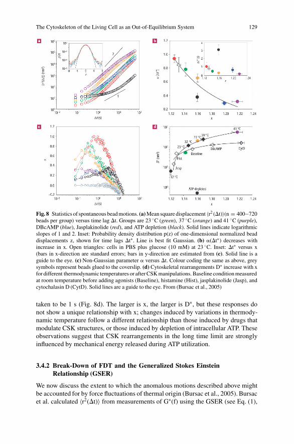
The Cytoskeleton of the Living Cell as an Out-of-Equilibrium System 129
Fig. 8 Statistics of spontaneous bead motions. (a) Mean square displacement 〈r2 (�t)〉(n = 400−720beads per group) versus time lag �t. Groups are 23 ◦C (green), 37 ◦C (orange) and 41 ◦C (purple),DBcAMP (blue), Jasplakinolide (red), and ATP depletion (black). Solid lines indicate logarithmicslopes of 1 and 2. Inset: Probability density distribution p(z) of one-dimensional normalized beaddisplacements z, shown for time lags �t∗. Line is best fit Gaussian. (b) �(�t∗) decreases withincrease in x. Open triangles: cells in PBS plus glucose (10 mM) at 23 ◦C. Inset: �t∗ versus x(bars in x-direction are standard errors; bars in y-direction are estimated from (c). Solid line is aguide to the eye. (c) Non-Gaussian parameter � versus �t. Colour coding the same as above, greysymbols represent beads glued to the coverslip. (d) Cytoskeletal rearrangements D∗ increase with xfor different thermodynamic temperatures or after CSK manipulations. Baseline condition measuredat room temperature before adding agonists (Baseline), histamine (Hist), jasplakinolide (Jasp), andcytochalasin D (CytD). Solid lines are a guide to the eye. From (Bursac et al., 2005)
taken to be 1 s (Fig. 8d). The larger is x, the larger is D∗, but these responses donot show a unique relationship with x; changes induced by variations in thermody-namic temperature follow a different relationship than those induced by drugs thatmodulate CSK structures, or those induced by depletion of intracellular ATP. Theseobservations suggest that CSK rearrangements in the long time limit are stronglyinfluenced by mechanical energy released during ATP utilization.
3.4.2 Break-Down of FDT and the Generalized Stokes EinsteinRelationship (GSER)
We now discuss the extent to which the anomalous motions described above mightbe accounted for by force fluctuations of thermal origin (Bursac et al., 2005). Bursacet al. calculated 〈r2(�t)〉 from measurements of G∗(f) using the GSER (see Eq. (1),

130 G. Lenormand et al.
Fig. 9 Breakdown of thegeneralized Stokes Einsteinrelationship. Local slope �(i.e., 〈r2(�t)〉 ∼ �t�)determined from directobservations of 〈r2(�t)〉(circles) and from predictionsbased on GSER andmeasurements of G∗(f)(squares), under baselinecondition (green) and afterATP depletion (black).Adapted from (Bursacet al., 2005)
Section 2.2.1). They focused on the local exponent � (i.e., 〈r2(�t)〉 ∼ �tθ ), andcompared these slopes with those measured directly from spontaneous bead motions(as in Fig. 8a). Measurements approach the predictions from GSER for the small-est measured time lags (Fig. 9). But for longer times, predictions and data divergestrongly, indicating a clear breakdown of GSER. The breakdown of the GSER im-plies that the system is away from thermodynamic equilibrium, and that mechanicalenergy dissipation has become uncoupled from force fluctuations of thermal origin.When ATP in the cell is largely depleted, a closer correspondence between dataand predictions is observed, however, and breakdown of the GSER is substantiallydelayed (Fig. 9).
3.5 Law 5: Laws 1-4 are Interconnected and Biologically Regulated
We have shown above that the parameter x is directly linked to the cytoskeletalprestress (Law 1, Fig. 3b). The parameter x controls the CSK’s position along a con-tinuous spectrum of solid-like versus and fluid-like states (Law 2, Fig. 5). This verysame parameter x predicts shear-softening and the rate of recovery (Law 3, Fig. 7cand d). Although it does not show an entirely unique relationship, the parameter x isthe main factor controlling the rate of cytoskeletal structural reorganization and theintermittent non-Gaussian nature of its statistics (Law 4, Fig. 8d). These observa-tions, taken together, lead to the 5th law, which is that a cell modulates its mechan-ical behaviour mainly by modifying the sole parameter x. Biological interventionsseem to modify CSK dynamics only to the extent that they affect x. Although x israther easy to measure, what it means mechanistically is quite a different matter.As described below, important clues may come from an unexpected and apparentlyunconnected field, namely soft condensed matter physics.
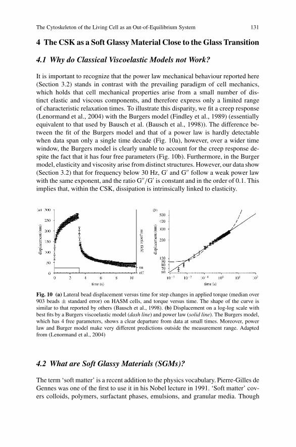
The Cytoskeleton of the Living Cell as an Out-of-Equilibrium System 131
4 The CSK as a Soft Glassy Material Close to the Glass Transition
4.1 Why do Classical Viscoelastic Models not Work?
It is important to recognize that the power law mechanical behaviour reported here(Section 3.2) stands in contrast with the prevailing paradigm of cell mechanics,which holds that cell mechanical properties arise from a small number of dis-tinct elastic and viscous components, and therefore express only a limited rangeof characteristic relaxation times. To illustrate this disparity, we fit a creep response(Lenormand et al., 2004) with the Burgers model (Findley et al., 1989) (essentiallyequivalent to that used by Bausch et al. (Bausch et al., 1998)). The difference be-tween the fit of the Burgers model and that of a power law is hardly detectablewhen data span only a single time decade (Fig. 10a), however, over a wider timewindow, the Burgers model is clearly unable to account for the creep response de-spite the fact that it has four free parameters (Fig. 10b). Furthermore, in the Burgermodel, elasticity and viscosity arise from distinct structures. However, our data show(Section 3.2) that for frequency below 30 Hz, G′ and G′′ follow a weak power lawwith the same exponent, and the ratio G′′/G′ is constant and in the order of 0.1. Thisimplies that, within the CSK, dissipation is intrinsically linked to elasticity.
Fig. 10 (a) Lateral bead displacement versus time for step changes in applied torque (median over903 beads ± standard error) on HASM cells, and torque versus time. The shape of the curve issimilar to that reported by others (Bausch et al., 1998). (b) Displacement on a log-log scale withbest fits by a Burgers viscoelastic model (dash line) and power law (solid line). The Burgers model,which has 4 free parameters, shows a clear departure from data at small times. Moreover, powerlaw and Burger model make very different predictions outside the measurement range. Adaptedfrom (Lenormand et al., 2004)
4.2 What are Soft Glassy Materials (SGMs)?
The term ‘soft matter’ is a recent addition to the physics vocabulary. Pierre-Gilles deGennes was one of the first to use it in his Nobel lecture in 1991. ‘Soft matter’ cov-ers colloids, polymers, surfactant phases, emulsions, and granular media. Though

132 G. Lenormand et al.
very different in structure, the mechanical behaviour of each of these substances issurprisingly alike. All are very soft (in the range of Pa to kPa), both the storage,G′, and the loss, G′′, moduli increase with the same weak power law dependenceson frequency, and the ratio G′′/G′ is frequency insensitive and in the order of 0.1(Sollich, 1998). The fact that similar anomalous rheology should be seen in sucha wide range of soft materials suggests a common cause (Sollich et al., 1997).Sollich et al. argued that ‘glassy’ dynamics is a natural consequence of two proper-ties shared by all soft materials: structural disorder and metastability.
4.3 Sollich’s Soft Glassy Rheology (SGR) Model
Sollich developed a theory of soft glassy rheology (SGR) using an earlier work byBouchaud (Bouchaud, 1992; Sollich, 1998; Sollich et al., 1997). The SGR modelis a phenomenological model that aims to explain the main features of SGM rhe-ology. To be reasonably generic, it incorporates only a minimal number of featurescommon to all SGMs, leaving aside as much system specific details as possible.One important feature is the ‘glassiness’ i.e., the effects of structural disorder andmetastability. This feature is modelled using a fairly intuitive picture of a glass(Fig. 11): it consists of local ‘elements’ that are trapped in ‘cages’ formed by theirneighbours so that they cannot move. Occasionally, however, a rearrangement ofthe elements may be possible, due to thermal activation, for example. In SGMshowever, it is unlikely that thermal motion alone is enough to archive completestructural relaxation. It is assumed instead that the ‘activation’ in SGMs is due to‘interactions’: a rearrangement somewhere in the material can propagate and cause
Fig. 11 Potential well picture of the dynamics of the SGR model. Note that the relative horizontaldisplacement of the quadratic potential wells is arbitrary; each has its own independent zero for thescale of the local strain �. The solid vertical bars indicate the energy dissipated in the ‘hops’ (yieldevents) from 1 to 2 and 3 to 4, respectively. When a particle hops, the stored elastic energy E =12 k�2 dissipates, giving rise to the link between G′ and G′′. Adapted from (Sollich, 1998). Reprintedfigure with permission from Sollich P., Physical Review E, 58, 738–759, 1998). Copyright (2007)by the American Physical Society

The Cytoskeleton of the Living Cell as an Out-of-Equilibrium System 133
rearrangements elsewhere. In a mean-field spirit, this coupling between elements isrepresented by an effective temperature x.
To describe rheology and flow, the SGR incorporates a local strain variable �.As the system is sheared, a constitutive element first deform elastically from a localequilibrium configuration, giving rise to a stored elastic energy (Fig. 11). This con-tinues up to a yield point, �y, whereupon rearrangement occurs. The yield energy isE = 1
2 k�y2, where k is an elastic constant. The effects of structural disorders are
modelled by assuming a distribution of yield energies E, rather than a single valuecommon to all elements. While the SGR model assumes a spatially homogeneousstrain rate, it does admit inhomogeneities in the local strain � and stress � = k�.These inhomogeneities arise because different elements generally yield at differenttimes. Therefore to describe the state of the system at a given time, one now needsto know the joint probability P(E, �, t) of finding an element with a yield energy Eand a local strain � at time t. Within the SGR model this probability evolves in timeaccording to:
�t P = −��� P − �0 Pe−[E−k�2 ]/x + �(t)ρ(E)δ(�), (4)
where
�(t) = �0
∫dE d� P(E, �, t)e−[E−k�2/2]/x, (5)
�0 is the transition rate constant, and x is the effective temperature. The macroscopicstress is given by the average of the local stress, �(E) = k
∫�P(E, �, t)d Ed�.
These three equations, taken with the distribution of trap depths, �(E) ∼e−E, definethe SGR model.
4.4 SGR Applied to the Living Cell
Remarkably, in the limit that the frequency is small compare to �0, the SGR modelleads directly to the structural damping equation (Eq. 3). If Sollich’s theory is ap-plied to the CSK, then the parameter x corresponds to the noise temperature of thecytoskeletal matrix, and represent the amount of ‘jostling’ within the CSK relativeto the depth of energy wells, G0 is the CSK stiffness at the glass transition, andf0 corresponds to �0, and is the maximum rate at which cytoskeletal elements canescape their traps. It is important to note that SGR does not invoke a viscosity.Energy dissipation in SGR theory comes not at all from a viscous process but fromelastic strain energy that is stored, and, upon a hopping transition, irreversibly lost.Finally note that when the CSK is modulated using specific drugs, we hypothesizethat changes arise from modulation of x, and that we assume the glass transitiontemperature, xg, to remain constant.
A physical picture capturing these dynamics uses an energy landscape to de-scribe all possible configurations of the CSK. A cytoskeletal element (e.g., structural

134 G. Lenormand et al.
protein or protein complex) finds itself trapped in an energy well. Trapping mightarise from physical interactions such as molecular crowding, attractive interactions,or weak chemical bonding. Thermal energy is typically insufficient to push the sys-tem over energy barriers into different configurations; this is analogous to a jammedstate (Liu and Nagel, 1998; Trappe et al., 2001). Through intermittent transitionsfrom one metastable state to another, the system evolves slowly (ages) into morestable configurations. Imposed mechanical stress can provide sufficient energy tobring the system to a new configuration, thus pushing the system farther from ther-modynamic equilibrium and resetting this evolution (rejuvenation). Applied to theliving cell, this hypothesis implies that molecular crowding and approach to kineticarrest are key attributes of CSK dynamics.
5 Open Questions
5.1 What is the Role of ATP?
Sollich interprets the effective temperature x as reflecting jostling of elements (byan unidentified but non-thermal mechanism) relative to the depth of energy wells.It appears an interesting question whether the ambiguity surrounding x might beresolved in the case of the living cell where the mechanical energy released by ATPhydrolysis is quite large. In the living cell, the energy derived from hydrolysis of thegamma phosphate bond of ATP is roughly 25 kBT per event (Howard, 2001); andfor one cubic micron of cytoplasm of a typical cell, these events occur at a rate ofmore than 104/s (Gunst and Fredberg, 2003). Vesicle transport and any local motionscaused by proteins that undergo cyclic conformational changes, such as molecularmotors, contribute to the intracellular molecular agitation. Associated local motionsare quite large, in the range 1–10 nm, which is comparable to molecular dimensionsand molecular spacing.
ATP-dependent rearrangements of the CSK modify the configurations them-selves, providing an alternate means of exploring new configurations; ATP hydrol-ysis can drive both protein conformational changes and polymerization/ depoly-merization cycles, either of which could conceivably resolve constraints and drivestructural rearrangements. This is radically different from traditional glassy systemswhich remain trapped in a configuration for increasingly long times because onlythermal energy is available to allow the system to evolve. Interestingly, x does notseem to be affected by ATP depletion (Fig. 8d and (Bursac et al., 2005)). But at thesame time, it is possible that ATP also acts to tilt the energy landscape in such a wayas to decrease energy barriers.
At the level of physics, whether E is an energy is a question of much debate. InSollich’s theory, <E> (the ensemble average of E over the probability density of E)is not a conserved quantity. This is not a fatal flaw in non-isolated systems whichare in weak contact with an external heat reservoir where <E> can either increase

The Cytoskeleton of the Living Cell as an Out-of-Equilibrium System 135
or decrease or stay the same, depending on the initial conditions of the preparedstate. This same idea is true in the Sollich’s theory – the evolution of <E> mirrorsthat of the evolution of the probability density of E. If the initial state is preparedin a high <E> configuration (perhaps associated with fluidization), then <E> willgradually decrease, and conversely, if prepared in a low <E> state, than <E> willgradually increase, both behaviors tending toward the steady state <E>, and withan approach that is temporally a power law.
5.2 What is the Meaning of the Common Intersection?
The existence of a common intersection at high frequency, f0, remains an empiricalfinding with no known mechanistic basis. Nonetheless it is highly consequential be-cause it accounts for the fact that all G′ data can be described by only one parameter,x, and therefore collapse onto unifying master relationships. Statistical analysis ofour data suggests that f0 did not vary with drug treatments and possibly not evenacross cell type (Fabry et al., 2003). In Sollich’s theory, f0 is identified as beingthe maximum rate at which cytoskeletal elements can escape their traps. However,for soft glassy materials in general, and for the living cells in particular, the factorsthat determine f0 remain unclear, and why f0 is invariant is not explained by SGRtheory.
Factors that are present in all cells are the presence of water and molecular crowd-ing, with an average distance between macromolecules of only 2 nm, or roughly10 water molecules across. Using microwave dielectric spectroscopy, Suzuki et al.have measured the rotational mobility of water in the vicinity of proteins, and theyobtained a relaxation frequency ranging from a few GHz to 40 GHz (Fuller andRand, 1999; Kabir et al., 2003; Suzuki et al., 2004). This range of relaxation fre-quencies coincides with the range of estimates for the intersection frequency f0. Wespeculate that if the cell were perturbed mechanically at a frequency correspond-ing to the rotational relaxation frequency of water, the water might dominate themechanical response of the system and differences in stiffness with various CSKinterventions might vanish. Whether this mechanism accounts for the observed in-tersection remains a matter of speculation.
Interestingly, Kramers-Smoluchowski theory of the overdamped unbinding ki-netics of weakly bonded structures involves a diffusive flux over an energy barrier,with an attempt frequency �0 = D/1c1ts, where D is the diffusion constant relatedto molecular damping, and the length scales lc and lts are the thermal widths ofthe bound states and of the barrier respectively (Evans, 2001). This theory is usedto describe force spectroscopy of single molecular bonds, and typically gives �0
in the order of 109–1010 Hz, which is consistent with the value of f0 extrapolatedfrom the measurements of G∗(f) (Fabry et al., 2001a, 2003; Lenormand et al., 2004;Puig-de-Morales et al., 2004). The striking similarity between f0 and �0 could meanthat scale-free rheology is a consequence of proteins unfolding under forces. Thispoint is further discussed in the following section.

136 G. Lenormand et al.
5.3 Protein Unfolding and Glassy Rheology
Glassy dynamics come into play at the level of forced unfolding of isolated pro-tein domains (Brujic et al., 2006). Using single-molecule force-clamp spectroscopyto measure the kinetics of unfolding of the isolated ubiquitin molecule, Brujicet al. have shown that the distribution of unfolding rates is surprisingly broad andfollows a power law with no characteristic mean. Structural relaxations are gov-erned by an exponential distribution of energy well depths with a distribution of5–10 kBT. Corresponding interconversion between conformations is scale-free andfails to conform to the modified Arrhenius form. Accordingly, conformations mayunfold only at sufficiently high forces that barriers become comparable to kBT butotherwise remain frozen into a given distribution and away from thermodynamicequilibrium.
5.4 What Can We Learn from Reconstituted F-Actin Networks?
In the past few years, reconstituted cytoskeletal protein networks have been shownto mimic many aspects of the mechanical properties of the cell, providing new in-sight into the origin of cellular behaviours (Kasza et al., 2007). Filament length andcrosslinking protein concentration in networks of F-actin are among the most widelystudied (Gardel et al., 2004; Liu et al., 2006). Interestingly, with increasing appliedstress, these networks stiffen. This stress-stiffening has important implications: themagnitude of their linear elasticity is typically orders of magnitude less than that ofcells, but when prestressed into the nonlinear regime the elasticity of these networksdramatically increases and approaches that of cells (Gardel et al., 2006). Recently,a reversible stress-softening behaviour in a reconstituted actin network has been re-ported (Chaudhuri et al., 2007). These reconstituted networks provide ideal systemsto study the complex mechanical behaviours of the living cell, and in particular theinteraction between prestress and power law rheology (Stamenovic, 2006).
6 Concluding Remarks
The CSK of the living cell exhibits physical properties and remodelling dynamicswith all the same signatures as inert condensed systems, although with importantdifferences as well. In these systems, the transition between solid-like and liquid-like states is not at all understood, and represents an outstanding unsolved problemin the field of soft condensed matter. Framing CSK dynamics in this peculiar con-text brings trapping, intermittency, and approach to kinetic arrest as key featuresof collective protein–protein interactions. These generic features represent novelmesoscale mechanisms that might link integrative CSK functions to underlyingmolecular events.

The Cytoskeleton of the Living Cell as an Out-of-Equilibrium System 137
References
A-Hassan, E., Heinz, W. F., Antonik, M. D., D’Costa, N. P., Nageswaran, S., Schoenenberger, C. A.and Hoh, J. H., 1998, Relative microelastic mapping of living cells by atomic force microscopy,Biophys. J. 74, 1564–78.
Alcaraz, J., Buscemi, L., Grabulosa, M., Trepat, X., Fabry, B., Farre, R. and Navajas, D., 2003,Microrheology of human lung epithelial cells measured by atomic force microscopy, Biophys.J. 84, 2071–9.
Alcaraz, J., Buscemi, L., Puig-De-Morales, M., Colchero, J., Baro, A. M. and Navajas, D., 2002,Correction of microrheological measurements of soft samples with atomic force microscopyfor the hydrodynamic drag on the cantilever, Langmuir 18, 716–21.
An, S. S., Fabry, B., Mellema, M., Bursac, P., Gerthoffer, W. T., Kayyali, U. S., Gaestel, M., Shore,S. A. and Fredberg, J. J., 2004, Role of heat shock protein 27 in cytoskeletal remodeling of theairway smooth muscle cell, J. Appl. Physiol. 96, 1701–13.
Balland, M., Desprat, N., Icard, D., Fereol, S., Asnacios, A., Browaeys, J., Henon, S. and Gallet,F., 2006, Power laws in microrheology experiments on living cells: comparative analysis andmodelling, Phys. Rev. E. 74, 021911.
Balland, M., Richert, A. and Gallet, F., 2005, The dissipative contribution of myosin II in thecytoskeleton dynamics of myoblasts, Eur. Biophys. J. 34, 255–61.
Bausch, A. R., Ziemann, F., Boulbitch, A. A., Jacobson, K. and Sackmann, E., 1998, Local mea-surements of viscoelastic parameters of adherent cell surfaces by magnetic bead microrheome-try, Biophys. J. 75, 2038–49.
Bissig, H., Romer, S., Cipelletti, L., Trappea, V. and Schurtenbergera, P., 2003, Inter-mittent dynamics and hyper-aging in dense colloidal gels, Phys. Chem. Comm. 6,21–3.
Bouchaud, J., 1992, Weak ergodicity breaking and aging in disordered systems, J. Phys. I. 2,1705–13.
Brujic, J., Hermans, R., Walther, K. and Fernandez, J. M., 2006, Single-molecule force spec-troscopy reveals signatures of glassy dynamics of the energy landscape of ubiquitin, NaturePhysics. 2, 282–6.
Bursac, P., Fabry, B., Trepat, X., Lenormand, G., Butler, J. P., Wang, N., Fredberg, J. J. and An,S. S., 2007, Cytoskeleton dynamics: fluctuations within the network, Biochem. Biophys. Res.Commun. 355, 324–30.
Bursac, P., Lenormand, G., Fabry, B., Oliver, M., Weitz, D. A., Viasnoff, V., Butler, J. P. andFredberg, J. J., 2005, Cytoskeletal remodelling and slow dynamics in the living cell, Nat. Mater.4, 557–61.
Butler, J. P., Tolic-Norrelykke, I. M., Fabry, B. and Fredberg, J. J., 2002, Traction fields, moments,and strain energy that cells exert on their surroundings, Am. J. Physiol. Cell Physiol. 282,C595–605.
Byfield, F. J., Aranda-Espinoza, H., Romanenko, V. G., Rothblat, G. H. and Levitan, I., 2004,Cholesterol depletion increases membrane stiffness of aortic endothelial cells, Biophys. J. 87,3336–43.
Chaudhuri, O., Parekh, S. H. and Fletcher, D. A., 2007, Reversible stress softening of actin net-works, Nature 445, 295–8.
Cipelletti, L., Ramos, L., Manley, S., Pitard, E., Weitz, D. A., Pashkovski, E. E. and Johans-son, M., 2003, Universal non-diffusive slow dynamics in aging soft matter, Faraday Discuss.123, 237–51.
Cloitre, M., Borrega, R. and Leibler, L., 2000, Rheological aging and rejuvenation in microgelpastes, Phys. Rev. Lett. 85, 4819–22.
Crick, F. H. C. and Hughes, A. F. W., 1950, The physical properties of cytoplasm, Exp. Cell. Res.1, 37–80.
Crocker, J. C., Valentine, M. T., Weeks, E. R., Gisler, T., Kaplan, P. D., Yodh, A. G. andWeitz, D. A., 2000, Two-point microrheology of inhomogeneous soft materials, Phys. Rev.Lett. 85, 888–91.

138 G. Lenormand et al.
Dahl, K. N., Engler, A. J., Pajerowski, J. D. and Discher, D. E., 2005, Power-law rheology ofisolated nuclei with deformation mapping of nuclear substructures, Biophys. J. 89, 2855–64.
Dembo, M. and Wang, Y. L., 1999, Stresses at the cell-to-substrate interface during locomotion offibroblasts, Biophys. J. 76, 2307–16.
Deng, L., Trepat, X., Butler, J. P., Millet, E., Morgan, K. G., Weitz, D. A. andFredberg, J., 2006, Fast and slow dynamics of the cytoskeleton, Nat. Mater. 5, 636–40.
Derec, C., Ducouret, G., Ajdari, A. and Lequeux, F., 2003, Aging and nonlinear rheology in sus-pensions of polyethylene oxide-protected silica particles, Phys. Rev. E 67, 061403.
Desprat, N., Richert, A., Simeon, J. and Asnacios, A., 2005, Creep function of a single living cell,Biophys. J. 88, 2224–33.
Dimitriadis, E. K., Horkay, F., Maresca, J., Kachar, B. and Chadwick, R. S., 2002, Determinationof elastic moduli of thin layers of soft material using the atomic force microscope, Biophys. J.82, 2798–810.
Discher, D. E., Janmey, P. and Wang, Y. L., 2005, Tissue cells feel and respond to the stiffness oftheir substrate, Science 310, 1139–43.
Engler, A. J., Sen, S., Sweeney, H. L. and Discher, D. E., 2006, Matrix elasticity directs stem celllineage specification, Cell 126, 677–89.
Evans, E., 2001, Probing the relation between force – lifetime – and chemistry in single molecularbonds, Annu. Rev. Biophys. Biomol. Struct. 30, 105–28.
Fabry, B., Maksym, G. N., Butler, J. P., Glogauer, M., Navajas, D. and Fredberg, J. J., 2001a,Scaling the microrheology of living cells, Phys. Rev. Lett. 87, 148102.
Fabry, B., Maksym, G. N., Shore, S. A., Moore, P. E., Panettieri, R. A., Jr., Butler, J. P. and Fred-berg, J. J., 2001b, Time course and heterogeneity of contractile responses in cultured humanairway smooth muscle cells, J. Appl. Physiol. 91, 986–94.
Fabry, B., Maksym, G. N., Butler, J. P., Glogauer, M., Navajas, D., Taback, N. A., Millet, E. J.and Fredberg, J. J., 2003, Time scale and other invariants of integrative mechanical behavior inliving cells, Phys. Rev. E. 68, 041914.
Findley, W. N., Lai, J. S. and Onaran, K., 1989, Creep and Relaxation of Nonlinear ViscoelasticMaterials with an Introduction to Linear Viscoelasticity, Mineola, New York: Dover Publica-tions, Inc.
Fredberg, J. J. and Stamenovic, D., 1989, On the imperfect elasticity of lung tissue, J. Appl. Phys-iol. 67, 2408–19.
Fuller, N. and Rand, R. P., 1999, Water in actin polymerization, Biophys. J. 76, 3261–6.Gardel, M. L., Nakamura, F., Hartwig, J. H., Crocker, J. C., Stossel, T. P. and Weitz, D. A., 2006,
Prestressed F-actin networks cross-linked by hinged filamins replicate mechanical propertiesof cells, Proc. Natl. Acad. Sci. U S A 103, 1762–7.
Gardel, M. L., Shin, J. H., MacKintosh, F. C., Mahadevan, L., Matsudaira, P. and Weitz, D. A.,2004, Elastic behavior of cross-linked and bundled actin networks, Science 304, 1301–5.
Gittes, F. and MacKintosh, F. C., 1998, Dynamic shear modulus of a semiflexible polymer network,Phys. Rev. E. 58, R1241–4.
Gunst, S. J. and Fredberg, J. J., 2003, The first three minutes: smooth muscle contraction, cy-toskeletal events, and soft glasses, J. Appl. Physiol. 95, 413–25.
Gunst, S. J., Meiss, R. A., Wu, M. F. and Rowe, M., 1995, Mechanisms for the mechanical plasticityof tracheal smooth muscle, Am. J. Physiol. 268, C1267–76.
Henon, S., Lenormand, G., Richert, A. and Gallet, F., 1999, A new determination of the shearmodulus of the human erythrocyte membrane using optical tweezers, Biophys. J. 76, 1145–51.
Hoffman, B. D., Massiera, G., Van Citters, K. M. and Crocker, J. C., 2006, The consensus mechan-ics of cultured mammalian cells, Proc. Natl. Acad. Sci. U S A 103, 10259–64.
Howard, J., 2001, Mechanics of Motor Proteins and the Cytoskeleton, Sunderland, Massachusetts:Sinauer Associates, Inc.
Ingber, D. E., 2006, Cellular mechanotransduction: putting all the pieces together again, Faseb. J.20, 811–27.
Ingber, D. E., Dike, L., Hansen, L., Karp, S., Liley, H., Maniotis, A., McNamee, H., Mooney, D.,Plopper, G., Sims, J. and et al., 1994, Cellular tensegrity: exploring how mechanical changes in

The Cytoskeleton of the Living Cell as an Out-of-Equilibrium System 139
the cytoskeleton regulate cell growth, migration, and tissue pattern during morphogenesis, Int.Rev. Cytol. 150, 173–224.
Janmey, P. A. and Weitz, D. A., 2004, Dealing with mechanics: mechanisms of force transductionin cells, Trends Biochem. Sci. 29, 364–70.
Jones, W. R., Ting-Beall, P. H., Lee, G. M., Kelley, S. S., Hochmuth, R. M. and Guilak, F., 1999,Alterations in the Young’s modulus and volumetric properties of chondrocytes isolated fromnormal and osteoarthritic human cartilage, J. Biomech. 32, 119–27.
Kabir, S. R., Yokoyama, K., Mihashi, K., Kodama, T. and Suzuki, M., 2003, Hyper-mobile wateris induced around actin filaments, Biophys. J. 85, 3154–61.
Karcher, H., Lammerding, J., Huang, H., Lee, R. T., Kamm, R. D. and Kaazempur-Mofrad, M. R.,2003, A three-dimensional viscoelastic model for cell deformation with experimental verifica-tion, Biophys. J. 85, 3336–49.
Kasza, K. E., Rowat, A. C., Liu, J., Angelini, T. E., Brangwynne, C. P., Koenderink, G. H. andWeitz, D. A., 2007, The cell as a material, Curr. Opin. Cell Biol. 19, 101–7.
Kob, W., Donati, C., Plimpton, S. J., Poole, P. H. and Glotzer, S. C., 1997, Dynamical hetero-geneities in a supercooled Lennard-Jones liquid, Phys. Rev. Lett. 79, 2827–30.
Lau, A. W. C., Hoffman, B. D., Davies, A., Crocker, J. C. and Lubensky, T. C., 2003, Mi-crorheology, stress fluctuations, and active behavior of living cells, Phys. Rev. Lett. 91,198101.
Laudadio, R. E., Millet, E. J., Fabry, B., An, S. S., Butler, J. P. and Fredberg, J. J., 2005, Rat airwaysmooth muscle cell during actin modulation: rheology and glassy dynamics, Am. J. Physiol.Cell. Physiol. 289, C1388–95.
Lenormand, G., Bursac, P., Butler, J. P. and Fredberg, J. J., 2007a, Out-of-equilibrium dynamics inthe cytoskeleton of the living cell, Phys. Rev. E. 76, 041901.
Lenormand, G., Chopin, J., Bursac, P., Fredberg, J. J. and Butler, J. P., 2007b, Directional mem-ory and caged dynamics in cytoskeletal remodelling, Biochem. Biophys. Res. Commun. 360,797–801.
Lenormand, G., Millet, E., Fabry, B., Butler, J. P. and Fredberg, J. J., 2004, Linearity and time-scaleinvariance of the creep function in living cells, J. R. Soc. Interface 1, 91–7.
Levine, A. J. and Lubensky, T. C., 2001, Two-point microrheology and the electrostatic analogy,Phys. Rev. E 65, 11501.
Liu, A. J. and Nagel, S. R., 1998, Jamming is not just cool any more, Nature 396, 21–22.Liu, J., Gardel, M. L., Kroy, K., Frey, E., Hoffman, B. D., Crocker, J. C., Bausch, A. R. and
Weitz, D. A., 2006, Microrheology probes length scale dependent rheology, Phys. Rev. Lett.96, 118104.
Mason, T. G., 2000, Estimating the viscoelastic moduli of complex fluids using the generalizedStokes-Einstein equation, Rheologica. Acta. 39, 371–8.
Mason, T. G. and Weitz, D. A., 1995a, Linear Viscoelasticity of Colloidal Hard Sphere Suspensionsnear the Glass Transition, Phys. Rev. Lett. 75, 2770–3.
Mason, T. G. and Weitz, D. A., 1995b, Optical measurements of frequency-dependent linear vis-coelastic moduli of complex fluids, Phys. Rev. Lett. 74, 1250–3.
Matthews, B. D., Overby, D. R., Alenghat, F. J., Karavitis, J., Numaguchi, Y., Allen, P. G. and In-gber, D. E., 2004, Mechanical properties of individual focal adhesions probed with a magneticmicroneedle, Biochem. Biophys. Res. Commun. 313, 758–64.
Mijailovich, S. M., Kojic, M., Zivkovic, M., Fabry, B. and Fredberg, J. J., 2002, A finiteelement model of cell deformation during magnetic bead twisting, J. Appl. Physiol. 93,1429–1436.
Mizuno, D., Tardin, C., Schmidt, C. F. and Mackintosh, F. C., 2007, Nonequilibrium mechanics ofactive cytoskeletal networks, Science 315, 370–3.
Nash, G. B. and Gratzer, W. B., 1993, Structural determinants of the rigidity of the red cell mem-brane, Biorheology 30, 397–407.
Ohayon, J., Tracqui, P., Fodil, R., Fereol, S., Laurent, V. M., Planus, E. and Isabey, D., 2004,Analysis of nonlinear responses of adherent epithelial cells probed by magnetic bead twisting:A finite element model based on a homogenization approach, J. Biomech. Eng. 126, 685–98.

140 G. Lenormand et al.
Overby, D. R., Matthews, B. D., Alsberg, E. and Ingber, D. E., 2005, Novel dynamic rheologi-cal behavior of individual focal adhesions measured within single cells using electromagneticpulling cytometry, Acta. Biomater. 1, 295–303.
Pratusevich, V. R., Seow, C. Y. and Ford, L. E., 1995, Plasticity in canine airway smooth muscle,J. Gen. Physiol. 105, 73–94.
Puig-de-Morales, M., Millet, E., Fabry, B., Navajas, D., Wang, N., Butler, J. P. and Fredberg, J. J.,2004, Cytoskeletal mechanics in adherent human airway smooth muscle cells: probe specificityand scaling of protein-protein dynamics, Am. J. Physiol. Cell. Physiol. 287, C643–54.
Puig-de-Morales-Marinkovic, M., Turner, K. T., Butler, J. P., Fredberg, J. J. and Suresh, S.,2007, Viscoelasticity of the human red blood cell, Am. J. Physiol. Cell Physiol. 293,C597–605.
Ramos, L. and Cipelletti, L., 2001, Ultraslow dynamics and stress relaxation in the aging of a softglassy system, Phys. Rev. Lett. 87, 245503.
Rico, F., Roca-Cusachs, P., Gavara, N., Farre, R., Rotger, M. and Navajas, D., 2005, Probing me-chanical properties of living cells by atomic force microscopy with blunted pyramidal cantilevertips, Phys. Rev. E Stat. Nonlin. Soft Matter Phys. 72, 021914.
Roca-Cusachs, P., Almendros, I., Sunyer, R., Gavara, N., Farre, R. and Navajas, D., 2006, Rheologyof passive and adhesion-activated neutrophils probed by atomic force microscopy, Biophys. J.91, 3508–18.
Rowat, A. C., Lammerding, J. and Ipsen, J. H., 2006, Mechanical properties of the cell nucleus andthe effect of emerin deficiency, Biophys. J. 91, 4649–64.
Sato, M., Theret, D. P., Wheeler, L. T., Ohshima, N. and Nerem, R. M., 1990, Application of the mi-cropipette technique to the measurement of cultured porcine aortic endothelial cell viscoelasticproperties, J. Biomech. Eng. 112, 263–8.
Schmid-Schonbein, G. W., Sung, K. L., Tozeren, H., Skalak, R. and Chien, S., 1981, Passive me-chanical properties of human leukocytes, Biophys. J. 36, 243–256.
Smith, B. A., Roy, H., De Koninck, P., Grutter, P. and De Koninck, Y., 2007, Dendritic spineviscoelasticity and soft-glassy nature: balancing dynamic remodeling with structural stability,Biophys. J. 92, 1419–30.
Smith, B. A., Tolloczko, B., Martin, J. G. and Grutter, P., 2005, Probing the viscoelastic behaviorof cultured airway smooth muscle cells with atomic force microscopy: stiffening induced bycontractile agonist, Biophys. J. 88, 2994–3007.
Smith, P. G., Deng, L., Fredberg, J. J. and Maksym, G. N., 2003, Mechanical strain increases cellstiffness through cytoskeletal filament reorganization, Am. J. Physiol. Lung. Cell Mol. Physiol.285, L456–63.
Sollich, P., 1998, Rheological constitutive equation for a model of soft glassy materials, Phys. Rev.E. 58, 738–59.
Sollich, P., Lequeux, F., Hebraud, P. and Cates, M. E., 1997, Rheology of soft glassy materials,Phys. Rev. Lett. 78, 2020–3.
Stamenovic, D., 2006, Cell mechanics: two regimes, maybe three? Nat. Mater 5, 597–8.Stamenovic, D. and Coughlin, M. F., 1999, The role of prestress and architecture of the cytoskele-
ton and deformability of cytoskeletal filaments in mechanics of adherent cells: a quantitativeanalysis, J. Theor. Biol. 201, 63–74.
Stamenovic, D., Rosenblatt, N., Montoya-Zavala, M., Matthews, B. D., Hu, S., Suki, B., Wang, N.and Ingber, D. E., 2007, Rheological behavior of living cells is timescale-dependent, Biophys.J. 93, L39–41.
Stamenovic, D., Suki, B., Fabry, B., Wang, N. and Fredberg, J. J., 2004, Rheology of airwaysmooth muscle cells is associated with cytoskeletal contractile stress, J. Appl. Physiol. 96,1600–5.
Struick, L. C. E., 1978, Physical Aging in Amorphous Polymers and Other Materials, Houston,Texas: Elsevier.
Suzuki, M., Kabir, S. R., Siddique, M. S., Nazia, U. S., Miyazaki, T. and Kodama, T., 2004,Myosin-induced volume increase of the hyper-mobile water surrounding actin filaments,Biochem. Biophys. Res. Commun. 322, 340–6.

The Cytoskeleton of the Living Cell as an Out-of-Equilibrium System 141
Theret, D. P., Levesque, M. J., Sato, M., Nerem, R. M. and Wheeler, L. T., 1988, The application ofa homogeneous half-space model in the analysis of endothelial cell micropipette measurements,J. Biomech. Eng. 110, 190–9.
Thoumine, O., Cardoso, O. and Meister, J. J., 1999, Changes in the mechanical properties of fi-broblasts during spreading: a micromanipulation study, Eur. Biophys. J. 28, 222–34.
Thoumine, O. and Ott, A., 1997, Time scale dependent viscoelastic and contractile regimes infibroblasts probed by microplate manipulation, J. Cell Sci. 110 ( Pt 17), 2109–16.
Trappe, V., Prasad, V., Cipelletti, L., Segre, P. N. and Weitz, D. A., 2001, Jamming phase diagramfor attractive particles, Nature 411, 772–5.
Trepat, X., Deng, L., An, S. S., Navajas, D., Tschumperlin, D. J., Gerthoffer, W. T., Butler, J. P.and Fredberg, J. J., 2007, Universal physical responses to stretch in the living cell, Nature 447,592–5.
Tseng, Y., Kole, T. P. and Wirtz, D., 2002, Micromechanical mapping of live cells by multiple-particle-tracking microrheology, Biophys. J. 83, 3162–76.
Tseng, Y. and Wirtz, D., 2001, Mechanics and multiple-particle tracking microheterogeneity ofalpha-actinin-cross-linked actin filament networks, Biophys. J. 81, 1643–56.
Valentine, M. T., Perlman, Z. E., Mitchison, T. J. and Weitz, D. A., 2005, Mechanical properties ofXenopus egg cytoplasmic extracts, Biophys. J. 88, 680–9.
Van Citters, K. M., Hoffman, B. D., Massiera, G. and Crocker, J. C., 2006, The role of F-actin andmyosin in epithelial cell rheology, Biophys J. 91, 3946–56.
Wang, N., Butler, J. P. and Ingber, D. E., 1993, Mechanotransduction across the cell surface andthrough the cytoskeleton, Science 260, 1124–7.
Wang, N., Naruse, K., Stamenovic, D., Fredberg, J. J., Mijailovich, S. M., Tolic-Norrelykke, I. M.,Polte, T., Mannix, R. and Ingber, D. E., 2001, Mechanical behavior in living cells consistentwith the tensegrity model, Proc. Natl. Acad. Sci. U S A 98, 7765–70.
Wang, N., Tolic-Norrelykke, I. M., Chen, J., Mijailovich, S. M., Butler, J. P., Fredberg, J. J. andStamenovic, D., 2002, Cell prestress. I. Stiffness and prestress are closely associated in adherentcontractile cells, Am. J. Physiol. Cell Physiol. 282, C606–16.
Weeks, E. R., Crocker, J. C., Levitt, A. C., Schofield, A. and Weitz, D. A., 2000, Three-dimensionaldirect imaging of structural relaxation near the colloidal glass transition, Science 287, 627–31.
Wottawah, F., Schinkinger, S., Lincoln, B., Ananthakrishnan, R., Romeyke, M., Guck, J. and Kas,J., 2005, Optical rheology of biological cells, Phys. Rev. Lett. 94, 098103.
Yamada, S., Wirtz, D. and Kuo, S. C., 2000, Mechanics of living cells measured by laser trackingmicrorheology, Biophys. J. 78, 1736–47.
Yanai, M., Butler, J. P., Suzuki, T., Sasaki, H. and Higuchi, H., 2004, Regional rheological differ-ences in locomoting neutrophils, Am. J. Physiol. Cell. Physiol. 287, C603–11.


Unexpected Linkage Between Unstirred Layers,Exclusion Zones, and Water
Gerald H. Pollack and James Clegg
Abstract We make the case that the unstirred layers of classical physiology arisefrom the influence of surfaces on the structure, and therefore the properties, ofcontiguous water. Traditionally, unstirred layers have been thought to arise merelyout of stagnant volumes adjacent to membranes and other surfaces. These volumeswould have to extend tens to hundreds of micrometers in order to account for the ob-served effects. On the other hand, charged and hydrophilic surfaces have been shownto impact water out to surprising distances, on the order of hundreds of micrometers.We present evidence that it is this water-structuring effect that is responsible for theunstirred layer.
Keywords Unstirred layers · exclusion zones · water structure
1 Unstirred Layers (USLs)
It is generally agreed that unstirred layers (USLs) are more or less static fluid regionsadjacent to membranes, and probably many other surfaces, within which thermalconvection or density gradients do not cause significant mixing (Barry and Dia-mond, 1984). Even when the surrounding fluid is vigorously stirred this seeminglystatic fluid remains, at least to some extent, and there is a relationship between thethickness of USLs (�) and the intensity of stirring of the surrounding fluid (Barryand Diamond, 1984).
USLs are of critical importance in a wide variety of physiological processes, no-tably those involving solute transport, enzyme and other chemical reactions on or nearthe cell surface. Membrane electrochemical potentials may also be affected becauseconcentrations of ions and other solutes adjacent to the plasma membrane are not thesame as those in the bulk, an important point to which we will return. The literatureon USLs is massive, including research in physical chemistry where the USLs havebeen referred to as boundary layers (House, 1974; Barry and Diamond, 1984). These
G.H. PollackDepartment of Bioengineering, Box 355061, University of Washington, Seattle WA, 98195, USAe-mail: [email protected]
G.H. Pollack, W.-C. Chin (eds.), Phase Transitions in Cell Biology,C© Springer Science+Business Media B.V. 2008
143

144 G.H. Pollack, J. Clegg
layers have been implicated in many processes, and in a wide variety of systems suchas the gills of mussels (Wright et al., 1980), the human jejunum (Pappenheimer,2001),monolayer cultures of mammalian cells (Spivak et al., 2006), rainbow-trout embryosand larvae (Ciuhandu et al., 2007) and a giant-celled alga (Kim et al., 2006). We makeno pretence about a thorough review of this literature but will attempt to point outfeatures that are salient to the topics covered in this essay.
A brief examination of the history of USLs seems worthwhile. In an importantearly review, Dainty and House (1966) state that the USL concept was originallydeveloped by Noyes and Whitney in 1897 and further in 1904 by Nernst. However,based on the translation of Wilhelm Pfeffer’s 1877 book, Osmotische Unteruchun-gen (Kepner and Stadelmann, 1985), it appears that Pfeffer described USLs wellbefore those authors. Pfeffer clearly emphasized the critical role played by USLs inhis extensive research on osmosis and related processes. Key papers on USLs in theearly to mid-1900s have been considered or reviewed by Teorell (1936) Ginzbergand Katchalsky (1963) and Dick (1959). The review by Barry and Diamond, (1984)is definitive in our opinion, with excellent overall coverage of USLs. More recently,work from the laboratory of Peter Pohl considers the wide-ranging significance andimplications of USLs and their relation to what is called reaction layers, RLs (seeAntonenko et al., 1996; Pohl et al., 1998). RLs are solution layers adjacent to cellmembranes within which chemical reactions, including enzymatic ones, take place.These layers have been described previously (see Gutnecht and Tosteson,1973;Verkman and Dix, 1984) but particularly by Antonenko et al. (1996). We mentionthem here to draw attention to the important relationship between USLs and RLsand, as we will propose, the importance of water in the operation of both layers.
A key feature of USLs concerns their thickness—the distance from membranesand other surfaces over which mechanical stirring is not effective. An excellentsource of information on USL thickness (�) is the review by Barry and Diamond(1984). Without stirring, � exceeds 300 μm, depending on the nature of the interface,and can even extend over several millimeters from the surface. Despite the mostvigorous stirring, � remains near 10 μm, obviously a huge distance in molecularterms. The view is widely held that the USL is not stationary but a region of slowlaminar flow, parallel to the interface, in which solute transport occurs by diffusionalone (Dainty and House, 1966).
A key question arises: what generates the USL? A logical possibility is that thephysical properties of the USL are somehow different from those of regions beyond.To our knowledge the “structure” of water in such interfacial regions has not beenconsidered in this connection. We wish to raise this possibility, that the influenceof the adjacent “solid” surface, membrane, or otherwise, alters the structure andproperties of the interfacial water in such a way that the USL is generated.
2 Cellular Water
Studies on the properties and structure of water in cells and adjacent to surfaceshave been many, and the topic has had a long and often controversial history. Itis not appropriate here to review that history, except to note certain points. Cell

Unexpected Linkage Between Unstirred Layers, Exclusion Zones, and Water 145
biology seems to have proceeded throughout its history on the assumption that allbut a small fraction (perhaps 5%) of total cell water exhibited the properties of bulkwater, and that the cytoplasm could be treated as an ordinary, although non-ideal,aqueous solution (see Clegg, 1982). Some have objected to that view over the years,notably Gilbert Ling, Walter Drost-Hansen, and Aphanasij Troschin, who proposedthat most and probably all of cell water is different. Those three scientists began theirwork in the 1950s, and two continue their work today (see Pollack et al., 2006). Ahigh point arrived in the late 1960s when both Freeman Cope (1969), and CarltonHazlewood and colleagues (1969), published papers using pulsed NMR technology.They concluded that the structure of most or even all of water in skeletal muscle andbrain was not that of the bulk liquid.
Much attention was given to these results, supporting the positions of Ling,Drost-Hansen and Troschin, but it was not very long until opposition to those viewsarose (see Kolata, 1976). What followed was a contentious period spanning some 10years of claim and counterclaim (considered to some extent in works by Hazlewood,1973; Franks and Mathias, 1982; Clegg and Drost-Hansen, 1991; Drost-Hansen andClegg, 1979; Ling, 2001; Mentre, 2001; Pollack, 2001; Pollack et al., 2006). As ofthis writing research on cell water has slowed greatly, with a few notable exceptions(see articles in Mentre, 2001; Pollack, 2001; Pollack et al., 2006) although interestin the subject continues (for example, Leterrier, 2001; Shepherd, 2006). The versiongiven in textbooks and the general literature is not much different than it was acentury ago: almost all of cell water is considered to be essentially the same as thatin the bulk liquid. But there remains a dissenting group, albeit small, that believesthe last word has yet to be said; we are among that group.
That surfaces alter the structure of water immediately adjacent to them is wellestablished for very short distances, equivalent to two or three molecular layers ofwater. But it is our impression that very few (other than those named above) wouldaccept the possibility that such influences might extend over distances of tens orhundreds of nanometers, let alone micrometers. However, in the sections that followwe will make a case for just that; namely, that the existence and properties of USLsarise from the influence of membranes and other “solid” surfaces on the structure ofwater adjacent to them.
3 Exclusion Zones (EZs)
Motivated independently from unstirred layer considerations, studies of the physicalbehavior of this near-surface aqueous zone were begun several years ago (Zheng andPollack, 2003). These studies were in fact broader than just examining the unstirredlayer next to cells: they dealt with the nature of aqueous regions adjacent to manyhydrophilic surfaces.
A half-century old review of the solid-liquid interface (Henniker, 1949) citedmore than 100 studies showing a surface influence extending into the liquid farbeyond the two or three molecular layers generally implied; and, a later studyshowed that on polished quartz, ordered water could extend out to some 600

146 G.H. Pollack, J. Clegg
molecular layers from the surface (Pashley and Kitchener, 1979). For many decadesDrost-Hansen has mustered evidence that similar distances are involved in a widevariety of surface systems, the adjacent water being referred to as vicinal water (seeDrost-Hansen, 2006). These reports, and many references therein, indicate consid-erably more structuring than has generally been accepted. Hence, positions weretaken on both sides of the issue.
The experimental approach taken by Zheng and Pollack (2003) was to explore thebehavior of colloidal particles next to hydrophilic surfaces. Since it is energeticallyfavorable for colloidal particles and solutes to reside outside of structured regions sothey can more readily hydrate, attempts were made to examine solute partitioning inthe vicinity of interfaces. If such solutes are preferentially excluded from the interfa-cial zone, the presumption was that this zone was likely to be structured differentlyfrom ordinary bulk water.
Initial results seemed astonishing (Zheng and Pollack, 2003). A polyvinyl al-cohol gel was placed in a chamber, and the edge of the gel was visualized in anoptical microscope. A suspension of latex microspheres was poured over the gel,filling the chamber. Immediately, microspheres began migrating away from the gelsurface, into the water. Within several minutes a microsphere-free zone could beseen extending some 50–100 μm from the gel surface (Fig. 1). The result impliedthat the exclusion zone could quite possibly extend by as much as 100 μm from thegel surface.
In retrospect, this observation should not have been quite so astonishing. As men-tioned, Drost-Hansen has long argued that the properties of water adjacent to sur-faces, vicinal water, were different from the bulk, and that these properties extendedover great distances from surfaces, in some cases micrometers (see Drost-Hansen,
PVA gel
100 µ m
microspheres
microspheres
EZ
Fig. 1 Exclusion zones (dark, “EZ”) on either side of a polyvinyl alcohol gel. Microspheres (upperleft and lower right) are excluded from the region close to the gel. Cloud-like bright areas areartifactual reflections

Unexpected Linkage Between Unstirred Layers, Exclusion Zones, and Water 147
200 µm
PAA gel
Fig. 2 Exclusion zones (dark) on either side of a polyacrylic acid gel. Microspheres translatedfrom the gel surface appear at the far right. The vertical bright stripe in the middle of the PAA gelis an optical artifact
2006). Further, those observations seemed in concordance with the many results ob-tained during the first half of the previous century, as reviewed by Henniker (1949).But the fact remains that these observations and proposals have had little effect oncurrent views concerning interfacial water.
Even more remarkable than the 100-μm EZ seen with polyvinyl alcohol gels wasthe exclusion zone seen with the more highly charged polyacrylic-acid gels. There,the microsphere-exclusion zone could easily extend from the gel surface by 250 μmor more (Fig. 2).
It was not only microspheres that were excluded from the region next to gels.Many suspended or dissolved species were excluded, ranging in size from particlessuch as sand and silt, to biological entities including bacteria and erythrocytes,down to much smaller species such as proteins and low molecular weight dyes. Allof those were excluded (Zheng et al., 2006). The kinds of surfaces that producedsuch exclusion also ranged broadly. They included a series of hydrogels, variouspolymeric surfaces including Nafion, and biological surfaces including vascular en-dothelium, seaweed, and various plant roots. Muscle, for example, produced an EZof several hundred micrometers (Zheng et al., 2006). Hence, the exclusion phe-nomenon seems rather general, and is not the quirk of a particular solute juxtaposednext to a particular surface.
It was after those experiments had been largely completed that we came uponthe study by Green and Otori (1970) which linked surfaces and microspheres withunstirred layers. Curious about the nature of the unstirred layer around the eyecornea, those investigators exposed the cornea to suspensions of polystyrene latexmicrospheres in much the same manner as we had done more recently with otherhydrophilic surfaces. The results were similar. Green and Otori found microsphere-exclusion zones on the order of 350 μm. They found exclusion not only adjacentto the natural cornea, but also next to artificial contact lenses – results virtually

148 G.H. Pollack, J. Clegg
identical to what we found next to polyNIPAM, the gel used to fabricate contactlenses. Clearly, the results of Green and Otori and the more recent results obtainedin the Pollack laboratory are for all intents and purposes indistinguishable. The un-stirred layer of classical physiology and the solute-exclusion zone seem to be oneand the same.
4 Nature of Unstirred Layers and Exclusion Zones
What is the nature of the water within USLs and EZs? One possibility is that it isidentical to bulk water, and that exclusion merely derives from proximity to surfaces.Because of entropic effects, solutes should be in lower concentrations in regions“close to” surfaces. Such effects are generally considered to extend out to several,or even tens of molecular layers of water. But, here the distances under considerationare far greater: a span of 250–300 μm is equivalent to a stack of about a million watermolecules. Such numbers are far too large to fall into the realm of surface-entropiceffects.
A second possibility is that the water in this zone may be impacted by the surface;i.e., that the surface influences water structure out to distances considerably fartherthan expected. The results described above are fully consistent with that possibility,but they do not prove it. More recent results, on the other hand, lend further supportto the possibility that water in the exclusion zone is physically different from bulkwater.
A significant indication of water-structural differences in the vicinal zone derivesfrom studies using UV-Vis spectrometry (Zheng et al., 2006). Those studies involvedNafion, whose surface generates an appreciable exclusion zone. A sheet of Nafionwas glued to the inside face of one of the four vertical walls of a standard cuvette,which was then filled with distilled, deionized water. The spectrometer was modi-fied such that a narrow slit of light could sample zones parallel to, and at variabledistances from, the Nafion face.
Far from the Nafion surface, the absorption spectrum was indistinguishable fromthat of bulk water. As the narrow sampling window came progressively closer tothe Nafion surface, a new absorption peak at ∼270 nm, could be detected. The peakbecame prominently visible at the exclusion-zone boundary, ∼ 500 μm from theNafion surface. It grew further as the window came closer to the surface, ultimatelydominating the pattern (Fig. 3). Since no absorption peaks at 270 nm are foundin bulk water, the presence of a strong “anomalous” absorption peak implies thatexclusion-zone water differs appreciably from bulk water.
Artifacts in such measurements are possible: namely, some leaching of polymerfrom the Nafion could be responsible for this absorption. Control experiments havebeen carried out to determine whether a similar absorption peak occurs in situationsin which water structuring is anticipated. Indeed, various concentrated salt solutionsand sugar solutions showed similar absorption peaks (Chai et al., 2008). When thesalts were first heat-purified in order to remove volatile hydrocarbon contaminants,

Unexpected Linkage Between Unstirred Layers, Exclusion Zones, and Water 149
Fig. 3 UV-Vis absorption spectrum as a function of distance from the Nafion surface
the absorption peaks grew more intense. Hence, these absorption peaks do not ap-pear to be artifacts, but may be a genuine “signature” of water structure.
Several other physical features of exclusion-zone water differ from their coun-terparts in bulk water. These include shorter NMR-T2 relaxation times, reducedinfrared radiation, and a steep electrical potential gradient (Zheng et al., 2006).Unpublished experiments also show different optical refractive features, increasedviscosity, and higher density.
Taken together, these observations imply that water in the exclusion zone is phys-ically, and perhaps chemically different from ordinary bulk water. In particular, thereduced infrared radiation and shorter T2 values imply that water in USLs and EZsmay be structurally more stable than bulk water—perhaps partially liquid crystallinein nature. In such a case it would be clear why colloids and solutes might be ex-cluded: energetically, they prefer to lodge in the region of bulk water, where theycan more easily hydrate. It also accounts for the cornea and contact lens observationsof Green and Otori (1970), who found that microspheres did not invade the unstirredlayer. Apparently, the microspheres avoided the region because it was structured ina way that did not admit them.
Thus, the unstirred layers of classical physiology appear to be the same as exclu-sion zones found next to many hydrophilic surfaces. Water in this zone is extensivelyimpacted by the surface, and the resulting structure tends to exclude particles and so-lutes. On the other hand, important questions remain. One of the issues to be studiedin more detail is the degree to which ions and small solutes penetrate the exclusionzone. To our knowledge this has not yet been explored directly. Although largersolutes and colloids seem fully excluded from such zones, it is not yet clear whetherthe smallest solutes are fully excluded, partially excluded, or not excluded. Giventhe striking similarities thus far established between unstirred layers and exclusionzones, there is reason to be optimistic that the “limited diffusion” of small solutesthrough the USL may be seen as well in the exclusion zone, but such hypothesesremain to be tested.

150 G.H. Pollack, J. Clegg
5 Ultrastructural Evidence for Exclusion Zones
Mollenhauer and Morre (1978) seem to have been the first to point out that zonesof exclusion (their term) can be detected at the ultrastructural level in eukaryoticcells using transmission electron microscopy. They defined these zones as “differ-entiated regions of cytoplasm in which ribosomes, glycogen and organelles such asmitochondria, plastids or microbodies are scarce or absent.” They also list other cellstructures that are excluded. Our impression is that exclusion zones are consistentlyfound adjacent to various membranes or cytoskeletal structures. These include actinfilaments (Kamitsubo, 1972), and microtubules (Stebbings and Willison, 1973;Stebbings and Hunt, 1982), and they persist even when most of the water hasbeen osmotically removed (Albrecht-Buehler and Bushnell, 1982). Thus, we believethese ultrastructural exclusion zones are comparable to the USLs and EZs we havediscussed in this paper, extending their importance to the in vivo case.
6 Concluding Comments
The USL of classical physiology has long been known and studied. That trail hasgone on with little or no attention given to what generates it. In this essay we supportthe view that the properties and structure of both the USL and EZ arise because ofthe influence of surfaces on the properties and structure of water in its vicinity. Thiswater is somehow altered. The alteration may be profound—enough so that it hasbeen considered to constitute a “fourth phase” of water (Hardy, 1932). The viewthat “boundary water” is altered is supported by evidence briefly summarized here,much of it coming from work done over the last 50 years, indicating that the influ-ence of surfaces extends over large distances, exceeding 100 μm. That contention,standing in opposition to the conventional wisdom, indicates that the role of water inand around cells and tissues is vastly more important than the teachings of currenttextbooks permit. As always, the evidence will prevail, and the future will decidethat outcome.
References
Albrecht-Buehler G, Bushnell A (1982) Reversible compression of the cytoplasm. Exp Cell Res140:173–189
Antonenko YN, Pohl P, Rosenfeld E (1996) Visualization of the reaction layer in the immediatemembrane vicinity. Arch Biochem Biophys 333:225–232
Barry PH, Diamond JM (1984) Effects of unstirred layers on membrane phenomena. Physiol Rev64:763–871
Chai, B-H, Zheng, J-M, Zhao, Q, Pollack, GH (2008) Spectroscopic studies of solutes in aqueoussolution. J. Phys. Chem A 112:2242–2247
Ciuhandu CS, Wright PA, Goldberg JI, Stevens ED (2007) Parameters influencing the dissolvedoxygen in the boundary layer of rainbow trout (Oncorhynchus mykiss) embryos and larvae.J Exp Biol 210:1435–1445

Unexpected Linkage Between Unstirred Layers, Exclusion Zones, and Water 151
Clegg JS (1982) Alternative views on the role of water in cell function. In: Franks F, Mathias SF(ed.) Biophysics of Water. John Wiley and Sons, New York, pp. 365–385
Clegg JS, Drost-Hansen W (1991) On the biochemistry and cell physiology of water. In:Hochachka PW, Mommsen TP (ed.), Biochemistry and Molecular Biology of Fishes, Elsevier,Amsterdam, pp. 1–23
Cope F (1969) Nuclear magnetic resonance evidence using D2O for structured water in muscle andbrain. Biophys J 9:303–319
Dainty J, House CR (1966) “Unstirred layers” in frog skin. J Physiol 182:66–78Dick DAT (1959) Osmotic properties of living cells. Int Rev Cytol 8:387–448Drost-Hansen W (2006) Vicinal hydration of biopolymers: cell biological consequences. In:
Pollack GH, Cameron IL, Wheatley DN (ed.). Water and the Cell. Springer-Verlag, Berlin,pp. 175–217
Drost-Hansen W, Clegg JS (ed.) (1979) Cell Associated Water. Ascademic Press, New York, p. 440Franks F, Mathias SF (ed.) (1982) Biophysics of Water. John Wiley and Sons, New York, p. 400Ginzberg BZ, Katchalsky A (1963) The frictional coefficients of the flows of non-electrolytes
through artificial membranes. J Gen Physiol 47:403–418Green K, Otori T (1970) Direct measurement of membrane unstirred layers. J Physiol 207:93–102Gutknecht, J, Tosteson, DC (1973). Diffusion of weak acids across lipid bilayer membranes: effects
of chemical reactions in the unstirred layers. Science 182:1258–1261Hardy, W (1932) Problems of the boundary state. Phil Trans Roy Soc Lon Ser A 230:1–37Hazlewood CF (ed.) (1973) Physicochemical state of ions and water in living tissues and model
systems. Ann NY Acad Sci 204:5–631Hazlewood CF, Nichols BL, Chamberlain NF (1969) Evidence for the existence of a minimum of
two phases of ordered water in skeletal muscle. Nature 222:747–750Henniker, JC (1949) The depth of the surface zone of a liquid. Rev Mod Phys 21(2):322–341House CR (1974) Water Transport in Cells. Arnold Press, London, p. 276Kamitsubo, E (1972) Motile protoplasmic fibrils in cells of the Characae. Protoplasma 74:53–70Kepner GR, Stadelmann EJ (1985) Translation of Osmotische Untersuchungen. Studien zur
Zellemechanik. Van Nostrand Reinhold, New York, p. 267Kim Y, Ye Q, Reinhardt H, Steudle E (2006) Further quantification of the role of internal stirred
layers during the measurement of transport coefficients in giant internodes of Chara by a newstop-flow technique. J Exp Bot 57:4133–4144
Kolata G (1976) Water structure and ion binding: a role in cell physiology? Science 192:1220–1222Leterrier JF (2001) Water and the cytoskeleton. Cell Mol Biol (Noisy-le-grand) 47:901–923Ling GN (2001) Life at the Cell and Below-Cell Level. Pacific Press, New York, p. 373Mentre P (ed.) (2001) Water in the cell. Cell Mol Biol 47:709–970Mollenhauer HH, Morre DJ (1978) Structural compartmentation of the cytosol: zones of exclusion,
zones of adhesion, cytoskeletal and intercisternal elements. In: Roodyn DB (ed.) SubcellularBiochemistry, vol. 5, Plenum Press, New York, pp. 327–362
Pappenheimer JR (2001) Role of pre-epithelial “unstirred” layers in absorption of nutrients fromthe human jejunum. J Membrane Biol 179:185-204
Pashley RM, Kitchener JA (1979) Surface forces in adsorbed multilayers of water on quartz. J CollInterface Sci 71: 491–500
Pohl P, Saparov SM, Antonenko YN (1998) The size of the unstirred layer as a function of thesolute diffusion coefficient. Biophys J 75:1403–1409
Pollack GH (2001) Cells, Gels and the Engines of Life. Ebner and Sons, Seattle, p. 305Pollack GH, Cameron IL, Wheatley DN (ed.) (2006) Water and the Cell. Springer-Verlag, Berlin,
pp. 165–174.Shepherd VA (2006) The cytomatrix as a cooperative system of macromolecular and water net-
works. Curr Top Dev Biol 75:171–233Spivak CE, Oz M, Beglan CL, Shrager RI (2006) Diffusion delays and unstirred layer effects at
monolayer cultures of Chinese hamster ovary cells. Cell Biochem Biophys 45:43–58Stebbings H and Hunt C (1982) The nature of the clear zone around microtubules. Cell and Tissue
Res 227:609–617

152 G.H. Pollack, J. Clegg
Stebbings H, Willison JHM (1973) Structure of microtubules: a study of freeze-etched and neg-atively stained microtubules from the ovaries of Notonecta. Z. Zellforsch u Mikrosc Anat138(3):387–396
Teorell T (1936) A method of studying conditions within diffusion layers. J Biol Chem113:735–748
Verkman AS, Dix JA (1984) Effect of unstirred layers on binding and reaction kinetocs at a mem-brane surface. Anal Biochem 142:109–116
Wright SH, Becker SA, Stephens GC (1980) Influence of temperature and unstirred layers on thekinetics of glycine transport in isolated gills of Mytilus californianus. J Exp Zool 214:27–35
Zheng, JM, Pollack, GH (2003) Long range forces extending from polymer surfaces. Phys Rev E.:68:031408
Zheng, J-M, Chin, W-C, Khijniak, E, Khijniak, E, Jr, Pollack, GH (2006) Surfaces and InterfacialWater: Evidence that hydrophilic surfaces have long-range impact. Adv. Colloid Interface Sci.127:19–27

“Autothixotropy” of Water – An UnknownPhysical Phenomenon, and its PossibleImportance for the Cytoskeleton
Bohumil Vybıral and Pavel Voracek
Abstract A series of until now unknown phenomena ongoing in water was discov-ered in laboratory experiments, which made gravimetric measurements, with thenecessarily extreme precision, impossible. This behaviour of water, which we callautothixotropy, is the subject of the presented report on the experimental research.We also propose a possible explanation. Finally, the dependence of the manifestationof autothixotropy on the degree of molecular translative freedom is mentioned andits possible relevance to the cytoskeleton is pointed out.
Keywords Water · quasi-staticfriction · autothixotropy · cytoskeleton · ion-kernels
1 Laboratory Observations
From 1978 to 1986 a series of measurements were performed (Vybıral, 1987, 1989)in order to verify the gravitational law in fluids as deduced by Horak (1984). In1978 a peculiar phenomenon was observed in the measurements which compelledthe author to use another method.
In a private communication between the authors, the new – then disturbing – phe-nomenon was discussed and a series of experiments focusing on this phenomenonwere proposed. In the Department of Physics of Pedagogical Faculty of Universityof Hradec Kralove, the original experimental device was reconstructed with the pur-pose of creating the best conditions for the observation of the phenomenon (Fig. 1).
After objects immersed in the water have been at rest for one or several days,seven qualitatively different phenomena can be observed:
B. VybıralDepartment of Physics, Pedagogical Faculty, University of Hradec Kralove, Rokitanskeho 62,CZ-500 03 Hradec Kralove, Czech Republice-mail: [email protected]
This article appeared, in its preliminary form, in July 2003 on the web at the address:http://arxiv.org/abs/physics/0307046
G.H. Pollack, W.-C. Chin (eds.), Phase Transitions in Cell Biology,C© Springer Science+Business Media B.V. 2008
153

154 B. Vybıral, P. Voracek
Fig. 1 Schematic drawing of the experimental device: 1 – fine turning hanger; 2 – phosphor-bronzefilament (cross-section 0.20 mm × 0.025 mm, length 0.811 m); 3 – stainless steel plate (b. 60 mm,h. 50 mm, t. 1 mm); 4 – distilled air-free water; 5 – heat-insulated closed sterilized vessel (innerdiameter 150 mm)
(1) When the hanger is rotated by a certain angle, the plate immersed in the wa-ter remains in practically the same position, in spite of the twisting tensionarising in the thin filament. When a certain critical angle is reached, the platewill rotate – relatively quickly – into a new neutral position determined by thehanger, which means into the position where the filament is relaxed (i.e. withno torsion). If the rotation of the hanger is interrupted before the critical angleis reached, a “creep” toward a new neutral position can be observed after somedays or weeks. When a smooth-surfaced cylinder, capable of rotating around itsown axis, was used instead of the plate, these phenomena were not observed.
(2) Another phenomenon, though – in the clarity of appearance – weaker than thephenomenon mentioned above, can simultaneously be observed: an immediate

“Autothixotropy” of Water – An Unknown Physical Phenomenon 155
rotation of the plate in the direction of the rotation of the hanger; nevertheless,the angle of the immediate rotation is one or two orders of magnitude less thanthe angle in phenomenon (1).
(Using an older device, the strongly dampened oscillations could be observedaround the new equilibrium position (i.e. rest-position with twisting tension inthe filament) after a mechanical pulse had been given to the plate. Such os-cillations were directly observed by Voracek in a very rough experiment per-formed in March 1991 (not published) and (after a personal report) discoveredby Vybıral when reanalyzing observational material from 1978.)
(3) The critical angle of rotation in phenomenon (1) is dependent on the period oftime the water has been at rest. This angle increases with time, starting fromvirtually zero. (The critical angle can reach values of several tens of degrees!)
(4) If the plate is only partially immersed, the critical angle is significantly greaterthan when it is immersed completely.
(5) In the case of partial immersion, the phenomenon analogous to (2) is much moreprominent.
(Both phenomena (4) and (5) are time-dependent as described in (3) despitethe time-invariance of the surface tension which we also tested.)
(6) If the water is stirred – not too thoroughly – after having been at rest for sev-eral days, then, when being again at rest, the critical angle increases from zeroquicker than when new “fresh” water is used.
(7) The critical angle is significantly augmented and the phenomenon appears ear-lier if the (distilled) water is boiled (thus deaerated) before the experiment isstarted.
Regarding the convincing prominence of observed phenomena, we consider thatthey are significant enough in order to claim they are real, despite the performedexperiments being only qualitative. Our first attempts in carrying out quantitativeexperiments with any acceptable precision were not successful. This was due to atoo large dispersion of the measured values; much more sophisticated laboratoryequipment than we could acquire, as well as more strict measurement conditionsthan those we could guarantee, were necessary. In spite of many problems, theexperiments have since been performed (Vybıral, 2006). At that occasion it wasincidentally discovered that the phenomena never appeared in the water which wasdeionized.
In accordance with the generally accepted terminology, the above described com-plex of phenomena could appropriately be called the autothixotropy of water.
2 Proposed Explanation of the Phenomena
Looking for a preliminary interpretation of our observations, a hypothesis basedon “ephemeral polymerisation” of the water seems to be plausible. The existenceof such a weak/false polymerisation was suspected decades ago, both defendedand denied by experts. If the ephemeral polymerisation of water is the cause ofthe observed phenomena, it indicates that dipole water molecules are establishing

156 B. Vybıral, P. Voracek
chains or a network; first as minute complexes and thereafter combining succes-sively with one another. The structure then becomes more and more dense whileoscillating at a certain amplitude on a scale of molecules. Such a structure will berelatively fragile, susceptible perhaps even to differences in the concentration ofmaterials dissolved in water, at different points inside the vessel. Brownian motioncan be observed in the case of a conglomerate of molecules having a non-polarcharacter, owing to collisions with molecules of water oscillating in the establishednetwork. A stirring – not too thorough – of the “old” water seems to preserveparts of the network, making the subsequent “dipole polymerisation” quicker than itwould be in the “fresh” (i.e. well stirred) water. Further, the established structurehas a certain degree of elasticity. If the water is deaerated by boiling, no dis-solved air (gases) has a disturbing influence either on the developing process or in-tegrity of the structures; consequently, the phenomenon appears earlier and is morepronounced.
Our observations are consistent with the recently published results of Wernetet al. (2004).
3 Dependence of the Manifestation of Autothixotropyon the Degree of Molecular Translative Freedomand its Possible Influence on the Cytoskeleton
A manifestation of the autothixotropy of water depends – among others – on thedegree of freedom of the translative motion of its molecules. The freedom is lim-ited close to the boundary between the water and some other environment, e.g. asolid body or the air-atmosphere over the surface of the water. The freedom of themolecular motion is then limited relatively very deep into the water-body, not onlyin one molecular layer but, perhaps, on the scale of several hundred multiples of itsthickness or more. The limited degree of freedom, depending on the number of freespace-dimensions less than three, appears as follows:
(1) In the situation where the free motion is limited to two space-dimensionsi.e. – more or less – to a plane, one can find its relevant manifestation in thephenomena (4) and (5) described in the above Section on laboratory experi-ments.
(2) When a thin capillary tube is used, the free molecular transitive motion is lim-ited in practice to just one dimension, resulting in the apparently undeniablephenomenon of the polywater, observed decades ago, at that time claimed to bea sensational discovery, which – however – soon after proved to be false.
(3) When the transitive freedom is limited in all possible directions, i.e. in all threespace-dimensions, the manifestation of the autothixotropy must – quite logi-cally – become very prominent and influential. Such a situation occurs in smallcellular spaces and possibly significantly influences, or even determines, therigidity of the cytoskeleton.

“Autothixotropy” of Water – An Unknown Physical Phenomenon 157
Note
Actually two diametrically different results sustained by serious observations exist:According to the first one (Cowan et al., 2005), the clusters in the water have aduration of less than one hundred femtoseconds, while, according to the second one(made by the authors), the clusters are growing to the webs on the time scale of days.Since the webs never arose in the water which was deionized, we believe that thepurity of the water can be a decisive factor; we must admit that the distilled waterused was not perfectly pure and could have been significantly contaminated by saltions, even if only to a very minute degree.
From a comparison of experiments with distilled water and deionized dis-tilled water, it is possible to deduce that kernels of macroscopic clusters of watermolecules are the salt ions contained in water.
Motto a posteriori: If two different observations seem to be mutually incom-patible within the frame of an established theory, the most probable explanation isnot that one of the observations must be wrong, but that the theory is wrong or –at least – incomplete, and that the observations merely discovered that it was notself-consistent.
References
Cowan, M. L. et al. (2005) Nature 434, pp. 199–202.Horak, Z. (1984) Astrophys. Space Sci. 100, pp. 1–11.Vybıral, B. (1987) Astrophys. Space Sci. 138, pp. 87–98.Vybıral, B. (1989) In: Sbornık Pedagogicke Fakulty 54, Fyzika, pp. 307–318, SPN, Praha,
(in Czech).Vybıral, B. (2006) In: G. Pollack et al.(eds.), Water and the Cell (Chapter 15, pp. 299–314: The
Comprehensive Experimental Research on the Autothixotropy of Water), Springer, Dordrecht.Wernet, Ph. et al. (2004) Science 304, pp. 995–999.


Propagation of Volume Phase Transitionsas a Possible Mechanism for Movementin Biological Systems
L. Yeghiazarian and R. Lux
Abstract Scientists frequently use technical terms to describe biological systemsand functions. By virtue of technical analogy, these terms provide us with insightinto the mechanisms that drive biological systems, and often guide us in explo-ration of little understood phenomena. In this article, we go from a well understoodengineered system to less understood biological ones. We apply our knowledge ofpolymer gel based devices, to motility principles of two microorganisms: Vorticel-lid ciliates and non-flagellated cyanobacterium Synechococcus. We propose thatcontraction of Vorticellid and swimming of Synechococcus are based on the samemechanism that drives the movement of polymer gels, namely the propagating vol-ume phase transitions.
Keywords Gel · polymer · volume phase transition · Vorticellid · Synechococcus ·swimming · contraction mechanism
1 Introduction
The term “biomimetic” is commonly used to describe a technology that is built aftera certain biological organism, feature, or function. It implies therefore that the flowof knowledge and inspiration between “bio” and engineering is unidirectional: from“bio” to engineering. While there can be little argument about the truthfulness ofthis statement, it is worth noting the existence of information and technology flowin the opposite direction, i.e. from engineering to “bio”. Let us explore this idea ingreater detail.
The flow of knowledge from engineering to “bio” often takes a descriptiveshape, with examples abundant in cell biology and micro- and nanotechnology.We commonly use terms such as “engines, locomotives, motors, actuators, sensors,machines, fuel”, etc. to describe biological structures such as molecular motors and
L. YeghiazarianDepartment of Bioengineering, currently at the Department of Biostatistics,School of Public Health, UCLA, Los Angeles, CA, USA
G.H. Pollack, W.-C. Chin (eds.), Phase Transitions in Cell Biology,C© Springer Science+Business Media B.V. 2008
159

160 L. Yeghiazarian, R. Lux
ion pumps, and their functionality. Cell membrane processes are routinely expressedin terms such as conductivity, insulation and capacitance, most commonly encoun-tered in electrical engineering. Perhaps the most famous example is the comparisonof F1F0-ATPase, an enzyme that produces ATP in mitochondria and in the majorityof microorganisms, to a rotary engine. We refer the reader to an excellent review byKnoblauch and Peters (Knoblauch and Peters 2004) for an in-depth discussion ofthese issues.
Even though such comparisons appear on the surface purely descriptive, by virtueof technical analogy they do nonetheless provide us with explanatory interpreta-tion and insight into the actual mechanisms that drive these biological entities. Theanalogy is therefore not just morphological, but also functional. Taking one stepfurther, these analogies often guide us in our investigation of phenomena that arenot fully understood. Our knowledge of the principles that form the basis of engi-neering creations may eventually lead us to the yet uncovered principles underlyingtheir biological “counterparts”.
In step with these ideas, we describe here some of the mechanisms of mobilityrecently investigated in artificial hydrogels, and explore the possibility that similarmechanisms are responsible for movement in two biological organisms: Vorticellidciliates and the non-flagellated motile cyanobacterium Synechococcus.
2 Gels and Volume Phase Transitions
Gels consist of solvent and a cross-linked polymer matrix. If the solvent used iswater, they are called hydrogels. The polymer can be of natural (polysaccharides,proteins) or synthetic (poly (vinyl alcohol), poly (hydroxyethyl methacrylate)) ori-gin (Yamauchi 2001).
Apart from being used in desserts (think Jell-O), gels are becoming increasinglypopular and extensively investigated in materials science, engineering, biotechnol-ogy and pharmaceuticals. One of the most researched properties of gels is theircapability to shrink and swell thus changing their volume several hundred-fold inresponse to changes in environmental stimuli (Fig. 1). As the gel shrinks, the solventis removed from its polymer matrix; during swelling the gels re-absorb the solvent.Stimuli include temperature, pH, ion concentration etc. Under some conditions, thisdramatic change in volume is discontinuous, i.e. the gel swells or shrinks in responseto an infinitesimal change at the critical value of an environmental parameter (Fig. 2)(Hirotsu et al. 1987, Matsuo and Tanaka 1988). This property is called a volumephase transition – a phenomenon that might be the mechanism behind motility ofsome biological organisms, as we will discuss later.
Generally, volume phase transitions occur due to imbalance in (1) polymer-polymer affinity as mediated by the solvent, (2) elasticity of the cross-linked poly-mer network, and (3) presence of ions driven forces, or pressures, acting within thegel (Hirotsu et al. 1987). Polymer-polymer affinity ultimately stems from mixingbetween the polymer network and the solvent. These interactions give rise to a forcethat tends to expand or contract the gel depending on the chemical and electrical

Propagation of Volume Phase Transitions in Biological Systems 161
Fig. 1 A drawing of a gel ina swollen and collapsed state.The gel is capable ofreversibly changing itsvolume in response to aninfinitesimal change at thecritical value of anenvironmental parameter
properties of the participating molecules. This force is countered by the elasticity ofthe polymer network. The presence of ions changes the balance of forces dependingon interactions of ions with other molecular components of the gel (Tanaka 1981).
As an example of interplay between the three forces, consider thermosensitivegels such as the widely used N-isopropylacrylamide (NIPA) gels. NIPA monomershave hydrophobic side chain groups that are shielded by water cages at lower tem-peratures. As the temperature is increased, the ordered structure of water molecules
33
Tem
pera
ture
0.1 0.2 0.5 1 2 5 10 20 50 100Swelling ratio (swollen volume/collapsed volume)
Fig. 2 A graph showing the dependence of the swelling ratio on ambient temperature for a temper-ature sensitive N-isopropylacrylamide (NIPA) gel (drawing based on work of Hirotsu et al. 1987;Tanaka 1981). The gel is swollen at low temperatures. The volume decreases dramatically atthe critical temperature of approximately 33◦C, as the gel undergoes a volume phase transition.A discontinuity is observed in the swelling ratio at the critical temperature: it jumps by an order ofmagnitude. In some cases the volume may change by two or more orders of magnitude

162 L. Yeghiazarian, R. Lux
around the gel network breaks, the polymer network aggregates, the gel collapsesand the water is removed from the gel (Annaka et al. 2000). NIPA gels swell attemperatures below about the critical 33◦ C and collapse above it.
Another example of environmentally sensitive gels are ion-responsive hydrogels.These gels contain groups that selectively react with particular ions. As ions bind tothese groups, the balance of forces is tilted, and the gel changes its volume accord-ingly (van der Linden et al. 2003).
We have harvested the capability of NIPA gels to undergo volume phase tran-sitions, in the development of a mobile thermosensitive gel-based device. Themovement was initiated and sustained by capitalizing on volume phase transitionscontrollably induced in the gel (Yeghiazarian et al. 2005).
The device consists of a cylindrical NIPA gel confined in a glass tube. The basicunderlying principle of directional movement lies in the propagation of a volumephase transition along the length of the gel. As mentioned before, at room tempera-ture the gel is swollen. As we heat the gel from one end, it collapses. We then movethe source of heat along the body of the gel, and as we do so, the gel continuesshrinking in the same direction. Once the gel has collapsed, we start cooling it fromthe same end. It swells and pushes the rest of the gel forward until it swells back toits original volume. It should be mentioned that each segment of the gel swells and
Fig. 3 A schematic of an experiment demonstrating linear displacement of the center of mass of apolymer gel within a glass capillary. It is achieved by propagating a volume phase transition alongthe body of the gel in the desired direction of movement. Drawing courtesy of Wiesner group,Cornell University

Propagation of Volume Phase Transitions in Biological Systems 163
shrinks isotropically, i.e. in equal proportions in all directions. The schematic of ourexperiment (Fig. 3) shows a significant displacement of the gel after several cyclesof heating and cooling.
Gels that move by propagating volume phase transitions can be seen as havingwaves travel along their bodies. We will come back to the idea of traveling waveswhen we discuss the movement mechanisms of non-flagellated cyanobacteriumSynechococcus. We are now ready to explore the motility of our two target organ-isms, the Vorticellid ciliates and the non-flagellated cyanobacterium Synechococcus.We will present evidence for the idea that these organisms might be using a mecha-nism similar to propagating volume phase transitions.
3 Contraction of Spasmoneme in Vorticellid Ciliates
Motility in most eukaryotic organisms is associated with hydrolysis of ATP, theuniversal energy source. There are some organisms however, that are ATP inde-pendent yet highly efficient in terms of movement (Knoblauch and Peters 2004).Perhaps some of the best-known examples of such independence are Vorticellidciliates such as Vorticella, Carchesium, and Zoothamnium. These are single cell or-ganisms consisting of a zooid and a long stalk. They attach themselves to substrateswith the stalk (Fig. 4). They have drawn a lot of interest because of the contractilecapability of the stalk: it coils in an all-or-nothing fashion in response to environ-mental hazards such as predators. The stalk coiling is a result of contraction of arod-shaped intracellular helical organelle, the spasmoneme which resides within thestalk.
In Sections 3.1–3.2 we summarize some of the known characteristics ofVorticella convallaria and Giant zoothamnium (Moriyama et al. 1998; Moriyamaet al. 1999). We comment on these characteristics and discuss hypotheses of con-traction mechanisms in Section 3.3.
Fig. 4 A phase contrast microscope image of a living Vorticella shown in extended (left) andcontracted (right) state. Image courtesy of Dr. Rick Gillis, http://www.bioweb.uwlax.edu/zoolab/

164 L. Yeghiazarian, R. Lux
3.1 Spasmoneme Composition
The biochemical features of the spasmoneme reflect the composition and behaviorof ion-sensitive gels. Even though the spamoneme appears to contain organelles in-cluding endoplasmatic reticulum (Moriyama et al. 1999), the main “building block”of the spasmoneme is spasmin. This 20 kDa protein forms 2–4 nm wide weaklycross-linked filaments that appear to be roughly aligned. Binding of Ca2+ ionsrenders spasmin hydrophobic and results in the collapse of the spasmin network(Mahadevan and Matsudaira 2000) Since the dry volume of the spasmoneme, aswell as the number of filaments and crosslinks, do not change in the extended andcollapsed state (Moriyama et al. 1999), the structure of the spasmin network maynot be altered upon addition or removal of Ca2+. The overall spasmoneme con-figuration is helical, which causes it to rotate during contraction (Mahadevan andMatsudaira 2000).
3.2 Characteristics of Contraction and EnvironmentalRequirements for Contractility
The contraction is driven by changes in Ca2+ concentration in the environment,and is fully reversible. No ATP or pH dependency has been found. Contraction andextension cycles can be repeated many times in vitro by simply varying the Ca2+
concentration (Moriyama et al. 1999). The critical Ca2+ concentration threshold isabout 0.2 M, saturating at 1 M. While contraction takes only a few milliseconds,the extension time is much longer (a few seconds).
When exposed to Ca2+, the stalk begins coiling near the zooid, and coilingspreads distally (Moriyama et al. 1998). The stalk contracts up to 50% of its orig-inal length and diameter, achieving maximum speeds of 8–9 cm/s. The fact thatboth length and diameter change in same proportion, means that the spasmonemecontracts isotropically, i.e. equally in all directions. The total volume decreases dra-matically by up to 86% of the volume in the extended state (Moriyama et al. 1999).The stalk itself in the extended state is about 2–3 mm long. A car moving withequivalent velocity would be traveling at an astonishing 260,000 km/h.
The force of contraction is calculated to be on the order of a millidyne, and thepower generated during contraction is several milliergs per second. This makes thespasmoneme one of the most powerful biological engines in terms of specific powerper unit mass (Mahadevan and Matsudaira 2000).
3.3 Mechanisms of Contraction Initiation and Propagation
The exact mechanisms of contraction initiation and propagation remain unclear. Itdoes not appear to be caused by a sliding mechanism like in muscles (Moriyamaet al. 1999). Muscle contraction is driven by energy liberated from ATP, while thespasmoneme is entirely ATP-independent.

Propagation of Volume Phase Transitions in Biological Systems 165
The currently accepted working hypothesis is based on the fact that the con-traction is induced by Ca2+, and that spasmoneme contains Ca2+ binding proteins.The spasmoneme has been described as a supramolecular spring with rubber-likeelasticity that contracts due to imbalance between osmotic and entropic forces, justlike in a polyelectrolyte gel. The energy is stored by electrostatic repulsion betweenthe negatively charged filaments. The charges are neutralized in the presence ofCa2+, and the spring collapses entropically (Moriyama et al. 1998; Mahadevan andMatsudaira 2000). Ca2+ is hypothesized to be contained in membranous tubuleswithin the spasmoneme, and globally released from these stores. This propositionhas not been proven (Moriyama et al. 1998).
We would like to build on this theory by pointing out several similarities in obser-vations of polymer gel and spasmoneme behavior, and by offering inferences basedon our working knowledge of gel-based devices.
The facts presented in Sections 3.1–3.2 provide us with very valuable informa-tion. It appears that the spasmoneme is a cross-linked network of spasmin proteins.Since the dry volume of the spasmoneme does not change as it swells and collapses,the change in volume can be attributed to the solvent entering and leaving the spas-min network. It is also reasonable to assume that since the number of filaments andcross-links remains constant throughout, the network structure doesn’t change, anda polymerization/de-polymerization mechanism (Mogilner and Oster 1996) can beexcluded.
We also note that the spasmin network is responsive to the presence of Ca2+
ions, which indicates that we are dealing with an ion-sensitive system. It is knownthat spasmin contains groups that become hydrophobic when bound to Ca2+ ions.Moreover, the system undergoes critical changes as the concentration of Ca2+ ionscrosses over a certain threshold, in this case 0.2 M. The result of this crossover isa discontinuous change in the spasmoneme volume. In the light of our knowledgeof polymer gel behavior, we could speculate that the spasmoneme is actually anion-sensitive protein gel capable of volume phase transitions.
Let us now look into a seemingly minor observation of difference between thecontraction and extension times of the spasmoneme. The contraction time is signif-icantly shorter than the extension time. Such discrepancy is typical of polymer gels,which rely on diffusion to change their volume. Kinetics of shrinking and swellingare quite complex, and have been explored in detail by Matsuo and Tanaka (Matsuoand Tanaka 1988). Here it is sufficient to say that there is a power law relationshipbetween the time required to resume the original volume and the gel radius, withdifferent exponents for shrinking and swelling. As a rough approximation however,the time is considered proportional to the squared gel radius (Tanaka et al. 1980). Aninteresting consequence of this relationship is that the time it takes the gel to changeits volume decreases dramatically as the gel radius gets smaller. For example, if 1cm diameter gel needed 36 days to swell, a gel of 1m radius would swell in 3seconds (van der Linden et al. 2003). This acceleration of time constants on smallerscales may account for the large speed with which the spasmoneme collapses thatwe mentioned in Section 3.2. Exact calculations of time constants and attainablevelocities (Matsuo and Tanaka 1988; Yeghiazarian et al. 2005) require knowledge

166 L. Yeghiazarian, R. Lux
of several spasmoneme parameters, in particular the collective diffusion coefficient,that are not currently available.
Next, recall that the contraction originates near the zooid and then spreads alongthe stalk. This might be an indicator that spasmoneme coiling is due to a propagationof a volume phase transition along its length. This also indicates that Ca2+ maynot necessarily be released simultaneously from all storage locations. If Ca2+ werereleased globally at the same time, the entire body of the stalk would coil simulta-neously. That has not been observed.
Finally, let us turn towards the last two issues: the reversibility and the isotropicnature of volume change. In polymer gels, volume changes due to phase transitionshave been shown to be fully reversible in response to a change in the environmentalconditions. The same is true about reversibility of spasmoneme volume in responseto concentration of Ca2+.
As we have mentioned earlier, polymer gels exhibit isotropic swelling andshrinking. This means that a segment, or an entire gel, whichever is subjected toan environmental stimulation, will change its volume in the same proportion in alldirections. For example, both length and diameter will shrink or swell by 50%. Prop-agation of volume changes is then possible by exposing individual segments of thegel sequentially and controllably to stimulation, however each individual segmentwould swell/shrink isotropically. When the entire gel is swollen or collapsed, theproportional change from its original dimensions is the same in all directions. Sincewe know that the final volume change in the spasmoneme is isotropic, and that thecoiling begins at a certain point and then spreads along the stalk, we could concludethat the spasmoneme might be undergoing a traveling volume phase transition.
To wrap up the discussion of spasmonemes, the initiation mechanism is un-known. Possibly in response to presence of a predator, Ca2+ ions are locally releasedinto the spasmin network at the point closest to the zooid. Ca2+ ions bind to spasminstrands and render them hydrophobic. Water begins escaping the spasmoneme, thevolume starts decreasing near the zooid and the stalk starts coiling. Contraction isinduced, and as more Ca2+ is being released, the contraction travels along the lengthof the stalk.
Next, we look into the motility mechanism of the non-flagellated cyanobacteriumSynechococcus. As we will see, even though Synechococcus motility is seeminglyvery different from the contractile properties of the Vorticellid spasmoneme, we willpropose that propagating volume phase transitions may be the mechanism used bySynechococcus to swim.
4 Swimming Capability of the Non-FlagellatedCyanobacterium Synechococcus
We will first present the known facts about the cyanobacterium Synechococcus(Section 4.1). We will follow with existing hypotheses on its motility mechanism(Section 4.2), and propose that volume phase transitions could be implicated in theswimming capability of this organism (Section 4.3).

Propagation of Volume Phase Transitions in Biological Systems 167
4.1 Structure and Characteristics
The cyanobacterium Synechococcus is a unicellular rod-shaped bacterium thatswims without the aid of flagella or any other visible swimming organelles. Themechanism of motility of this organism is still unclear. Synechococcus is about0.7–0.9 μm in diameter and 1.25–2.5 μm in length (Waterbury et al. 1985). Thecells swim in relatively straight paths, reaching velocities of about 25 μm/s. Lineartranslocation is accompanied by cell rotation. The speed seems to be independentof light intensity, ATP concentration, or electric field (Pitta and Berg 1995).
It was discovered that motility was not possible in the absence of sodium andcalcium ions (Willey et al. 1987; Pitta et al. 1997). The cells stopped moving oncethe external sodium concentration dropped below 10 mM. Sodium is proposed tobe the most probable energy source for motility, as changes in cell velocity corre-lated with changes in sodium concentration. This indicated that the conversion ofenergy to produce mechanical motion took place in the cytoplasmic membrane ofthe bacterium, as is the case with flagellated organisms.
It was also found that motility was dependent on the presence of Ca2+ ions, andno divalent atoms could replace Ca2+. The threshold concentration for Ca2+wasfound to be 1 mM, below which motility was arrested.
Prior to the confirmation of an essential role of Ca2+ in swimming of Syne-chococcus, a remarkable Ca2+–binding, negatively charged 130 kDa polypeptideprotein SwmA that covers the entire cell surface, was discovered. When the externallayer containing SwmA was removed, the organism lost its ability to propel itselflinearly, however it was still able to rotate (Brahamsha 1996). It was hypothesizedthat the generation of torque and thrust was independent. It was further found thatSwmA formed a layer, called the S-layer, covering the cell. The S-layer was absentin mutated non-swimming cells. Moreover, the presence of the S-layer was the onlystructural difference between the swimming and non-swimming cells (McCarrenet al. 2005).
Some Synechococcus strains possess an interesting structural detail on the cellsurface, namely short fibrils, or spicules, that extended up to 150 nm into thesurrounding liquid, and also inwards towards the inner cell membrane (Samuelet al. 2001). The discovering team suggested that the spicules might be part of thecell’s motility mechanism. One possibility was that any oscillations of the cell mem-brane could be transmitted to the spicules resulting in a rowing motion. Interestingly,experiments with various strains of Synechococcus later revealed the existence ofwild-type cells without spicules, that were capable of swimming. These results sug-gested that spicules in general were not required for motility (McCarren et al. 2005).
4.2 Traveling Waves as a Possible Motility Mechanism
In search of a mechanism responsible for swimming of Synechococcus and intriguedby the absence of any detectable motility structures, researchers turned to mechan-ics of deformations. The theory of self-propulsion of deformable bodies in liquids

168 L. Yeghiazarian, R. Lux
has been around for quite some time. Miloh and Galper calculated sequences ofgeometric shapes that would sustain self-propulsion for general bodies (Miloh andGalper 1993). It was later proposed that Synechococcus might swim by using trav-eling surface waves. In order to attain the actual velocities of about 25 m/s, theorganism would have to propagate waves of 0.02 m in amplitude, 0.2 m in lengthand 160 m/s speed (Ehlers et al. 1996; Stone and Samuel 1996).
Pitta and colleagues (Pitta et al. 1997) suggested that local swelling of the cellenvelope propagating down the cell body might be responsible for traveling waves.They also indicated that the waves with parameters derived by Ehlers, Stone andcolleagues (Ehlers et al. 1996; Stone and Samuel 1996) would not be detectableunder a standard phase-contrast microscope. Indeed, no visible deformations havebeen reported in the literature so far.
Oster and his group proposed that undulations of the cell body could be causedby local bending of postulated elastic mechanochemical filaments located at theinner membrane of the cell. They incorporated the discovery of spicules in someSynechococcus strains into their theory and proposed that if spicules are attachedto the filaments, then filament bending would make the spicules move and inducecilia-like undulations (Wolgemuth et al. 2003).
We believe that swimming by body deformation in the form of surface wavesis a plausible hypothesis for Synechococcus motility. Local swelling as a motilitymechanism has been proposed by Koch for gliding microorganisms (Koch 1990).Pitta and colleagues (Pitta et al. 1997) suggested that this idea should be consideredamong others. Here we hypothesize that traveling waves might be due to volumephase transitions propagating along the cell surface.
While Synechococcus is much less studied than Vorticellids, many of the fac-tors necessary for sustained volume phase transitions are present nonetheless. Weobserve an all-or-nothing behavior with respect to Ca2+ ions. A Ca2+-binding pro-tein layer (the S-layer) with alternating hydrophobic and hydrophilic regions hasbeen found to cover the entire cell surface, and the presence of the S-layer is the onlydifference between the swimming wild-type and non-swimming mutants discoveredso far. In a possible scenario, transient conformational changes of a cell surfaceprotein (SwmA) could be triggered by sequential binding of Ca2+. The sequenceand propagation of these conformational changes could be regulated by controlledrelease of intracellular Ca2+ through specific channels along the cell body. Theconformational changes of SwmA could lead to a localized decrease of volume,and therefore a local shape change of the cell. The localized volume change wouldtravel along the cell body thus generating traveling waves. If Ca2+ is found to renderSwmA hydrophobic, then in a somewhat modified scenario the shape change couldbe induced by a local decrease in volume and therefore a local contraction of thecell wall.
The all-or-nothing characteristic of the cell motility with respect to Ca2+ ionsindicates that the localized volume change is likely to be due to a discontinuous vol-ume phase transition. As we have mentioned earlier, discontinuity is exhibited in anabrupt change following an infinitesimal variation of the controlling environmentalparameter, in this case Ca2+ concentration.

Propagation of Volume Phase Transitions in Biological Systems 169
We would like to reiterate that this microorganism is not well studied. UnlikeVorticellids, Synechococcus does not have easily identifiable motility organelles thatcould be isolated and studied in greater detail and under various conditions. Much ofthe available information lends itself only to theoretical hypotheses that have not yetbeen experimentally verified. Until more solid experimental evidence emerges, thetheory of traveling waves remains the only plausible explanation of the swimmingcapabilities of this amazing microorganism.
5 Conclusions
We began this piece with a discussion of analogies between the engineering andbiological worlds. While examples of biomimetically engineered structures anddevices are abundant, scientists are now more and more often using technical termsto describe biological systems. This indicates that the flow of ideas goes not onlyfrom the biological world to the engineering one, but in the opposite direction aswell. A very interesting feature of this trend is that by applying our technical knowl-edge to biological objects and phenomena, we are actually going from much betterunderstood systems towards the less understood ones. By finding similarities in theway technical and biological systems behave, we hope to enhance our explorationsof biological systems with engineering knowledge.
In the spirit of these ideas, we looked at the motility mechanisms of two groups oforganisms – Vorticellids and Synechococcus. Based on our recent work on polymergel-based devices and using our knowledge of propagating volume phase transitionsas a mechanism to drive synthetic polymer gels, we proposed that the same mecha-nism could be responsible for contractions of Vorticellids and swimming capabilityofSynechococcus. While information currently available is very much in accord withthe propagating volume phase transitions being the actual mechanism behind themotility of these microorganisms, more experimental evidence is needed to confirmor disprove this hypothesis.
Acknowledgments The authors would like to thank Ulrich Wiesner and Claude Cohen of CornellUniversity for stimulating discussions and critical reading of the manuscript.
References
Annaka, M., K. Motokawa, S. Sasaki, T. Nakahira, H. Kawasaki, H. Maeda, and Y. Tominaga.2000. Salt-induced volume phase transition of poly (N-isopropylacrylamide) gel. J. Chem.Phys. 113:5980.
Brahamsha, B. 1996. An abundant cell-surface polypeptide is required for swimming by the non-flagellated marine cyanobacterium Synechococcus. Proceedings of the National Academy ofSciences of the United States of America 93:6504–6509.
Ehlers, K. M., A. D. T. Samuel, H. C. Berg, and R. Montgomery. 1996. Do cyanobacteria swimusing traveling surface waves? Proceedings of the National Academy of Sciences of the UnitedStates of America 93:8340–8343.

170 L. Yeghiazarian, R. Lux
Hirotsu, S., Y. Hirokawa, and T. Tanaka. 1987. Volume-phase transitions of ionizedN-isopropylacrylamide gels. Journal of Chemical Physics 87:1392–1395.
Knoblauch, M., and W. S. Peters. 2004. Biomimetic actuators: where technology and cell biologymerge. Cellular and Molecular Life Sciences 61:2497–2509.
Koch, A. L. 1990. The Sacculus Contraction Expansion Model for Gliding Motility. Journal ofTheoretical Biology 142:95–112.
Mahadevan, L., and P. Matsudaira. 2000. Motility powered by supramolecular springs and ratchets.Science 288:95–99.
Matsuo, E. S., and T. Tanaka. 1988. Kinetics of discontinuous volume-phase transitions of gels.Journal of Chemical Physics 89:1695–1703.
McCarren, J., J. Heuser, R. Roth, N. Yamada, M. Martone, and B. Brahamsha. 2005. Inactivationof swmA results in the loss of an outer cell layer in a swimming Synechococcus strain. Journalof Bacteriology 187:224–230.
Miloh, T., and A. Galper. 1993. Self-Propulsion of General Deformable Shapes in a Perfect Fluid.Proceedings of the Royal Society of London Series a-Mathematical Physical and EngineeringSciences 442:273–299.
Mogilner, A., and G. Oster. 1996. Cell motility driven by actin polymerization. Biophysical Journal71:3030–3045.
Moriyama, Y., S. Hiyama, and H. Asai. 1998. High-speed video cinematographic demonstrationof stalk and zooid contraction of Vorticella convallaria. Biophysical Journal 74:487–491.
Moriyama, Y., H. Okamoto, and H. Asai. 1999. Rubber-like elasticity and volume changes in theisolated spasmoneme of giant Zoothamnium sp. under Ca2+-induced contraction. BiophysicalJournal 76:993–1000.
Pitta, T. P., and H. C. Berg. 1995. Self-Electrophoresis Is Not the Mechanism for Motility in Swim-ming Cyanobacteria. Journal of Bacteriology 177:5701–5703.
Pitta, T. P., E. E. Sherwood, A. M. Kobel, and H. C. Berg. 1997. Calcium is required for swimmingby the nonflagellated cyanobacterium Synechococcus strain WH8113. Journal of Bacteriology179:2524–2528.
Samuel, A. D. T., J. D. Petersen, and T. S. Reese. 2001. Envelope structure of Synechococcus sp.WH8113, a nonflagellated swimming syanobacterium. BMC Microbiology 1.
Stone, H. A., and A. D. T. Samuel. 1996. Propulsion of microorganisms by surface distortions.Physical Review Letters 77:4102–4104.
Tanaka, T. 1981. Gels Scientific American 244:124–137.Tanaka, T., D. Fillmore, S.-T. Sun, I. Nishio, G. Swislow, and A. Shah. 1980. Phase transtions in
ionic gels. Physical Review Letters 45:1636–1639.van der Linden, H. J., S. Herber, W. Olthuis, and P. Bergveld. 2003. Stimulus-sensitive hydrogels
and their applications in chemical (micro) analysis. Analyst 128:325–331.Waterbury, J. B., J. M. Willey, D. G. Franks, F. W. Valois, and S. W. Watson. 1985. A Cyanobac-
terium Capable of Swimming Motility. Science 230:74–76.Willey, J. M., J. B. Waterbury, and E. P. Greenberg. 1987. Sodium-Coupled Motility in a Swimming
Cyanobacterium. Journal of Bacteriology 169:3429–3434.Wolgemuth, C. W., O. Igoshin, and G. Oster. 2003. The motility of mollicutes. Biophysical Journal
85:828–842.Yamauchi. 2001. Gels: introduction. In Gels handbook, Y. Osada (Ed.). Academic Press.Yeghiazarian, L. L., S. Mahajan, C. D. Montemagno, C. Cohen, and U. Wiesner. 2005. Directed
motion and cargo transport through propagation of polymer-gel volume phase transitions.Advanced Materials 17:1869–1873.

Cell Plasma Membranes and Phase Transitions
Mark M. Banaszak Holl
Abstract Cell plasma membrane phase plays a large role in membrane traffickingand signaling. The role that membrane phase plays in cell function, current hypothe-ses concerning the size and time duration of the phase transitions, and the role indisease are discussed.
Keywords Cell · plasma membrane · lipid raft · gel · fluid mosaic model
Cellular plasma membranes consist of a variety of molecules including lipids,cholesterol, and protein. For decades, it has been noted that these materials exhibitliquid crystalline behavior and that the combination of oriented structure, flow, andlability are important characteristics for optimal functioning of the plasma mem-brane. (Bernal, 1933; Stewart, 1961) Indeed, these are the key physical characteris-tics which were considered in the development of the Fluid Mosiac Model of the cellplasma membrane. (Singer and Nicolson, 1972) In addition to oriented structure,flow, and lability, liquid crystalline cell plasma membranes have another intriguingproperty. Namely, the membranes are often just above the temperature for transitionto the gel phase. This phase transition is likely of great significance in the homeosta-sis of raft structures in the cell membrane. (Simons and Ikonen, 1997; Vereb et al.,2003) Lipid raft structures, localized regions enriched with species such as choles-terol, glycolipids, and sphingolipids, (Simons and Ikonen, 1997; Simons, and Vaz,2004) are proposed to play important roles in a great variety of membrane traffick-ing and signaling events. In addition, the phase transition of the plasma membranefrom a majority liquid crystalline state to a majority or completely gel state mayplay an important role in suspending all membrane trafficking activities. The subtleinterplay between membrane fluid and gel was succinctly summarized by Mouritsenas follows:
M.M. Banaszak HollDepartment of Chemistry and Program in Macromolecular Science and Engineering, MichiganNanotechnology Institute for Medicine and Biological Science, 930 N. University Avenue,University of Michigan, Ann Arbor, MI 48109-1055
G.H. Pollack, W.-C. Chin (eds.), Phase Transitions in Cell Biology,C© Springer Science+Business Media B.V. 2008
171

172 M.M. Banaszak Holl
“Biological activity almost universally requires that membranes be in their fluid state; inmany cases, just above the transition point” (Mouritsen, 1987)
Although the “transition point” described by Mouritsen may in fact be rather broadfor a cell membrane (tens of degrees), it is well known that gel phase membranes ex-hibit dramatically decreased permeability and that the rates of active transport eventssuch as endo or exocytosis decrease dramatically and frequently cease altogether.(Chapman, 1975; McElhaney, 1984) The evidence that the membrane phase transi-tions are important has been gathering for over 70 years. However, papers are stillfrequently published that ignore the importance and/or the existence of membranephase transition on membrane trafficking events. It remains common for authors tofocus on enzymatic cascades.
The texts used to train generations of biochemists and cell biologists provide aninteresting lens through which to examine the evolution of understanding regardingthe role of cell plasma membrane phase transitions. For example, the 2nd Edition ofBiochemistry by Stryer, published in 1981, lists seven common features of biologi-cal membranes. (Stryer, 1981) Phase transitions are not mentioned and it is stressedthat the membranes are fluid structures. The 2nd Edition of Biochemistry by Voetand Voet published in 1995 has a list of key properties of lipid aggregates. (Voetand Voet, 1995) Once again it is stressed that lipid bilayers are two-dimensionalfluids although there is also section describing the variation of fluidity as a func-tion of temperature and the phase change to the gel state is now explicitly men-tioned. However, the focus remains on how fluidity allows activity not on howphase transitions might serve as a biological mechanism for modulating action.The 4th Edition of Molecular Biology of the Cell by Alberts et al. published in2002 also highlights both the fluidity as well as the temperature and compositiondependence of the fluidity. (Alberts et al., 2002) The focus is on how cells canmaintain a fluid structure at a variety of temperatures by varying membrane com-position. Once again the potential role of global phase transitions in modulatingoverall behavior is not addressed. However, the role of lipid rafts (local changesin membrane composition) in biological activity is introduced. The 3rd Edition ofBiochemistry by Hames and Hooper published in 2005 focuses on the fluid mo-saic model and also includes explicit discussion of the fluid to gel phase transition,the role of cholesterol, and the importance of lipid rafts in cell signaling. (Hamesand Hooper, 2005) In summary, using textbooks as a window on our understand-ing of membrane function, we don’t see a role proposed for the transition of anentire cell membrane from liquid to gel state but do find discussions in terms oflocalized gel phase regions. A detailed discussion of how regulation and homeosta-sis of phase (e.g. lipid rafts or domains) modulates biological activity is still notfound.
The textbooks reflect the lack of consensus in the tertiary scientific literatureregarding the variety of roles which cell plasma membranes may play. However,a survey of the primary scientific literature yields many original scientific studiesthat bear on these questions. In addition, the secondary literature provides a numberof review articles and book chapters. The remainder of this review will focus on

Cell Plasma Membranes and Phase Transitions 173
generalized models of membrane structure and a number of key experimental caseswhere substantial evidence regarding the role of cell plasma phase transitions in cellfunction has been discovered.
1 Lipid Domains or Rafts and Cell MembraneStructure – Updates to the Singer-Nicolson Model
The Fluid Mosaic Model of cell membranes proposed by Singer and Nicolson in1972 had a dramatic impact upon the scientific understanding of cell membranes.(Singer and Nicolson, 1972) They described the cell membrane by saying
“In particular, the mosaic appears to be a fluid or dynamic one and, for many purposes, isbest thought of as a two-dimensional oriented viscous solution.”
Two general consequences of this model were stressed: (1) long range order onthe order of 100–200 nm did not typically exist (2) under physiological conditionsfunctional cell membranes would be in fluid state rather than in crystalline (gelled)state. The general principles of this model have served the biological and biochemi-cal communities well in interpreting many important properties of cell membranes.However, an increasing number of experimental observations have suggested thatparts of the membrane are not in the fluid state and that phase transitions mayplay an important role. An interesting comment in the original Singer and Nicolsonreport pointed to such possibilities when they discussed the broad temperature in-terval of the liquid to gel phase transition and noted that the breadth suggests only∼100 lipids are involved in the cooperative unit. With hindsight, this value couldbe taken to suggest a raft domain size of ∼30 nm2 given an average lipid sizeof ∼60 A2 and assuming the cooperative unit is bilayer in nature. (Klauda et al.,2006) This estimate suggests a larger raft domain if the protein components ofthe membrane are also taken into account (up to ∼60 nm2 since the membraneis roughly 50% protein) or if the domain measured by calorimetry is monolayer innature (once again ∼60 nm2). Based upon the experimental evidence available at thetime, and upon additional evidence that has accumulated over the last 35 years, theFluid Mosaic Model is best considered an important limiting case of cell membranebehavior.
At the same time Singer and Nicholson were developing the Fluid Mosaic Modelanother researcher, Chapman, was performing a series of experiments that focusedon the phase transition characteristics of cell membranes. His group’s work, andthe efforts of many other groups, was summarized in a major review published in1975. (Chapman, 1975) The remarkable correspondence between the phase tran-sition temperature of model lipid bilayer systems and that observed for cells wasnoted (as it had been in the Singer/Nicolson paper). The review discussed a widevariety of experimental data and ended with a section discussing the significance oflipid phase transitions for biological systems. It was noted that modulation of mem-brane fluidity, or in other words the extent of gelled membrane regions, would affecta number of important cell characteristics including membrane permeability and

174 M.M. Banaszak Holl
signaling. The article ended with a prescient cautionary note regarding applicationsof cryobiology and the potential impact of lipid phase transitions upon the dataobtained.
Papers published in this millennium indicate that the concept of lipid membranedomains, or lipid rafts, has gained wide acceptance. (Edidin, 2003; Jacobson et al.,2007; Kusumi et al., 2005; Lommerse et al., 2004; Mayor and Rao, 2004; Mukherjeeand Maxfield, 2004; Munro, 2003; Parton and Hancock, 2004; Simons, and Vaz,2004) In 2003, a major review article addressed the role of lipid rafts, and theresulting compartmentalization of membrane components, and the impact of theseideas on the Fluid Mosaic Model. (Vereb et al., 2003) Single particle tracking experi-ments, (Edidin et al., 1994; Kusumi et al., 1993) electron microscopy, (Damjanovichet al., 1995) confocal laser-scanning microscopy, (Vereb et al., 2000) and othertechniques (Ishitsuka et al., 2005; Miersch and Mutus, 2007) suggest domains onthe order of 30,000–500,000 nm2. Photonic force microscopy experiments suggestraft sizes of < 2000 nm2 that form aggregates to generate the larger domains. (Pralleet al., 2000) Note that all of these estimates are substantially larger than the size ofthe cooperativity domain for pure lipid membranes (∼30–60 nm2) predicted by thebreadth of the fluid/gel phase transition as noted in the Singer-Nicolson paper. Thisis consistent with the view that lipid rafts contain a variety of components includingcholesterol, glycolipids, sphingolipids, and protein and the lipid rafts are not simplythe cooperativity domains present in pure lipid systems. Estimates of the lifetime ofthese dynamically restructured domains is estimated to be the on the order of tensof seconds (Dietrich et al., 2002) with fast exchange occurring between the gel andfluid phase domains. The review concludes that an update to the Singer-Nicolsonparadigm is needed and the new model should be a “dynamically structured mosaicmodel.” In other words, the Singer-Nicolson model needs the specific inclusion ofdynamic phase transitions of the cell membrane to provide the best explanation ofthe currently available experimental data.
2 Global Cell Plasma Membrane Phase Change – When Mightthe Entire Cell Plasma Membrane Enter the Gel State?
The importance of a fluid phase membrane for cell function is a central dogma ofstructural cell biology. (Alberts et al., 2002; Vereb et al., 2003) However, mightthe global transition of the cell plasma membrane to the gel state serve a usefulbiological function? This could provide an interesting cellular “switch” for impor-tant membrane properties such as permeability, receptor clustering, or changes insurface area required by changes in cell volume. One event during which such aglobal switch might be useful is apoptosis or programmed cell death. (Bowen et al.,1998) Early in this process, the cytoplasm appears to undergo a gel phase transitionin which the volume of the cell decreases by as much as ∼45%. (Hessler et al., 2005)Typically termed apoptotic volume decrease or AVD, water is expelled and the con-centration of protein in the cytosol increases dramatically. (Bortner and Cidlowski,

Cell Plasma Membranes and Phase Transitions 175
1998; Bortner and Cidlowski, 2002; Lang et al., 2000; Maeno et al., 2000; Okadaand Maeno, 2001; Vu et al., 2001; Yu et al., 2001; Yu and Choi, 2000) A volumechange of this magnitude requires a dramatic ∼25% change in surface area of thecell. This could be accomplished by incorporating the cell membrane componentsinto the cell (further increasing the concentration) or releasing the materials to thesurroundings (avoided during apoptosis to avoid inducing inflammation). Anotherpossibility is the cell plasma membrane undergoing a liquid to gel phase transition.The lipids occupy 60 A2 in liquid phase membrane and only 45 A2 in a gel phasemembrane. (Klauda et al., 2006) Thus, a cell plasma membrane phase transitioncan accommodate a ∼25% change in cell surface area, roughly what is requiredfor an AVD of ∼45%. Later steps in the apoptosis cascade, such as phosphatidylserine (PS) flipping from the inner to outer leaflet and bleb formation, would seemto require a fluid membrane, or at least domains of fluidity. It is interesting to notealong these lines that sphingomyelin depletion concomitant with cholesterol effluxhas been proposed to help fluidize the membrane prior to PS slipping. (Tepper et al.,2000)
3 Local Cell Plasma Membrane Phase Change
The importance of the topic of phase transitions in cell membranes is partially indi-cated by the > 3000 articles published mentioning “lipid rafts” since 1997 (Fig. 1).This particular aspect of phase transitions in cell membranes has garnered substan-tial attention because of the role the lipid rafts play in cell function, particularlysignal transduction and material transport across the membrane. The combinationof nanometer size, time dynamics, and cell complexity and diversity has kept thestudy of lateral membrane phase heterogeneities a challenging endeavor. It remainsa key challenge in the nascent field of nanobiology.
Local phase changes in cellular plasma membrane are believed to be importantfor a great variety of cell functions. (Blazyk and Steim, 1972; Brown and London,1998) Evidence for the importance of local membrane phase for trafficking ma-terials in and out of the cell is substantial. A term in common parlance for theselocal regions is “lipid raft.” It is proposed that raft mediated mechanisms play an
Fig. 1 Publications on thetopic of lipid rafts 1997–2006
0
100
200
300
400
500
600
700
800
2006200520042003200220012000199919981997Year
Num
ber
of P
ublic
atio
ns

176 M.M. Banaszak Holl
important role for some forms of endocytosis. (Allen et al., 2007; Hammond et al.,2005; Jacobson et al., 2007; Munro, 2003; Nichols, 2003; Parton and Richards,2003; Pelkmans et al., 2004; Simons and Ikonen, 1997) Caveolin may also serve tostabilize ∼50 nm domains (Anderson et al., 1992; Pelkmans et al., 2004; Rejmanet al., 2004) and this may also be a gelled region. Lipid rafts are also proposed toplay an important role in great variety of cell signaling events. (Callera et al., 2007;Freeman et al., 2005; Holowka et al., 2005; Karnell and Monroe, 2006; McFarlandet al., 2006; Plowman and Hancock, 2005; Simons and Toomre, 2000; Yang et al.,2004) Immunoglobulin E (IgE), T-cell antigen receptor (TCR), glial-cell-derivedneurotrophic factor (GDNF), Ras, and Hedgehog signaling have all had raft-basedmechanisms proposed. (Simons and Toomre, 2000) Evidence regarding lipid raftsin intracellular membranes is scant and there are far fewer reports to date relatinglipid rafts to exocytosis events. (Chintagari et al., 2006; Musch et al., 2004; Salaunet al., 2004; Xia et al., 2004) However, a number of reports have recently appearedconnecting SNARE protein and exocytois with lipid rafts. (Kay et al., 2006; Puriand Roche, 2002; Puri and Roche, 2006; Salaun et al., 2005) Lipid rafts are alsobelieved to play important roles in cell adhesion and motility.
4 Biological Systems as Testing Grounds for Phase Hypotheses
Pure lipid bilayers, whether in the form of vesicles or on supports such as glass ormica, are convenient models that are easily formed. Bilayers containing cholesterol,sphingolipids, and/or protein can also be constructed. These have well defined phasetransition characteristics that can be quantitatively measured. However, there is con-cern that these oversimplified systems may yield too simplistic a picture of the mem-brane behavior of living cells. Researchers employing these convenient biophysicalmodels need to keep in mind the admonition attributed to Einstein, “Everythingshould be made as simple as possible, but not simpler.” One method to simplify thecomplex problem of cell membrane behavior is the selection of simpler organisms.In this regard, Acholeplasma laidlawii has been studied in great detail. (McElhaney,1984)
Acholeoplasma laidlawii (A. laidlawii) belongs to a group of fermentive my-coplasmas that are readily grown in the laboratory. These prokaryotic cells lack acell wall and internal membranes. The cell plasma membrane therefore containsthe vast majority of the cell’s lipid and much of the cellular protein. The cellsare readily lysed to yield the plasma membranes alone. (Razin, 1979) This systemhas been amenable to a wide variety of techniques including differential scanningcalorimetry and differential thermal analysis, X-ray diffraction, nuclear magneticresonance, electron spin resonance, infrared spectroscopy, (Cameron et al., 1985)and light scattering. The cells were used for the seminal studies that demonstratedgel-to-liquid lipid phase transitions occur for living A. laidlawii cells as well asthe isolated membranes. (Melchior et al., 1970; Reinert and Steim, 1970; Steimet al., 1969) Exposure of the cells to exogenous lipid allows variation of plasma

Cell Plasma Membranes and Phase Transitions 177
membrane lipid content with a concomitant change in the gel-to-liquid phase transi-tion temperature. (McElhaney, 1974a,b; Silvius and McElhaney, 1978) Furthermore,it was found that the cells could be successfully grown at a variety of temperaturesprovided that the lipid composition was varied so as to keep the liquid-to-gel phasetransition just below the growth temperature.
An interesting variant of the relationship of temperature and plasma membranelipid composition was carried out by analyzing the hypothalamus and brain of gar-den lizards, Calotes versicolor, acclimatized to 16, 26, and 36◦C for a period ofthirty days. (Durairaj and Vijayakumar, 1984) The lizards provide an interesting testanimal because they have no physiological thermoregulation but must control mem-brane properties by varying membrane composition. The lizards exhibited dramaticchanges in the concentration of major phospholipids such as phosphatidylcholine,phosphatidylethanolamine, phosphatidylinositol, cardiolipin, and sphingomyelin.Temperature acclimation has been observed to vary the lipid composition of carpmuscle microsomes (Yang and Endo, 1994) and erythrocytes. (Dey and Farkas,1992) Similarly, a season variation has been noted in the lipid composition in thebrains of Rainbow trout (Clarias batrachus). (Roy et al., 1997)
5 Hypothesized Roles of Membrane Phase in Disease
In 1785, Benjamin Rush reported that alcohol consumption reduced immunity toinfection. (Rush, 1785) Subsequent detailed studies have confirmed this observation.Recent reports indicate that one pathway of ethanol action is disruption of lipidrafts and that this directly modulates immune function. (Szabo et al., 2007) Lipidrafts have been implicated in prion disease as the location of the post-translationalconformation conversion of the normal prion protein to the infectious form. (Taylorand Hooper, 2006) Lipid rafts may play a role in Alzheimer’s disease as evidencesuggest that amyloid precursor protein processing may occur at rafts and that therafts may be involved in the aggregation of amyloid-ß peptide to form placques.(Cordy et al., 2006) The infection of human erythrocytes by the malarial parasitecauses membrane sorting and signaling events that rely on proteins present in lipidrafts. (Murphy et al., 2006) Defective or faulty signaling associated with lipid raftshas also been hypothesized as playing a role in prostate cancer (Freeman et al.,2005) and in hypertension. (Callera et al., 2007)
6 Summary
Phase transitions play a major role in cell plasma membrane function. Variationof overall plasma membrane composition, frequently just above the liquid-to-gelphase transition, is an important whole membrane property for many cells. In ad-dition, control over the phase of localized patches of membrane is important forkey cell functions involving internalization and excretion of material and signaling.

178 M.M. Banaszak Holl
Continued efforts to understand the interplay between membrane phase and enzy-matic activity will lead to a better understanding of biology and greater insight intodisease mechanisms.
References
Alberts B, Johnson A, Lewis J, Raff M, Roberts K, and Walter P. 2002. Molecular Biology of theCell. Garland Science, New York.
Allen JA, Halverson-Tamboli RA, and Rasenick MM. 2007. Lipid raft microdomains and neuro-transmitter signalling. Nature Reviews Neuroscience 8: 128–140.
Anderson RGW, Kamen BA, Rothberg KG, and Lacey SW. 1992. Potocytosis: Sequestration andtransport of small molecules by Caveolae. Science 255: 410–411.
Bernal JD. 1933. Liquid crystals and anisotropic melts. A general discussion. Transactions of theFaraday Society 29: 1082.
Blazyk JF, and Steim JM. 1972. Phase-Transitions in Mammalian Membranes. Biochimica Et Bio-physica Acta 266: 737–741.
Bortner CD, and Cidlowski JA. 1998. A necessary role for cell shrinkage in apoptosis. BiochemicalPharmacology 56: 1549–1559.
Bortner CD, and Cidlowski JA. 2002. Apoptotic volume decrease and the incredible shrinking cell.Cell Death and Differentiation 9: 1307–1310.
Bowen ID, Bowen SM, and Jones AH. 1998. Mitosis and Apoptosis. Chapman & Hall, London.Brown DA, and London E. 1998. Functions of lipid rafts in biological membranes. Annual Review
of Cell and Developmental Biology 14: 111–136.Callera GE, Montezano ACI, Yogi A, Tostes RCA, and Touyz RM. 2007. Vascular signaling
through cholesterol-rich domains: implications in hypertension. Current Opinion in Nephrologyand Hypertension 16: 90–104.
Cameron DG, Martin A, Moffatt DJ, and Mantsch HH. 1985. Infrared Spectroscopic Study of theGel to Liquid-Crystal Phase-Transition in Live Acholeplasma-Laidlawii Cells. Biochemistry24: 4355–4359.
Chapman D. 1975. Phase-Transitions and Fluidity Characteristics of Lipids and Cell-Membranes.Quarterly Reviews of Biophysics 8: 185–235.
Chintagari NR, Jin N, Wang PC, Narasaraju TA, Chen JW, and Liu L. 2006. Effect of cholesteroldepletion on exocytosis of alveolar type II cells. American Journal of Respiratory Cell andMolecular Biology 34: 677–687.
Cordy J, Hooper N, and Turner A. 2006. The involvement of lipid rafts in Alzheimer’s disease.Molecular Membrane Biology 23: 111–122.
Damjanovich S, Vereb G, Schaper A, Jenei A, Matko J, Starink JPP, Fox GQ, Arndtjovin DJ,and Jovin TM. 1995. Structural Hierarchy in the Clustering of Hla Class-I Molecules in thePlasma-Membrane of Human Lymphoblastoid-Cells. Proceedings of the National Academy ofSciences of the United States of America 92: 1122–1126.
Dey I, and Farkas T. 1992. Temperature Shifts Induce Adaptive-Changes in the Physical State ofCarp (Cyprinus-Carpio L) Erythrocyte Plasma-Membranes Invitro. Fish Physiology and Bio-chemistry 10: 347–355.
Dietrich C, Yang B, Fujiwara T, Kusumi A, and Jacobson K. 2002. Relationship of lipid raftsto transient confinement zones detected by single particle tracking. Biophysical Journal 82:274–284.
Durairaj G, and Vijayakumar I. 1984. Temperature-Acclimation and Phospholipid Phase-Transition in Hypothalamic Membrane Phospholipids of Garden Lizard, Calotes-Versicolor.Biochimica Et Biophysica Acta 770: 7–14.
Edidin M. 2003. The state of lipid rafts: From model membranes to cells. Annual Review ofBiophysics and Biomolecular Structure 32: 257–283.

Cell Plasma Membranes and Phase Transitions 179
Edidin M, Zuniga MC, and Sheetz MP. 1994. Truncation Mutants Define and Locate Cytoplas-mic Barriers to Lateral Mobility of Membrane-Glycoproteins. Proceedings of the NationalAcademy of Sciences of the United States of America 91: 3378–3382.
Freeman MR, Cinar B, and Lu ML. 2005. Membrane rafts as potential sites of nongenomic hor-monal signaling in prostate cancer. Trends in Endocrinology and Metabolism 16: 273–279.
Hames D, and Hooper N. 2005. Biochemistry. Taylor & Francis, New York.Hammond AT, Heberle FA, Baumgart T, Holowka D, Baird B, and Feigenson GW. 2005.
Crosslinking a lipid raft component triggers liquid ordered-liquid disordered phase separationin model plasma membranes. Proceedings of the National Academy of Sciences of the UnitedStates of America 102: 6320–6325.
Hessler JA, Budor A, Putchakayala KG, Mecke A, Rieger D, Banaszak Holl MM, Orr BG,Bielinska A, Beals J, and Baker JR. 2005. An atomic force microscopy study of early mor-phological changes during apoptosis. Langmuir 21: 9280–9286.
Holowka D, Gosse JA, Hammond AT, Han XM, Sengupta P, Smith NL, Wagenknecht-Wiesner A,Wu M, Young RM, and Baird B. 2005. Lipid segregation and IgE receptor signaling: A decadeof progress. Biochimica Et Biophysica Acta-Molecular Cell Research 1746: 252–259.
Ishitsuka R, Sato SB, and Kobayashi T. 2005. Imaging lipid rafts. Journal of Biochemistry 137:249–254.
Jacobson K, Mouritsen OG, and Anderson RGW. 2007. Lipid rafts: at a crossroad between cellbiology and physics. Nature Cell Biology 9: 7–14.
Karnell FG, and Monroe JG. 2006. The role of membrane lipids in the regulation of immune cellactivity. Transfusion Medicine and Hemotherapy 33: 8–17.
Kay JG, Murray RZ, Pagan JK, and Stow JL. 2006. Cytokine secretion via cholesterol-richlipid raft-associated SNAREs at the phagocytic cup. Journal of Biological Chemistry 281:11949–11954.
Klauda JB, Kucerka N, Brooks BR, Pastor RW, and Nagle JF. 2006. Simulation-based methods forinterpreting X-ray data from lipid bilayers. Biophysical Journal 90: 2796–2807.
Kusumi A, Nakada C, Ritchie K, Murase K, Suzuki K, Murakoshi H, Kasai RS, Kondo J, andFujiwara T. 2005. Paradigm shift of the plasma membrane concept from the two-dimensionalcontinuum fluid to the partitioned fluid: High-speed single-molecule tracking of membranemolecules. Annual Review of Biophysics and Biomolecular Structure 34: 351–371.
Kusumi A, Sako Y, and Yamamoto M. 1993. Confined Lateral Diffusion of Membrane-Receptorsas Studied by Single-Particle Tracking (Nanovid Microscopy) – Effects of Calcium-InducedDifferentiation in Cultured Epithelial-Cells. Biophysical Journal 65: 2021–2040.
Lang F, Ritter M, Gamper N, Huber S, Fillon S, Tanneur V, Lepple-Wienhues A, Szabo I, andGulbins E. 2000. Cell volume in the regulation of cell proliferation and apoptotic cell death.Cellular Physiology and Biochemistry 10: 417–428.
Lommerse PHM, Spaink HP, and Schmidt T. 2004. In vivo plasma membrane organization: resultsof biophysical approaches. Biochimica Et Biophysica Acta-Biomembranes 1664: 119–131.
Maeno E, Ishizaki Y, Kanaseki T, Hazama A, and Okada Y. 2000. Normotonic cell shrinkagebecause of disordered volume regulation is an early prerequisite to apoptosis. Proceedings ofthe National Academy of Sciences of the United States of America 97: 9487–9492.
Mayor S, and Rao M. 2004. Rafts: Scale-dependent, active lipid organization at the cell surface.Traffic 5: 231–240.
McElhaney RN. 1974a. Effect of alterations in physical state of membrane lipids on ability ofAcholeplasma-Laidlawii-B to grow at various temperatures. Journal of Molecular Biology 84:145–157.
McElhaney RN. 1974b. Effect of membrane-lipid phase transitions on membrane structure and onthe growth of Acholeplasma laidlawii B. Journal of Supramolecular Structure 2.
McElhaney RN. 1984. The structure and function of the Acholeplasma Laidlawii plasma mem-brane. Biochimica et Biophysica Acta 779: 1–42.
McFarland MJ, Terebova EA, and Barker EL. 2006. Detergent-resistant membrane microdomainsin the disposition of the lipid signaling molecule anandamide. American Association ofPharmaceutical Scientists Journal 8: E95–E100.

180 M.M. Banaszak Holl
Melchior DL, Morowitz HJ, Sturteva JM, and Tsong TY. 1970. Characterization of Plasma Mem-brane of Mycoplasma-Laidlawii. 7. Phase Transitions of Membrane Lipids. Biochimica Et Bio-physica Acta 219: 114–122.
Miersch S, and Mutus B. 2007. Membrane lipid domains: Techniques for visualization and char-acterization. Current Analytical Chemistry 3: 81–92.
Mouritsen OG. 1987. Phase transitions in biological membranes. Annals of the New York Academyof Science 491: 166–169.
Mukherjee S, and Maxfield FR. 2004. Membrane domains. Annual Review of Cell and Develop-mental Biology 20: 839–866.
Munro S. 2003. Lipid rafts: Elusive or illusive? Cell 115: 377–388.Murphy S, Hiller NL, Harrison T, and Lomasney J. 2006. Lipid rafts and malaria parasite infection
of erythrocytes. Molecular Membrane Biology 23: 81–88.Musch MW, Koomoa DLT, and Goldstein L. 2004. Hypotonicity-induced exocytosis of the skate
anion exchanger skAE1 – Role of lipid raft regions. Journal of Biological Chemistry 279:39447–39453.
Nichols B. 2003. Caveosomes and endocytosis of lipid rafts. Journal of Cell Science 116:4707–4714.
Okada Y, and Maeno E. 2001. Apoptosis, cell volume regulation and volume-regulatory chloridechannels. Comparative Biochemistry and Physiology a-Molecular and Integrative Physiology130: 377–383.
Parton RG, and Hancock JF. 2004. Lipid rafts and plasma membrane microorganization: insightsfrom Ras. Trends in Cell Biology 14: 141–147.
Parton RG, and Richards AA. 2003. Lipid rafts and caveolae as portals for endocytosis: Newinsights and common mechanisms. Traffic 4: 724–738.
Pelkmans L, Burli T, Zerial M, and Helenius A. 2004. Caveolin-stabilized membrane domains asmultifunctional transport and sorting devices in endocytic membrane traffic. Cell 118: 767–780.
Plowman SJ, and Hancock JF. 2005. Ras signaling from plasma membrane and endomembranemicrodomains. Biochimica Et Biophysica Acta-Molecular Cell Research 1746: 274–283.
Pralle A, Keller P, Florin EL, Simons K, and Horber JKH. 2000. Sphingolipid-cholesterol raftsdiffuse as small entities in the plasma membrane of mammalian cells. Journal of Cell Biology148: 997–1007.
Puri N, and Roche PA. 2002. Lipid raft association of SNAP-23 and other SNARE proteins: im-plications for regulated exocytosis from RBL mast cells. Molecular Biology of the Cell 13:506A–506A.
Puri N, and Roche PA. 2006. Ternary SNARE complexes are enriched in lipid rafts during mastcell exocytosis. Traffic 7: 1482–1494.
Razin S. 1979. In: Barile MF and Razin S (eds.), The Mycoplasmas. New York: Academic Press.188–211.
Reinert JC, and Steim JM. 1970. Calorimetric Detection of a Membrane-Lipid Phase Transition inLiving Cells. Science 168: 1580–1582.
Rejman J, Oberle V, Zuhorn IS, and Hoekstra D. 2004. Size-dependent internalization of particlesvia the pathways of clathrin-and caveolae-mediated endocytosis. Biochemical Journal 377:159–169.
Roy R, Das AB, and Ghosh D. 1997. Regulation of membrane lipid bilayer structure during sea-sonal variation: A study on the brain membranes of Clarias batrachus. Biochimica Et Biophys-ica Acta-Biomembranes 1323: 65–74.
Rush B. 1785. An inquiry into the effects of ardent spirits upon the human body and mind. QuaterlyJournal of Studies on Alcohol 4: 321.
Salaun C, Gould GW, and Chamberlain LH. 2005. Lipid raft association of SNARE proteins regu-lates exocytosis in PC12 cells. Journal of Biological Chemistry 280: 19449–19453.
Salaun C, James DJ, and Chamberlain LH. 2004. Lipid rafts and the regulation of exocytosis.Traffic 5: 255–264.
Silvius JR, and McElhaney RN. 1978. Growth and membrane lipid properties of Acholeplasma-Laidlawii-B lacking Fatty-Acid heterogeneity. Nature 272: 645–647.

Cell Plasma Membranes and Phase Transitions 181
Simons K, and Ikonen E. 1997. Functional rafts in cell membranes. Nature 387: 569–572.Simons K, and Toomre D. 2000. Lipid rafts and signal transduction. Nature Reviews Molecular
Cell Biology 1: 31–39.Simons K, and Vaz WLC. 2004. Model systems, lipid rafts, and cell membranes. Annual Review
of Biophysics and Biomolecular Structure 33: 269–295.Singer SJ, and Nicolson GL. 1972. Fluid mosaic model of structure of Cell-Membranes. Science
175: 720–731.Steim JM, Tourtell ME, Reinert JC, McElhaney RN, and Rader RL. 1969. Calorimetric Evidence
for Liquid-Crystalline State of Lipids in a Biomembrane. Proceedings of the National Academyof Sciences of the United States of America 63: 104–109.
Stewart GT. 1961. Mesomorphic forms of lipid in structure of normal and Atheromatous Tissues.Journal of Pathology and Bacteriology 81: 385–393.
Stryer L. 1981. Biochemistry. W.H. Freeman and Company, New York.Szabo G, Dolganiuc A, Dai Q, and Pruett SB. 2007. TLR4, ethanol, and lipid rafts: A new mech-
anism of ethanol action with implications for other receptor-mediated effects. Journal of Im-munology 178: 1243–1249.
Taylor D, and Hooper N. 2006. The prion protein and lipid rafts. Molecular Membrane Biology23: 89–99.
Tepper AD, Ruurs P, Wiedmer T, Sims PJ, Borst J, and van Blitterswijk WJ. 2000. Sphingomyelinhydrolysis to ceramide during the execution phase of apoptosis results from phospholipidscrambling and alters cell-surface morphology. Journal of Cell Biology 150: 155–164.
Vereb G, Matko J, Vamosi G, Ibrahim SM, Magyar E, Varga S, Szollosi J, Jenei A, Gaspar R,Waldmann TA, and Damjanovich S. 2000. Cholesterol-dependent clustering of IL-2R alphaand its colocalization with HLA and CD48 on T lymphoma cells suggest their functional asso-ciation with lipid rafts. Proceedings of the National Academy of Sciences of the United Statesof America 97: 6013–6018.
Vereb G, Szollosi J, Matko J, Nagy P, Farkas T, Vigh L, Matyus L, Waldmann TA, andDamjanovich S. 2003. Dynamic, yet structured: The cell membrane three decades after theSinger-Nicolson model. Proceedings of the National Academy of Sciences of the United Statesof America 100: 8053–8058.
Voet D, and Voet JG. 1995. Biochemistry. John Wiley & Sons, New York.Vu CCQ, Bortner CD, and Cidlowski JA. 2001. Differential involvement of initiator caspases in
apoptotic volume decrease and potassium efflux during Fas- and UV-induced cell death. Jour-nal of Biological Chemistry 276: 37602–37611.
Xia FZ, Gao XD, Kwan E, Lam P, Chan LL, Shen L, Gaisano HY, and Tsushima RG. 2004.Lipid raft regulation of beta-cell ion channel gating and exocytosis. Biophysical Journal 86:170A–170A.
Yang SA, and Endo K. 1994. Acclimation temperature affects activities of 5′-Nucleotidase andAcid-Phosphatase and lipid and Fatty-Acid composition in carp muscle microsomes. Journalof Food Science 59: 1009–1012.
Yang XL, Xiong WC, and Mei L. 2004. Lipid rafts in neuregulin signaling at synapses. Life Sci-ences 75: 2495–2504.
Yu SP, Canzoniero LMT, and Choi DW. 2001. Ion homeostasis and apoptosis. Current Opinion inCell Biology 13: 405–411.
Yu SP, and Choi DW. 2000. Ions, cell volume, and apoptosis. Proceedings of the National Academyof Sciences of the United States of America 97: 9360–9362.

Index
Aactin dynamics, 111action potential, 10–19, 77, 83acute pancreatitis, 51, 52, 59, 60airway smooth muscle, 111, 114, 123alcohol, 51, 60, 86, 146, 147, 160, 177apoptosis, 63, 68, 70, 174, 175apoptotic, 63, 69, 70, 174ATP (Adenosine triphosphate), 111, 118, 119,
127–130, 134, 160, 163, 164, 167auto/paracrine signalling, 23, 24autocrine relay, 23, 39autothixotropy, 153, 155, 156
Bbiological membranes, 73–75, 77, 78, 172
CCa2+, 1–4, 6, 7, 10–12, 14, 15, 17, 19, 33,
51–61, 75, 78, 80, 82, 164–168calcium, 12, 19, 52–54, 56, 60, 73, 75, 83, 167cation-exchange, 1, 2, 7chain-ordering phase transition, 73cholesterol, 119, 171, 172, 174–176collagen, 43, 45–48, 96, 97, 102contraction mechanism, 159, 163covalent bonds, 68, 95–106critical phenomena, 23, 96crowding (crowded), 48, 49, 112, 134, 135cytokines, 24cytoplasm, 48, 49, 54, 65, 68–70,113, 116,
134, 145, 150, 174cytoskeleton (cytoskeletal), 76, 111–136, 150,
153, 156
Ddegeneration, 63diffusional or nondirectional transport, 24diffusive messengers, 23
directed percolation, 23, 25, 32, 33, 35–39divalent-monovalent cation-exchange, 1, 2, 7, 9dynamics, 28–33, 35, 38, 95, 97, 98, 100,
111–113, 118–124, 128, 130, 132–134,136, 175
Eenzymes, 52, 59, 67, 95–97, 99, 101, 103, 104,
106, 108, 143, 160ephemeral polymerisation, 155ethanol, 14, 15, 51, 52, 54, 56–58, 60, 61, 80,
85, 177exclusion zones, 143–150Exocytosis, 73, 77, 80, 81, 83, 86, 88, 89, 172,
176
Ffluid mosaic model, 171–174function of sleep, 73
Ggel, 1–13, 15, 19, 32, 67–70, 75, 95–108,
146–148, 159–163, 165, 166, 169, 171–177gelatin gels, 95, 97, 100, 104general anaesthesia, 73, 79, 87granules, 51–61, 65
Hhelical form, 1high-pressure neurological syndrome, 73, 86homeothermy, 73, 88hydration subcompartments, 43, 49hydrogen bonding, 44, 45hydrogen bonds, 1, 44
Iintracellular Ca2+, 51, 52, 59, 168intracellular perfusion, 10–12, 19ion exchange, 51–61
183

184 Index
Jjamming, 111
Llight- and electron-microscopy, 63, 66, 69lipid bilayer, 74, 172, 173, 176lipid raft, 171, 172, 174–176, 177Lipids, 73, 81, 83, 84, 171, 173, 175
Mmagnetic twisting cytometry, 111, 113MAPK pathway, 24matrix, 51, 56, 58, 60, 61, 67, 68, 74, 96, 108,
109, 111, 114, 122, 125, 133, 160messenger molecules, 24–28, 32, 36, 39microsphere, 117, 146, 147, 149mode of death, 63molecular machines, 73–75monolayer, 44, 46, 48, 76, 83, 144, 173MPTD machine, 73–75, 77–86, 88–90myosin, 119
NNafion, 147–149nerve conduction, 18nerve excitation, 1–20nerve fiber, structural phase transition in, 1neuron, dark, 63–66, 68, 69nonequilibrium phase transition, 23–39
Ooscillation, 37, 89, 155, 167
Ppancreatitis, 51, 52, 59, 60paracrine cell-to-cell communication, 23peptide bonds, 96phase of water, 43, 50phase transition, 1, 23–39, 43, 44, 63–70,
73–90, 95, 96, 101, 102, 104, 108,159–169, 171–178
plasma membrane, 49, 60, 68, 81, 83, 119,143, 171–177
polyanionic gels, 1, 2, 7polyanionic hydrogels, 1, 10
polymer, 6–8, 60, 74, 95–109, 148, 159–162,166, 169
pressure-reversal of anaesthesia, 73, 86protease, 53, 95–97, 100–106, 108proteolysis (proteolytic), 24, 52, 59, 96, 97,
100, 102, 108
Rrandom coil, 1, 105, 107recovery, 7, 8, 63, 68, 73, 89, 125, 127, 128,
130
Ssignal transmission, 23, 37, 175soft glassy rheology, 111–113, 132, 133, 135soft matter, 95, 131solid-liquid interface, 145solvent, 1, 14, 48, 160, 165spasmoneme, 163–166spontaneous bead motion, 111, 128–130stochastic models, 23, 24structural phase transition, in nerve fiber, 1swimming, 159, 166–169synaptic exocytosis, 73, 77, 81, 88, 89Synechococcus, 159, 160, 163, 166–169
Ttendon, 43, 45–49thermoregulation, 73, 84, 85, 88, 177transglutaminases, 95–104, 108trypsin, 51–61trypsin activation, 51–61
Uunstirred layers (USL), 143–159
Vvolume phase transition, 159–169vorticellid, 159, 160, 163, 166, 168, 169
Wwater bridge, 44–46, 48
Zzymogen granules, 51–61