Tubule-specific ablation of endogenous β-catenin aggravates acute kidney injury in mice
20
Tubule-specific ablation of endogenous β-catenin aggravates acute kidney injury in mice Dong Zhou 1,2 , Yingjian Li 1 , Lin Lin 1 , Lili Zhou 1,3 , Peter Igarashi 4 , and Youhua Liu 1 1 Department of Pathology, University of Pittsburgh School of Medicine, Pittsburgh, Pennsylvania 2 Department of Nephrology, The First Affiliated Hospital, Nanjing University of Chinese Medicine Nanjing, China 3 Division of Nephrology, Nanfang Hospital, Southern Medical University, and Guangdong Provincial Institute of Nephrology, Guangzhou, China 4 Department of Medicine, University of Texas Southwestern Medical Center, Dallas, Texas Abstract β-catenin is a unique intracellular protein functioning as an integral component of the cell-cell adherens complex and a principal signaling protein mediating canonical Wnt signaling. Little is known about its function in adult kidneys in the normal physiologic state or after acute kidney injury (AKI). To study this, we generated conditional knockout mice in which the β-catenin gene was specifically disrupted in renal tubules (Ksp-β-cat−/−). These mice were phenotypically normal with no appreciable defects in kidney morphology and function. In the absence of β- catenin, γ-catenin functionally substituted it for E-cadherin binding, thereby sustaining the integrity of epithelial adherens junctions in the kidneys. In AKI induced by ischemia reperfusion or folic acid, the loss of tubular β-catenin substantially aggravated renal lesions. Compared with controls, Ksp-β-cat−/− mice displayed higher mortality, elevated serum creatinine and more severe morphologic injury. Consistently, apoptosis was more prevalent in kidneys of the knockout mice, which was accompanied by increased expression of p53 and Bax, and decreased phosphorylated Akt and survivin. In vitro, activation of β-catenin by Wnt1 or stabilization of β- catenin protected tubular epithelial cells from apoptosis, activated Akt, induced survivin, and repressed p53 and Bax expression. Hence, endogenous β-catenin is pivotal for renal tubular protection after AKI by promoting cell survival through multiple mechanisms. Keywords Wnt; β-catenin; acute kidney injury; apoptosis Introduction Acute kidney injury (AKI), often resulting from ischemic, toxic and septic insults, is a common kidney disorder associated with high rate of morbidity and mortality. 1, 2 Although a number of cellular events may contribute to the pathogenesis of AKI, excessive apoptosis of renal tubular epithelial cells is increasingly recognized as a major mechanism leading to tubular collapse and an abrupt decline in kidney function. 3 Tubular cell apoptosis is tightly To whom correspondence should be addressed: Youhua Liu, Ph.D, Department of Pathology, University of Pittsburgh, S-405 Biomedical Science Tower, 200 Lothrop Street, Pittsburgh, PA 15261. Phone: (412) 648-8253. Fax: (412) 648-1916. [email protected]. Disclosures None. NIH Public Access Author Manuscript Kidney Int. Author manuscript; available in PMC 2013 March 01. Published in final edited form as: Kidney Int. 2012 September ; 82(5): 537–547. doi:10.1038/ki.2012.173. NIH-PA Author Manuscript NIH-PA Author Manuscript NIH-PA Author Manuscript
Transcript of Tubule-specific ablation of endogenous β-catenin aggravates acute kidney injury in mice

Tubule-specific ablation of endogenous β-catenin aggravatesacute kidney injury in mice
Dong Zhou1,2, Yingjian Li1, Lin Lin1, Lili Zhou1,3, Peter Igarashi4, and Youhua Liu1
1Department of Pathology, University of Pittsburgh School of Medicine, Pittsburgh, Pennsylvania2Department of Nephrology, The First Affiliated Hospital, Nanjing University of Chinese MedicineNanjing, China3Division of Nephrology, Nanfang Hospital, Southern Medical University, and GuangdongProvincial Institute of Nephrology, Guangzhou, China4Department of Medicine, University of Texas Southwestern Medical Center, Dallas, Texas
Abstractβ-catenin is a unique intracellular protein functioning as an integral component of the cell-celladherens complex and a principal signaling protein mediating canonical Wnt signaling. Little isknown about its function in adult kidneys in the normal physiologic state or after acute kidneyinjury (AKI). To study this, we generated conditional knockout mice in which the β-catenin genewas specifically disrupted in renal tubules (Ksp-β-cat−/−). These mice were phenotypicallynormal with no appreciable defects in kidney morphology and function. In the absence of β-catenin, γ-catenin functionally substituted it for E-cadherin binding, thereby sustaining theintegrity of epithelial adherens junctions in the kidneys. In AKI induced by ischemia reperfusionor folic acid, the loss of tubular β-catenin substantially aggravated renal lesions. Compared withcontrols, Ksp-β-cat−/− mice displayed higher mortality, elevated serum creatinine and moresevere morphologic injury. Consistently, apoptosis was more prevalent in kidneys of the knockoutmice, which was accompanied by increased expression of p53 and Bax, and decreasedphosphorylated Akt and survivin. In vitro, activation of β-catenin by Wnt1 or stabilization of β-catenin protected tubular epithelial cells from apoptosis, activated Akt, induced survivin, andrepressed p53 and Bax expression. Hence, endogenous β-catenin is pivotal for renal tubularprotection after AKI by promoting cell survival through multiple mechanisms.
KeywordsWnt; β-catenin; acute kidney injury; apoptosis
IntroductionAcute kidney injury (AKI), often resulting from ischemic, toxic and septic insults, is acommon kidney disorder associated with high rate of morbidity and mortality.1, 2 Althougha number of cellular events may contribute to the pathogenesis of AKI, excessive apoptosisof renal tubular epithelial cells is increasingly recognized as a major mechanism leading totubular collapse and an abrupt decline in kidney function.3 Tubular cell apoptosis is tightly
To whom correspondence should be addressed: Youhua Liu, Ph.D, Department of Pathology, University of Pittsburgh, S-405Biomedical Science Tower, 200 Lothrop Street, Pittsburgh, PA 15261. Phone: (412) 648-8253. Fax: (412) 648-1916. [email protected].
DisclosuresNone.
NIH Public AccessAuthor ManuscriptKidney Int. Author manuscript; available in PMC 2013 March 01.
Published in final edited form as:Kidney Int. 2012 September ; 82(5): 537–547. doi:10.1038/ki.2012.173.
NIH
-PA Author Manuscript
NIH
-PA Author Manuscript
NIH
-PA Author Manuscript

controlled by the delicate balance between pro- and anti-apoptotic forces, in which Aktkinase, p53 transcription factor, and pro-apoptotic Bax are key players.2, 3 In this context,elucidation of their regulation in vivo is essential for understanding of the pathogenesis ofAKI, as well as for designing rational intervention strategies.
β-catenin is unique intracellular protein that possesses distinctive, dual functions. In additionto playing a role in establishing cell-cell adhesion as an integral component of the adherensjunction complex, β-catenin is also the principal signaling protein that mediates thecanonical Wnt signaling.4, 5 As a structural protein in the cell adherens complex, β-cateninphysically binds to and bridges E-cadherin to actin cytoskeleton, thereby stabilizing celladherens junction. As a signaling protein, β-catenin is controlled in the cytoplasm undernormal resting conditions, and constitutively undergoes phosphorylation and subsequentubiquitin-mediated degradation. However, upon activation by its upstream signaling, β-catenin is stabilized and translocates into the nucleus, where it binds to members of the T-cell factor (TCF)/lymphoid enhancer-binding factor (LEF) family of transcription factors,and drive the expression of its target genes. Aside from Wnt signaling, β-catenin activationis also regulated by other signal pathways as well, such as integrin-linked kinase (ILK) andTGF-β1.6–9 In that regard, β-catenin could serves as a converging downstream effector thatmediates the actions of multiple key intracellular signaling. Not surprisingly, extensivestudies have demonstrated that β-catenin is essential in regulating diverse arrays of biologicprocesses such as organ development, tissue homeostasis and injury repair.5, 10
Activation of β-catenin signaling in a temporally and spatially controlled fashion isindispensible for nephron formation and kidney development.11, 12 Inappropriate activationof β-catenin, however, has been shown to implicate in the pathogenesis of various chronickidney diseases such as obstructive nephropathy, diabetic nephropathy, adriamycinnephropathy, polycystic kidney disease and chronic allograft nephropathy.13–17 Theseresults suggest that a properly controlled β-catenin signaling is necessary and essential forthe maintenance of kidney tissue integrity and homeostasis.18–20 β-catenin is ubiquitouslyexpressed, at low level, in normal kidneys.14 However, little is known about its function inadult kidneys in normal physiologic setting. Furthermore, whether it plays any role inregulating tissue damage or protection after AKI is completely unknown.
In this study, we studied β-catenin expression in mouse models of AKI, and investigated itsfunction in regulating tubular cell injury/survival in conditional knockout mice in which β-catenin is specifically ablated in renal tubules. Our results suggest that endogenous β-cateninis crucial for renal tubular protection after AKI, which is primarily mediated by promotingcell survival through multiple mechanisms.
ResultsInduction of β-catenin in renal tubules after acute kidney injury
We first examined the expression of β-catenin in AKI induced by folic acid, a modelcharacterized by renal tubule injury, cell apoptosis and acute renal failure.21–23
Immunohistochemical staining demonstrated an increased β-catenin protein in renal tubulesat 2 days after folic acid injection (Figure 1b), compared with the controls (Figure 1a).Cytoplasmic and nuclear localization of β-catenin was clearly evident in renal tubules(Figure 1b, arrowheads in the boxed area), indicating its activation after AKI. To confirmthis finding, we quantitatively assessed renal β-catenin abundance by utilizing Western blotanalysis of whole kidney lysates. As shown in Figure 1, c and d, more than 6-fold inductionof β-catenin abundance was observed in the injured kidneys after folic acid injection,compared to the controls (Figure 1d). Similar induction of β-catenin protein was alsoobserved in the AKI induced by renal ischemia/reperfusion injury (IRI) (data not shown).
Zhou et al. Page 2
Kidney Int. Author manuscript; available in PMC 2013 March 01.
NIH
-PA Author Manuscript
NIH
-PA Author Manuscript
NIH
-PA Author Manuscript

To determine tubular segment-specificity of β-catenin induction in AKI, we employeddouble immunostaining for β-catenin (green) and various tubular markers (red) in thekidneys after folic acid injection. As illustrated in Figure 1e, β-catenin protein wasapparently detectable in all tubular segments, including proximal tubule, cortical thickascending limb, distal tubule and collecting duct epithelia in AKI after folic acid injection.
Mice with tubule-specific ablation of β-catenin are phenotypically normalWe sought to determine the potential role of endogenous β-catenin in normal renalphysiology and in regulating tissue injury in AKI. To this end, we generated conditionalknockout mice in which β-catenin gene is specifically disrupted in renal tubules by utilizingthe Cre-LoxP system. Homozygous β-catenin floxed mice were mated with Ksp-Cretransgenic mice expressing Cre recombinase under the control of Ksp-cadherin promoter. Asshown in Figure 2, a and b, conditional knockout mice with tubule-specific ablation ofendogenous β-catenin (designated as Ksp-β-cat−/−) were generated (genotype: β-catfl/fl,Cre) (Figure 2b, lane 2). Age- and sex-matched β-catenin floxed mice (genotype: β-catfl/fl)from the same litters were used as controls (Figure 2b, lane 1). To confirm conditionalablation of β-catenin, we examined its expression in the kidneys after folic acid injection,since renal β-catenin was induced in that setting (Figure 1). As shown in Figure 2c, Westernblot analyses of whole kidney lysates revealed a marked reduction of renal β-catenin proteinin Ksp-β-cat−/− mice, comparing with the controls. Immunohistochemical staining for β-catenin also showed a tubule-specific reduction of β-catenin protein in the kidneys of Ksp-β-cat−/− mice (Figure 2, d and e).
Mice with tubule-specific deletion of β-catenin (Ksp-β-cat−/−) were phenotypically normal.There was no appreciable abnormality in kidney morphology of Ksp-β-cat−/− mice (Figure3a). Ksp-β-cat−/− mice exhibited similar body weights as the controls (Figure 3b). Kidneyfunction, as reflected by serum creatinine and urinary albumin, was indistinguishablebetween Ksp-β-cat−/− mice and the controls (Figure 3, c and d).
As β-catenin is known to bind to E-cadherin and mediates epithelial adherent interaction,this prompted us to investigate the reason why ablation of this protein has not resulted in adefect at epithelial adherens junctions. As shown in Figure 3e, co-immunoprecipitationdemonstrates that β-catenin physically interacted with E-cadherin in the control kidneys. Asexpected, this interaction of β-catenin/E-cadherin was reduced in the kidneys of Ksp-β-cat−/− mice (Figure 3e). Interestingly, the interaction between E-cadherin and γ-catenin, anothermember of the catenin family, was markedly induced in Ksp-β-cat−/− kidneys, compared tothe controls (Figure 3e). The protein levels of γ-catenin and E-cadherin, however, were notchanged in the Ksp-β-cat−/− kidneys (Figure 3f), indicating that the increased association ofE-cadherin/γ-catenin is not due to their upregulation. These results suggest that in theabsence of β-catenin, γ-catenin functionally substitutes for its role in maintaining theintegrity of epithelial cell-cell adherens junctions.
Tubule-specific ablation of β-catenin aggravates acute kidney injuryWe next examined the effects of β-catenin ablation on acute tubular injury after folic acidinjection. Of interest, nine out of eighteen Ksp-β-cat−/− mice died (50% mortality rate)within 2 days after folic acid administration, whereas only four out of seventeen controlmice deceased (23.5% mortality rate) in the same period under the identical conditions. Thissuggests that loss of endogenous β-catenin increases mortality rate in mice after AKI. In thesurviving mice, serum creatinine levels at 2 days after folic acid were significantly higher inKsp-β-cat−/− mice than that in the controls (Figure 4a). Accordingly, Ksp-β-cat−/− kidneysexhibited more severe morphological injury, particularly in the outer stripe of out medullaregion, characterized by loss of brush border, tubular cell depletion and cast formation in the
Zhou et al. Page 3
Kidney Int. Author manuscript; available in PMC 2013 March 01.
NIH
-PA Author Manuscript
NIH
-PA Author Manuscript
NIH
-PA Author Manuscript

lumen (Figure 4b, yellow asterisks). Quantitative assessment of kidney morphological injurybetween control and Ksp-β-cat−/− groups at 2 days after folic acid injection is presented inFigure 4c. Together, it is clear that loss of endogenous β-catenin aggravates tubular lesionsand acute kidney failure induced by folic acid.
Ablation of β-catenin promotes tubular cell apoptosis and Bax expressionTo explore the mechanism underlying the cytoprotective role of endogenous β-catenin inAKI, we further examined apoptotic cell death in the kidneys of control and Ksp-β-cat−/−mice after folic acid injection. As shown in Figure 5a, TUNEL staining revealedconsiderable apoptosis in both cortical and medullar regions of the kidneys in control miceat 2 days after folic acid administration. However, the frequency of apoptosis in the Ksp-β-cat−/− kidneys was significantly higher than that in the controls under same conditions(Figure 5a, arrows). Quantitative data on apoptotic cells in both cortical and medullarregions of control and Ksp-β-cat−/− mice are presented in Figure 5b. These results suggestthat tubule-specific loss of β-catenin exacerbates kidney injury by promoting apoptosis.
We further examined renal expression and distribution of Bax, a pro-apoptotic member ofBcl-2 family, in control and Ksp-β-cat−/− mice, since it is a central player in mediatingmitochondrial dysfunction and cell apoptosis.24, 25 As shown in Figure 5, c and d, Baxprotein was markedly increased in the kidneys of Ksp-β-cat−/− mice at 2 days after folicacid injection, when compared to the controls. Immunohistochemical staining also revealeda substantial increase of Bax protein in renal tubules in the kidneys of Ksp-β-cat−/− mice(Figure 5e, arrowheads).
Ablation of endogenous β-catenin activates multiple pro-apoptotic pathwaysTo elucidate the upstream signaling that is responsible for Bax induction in Ksp-β-cat−/−mice, we further examined renal expression of p53, a tumor suppressor protein thatpromotes apoptosis by regulating Bax expression.26 As shown in Figure 6, a and b, p53protein was significantly upregulated in the kidneys of Ksp-β-cat−/− mice at 2 days afterfolic acid injection, comparing with the controls. These data suggest that p53 upregulationcould be a potential upstream signaling that leads to renal Bax induction in Ksp-β-cat−/−mice after injury.
Bax protein is also subjected to regulation by Akt-mediated phosphorylation.27 Therefore,we also examined the phosphorylation status of renal Akt in vivo. As shown in Figure 6c,tubule-specific loss of β-catenin substantially inhibited Akt phosphorylation at Serine 473 inthe Ksp-β-cat−/− mice, although total Akt abundance was unaltered (Figure 6, c and d).
We reasoned that in the setting of AKI, activated β-catenin might also directly control thetranscription of pro-survival genes. In that regards, previous studies indicate that survivin, amember of the inhibitors of apoptosis proteins (IAPs) family that promotes cell survival bypreventing apoptosis,28, 29 is a direct downstream target gene of β-catenin.30, 31 Hence, weexamined survivin mRNA expression in the kidneys by quantitative, real-time RT-PCR(qRT-PCR). As shown in Figure 6e, the steady-state level of survivin mRNA in Ksp-β-cat−/− mice at 2 days after folic acid injection was significantly lower than that in the controls.All together, as illustrated in Figure 6f, it becomes clear that loss of β-catenin stimulatesmultiple signaling pathways leading to tubular cell apoptosis after AKI.
Loss of tubular β-catenin also aggravates AKI induced by ischemia/reperfusion injuryWe also investigated the cytoprotective role of endogenous β-catenin by utilizing anothermodel of AKI, renal ischemia/reperfusion injury (IRI). At 1 day after IRI, all control mice (n= 4) survived, while one out of four Ksp-β-cat−/− mice (n = 4) died. As shown in Figure 7a,
Zhou et al. Page 4
Kidney Int. Author manuscript; available in PMC 2013 March 01.
NIH
-PA Author Manuscript
NIH
-PA Author Manuscript
NIH
-PA Author Manuscript

serum creatinine levels at 1 day after IRI were significantly higher in Ksp-β-cat−/− micethan that in the controls. Ksp-β-cat−/− kidneys also showed more severe morphologicalinjury, characterized by loss of brush border and tubular cell loss (Figure 7, b and c).Similarly, TUNEL staining also exhibited more apoptosis in the kidneys after IRI in Ksp-β-cat−/− mice than that in the controls (Figure 7, d and e). Renal expression of Bax proteinwas markedly increased in the kidneys of Ksp-β-cat−/− mice at 1 day after IRI, compared tothe controls (Figure 7, f and g). In short, these results indicate that loss of endogenous β-catenin exacerbates ischemic AKI as well.
Activation of β-catenin protects tubular cells against apoptosis in vitroTo provide direct evidence that links the loss of β-catenin to tubular cell apoptosis, wefinally investigated the potential role of β-catenin activation in regulating tubular cellsurvival after injury by using in vitro system. For activating endogenous β-catenin, humanproximal tubular epithelial cells (HKC-8) were transfected with the expression vectorencoding Wnt1, the prototype member of Wnt family that activates β-catenin via canonicalpathway. Previous studies have shown that ectopic expression of Wnt1 causes endogenousβ-catenin activation in HKC-8 cells.32 As shown in Figure 8, a and b, significant apoptosiswas observed in HKC-8 cells after treatment with staurosporine (STS), a potent apoptosisinducer,33, 34 for a short period of incubation, as illustrated by TUNEL staining. However,transfection of Wnt1 expression vector (pHA-Wnt1) completely protected HKC-8 cells fromSTS-induced apoptosis under same conditions (Figure 8, a and b).
Wnt1 also induced survivin mRNA expression in tubular epithelial cells, as demonstrated byqRT-PCR (Figure 8c). As shown in Figure 8d, tubular cell apoptosis induced by STS wasassociated with Bax induction in HKC-8 cells. However, ectopic expression of Wnt1substantially abolished Bax induction in HKC-8 cells (Figure 8d). Consistent with the invivo data, activation of endogenous β-catenin by Wnt1 also promoted Akt phosphorylationand inhibited p53 expression in tubular cells after injury (Figure 8e). Similarly, ectopicexpression of exogenous β-catenin by transfecting of HKC-8 cells with N-terminallytruncated, stabilized β-catenin expression vector (pDel-β-cat) also prevented STS-inducedapoptosis (Figure 8f), induced survivin mRNA expression (Figure 8g) and abolished Baxinduction (Figure 8h). These data suggest that activation of β-catenin in vitro is sufficient forprotecting tubular epithelial cells from apoptosis by multiple mechanisms.
DiscussionBy generating conditional knockout mouse model in which β-catenin is selectively ablatedin renal tubules, the present study represents the first attempt to elucidate the function ofendogenous β-catenin in normal renal physiology and in regulating tubular cell damage/survival after acute injury. Our data indicate that activation of β-catenin in the setting ofAKI is advantageous and renal protective, as tubule-specific loss of endogenous β-cateninresults in higher mortality rate, elevated serum creatinine and worsened morphologic lesionsafter folic acid and ishemia/reperfusion injury (Figures 4 and 7). Furthermore, loss of β-catenin also leads to an increased tubular cell apoptosis after AKI, which is associated withan increased renal expression of p53 and Bax, and decreased Akt phosphorylation andsurvivin expression (Figures 5 and 6). It should be pointed out that the difference in renalinjury and kidney dysfunction between control and Ksp-β-cat−/− groups is mostly likelyunderestimated, because more mice with the most severe AKI died selectively in the Ksp-β-cat−/− group, compared to the controls. These results clearly indicate that activation of β-catenin in renal tubules after AKI is a defensive response of the kidneys in an attempt toprotect against the catastrophic damage to tubule cells.
Zhou et al. Page 5
Kidney Int. Author manuscript; available in PMC 2013 March 01.
NIH
-PA Author Manuscript
NIH
-PA Author Manuscript
NIH
-PA Author Manuscript

Kidney tubular cells are susceptible to various toxic and metabolic injuries to undergoapoptotic cell death, mainly by a mitochondria-dependent pathway. In AKI induced by folicacid or IRI, tubular cell apoptosis is a major pathogenic mechanism leading to acute renalfailure.3, 22 Because β-catenin is a survival factor in vitro for tubular epithelial cells (Figure8),35 loss of β-catenin would eradicate an endogenous survival mechanism that normallysafeguards renal tubules, leading to an exaggerated apoptosis after injury. This notion issubstantiated experimentally in two models of AKI induced by folic acid (Figure 5) and IRI(Figure 7). However, we cannot exclude the possibility that endogenous β-catenin may alsoaffect other forms of tubular cell death such as necrosis and necroptosis after injury.3, 36
Comparing to renal medulla, only a fraction of proximal tubules in the cortical region inKsp-β-cat−/− kidneys exhibited an increased TUNEL staining and Bax induction (Figure 5).This is probably related to the unique expression pattern of Cre recombinase in Ksp-Cremice,37, 38 which closely imitates endogenous Ksp-cadherin, a tissue-specific member of thecadherin family of cell adhesion molecules.37, 39 Although Cre is expressed in all segmentsof the nephron and collecting ducts in Ksp-Cre mice, not all proximal tubules expressit.37, 38 In fact, earlier studies using reporter gene indicate that only 21% of proximal tubularcells, but more than 92% of distal tubular and collecting duct cells, actually expresssufficient Cre to mediate the deletion of a floxed DNA segment.38 This could result in anincomplete ablation of β-catenin in selective proximal tubules, thereby contributing to thediscriminatory apoptosis and Bax induction in the cortical region of the Ksp-β-cat−/−kidneys.
The present study indicates that activation of β-catenin may promote tubular epithelial cellsurvival by a multitude of mechanisms in vivo. Loss of β-catenin results in Bax induction inrenal tubules after injury, suggesting that Bax, a pro-apoptotic member of Bcl-2 familyproteins, may play a critical role in mediating pro-apoptotic effect of β-catenin ablation invivo. This notion is in line with previous in vitro studies,35 and is substantiated by theobservations that either activation of endogenous β-catenin by expressing Wnt1 or ectopicexpression of exogenous β-catenin effectively prevents Bax induction and tubular epithelialcell apoptosis after incubation with staurosporine, an apoptosis inducer (Figure 8). It isconceivable that p53, a tumor suppressor with pro-apoptotic activity, could be an upstreamregulator that is responsible for Bax induction, as it is induced in Ksp-β-cat−/− kidneys aswell after folic acid injury (Figure 6). It has been shown that p53, as a transcription factor,regulates Bax by controlling its transcription.40 Furthermore, p53 also exerts its pro-apoptotic activity in a transcription-independent fashion by interacting with Bax, whichresults in promotion of Bax activation as well as its insertion into the mitochondrialmembrane.40, 41
Loss of β-catenin also leads to reduction of Akt phosphorylation and survivin expression,two pro-survival signals. Akt, a protein kinase activated by phosphatidylinositol 3-kinase(PI3K), is an essential regulator of cell survival/apoptosis.42 Although β-catenin mightactivate Akt through stimulating its upstream PI3K and/or inducing its expression,35 ourresults indicate that β-catenin ablation inhibits Akt phosphorylation, but does not affect Aktabundance in vivo (Figure 6). As Akt can phosphorylate Bax leading to its inactivation, areduced Akt phosphorylation would enhance the pro-apoptotic activity of Bax. Similarly,survivin, a member of the IAPs family proteins that promotes cell survival by inhibitingcaspase activity, is a direct transcription target of β-catenin.31, 43 Therefore, loss of β-catenin inevitably reduces the expression of this survival gene. Taken together, assummarized in Figure 6f, deletion of β-catenin in a tubule-specific fashion leads to anincreased apoptosis after AKI by multiple mechanisms. On one hand, β-catenin ablationcauses p53 induction and Akt inhibition, resulting in Bax induction and activation,respectively, which lead to subsequent activation of caspases (Figure 6f). On the other hand,
Zhou et al. Page 6
Kidney Int. Author manuscript; available in PMC 2013 March 01.
NIH
-PA Author Manuscript
NIH
-PA Author Manuscript
NIH
-PA Author Manuscript

loss of β-catenin reduces survivin expression, thereby effectively eliminating the negativeinhibitor of caspases (Figure 6f). Undoubtedly, these effects resulted from loss of β-cateninwould make tubular cells extremely vulnerable to injury, leading to an enhanced tubular cellapoptosis. In addition, as Akt is also involved in promoting tubular cell proliferation,44
reduced Akt activation in β-catenin-deficient tubules may contribute to AKI via an impairedrenal regeneration.
It is interesting to point out that β-catenin is also important in mediating cell-cell adhesion,and is a constituent of adherens junctions where it links E-cadherin to the actin cytoskeleton.However, tubule-specific ablation of β-catenin seems not to cause any phenotypicabnormality, suggesting that β-catenin is dispensable for maintaining the tubular integrityand homeostasis in adult kidneys. This observation is also in line with recent reportsdemonstrating that genetic deletion of β-catenin in various tissues including glomerularpodocytes, hepatocytes and cardiomyocytes does not cause significant pathologiclesions.13, 45–47 Although β-catenin is a component of cell adherens junction complex, ourstudies indicate that its function as a structural protein can be substituted by γ-catenin, alsoknown as plakoglobin, a structurally related protein that also binds to E-cadherin (Figure3e).46, 47
In summary, we report herein that β-catenin is induced in mouse models of AKI induced byfolic acid or IRI, and loss of endogenous β-catenin in a tubule-specific fashion aggravateskidney injury by promoting apoptosis via multiple mechanisms. These findings suggest thatrenal activation of β-catenin after AKI is a protective response in attempt to minimize celldamage. This seems in sharp contrast to the setting of chronic kidney diseases, in whichsustained activation of β-catenin after chronic injury is shown to lead to tubular epithelial-mesenchymal transition (EMT) and renal fibrogenesis.17, 19, 20 Therefore, a betterunderstanding of the role and mechanism of endogenous β-catenin signaling in differentsettings would be essential for designing rational strategies for therapeutic interventions.Clearly, more studies are needed in this area.
Materials and methodsMice and Genotyping
Homozygous β-catenin floxed mice (C57BL/6J background) were obtained from theJackson Laboratories (Bar Harbor, ME), as described previously.13 Transgenic mice thatexpressed Cre recombinase under the control of kidney-specific Ksp-cadherin promoter(ksp-Cre) was reported elsewhere.37 By mating β-catenin floxed mice with Ksp-Cretransgenic mice, conditional knockout mice (Ksp-β-cat−/−) in which β-catenin gene wasspecifically disrupted in renal tubular epithelial cells (genotype β-catfl/fl, Cre+/−) werecreated. These mice were cross-bred with homozygous β-catenin floxed mice (genotype β-catfl/fl) to generate offspring with 50% Ksp-β-cat−/− mice and 50% control mice (β-cateninfloxed mice) within the same litters. A routine PCR protocol was used for genotyping of tailDNA samples with the following primer pairs: Cre transgene, 5′-AGG-TGT-AGA-GAA-GGC-ACT-TAGC-3′ and 5′-CTA-ATC-GCC-ATC-TTC-CAG-CAG-G-3′, whichgenerated a 411-bp fragment; and β-catenin genotyping, 5′-AAGGTA-GAG-TGA-TGA-AAG-TTG-TT-3′ and 5′-CAC-CAT-GTCCTC-TGT-CTA-TTC-3′, which yielded 324-bpband for the floxed alleles. All animals were born normally at the expected Mendelianfrequency; and they were normal in size and did not display any gross physical or behavioralabnormalities. Animal experiments were approved by the Institutional Animal Care and UseCommittee at the University of Pittsburgh.
Zhou et al. Page 7
Kidney Int. Author manuscript; available in PMC 2013 March 01.
NIH
-PA Author Manuscript
NIH
-PA Author Manuscript
NIH
-PA Author Manuscript
Mouse models of Acute Kidney InjuryAcute kidney injury in mice was induced by a single intraperitoneal injection of folic acid(Sigma, St. Louis, MO) dissolved in 150 mM sodium bicarbonate (vehicle) at 250 mg/kgbody weight, as described previously in rats.21 At 48 hours after injection, mice weresacrificed, and serum and kidney samples were collected for various analyses. We alsoutilized another AKI model by inducing renal ischemia/reperfusion injury (IRI).36 Briefly,after mice were anesthetized, a midline abdominal incision was made and bilateral renalpedicles were clipped for 35 minutes using microaneurysm clamps. After removal of theclamps, reperfusion of the kidneys was visually confirmed. The incision was then closed andthe animal was allowed to recover. During the ischemic period, body temperature wasmaintained between 35~37.5°C using a temperature-controlled heating system. Blood andtissue samples were obtained at 24 hours post-IRI.
Determination of Serum CreatinineSerum was collected from mice at 48 hours after folic acid injection. Serum creatinine levelwas determined by use of a QuantiChrom creatinine assay kit, according to the protocolsspecified by the manufacturer (BioAssay Systems, Hayward, CA). The level of serumcreatinine was expressed as milligrams per 100 ml (dl).
Histology and Immunohistochemical StainingParaffin-embedded mouse kidney sections (3-μm thickness) were prepared by a routineprocedure. The sections were stained with hematoxylin-eosin (HE), periodic acid-Schiff(PAS) reagent by standard protocol. Immunohistochemical staining was performedaccording to the established protocol as described previously.19 The antibodies used were asfollows: rabbit polyclonal to β-catenin (ab15180; Abcam, Cambridge, MA) and rabbitpolyclonal to Bax (sc-493; Santa Cruz Biotechnology, Santa Cruz, CA).
Immunofluorescence Staining and Confocal MicroscopyKidney cryosections were fixed with 3.7% paraformalin for 15 min at room temperature andimmersed in 0.2% Triton X-100 for 10 min. After blocking with 10% donkey serum in PBSfor 1 hour, slides were double immunostained with anti-β-catenin and one of the followingantibodies: anti-aquaporin 1 (AQP1; sc-9878, Santa Cruz Biotechnology, Santa Cruz, CA),anti-aquaporin 3 (AQP3; AB2219, Millipore, Billerica, MA), anti-Tamm-Horsfallprotein(THP; sc-19554, Santa Cruz Biotechnology), or anti-Thiazide-sensitive NaclCotransporter(NCC; AB3553, Millipore) to determine β-catenin expression in differenttubule segments, as described previously.48 To visualize the primary antibodies, slides werestained with cyanine Cy2- or Cy3-conjugated secondary antibodies (JacksonImmunoResearch Laboratories, West Grove, PA). Stained slides were viewed under a LeicaTCS-SL confocal microscope equipped with a digital camera (Buffalo Grove, IL).
Detection of Apoptotic CellsApoptotic cell death was determined by using terminal deoxynucleotidyl transferase–mediated dUTP nick-end labeling (TUNEL) staining with DeadEnd Colorimetric orFluorometric Apoptosis Detection System (Promega, Madison, WI), as describedpreviously.49
Real-Time RT-PCRTotal RNA isolation and real-time RT-PCR were carried out by the procedures describedpreviously.20 Briefly, the first strand cDNA synthesis was carried out by using a reversetranscription system kit according to the instructions of the manufacturer (Promega).Quantitative, real-time RT-PCR (qRT-PCR) was performed on ABI PRISM 7000 sequence
Zhou et al. Page 8
Kidney Int. Author manuscript; available in PMC 2013 March 01.
NIH
-PA Author Manuscript
NIH
-PA Author Manuscript
NIH
-PA Author Manuscript

detection system (Applied Biosystems, Foster City, CA) as described previously.20 Thesequences of the primer pairs were as follows: mouse survivin, 5-GTT TGA GTC GTC TTGGCG-3 (sense) and 5-TCA GGC TCG TTC TCG GTA-3 (anti-sense); human survivin, 5-GCA CCA CTT CCA GGG TTT ATT C-3 (sense) and 5-TCT CCT TTC CTA AGA CATTGC TAA GG-3. PCR was run by using standard conditions. The mRNA levels of variousgenes were calculated after normalizing with β-actin.
Western Blot AnalysisKidney tissues were lysed with radioimmune precipitation assay (RIPA) buffer containing1% NP-40, 0.1% SDS, 100 μg/ml PMSF, 1% protease inhibitor cocktail, and 1%phosphatase I and II inhibitor cocktail (Sigma) in PBS on ice. The supernatants werecollected after centrifugation at 13,000×g at 4°C for 15 min. Protein expression wasanalyzed by Western blot analysis as described previously.33 The primary antibodies usedwere as follows: anti-β-catenin (#610154; BD Transduction Laboratories, San Jose, CA),anti-cleaved caspase-3 (#9661), anti-Akt (#4685), anti-phospho-Akt (Ser473) (#4060), anti-E-cadherin ((#3195), anti-γ-catenin (#2309) (Cell Signaling Technology, Danvers, MA),anti-Bax (sc-493), anti-p53 (sc-6243), anti-actin (sc-1616) (Santa Cruz Biotechnology), andanti-GAPDH (AM4300; Ambion, Austin, TX).
Co-immunoprecipitationCo-immunoprecipitation was carried out by using an established method.50 Briefly, kidneysfrom control and Ksp-β-cat−/− mice were lysed on ice in 1 ml of non-denaturing lysis bufferthat contained 1% Triton X-100, 0.01 M Tris-HCl (pH 8.0), 0.14 M NaCl, 0.025%NaN3,1% protease inhibitors cocktail, and 1% phosphatase inhibitors cocktail I and II (Sigma).After preclearing with normal IgG, kidney lysates (0.5 mg of protein) were incubatedovernight at 4°C with 2 μg of anti-E-cadherin (#610182; BD Transduction Laboratories, SanJose, CA), followed by precipitation with 100 μl of protein A/G Plus-agarose for 3 hours at4°C. The precipitated complexes were separated on SDS-PAGE and immunoblotted withspecific antibodies against E-cadherin (#3195; Cell Signaling Technology), β-catenin(#610154; BD Transduction Laboratories) and γ-catenin (#2309, Cell SignalingTechnology), respectively.
Cell Culture and TransfectionHuman proximal tubular epithelial cell line (HKC, clone 8) was provided by Dr. L. Racusenof the Johns Hopkins University (Baltimore, MD). Cells were maintained as describedpreviously.9 Serum-starved HKC-8 cells were transfected with Wnt1 expression vector(pHA-Wnt1), N-terminally truncated, constitutively active β-catenin expression vector(pDel-β-cat), or empty plasmid pcDNA3, respectively. For some experiments, thetransfected cells were collected at 24 and 48 hours after transfection, and total RNA wasprepared for qRT-PCR analysis. The transfected cells were also treated at 48 hours aftertransfection with 1 μM staurosporine (S4400; Sigma, St. Louis, MO) for various periods oftime as indicated to induce apoptosis, and then subjected to TUNEL staining and Westernblot analyses, respectively.
Statistical AnalysesAll data were expressed as mean ± SEM. Statistical analysis of the data was performedusing SigmaStat software (Jandel Scientific Software, San Rafael, CA). Comparisonbetween groups was made using one-way ANOVA, followed by the Student-Newman-Keulstest. P < 0.05 was considered significant.
Zhou et al. Page 9
Kidney Int. Author manuscript; available in PMC 2013 March 01.
NIH
-PA Author Manuscript
NIH
-PA Author Manuscript
NIH
-PA Author Manuscript

AcknowledgmentsThis work was supported by National Institutes of Health grant DK064005, 973 Program Grant 2012CB517700,and National Natural Science Foundation of China Grant 81130011.
References1. Bonventre JV, Weinberg JM. Recent advances in the pathophysiology of ischemic acute renal
failure. J Am Soc Nephrol. 2003; 14:2199–2210. [PubMed: 12874476]
2. Pabla N, Dong Z. Cisplatin nephrotoxicity: mechanisms and renoprotective strategies. KidneyInternational. 2008; 73:994–1007. [PubMed: 18272962]
3. Havasi A, Borkan SC. Apoptosis and acute kidney injury. Kidney Int. 2011; 80:29–40. [PubMed:21562469]
4. Angers S, Moon RT. Proximal events in Wnt signal transduction. Nat Rev Mol Cell Biol. 2009;10:468–477. [PubMed: 19536106]
5. MacDonald BT, Tamai K, He X. Wnt/beta-catenin signaling: components, mechanisms, anddiseases. Dev Cell. 2009; 17:9–26. [PubMed: 19619488]
6. Liu Y. New insights into epithelial-mesenchymal transition in kidney fibrosis. J Am Soc Nephrol.2010; 21:212–222. [PubMed: 20019167]
7. Wang D, Dai C, Li Y, et al. Canonical Wnt/β-catenin signaling mediates transforming growthfactor-β 1-driven podocyte injury and proteinuria. Kidney Int. 2011; 80:1159–1169. [PubMed:21832980]
8. Kang YS, Li Y, Dai C, et al. Inhibition of integrin-linked kinase blocks podocyte epithelial-mesenchymal transition and ameliorates proteinuria. Kidney Int. 2010; 78:363–373. [PubMed:20505657]
9. Li Y, Tan X, Dai C, et al. Inhibition of integrin-linked kinase attenuates renal interstitial fibrosis. JAm Soc Nephrol. 2009; 20:1907–1918. [PubMed: 19541809]
10. Lade AG, Monga SP. Beta-catenin signaling in hepatic development and progenitors: which waydoes the WNT blow? Dev Dyn. 2011; 240:486–500. [PubMed: 21337461]
11. Schmidt-Ott KM, Barasch J. WNT/beta-catenin signaling in nephron progenitors and theirepithelial progeny. Kidney Int. 2008; 74:1004–1008. [PubMed: 18633347]
12. Karner CM, Chirumamilla R, Aoki S, et al. Wnt9b signaling regulates planar cell polarity andkidney tubule morphogenesis. Nat Genet. 2009; 41:793–799. [PubMed: 19543268]
13. Dai C, Stolz DB, Kiss LP, et al. Wnt/beta-catenin signaling promotes podocyte dysfunction andalbuminuria. J Am Soc Nephrol. 2009; 20:1997–2008. [PubMed: 19628668]
14. He W, Dai C, Li Y, et al. Wnt/beta-catenin signaling promotes renal interstitial fibrosis. J Am SocNephrol. 2009; 20:765–776. [PubMed: 19297557]
15. von Toerne C, Schmidt C, Adams J, et al. Wnt pathway regulation in chronic renal allograftdamage. Am J Transplant. 2009; 9:2223–2239. [PubMed: 19681821]
16. Surendran K, Schiavi S, Hruska KA. Wnt-dependent beta-catenin signaling is activated afterunilateral ureteral obstruction, and recombinant secreted frizzled-related protein 4 alters theprogression of renal fibrosis. J Am Soc Nephrol. 2005; 16:2373–2384. [PubMed: 15944336]
17. Liu Y. Cellular and molecular mechanisms of renal fibrosis. Nat Rev Nephrol. 2011; 7:684–696.[PubMed: 22009250]
18. Nelson PJ, von Toerne C, Grone HJ. Wnt-signaling pathways in progressive renal fibrosis. ExpertOpin Ther Targets. 2011; 15:1073–1083. [PubMed: 21623684]
19. He W, Kang YS, Dai C, et al. Blockade of Wnt/β-catenin signaling by paricalcitol amelioratesproteinuria and kidney injury in adriamycin nephropathy. J Am Soc Nephrol. 2011; 22:90–103.[PubMed: 21030600]
20. Hao S, He W, Li Y, et al. Targeted inhibition of β-catenin/CBP signaling ameliorates renalinterstitial fibrosis. J Am Soc Nephrol. 2011; 22:1642–1653. [PubMed: 21816937]
21. Liu Y, Tolbert EM, Lin L, et al. Up-regulation of hepatocyte growth factor receptor: anamplification and targeting mechanism for hepatocyte growth factor action in acute renal failure.Kidney Int. 1999; 55:442–453. [PubMed: 9987069]
Zhou et al. Page 10
Kidney Int. Author manuscript; available in PMC 2013 March 01.
NIH
-PA Author Manuscript
NIH
-PA Author Manuscript
NIH
-PA Author Manuscript

22. Bengatta S, Arnould C, Letavernier E, et al. MMP9 and SCF protect from apoptosis in acutekidney injury. J Am Soc Nephrol. 2009; 20:787–797. [PubMed: 19329763]
23. Dai C, Yang J, Liu Y. Single injection of naked plasmid encoding hepatocyte growth factorprevents cell death and ameliorates acute renal failure in mice. J Am Soc Nephrol. 2002; 13:411–422. [PubMed: 11805170]
24. Wei MC, Zong WX, Cheng EH, et al. Proapoptotic BAX and BAK: a requisite gateway tomitochondrial dysfunction and death. Science. 2001; 292:727–730. [PubMed: 11326099]
25. Ruiz-Vela A, Opferman JT, Cheng EH, et al. Proapoptotic BAX and BAK control multipleinitiator caspases. EMBO Rep. 2005; 6:379–385. [PubMed: 15776018]
26. Kojima K, Shimanuki M, Shikami M, et al. Cyclin-dependent kinase 1 inhibitor RO-3306enhances p53-mediated Bax activation and mitochondrial apoptosis in AML. Cancer Sci. 2009;100:1128–1136. [PubMed: 19385969]
27. Gardai SJ, Hildeman DA, Frankel SK, et al. Phosphorylation of Bax Ser184 by Akt regulates itsactivity and apoptosis in neutrophils. J Biol Chem. 2004; 279:21085–21095. [PubMed: 14766748]
28. Altieri DC. Survivin and IAP proteins in cell-death mechanisms. Biochem J. 2010; 430:199–205.[PubMed: 20704571]
29. Yamamoto H, Ngan CY, Monden M. Cancer cells survive with survivin. Cancer Sci. 2008;99:1709–1714. [PubMed: 18537980]
30. Zhang T, Otevrel T, Gao Z, et al. Evidence that APC regulates survivin expression: a possiblemechanism contributing to the stem cell origin of colon cancer. Cancer Res. 2001; 61:8664–8667.[PubMed: 11751382]
31. Henderson WR Jr, Chi EY, Ye X, et al. Inhibition of Wnt/β-catenin/CREB binding protein (CBP)signaling reverses pulmonary fibrosis. Proc Natl Acad Sci U S A. 2010; 107:14309–14314.[PubMed: 20660310]
32. He W, Tan R, Dai C, et al. Plasminogen activator inhibitor-1 is a transcriptional target of thecanonical pathway of Wnt/β-catenin signaling. J Biol Chem. 2010; 285:24665–24675. [PubMed:20519507]
33. Hu K, Lin L, Tan X, et al. tPA protects renal interstitial fibroblasts and myofibroblasts fromapoptosis. J Am Soc Nephrol. 2008; 19:503–514. [PubMed: 18199803]
34. Alves da Costa C, Mattson MP, Ancolio K, et al. The C-terminal fragment of presenilin 2 triggersp53-mediated staurosporine-induced apoptosis, a function independent of the presenilinase-derived N-terminal counterpart. J Biol Chem. 2003; 278:12064–12069. [PubMed: 12556443]
35. Wang Z, Havasi A, Gall JM, et al. Beta-catenin promotes survival of renal epithelial cells byinhibiting Bax. J Am Soc Nephrol. 2009; 20:1919–1928. [PubMed: 19696224]
36. Linkermann A, Brasen JH, Himmerkus N, et al. Rip1 (receptor-interacting protein kinase 1)mediates necroptosis and contributes to renal ischemia/reperfusion injury. Kidney Int. 2012Advance online publication. 10.1038/ki.2011.1450
37. Shao X, Somlo S, Igarashi P. Epithelial-specific Cre/lox recombination in the developing kidneyand genitourinary tract. J Am Soc Nephrol. 2002; 13:1837–1846. [PubMed: 12089379]
38. Li L, Zepeda-Orozco D, Black R, et al. Autophagy is a component of epithelial cell fate inobstructive uropathy. Am J Pathol. 2010; 176:1767–1778. [PubMed: 20150430]
39. Igarashi P. Kidney-specific gene targeting. J Am Soc Nephrol. 2004; 15:2237–2239. [PubMed:15284310]
40. Galluzzi L, Morselli E, Kepp O, et al. Targeting p53 to mitochondria for cancer therapy. CellCycle. 2008; 7:1949–1955. [PubMed: 18642442]
41. Moll UM, Wolff S, Speidel D, et al. Transcription-independent pro-apoptotic functions of p53.Curr Opin Cell Biol. 2005; 17:631–636. [PubMed: 16226451]
42. Vasudevan KM, Garraway LA. AKT signaling in physiology and disease. Curr Top MicrobiolImmunol. 2010; 347:105–133. [PubMed: 20549472]
43. Kim PJ, Plescia J, Clevers H, et al. Survivin and molecular pathogenesis of colorectal cancer.Lancet. 2003; 362:205–209. [PubMed: 12885482]
44. Zhuang S, Schnellmann RG. Suramin promotes proliferation and scattering of renal epithelial cells.J Pharmacol Exp Ther. 2005; 314:383–390. [PubMed: 15833899]
Zhou et al. Page 11
Kidney Int. Author manuscript; available in PMC 2013 March 01.
NIH
-PA Author Manuscript
NIH
-PA Author Manuscript
NIH
-PA Author Manuscript

45. Heikkila E, Juhila J, Lassila M, et al. β-Catenin mediates adriamycin-induced albuminuria andpodocyte injury in the adult mouse kidneys. Nephrol Dial Transplant. 2010; 25:2437–2446.[PubMed: 20237062]
46. Zhou J, Qu J, Yi XP, et al. Upregulation of γ-catenin compensates for the loss of beta-catenin inadult cardiomyocytes. Am J Physiol Heart Circ Physiol. 2007; 292:H270–276. [PubMed:16936006]
47. Wickline ED, Awuah PK, Behari J, et al. Hepatocyte γ-catenin compensates for conditionallydeleted β-catenin at adherens junctions. J Hepatol. 2011; 55:1256–1262. [PubMed: 21703193]
48. Li Y, Wen X, Liu Y. Tubular cell dedifferentiation and peritubular inflammation are coupled bythe transcription regulator Id1 in renal fibrogenesis. Kidney Int. 2012 Advance online publication.10.1038/ki.2011.469
49. Dai C, Saleem MA, Holzman LB, et al. Hepatocyte growth factor signaling ameliorates podocyteinjury and proteinuria. Kidney Int. 2010; 77:962–973. [PubMed: 20375988]
50. Dai C, Stolz DB, Bastacky SI, et al. Essential role of integrin-linked kinase in podocyte biology:bridging the integrin and slit diaphragm signaling. J Am Soc Nephrol. 2006; 17:2164–2175.[PubMed: 16837631]
Zhou et al. Page 12
Kidney Int. Author manuscript; available in PMC 2013 March 01.
NIH
-PA Author Manuscript
NIH
-PA Author Manuscript
NIH
-PA Author Manuscript

Figure 1. Up-regulation of renal β-catenin in mouse model of acute kidney injury induced byfolic acid(a, b) Immunohistochemical staining shows the expression and localization of β-catenin inthe kidneys at 2 days after folic acid injection. Arrowheads indicate positive staining. Boxedarea was enlarged. Scale bar, 50 μm. (c, d) Western blots demonstrate renal β-cateninprotein levels at 2 days after folic acid injection. Representative Western blot (c) andquantitative data (d) are presented. Numbers (1, 2 and 3) indicate each individual animal in agiven group. **P < 0.01 versus vehicle controls (n = 4). (e) Co-staining for β-catenin andtubular segment-specific markers in the injured kidneys after folic acid injection.Immunofluorescence staining demonstrated the co-staining of β-catenin (green) and varioustubular markers (red) in the kidneys at 2 days after folic acid injection. Segment-specifictubular markers used are as follows: proximal tubule, aquaporin-1 (AQP1); cortical thickascending limb, Tamm-Horsfall glycoprotein (THP); distal tubule, thiazide-sensitive NaClcotransporter (TSC)/NCC; and collecting duct, aquaporin-3 (AQP3). Arrowheads indicate β-catenin-positive tubules.
Zhou et al. Page 13
Kidney Int. Author manuscript; available in PMC 2013 March 01.
NIH
-PA Author Manuscript
NIH
-PA Author Manuscript
NIH
-PA Author Manuscript

Figure 2. Generation of conditional knockout mice with tubule-specific ablation of endogenousβ-catenin(a) Experimental design shows the strategy of cross-breeding of the β-catenin floxed mice(β-catfl/fl) with Cre transgenic mice under the control of Ksp-cadherin promoter (Ksp-Cre).Black boxes indicate the exons of β-catenin gene. Orange boxes denote LoxP site. (b)Genotyping of the mice by PCR analysis of genomic DNA. Lane 1 shows the genotyping ofthe control mice used in this study (genotype: β-catfl/fl), whereas lane 2 denotes thegenotyping of the tubule-specific β-catenin knockout mice (genotype: β-cateninfl/fl,Cre),designated as Ksp-β-cat−/−. (c) Western blot analyses demonstrated a substantial reductionof renal β-catenin protein in Ksp-β-cat−/− mice. Kidney lysates were made from control andKsp-β-cat−/− mice at 2 days after folic acid injection, and immunoblotted with specificantibodies against β-catenin and GAPDH, respectively. Numbers (1, 2 and 3) indicate eachindividual animal in a given group. (d, e) Representative micrographs show renal β-cateninstaining in the control (d) and Ksp-β-cat−/− (e) mice at 2 days after folic acid injection.Boxed areas in cortex and medulla regions are enlarged. Arrowheads indicate renal tubules.Scale bar, 50 μm.
Zhou et al. Page 14
Kidney Int. Author manuscript; available in PMC 2013 March 01.
NIH
-PA Author Manuscript
NIH
-PA Author Manuscript
NIH
-PA Author Manuscript

Figure 3. Mice with tubule-specific ablation of endogenous β-catenin are phenotypically normal(a) Representative micrographs show the morphology of control and Ksp-β-cat−/− kidneys.Cortical and medullar regions were shown. Scale bar, 50 μm. (b–d) There was no differencein body weight (b), serum creatinine (c) and urinary albumin level (d) between control andKsp-β-cat−/− mice (n = 3). (e) γ-catenin functionally compensates for the lost β-catenin atadherens junctions by augmenting interaction with E-cadherin in the kidneys of Ksp-β-cat−/− mice. Kidney lysates from control and Ksp-β-cat−/− mice were immunoprecipitated withanti-E-cadherin antibody, followed by immunoblotting with antibodies against β-catenin, γ-catenin and E-cadherin, respectively. (f) Loss of tubular β-catenin did not affect theexpression of γ-catenin and E-cadherin in the kidneys. Kidney lysates were immunoblottedwith antibodies against β-catenin, γ-catenin, E-cadherin and actin, respectively.
Zhou et al. Page 15
Kidney Int. Author manuscript; available in PMC 2013 March 01.
NIH
-PA Author Manuscript
NIH
-PA Author Manuscript
NIH
-PA Author Manuscript

Figure 4. Tubule-specific ablation of β-catenin aggravates acute kidney injury induced by folicacid in mice(a) Serum creatinine level in control and Ksp-β-cat−/− mice at 2 days after folic acidinjection. *P < 0.05 (n = 9–10). (b, c) Morphological injury assessed in the PAS-stainedkidney sections in control and Ksp-β-cat−/− mice. Representative micrographs of thekidneys at 2 days after folic acid injection (b) and quantitative assessment of injury (c) arepresented. Yellow asterisks in the enlarged boxed areas indicate injured tubules. **P < 0.01(n = 4).
Zhou et al. Page 16
Kidney Int. Author manuscript; available in PMC 2013 March 01.
NIH
-PA Author Manuscript
NIH
-PA Author Manuscript
NIH
-PA Author Manuscript

Figure 5. Tubule-specific ablation of β-catenin promotes apoptosis and Bax expression afteracute kidney injury(a, b) Tubule-specific loss of β-catenin aggravates apoptosis in acute kidney injury. (a)Representative micrographs show apoptotic cell death detected by TUNEL staining. Arrowsin the enlarged boxed areas indicate apoptotic cells. Scale bar, 50 μm. (e) Quantitativedetermination of apoptotic cells in renal cortex and medulla regions at 2 days after folic acidinjection. Data are presented as numbers of apoptotic cells per high power field (HPF). **P< 0.01 (n = 4). (c, d) Loss of β-catenin promoted renal Bax protein expression.Representative Western blot data (c) and quantitative analysis (d) are presented. Numbers(1, 2, 3 and 4) indicate each individual animal in a given group. *P < 0.05 (n = 4). (e)Representative micrographs showed immunohistochemical staining for Bax in the kidneys at2 days after folic acid injection. Arrows indicate Bax-positive tubules in renal cortex andmedulla. Scale bar, 50 μm.
Zhou et al. Page 17
Kidney Int. Author manuscript; available in PMC 2013 March 01.
NIH
-PA Author Manuscript
NIH
-PA Author Manuscript
NIH
-PA Author Manuscript
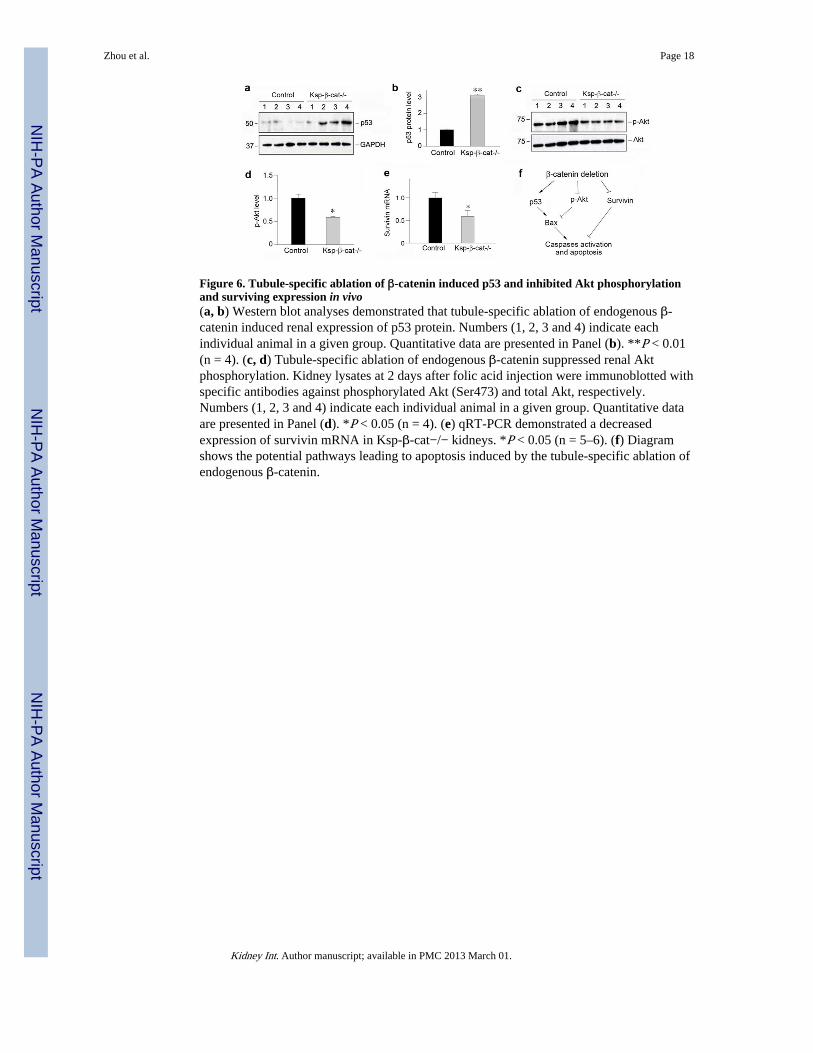
Figure 6. Tubule-specific ablation of β-catenin induced p53 and inhibited Akt phosphorylationand surviving expression in vivo(a, b) Western blot analyses demonstrated that tubule-specific ablation of endogenous β-catenin induced renal expression of p53 protein. Numbers (1, 2, 3 and 4) indicate eachindividual animal in a given group. Quantitative data are presented in Panel (b). **P < 0.01(n = 4). (c, d) Tubule-specific ablation of endogenous β-catenin suppressed renal Aktphosphorylation. Kidney lysates at 2 days after folic acid injection were immunoblotted withspecific antibodies against phosphorylated Akt (Ser473) and total Akt, respectively.Numbers (1, 2, 3 and 4) indicate each individual animal in a given group. Quantitative dataare presented in Panel (d). *P < 0.05 (n = 4). (e) qRT-PCR demonstrated a decreasedexpression of survivin mRNA in Ksp-β-cat−/− kidneys. *P < 0.05 (n = 5–6). (f) Diagramshows the potential pathways leading to apoptosis induced by the tubule-specific ablation ofendogenous β-catenin.
Zhou et al. Page 18
Kidney Int. Author manuscript; available in PMC 2013 March 01.
NIH
-PA Author Manuscript
NIH
-PA Author Manuscript
NIH
-PA Author Manuscript

Figure 7. Loss of tubular β-catenin aggravates AKI in renal ischemia/reperfusion injury model(a) Serum creatinine level in control and Ksp-β-cat−/− mice at 1 day after renal ischemia/reperfusion injury. *P < 0.05 (n=3–4). (b) Representative micrographs of the kidneys incontrol and Ksp-β-cat−/− mice at 1 day after renal ischemia/reperfusion injury. Yellowasterisks in the enlarged boxed areas indicate injured tubules. Scale bar, 50 μm. (c)Quantitative assessment of renal injury. Injury score (% of injured tubules) are presented.**P < 0.01 (n=3). (d, e) Tubule-specific ablation of β-catenin aggravates apoptosis afterrenal ischemia/reperfusion injury. (d) Representative micrographs show apoptotic cell deathdetected by TUNEL staining. White arrows indicate apoptotic cells. (e) Quantitativedetermination of apoptotic cells in renal cortical and medullar regions at 1 day after renalischemia/reperfusion injury. Data are presented as numbers of apoptotic cells per high powerfield (HPF). **P < 0.01 (n = 3). (f, g) Loss of β-catenin promoted renal Bax proteinexpression in ischemic AKI. Western blot (f) and quantitative analysis (g) are presented.Numbers (1, 2 and 3) indicate each individual animal in a given group. *P < 0.05 (n= 3).
Zhou et al. Page 19
Kidney Int. Author manuscript; available in PMC 2013 March 01.
NIH
-PA Author Manuscript
NIH
-PA Author Manuscript
NIH
-PA Author Manuscript
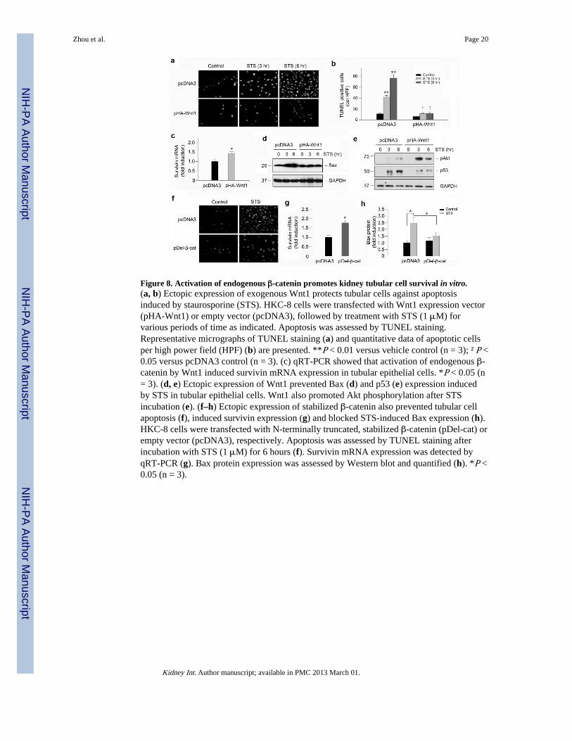
Figure 8. Activation of endogenous β-catenin promotes kidney tubular cell survival in vitro.(a, b) Ectopic expression of exogenous Wnt1 protects tubular cells against apoptosisinduced by staurosporine (STS). HKC-8 cells were transfected with Wnt1 expression vector(pHA-Wnt1) or empty vector (pcDNA3), followed by treatment with STS (1 μM) forvarious periods of time as indicated. Apoptosis was assessed by TUNEL staining.Representative micrographs of TUNEL staining (a) and quantitative data of apoptotic cellsper high power field (HPF) (b) are presented. **P < 0.01 versus vehicle control (n = 3); †P <0.05 versus pcDNA3 control (n = 3). (c) qRT-PCR showed that activation of endogenous β-catenin by Wnt1 induced survivin mRNA expression in tubular epithelial cells. *P < 0.05 (n= 3). (d, e) Ectopic expression of Wnt1 prevented Bax (d) and p53 (e) expression inducedby STS in tubular epithelial cells. Wnt1 also promoted Akt phosphorylation after STSincubation (e). (f–h) Ectopic expression of stabilized β-catenin also prevented tubular cellapoptosis (f), induced survivin expression (g) and blocked STS-induced Bax expression (h).HKC-8 cells were transfected with N-terminally truncated, stabilized β-catenin (pDel-cat) orempty vector (pcDNA3), respectively. Apoptosis was assessed by TUNEL staining afterincubation with STS (1 μM) for 6 hours (f). Survivin mRNA expression was detected byqRT-PCR (g). Bax protein expression was assessed by Western blot and quantified (h). *P <0.05 (n = 3).
Zhou et al. Page 20
Kidney Int. Author manuscript; available in PMC 2013 March 01.
NIH
-PA Author Manuscript
NIH
-PA Author Manuscript
NIH
-PA Author Manuscript