Mesenchymal stem cell-conditioned medium accelerates regeneration of human renal proximal tubule...
6
Experimental and Toxicologic Pathology 65 (2013) 595–600 Contents lists available at SciVerse ScienceDirect Experimental and Toxicologic Pathology jo ur nal homepa ge: www.elsevier.de/etp Mesenchymal stem cell-conditioned medium accelerates regeneration of human renal proximal tubule epithelial cells after gentamicin toxicity Reza Moghadasali a,b,c,1 , Henricus A.M. Mutsaers c,d,1 , Mahnaz Azarnia a , Nasser Aghdami b , Hossein Baharvand b , Ruurd Torensma e , Martijn J.G. Wilmer c , Rosalinde Masereeuw c,∗ a Department of Biology, Tarbiat Moallem University, Tehran, Iran b Department of Stem Cells and Developmental Biology, Cell Sciences Research Center, Royan Institute for Stem Cell Biology and Technology, ACECR, Tehran, Iran c Department of Pharmacology and Toxicology, Radboud University Nijmegen Medical Centre, Nijmegen, The Netherlands d Department of Physiology, Radboud University Nijmegen Medical Centre, Nijmegen, The Netherlands e Department of Tumor Immunology, Radboud University Nijmegen Medical Centre, Nijmegen, The Netherlands a r t i c l e i n f o Article history: Received 30 January 2012 Accepted 4 June 2012 Keywords: Conditionally immortalized human proximal tubule epithelial cell Conditioned medium Gentamicin Mesenchymal stem cells Nephrotoxicity Regeneration a b s t r a c t Bone marrow-derived mesenchymal stem cells (MSCs) have the capacity to regenerate renal tubule epithelia and repair renal function without fusing with resident tubular cells. The goal of the present project was to investigate the role of MSCs secreted cytokines on tubule cell viability and regeneration after a toxic insult, using a conditionally immortalized human proximal tubule epithelial cell (ciPTEC) line. Gentamicin was used to induce nephrotoxicity, and cell viability and migration were studied in absence and presence of human MSC-conditioned medium (hMSC-CM) i.e. medium containing soluble factors produced and secreted by MSCs. Exposure of ciPTEC to 0–3000 g/ml gentamicin for 24 h caused a significant dose-dependent increase in cell death. We further demonstrated that the nephrotoxic effect of 2000 g/ml gentamicin was recovered partially by exposing cells to hMSC-CM. Moreover, exposure of ciPTEC to gentamicin (1500–3000 g/ml) for 7 days completely attenuated the migratory capacity of the cells. In addition, following scrape-wounding, cell migration of both untreated and gentamicin-exposed cells was increased in the presence of hMSC-CM, as compared to exposures to normal medium, indicat- ing improved cell recovery. Our data suggest that cytokines secreted by MSCs stimulate renal tubule cell regeneration after nephrotoxicity. © 2012 Elsevier GmbH. All rights reserved. 1. Introduction The proximal tubule epithelium in the kidney is essential in the clearance of endogenous waste products, exogenously adminis- tered compounds, such as drugs, as well as environmental toxicants (Ferguson et al., 2008; Mutsaers et al., 2011). Due to the unique physiological role of the kidney, the tubular epithelium is exposed to high concentrations of potential toxic compounds and there- fore a frequent target of injury due to toxicity. This can lead to acute kidney injury (AKI) or acute renal failure (ARF), which is a frequently occurring clinical problem that affects up to 7% of hos- pitalized patients. ARF is potentially reversible; however, it can be a determining element of multiple organ failure as well. Hence, the mortality rate in hospital-acquired ARF ranges from 30 to 80% (Schrier et al., 2004). ∗ Corresponding author at: Department of Pharmacology and Toxicology (149), Radboud University Nijmegen Medical Centre, P.O. Box 9101, 6500 HB Nijmegen, The Netherlands. Tel.: +31 24 3613730; fax: +31 24 3614214. E-mail address: [email protected] (R. Masereeuw). 1 These authors contributed equally to this work. While tubular cell death due to necrosis or apoptosis is widespread following injury, less severely injured cells can survive and are believed to be the principal source for tubule regeneration (Humphreys et al., 2011; Lin et al., 2005). Surviving tubular cells can spread and migrate to cover the exposed areas of the base- ment membrane followed by redifferentiation into an epithelial phenotype (Duffield et al., 2005). Additionally, resident epithe- lial cells may lose their characteristic features of mature renal tubular epithelia, e.g. the apical brush border membrane and tight-junctions. As a consequence, the cells acquire a flat and more spread morphological phenotype, resembling undifferenti- ated mesenchyme or fibroblasts. This loss in phenotype, via the process of epithelial-to-mesenchymal transition (EMT), is sup- posed to be an essential factor in promoting regeneration through activation of cell migration and proliferation pathways (Guo and Cantley, 2010; Witzgall et al., 1994). Yet, such resolution may occur only when the tubular basement membrane is still intact (Fragiadaki and Mason, 2011). In addition to EMT, bone marrow-derived mesenchymal stem cells (MSCs) may have the capacity to migrate to the injured kid- ney and contribute to tubule epithelium regeneration and renal function repair without fusing with resident tubular cells (Huls 0940-2993/$ – see front matter © 2012 Elsevier GmbH. All rights reserved. http://dx.doi.org/10.1016/j.etp.2012.06.002
-
Upload
royaninstitute -
Category
Documents
-
view
2 -
download
0
Transcript of Mesenchymal stem cell-conditioned medium accelerates regeneration of human renal proximal tubule...

Mo
RHa
b
c
d
e
ARA
KCpCGMNR
1
tt(ptfafpat(
RT
0h
Experimental and Toxicologic Pathology 65 (2013) 595– 600
Contents lists available at SciVerse ScienceDirect
Experimental and Toxicologic Pathology
jo ur nal homepa ge: www.elsev ier .de /e tp
esenchymal stem cell-conditioned medium accelerates regenerationf human renal proximal tubule epithelial cells after gentamicin toxicity
eza Moghadasali a,b,c,1, Henricus A.M. Mutsaersc,d,1, Mahnaz Azarniaa, Nasser Aghdamib,ossein Baharvandb, Ruurd Torensmae, Martijn J.G. Wilmerc, Rosalinde Masereeuwc,∗
Department of Biology, Tarbiat Moallem University, Tehran, IranDepartment of Stem Cells and Developmental Biology, Cell Sciences Research Center, Royan Institute for Stem Cell Biology and Technology, ACECR, Tehran, IranDepartment of Pharmacology and Toxicology, Radboud University Nijmegen Medical Centre, Nijmegen, The NetherlandsDepartment of Physiology, Radboud University Nijmegen Medical Centre, Nijmegen, The NetherlandsDepartment of Tumor Immunology, Radboud University Nijmegen Medical Centre, Nijmegen, The Netherlands
a r t i c l e i n f o
rticle history:eceived 30 January 2012ccepted 4 June 2012
eywords:onditionally immortalized humanroximal tubule epithelial cellonditioned mediumentamicin
a b s t r a c t
Bone marrow-derived mesenchymal stem cells (MSCs) have the capacity to regenerate renal tubuleepithelia and repair renal function without fusing with resident tubular cells. The goal of the presentproject was to investigate the role of MSCs secreted cytokines on tubule cell viability and regenerationafter a toxic insult, using a conditionally immortalized human proximal tubule epithelial cell (ciPTEC)line. Gentamicin was used to induce nephrotoxicity, and cell viability and migration were studied inabsence and presence of human MSC-conditioned medium (hMSC-CM) i.e. medium containing solublefactors produced and secreted by MSCs. Exposure of ciPTEC to 0–3000 �g/ml gentamicin for 24 h causeda significant dose-dependent increase in cell death. We further demonstrated that the nephrotoxic effect
esenchymal stem cellsephrotoxicityegeneration
of 2000 �g/ml gentamicin was recovered partially by exposing cells to hMSC-CM. Moreover, exposure ofciPTEC to gentamicin (1500–3000 �g/ml) for 7 days completely attenuated the migratory capacity of thecells. In addition, following scrape-wounding, cell migration of both untreated and gentamicin-exposedcells was increased in the presence of hMSC-CM, as compared to exposures to normal medium, indicat-ing improved cell recovery. Our data suggest that cytokines secreted by MSCs stimulate renal tubule cell
toxic
regeneration after nephro. Introduction
The proximal tubule epithelium in the kidney is essential inhe clearance of endogenous waste products, exogenously adminis-ered compounds, such as drugs, as well as environmental toxicantsFerguson et al., 2008; Mutsaers et al., 2011). Due to the uniquehysiological role of the kidney, the tubular epithelium is exposedo high concentrations of potential toxic compounds and there-ore a frequent target of injury due to toxicity. This can lead tocute kidney injury (AKI) or acute renal failure (ARF), which is arequently occurring clinical problem that affects up to 7% of hos-italized patients. ARF is potentially reversible; however, it can be
determining element of multiple organ failure as well. Hence,he mortality rate in hospital-acquired ARF ranges from 30 to 80%Schrier et al., 2004).
∗ Corresponding author at: Department of Pharmacology and Toxicology (149),adboud University Nijmegen Medical Centre, P.O. Box 9101, 6500 HB Nijmegen,he Netherlands. Tel.: +31 24 3613730; fax: +31 24 3614214.
E-mail address: [email protected] (R. Masereeuw).1 These authors contributed equally to this work.
940-2993/$ – see front matter © 2012 Elsevier GmbH. All rights reserved.ttp://dx.doi.org/10.1016/j.etp.2012.06.002
ity.© 2012 Elsevier GmbH. All rights reserved.
While tubular cell death due to necrosis or apoptosis iswidespread following injury, less severely injured cells can surviveand are believed to be the principal source for tubule regeneration(Humphreys et al., 2011; Lin et al., 2005). Surviving tubular cellscan spread and migrate to cover the exposed areas of the base-ment membrane followed by redifferentiation into an epithelialphenotype (Duffield et al., 2005). Additionally, resident epithe-lial cells may lose their characteristic features of mature renaltubular epithelia, e.g. the apical brush border membrane andtight-junctions. As a consequence, the cells acquire a flat andmore spread morphological phenotype, resembling undifferenti-ated mesenchyme or fibroblasts. This loss in phenotype, via theprocess of epithelial-to-mesenchymal transition (EMT), is sup-posed to be an essential factor in promoting regeneration throughactivation of cell migration and proliferation pathways (Guo andCantley, 2010; Witzgall et al., 1994). Yet, such resolution mayoccur only when the tubular basement membrane is still intact(Fragiadaki and Mason, 2011).
In addition to EMT, bone marrow-derived mesenchymal stemcells (MSCs) may have the capacity to migrate to the injured kid-ney and contribute to tubule epithelium regeneration and renalfunction repair without fusing with resident tubular cells (Huls

5 d Tox
ertafimaBMm
stiduai(gaC
2
2
tbamcDsUsf5belfdicbmmciage
2
abetflo
96 R. Moghadasali et al. / Experimental an
t al., 2010). The exact mechanism through which MSCs mitigateenal damage is not fully elucidated. One possible explanation ishat MSCs produce cytokines and growth factors that promotenti-inflammatory, immunosuppressive, anti-apoptotic and proli-erative effects (Humphreys and Bonventre, 2008). This hypothesiss supported by other studies demonstrating that conditioned
edium, in which MSCs secreted growth factors and cytokinesre present, could ameliorate renal tubular injury (Humphreys andonventre, 2008). Yet, most studies that investigated the role ofSCs in the regeneration of kidney injury used rodent or otherammalian animal models.Therefore, this study was designed to investigate whether
oluble factors produced and secreted by MSCs could provide pro-ection against gentamicin-induced nephrotoxicity using a humann vitro model. We hypothesized that human MSC-derived con-itioned medium would prevent renal proximal tubular cells tondergo nephrotoxicant-induced cell death thereby protectinggainst tubular injury. We used a recently developed conditionallymmortalized human proximal tubular epithelial cell line (ciPTEC)Wilmer et al., 2009), which was exposed to the nephrotoxic drugentamicin, and studied the cell viability and cell migration inbsence and presence of human MSC-conditioned medium (hMSC-M).
. Materials and methods
.1. Proximal tubular epithelial cells
The ciPTEC line was generated by culturing cells exfoliated inhe urine of a healthy volunteer, followed by immortalization usingoth the temperature-sensitive mutant U19tsA58 of SV40 large Tntigen (SV40T) and the essential catalytic subunit of human telo-erase (hTERT), as previously described (Wilmer et al., 2009). The
ells were cultured in ciPTEC medium containing phenol red-freeMEM/F12 medium (Gibco/Invitrogen, Breda, the Netherlands)
upplemented with 10% (v/v) fetal calf serum (FCS; MP Biomedicals,den, the Netherlands), insulin (5 �g/ml), transferrin (5 �g/ml),
elenium (5 ng/ml), hydrocortisone (36 ng/ml), epithelial growthactor (10 ng/ml) and tri-iodothyronine (40 pg/ml) at 33 ◦C in a% (v/v) CO2 atmosphere. Propagation of cells was maintainedy subculturing the cells at a dilution of 1:3 to 1:6 at 33 ◦C. Forxperiments, cells were cultured at 33 ◦C to 40% confluency, fol-owed by maturation for 7 days at 37 ◦C during which the cellsormed a confluent monolayer. Furthermore, expression of SV40Tecreases gradually in ciPTEC cultured at 37 ◦C, minimizing the
nfluence of the transfection on cellular phenotype and allowingells to differentiate. Routinely, cell morphology was monitoredy phase-contrast microscopy at each passage and showed noarked difference from passage 35 up to 40, during which experi-ents were performed. Moreover, it was previously reported that
iPTECs maintain their proximal tubular characteristics, includ-ng expression of CD13, ZO-1, megalin-mediated albumin uptake,nd functional expressions of organic cation transporter 2 and P-lycoprotein over a prolonged period of culturing time (Wilmert al., 2009).
.2. Stem cell isolation and culture, and preparation of hMSC-CM
Bone marrow-derived MSCs were isolated from healthy donorst the Radboud University Nijmegen Medical Centre, as describedefore (Jansen et al., 2010). In short, MSCs were isolated from
ach bone marrow sample by Ficoll density gradient centrifuga-ion. Afterwards, the cells were seeded into polystyrene cultureasks (Becton Dickinson, Bedford, Mass, United States) at a densityf 1 × 10⁶ cells/cm2 in alpha-Minimum Essential Medium (alphaicologic Pathology 65 (2013) 595– 600
MEM), with 100 U/ml penicillin, 0.1 mg/ml streptomycin (GibcoBRL), 2 �M l-glutamine (Gibco BRL) and 10% (v/v) FCS selectedfor MSC growth (Hyclone, characterized, Lot ALF 14015). Cul-tures, maintained in a humidified atmosphere with 5% (v/v) CO2at 37 ◦C, had their medium changed twice weekly thereafter. Onreaching 60–80% confluency, adherent cells were detached aftertreatment with 0.05% (v/v) trypsin/1 �M EDTA solution (Gibco,BRL) for reseeding at 103 cells per/cm2. To obtain human MSC-conditioned medium (hMSC-CM), cells were cultured in DMEMmedium containing low glucose (1 g/l), supplemented with peni-cillin (100 U/ml), streptomycin (100 �g/ml) and 10% (v/v) FCSof a selected batch. We cultured hMSCs to confluency andcollected the medium after approximately 24–48 h after refresh-ing medium. hMSC-CM was centrifuged at 800 × g for 5 min toremove detached MSCs and stored at −80 ◦C until further use,hMSC-CM was diluted (1:1) in ciPTEC culture medium prior toincubation.
2.3. Cell viability
Sensitivity of cells to gentamicin (100–3000 �g/ml) was deter-mined by a standard spectrophotometric 3-(4,5-dimethylthiazole-2-yl)-2,5 diphenyltetrazolium bromide (MTT; Sigma) assay.ciPTECs were grown in 96-well plates and exposed to gentami-cin for 24 h in 100 �l of ciPTEC medium (control) or hMSC-CM.Next, medium was removed, 20 �l preheated (37 ◦C) MTT solu-tion (5 mg/ml PBS) was added and cells were incubated for 4 h at37 ◦C. Afterwards, MTT solution was removed, followed by the addi-tion of 200 �l DMSO. The extinction of the solution was measuredat 570 nm using a Benchmark Plus Microplate Spectrophotometer(Biorad, Veenendaal, the Netherlands).
2.4. Cell migration
The effect of hMSC-CM on migration of unexposed andgentamicin-exposed ciPTECs was determined using the scratchassay. Cells were cultured in 6-well plates and the confluent mono-layers were treated with ciPTEC medium, hMSC-CM or gentamicin(1500–3000 �g/ml) for 24 h followed by scrape-wounding the cellsusing a plastic pipette tip. Following scraping, the medium contain-ing detached cells was removed and replaced with either ciPTECmedium without or with gentamicin (1500–3000 �g/ml) or hMSC-CM, and incubated until the monolayer was restored (5–7 days).To replenish nutrients, medium was refreshed every 2 days. Cellmigration and cell monolayer recovery was studied by phase con-trast microscopy over time, as described in detail by Liang et al.(2007). Length of scratching area was measured on stored imagesusing ImageJ software (U.S. National Institutes of Health, Bethesda,MD, USA).
2.5. Statistics
Statistics were performed using GraphPad Prism 5.02 via aOne-way analysis of variance (ANOVA) followed by the Dunnett’sor Bonferroni’s Multiple Comparison Test. Differences betweengroups were considered to be statistically significant when P < 0.05.
3. Results
3.1. Gentamicin-induced cell death can be partially rescued byhMSC-CM
First the cytotoxic effect of gentamicin without co-exposureto hMSC-CM was evaluated, after which the protective effect ofhMSC-CM on a toxic concentration of gentamicin was studied(see Fig. 1A for experimental design). A dose-dependent reduction

R. Moghadasali et al. / Experimental and Toxicologic Pathology 65 (2013) 595– 600 597
Fig. 1. Reduction in cell viability of ciPTECs exposed to gentamicin and partial recovery by hMSC-CM. Cell viability was determined using the MTT assay and data are shownas the percentage of viable cells. (A) Schematic overview of experimental set-up. Cells were plated at 33 ◦C at day-8 and transferred to 37 ◦C to enable maturation. Mediumwas replaced every other day. At day 0, medium was replaced with ciPTEC medium supplemented with gentamicin for 24 h. At day 1, an MTT assay was performed. (B) Therei reaset 01 as
ifogean
3g
mifcebsmFwmssghawcsrd
s a significant increase in cell death with increasing doses of gentamicin. (C) The inco hMSC-CM. Cells incubated with ciPTEC medium are indicated as control. **P < 0.0
n cell viability was observed after exposing cells to gentamicinor 24 h as assessed by the MTT assay, with an apparent LC50f 1765 ± 25 �g/ml (Fig. 1B). At a concentration of 2000 �g/ml,entamicin reduced cell viability by 53% (P < 0.001). In the pres-nce of hMSC-CM, viability was only reduced by 33%, suggesting
protective effect of conditioned medium on gentamicin-inducedephrotoxicity (Fig. 1C).
.2. Effect of the hMSC-CM on migration of ciPTECs exposed toentamicin
The scratch assay is a straightforward method to study celligration in vitro (see Fig. 2A for experimental set-up). Long term,
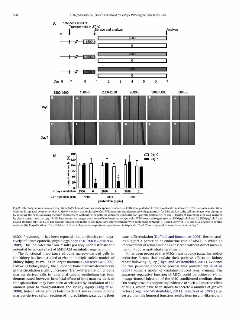
.e. 7 days, exposure of ciPTEC to high doses of gentamicin, rangingrom 1500 to 3000 �g/ml, completely attenuated the migratoryapacity of the cells (Fig. 2B, D, F, H, J and L). However, ciPTECsxposed to gentamicin (1500 and 2000 �g/ml) for 24 h, followedy a change toward fresh medium without the nephrotoxicant,howed a tremendous increase in cell migration and cell transfor-ation with monolayer restoration for up to 80% (Fig. 2C, E, I and K).
or the highest concentration tested, 3000 �g/ml, no cell migrationas observed during 7 days using fresh medium after 24 h genta-icin exposure, suggesting severe toxicity (Fig. 2G and M). Fig. 2N
hows a summary of three independent experiments, demon-trating that restoration of the monolayer was possible after 24 hentamicin exposure at concentrations up to 2000 �g/ml. WhenMSC-CM was used instead of normal medium the recovery ratefter intermediate toxic concentration of gentamicin (1500 �g/ml)as accelerated by 25% on day 5 (Fig. 3; P < 0.05). Treatment with
onditioned medium solely resulted in a 96% restoration of thecratch area on day 3 and 100% on day 5, whereas normal mediumesulted in a restoration of the scratch wound by 87% and 96% onay 3 and 5, respectively.
in cell death by 2000 �g/ml gentamicin was partially restored by co-exposing cellscompared to control; #P < 0.01 as compared to 2000 �g/ml gentamicin.
4. Discussion
The results of the present study indicate that MSC-derivedconditioned medium increased cell viability and accelerated cellmigration after gentamicin-induced cell toxicity in a unique humanproximal tubular epithelial cell line. Gentamicin is well known forits nephrotoxicity in various in vitro and in vivo investigations,which hampers its clinical use. In accordance with previous studiesusing Madin–Darby canine kidney cells type II (Notenboom et al.,2006), we observed a concentration-dependent increase in cell tox-icity after gentamicin exposure, indicating that ciPTECs are an ideal,human-derived, tool to study gentamicin-induced renal toxicity.Concentrations of ≥2000 �g/ml gentamicin and multiple days dos-ing of ciPTECs caused irreversible cell damage and a lack of abilityto migrate into a scratch-wounded area.
Wound healing assays have been carried out in tissue culturestudies to estimate the migration and proliferation rates of cellsand the impact of culture conditions on these parameters. Uponscratching a line through a monolayer, the open gap is inspectedmicroscopically over time as the cells move in and fill the damagedarea. This healing can take from several hours to over a day depend-ing on the cell type, conditions, and the extent of the woundedregion. Here, the ciPTEC monolayer was almost completely recov-ered within five days using normal medium. This process wassignificantly accelerated when cells were exposed to hMSC-CM.In presence of gentamicin, no cell monolayer regeneration wasobserved for up to seven days, however, when cells were exposedfor 24 h to gentamicin followed by a replacement with normalmedium or hMSC-CM the monolayer was restored in a great partwithin seven days. Again, the recovery rate was accelerated by 25%
by hMSC-CM.During this study, hMSC-CM contained penicillin and strepto-mycin, due to the cell culture requirements of primary human
598 R. Moghadasali et al. / Experimental and Toxicologic Pathology 65 (2013) 595– 600
Fig. 2. Effect of gentamicin on cell migration. (A) Schematic overview of experimental set-up. Cells were plated at 33 ◦C at day-8 and transferred to 37 ◦C to enable maturation.Medium is replaced every other day. At day 0, medium was replaced with ciPTEC medium supplemented with gentamicin for 24 h. At day 1, the cell monolayer was disruptedby scraping the cells, following medium replacement without (0) or with the indicated concentration (�g/ml) gentamicin. At day 7, length of scratching area was analyzedby phase contrast microscopy. (B–M) Representative images are shown of confluent monolayers of ciPTECs exposed to gentamicin (1500 �g/ml (B and C), 2000 �g/ml (D andE treatmm rmed
Mt2p
tkFimdta2m
) and 3000 �g/ml (F and G)). The wound-induced cell motility was measured afteredium (0). Magnification, 10×. (N) Mean of three independent experiments perfo
SCs. Previously, it has been reported that antibiotics can nega-ively influence epithelial physiology (Shen et al., 2003; Zietse et al.,009). This indicates that our results possibly underestimate theotential beneficial effect of hMSC-CM on tubular regeneration.
The functional importance of bone marrow-derived cells inhe kidney has been studied in vivo in multiple rodent models ofidney injury as well as in larger mammals (Masereeuw, 2009).ollowing kidney injury, the number of bone marrow-derived cellsn the circulation slightly increases. Trans-differentiation of bone
arrow-derived cells to functional tubular epithelium has beenemonstrated, however, beneficial effects of bone marrow-derived
ransplantations may have been accelerated by irradiation of thenimals prior to transplantation and kidney injury (Fang et al.,008). Indeed, other groups failed to detect any evidence of bonearrow-derived cells in sections of injured kidneys, excluding theirent with gentamicin without (H, J, and L) or with (I, K, and M) a change to control in triplicate. *P < 0.05 as compared to same treatment on day 0.
trans-differentiation (Duffield and Bonventre, 2005). Recent stud-ies support a paracrine or endocrine role of MSCs, in which animprovement of renal function is observed without direct involve-ment in tubular epithelial engraftment.
It has been proposed that MSCs must provide paracrine and/orendocrine factors that explain their positive effects on kidneyrepair following injury (Togel and Westenfelder, 2011). Evidencefor this paracrine/endocrine process was provided by Bi et al.(2007), using a model of cisplatin-induced renal damage. Theapparent reparative function of MSCs could be achieved via anintraperitoneal injection of the MSC-conditioned medium alone.
Our study provides supporting evidence of such a paracrine effectof MSCs, which have been shown to secrete a number of growthfactors (Togel and Westenfelder, 2011). Imberti et al. (2007) sug-gested that this humoral function results from insulin-like growth
R. Moghadasali et al. / Experimental and Toxicologic Pathology 65 (2013) 595– 600 599
Fig. 3. MSC-derived conditioned medium increases cell migration. (A) Schematic overview of experimental set-up. Cells were plated at 33 ◦C at day-8 and transferred to37 ◦C to enable maturation. Medium is replaced every other day. At day 0, medium was replaced with hMSC-CM or control medium in presence or absence of gentamicin(1500 �g/ml) for 24 h. At day 1, the cell monolayer was disrupted by scraping the cells, following medium replacement with either hMSC-CM or ciPTEC medium (control).At day 3 and 5, length of scratching area was analyzed by phase contrast microscopy. (B–G) Representative images of confluent monolayers of ciPTECs exposed to medium(control; B), hMSC-CM (E), and gentamicin for 24 h and then were scratched by a plastic pipette. The wound-induced cell motility was measured after recovery in controlm depent
foffli2tcg
edium (C and D) and hMSC-CM (F and G). Magnification, 10×. (H) Mean of three inreatment; #P < 0.05 hMSC-CM as compared to ciPTEC medium on same day.
actor 1 (IGF1), whereas others attributed it to a combinationf hepatocyte growth factor (HGF), IGF1 and epidermal growthactor. Vascular endothelial growth factor can be an additionalactor in the renoprotection by MSCs (Togel et al., 2009). It hasong been known that IGF1 and HGF can play reparative rolesn the kidney following acute injury (Humphreys and Bonventre,
008). Bone morphogenetic protein 7 has also been implicated inhe protection against fibrosis (Zeisberg and Kalluri, 2008). Theurrent study, demonstrating hMSC-CM accelerates repair afterentamicin-induced nephrotoxicity in ciPTECs, is in concordancedent experiments performed in triplicate. **P < 0.001 as compared to day 0 of same
with the theory that not MSCs themselves, but secretory proteinsdominate regeneration processes. Moreover, the human origin ofciPTECs in this study underscores the promising effective thera-peutic interventions using MSCs in tissue regeneration in case ofARF and AKI. Still, a number of uncertainties exist in understand-ing which (patho)physiological triggers cause activation of MSCs
and the subsequent production of soluble factors. Most likely, theinflammatory conditions after an injury provide key signals. Togeland Westenfelder (2011) suggested that MSCs exert their renalprotection through inhibition of proinflammatory cytokines. This
6 d Tox
iisIrsFf
lrhattio
A
eIw
R
B
D
D
F
F
F
G
00 R. Moghadasali et al. / Experimental an
mplies that the reparative role of MSCs may be multifactorial andnclude production of anti-inflammatory cytokines to limit apopto-is, enhance proliferation and dampen the inflammatory response.n addition, the renal interstitium itself likely contributes to theenal regeneration process as well, by controlling fibrosis along-ide preserving the kidney’s architecture (Kaissling and Le, 2008).uture in vivo studies should be directed at identifying the multipleactors that contribute to renal restoration after acute injury.
In conclusion, hMSC-conditioned medium accelerates mono-ayer restoration after gentamicin-induced toxicity in a humanenal proximal tubular epithelial cell line, demonstrating thatuman MSCs can play an important role in renal repair mechanismsfter acute injury. These findings warrant further investigation intohe specific set of cytokines and growth factors excreted by MSCshat contribute to the positive effect of these cells on renal repairn vivo. Identification of these factors could aid in the developmentf novel therapies.
cknowledgements
Financial support was obtained from the Ministry of Sci-nce, Research and Technology of the Islamic Republic ofran, and the Dutch Kidney Foundation (grant number IK08.03;
ww.nierstichting.nl).
eferences
i B, Schmitt R, Israilova M, Nishio H, Cantley LG. Stromal cells protect againstacute tubular injury via an endocrine effect. Journal of the American Societyof Nephrology 2007;18:2486–96.
uffield JS, Bonventre JV. Kidney tubular epithelium is restored without replacementwith bone marrow-derived cells during repair after ischemic injury. KidneyInternational 2005;68:1956–61.
uffield JS, Park KM, Hsiao LL, Kelley VR, Scadden DT, Ichimura T, et al. Restorationof tubular epithelial cells during repair of the postischemic kidney occurs inde-pendently of bone marrow-derived stem cells. Journal of Clinical Investigation2005;115:1743–55.
ang TC, Otto WR, Jeffery R, Hunt T, Alison MR, Cook HT, et al. Exogenous bonemarrow cells do not rescue non-irradiated mice from acute renal tubular damagecaused by HgCl2, despite establishment of chimaerism and cell proliferation inbone marrow and spleen. Cell Proliferation 2008;41:592–606.
erguson MA, Vaidya VS, Bonventre JV. Biomarkers of nephrotoxic acute kidneyinjury. Toxicology 2008;245:182–93.
ragiadaki M, Mason RM. Epithelial-mesenchymal transition in renalfibrosis—evidence for and against. International Journal of ExperimentalPathology 2011;92:143–50.
uo JK, Cantley LG. Cellular maintenance and repair of the kidney. Annual Reviewof Physiology 2010;72:357–76.
icologic Pathology 65 (2013) 595– 600
Huls M, Schoeber JP, Verfaillie CM, Luttun A, Ulloa-Montoya F, Menke AL, et al.Deficiency of either P-glycoprotein or breast cancer resistance protein protectagainst acute kidney injury. Cell Transplantation 2010;19:1195–208.
Humphreys BD, Bonventre JV. Mesenchymal stem cells in acute kidney injury.Annual Review of Medicine 2008;59:311–25.
Humphreys BD, Czerniak S, DiRocco DP, Hasnain W, Cheema R, Bonventre JV.Repair of injured proximal tubule does not involve specialized progenitors.Proceedings of the National Academy of Sciences of the United States of America2011;108:9226–31.
Imberti B, Morigi M, Tomasoni S, Rota C, Corna D, Longaretti L, et al. Insulin-likegrowth factor-1 sustains stem cell mediated renal repair. Journal of the AmericanSociety of Nephrology 2007;18:2921–8.
Jansen BJ, Gilissen C, Roelofs H, Schaap-Oziemlak A, Veltman JA, Raymakers RA,et al. Functional differences between mesenchymal stem cell populations arereflected by their transcriptome. Stem Cells and Development 2010;19:481–90.
Kaissling B, Le HM. The renal cortical interstitium: morphological and functionalaspects. Histochemistry and Cell Biology 2008;130:247–62.
Liang CC, Park AY, Guan JL. In vitro scratch assay: a convenient and inexpensivemethod for analysis of cell migration in vitro. Nature Protocols 2007;2:329–33.
Lin F, Moran A, Igarashi P. Intrarenal cells, not bone marrow-derived cells, are themajor source for regeneration in postischemic kidney. Journal of Clinical Inves-tigation 2005;115:1756–64.
Masereeuw R. Contribution of bone marrow-derived cells in renal repair after acutekidney injury. Minerva Urologica e Nefrologica 2009;61:373–84.
Mutsaers HA, van den Heuvel LP, Ringens LH, Dankers AC, Russel FG, Wetzels JF,et al. Uremic toxins inhibit transport by breast cancer resistance protein andmultidrug resistance protein 4 at clinically relevant concentrations. PLoS One2011;6:e18438.
Notenboom S, Wouterse AC, Peters B, Kuik LH, Heemskerk S, Russel FG, et al.Increased apical insertion of the multidrug resistance protein 2 (MRP2/ABCC2) inrenal proximal tubules following gentamicin exposure. Journal of Pharmacologyand Experimental Therapeutics 2006;318:1194–202.
Schrier RW, Wang W, Poole B, Mitra A. Acute renal failure: definitions, diag-nosis, pathogenesis, and therapy. Journal of Clinical Investigation 2004;114:5–14.
Shen MR, Chou CY, Chiu WT. Streptomycin and its analogues are potent inhibitorsof the hypotonicity-induced Ca2
+ entry and Cl− channel activity. FEBS Letters2003;554:494–500.
Togel F, Cohen A, Zhang P, Yang Y, Hu Z, Westenfelder C. Autologous and allogeneicmarrow stromal cells are safe and effective for the treatment of acute kidneyinjury. Stem Cells and Development 2009;18:475–85.
Togel F, Westenfelder C. The role of multipotent marrow stromal cells (MSCs) intissue regeneration. Organogenesis 2011;7:96–100.
Wilmer MJ, Saleem MA, Masereeuw R, Ni L, van d V, Russel FG, et al. Novelconditionally immortalized human proximal tubule cell line expressing func-tional influx and efflux transporters. Cell and Tissue Research 2009;339:449–57.
Witzgall R, Brown D, Schwarz C, Bonventre JV. Localization of proliferating cellnuclear antigen, vimentin, c-Fos, and clustering in the postischemic kidney.Evidence for a heterogenous genetic response among nephron segments, anda large pool of mitotically active and dedifferentiated cells. Journal of Clinical
Investigation 1994;93:2175–88.Zeisberg M, Kalluri R. Fibroblasts emerge via epithelial-mesenchymal transition inchronic kidney fibrosis. Frontiers in Bioscience 2008;13:6991–8.
Zietse R, Zoutendijk R, Hoorn EJ. Fluid, electrolyte and acid-base disorders associatedwith antibiotic therapy. Nature Reviews Nephrology 2009;5:193–202.