Exposure to an Enriched Environment Accelerates Recovery from Cerebellar Lesion
16
Exposure to an Enriched Environment Accelerates Recovery from Cerebellar Lesion Francesca Foti & Daniela Laricchiuta & Debora Cutuli & Paola De Bartolo & Francesca Gelfo & Francesco Angelucci & Laura Petrosini # Springer Science+Business Media, LLC 2010 Abstract The exposure to enriched environments allows the maintenance of normal cognitive functioning even in the presence of brain pathology. Up until now, clinical and experimental studies have investigated environmental effects mainly on the symptoms linked to the presence of neuro-degenerative diseases, and no study has yet analyzed whether prolonged exposure to complex environments allows modifying the clinical expression and compensation of deficits of cerebellar origin. In animals previously exposed to complex stimulations, the effects of cerebellar lesions have been analyzed to verify whether a prolonged and intense exposure to complex stimulations affected the compensation of motor and cognitive functions following a cerebellar lesion. Hemicerebellectomized or intact animals housed in enriched or standard conditions were adminis- tered spatial tests. Postural asymmetries and motor behavior were also assessed. Exposure to the enriched environment almost completely compensated the effects of the hemi- cerebellectomy. In fact, the motor and cognitive perform- ances of the enriched hemicerebellectomized animals were similar to those of the intact animals. The plastic changes induced by enhanced mental and physical activity seem to provide the development of compensatory responses against the disrupting motor and cognitive consequences of the cerebellar damage. Keywords Hemicerebellectomy . Cerebellar compensation . Postural and locomotor behavior . Spatial tests Introduction Cognitive enrichment plays an important role in maintaining cognitive performance even in the presence of brain damage [1–3]. It has been reported that even though cognitive enrichment does not prevent the onset of neuro-degenerative diseases, it may provide protection against the expression of clinical symptoms [4–8]. Thus, environmental complexity fosters the development of neuroplasticity properties that allow normal motor and cognitive functioning even in the presence of brain pathology. Up until now, the experimental studies on the behavioral effects of environmental enrich- ment have been performed in models of dementia-like neurodegeneration [9–13]. However, no study has yet addressed the question as to whether prolonged exposure to complex environments beneficially affects the clinical expression and compensation of deficits of cerebellar origin. Cerebellar damage elicits motor disorders in muscle coordination, balance and muscle strength [14, 15] as well as significant impairments in a variety of cognitive, emotional, and affective functions [16–18]. In both humans and animals, most motor symptoms of cerebellar origin following surgical ablation or stroke injury gradually and efficiently compensate over time. In the compensated state, faceted cognitive impairment, which affects memory, atten- tion, visuo-spatial abilities, and executive functions, is present together with stable motor symptomatology charac- terized by some ataxic, dysmetric, and asthenic symptoms [17, 19–26]. Whereas, the pharmacological treatment of cerebellar compensation is largely unsatisfactory, physical F. Foti : D. Laricchiuta : D. Cutuli : P. De Bartolo : F. Gelfo : F. Angelucci : L. Petrosini IRCCS Santa Lucia Foundation, Rome, Italy F. Foti : D. Laricchiuta : D. Cutuli : P. De Bartolo : L. Petrosini (*) Department of Psychology, University “Sapienza” of Rome, Via dei Marsi 78, 00185, Rome, Italy e-mail: [email protected] Cerebellum DOI 10.1007/s12311-010-0236-z
Transcript of Exposure to an Enriched Environment Accelerates Recovery from Cerebellar Lesion

Exposure to an Enriched Environment Accelerates Recoveryfrom Cerebellar Lesion
Francesca Foti & Daniela Laricchiuta & Debora Cutuli &Paola De Bartolo & Francesca Gelfo &
Francesco Angelucci & Laura Petrosini
# Springer Science+Business Media, LLC 2010
Abstract The exposure to enriched environments allowsthe maintenance of normal cognitive functioning even inthe presence of brain pathology. Up until now, clinical andexperimental studies have investigated environmentaleffects mainly on the symptoms linked to the presence ofneuro-degenerative diseases, and no study has yet analyzedwhether prolonged exposure to complex environmentsallows modifying the clinical expression and compensationof deficits of cerebellar origin. In animals previouslyexposed to complex stimulations, the effects of cerebellarlesions have been analyzed to verify whether a prolongedand intense exposure to complex stimulations affected thecompensation of motor and cognitive functions following acerebellar lesion. Hemicerebellectomized or intact animalshoused in enriched or standard conditions were adminis-tered spatial tests. Postural asymmetries and motor behaviorwere also assessed. Exposure to the enriched environmentalmost completely compensated the effects of the hemi-cerebellectomy. In fact, the motor and cognitive perform-ances of the enriched hemicerebellectomized animals weresimilar to those of the intact animals. The plastic changesinduced by enhanced mental and physical activity seem toprovide the development of compensatory responsesagainst the disrupting motor and cognitive consequencesof the cerebellar damage.
Keywords Hemicerebellectomy . Cerebellarcompensation . Postural and locomotor behavior . Spatialtests
Introduction
Cognitive enrichment plays an important role in maintainingcognitive performance even in the presence of brain damage[1–3]. It has been reported that even though cognitiveenrichment does not prevent the onset of neuro-degenerativediseases, it may provide protection against the expression ofclinical symptoms [4–8]. Thus, environmental complexityfosters the development of neuroplasticity properties thatallow normal motor and cognitive functioning even in thepresence of brain pathology. Up until now, the experimentalstudies on the behavioral effects of environmental enrich-ment have been performed in models of dementia-likeneurodegeneration [9–13]. However, no study has yetaddressed the question as to whether prolonged exposure tocomplex environments beneficially affects the clinicalexpression and compensation of deficits of cerebellar origin.
Cerebellar damage elicits motor disorders in musclecoordination, balance and muscle strength [14, 15] as wellas significant impairments in a variety of cognitive,emotional, and affective functions [16–18]. In both humansand animals, most motor symptoms of cerebellar originfollowing surgical ablation or stroke injury gradually andefficiently compensate over time. In the compensated state,faceted cognitive impairment, which affects memory, atten-tion, visuo-spatial abilities, and executive functions, ispresent together with stable motor symptomatology charac-terized by some ataxic, dysmetric, and asthenic symptoms[17, 19–26]. Whereas, the pharmacological treatment ofcerebellar compensation is largely unsatisfactory, physical
F. Foti :D. Laricchiuta :D. Cutuli : P. De Bartolo : F. Gelfo :F. Angelucci : L. PetrosiniIRCCS Santa Lucia Foundation,Rome, Italy
F. Foti :D. Laricchiuta :D. Cutuli : P. De Bartolo :L. Petrosini (*)Department of Psychology, University “Sapienza” of Rome,Via dei Marsi 78,00185, Rome, Italye-mail: [email protected]
CerebellumDOI 10.1007/s12311-010-0236-z
therapy and neuro-rehabilitation have been found of someefficacy, although the basis of the effects of these treatmentsis still unknown [27–29]. In this regard, it is important toanalyze whether compensation of cerebellar symptoms maybe positively influenced by previous exposure to an enrichedenvironment. In other words, does exposure to complexstimulation may accelerate compensation of cerebellarimpairments? To clarify this issue, we studied whether earlyand long-lasting exposure to an enriched environment affectsthe motor and cognitive symptoms evoked by a hemi-cerebellectomy (HCb). The beneficial effects of the exposureto an enriched environment can be demonstrated only if thepreviously enriched hemicerebellectomized (HCbed) animalsexhibit an accelerated compensation of motor and cognitiveimpairments in comparison to standard-housed HCbedanimals.
Materials and Methods
Animals
Forty adult male Wistar rats (250–300 g; Harlan, Italy)were used in the present research. The animals were keptunder standard conditions (temperature, 22°C±2; relativehumidity, 60%±10) with food and water ad libitum on a12/12-h dark/light cycle (light on between 0700 and1900 hours). Rats were assigned to four experimentalgroups. The first group comprised enriched-housed hemi-cerebellectomized rats (EH group), the second groupcomprised standard-housed hemicerebellectomized rats(SH group), the third group comprised enriched-housedcontrol rats (EC), and the fourth group comprised standard-housed control rats (SC group). The animals were main-tained according to the guidelines for ethical conductdeveloped by the European Communities Council Directiveof November 24, 1986 (86/609/EEC). All efforts weremade to minimize pain or discomfort of the animals.
Housing Conditions
On the 21st postnatal day, an even number of malelittermates of the same dam were randomly assigned toone of two experimental groups. The first group was rearedin enriched conditions and the other in standard conditions.
The enriched rats were housed in groups of ten animals in alarge cage (100×50×80 cm) with an extra level constructed ofgalvanized wire mesh and connected by ramps to create twointerconnected levels. The cage contained wood shavings, arunning wheel, a shelter (a house-shaped toy with a concaveopening in which the rat could enter), colored plastic toys (redor green small balls, little bells, jingle noise-maker playthings,and ropes), and small objects (transparent rat igloo, colored
bricks, cubes, tunnels, a mirror, and platform). Throughout theenrichment period, the shelter and running wheel were kept inthe cage but the toys and constructions were changed twice aweek. Once a week, the feeding boxes and water bottles weremoved to different areas of the cage to encourage explorativebehaviors. Furthermore, each enriched animal was handleddaily for at least 10 min.
The rats reared in standard conditions were pair-housedin a standard cage (42×26×18 cm) containing woodshavings but no objects. Feeding boxes and water bottleswere kept in the same position. These animals received theusual care provided by the animal facilities staff but noparticular or prolonged manipulation. This procedure didnot result in impoverished rearing because the standardanimals were accustomed to human contact. Both groups ofanimals received the same type of food.
Surgery
On the 75th postnatal day, ten enriched- and ten standard-housed rats received a right HCb. Rats were anesthetized withZoletil 100 (Tiletamine and Zolazepam: 50 mg/kg i.p.—Virbac s.r.l., Milan, Italy) and Rompun (Xylazine: 10 mg/Kgi.p.—Bayer s.p.a., Milan, Italy). A craniotomy was performedover the right hemicerebellum. The dura was excised, and theright cerebellar hemisphere and hemivermis as well as thefastigial, interpositus, and dentate cerebellar nuclei of the rightside were ablated by suction. Care was taken not to lesion theextra-cerebellar structures. The cavity was filled with sterilegel foam, the wound edges were sutured and the animals wereallowed to recover from anesthesia and surgical stress. Thecontrol animals belonging to the sham surgery groups wereanesthetized to perform the craniotomy over the cerebellarstructures, but neither excision of meningeal membranes norcerebellar ablation was performed. The wound edges werethen sutured and the animals were allowed to recover fromanesthesia and surgical stress. They were maintained in theirrespective housing conditions for the entire testing period.
Experimental Groups
Four of the hemicerebellectomized rats died during surgeryor behavioral testing. Furthermore, data obtained from twolesioned animals that completed the behavioral testing werediscarded because lesion verification indicated lesionincorrectness. Thus, the EH and SH groups were comprisedof seven lesioned animals each. The EC and SC groupswere comprised of ten sham-lesioned rats. Postural evalu-ation started at 24 h after HCb, and it was performed atvariable time intervals to the end of the behavioral testing.Three weeks after surgery, the animals were behaviorallytested in the Morris water maze (MWM), radial arm maze(RAM), and open field (OF) (Fig. 1).
Cerebellum

Neurological Evaluation
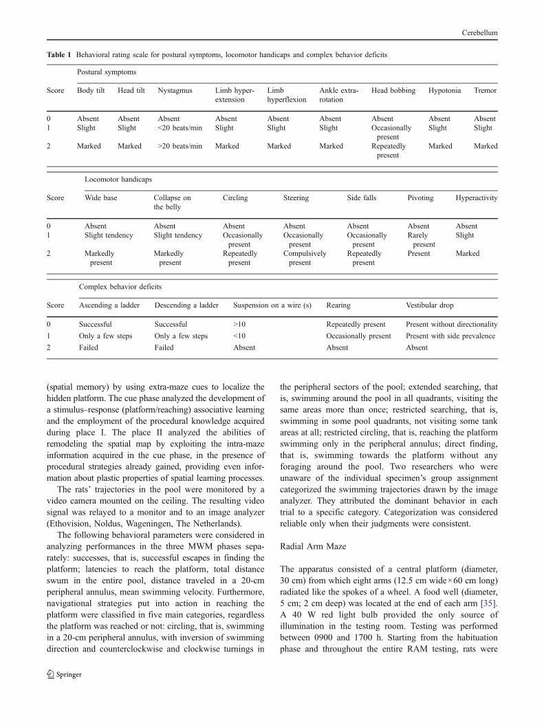
Postural symptoms, locomotor handicaps and complex behav-ior deficits were assessed by means of a behavioral rating scaledescribed elsewhere in detail [30, 31]. Neurological evalua-tions were scored by an examiner unaware of housingconditions the animals were submitted to. Presence or absenceof head and body tilt, hyperflexion, or hyper-extension offore- and hind-limbs in relation to the trunk, ankle extra-rotation, hypotonia, eye nystagmus, head oscillations (bob-bing), and tremor were evaluated. Some characteristics oflocomotion, namely, wide-based, collapsing on the belly,steering, circling, pivoting, and falling to the side, were alsoanalyzed. Finally, complex motor skills, such as ascending aladder and suspension on a wire as well as vestibular dropreaction and rearing behavior, were assessed. Video recordswere taken throughout the entire testing cycle and were usedto supplement direct behavioral observations. A score from 0(complete absence of deficit) to 2 (presence of the symptomto the highest degree) was assigned to each symptomaccording to its degree of severity, as described in Table 1.
Morris Water Maze
The rats were placed in a circular white pool (diameter140 cm) located in a normally equipped laboratory room,
uniformly lighted by four neon lamps (40 W each). Extra-maze spatial cues were on the walls and held in constantspatial relations throughout the experiments. The pool wasfilled with 24°C water (60 cm deep), made opaque by theaddition of 2 l of milk. An escape platform (diameter 10 cm)submerged 2 cm below or elevated 2 cm above the water levelwas placed in the middle of one cardinal quadrant located30 cm from the pool walls. Testing was performed between0900 and 1700 hours. The rat was released into the water fromrandomly varied starting points and was allowed to swimaround to find the platform. If this did not occur within 120 s,the experimenter guided it there. At the end of each trial the ratwas left on the platform for 30 s. Each rat was submitted totwo sessions of four trials per day, with a 3-min inter-trial and4-h inter-session intervals. In the first four sessions, theplatformwas hidden in the northwest quadrant (place I); in thenext two sessions, the platform was kept visible in thenortheast quadrant (cue phase); in the final four sessions, theplatform was hidden in the northeast quadrant (place II) [32–34]. The MWM protocol we used allowed to investigatenumerous components of spatial function. The place Ianalyzed first the general procedures such as the inhibitingnon-adaptive behaviors, as scrabbling at pool walls, then thesequence of navigational strategies (spatial procedurallearning) put into action to explore the pool and to find theplatform as well as the ability of building a spatial map
SC
SH
EC
EH
Neurological evaluation
0 21 75 PND11796 103 110
MWM RAM OF
f-b f-c food deprivation
1 week
Pl C Pl I II
Sham surgery
standard housing
environmental enrichment
HCb
Fig. 1 Diagram describing thebehavioral procedure and theglobal timing of the experimen-tal design of the four groupssince animals’ birth to the 117thpostnatal day (PND). Enrich-ment period, surgery, neurologi-cal evaluation, and behavioraltesting are indicated. MWMMorris water maze (Pl I place I,C cue, Pl II place II, phases).RAM radial arm maze (f-b full-baited, f-c forced-choice, proce-dures), OF open field
Cerebellum
(spatial memory) by using extra-maze cues to localize thehidden platform. The cue phase analyzed the development ofa stimulus–response (platform/reaching) associative learningand the employment of the procedural knowledge acquiredduring place I. The place II analyzed the abilities ofremodeling the spatial map by exploiting the intra-mazeinformation acquired in the cue phase, in the presence ofprocedural strategies already gained, providing even infor-mation about plastic properties of spatial learning processes.
The rats’ trajectories in the pool were monitored by avideo camera mounted on the ceiling. The resulting videosignal was relayed to a monitor and to an image analyzer(Ethovision, Noldus, Wageningen, The Netherlands).
The following behavioral parameters were considered inanalyzing performances in the three MWM phases sepa-rately: successes, that is, successful escapes in finding theplatform; latencies to reach the platform, total distanceswum in the entire pool, distance traveled in a 20-cmperipheral annulus, mean swimming velocity. Furthermore,navigational strategies put into action in reaching theplatform were classified in five main categories, regardlessthe platform was reached or not: circling, that is, swimmingin a 20-cm peripheral annulus, with inversion of swimmingdirection and counterclockwise and clockwise turnings in
the peripheral sectors of the pool; extended searching, thatis, swimming around the pool in all quadrants, visiting thesame areas more than once; restricted searching, that is,swimming in some pool quadrants, not visiting some tankareas at all; restricted circling, that is, reaching the platformswimming only in the peripheral annulus; direct finding,that is, swimming towards the platform without anyforaging around the pool. Two researchers who wereunaware of the individual specimen’s group assignmentcategorized the swimming trajectories drawn by the imageanalyzer. They attributed the dominant behavior in eachtrial to a specific category. Categorization was consideredreliable only when their judgments were consistent.
Radial Arm Maze
The apparatus consisted of a central platform (diameter,30 cm) from which eight arms (12.5 cm wide×60 cm long)radiated like the spokes of a wheel. A food well (diameter,5 cm; 2 cm deep) was located at the end of each arm [35].A 40 W red light bulb provided the only source ofillumination in the testing room. Testing was performedbetween 0900 and 1700 h. Starting from the habituationphase and throughout the entire RAM testing, rats were
Table 1 Behavioral rating scale for postural symptoms, locomotor handicaps and complex behavior deficits
Postural symptoms
Score Body tilt Head tilt Nystagmus Limb hyper-extension
Limbhyperflexion
Ankle extra-rotation
Head bobbing Hypotonia Tremor
0 Absent Absent Absent Absent Absent Absent Absent Absent Absent1 Slight Slight <20 beats/min Slight Slight Slight Occasionally
presentSlight Slight
2 Marked Marked >20 beats/min Marked Marked Marked Repeatedlypresent
Marked Marked
Locomotor handicaps
Score Wide base Collapse onthe belly
Circling Steering Side falls Pivoting Hyperactivity
0 Absent Absent Absent Absent Absent Absent Absent1 Slight tendency Slight tendency Occasionally
presentOccasionallypresent
Occasionallypresent
Rarelypresent
Slight
2 Markedlypresent
Markedlypresent
Repeatedlypresent
Compulsivelypresent
Repeatedlypresent
Present Marked
Complex behavior deficits
Score Ascending a ladder Descending a ladder Suspension on a wire (s) Rearing Vestibular drop
0 Successful Successful >10 Repeatedly present Present without directionality
1 Only a few steps Only a few steps <10 Occasionally present Present with side prevalence
2 Failed Failed Absent Absent Absent
Cerebellum

food-restricted to decrease their weight by 20%. Thehabituation phase, during which the rat was allowed tofreely explore the maze (in which a piece of Purina chowhad been placed in each arm) for 10 min a day, was carriedout 2 days before the full-baited maze procedure.
Full-Baited Maze Procedure All maze arms were baited witha piece of Purina chow prior to each session. The rat wasplaced on the central platform. The task goal was to collect theeight rewards. This aim could be reached through a maximalnumber of 16 entries. Once all the eight rewards were baitedor the allowed 16 visits were made the animals were removedby the maze and put in their cages. Any RAM session wascomprised only one trial. The animals were submitted to twosessions a day for five consecutive days. The inter-sessioninterval was 4 h. The apparatus between subjects was cleanedwith a 70% solution of ethanol.
The following parameters were considered: total errors(number of re-visited arms divided by total number ofvisits, correct and incorrect, ×100); mean spatial span (thelongest sequence of correctly visited arms displayed in eachsession); perseverations (sum of consecutive entries in thesame arm or in a fixed sequence of a maximum of threetray arms in the ten sessions. Perseverations on more thanthree trays were never observed); number of 45° angles(entries in adjacent arms); 45° angle span (the longestsequence of 45° angles made during each session).
Forced-Choice Procedure All animals were submitted to theforced-choice paradigm 48 h after the preceding protocolended. In the first phase, only four arms (for example, arms 1,3, 4, and 7) were opened and baited; the others arms remainedclosed. The baited arms were separated by different angles toprevent the animal from reaching the solution by adopting astereotyped pattern. The rat was allowed to explore the openarms. Then, it spent 60 s in its cage before being returned to themaze. In the second phase, the rat was allowed free access to alleight arms, but only the four previously closed arms werebaited. This task was repeated for five consecutive days with adifferent configuration of arms closed each day to avoid anyfixed search pattern.
The parameter considered was working memory errors,considered as re-entries into already visited arms. In the secondphase, this parameter was further broken down into two errorsubtypes: across-phase errors, defined as entries into an armentered during the first phase; and within-phase errors, definedas re-entries into an arm visited earlier in the same session.
Open Field
The apparatus consisted of a circular arena (diameter,140 cm) delimited by a 30-cm high wall. A 40 W red light
bulb provided the only source of illumination in the testingroom. Testing was performed between 0900 and 1700 h.Forty-eight hours after the preceding RAM test, each ratwas gently placed in the periphery of the arena facingtoward the center of the arena and allowed to move freelyin the empty open field. The starting point was the southpoint for all animals. The baseline level of activity wasmeasured during a 6-min period. All testing was recordedby a video camera; the signal was relayed to a monitor andto the previously described image analyzer. The apparatusbetween subjects was cleaned with a 70% solution ofethanol. The following emotional and motor parameterswere analyzed: number of defecation boluses, motionlesstime, rearings, total distance (in cm) traveled in the arena,percentage of the distance traveled in exploring a 20-cmperipheral annulus (peripheral distance), percentage of thetotal distance traveled in exploring a central area of 35 cmof radius, and central area crossings, that is the number ofentries into the central area of 35 cm of radius.
Histological Controls
When the behavioral testing was finished, the HCbedanimals were deeply anesthetized and transcardially per-fused with saline followed by 4% buffered formalin. Theextent of the cerebellar lesion was determined from Nissl-stained 50-μm frozen sections. Animals were included inthe present study if they had received a complete right HCbwith total ablation of deep nuclei (Fig. 2a). In all casesreported here, the left side of the cerebellum and all extra-cerebellar structures were spared, except for the dorsal capof the right Deiters’ nucleus which in some cases wasslightly affected. The variability in the extent of thefloccular and vermian lesions was considered not influenc-ing, because in all cases these structures were functionallydisconnected due to the ablation of the cerebellar pedunclesand deep nuclei of the right side. To verify variability of theextent of cerebellar lesion, Nissl-stained coronal sections atthe same representative levels from bregma (−10.04,−11.60, and −13.24 mm) were selected for each animal.Lesion boundaries were then drawn on schematic drawingsof corresponding atlas tables [36] and the areas ablatedcomputed by the software ImageJ (version 1.42q).
Statistical Analysis
The data were first tested for normality (Shapiro–Wilk’stest) and homoscedasticity (Levene’s test). All data pre-sented as the mean ± SEM were analyzed by one-, two-orthree-way analyses of variance (ANOVAs). The two-wayANOVAs were performed by applying either the mixedmodel for independent variable (group) and repeatedmeasures (day) or the model for two independent variables
Cerebellum

(housing×lesion). Three-way ANOVAs (housing x lesion xsession or strategy) were also performed. These analyseswere followed by Newman–Keuls test. All analyses wereperformed by using Statistica 7.0 for Windows and thesignificance level was established at p≤0.05.
Results
Verification of Cerebellar Lesion
The mean percentages of cerebellar ablation were calculatedfor both SH and EH groups (at −10.04 mm from bregma,SH=49.9%±0.16; EH=50.3%±0.16; at −11.60 mm frombregma, SH=45.6%±0.35; EH=44.4%±0.37; at −13.24 mmfrom bregma, SH=50.4%±0.22; EH=49.6%±0.19). One-way ANOVA on the mean percentages of ablation revealed nosignificant differences between groups (F1, 12=0.32; p=0.58)(Fig. 2b).
Neurological Evaluation
Figure 3 depicts the time courses of compensation of thecerebellar symptoms of the HCbed groups (EH and SH)together with the baseline of the postural evaluation ofsham-operated control groups (EC and SC). All animalswere evaluated by means of the rating scale described inTable 1. However, since it evaluated symptoms andasymmetries, the sham-operated animals obtained alwaysscores of 0 (complete absence of deficits). This kind ofevaluation prevented the inclusion of the scores obtained bythe sham-operated groups in the ANOVAs.
As shown in Fig. 3, dramatic effects of housing conditionswere observed on postural, locomotor and complex behaviorsof the two groups of lesioned animals. Twenty-four hoursafter the HCb, both EH and SH groups exhibited postural andmotor impairments of the same severity. From the secondpost-operative day onward, the EH group exhibited signifi-cantly less impaired posture (Fig. 3a), locomotion (Fig. 3b)and complex behaviors (Fig. 3c) compared to the SH group.As for the postural symptoms, a two-way ANOVA (group×day) revealed significant group (F1, 12=64.1; p<0.00001) andday (F11,132=70.6; p<0.00001) effects. Interaction was alsosignificant (F11, 132=14.7; p<0.00001). Post hoc comparisonsshowed that the two HCbed groups were similarly impaired24 h after the lesion but obtained significantly different scoresfrom the 2nd to the 28th day (Fig. 3a). As for the locomotorhandicaps, a two-way ANOVA (group×day) revealed signif-icant group (F1, 12=71.4; p<0.00001) and day (F11, 132=41.7;p<0.00001) effects. Interaction was also significant (F11,132=8.4, p<0.00001). Once again, post hoc comparisons showedthat the two HCbed groups were similarly impaired 24 h afterthe lesion but obtained significantly different scores from the2nd to the 21st day (Fig. 3b). The two HCbed groups weresignificantly different even in putting complex behaviors intoaction. A two-way ANOVA (group×day) revealed significantgroup (F1, 12=993.1; p<0.00001) and day (F11, 132=93.2; p<0.00001) effects. Interaction was also significant (F11, 132=19.8; p<0.00001). Interestingly, post hoc comparisonsshowed that 6 weeks after the cerebellar lesion SH animals
Fig. 2 a Nissl-stained coronal section through cerebellum and brainstem in a HCbed rat. Note the total absence of the right hemi-cerebellum and the sparing of any extra-cerebellar structure. Scale bar,2 mm. b Schematic drawings of the cerebellum and brainstemat −10.04, −11.60, and −13.24 mm from bregma illustrating thereconstruction of minimal (dark gray) and maximal (light gray)extension of the lesion in SH and EH groups. Note that the ablationsof the right cerebellar hemisphere totally include the ipsilateral deepnuclei (F fastigial, I interpositus, D dentate, nuclei). Deep nuclei areconversely spared on the left side
Cerebellum

did not yet reach the same level of compensation of thecomplex behaviors of the EH group (Fig. 3c).
As awhole, these findings provide evidence of the beneficialeffects of housing in an enriched environment on all facets ofmotor performance in the presence of a cerebellar lesion.
Morris Water Maze
The ANOVAs for all parameters are reported in Table 2. Allexperimental groups explored the pool and found theplatform with latencies progressively lower, as the sessionswent by. While in place I both groups of enriched animalsdisplayed a significantly higher number of successes (EC=3.8±0.2; EH=3.9±0.2) in comparison to the non-enrichedgroups (SC=3.7±0.3; SH=3.5±0.3), and in the cue andplace II phases all animals succeeded in mastering the taskwith a 100% of successes.
Figure 4a shows no differences in latency values amonggroups except in the place II, where the SH displayed thehighest mean latencies in comparison to the remaininggroups (post hoc comparisons: SH vs. EH, SC, or EC: p≤0.0005). Similarly, when total distances swum to reach theplatform were analyzed in the three MWM phases, EH, SC,and EC animals traveled similar distances, while SH
animals swam the longest distances (post hoc comparisons:SH vs. EH, SC, or EC: at least p<0.05) (Fig. 4b).
By analyzing the percentages of distance traveled in theperipheral annulus, we observed that while during place IEH and SH groups displayed higher percentages than theun-lesioned groups (post hoc comparisons: EH or SH vs.SC or EC: at least p<0.05), in place II EH groupinterestingly exhibited a percentage of peripheral travelingnot different from SC group (Fig. 4c).
In place I, EC group displayed the highest velocityvalues (post hoc comparison: EC vs. EH, SH, or SC: atleast p<0.0005) while EH group displayed velocity valuesnot different from SH and SC groups. The SH group that inplace I swam significantly more quickly than SC (post hoccomparison: p<0.01) in place II swan as quickly as ECgroup. In place II, the velocities of EH group did not differfrom those of SC group, but were significantly lower thanthose of EC and SH groups (post hoc comparison: EH orSC vs. EC or SH: at least p<0.05) (Fig. 4d).
An analysis of the explorative strategies the animals put intoaction in performing the task revealed interesting differencesamong groups and phases (Fig. 4e). In place I phase, SHanimals were unable to orientate their bodies to reach theplatform and consequently swam in circles around the
NEUROLOGICAL EVALUATION
Postural symptoms
0
5
10
15
20
Def
icit
sco
re
EH
SH***
***
**
a
Locomotor handicaps
0
2
4
6
8
10
12
Def
icit
sco
re
n.s.
*
b
Complex behavior deficits
0
2
4
6
8
10
12
Days after surgery
Def
icit
sco
re
*
c
n.s.
n.s.
1 2 3 4 7 9 12 14 21 28 35 42
n.s.
1 2 3 4 7 9 12 14 21 28 35 42
n.s.
***
1 2 3 4 7 9 12 14 21 28 35 42
***
14
SC/EC
Fig. 3 Neurological evaluationof a postural symptoms, b loco-motor handicaps, and c complexbehavior deficits in enrichedhemicerebellectomized (EH;n=7) and standard-housed hem-icerebellectomized (SH; n=7)rats together with the baseline ofthe evaluation of enriched sham-operated (EC; n=10) andstandard-housed sham-operated(SC; n=10) control groups.Abscissa: time in days aftersurgery. Ordinate: degree ofseverity of symptoms evaluatedaccording to the rating scaledescribed in Table 1. Data arepresented as means±SEM.Asterisks indicate the signifi-cance level of the post hoccomparisons between groups:*p<0.05; **p<0.005;***p<0.001
Cerebellum

Tab
le2
Statistical
comparisons
(three-w
ayANOVAs)
ofbehavioral
respon
sesin
theMWM
Param
eters
Hou
sing
Lesion
Session
H×L
H×S
L×S
H×L×S
F(df,1
and30
)p
F(df,1
and30
)p
F(df,3
and90
)p
F(df,1
and30
)p
F(df,3
and90
)p
F(df,3
and90
)p
F(df,3
and90
)p
Place
I
Successes
7.24
<0.05
0.23
n.s.
12.51
<0.00
001
3.24
n.s.
0.91
n.s.
0.33
n.s.
0.74
n.s.
Latencies
39.16
<0.00
018.85
<0.01
46.75
<0.00
001
1.15
n.s.
0.99
n.s.
0.03
n.s.
0.62
n.s.
Total
distance
18.44
<0.00
058.13
<0.01
16.17
<0.00
001
8.65
<0.01
2.45
n.s.
1.87
n.s.
0.10
n.s.
Peripheral
distance
13.37
<0.00
145
.27
<0.00
001
16.97
<0.00
001
6.75
<0.01
3.95
<0.01
6.55
<0.00
051.76
n.s.
Velocity
18.22
<0.00
052.20
n.s.
30.13
<.000
0135
.90
<0.00
001
3.44
<0.05
5.87
<0.00
51.75
n.s.
Strategies
0n.s.
0n.s.
8.34
<0.00
001
5.65
<0.05
6.54
<0.00
016.18
<0.00
056.09
<0.00
05
Cue
Successes
0.03
n.s.
0.03
n.s.
0.03
n.s.
2.28
n.s.
2.28
n.s.
2.28
n.s.
0.03
n.s.
Latencies
5.11
<0.05
5.20
<0.05
9.84
<0.00
51.48
n.s.
0.34
n.s.
2.44
n.s.
1.46
n.s.
Total
distance
6.07
<0.05
7.36
<0.05
2.15
n.s.
5.12
<0.05
2.50
n.s.
6.81
<0.01
0.75
n.s.
Peripheral
distance
26.46
<0.00
005
42.08
<0.00
001
0.23
n.s.
0.37
n.s.
3.34
n.s.
0.11
n.s.
0.24
n.s.
Velocity
0.71
n.s.
2.26
n.s.
26.94
<0.00
005
1.84
n.s.
0.07
n.s.
1.67
n.s.
0.16
n.s.
Strategies
0.47
n.s.
0.07
n.s.
22.74
<0.00
001
0.47
n.s.
6.15
<0.00
053.79
<0.01
2.27
n.s.
Place
II
Successes
0.46
n.s.
0.46
n.s.
0.75
n.s.
2.88
n.s.
2.16
n.s.
2.16
n.s.
0.75
n.s.
Latencies
11.28
<0.00
518
.38
<0.00
056.0
<0.00
17.65
<0.01
2.12
n.s.
2.13
n.s.
1.96
n.s.
Total
distance
15.91
<0.00
0518
.20
<0.00
052.04
n.s.
9.43
<0.00
50.23
n.s.
1.49
n.s.
0.82
n.s.
Peripheral
distance
27.49
<0.00
005
50.40
<0.00
001
1.99
n.s.
0.00
5n.s.
0.30
n.s.
0.93
n.s.
2.82
<0.05
Velocity
0.03
n.s.
1.30
n.s.
4.65
<0.00
523
.87
<0.00
005
3.45
<0.05
3.63
<0.05
2.76
<0.05
Strategies
0.06
n.s.
0.06
n.s.
75.93
<0.00
001
2.07
n.s.
15.41
<0.00
001
18.21
<0.00
001
7.88
<0.00
001
Cerebellum

periphery. Then, they displayed higher percentages of circlingand lower percentages of finding in comparison to theremaining groups. Conversely, EH group exhibited percen-tages of circling and finding not different to SC and highestpercentages of restricted circling. EC group displayed thehighest percentages of Finding since this first phase(Fig. 4e1). In the cue phase, no significant difference amonggroups in the five navigational strategies was found(Fig. 4e2). In the place II phase, while SH group persistedin displaying high percentages of strategies of peripheralexploration (circling and restricted circling) as well as lowpercentages of finding, EH group increased its percentages offinding, as the SC group, and dramatically decreased thepercentage of restricted circling (Fig. 4 e3).
Summing up, EH animals learned MWM task similarly toSC animals, with the only difference of a more evidentperipheral exploration in the first phase of the task. It isimportant to underline that even in the initial Place I the EHrats did not exhibit the compulsive circling typically displayedby HCbed animals. It is possible that the early recovery ofpostural and locomotor deficits depicted in Fig. 3 contributedto the improved performances of EH animals in the MWM.
Radial Arm Maze
Full-Baited Procedure The EH, EC and SC animalssignificantly reduced their errors as the sessions went by(one-way ANOVA, EH: F9, 54=2.4; p<0.05; EC: F9, 81=
Mea
n ti
me
(sec
)
Latencies a
Mea
n di
stan
ce (
cm)
Total distance b
Per
cent
age
Peripheral distance c
Velocity d
Mea
n ve
loci
ty (
cm/s
ec)
0
20
40
60
80
100
1 2 3 4 1 2 1 2 3 4
0200400600800
100012001400
1 2 3 4 1 2 1 2 3 4
0
20
40
60
80
100
1 2 3 4 1 2 1 2 3 4
0
10
20
30
40
1 2 3 4 1 2 1 2 3 4
Place I Place II Cue
EH
SH EC
SC
Place I
Cue
0
20
40
60
80
Place II
0
20
40
60
80
EH SH EC SC
C ES RS FRC
e1
e2
e3
Per
cent
age
Per
cent
age
Per
cent
age
MORRIS WATER MAZE
0
20
40
60
80
*** ***
*
***
**
**
* **
*
*** *
* ***
* *** * ***
Navigational strategies
Fig. 4 Effects of hemicerebel-lectomy and environmental en-richment on the Morris WaterMaze performance analyzed inthe three phases. Latencies (a),total distance (b) peripheral dis-tance, (c) and swimming veloc-ity (d) are depicted. Asterisks atthe right side of the graphsindicate post hoc comparisonsbetween groups: *p<0.05; ***p<0.0005. In (e), navigationalstrategies exhibited in the threeMWM phases are shown.Asterisks inside the graphs in-dicate the post hoc comparisonsbetween groups: *p<0.05; **p<0.01; ***p<0.0005. The circu-lar figurines under the graphsillustrate the typical explorativepatterns of the five main navi-gational strategies. The black-filled circles indicate platformposition. C circling strategy, ESextended searching, RS restrict-ed searching, RC restricted cir-cling, F finding. Vertical barsindicate SEM. In this and thefollowing figures: EH enrichedhemicerebellectomized animals,SH standard-housed hemicere-bellectomized animals, ECenriched sham-operated con-trols, SC standard-housed sham-operated controls
Cerebellum

5.5; p<0.00001; SC: F9, 81=2.5; p<0.05) unlike the SH(F9, 54=1.3; p=0.3) (Fig. 5a). A three-way ANOVA(housing×lesion×session) on total errors revealed signifi-cant housing (F1, 30=82.7; p<0.00001), lesion (F1, 30=54.6; p<0.00001) and session (F9, 270=4.6; p<0.00005)effects; some first-order interactions were significant(housing×session: F9, 270=2.4; p<0.05; lesion×session:F9, 270=1.9; p=0.05).
EH, EC and SC groups progressively lengthened theirspatial span (one-way ANOVA, EH: F9, 54=2.1; p<0.05; EC:F9, 81=4.1; p<0.0005; SC: F9, 81=2.1; p<0.05), unlike theSH group (F9, 54=0.8; p=0.6). A three-way ANOVA(housing×lesion×session) on spatial span values revealedsignificant housing (F1, 30=41.6; p<0.00001), lesion (F1, 30=45.1; p<0.00001), and session (F9, 270=4.3; p<0.00005)
effects; the first-order interaction housing×session (F9, 270=2.1; p<0.05) was also significant (Fig. 5b).
Perseverative errors were present mainly in the SH group(X=5.43±1.63) and almost lacking in the remaining groups(EH: X=1.29±0.52; EC: X=0.62±0.26; SC: X=0.35±0.13).Given the scattered occurrence of perseverations, weanalyzed perseverative errors not considering session effect.A two-way ANOVA (housing×lesion) revealed significanthousing (F1, 30=7.1; p<0.05) and lesion (F1, 30=15.6; p<0.0005) effects. Also, interaction was significant (F1, 30=9.2; p<0.01) (post hoc comparisons: SH vs. EH, SC, or EC:at least p<0.0005).
To analyze the procedural strategies used to explore themaze, we considered the number of 45° angles made invisiting the eight arms that is the entries in adjacent arms. A
RADIAL ARM MAZE
0
20
40
60
Mea
n er
rors
Total errors a
Span b
0
4
8
45° angles c
0
20
40
60
80
100
0
2
4
6
45° angle span d
Sessions
0
4
8
Mea
n er
rors
First phase errors e
0
8
16
Second phase errors f
Mea
n er
rors
0
8
16
Mea
n er
rors
Across-phase errors g
0
8
16
1 2 3 4 5 6 7 8 9 10
1 2 3 4 5 6 7 8 9 10
1 2 3 4 5 6 7 8 9 10
1 2 3 4 5 6 7 8 9 10
1 2 3 4 5
1 2 3 4 5
1 2 3 4 5
1 2 3 4 5
Mea
n er
rors
Within-phase errors h
Sessions
Full-baited procedure Forced-choice procedure
Per
cent
age
Vis
its
Vis
its
SC SH EC EH
** ** *
**
Fig. 5 Effects of hemicerebel-lectomy and environmental en-richment on the Radial ArmMaze performance. Total errors(a), span (b), 45° angles (c), and45° angle span (d) in the full-baited procedure are depicted.Working memory errors in first(e) and in second (f) phases ofthe forced-choice procedure aredepicted. The working memoryerrors of second phase are dis-tinguished in across- (g) andwithin- (h) phase errors. Theasterisks at the right side of thegraphs indicate the post hoccomparisons between groups:*p<0.05; **p<0.001; ***p<0.0005. Vertical bars indicateSEM
Cerebellum

three-way ANOVA (housing x lesion x session) on 45°angles revealed significant housing (F1, 30=27.9; p<0.00001), lesion (F1, 30=37.6; p<0.00001), and session(F9, 270=2.3; p<0.05) effects; the first-order interactionhousing×session was also significant (F9, 270=4.9; p<0.00001) (Fig. 5c).
To determine whether a chaining strategy was used, the45° angle span was analyzed. A three-way ANOVA(housing×lesion×session) on 45° angle span revealedsignificant housing (F1, 30=8.6; p<0.01), lesion (F1, 30=31.4; p<0.00001), and session (F9, 270=2.2; p<0.05)effects; some first-order interactions were significant(housing×session: F9, 270=2.7; p<0.01; lesion×session:F9, 270=2.8; p<0.005) (Fig. 5d).
Forced-Choice Procedure In the forced-choice paradigmwith selectively baited arms, any regular search pattern wasdiscouraged by the irregularity and variation of the baitedarms distribution session by session. Thus, success in visitingonly rewarded arms depended on remembering the alreadyvisited arms, rather than putting into action specific searchpatterns. This feature of the protocol rendered possible todistinguish procedural from working memory components. Athree-way ANOVA (housing×lesion×session) on workingmemory errors of the first phase in which only four arms wereopened revealed significant housing (F1, 30=31.4; p<0.00001), lesion (F1, 30=15.5; p<0.0005), and session (F4,
120=2.6; p<0.05) effects; first-order interaction housing×lesion was also significant (F1, 30=5.5; p<0.05; post hoccomparisons: EH vs. SH p<0.001; EH vs. EC p=0.3; EH vs.SC p=0.3; SH vs. EC or SC at least p<0.0005) (Fig. 5e).
A three-way ANOVA (housing×lesion×session) onworking memory errors of the second phase when all eightarms were opened revealed significant housing (F1, 30=45.2; p<0.000001), lesion (F1, 30=28.8; p<0.00001), andsession (F4, 120=2.4; p=0.05) effects; first-order interactionhousing×lesion was also significant (F1, 30=4.9; p<0.05).In fact, performance of EH group was better than that of SHgroup (post hoc comparisons: p<0.001), it was worse thanthat of EC group (p<0.05) and did not differ from that ofSC group (Fig. 5f). The errors of the second phase werefurther divided in across-phase and within-phase errors. Athree-way ANOVA (housing×lesion×session) on across-phase errors revealed significant housing (F1, 30=30.6; p<0.00001) and lesion (F1, 30=21.2; p<0.0001) effects. Theother main effect and all interactions were not significant(Fig. 5g). A similar three-way ANOVA (housing×lesion×session) on within-phase errors revealed significant housing(F1, 30=52.1; p<0.00001), lesion (F1, 30=30.0; p<0.00001), and session (F4, 120=5.9; p<0.0005) effects.Some first-order interactions were also significant (hous-ing×lesion: F1, 30=11.8; p<0.005 (post hoc comparisons:EH vs. SH p<0.001; EH vs. EC p=0.1; EH vs. SC p=0.2;
SH vs. EC or SC at least p<0.0005); lesion×session: F4,
120=2.6; p<0.05). The second-order interaction was alsosignificant (F4, 120=3.1; p<0.05) (Fig. 5h).
Summing up, in the forced-choice paradigm of RAMtask EH animals behaved as SC animals in most parametersconsidered and even in some parameters reached theoptimal performances of EC animals.
Open Field
When exploring the OF arena, all animals traveled similartotal distances as revealed by a two-way ANOVA (housing xlesion). This analysis failed to reveal any significant effect onhousing (F1, 30=0.5; p=0.5) or lesion (F1, 30=1.3; p=0.3)factors. Also, interaction was not significant (F1, 30=2.1;p=0.2). The percentages of the distance traveled inexploring a peripheral annulus (peripheral distance) weresignificantly different among groups. A two-way ANOVA(housing×lesion) revealed significant housing effect (F1,
30=8.7; p<0.01) and interaction housing×lesion (F1, 30=7.3; p<0.05). Lesion effect was not significant (F1, 30=0.2; p=0.6). Post hoc comparisons revealed that the EHanimals’ percentages of peripheral distance were notsignificantly different from those of the un-lesioned SCand EC animals and significantly lower than those of theSH animals (p<0.005) that traveled almost exclusively inthe peripheral annulus (Fig. 6a). The percentage ofperipheral distance displayed by SH animals was signif-icantly (p=0.05) higher than that of SC group. Thisbehavior of SH group could be related to defectiveexploration as well as to increased anxiety levels. Thetotal distances traveled in the central area were affected byhousing condition, as revealed by a two-way ANOVA(housing×lesion; housing effect: F1, 30=17.0; p<0.0005;lesion effect: F1, 30=0.5; p=0.5; interaction: F1, 30=2.2;p=0.1) (Fig. 6a). The same pattern was observed whencentral area crossings were considered (two-way ANOVA:housing effect: F1, 30=6.9; p<0.05; lesion effect: F1, 30=0.2; p=0.6; interaction: F1, 30=3.1; p=0.09). Rearingbehavior was computed on data exhibited by EH, EC,and SC groups, since SH animals displayed no rearing atall because of the severity of their postural impairment(Fig. 6b). One-way ANOVA revealed that EH groupexhibited a number of rearings not significantly differentfrom both un-lesioned SC and EC groups (F2, 24=1.7;p=0.2).
No differences among groups were found as fordefecations (two-way ANOVA: housing effect: F1, 30=1.0;p=0.3; lesion effect: F1, 30=1.6; p=0.2; interaction: F1, 30=2.3; p=0.1). Finally, when the motionless time wasconsidered, a two-way ANOVA (housing×lesion) revealedsignificant housing (F1, 30=25.3; p<0.00005) and lesion
Cerebellum

(F1, 30=29.1; p<0.0001) effects. Also, interaction wassignificant (F1, 30=8.9; p<0.01) (Fig. 6c).
Discussion
Thus far, clinical and experimental studies have examinedthe factors that stabilize and improve the cognitivesymptoms following cortical neuro-degenerative damage[4, 9, 37]. We wondered whether environmental complexitymight promote motor and cognitive functioning also in thepresence of cerebellar pathologies.
The present results demonstrate that long-term exposureto an enriched environment exerts beneficial effects onmotor, cognitive and emotional symptoms of cerebellarorigin. Exposure to complex stimulations almost complete-ly compensated the HCb effects and rendered the perform-ances of EH animals similar to those of intact animals. It isnoteworthy that the outstanding compensation of the EHanimals was not due to a less severe initial impairment. Infact, both EH and SH groups exhibited similar initial valuesin almost all parameters of the postural and cognitivetesting. Namely, housing in complex situations acceleratedcompensation of postural and locomotor deficits of cere-
bellar origin; it improved both working and long-termmemory; it allowed rapid transition from one competenceto another (e.g., from procedural to mnesic competence); itameliorated functions linked to declarative (localizatory)memory; it improved exploratory strategies; and it de-creased anxiety levels.
First of all, we considered the possibility that theimprovements observed in the enriched lesioned animalswere due to changes in motivation rather than to specificeffects on cognitive and motor functions. However, theenvironmental enrichment did not appear to have affectedmotivational components by modifying the salience ofreward/punishment and explorative tendencies. In fact,regardless of whether or not they were lesioned or enriched,all animals promptly climbed onto the MWM platform,consumed the food pellets in RAM and searched through-out the environments (MWM pool, radial maze, or OFarena).
A primary feature of HCb symptomatology is themaximum degree of severity of the postural and locomotorimpairment present one day after the lesion, whichdecreases progressively as time goes by. The time courseof the compensation of the motor cerebellar symptoms wasfavorably influenced by the previous exposure to enriched
EH SH EC SC
EH
SH
EC
SC
0
40
80
120
160
0
5
10
15
20
EH SH EC SC
Motionless time
Groups
Mea
n ti
me
(sec
)
cRearings
Groups
Mea
n nu
mbe
r
bn.s.
******
OPEN FIELD TEST
SH SCEH
Explorative trajectoriesaEC
TD(cm)
PD(%)
2017.1 ± 479.1 1933.3 ± 296.8 2143.3 ± 178.6
74.2 ± 6.1 84.8 ± 5.4
8.6 ± 1.8 3.7 ± 1.8CD(%)
CC(n) 4.7 ± 1.7 2.4 ± 1.2
1362.9 ± 196.2
83.7 ± 1.5
7.5 ± 0.9
3.3 ± 0.6
98.3 ± 1.3
0.5 ± 0.5
0.1 ± 0.1
Fig. 6 Effects of hemicerebel-lectomy and environmental en-richment on the Open Fieldperformance. In (a), the explor-ative trajectories traveled by EH,SH, EC, and SC rats in explor-ing the arena are reported. Totaldistances (TD), percentages ofperipheral distance (PD), ofcentral distance (CD), and cen-tral crossings (CC) are alsoindicated. Rearings (b) and mo-tionless time (c) displayed bythe four experimental groups areillustrated. In (b–c), asterisksinside the graphs indicate thepost hoc comparisons betweengroups: ***p<0.0005. Verticalbars indicate SEM
Cerebellum

environment. As repeatedly reported in the literature aboutthe behavioral effects of the exposure to enriched environ-ments [9, 38–44], rearing in enriched conditions promotesphysical activity by encouraging exploratory movements toforage the large cages, fine manipulation skills linked to theinspection of the repeatedly renewed objects, and generalmotor activity in using running wheels. All motor compo-nents are beneficially influenced by enrichment condition.In the present research, the complex behaviors requiringlimb coordination, eumetric movements, muscle strengthand subtle balance control were particularly well-compensated in EH animals. Consistently, in rats recoveredfrom motor effects of cerebellar ablations the complexskills were more affected than postural and locomotorbehaviors by the treatment with NMDA antagonists thatdisrupted the compensated state [29, 30].
The present results on the effects of environmentalenrichment on motor deficits of cerebellar origin complete-ly fit with data obtained in other models of brainpathologies. Exposure to an enriched environment amelio-rated motor coordination deficits and spatial learning in thefemale mice of a murine model of the Rett’s syndrome, anautistic spectrum developmental disorder characterized bysevere behavioral and neuropathological deficits [39, 45,46]. Furthermore, research on the R6/1 transgenic micemodel of Huntington’s disease (HD) demonstrated thatexposure of HD mice to an enriched environment delayedthe onset and progression of disease [41, 47–49]. Further-more, environmental enrichment improved behavioralperformances in models of dementia-like neurodegenera-tion [10–12, 50].
The enhanced stimulations provided by the enrichedenvironment influenced not only motor but also cognitivefunction, in accordance with clinical studies reporting thatmore physical exercise and cognitive engagement in youthare associated with maintenance of memory function inmidlife [51, 52].
In the MWM, the EH animals exhibited improvedmnesic and mapping abilities (hidden platform) as well asincreased attention to contextual cues (visible platform).Moreover, they exhibited reduced peripheral swimming andefficient navigational strategies, as indicated by the highpercentage of finding strategy they used to reach theplatform. In general, the spatial behavior severely impairedby cerebellar lesions in the MWM [33, 34, 53–56] wasthoroughly recovered by EH animals that behave as intactrats. One possible explanation for the improved functionalrecovery in EH rats could take into account their improvedmotor performances as well as improved abilities of visuo-motor coordination. In fact, EH animals were able toorientate their bodies in the pool because of theiraccelerated postural recovery. However, the tuned naviga-tional strategies the EH animals exhibited in the phases in
which the platform was hidden (places I and II) seem tosupport even improved cognitive performances.
In the RAM, the EH group made fewer errors andperseverations and more entries in consecutive arms thanthe SH group in the full-baited procedure and performedsignificantly less errors than the SH rats even in the forced-choice procedure. Thus, depending on the context the EHanimals reorganized their strategies and shifted from usingspatial procedures to applying mnesic competencies. Thepresent data fit with the improved mnesic and proceduralperformances displayed by enriched intact [57] or cholin-ergically depleted animals [12].
In the OF, the EH group traveled the peripheral sectors notdifferently from the un-lesioned EC and SC groups. This kindof explorative pattern is in line with the reduced percentagesof Circling displayed by EH, EC, and SC rats in the MWM.Furthermore, EH and EC animals similarly traveled andcrossed the central area of the arena. Noteworthy, a patternfeatured by increased central traveling is retained to be linkedto reduced anxiety levels [58, 59]. Thus, it is possible that theameliorative effects of the environmental enrichment weobserved on various parameters of the behavioral tasks wererelated to diminished levels of anxiety of animals exposed toa complex environment [60]. Accordingly, the prolongedexperience of enrichment may contribute to the improvedperformance of EH animals not only potentiating thecognitive competencies but also by reducing the anxietylevels. Even the motionless time displayed by EH, EC andSC animals overlapped. The number of rearings exhibited byEH animals markedly differed from that of SH animals.Typically, HCbed animals show almost no rearings becausethey have difficulty keeping their balance standing on theirhind-limbs [61]. Notably, the EH animals produced the samenumber of rearings as the un-lesioned animals, once againindicating their almost complete compensation of the HCbmotor deficits.
Summing up, besides symmetrical and coordinatedpostural and locomotor behaviors, the EH rats exhibitedadvanced working memory and planning activities, analmost complete absence of perseverations, rapid acquisi-tion and flexible use of efficient explorative strategies andof procedural abilities. These functions are typicallymediated by the frontal and cerebellar circuits. Extensiveanatomo-functional connections [62] support the interplaybetween the cerebellar and frontal areas that interact inexecutive control of voluntary behavior [63–65]: thecerebellum permits acquisition of efficient proceduralcompetencies and the frontal cortex provides flexibilityamong already acquired and stored solutions [66–68].Previously, we demonstrated that in rats HCb provokeslack of flexibility in changing strategy, impossibility toinhibit patently wrong responses [69–71], deficits due toimpairment in adapting behavior to context. The evidence
Cerebellum

that the EH animals displayed no “frontal-like” deficits asperseverations and lack of attention to novel stimuli, as wellas no deficient working memory, could be an index of anefficient use of cerebello-frontal networks. One of the mostextensively replicated findings in enriched rodents is themodulation of neurotrophin expression [48, 72–74] retainedrelated to the improved performances of the enrichedanimals [75]. We recently demonstrated that rats enrichedwith the very same protocol used in the present researchexhibited marked up-regulation of the levels of BDNF inthe frontal cortex and cerebellum [76, 77]. Althoughspeculative, the relation between the increased expressionof neurotrophins in the cerebello-frontal areas and theimproved abilities related to these areas in the enrichedanimals is noteworthy.
To our knowledge, this is the first attempt at demonstratingthat early and prolonged exposure to an enriched environmentstrikingly improves compensation of deficits of cerebellarorigin. Further studies are needed to discover the mechanismsthrough which exposure to complex stimulations mayrepresent an endogenous device to ameliorate motor andcognitive performance in the presence of brain damage. Themolecular changes underlying experience-dependent synapticplasticity should be investigated in future research to developnew therapeutic approaches not only for brain disorders ingeneral but also for cerebellar pathologies in particular.
Acknowledgements This work was supported by MIUR grants to LP.
Conflicts of interest All the authors have approved the manuscriptthat is enclosed and fully disclose all conflicts of interest.
References
1. Borenstein AR, Copenhaver CI, Mortimer JA. Early-life riskfactors for Alzheimer disease. Alzheimer Dis Assoc Disord.2006;20:63–72.
2. McDowell I, Xi G, Lindsay J, Tierney M. Mapping theconnections between education and dementia. J Clin Exp Neuro-psychol. 2007;29:127–41.
3. Green CS, Bavelier D. Exercising your brain: a review of humanbrain plasticity and training-induced learning. Psychol Aging.2008;23:692–701.
4. Buckner RL. Memory and executive function in aging and AD:multiple factors that cause decline and reserve factors thatcompensate. Neuron. 2004;44:195–208.
5. Arendash GW, Garcia MF, Costa DA, Cracchiolo JR, Wefes IM,Potter H. Environmental enrichment improves cognition in agedAlzheimer’s transgenic mice despite stable beta-amyloid deposi-tion. NeuroReport. 2004;15:1751–4.
6. Artola A, von Frijtag JC, Fermont PC, Gispen WH, Schrama LH,Kamal A, et al. Long-lasting modulation of the induction of LTDand LTP in rat hippocampal CA1 by behavioural stress andenvironmental enrichment. Eur J Neurosci. 2006;23:261–72.
7. Ngandu T, von Strauss E, Helkala EL, Winblad B, Nissinen A,Tuomilehto J, et al. Education and dementia: what lies behind theassociation? Neurology. 2007;69:1442–50.
8. Normann C, Berger M. Neuroenhancement: status quo andperspectives. Eur Arch Psychiatry Clin Neurosci. 2008;258:110–4.
9. Nithianantharajah J, Hannan AJ. The neurobiology of brain andcognitive reserve: mental and physical activity as modulators ofbrain disorders. Prog Neurobiol. 2009;89:369–82.
10. Jankowsky JL, Melnikova T, Fadale DJ, Xu GM, Slunt HH,Gonzales V, et al. Environmental enrichment mitigates cognitivedeficits in a mouse model of Alzheimer’s disease. J Neurosci.2005;25:5217–24.
11. Berardi N, Braschi C, Capsoni S, Cattaneo A, Maffei L.Environmental enrichment delays the onset of memory deficitsand reduces neuropathological hallmarks in a mouse model ofAlzheimer-like neurodegeneration. J Alzheimers Dis.2007;11:359–70.
12. Mandolesi L, De Bartolo P, Foti F, Gelfo F, Federico F, LeggioMG, et al. Environmental enrichment provides a cognitive reserveto be spent in the case of brain lesion. J Alzheimers Dis.2008;15:11–28.
13. Petrosini L, De Bartolo P, Foti F, Gelfo F, Cutuli D, Leggio MG,et al. On whether the environmental enrichment may providecognitive and brain reserves. Brain Res Rev. 2009;61:221–39.
14. Luciani L. Il cervelletto: nuovi studi di fisiologia normale epatologica. Firenze: Le Monnier; 1891.
15. Holmes G. The cerebellum of man. Brain. 1939;62:1–30.16. Schmahmann JD, Sherman JC. The cerebellar cognitive affective
syndrome. Brain. 1998;121:561–79.17. Schmahmann JD. Disorders of the cerebellum: ataxia, dysmetria
of thought, and the cerebellar cognitive affective syndrome. JNeuropsychiatry Clin Neurosci. 2004;16:367–78.
18. Schmahmann JD, Caplan D. Cognition, emotion and the cerebel-lum. Brain. 2006;129:290–2.
19. Thach WT. Context-response linkage. Int Rev Neurobiol.1997;41:599–611.
20. Thach WT. A role for the cerebellum in learning movementcoordination. Neurobiol Learn Mem. 1998;70:177–88.
21. Thach WT. On the mechanism of cerebellar contributions tocognition. Cerebellum. 2007;6:163–7.
22. Drepper J, Timmann D, Kolb FP, Diener HC. Non-motorassociative learning in patients with isolated degenerative cere-bellar disease. Brain. 1999;122:87–97.
23. Ito M. Historical review of the significance of the cerebellum andthe role of Purkinje cells in motor learning. Ann NY Acad Sci.2002;978:273–88.
24. Molinari M, Petrosini L, Misciagna S, Leggio MG. Visuospatialabilities in cerebellar disorders. J Neurol Neurosurg Psychiatry.2004;75:235–40.
25. Golla H, Thier P, Haarmeier T. Disturbed overt but normal covertshifts of attention in adult cerebellar patients. Brain.2005;128:1525–35.
26. Ziemus B, Baumann O, Luerding R, Schlosser R, Schuierer G,Bogdahn U, et al. Impaired working-memory after cerebellarinfarcts paralleled by changes in BOLD signal of a cortico-cerebellar circuit. Neuropsychologia. 2007;45:2016–24.
27. Amrani K, Dykes RW, Lamarre Y. Bilateral contributions to motorrecovery in the monkey following lesions of the deep cerebellarnuclei. Brain Res. 1996;740:275–84.
28. Armano S, Rossi P, Taglietti V, D’Angelo E. Long-termpotentiation of intrinsic excitability at the mossy fiber-granulecell synapse of rat cerebellum. J Neurosci. 2000;20:5208–16.
29. Casado M, Isope P, Ascher P. Involvement of presynaptic N-methyl-D-aspartate receptors in cerebellar long-term depression.Neuron. 2002;33:123–30.
30. Federico F, Leggio MG, Mandolesi L, Petrosini L. The NMDAreceptor antagonist CGS 19755 disrupts recovery followingcerebellar lesions. Restor Neurol Neurosci. 2006a;24:1–7.
Cerebellum

31. Centonze D, Rossi S, De Bartolo P, De Chiara V, Foti F, MusellaA, et al. Adaptations of glutamatergic synapses in the striatumcontribute to recovery from cerebellar damage. Eur J Neurosci.2008;27:2188–96.
32. Morris RG, Garrud P, Rawlins JN, O’Keefe J. Place navigationimpaired in rats with hippocampal lesions. Nature. 1982;297:681–3.
33. Petrosini L, Molinari M, Dell’Anna ME. Cerebellar contributionto spatial event processing: Morris water maze and T-maze. Eur JNeurosci. 1996;8:1882–96.
34. Federico F, Leggio MG, Neri P, Mandolesi L, Petrosini L. NMDAreceptor activity in learning spatial procedural strategies II. Theinfluence of cerebellar lesions. Brain Res Bull. 2006b;70:356–67.
35. Mandolesi L, Leggio MG, Graziano A, Neri P, Petrosini L. Cerebellarcontribution to spatial event processing: involvement in procedural andworking memory components. Eur J Neurosci. 2001;14:2011–22.
36. Paxinos G, Watson C. The rat brain in stereotaxic coordinates. 4thed. San Diego: Academic; 1998.
37. Milgram NW, Siwak-Tapp CT, Arahujo J, Head E. Neuro-protective effects of cognitive enrichment. Ageing Res Rev.2006;5:354–69.
38. Bennett EL. krech D, Rosenzweig MR. Reliability and regionalspecificity of cerebral effects of environmental complexity andtraining. J Comp Physiol Psychol. 1964;57:440–1.
39. van Praag H, Kempermann G, Gage FH. Neural consequences ofenvironmental enrichment. Nat Rev Neurosci. 2000;1:191–8.
40. Greenough WT, Volkmar FR, Juraska J. Effects of rearingcomplexity on dendritic branching in frontolateral and temporalcortex of the rat. Exp Neurol. 1973;41:371–8.
41. van Dellen A, Cordery PM, Spires TL, Blakemore C, Hannan AJ.Wheel running from a juvenile age delays onset of specific motordeficits but does not alter protein aggregate density in a mousemodel of Huntington’s disease. BMC Neurosci. 2008;9:34.
42. Hicks RR, Zhang L, Atkinson A, Stevenon M, Veneracion M,Seroogy KB. Environmental enrichment attenuates cognitivedeficits but does not alter neurotrophin gene expression in thehippocampus following lateral fluid percussion brain injury.Neuroscience. 2002;112:631–7.
43. Kempermann G, Kuhn HG, Gage FH. More hippocampal neuronsin adult mice living in an enriched environment. Nature.1997;386:493–5.
44. Kempermann G. The neurogenic reserve hypothesis: what is adulthippocampal neurogenesis good for? Trends Neurosci.2008;31:163–9.
45. Kondo M, Gray LJ, Pelka GJ, Christodoulou J, Tam PP, HannanAJ. Environmental enrichment ameliorates a motor coordinationdeficit in a mouse model of Rett syndrome–Mecp2 gene dosageeffects and BDNF expression. Eur J Neurosci. 2008;27:3342–50.
46. Nag N, Moriuchi JM, Peitzman CG, Ward BC, Kolodny NH,Berger-Sweeney JE. Environmental enrichment alters locomotorbehaviour and ventricular volume in Mecp2 1lox mice. BehavBrain Res. 2009;196:44–8.
47. van Dellen A, Blakemore C, Deacon R, York D, Hannan AJ.Delaying the onset of Huntington’s in mice. Nature.2000;404:721–2.
48. Spires TL, Grote HE, Varshney NK, Cordery PM, van Dellen A,Blakemore C, et al. Environmental enrichment rescues proteindeficits in a mouse model of Huntington’s disease, indicating apossible disease mechanism. J Neurosci. 2004;24:2270–6.
49. Lazic SE, Grote HE, Blakemore C, Hannan AJ, van Dellen A,Phillips W, et al. Neurogenesis in the R6/1 transgenic mousemodel of Huntington’s disease: effects of environmental enrich-ment. Eur J Neurosci. 2006;23:1829–38.
50. De Bartolo P, Leggio MG, Mandolesi L, Foti F, Gelfo F, FerlazzoF, et al. Environmental enrichment mitigates the effects of basalforebrain lesions on cognitive flexibility. Neuroscience.2008;154:444–53.
51. Richards M, Hardy R, Wadsworth ME. Does active leisure protectcognition? Evidence from a national birth cohort. Soc Sci Med.2003;56:785–92.
52. Richards M, Deary IJ. A life course approach to cognitive reserve:a model for cognitive aging and development? Ann Neurol.2005;58:617–22.
53. Leggio MG, Neri P, Graziano A, Mandolesi L, Molinari M,Petrosini L. Cerebellar contribution to spatial event processing:characterization of procedural learning. Exp Brain Res.1999;127:1–11.
54. Rondi-Reig L, Le Marec N, Caston J, Mariani J. The role ofclimbing and parallel fibers inputs to cerebellar cortex innavigation. Behav Brain Res. 2002;132:11–8.
55. Gasbarri A, Pompili A, Pacitti C, Cicirata F. Comparative effectsof lesions to the ponto-cerebellar and olivo-cerebellar pathways onmotor and spatial learning in the rat. Neuroscience.2003;116:1131–40.
56. Colombel C, Lalonde R, Caston J. The effects of unilateralremoval of the cerebellar hemispheres on spatial learning andmemory in rats. Brain Res. 2004;1004:108–15.
57. Leggio MG, Mandolesi L, Federico F, Spirito F, Ricci B, Gelfo F,et al. Environmental enrichment promotes improved spatialabilities and enhanced dendritic growth in the rat. Behav BrainRes. 2005;163:78–90.
58. Bambico FR, Cassano T, Dominguez-Lopez S, Katz N, WalkerCD, Piomelli D, et al. Genetic deletion of fatty acid amidehydrolase alters emotional behavior and serotonergic transmissionin the dorsal raphe, prefrontal cortex, and hippocampus. Neuro-psychopharmacology. 2010;35:2083–100.
59. Long LE, Chesworth R, Arnold JC, Karl T. A follow-up study:acute behavioural effects of Delta(9)-THC in female heterozygousneuregulin 1 transmembrane domain mutant mice. Psychophar-macology. 2010;211:277–89.
60. Harris AP, D’Eath RB, Healy SD. Environmental enrichmentenhances spatial cognition in rats by reducing thigmotaxis (wallhugging) during testing. Anim Behav. 2009;77:1459–64.
61. Mandolesi L, Leggio MG, Spirito F, Petrosini L. Cerebellarcontribution to spatial event processing: do spatial procedurescontribute to formation of spatial declarative knowledge? Eur JNeurosci. 2003;18:2618–26.
62. Middleton FA, Strick PL. Cerebellar projections to the prefrontalcortex of the primate. J Neurosci. 2001;21:700–12.
63. Ito M. Cerebellar circuitry as a neuronal machine. Prog Neurobiol.2006;78:272–303.
64. Ito M. Control of mental activities by internal models in thecerebellum. Nat Rev Neurosci. 2008;9:304–13.
65. Diamond A. Close interrelation of motor development andcognitive development and of the cerebellum and prefrontalcortex. Child Dev. 2000;71:44–56.
66. Hyder F, Phelps EA, Wiggins CJ, Labar KS, Blamire AM,Shulman RG. “Willed action”: a functional MRI study of thehuman prefrontal cortex during a sensorimotor task. Proc NatlAcad Sci USA. 1997;94:6989–94.
67. Spence SA, Hirsch SR, Brooks DJ, Grasby PM. Prefrontal cortexactivity in people with schizophrenia and control subjects.Evidence from positron emission tomography for remission of‘hypofrontality’ with recovery from acute schizophrenia. Br JPsychiatry. 1998;172:316–23.
68. Pochon JB, Levy R, Poline JB, Crozier S, Lehericy S, Pillon B, etal. The role of dorsolateral prefrontal cortex in the preparation offorthcoming actions: an fMRI study. Cereb Cortex. 2001;11:260–6.
69. Foti F, Mandolesi L, Cutuli D, Laricchiuta D, De Bartolo P,Gelfo F, et al. Cerebellar damage loosens the strategic use ofthe spatial structure of the search space. Cerebellum. 2010;9:29–41.
Cerebellum

70. De Bartolo P, Mandolesi L, Federico F, Foti F, Cutuli D, Gelfo F,et al. Cerebellar involvement in cognitive flexibility. NeurobiolLearn Mem. 2009;92:310–7.
71. Mandolesi L, Foti F, Cutuli D, Laricchiuta D, Gelfo F, De BartoloP, et al. Features of sequential learning in hemicerebellectomizedrats. J Neurosci Res. 2010;88:478–86.
72. Pham TM, Ickes B, Albeck D, Soderstrom S, Granholm AC,Mohammed AH. Changes in brain nerve growth factor levels andnerve growth factor receptors in rats exposed to environmentalenrichment for one year. Neuroscience. 1999;94:279–86.
73. Young D, Lawlor PA, Leone P, Dragunow M, During MJ.Environmental enrichment inhibits spontaneous apoptosis, pre-vents seizures and is neuroprotective. Nat Med. 1999;5:448–53.
74. Zajac MS, Pang TY, Wong N, Weinrich B, Leang LS, Craig JM,et al. Wheel running and environmental enrichment differentially
modify exon-specific BDNF expression in the hippocampus ofwild-type and pre-motor symptomatic male and female Hunting-ton’s disease mice. Hippocampus. 2010;20:621–36.
75. Ickes BR, Pham TM, Sanders LA, Albeck DS, Mohammed AH,Granholm AC. Long-term environmental enrichment leads toregional increases in neurotrophin levels in rat brain. Exp Neurol.2000;64:45–52.
76. Angelucci F, De Bartolo P, Gelfo F, Foti F, Cutuli D, Bossu P, etal. Increased concentrations of nerve growth factor and brain-derived neurotrophic factor in the rat cerebellum after exposure toenvironmental enrichment. Cerebellum. 2009;8:499–506.
77. Gelfo F, Cutuli D, Foti F, Laricchiuta D, De Bartolo P, CaltagironeC, et al. Enriched environment improves motor function andincreases neurotrophins in hemicerebellar lesioned rats. Neuro-rehabil Neural Repair. 2010. doi:10.1177/1545968310380926
Cerebellum