
Cerebellar Contributions to Adaptive Control of Saccades in Humans
20
Model of the Control of Saccades by Superior Colliculus and Cerebellum CHRISTIAN QUAIA, 1,2 PHILIPPE LEF ` EVRE, 1,3 AND LANCE M. OPTICAN 1 1 Laboratory of Sensorimotor Research, National Eye Institute, Bethesda, Maryland 20892-4435; 2 Dipartimento di Elettronica, Elettrotecnica ed Informatica, Universita ` degli Studi di Trieste, 34100 Trieste, Italy; 3 Laboratory of Neurophysiology and Center for Systems Engineering and Applied Mechanics, Universite ` Catholique de Louvain, B-1348 Louvain-La-Neuve, Belgium Quaia, Christian, Philippe Lefe `vre, and Lance M. Optican. Model of the control of saccades by superior colliculus and cerebellum. J. Neurophysiol. 82: 999 –1018, 1999. Experimental evidence indicates that the superior colliculus (SC) is important but neither necessary nor sufficient to produce accurate saccadic eye movements. Furthermore both clinical and experimental evidence points to the cerebellum as an indispensable component of the saccadic system. Accordingly, we have devised a new model of the saccadic system in which the characteristics of saccades are determined by the cooperation of two pathways, one through the SC and the other through the cerebellum. Both pathways are influenced by feedback information: the feedback determines the decay of activity for collicular neurons and the timing of the activation for cerebellar neurons. We have modeled three types of cells (burst, buildup, and fixation neurons) found in the intermedi- ate layers of the superior colliculus. We propose that, from the point of view of motor execution, the burst neurons and the buildup neurons are not functionally distinct with both providing a directional drive to the brain stem circuitry. The fixation neurons determine the onset of the saccade by disfacilitating the omnipause neurons in the brain stem. Excluding noise-related variations, the ratio of the horizontal to the vertical components of the collicular drive is fixed throughout the saccade (i.e., its direction is fixed); the duration of the drive is such that it always would produce hypermetric movements. The cerebellum plays three roles: first, it provides an additional directional drive, which improves the acceleration of the eyes; second, it keeps track of the progress of the saccade toward the target; and third, it ends the saccade by choking off the collicular drive. The drive provided by the cerebellum can be adjusted in direction to exert a directional control over the saccadic trajectory. We propose here a control mechanism that incorporates a spatial displacement integrator in the cerebellum; under such conditions, we show that a partial directional control arises automatically. Our scheme preserves the advantages of several pre- vious models of the saccadic system (e.g., the lack of a spatial-to- temporal transformation between the SC and the brain stem; the use of efference copy feedback to control the saccade), without incurring many of their drawbacks, and it accounts for a large amount of experimental data. INTRODUCTION The saccadic system (i.e., the neural system that controls the rapid eye movements called saccades) has attracted the atten- tion of many investigators during the last 40 years. Thanks to the combined efforts of so many researchers, a great deal of data are now available about the pattern of neural activity, the anatomy of functional connections, and the effects of lesions and electrical stimulation in several brain areas involved in controlling saccades. The availability of such a large database and the relative simplicity of the mechanical system to be controlled (the eye plant), has prompted models of the saccadic system to spring up like mushrooms on a damp forest floor. In 1975, a milestone in the history of saccadic modeling, the Robinson model, was published (Robinson 1975; Zee et al. 1976). The central idea of that model, inherited by almost all subsequent models of the saccadic system, was that saccades are controlled by a local feedback loop, which in Robinson’s model was used to compare the desired position of the eyes with an internal estimate of their actual position, thus produc- ing an estimate of the instantaneous (or dynamic) motor error. This model, as well as others derived from it, was mainly conceptual, and many of its building blocks were not closely associated with anatomic structures. However, the growth of anatomic and physiological knowledge, due to the large num- ber of experiments carried out after 1975 (largely prompted by the many predictions of Robinson’s model), impelled modelers to identify the different parts of their models with specific regions of the brain. Why a new model of the saccadic system? Although the concept that several brain structures cooperate to produce fast and accurate saccadic eye movements has long been widely accepted, models necessarily concentrate on a restricted subset of these structures. Initially models included only the brain stem circuitry, but soon the great amount of data available about the superior colliculus (SC) made it essential to find a role for this midbrain structure. Accordingly, models focused on the role played by the SC in controlling saccades and in determining the firing pattern observed in brain stem neurons. However, during the last 10 years, new experimental evidence has induced modelers to attribute an increasing im- portance to the SC. This trend has lead to the development of a fairly large family of models that impute to the SC a domi- nant role in determining saccade metrics, and that thus could be dubbed ‘‘colliculocentric’’ (Arai et al. 1994; Droulez and Berthoz 1988; Lefe `vre and Galiana 1992; Optican 1994; Van Opstal and Kappen 1993; Waitzman et al. 1991). One of the major problems with colliculocentric models is that they have difficulties in explaining why lesions of the SC do not result in large and enduring deficits. In particular, it is The costs of publication of this article were defrayed in part by the payment of page charges. The article must therefore be hereby marked ‘‘advertisement’’ in accordance with 18 U.S.C. Section 1734 solely to indicate this fact. 999
-
Upload
independent -
Category
Documents
-
view
0 -
download
0
Transcript of Cerebellar Contributions to Adaptive Control of Saccades in Humans
Model of the Control of Saccades by Superior Colliculus andCerebellum
CHRISTIAN QUAIA,1,2 PHILIPPE LEFEVRE,1,3 AND LANCE M. OPTICAN1
1Laboratory of Sensorimotor Research, National Eye Institute, Bethesda, Maryland 20892-4435;2Dipartimento diElettronica, Elettrotecnica ed Informatica, Universita` degli Studi di Trieste, 34100 Trieste, Italy;3Laboratory ofNeurophysiology and Center for Systems Engineering and Applied Mechanics, Universite` Catholique de Louvain,B-1348 Louvain-La-Neuve, Belgium
Quaia, Christian, Philippe Lefevre, and Lance M. Optican. Modelof the control of saccades by superior colliculus and cerebellum.J.Neurophysiol.82: 999–1018, 1999. Experimental evidence indicatesthat the superior colliculus (SC) is important but neither necessary norsufficient to produce accurate saccadic eye movements. Furthermoreboth clinical and experimental evidence points to the cerebellum as anindispensable component of the saccadic system. Accordingly, wehave devised a new model of the saccadic system in which thecharacteristics of saccades are determined by the cooperation of twopathways, one through the SC and the other through the cerebellum.Both pathways are influenced by feedback information: the feedbackdetermines the decay of activity for collicular neurons and the timingof the activation for cerebellar neurons. We have modeled three typesof cells (burst, buildup, and fixation neurons) found in the intermedi-ate layers of the superior colliculus. We propose that, from the pointof view of motor execution, the burst neurons and the buildup neuronsare not functionally distinct with both providing a directional drive tothe brain stem circuitry. The fixation neurons determine the onset ofthe saccade by disfacilitating the omnipause neurons in the brain stem.Excluding noise-related variations, the ratio of the horizontal to thevertical components of the collicular drive is fixed throughout thesaccade (i.e., its direction is fixed); the duration of the drive is suchthat it always would produce hypermetric movements. The cerebellumplays three roles: first, it provides an additional directional drive,which improves the acceleration of the eyes; second, it keeps track ofthe progress of the saccade toward the target; and third, it ends thesaccade by choking off the collicular drive. The drive provided by thecerebellum can be adjusted in direction to exert a directional controlover the saccadic trajectory. We propose here a control mechanismthat incorporates a spatial displacement integrator in the cerebellum;under such conditions, we show that a partial directional control arisesautomatically. Our scheme preserves the advantages of several pre-vious models of the saccadic system (e.g., the lack of a spatial-to-temporal transformation between the SC and the brain stem; the use ofefference copy feedback to control the saccade), without incurringmany of their drawbacks, and it accounts for a large amount ofexperimental data.
I N T R O D U C T I O N
The saccadic system (i.e., the neural system that controls therapid eye movements called saccades) has attracted the atten-tion of many investigators during the last 40 years. Thanks tothe combined efforts of so many researchers, a great deal ofdata are now available about the pattern of neural activity, the
anatomy of functional connections, and the effects of lesionsand electrical stimulation in several brain areas involved incontrolling saccades. The availability of such a large databaseand the relative simplicity of the mechanical system to becontrolled (the eye plant), has prompted models of the saccadicsystem to spring up like mushrooms on a damp forest floor.
In 1975, a milestone in the history of saccadic modeling, theRobinson model, was published (Robinson 1975; Zee et al.1976). The central idea of that model, inherited by almost allsubsequent models of the saccadic system, was that saccadesare controlled by a local feedback loop, which in Robinson’smodel was used to compare the desired position of the eyeswith an internal estimate of their actual position, thus produc-ing an estimate of the instantaneous (or dynamic) motor error.This model, as well as others derived from it, was mainlyconceptual, and many of its building blocks were not closelyassociated with anatomic structures. However, the growth ofanatomic and physiological knowledge, due to the large num-ber of experiments carried out after 1975 (largely prompted bythe many predictions of Robinson’s model), impelled modelersto identify the different parts of their models with specificregions of the brain.
Why a new model of the saccadic system?
Although the concept that several brain structures cooperateto produce fast and accurate saccadic eye movements has longbeen widely accepted, models necessarily concentrate on arestricted subset of these structures. Initially models includedonly the brain stem circuitry, but soon the great amount of dataavailable about the superior colliculus (SC) made it essential tofind a role for this midbrain structure. Accordingly, modelsfocused on the role played by the SC in controlling saccadesand in determining the firing pattern observed in brain stemneurons. However, during the last 10 years, new experimentalevidence has induced modelers to attribute an increasing im-portance to the SC. This trend has lead to the development ofa fairly large family of models that impute to the SC a domi-nant role in determining saccade metrics, and that thus could bedubbed ‘‘colliculocentric’’ (Arai et al. 1994; Droulez andBerthoz 1988; Lefe`vre and Galiana 1992; Optican 1994; VanOpstal and Kappen 1993; Waitzman et al. 1991).
One of the major problems with colliculocentric models isthat they have difficulties in explaining why lesions of the SCdo not result in large and enduring deficits. In particular, it is
The costs of publication of this article were defrayed in part by the paymentof page charges. The article must therefore be hereby marked ‘‘advertisement’’in accordance with 18 U.S.C. Section 1734 solely to indicate this fact.
999

well known (Schiller et al. 1980) that collicular ablationsimpair the ability to make saccades only for a brief time.Furthermore even in the acute phase of a collicular lesion, thetrajectory and speed of saccades can be affected without astriking loss of accuracy (Aizawa and Wurtz 1998; Quaia et al.1998a). Conversely, it has been shown that cerebellar lesions(e.g., Optican and Robinson 1980) induce permanent deficits,affecting dramatically the accuracy and consistency of sac-cades. Thus we feel that a model is needed that gives lessimport to the SC and gives more relevance to the role of thecerebellum in controlling saccades.
What is the role of the cerebellum in controlling saccades?
For decades, the role attributed to the cerebellum by the fewmodels of the saccadic system that considered it (e.g., Dean etal. 1994; Grossberg and Kuperstein 1989; Optican 1986; Op-tican and Miles 1985) has been to compensate for alterations ofthe oculomotor plant due to age or injury and to adjust thesaccadic command as a function of the orbital position, com-pensating for plant nonlinearities. Such an approach was jus-tified on the basis that cerebellar lesions impair the ability ofthe system to compensate for changes in the oculomotor plant(Optican and Robinson 1980) and induce saccadic dysmetria(e.g., Optican and Robinson 1980; Ritchie 1976; Robinson etal. 1993; Sato and Noda 1992b; Takagi et al. 1998), often as afunction of orbital position. In all those schemes, the assump-tion was made (implicitly or explicitly) that the extracerebellarpathway generates, using a feedback loop controller, a com-mand that is a fixed function of the desired displacement of theeyes; that command then is supplemented by a fixed (butadaptable over the long term) command produced by the cer-ebellum. Thus in those schemes the extracerebellar pathwayguarantees the consistency of saccades, whereas the cerebel-lum is responsible for their accuracy. The major failure of thisscheme is that it does not account for one of the most strikingeffects of cerebellar lesions: the increased variability of sac-cades. In fact, after cerebellar impairment, saccades not onlyloose their characteristic accuracy, becoming dysmetric (hy-permetric or hypometric depending on the cerebellar areasaffected by the lesion), but they also become subject to aconspicuous trial-to-trial variability, affecting both amplitudeand direction (e.g., Robinson 1995; Robinson et al. 1993;Takagi et al. 1998).
This last observation, which has been reported after bothpermanent and temporary lesions, clearly is inconsistent withthe cerebellar output being simply an adaptive function of thestarting orbital position and the desired displacement of theeyes. Accordingly, we propose that the cerebellar contributionis carefully tailored during each saccade to compensate forboth the characteristics of the oculomotor plant and the vari-ability present in the rest of the saccadic system during thepreparation and execution of the movement. In our model thecerebellar output is tailored in flight, because it is part of afeedback loop, functionally similar to that proposed by Rob-inson as the core component of the saccadic system. Thus inour scheme the cerebellum is responsible for both the accuracyand the consistency of saccades. The increased variabilityobserved after cerebellar lesions is simply due to the unmask-ing of variability inherently present in the rest of the saccadicsystem. Unmasking occurs because the mechanism (i.e., the
feedback loop) that normally compensates (at least partially)for the variability is itself impaired. Previous models did notinclude the cerebellum in the feedback path and thus could notaccount for the increased variability.
We will show here how the presence of two separate path-ways, one through the superior colliculus and the other throughthe cerebellum, can account for many of the properties of thesaccadic system and for a great deal of anatomic and physio-logical data as well as for the effects of lesions and electricalstimulation. In another paper (Lefe`vre et al. 1998), we pre-sented a distributed implementation of the model describedhere. In that paper, we used simulations to demonstrate that thismodel 1) produces normal saccades that lie on the so-calledmain sequence (Bahill et al. 1975),2) guarantees the accuracyof saccades regardless of their speed,3) replicates the patternsof activation observed in collicular burst, buildup, and fixationneurons as well as in fastigial oculomotor region (FOR) neu-rons,4) exerts a partial trajectory control, and5) replicates theeffects of sustained electrical stimulation of the SC (i.e., itgenerates staircases of saccades). The decision to present thedistributed implementation of the model as a separate paperwas motivated by our desire to focus here on the neurophysi-ological basis of the model in a paper of reasonable length.Nonetheless, we will present here some additional simulationsof that model, mainly to show how it can account for theeffects of collicular and cerebellar lesions. We also will discussthe implications and the advantages that this organization hasfor controlling eye movements, as well as its limits and pos-sible extensions. Finally particular care will be devoted toillustrating the predictions of the model and describing exper-imental tests that could corroborate or refute it.
Earlier accounts of this model appeared in abstract form(Lefevre et al. 1996; Optican et al. 1996; Quaia et al. 1996).
B A C K G R O U N D
To justify our choices in attributing roles to the large numberof cell types and anatomic interconnections that we are mod-eling, we now briefly describe a subset of the relevant litera-ture, pointing out inconsistencies in the data, some of theprevious modeling studies, and alternative interpretations. Be-cause this analysis is not a complete review of the pertinentliterature, we will refer to existing reviews for all the topics onwhich there is general agreement, concentrating our efforts onthe most controversial or least explored subjects.
Intermediate layers of the SC
Since the early 1970s, single-unit recordings (Schiller andStryker 1972; Wurtz and Goldberg 1971, 1972) and electricalstimulation experiments (Robinson 1972; Schiller and Stryker1972) indicated that the intermediate layers of the SC mustplay an important role in producing saccades. Cells in the SC(from now on we always refer implicitly to the intermediatelayers of the SC) are characterized by fairly large movementfields (i.e., the range of movements associated with activationof a neuron) (Sparks et al. 1976), which are organized topo-graphically (i.e., cells close together have similar movementfields). Neurons that discharge in correspondence with smallsaccades are located rostrally, whereas large movements areencoded in more caudal sites. Accordingly, electrical stimula-
1000 C. QUAIA, P. LEFE` VRE, AND L. M. OPTICAN

tion at rostral sites results in small saccades, whereas at morecaudal sites larger saccades are evoked. These results indicatethat the saccadic (or target) vector is spatially, and not tempo-rally, encoded on the SC; movements toward targets in the leftvisual hemifield are encoded in the right SC and vice-versa (fora review, see Guitton 1991; Sparks and Hartwich-Young 1989;Wurtz 1996).
Recently saccade-related neurons in the SC have been di-vided into three classes according to their pattern of activityand location: burst, buildup, and fixation neurons (Munoz andWurtz 1992, 1993a, 1995a; Wurtz and Optican 1994). Theburst neurons, as classified by Munoz and Wurtz (1995a), arecharacterized by a brisk discharge synchronized with saccadeonset, have a closed movement field (i.e., they discharge onlyfor saccades around an optimal vector), and are probably thesame cells described by Sparks and colleagues as saccade-related burst neurons (SRBNs) (Sparks 1978; Sparks and Mays1980). Fixation neurons, located in the rostral pole of the SC,behave in an opposite manner, i.e., they discharge during activefixation and pause during saccades in any direction (exceptsometimes they do not pause, or even burst, for small, contra-versive saccades). These cells pause immediately before theonset of a saccade and resume firing at the time of saccadetermination (Munoz and Wurtz 1993a). The third class of cellsis represented by the so-called buildup neurons (located amongand just below the burst neurons), which are characterized bya small buildup of activity preceding saccades (hence theirname) and have an open movement field (i.e., they discharge,albeit with different intensities, for all saccades in one directionlarger than a certain amplitude). Some, but not all, buildupcells are characterized by a burst occurring at saccade onset,similar to that of the burst cells. In the majority of buildupcells, this burst component has a closed movement field, sim-ilar to that of the burst neurons (see Munoz and Wurtz 1995a,their Figs. 7B and 8). One striking characteristic of the buildupneurons is that some of the activity (but not the burst compo-nent) in the buildup layer seems to spread rostrally across theSC during a saccade (Munoz and Wurtz 1995b). This obser-vation, based on the analysis of the time course of cells’discharge during saccades of different amplitude, is reminis-cent of the finding that in the cat the locus of collicularactivation appears to move rostrally during a saccade (Munozet al. 1991a), possibly encoding instantaneous gaze error spa-tially (Guitton et al. 1993; Munoz et al. 1991b).
Role of the SC in current models of the saccadic system
The function classically attributed to the SC is to provide thedesired displacement signal to the brain stem circuitry (e.g.,Grossberg and Kuperstein 1989; Scudder 1988; Tweed andVilis 1990). Thus in these schemes, the SC is outside the localfeedback loop that has been postulated to control saccades. Inmany of these models, the collicular output is processed by aspatial-to-temporal transformation (STT, a process or mecha-nism used to transform information from a spatial encoding toa temporal encoding), which converts the location of the acti-vated locus on the collicular map into a temporal signal en-coding the desired displacement of the eyes.
Recently the finding that there is a fairly good correlationbetween the level of activity of some collicular neurons and theresidual motor error prompted the development of a model
(Waitzman et al. 1991) in which the burst neurons encodemotor error with their temporal discharge. In this case, as wellas in similar models (Arai et al. 1994; Van Opstal and Kappen1993), the SC becomes part of the local feedback loop. One ofthe major advantages of these schemes is that they do notrequire an STT because the information that is encoded spa-tially on the SC (i.e., the desired displacement) is never con-verted into a temporal code and the dynamic motor error isencoded temporally in the brain stem as well as in the SC. Thelack of an STT, which is a feature of several other models aswell as the model presented here (see following text), is veryimportant, because it simplifies considerably the connectivityfrom the SC to the brain stem (Quaia and Optican 1997).
Unfortunately there are some major problems with thescheme proposed by Waitzman and colleagues: first of all,because it posits that only the level of collicular activation, butnot its spatial distribution, is under feedback control, it cannotaccount for the purposeful curvature of saccades [which is suchthat when the eyes are not headed in the correct direction theyare brought back toward the target (Becker and Ju¨rgens 1990;Erkelens and Sloot 1995; Erkelens and Vogels 1995). Thisbehavior is particularly prominent after collicular reversibleinactivation (Aizawa and Wurtz 1998)]. Another problem withthis scheme is that it does not explain why sustained electricalstimulation of the colliculus produces movements the ampli-tude of which is a function of the rostrocaudal position of theelectrode on the SC map (Pare´ et al. 1994; Robinson 1972;Stanford et al. 1996).
Because of these problems, we think it is unlikely that thecollicular burst neurons are part of a feedback loop used totightly control saccade amplitude. Nonetheless we think thatthe correlation between burst neuron discharge and dynamicmotor error is not just an epiphenomenon. In fact, when sac-cades are interrupted in midflight by electrical stimulation ofthe region containing omnipause neurons (OPNs), the burstneurons’ activity goes temporarily to zero (supposedly becauseof antidromic stimulation of collicular fixation neurons) andthen resumes a level of activity that is again compatible withthe encoding of dynamic motor error (Keller and Edelman1994). This last finding makes the hypothesis that the burstneurons’ discharge simply is preprogrammed very unlikely.
The peculiar characteristics of the buildup neurons’ dis-charge, and particularly the rostral spread of activity during asaccade, makes it tempting to ascribe to this class of neurons adistinct function (e.g., Wurtz and Optican 1994). In particular,it has been proposed (Optican 1994) that the displacement ofthe center of activity on the buildup layer could represent aninternal estimate of the progress of the saccade toward thetarget (i.e., functionally represent the output of a displacementintegrator). This role for the spread of activity is similar to therole attributed to the SC by models based on cat data (Droulezand Berthoz 1988; Lefe`vre and Galiana 1992).
Unfortunately, a close inspection of the pattern of activity ofmonkey buildup neurons reveals that an interpretation of thespread of activity as functionally important in controlling themovement is problematic. For example, to have a significanteffect, the change of spatial distribution of activity during asaccade should be quite dramatic. However, the activity thatspreads across the buildup layer during a saccade is only asmall fraction of the activity that is produced at the sitecorresponding to the target (often characterized by a burst, see
1001COLLICULAR AND CEREBELLAR SYNERGIES

preceding text). Thus the center of gravity of the activated areain the buildup layer does not change much during the move-ment (Anderson et al. 1998). One could argue that the spreadof activity over the SC map could have an effect by inducinga timely reactivation of the fixation neurons, contributing tostopping the movement. However, under this hypothesis, le-sions of the rostral pole of the colliculus are expected to inducedysmetria (in particular hypermetria), whereas such lesions donot seem to affect saccade amplitude (Munoz and Wurtz1993b). Thus even though it is certainly possible that thereactivation of the fixation zone plays a role in stabilizing thesystem, we think that it is unwarranted to attribute to it adominant role in the determination of saccade amplitude. Fi-nally it should be noted that this spread of activity begins wellbefore saccade onset (e.g., during a 50° saccade the 3° buildupcell gets activated between 100 and 50 ms before saccade onsetand reaches its maximal activation$20 ms before saccadeonset) (Munoz and Wurtz 1995b, Fig. 3). This observationmakes the hypothesis that the spread is controlled by feedbackinformation tightly related to the movement pretty unlikelyeven though it does not rule out less tight feedback schemes.
One final problem common to all colliculocentric models isthat they cannot easily account for some recent findings thatsuggest a dissociation between saccade metrics and the col-licular locus activated. For example, it has been shown that thecollicular movement fields are different when comparing visu-ally guided movements with saccades to remembered targets(Stanford and Sparks 1994). Analogous results have beenobtained using the averaging saccade task (Edelman and Keller1998), after adaptation induced with the double step paradigm(Frens and Van Opstal 1997; Goldberg et al. 1993), and whensaccades to moving targets are considered (Keller et al. 1996).In all these cases, the collicular locus activated appears to be afunction of the location of the target and not of the movementevoked. As will become clear later, these results, which chal-lenge the various colliculocentric models, are perfectly com-patible with our model; in fact, in our scheme the actualdisplacement of the eyes is determined by the cerebellum,which decides when to stop the movement.
Cerebellum
A great deal of evidence points toward lobuli VIc and VII ofthe cerebellar vermis as being involved in the control of sac-cadic eye movements. First of all, only very small currents areneeded to evoke saccades from this region (Noda and Fujikado1987), whereas much higher currents are needed to evokesaccades from nearby lobuli (Keller et al. 1983; Ron andRobinson 1973). Second, ablations of this area result in dys-metric movements (Ritchie 1976; Takagi et al. 1998). Finally,neurons in this area present saccade-related activity (Helmchenand Buttner 1995; Ohtsuka and Noda 1995; Sato and Noda1992a), whereas activity in neurons belonging to other vermallobuli is not modulated during saccades (Sato and Noda1992a). Unfortunately, there is not much agreement regardingthe pattern of saccade-related activity of these neurons.Whereas Ohtsuka and Noda (1995) reported that neurons in theoculomotor vermis produce an early burst for ipsilateral sac-cades and a late burst for contralateral movements, Helmchenand Buttner (1995) reported that the preferred direction (i.e.,
the direction associated with the early burst) is ipsilateral forhalf the cells and contralateral for the other half.
In turn the oculomotor vermis projects to an ellipsoidalregion in the caudal fastigial nucleus (Yamada and Noda1987), the so-called FOR. These projections are strictly ipsi-lateral and topographically organized (Carpenter and Batton1982; Courville and Diakiw 1976; Noda et al. 1990). Becausethe vermis does not project directly outside the cerebellum, thesignals present in the FOR determine the effect of the cerebel-lar vermis on saccades. Consequently any model that is con-cerned with the control of saccades by the cerebellum has togive strong import to the saccade-related discharge of the FORneurons. Fortunately there is general agreement on the patternof discharge of these neurons (Fuchs et al. 1993; Helmchen etal. 1994; Ohtsuka and Noda 1990, 1991). They produce anearly burst of activity for movements in one direction (pre-ferred direction) and a late burst, time-locked with the end ofthe movement, for saccades in the opposite direction. Thepreferred direction always has a contralateral horizontal com-ponent.
Model
In this section, we describe our model in detail. We firstoutline the structure of the model to provide a general idea ofthe role that the various areas play in the overall picture. Toavoid any misunderstanding, we stress that all the connectionsand patterns of activity described hereafter refer to our model,and we will indicate, by means of citations to the relevantliterature, when they are supported by experimental findings.Similarly, when we make assertions relative to the role playedby brain areas in controlling saccades, we refer to our model ofthe saccadic system not to the saccadic system itself, evenwhen this is not explicitly stated.
Overall structure of the model
In designing this model, we gave primary significance to thepatterns of saccade-related activity recorded from single cellsin the SC, in the cerebellum (especially the fastigial nuclei,which contain the cerebellar neurons that project to the brainstem saccadic circuitry), and in the brain stem. Using many ofthe known anatomic connections between these different areas,we have created a model in which the metric and dynamiccharacteristics of saccades are determined by the cooperationof two parallel pathways (Fig. 1). The first pathway (collicularpathway) involves the cerebral cortex (which provides thetarget location in retinotopic coordinates), the SC, the premotormedium-lead burst neurons (MLBNs) [which are divided intoexcitatory (EBN) and inhibitory (IBN) burst neurons] and themotoneurons (MNs) that innervate the extraocular muscles.The core structure of this pathway is the SC, which plays tworoles: first, it determines the onset of the saccade, by releasingthe excitation provided to the OPNs, which tonically inhibit(gate) the MLBNs in between saccades. Second, it drives theeyes toward the target. Thus this pathway provides ago signaland what we call adirectional drive.
The second pathway (cerebellar pathway) involves the ce-rebral cortex, the SC (which just relays the target information),the cerebellum (vermis lobuli VIc and VII and FOR), MLBNs,and MNs. The cerebellum, which is the central structure of this
1002 C. QUAIA, P. LEFE` VRE, AND L. M. OPTICAN

second pathway, plays three roles:1) it provides an additionaldirectional drive,2) it monitors the progress of the saccadetoward the target (acting as a displacement integrator, DI),adjusting its output to compensate for directional errors, and,when the eyes approach the target, and3) it chokes off thedrive provided by these two pathways to the motoneurons,ending the saccade. Thus this pathway also provides twosignals to the brain stem circuitry: adirectional driveand achokesignal.
As will become clear further on, there is a fundamentaldifference in our model between the collicular and the cere-bellar drives: whereas the first cannot change direction duringa saccade (i.e., the ratio between the horizontal and verticalcomponents of the collicular drive is fixed throughout themovement), the second is adjustable in direction.
Brain stem circuitry
The brain stem network that we use in our model is sup-ported by a great deal of experimental evidence and is essen-tially identical to that used in several other models. Thus herewe just briefly describe its fundamental aspects. Several re-views describing the evidence for the connections we use have
been published (e.g., Bu¨ttner-Ennever and Bu¨ttner 1988; Fuchset al. 1985; Hepp et al. 1989; Moschovakis et al. 1991).
The basic structure of the horizontal channel of the brainstem circuitry implemented in our model is represented in Fig.2. The muscles innervated to move the eyes in the horizontalplane (i.e., to rotate the eye ball around the vertical axis) are thelateral recti (LR), which rotate the left eye to the left and theright eye to the right (i.e., they rotate the eyes temporally), andthe medial recti (MR), which exert opposite effects (i.e., theyrotate the eyes nasally). When a conjugate movement of theeyes is produced, the LR of one eye and the MR of the othereye act as agonists (i.e., their tension is increased), whereas theother two muscles act as antagonists (i.e., their tension isdecreased). The innervation to the lateral recti is provided bymotoneurons (MN) located in the ipsilateral abducens (VI)nucleus; intermixed with these motoneurons are interneurons(IN), which presumably receive the same inputs and project tothe motoneurons of the contralateral MR, located in the con-tralateral oculomotor (III) nucleus. We modeled the eye plantas a second-order system, with time constants of 0.15 and0.005 s (Keller and Robinson 1972; Robinson 1973), and
FIG. 2. Brain stem circuitry for the generation of horizontal saccades.OPNs tonically inhibit excitatory (EBN) and inhibitory (IBN) burst neuronsbetween saccades. In turn, EBNs inhibit OPNs (through an inhibitory inter-neuron, not shown), keeping them off during saccades. EBNs excite directlythe MNs of the ipsilateral lateral rectus (LR) muscle and, indirectly throughinterneurons (IN), the MNs of the contralateral medial rectus (MR) muscle.Conversely the IBNs inhibit directly the MNs of the contralateral LR and,indirectly through INs, the MNs of the ipsilateral MR. Drive to each populationof MNs is determined by the difference between the activity of EBNs of oneside and IBNs on the other side of the brain stem.
FIG. 1. Overview of model structure. There are 2 major pathways, onethrough the superior colliculus (SC) and the other through the cerebellum. SCperforms 2 functions: it determines the onset of the saccade (Go) by causingthe omnipause neurons (OPNs) to release their inhibitory action (Gate) on themedium lead burst neurons (MLBNs). SC also provides an excitatory input(Drive) to the MLBNs. Cerebellum performs 3 functions: it provides anadditional drive to the MLBNs, it monitors the progress of the saccade byacting as a displacement integrator (DI), and it chokes off the drive to theMLBNs, ending the movement. Sum of the 2 drives (unless modulated by thechoke) is passed on to the motoneurons (MNs) and determines the velocity ofthe eyes. —, excitatory signals; - - -, inhibitory signals.
1003COLLICULAR AND CEREBELLAR SYNERGIES

because the tension exerted by a pair of muscles is a linearfunction of the difference in innervation between the agonistand the antagonist (Haustein 1989), we lumped the two mus-cles into one equivalent muscle.
Each side of the brain stem contains two populations ofMLBNs, one (EBNs) that excites the ipsilateral MNs and INsand another (IBNs) that inhibits contralateral MNs and INs.These populations of MLBNs are inhibited by OPNs (locatedacross the midline), which fire tonically during periods offixation and pause during saccades, thus acting as a gate. Inturn, MLBNs inhibit the OPNs, helping to keep them inactiveduring saccades. Because no direct projections from the IBNsto the OPNs have been found (Bu¨ttner-Ennever and Bu¨ttner1988), we assume that the EBNs inhibit the OPNs through aninterneuron.
The difference between the signal carried by the ipsilateralEBNs and that carried by the contralateral IBNs determines thevelocity of the horizontal component of the movement. Thisvelocity signal then is integrated by neurons located in the nucleusprepositus hypoglossi and in the vestibular nuclei (for clarity thispathway has been omitted in Fig. 2); the output of this neuralintegrator, which is fed to the motoneurons, is used to hold theeyes in an eccentric position at the end of the saccade.
The scheme for the vertical channel is organized similarly(e.g., Crawford and Vilis 1992), even though two pairs ofmuscles for each eye (vertical recti and obliques) are activatedduring vertical movements. For the sake of simplicity andbecause we consider only movements in Listing’s plane, wemodeled the vertical channel in the same way as the horizontalchannel (which is reasonable under the hypothesis described inQuaia and Optican 1998).
Superior colliculus
INPUTS TO BURST NEURONS. We have modeled four inputs tothe collicular burst neurons: the first input comes from thefrontal eye fields (FEF), and it encodes the location of thetarget for the impending saccade in retinotopic coordinates(saccadic command) by providing a topographically organizedexcitatory input to the SC. Each input fiber discharges maxi-mally for one saccade vector; its discharge decreases followinga Gaussian function as the direction of the movement deviatesfrom the preferred vector and following a log-Gaussian func-tion as the amplitude of the movement deviates from thepreferred vector. This is in agreement with recordings frommovement cells in FEF (Bruce and Goldberg 1985). The widthof the FEF movement fields is larger than that of collicularburst neurons, and we assume that they are narrowed byintracollicular on-center-off-surround connections (Grossberg1973, 1988), which determine the size of the burst neurons’movement fields.
Similarly, we modeled the temporal characteristics of thissignal as being less brisk than those of the collicular burstneurons; in particular, the FEF activity rises earlier comparedwith saccade onset, the activation outlasts the saccadic move-ment, and the activity does not decay much during the saccade(Fig. 3A). Such characteristics are compatible with recordingsfrom cortico-tectal neurons in FEF (Segraves and Park 1993),which probably are the movement cells studied by Bruce andGoldberg (1985).
The second input to the burst neurons (fixation command) is
provided by the collicular fixation neurons, which provide inhi-bition until just before saccade onset, when they turn off, allowingthe burst neurons to start discharging (Fig. 3B). These neuronsthen are reactivated around the end of the saccade. This is com-patible with recordings in the rostral pole of the SC (Everling et al.1998; Munoz and Wurtz 1993a). The relative weight of these firsttwo inputs determines, in our model, the onset of burst neurons’discharge, and thus the latency of the movement.
The third input to the burst neurons encodes, in a relativelysloppy way, the magnitude of the displacement since the be-ginning of the saccade. This signal, which we callfeedbackinhibition, inhibits the burst neurons, thus determining theobserved decay of activity as a function of dynamic motor error(Fig. 3C); as we will explain at length later, it does not need tobe particularly accurate. Such an extracollicular signal is nec-essary in our model to reproduce the results of Keller andEdelman (1994) and Waitzman et al. (1991), as also pointedout by Keller and colleagues (Anderson et al. 1998), but thereis no direct experimental evidence for (or against) the existenceof a feedback inhibition signal.
The fourth and final input to the collicular burst neurons alsocomes from the cortex, but, because it is weak and has minimaleffect on burst neurons’ discharge, we will describe it later. For
FIG. 3. Schematic of the temporal characteristics of the inputs to modelcollicular burst neurons.A: cortical saccadic command is a phasic excitatoryinput that starts firing before saccade onset and outlasts the movement.B:fixation command is a tonic inhibitory input that is switched off at thebeginning of the movement and is reactivated after the saccade is over.C:feedback inhibition input approximately encodes the progress of the saccadetoward the target.D: output of collicular burst neurons is determined by thesum of the 3 signals described above. It is a burst that starts just before saccadeonset and is almost over by saccade end.
1004 C. QUAIA, P. LEFE` VRE, AND L. M. OPTICAN

now it suffices to say that this fourth input is, in our model, thesource of the early activity observed in buildup cells. It will bemade clear later why we propose that the burst neurons receivethis input as well.
ACTIVITY OF SC BURST NEURONS. We modeled the output ofthe burst neurons as a burst of activity that starts just before thebeginning of the saccade and is almost over by the end of thesaccade (Fig. 3D). Thus in our model, the burst neurons areonly partially clipped, i.e., the neurons are still active at the endof the movement, even though at a fairly low level (;20% ofmaximum activation). The choice of keeping this residualactivity at the end of the movement is due to the experimentalfinding that, even though some burst cells are clipped (i.e., theactivity is over by saccade end), most burst cells (probably asmany as 70%) are only partially clipped (Munoz and Wurtz1995a; Waitzman et al. 1991). The presence of unclippedactivity is not a problem because, as stated above, in our modelthe collicular output does not encode dynamic motor error,which has to be zero at the end of the saccade. In fact, later onit will become clear that the presence of unclipped activity is anindispensable feature of the model.
It is important to point out that in our model the spatial char-acteristics of the first three inputs described in the preceding text(which essentially determine the activity of burst neurons, becausethe fourth input is very weak) are not under feedback control and,except for noise-related variations, donot change during a sac-cade. Consequently, in our scheme the activity in the burst layermaintains its spatial distribution throughout the saccade, and it ismodulated only in intensity by feedback signals. Accordingly,only the magnitude of the output of the burst cells changes duringthe saccade, and thus in our model, the purposeful curvature ofsaccades (which reflects a feedback-driven directional control)cannot be due to this collicular output.
OUTPUTS OF BURST NEURONS. In our model, the burst cellsexcite the contralateral MLBNs (both EBNs and IBNs) (seeChimoto et al. 1996), with weights that are a function of theposition of the cell on the collicular map (caudal sites havestronger projections than rostral sites), as originally proposedby Edwards and Henkel (1978). Cells in the lateral and medialpart of the SC project preferentially to vertical MLBNs,whereas cells along the central meridian project preferentiallyto horizontal MLBNs (see Grantyn et al. 1997). However, theinput provided by the SC to the MLBNs is a directional drivesignal and no spatial-to-temporal transformation (seeRole ofthe SC in current models of the saccadic system) is performed.Thus the input provided to the MLBNs by the SC can be thesame even if two different collicular loci are activated atdifferent levels (e.g., a 20° locus weakly activated comparedwith a 10° locus strongly activated). In contrast, by definitionthe output of an STT always must be different when differentloci are activated regardless of the level of activity.
Thus in our scheme, the SC burst cells provide a signal thatonly drives the eyes approximately in the right direction. Thedirection of the movement is determined by the lateromediallocation of the collicular site activated, whereas its speeddepends on (but is not strictly encoded by) the level of activa-tion of the burst neurons and the rostrocaudal location of theactive site. This last aspect is in agreement with results fromsingle unit recordings (Berthoz et al. 1986), collicular lesions(Aizawa and Wurtz 1998; Hikosaka and Wurtz 1985, 1986;
Lee et al. 1988; Quaia et al. 1998a), and electrical stimulationof the SC (Pare´ et al. 1994; Stanford et al. 1996).
In the model, the burst neurons also provide a topographicallyorganized input to the nucleus reticularis tegmenti pontis (NRTP)and to the pontine nuclei (see Thielert and Thier 1993), which inturn project heavily to the cerebellum. As we will describe later,we propose that the function of these projections is to relay to thecerebellum information regarding the target location, retaining thespatial code and thus avoiding the need for an STT. Finally, theburst neurons inhibit the fixation neurons, thus helping to keepthem off during the saccade.
INPUTS TO BUILDUP NEURONS. In our model, the second cor-tical input to the SC, which we call thesaccadic planinput andbriefly introduced in the previous section, is the source of theearly activation and of the rostral spread of activity in buildupneurons. We call this signal the saccadic plan because itindicates the presence and location of an area of interest in thevisual scene. Any such location is a potential target for asaccade, but a saccade to it is not necessarily generated. In ourmodel, this signal starts exciting buildup neurons soon after thetarget has been designated and is characterized by a perisac-cadic spread (i.e., a particular input fiber is activated later forlarger saccades in one direction). Recordings from lateral in-traparietal cortex (LIP) neurons projecting to the SC (Pare´ andWurtz 1997) revealed the presence of a signal that could be
FIG. 4. Schematic outlining the effect of a cortical predictive remapping ofthe saccadic plan input. If the cortical activity is remapped from the locuscorresponding to the position of the target to the foveal zone, starting;80 msbefore saccade onset, the effect on collicular buildup neurons is a pattern ofactivation that resembles a spread of activity toward the rostral pole of the SC.Note that this figure does not account for the other inputs to collicular neurons,which were shown in Fig. 3.
1005COLLICULAR AND CEREBELLAR SYNERGIES

compatible with these requirements. Actually, because of thebreadth of the cortical movement fields, there is no need for theinput to spread: all that is needed is a step-like remapping ofthe target from its initial eccentric position to a foveal position(Fig. 4, left).
In Fig. 4 (right), we show the effect of such a remapping of thesaccadic plan input on collicular buildup neurons (this must not beconfused with the actual pattern of activation of buildup neurons,shown in Fig. 5, which also is determined by other inputs). Onecharacteristic of this spread/remapping is that to start before sac-cade onset (seeBACKGROUND), it must be predictive and cannotdepend on feedback information regarding an ongoing movement.The presence of such a signal is supported by the findings ofpredictive target remapping in LIP by Goldberg and colleagues(Duhamel et al. 1992; Goldberg and Bruce 1990; Goldberg et al.1990; Quaia et al. 1998b). The onset of such remapping (80 msbefore saccade onset) is consistent with the timing of the spreadobserved in the SC. It also should be noted that such remappinghas been reported in FEFs only in visual neurons (Umeno andGoldberg 1997) and not in movement neurons, which in ourmodel carry the saccadic command input to the SC (and thuscannot show remapping).
Besides the saccadic plan input, in our model the buildupneurons receive three other inputs, described in a previoussection: the saccadic command, the fixation command, and thefeedback inhibition (Fig. 3).
ACTIVITY OF THE BUILDUP NEURONS. In Fig. 5 we show how,in our scheme, the spatial distribution of neuronal activityacross the SC changes before and during the movement. Herethe case of a horizontal saccade having a duration of;60 msis illustrated. As already pointed out, only the burst neuronsaround the optimal vector are activated during a saccade (Fig.5, left). The activation starts just before, and it peaks around,saccade onset (see also Fig. 3D); no change in the spatialdistribution of the activity occurs. The fixation neurons (Fig. 5,right, rostral neurons) are inactive during the saccade and areotherwise firing tonically (see also Fig. 3B). Buildup neuronsare instead characterized by the superposition of the burst andof the input described in Fig. 4, which produces a pattern ofactivation that resembles a rostrally directed spread of activity.It is important to note that because, in our model, feedbackinformation controls the strength of the burst input but not thespread (or remapping) of activity toward the rostral pole, thebuildup neurons cannot contribute to the goal-directed curva-ture of saccades (i.e., even if there is a change in spatialdistribution, it does not depend on the trajectory of the eyes andthus is not part of a trajectory control mechanism).
OUTPUTS OF THE BUILDUP NEURONS. In our scheme, thebuildup neurons project to the same recipients as the burstneurons. Thus they provide an excitatory input to MLBNs(directional drive), an inhibitory input to the collicular fixationneurons, and topographically organized inputs to NRTP andpontine nuclei. Thus we propose that, as far as movementexecution is concerned, buildup neurons are not functionallydifferent from burst neurons.
FIXATION NEURONS. In our model, the fixation neurons receivefive inputs: an excitatory visual input from targets on the fovea,an excitatory input that is related to the desire to keep the eyessteady (active fixation), an excitatory input from the caudalfastigial nucleus, an inhibitory input from the ipsilateral caudalSC (burst and buildup neurons), and an excitatory input fromthe contralateral rostral pole of the SC. Several investigatorshave provided experimental evidence that supports this scheme(e.g., May et al. 1990; Munoz and Istvan 1998; Munoz andWurtz 1993a).
The role of the fixation neurons is to provide ago signal forthe saccade. They carry out this role by turning off just beforethe beginning of each saccade, thus reducing the excitatoryinput of the OPNs and allowing the MLBNs to turn on and startthe saccade. In our model, the role of this gate circuitry istwofold: first, it stabilizes the circuit during periods of fixation,avoiding the onset of oscillations (Robinson 1975; Van Gis-bergen et al. 1981). Second, the presence of a gating mecha-nism allows the collicular signal to rise to its maximum justbefore saccade onset, thus providing the MLBNs with thestrongest possible drive, which in turn results in the maximumacceleration of the eyes (Scudder 1988).
Furthermore in our scheme, the fixation neurons are reacti-vated after the end of the saccade to help maintain fixation. Wehave shown elsewhere (Lefe`vre et al. 1998) that the diminishedactivation of the burst neurons and the increased overall acti-vation of the FOR at the end of the saccade is sufficient toinduce a timely reactivation of the fixation neurons.
In our model, the fixation neurons project to both the OPNsand to the collicular burst/buildup neurons; both these connec-tions are supported by experimental evidence (Bu¨ttner-Ennever
FIG. 5. Pattern of activation of collicular neurons (schematic). Spatial dis-tribution of activity in collicular burst neurons (left) is shown at different timesbefore and during a horizontal saccade (saccade onset5 0 ms, duration5 60ms). Only the activity in the row of neurons corresponding to horizontalsaccades/targets is shown.Right: activity of fixation (around the vertical lineindicating the rostral pole of the SC) and buildup neurons during the sameperiod is illustrated.
1006 C. QUAIA, P. LEFE` VRE, AND L. M. OPTICAN

and Horn 1994; Gandhi and Keller 1997; Munoz and Istvan1998; Pare´ and Guitton 1994). It must be noted that because theactivity of the fixation neurons is determined by the activity ofburst and buildup neurons, the onset time of the saccade is notunder direct voluntary control (even though it is possible tovoluntarily prevent the execution of a saccade).
DIFFERENCE BETWEEN BURST AND BUILDUP NEURONS. Physio-logical recordings indicate that the early activity observed incollicular neurons can vary, in the same cell, from a significantlevel to essentially zero activity, depending on the experimen-tal conditions, such as likelihood of appearance of a target inthe response field or initial eye position (Basso and Wurtz1997; Dorris et al. 1997; Pare´ and Munoz 1996). Thus if, as wepropose here, this same low level component confers to thebuildup neurons their open-movement field characteristics, thesame neuron could be classified as burst or buildup dependingon the conditions under which it is observed.
To account for these observations, in our model burst andbuildup neurons share the same inputs and constitute a singleclass of neurons. Neurons that receive a strong cortical sac-cadic commandDE show a strong burst of activity, whereasneurons that receive a weakerDE input produce a smaller burstor no burst at all. Similarly the stronger the cortical saccadicplan input, the larger the buildup (Fig. 6). The characteristics ofindividual neurons, which form a continuum, are then just theresult of the different relative contribution of the four inputsshown in Fig. 6.
INHIBITORY CONNECTIONS IN OUR MODEL OF THE SC. It must benoted (see Fig. 6) that in our scheme the inhibition from thefixation neurons acts on the saccadic command input at thedendritic level, shunting that signal, and not (or only weakly) onthe soma of the burst/buildup neurons. This arrangement allowsour buildup neurons to be active long before the saccade (whenthe fixation neurons are active) and to have a burst closely syn-
chronized with the saccade. Such connections have not beenshown experimentally, but under these conditions, it should bepossible to find a frequency of stimulation in the fixation zone thatwould prevent the occurrence of the burst but not the early activityin the buildup cells. Lower frequencies would not be sufficient toprevent the occurrence of the burst, and higher frequencies alsomight inhibit the early activity if a fraction of the inhibition acts atthe level of the soma. In fact, such a finding has been reportedrecently (Munoz and Istvan 1998).
The same consideration holds for the intracollicular excita-tion-inhibition that narrows the movement field of the collicu-lar burst; in our scheme these connections act at the level of theburst input, otherwise it would not be possible to have a narrowmovement field for the burst and a large movement field for thebuildup. Finally for the same reasons, in our scheme thefeedback inhibition also acts at the dendritic level (Fig. 6).
An alternative scheme (Grossberg et al. 1997; Optican 1994)posits that only the buildup neurons receive the saccadic planinput and that the burst neurons generate the burst from thebuildup activity using a winner-take-all network. The burstthen is imposed on the buildup neurons by the burst neurons,and there is resonant feedback between the two layers. In theseschemes, inhibition from the fixation neurons is provided onlyto the burst neurons and can be applied directly to the soma.Currently no experimental evidence conclusively differentiatesbetween these two schemes. Nonetheless both schemes arecompatible with the rest of our model and in particular with thefunction exerted by the cerebellum in controlling saccades.
Cerebellum
INPUTS. To keep track of how far the eyes have turned sincethe beginning of the saccade, the cerebellum needs accurateinformation about eye movements. In our model, the cerebel-lum obtains this information by monitoring the output of the
FIG. 6. Classification of collicular neu-rons. In our scheme, burst and buildup neu-rons are extremes of a continuum of neurons.The stronger the connections with the sac-cadic command input (e.g., from corticalfrontal eye fields), the stronger the burst; thestronger the connections with the saccadicplan input (e.g., from parietal cortex), thestronger the early activation and the spreadof activity. For this scheme to work, theinhibitory inputs should act at the dendriticlevel to shunt the saccadic command input.
1007COLLICULAR AND CEREBELLAR SYNERGIES

MLBNs (i.e., velocity efference copy). In support of this hy-pothesis, bilateral projections from regions containing MLBNsto the cerebellum have been reported (Noda et al. 1990; Thie-lert and Thier 1993; Yamada and Noda 1987), and MLBN-likeactivity has been recorded in mossy fibers (Kase et al. 1980;Ohtsuka and Noda 1992). However, in one study no directprojections from the MLBNs to the cerebellum have beenreported (Strassman et al. 1986a,b), thus an alternative wouldbe to extract the velocity signal from the burst-tonic signalprovided (presumably by the nucleus prepositus hypoglossi) tothe cerebellum, which also has been documented (Kase et al.1980).
The signals just described enable the cerebellum to act as adisplacement integrator (DI, Fig. 1); however, to generate thechoke signal at the appropriate time, the cerebellum also needsto know the desired amplitude of the movement. In ourscheme, this information is provided by the NRTP [where thedesired displacement is spatially coded (Crandall and Keller1985)], which we propose sends topographically organizedprojections to the cerebellum. In support of this hypothesis,recordings in mossy fibers (Kase et al. 1980; Ohtsuka andNoda 1992) revealed the presence of signals similar to thosereported by Crandall and Keller in NRTP. Alternatively suchsignals could be provided by the pontine nuclei [in particularthe dorsomedial pontine nuclei (DMPN), which receive strongprojections from the FEF and project heavily to the cerebellum(Noda et al. 1990)].
As we will describe in detail below, we propose that thecerebellum uses these two signals (eye velocity and desireddisplacement) to keep track of the residual motor error, en-abling it to issue the choke signal at the appropriate time.
ACTIVITY. The discharge characteristics of fastigial neuronshave played a significant role in guiding our modeling effort. Inour model, each fastigial neuron produces an early burst forsaccades in one direction (having a contralateral horizontalcomponent) and a late burst for saccades in the oppositedirection. The early burst occurring in the contralateral FORprovides, through crossing connections from the FOR to theMLBNs, an additional directional drive. Thus the sum of theFOR and the collicular inputs to MLBNs determines the initialdirection and speed of the saccade (Fig. 7). However, becauseof the relatively mild effects on initial acceleration of musci-mol injections in the FOR (Robinson et al. 1993), we posit that,at the very beginning of the saccade, the cerebellar contributionto the overall directional drive is not very intense (;20–30%of the total drive). Accordingly, in our model the collicularpathway is stronger than the cerebellar pathway.
In contrast to the early burst observed for saccades in thepreferred direction, a late burst is produced in correspondencewith saccades in the opposite direction. This burst occurs laterand later for larger and larger saccades (see Fuchs et al. 1993;Helmchen et al. 1994; Ohtsuka and Noda 1990, 1991); it hadbeen proposed that such a signal contributes to the decelerationof the eyes at the end of the movement (Fuchs et al. 1993;Noda 1991; Robinson 1995). In our model, this signal exerts amore fundamental role: we propose that this late burst isgenerated by the cerebellum to actually end the saccade whenthe eyes are approaching the target, similar to the proposal bySparks and Barton (1993); this function is performed in our
model by activating the IBNs contralateral to the movement(Fig. 7).
An important novel aspect of our scheme is that the earlyand late bursts are not two distinct bursts, but a single burst thatspreads from the contralateral to the ipsilateral FOR duringhorizontal saccades and within each FOR during verticalmovements. The major consequence of this spreading mecha-nism is that if the speed of the spread (which in our model iscontrolled by the vermis) is an appropriate function of thevelocity of the movement, the FOR acts as a spatial displace-ment integrator that keeps track of the residual motor error.The integration of the velocity signal is carried out by thecerebellum in the spatial as opposed to temporal, domain. Toperform a spatial integration of the velocity signal, some sort oftopographic organization has to exist (Optican 1995); accord-ingly in our model, the FOR is organized topographically.Under this assumption, there are regions of the FOR thatproject preferentially to vertical bursters and others that projectmore heavily to horizontal bursters; furthermore the preferreddirections of neurons spans the whole contralateral hemifield.In fact, recordings in the FOR appear to be compatible withthis scheme (Fuchs et al. 1993; Ohtsuka and Noda 1991).
Furthermore thanks to this topographical organization of the
FIG. 7. Contributions to the saccadic drive. At the beginning of the move-ment, on the side contralateral to the movement (e.g., left for a rightwardsaccade), both the SC and the cerebellum excite the EBNs that contact the MNs(both ipsilateral to the movement) of the agonist muscle. During the saccade,the cerebellum integrates (DI) the efference copy of the drive signal, and whenthe eyes approach the target, the ipsilateral fastigial neurons produce a chokesignal through the contralateral IBNs that inhibits the MNs of the agonistmuscle.
1008 C. QUAIA, P. LEFE` VRE, AND L. M. OPTICAN

FOR, a directional control over the saccade automaticallyarises. When a horizontal saccade starts, the activated area is inthe contralateral FOR at a location proportional to the ampli-tude of the movement (Fig. 8,top), and thus its contribution iscollinear with the collicular drive. As the saccade progresses,the activity spreads across the map; if the eyes are movingstraight toward the target (Fig. 8,middle,blue arrow), the FORactivity spreads into an area having the same amount of pro-jections to the upward and downward MLBNs (Fig. 8,left).However, if the saccade is bending away from a straighttrajectory, going for example upward (Fig. 8,middle, redarrow), the activity spreads toward an area that projects moreheavily to the downward MLBNs (Fig. 8,right), compensatingfor the directional error. Thus in our model the FOR exerts adirectional control over the saccade, redirecting the eyes to-ward the target, and even though the output of the collicularpathway is unidimensional, saccades can be curved purpose-fully. As the eyes approach the target, the activity reaches theother side of the FOR, and the choke signal is applied to thebrain stem circuitry (Fig. 7). Because the collicular drive to theEBNs is choked off by the cerebellar input acting on thecontralateral IBNs, and not on OPNs, the two components of a
saccade can terminate at different times as occasionally ob-served (Bahill and Stark 1977; Becker and Ju¨rgens 1990; Kinget al. 1986). Note that the spread of activity in the FOR is verydifferent from the spread of activity in the buildup layer of theSC, which in our model begins before the saccade and is notunder feedback control.
OUTPUTS. As indicated in the previous section, in our modelthe FOR projects to the contralateral MLBNs, stronger to theIBNs than to the EBNs. Experimental evidence supports thishypothesis (Gonzalo-Ruiz et al. 1988; Noda et al. 1990).
At the beginning of horizontal saccades, the FOR contralat-eral to the direction of the saccade produces a burst, excitingthe MLBNs ipsilateral to the saccade and thus supplying anadditional drive. In contrast, toward the end of the movementthe FOR ipsilateral to the direction of the saccade bursts, thusexciting the MLBNs contralateral to the saccade. The activityinduced in the contralateral EBNs is canceled out, at the levelof the MNs, by the activity still present in the ipsilateral IBNsbecause of the collicular drive. At the same time the ipsilateralFOR also excites the contralateral IBNs, with stronger weights(as supported by anatomic studies, see preceding text); this late
FIG. 8. Directional control by the fastigial oculomotor re-gion (FOR). Here the 2 fastigial nuclei are represented as asingle map. Neurons in the left half of the map drive the eyestoward the right (R), neurons in the top half of the map drive theeyes downward (D), etc. Pattern of activation of the fastigialnuclei during 2 saccades to the same target is representedschematically.Middle column: trajectory of the eyes is plottedfor a straight (blue) and for a curved (red) saccade. E, initial eyeposition; T, target position.Left: FOR activity during thestraight saccade.Right: FOR activity during the curved saccade.Initially the activity is the same in the 2 cases (as it dependsonly on the desired displacement of the eyes), but as the saccadeprogresses, the pattern of activation reflects the movement ofthe eyes so that when the eyes deviate upward the locus ofactivity spreads upward, increasing the downward componentof the FOR output and reducing the upward component.
1009COLLICULAR AND CEREBELLAR SYNERGIES

activity in the contralateral IBNs cancels out, at the level of theMNs, the activity present in the ipsilateral EBNs because of thecollicular pathway and of the contralateral FOR, thus stoppingthe saccade. In other words, the late excitation of the contralat-eral IBNs is used to choke off the activity still present in theipsilateral EBNs. We call this a choke and not a brake becausethe saccade is terminated by removing the pulse component ofthe drive to the agonist muscle, and not by activating theantagonist. Thus no cocontraction of the agonist-antagonistpair of muscles is produced. The same line of reasoning can beapplied to vertical and oblique saccades; however, in thosecases, the concepts of ipsilateral and contralateral are lost, andit is useful to visualize the two FORs as a single map.
It now becomes clear why we said earlier that the presenceof unclipped activity in the SC is an indispensable feature inour model: if the collicular drive was over at, or before, the endof the saccade there would be nothing left for the cerebellarpathway to choke off. Furthermore the lack of activity in thecaudal SC would cause the reactivation of the collicular fixa-tion neurons, which in turn would reactivate the OPNs, open-ing the gate and making the positive drive produced by thecontralateral FOR useless. When this happens, the accuracy ofthe saccade cannot be controlled by the cerebellum. Thus thecollicular pathway always must supply a drive that wouldproduce hypermetric saccades so that the cerebellum can turnthem into normometric movements by choking off the collicu-lar drive at the appropriate time. After the saccade has beenstopped in this way, the OPNs reactivate, stabilizing the sac-cadic circuit. Nevertheless in our model, neither the removal ofexcitatory input to the ipsilateral EBNs nor the reactivation ofthe OPNs is necessary to stop the movement.
Action of the vermis
As we already pointed out, in our scheme the desired dis-placement signal is delivered to the cerebellum by connectionsfrom the NRTP, which is characterized by a retinotopic orga-nization (i.e., cells have retinotopic response fields) (Crandalland Keller 1985). So, the earliest burst on the FOR is imposedby topographic inputs from NRTP (or from DMPN). However,in our scheme, the connections from NRTP (or from DMPN) tothe FOR need to be bilateral; this aspect, which is supported byexperimental evidence (Noda et al. 1990), is extremely impor-tant. In fact during small saccades, there is no time for theipsilateral burst to be generated by making the contralateralburst spread across the FOR under the effect of velocity feed-back. Thus in these conditions, the ipsilateral FOR, which inour model provides the choke, should start discharging beforethe onset of the saccade (this is in agreement with experimentalfindings) (see Fuchs et al. 1993, their Fig. 1).
Another reason for having bilateral projections from theNRTP to the FOR is related to the fact that the vermis, whichin our model controls the spread, can only disinhibit the FORneurons. So it is conceivable that the burst of the FOR neuronsis determined by a widespread excitatory input from the NRTP,controlled by a selective inhibition of FOR cells by the vermis(Fig. 9). At the beginning of the movement, the activity islocalized in the contralateral FOR (Fig. 9, solid annulus),whereas by the end of the movement the activity has spread tothe other FOR (Fig. 9, dashed annulus). The only relationshipto be learned to produce accurate saccades is the relationship
between the velocity of the movement [which is a function ofthe output of the MLBNs and the orbital position of the eyes(Collins 1975)] and the speed and direction of the spread. Italso should be noted that because in our model the SC encodes(spatially) the location of the target and not the desired dis-placement, eye position information also could be used by thecerebellum to implement the visuomotor transformationneeded to convert a target location from retinotopic coordinatesinto the displacement of the eyes required to foveate it (Klierand Crawford 1998).
Simulations
As pointed out in theINTRODUCTION, in another paper (Lefe`-vre et al. 1998) we presented a distributed implementation ofthe model described here. Now we briefly indicate how sensi-tive the model is to changes in its various parameters, and wepresent some additional simulations, showing how it can ac-count for the effects of collicular and cerebellar lesions. Thesimulations reported here were performed using MATLAB/SIMULINK (The Mathworks, Natick, MA) running on a Chal-lenge-L computer (Silicon Graphics, Mountain View, CA). Allthe details of the implementation are presented elsewhere(Lefevre et al. 1998); unless the contrary is stated explicitly,the simulations in both papers have been obtained using thesame values for the various parameters of the model.
Sensitivity of the model to changes in its parameters
As expected, our model is very sensitive to the relationshipbetween the MLBNs’ activity, which determines the speed ofthe eyes, and the speed of the spread of activity in the FOR. Ifthis relationship is not precise, saccades will not be accurate. Asecond important factor is the mapping from NRTP/DMPN tothe cerebellum. This mapping determines the area of the FORthat bursts at the beginning of the movement; the location ofthis area is also very important to ensure the accuracy of themovement.
On the other hand, the model’s saccade accuracy turned out
FIG. 9. Hypothetical mechanism for producing a spread of activity in theFOR. Nucleus reticularis tegmenti pontis (NRTP) could provide a widelydistributed input to the FOR, and the vermis then could sequentially disinhibitthe FOR. Inhibition is released initially in the contralateral FOR (solid annu-lus), whereas at the end of the movement the other FOR gets activated (dashedannulus). Location of the solid annulus is a function of the desired displace-ment DE and eye position, whereas the speed of the spread is a function ofvelocity feedback (E) and eye position.
1010 C. QUAIA, P. LEFE` VRE, AND L. M. OPTICAN
to be fairly insensitive to changes in the speed of the movementand thus to the weight of the connections between the SC andthe MLBNs; furthermore altering the feedback inhibition sig-nal to the SC has little effect on the metric of saccades.Similarly the weight of the connections between the FOR andthe MLBNs is not very important as long as the input to theIBNs (the choke) is strong enough to overcome the collicularinput to the EBNs (otherwise the movement would not stop).
Finally the OPNs deserve a special note: we have noticedthat even though under normal circumstances they play essen-tially no role in determining the characteristics of saccades,they can become very important when abnormal conditions areconsidered. For example, they can have important effects afterlesions or during electrical stimulation. Thus we suggest that itwould be interesting to study their behavior under these con-ditions or, for example, to study how a lesion of the OPNseffects electrically evoked saccades.
Effects of cerebellar lesions
Lesions of the oculomotor cerebellum have a large impacton the characteristics of saccades. Because in our implemen-tation we have focused on the role of the FOR and we have notdirectly addressed the issue of how the cerebellar cortex carriesout its function, we will describe here simulations of lesions ofthe FOR. All the simulations we show refer to the effects ofFOR lesions on a saccade to a target located 20° to the right ofthe center. In all figures the prelesion (control) saccades areindicated with a dashed line, whereas the postlesion saccadesare indicated with a solid line.
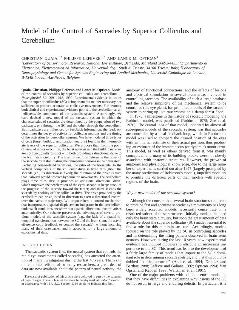
It has been shown (Robinson et al. 1993) that when thefastigial nuclei are lesioned bilaterally saccades become hyper-metric regardless of their direction. Furthermore their speed islower than expected for saccades of their size and even lowerthan the speed of normal (i.e., prelesion) saccades to the sametarget. To simulate these conditions with our model, we haveassumed that the effect of a lesion of the FOR is to attenuate itsoutput (because some of the FOR possibly is spared). Forexample, when we impose an attenuation of 60%, we obtainsaccades that are hypermetric (Fig. 10A) and slower (Fig. 10B)than normal just as reported in the literature. Effects on latencyby actual lesions seem to be very inconsistent; in our simula-tions, we observe a very small latency decrease due to adecrease in the excitatory drive provided by the FOR to thecollicular fixation neurons.
With unilateral lesions of the FOR, it is possible to evoke amuch larger range of effects (Ohtsuka et al. 1994; Robinson etal. 1993). First of all, ipsilateral saccades become hypermetric,while their velocity (at least for 20° saccades) slightly in-creases. Our simulations (performed by attenuating by 60% theoutput of the right FOR for a 20° rightward movement) are inagreement with such findings (Fig. 10,C andD). Converselyafter contralateral lesions, saccades become hypometric andslower. However, when we simulate this condition with ourmodel (using the same attenuation as before), we can repro-duce the slowing down,but not the hypometria (Fig. 10,E andF). This is due to the fact that we are assuming that altering theactivity in the contralateral FOR (the one that is active at thebeginning of the movement) does not affect in any way
FIG. 10. Simulation of the effects of FOR lesions on a 20° rightward saccade.A andB: eye position and velocity before (- - -)and after (—) a bilateral FOR lesion. In agreement with what has been observed experimentally, after the lesion saccades are slowerand larger than normal.C andD: effects of a lesion of the right FOR. Saccades ipsilateral to the lesion are bigger and faster thannormal, as reported experimentally.E andF: effects of a lesion of the left FOR. Only the decrease in speed, but not the hypometria,observed experimentally is replicated by our model (see text for comments). All results obtained by reducing the output of the FORby 60% (see text).
1011COLLICULAR AND CEREBELLAR SYNERGIES
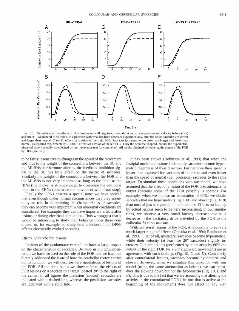
the functioning of the spatial integrator. Thus even though thesaccade starts slower, the choking signal supplied by the ipsi-lateral FOR is delivered later and the eyes land on target.However, it should be noted that the FOR projects to the NRTP(Noda et al. 1990), which in turn projects to the vermis,possibly disrupting the mechanism underlying the spatial inte-gration of the velocity signal and inducing an early activationof the choke (in our simulations, we only attenuated the outputof the cells). To clarify this issue, a better understanding of theNRTP-vermis interaction is needed.
Unilateral lesions of the FOR also affect vertical saccades,which become slightly hypermetric and bend toward the side ofthe injection (Robinson et al. 1993). Because of a large edgeeffect (due to the need to activate both collicular maps), thecurrent implementation of our model is not very well suited tosimulate vertical saccades. However, because of our model’sstructure, the effects of a lesion of the left FOR on an upwardsaccade are equivalent to the effects of a lesion of the upperhalf of the FOR (see Fig. 8) on a rightward saccade. Thisallows a vertical saccade to be simulated by interchanging ofhorizontal and vertical in our model. The results of such asimulation (Fig. 11) are very similar to what has been reportedin the literature (see Robinson et al. 1993, their Fig. 2). Inparticular, note that the saccade starts in the correct directionand then starts bending away from the target. Furthermore thesaccade is also slower (not shown), as reported by Robinsonand colleagues (1993).
Another study of unilateral injections of muscimol in thefastigial nuclei of the head-free cat (Goffart and Pelisson 1994)showed that ipsilateral saccadic deficits were compatible witha remapping of the target rather than with a generalized hy-permetria. In contrast, contralateral saccades were hypometric,as expected. Our model, in its present form, does not predictsuch results; this could be due to the disruption of some
additional mechanism (perhaps related to the removal of thetonic level of activity that is normally present in fastigialneurons, which we have not modeled here). Nonetheless itshould be noted that the effect of unilateral FOR lesions onvertical saccades, which is reproduced very well by our model,would be very difficult to explain with a theory that posits arole for the FOR in specifying the target.
Finally we previously pointed out that when the FOR islesioned the variability of saccades is considerably increased,both in amplitude and in direction. As pointed out in thepreceding text, this increased variability is incompatible withclassic models of cerebellar contribution that use only long-term adapted control signals. On the other hand, the increasedvariability is compatible with our model, where the cerebellumis the structure that accounts for both the accuracy and consis-tency of saccades. Because noise sources have not been in-cluded in this implementation of our model, we did not usesimulations to demonstrate this property. However, becausewithout a cerebellum our model of the saccadic system wouldsimply be a feed-forward controller, the results are obvious.
Effects of collicular lesions
Even though the SC is not necessary to produce saccadic eyemovements (Schiller et al. 1980), it is well known that itspartial chemical inactivation causes, at least in the acute phaseof the lesion, changes in all saccadic parameters. Typicaleffects of reversible partial deactivation of the SC are increasedlatency, decreased peak velocity, and dysmetria of the move-ments (Aizawa and Wurtz 1998; Hikosaka and Wurtz 1985,1986; Lee et al. 1988; Quaia et al. 1998a). Furthermore it hasbeen reported recently that the trajectory (Aizawa and Wurtz1998) and the initial velocity and direction (Quaia et al. 1998a)also can be affected systematically.
We have simulated a collicular lesion by attenuating theoutput of a region of the SC. We have reduced the activity ofone cell by 70%, of its 8 neighbors by 60%, and of thesuccessive 12 neighbors by 50% (both buildup and burst neu-rons were affected in the same way) with the central cellcorresponding to a 15° saccade at 45° of elevation. Then wehave looked at the effect of this lesion on a 10° and a 20°saccade, both at 45° of elevation. In another paper (Quaia et al.1998a), we suggested that the effects of SC lesions on theinitial direction of saccades can be accounted for if it isassumed that the lesion always causes a change in the hori-zontal drive larger than what would be expected given thelocation of the lesion. To include this assumption in our sim-ulations, we also have reduced the drive of the SC to thehorizontal MLBNs by 30%.
When, under the above mentioned conditions, a saccade to a20° target is simulated (Fig. 12,A and B), the eyes deviateupward and then curve back toward the target. However, thecompensation is only partial so that the saccade falls short ofthe target. The speed (both initial and peak) of the movementis considerably lower than in the control situation, even thoughthe amplitude of the movement is not much different. When a10° saccade is simulated (Fig. 12,C andD), a similar patternof curvature is observed, and again both peak and initial speedare considerably affected. However, in this case the eyes fallconsiderably short of the target.
All these characteristics are in agreement (at least qualita-
FIG. 11. Simulation of the effects of a lesion of the left FOR on thetrajectory of a 20° upward saccade. Late deviation from the normal trajectoryand the hypermetria are in agreement with experimental results.
1012 C. QUAIA, P. LEFE` VRE, AND L. M. OPTICAN

tively) with the results presented by Aizawa and Wurtz (1998);however, our simulations clearly fail to show the large changein latency which is a trademark of collicular lesions. Thisfailure is due principally to the fact that the cortical fixationinput provided to the collicular fixation neurons (see precedingtext) is, in our current implementation, removed abruptly andnot gradually. A more gradual removal of this input wouldmake the timing of the saccade onset more sensitive to thebalance between the activity of the burst/buildup neurons andthat of the fixation neurons, thus allowing a much larger spreadof latencies.
D I S C U S S I O N
We have presented a model in which saccades are generatedby the cooperation of two pathways, both influenced by feed-back information. In this sense, our model departs from theRobinsonian scheme that has dominated saccadic modeling forthe last 20 years, where the saccadic drive was generated by asingle feedback loop. The main concepts that characterize ourscheme are as follows:1) the saccade ends not because theMNs run out of drive from the EBNs but because that drive isactively choked off;2) only one part of the drive can becontrolled in direction;3) the cerebellar contribution dependson feedback information, and it is tailored carefully for eachmovement;4) no classical spatial-to-temporal transformation(which would produce a temporally coded dynamic motorerror) is performed; and5) the displacement integrator isimplemented in the spatial domain in the cerebellum.
Even though we have left out many other structures, bothcortical and subcortical, which certainly are involved in control-ling saccadic eye movement, we think that the areas we havemodeled are sufficient to reproduce at least the simplest saccadicbehavior. In the next few sections, we will compare the modelpresented here with other models recently proposed; because inprevious sections we already have described at length other mod-els of the collicular involvement in saccadic control, we will nowfocus on the role of the cerebellum. Finally we will indicate someexperiments that could be used to test our scheme and to shedfurther light on the saccadic system.
Types of cerebellar models
Models of cerebellar function can be divided into twogroups: those that are inspired by theories of learning in neuralnetworks and those that are inspired by principles of controltheory. Models of the first group stem from the early work byMarr (1969), Grossberg (1969), and Albus (1971); two of themost influential theories in this group are those of Houk, Barto,and colleagues (Barto et al. 1998; Berthier et al. 1993; Houk1989; Houk et al. 1996) and of Grossberg and colleagues(Contreras-Vidal et al. 1997; Grossberg and Kuperstein 1989).On the other hand, cerebellar models inspired by principles ofcontrol theory propose that the cerebellum functions as a modelof the controlled system and usually do not deal with adapta-tion and learning in the cerebellum. Some of these models(e.g., Jordan and Rumelhart 1992; Miall and Wolpert 1996;Miall et al. 1993) suggest that the cerebellum computes an
FIG. 12. Simulation of the effects of a collicularlesion, centered at 15° of amplitude and 45° ofelevation, on trajectory and eye velocity.A and B:effect on a 20° oblique saccade.C andD: effect ona 10° oblique saccade. In both cases, the results(decreased initial and peak velocity, increased cur-vature, dysmetria) are in qualitative agreement withexperimental data. See text for details about theparameters used to simulate the lesion.
1013COLLICULAR AND CEREBELLAR SYNERGIES
estimate of the effect that an outgoing motor signal will haveon the controlled plant (direct, or forward, models), whereasothers (e.g., Gomi and Kawato 1992; Kawato and Gomi 1992)estimate the motor outputs needed to generate a desired move-ment (inverse models).
Even though we took special care in reproducing the patternof activation of FOR neurons, our model is clearly moreinspired by principles of control theory than by theories ofnetwork learning. More precisely, we think that our schemecould be regarded as a forward model because the spatialintegration process (which we hypothesize takes place in thecerebellum) is used to predict when the eyes are approachingthe target, given the efference copy of the motor command.However, this signal is not fed back to the main controller, andthus it is different from classical control schemes. In this sense,it is closer to models like the one proposed by Grossberg andKuperstein (1989), where the cerebellum is embedded in a sideloop. One important advantage of learning theory models isthat, especially in their most recent versions (Barto et al. 1998;Contreras-Vidal et al. 1997), they make testable predictionsabout the pattern of activities in Purkinje cells and interneurons(especially basket cells) in the cerebellar cortex. This is cer-tainly a most desirable feature; unfortunately at this stage, ourmodel lacks this. However, we think that our model has certainadvantages (which will be outlined next) over existing modelsthat justify our decision to take another approach and to pro-pose a different model.
Comparison with other models of cerebellar involvement insaccadic control
Recently some models that address the role of the cerebel-lum in the in-flight control of saccades have appeared; how-ever, in only one of those models (Houk et al. 1992, 1996) isthe cerebellum part of the feedback loop. The theory proposedby Houk and colleagues posits that the Purkinje cells in thecerebellar cortex are trained to recognize particular configura-tions of the proprioceptive inputs (carried by the mossy fibers),and when these patterns occur, they fire to stop an ongoingmovement. Thus one of the roles that we propose here for thecerebellum is similar to the one proposed by Houk and col-leagues (i.e., to terminate the saccade when the eyes approachthe target), even though the mechanism used by the cerebellumto achieve this goal is very different in the two models. Thepattern-recognition mechanism proposed by Houk and col-leagues works well to control limb movement, where thedelays in the system are shorter than the duration of themovement and proprioceptive feedback can be used to track(and even predict) the ongoing movement (Barto et al. 1998).However, we think there are some fundamental problems inextending their model to the control of saccadic eye move-ments. First of all in Houk’s model, the movement is inter-rupted when a given final position, and not displacement, isattained. Thus the cerebellar cortex should work in head coor-dinates; however, it has been shown recently that saccadicadaptation, which almost certainly is controlled by the cere-bellum (Goldberg et al. 1993; Optican and Robinson 1980),occurs in oculocentric coordinates (Frens and van Opstal1994). Furthermore it is known that proprioceptive feedbackplays no role in the in-flight control of saccades (Guthrie et al.1983); one could argue that an internal estimate of the position
of the eyes could be used instead, but no signal encoding theposition of the eyes during saccades has been found in themossy fibers [in our model (Fig. 9), the eye position signal isused only before the movement to determine the displacementof the eyes required to foveate the target]. One could overcomethese problems by postulating the presence of a displacementintegrator in the brain stem, whose output then could be fed tothe cerebellum. However, to the best of our knowledge, such asignal has not been observed in the mossy fibers. Finally in itspresent form, the scheme proposed by Houk predicts a patternof activity for the FOR that mirrors the activity in the SC, i.e.,a burst of activity only for saccades in one direction, that is notcompatible with what is reported in the cerebellar literature.For these reasons, we think that even though Houk’s scheme isconsistent with data on limb control, it is at odds with somecrucial data regarding the saccadic system.
Another theory of cerebellar function is the one proposed byGrossberg and colleagues, both for saccadic (Grossberg andKuperstein 1989) and limb control (Contreras-Vidal et al.1997). One of the major differences between Houk’s andGrossberg’s models is that Grossberg proposes an extracer-ebellar loop to compute the residual motor error and to gener-ate a desired velocity signal, which then is fed to the cerebel-lum. Thus the cerebellum is part of a side loop, and it workswith velocity (as opposed to position) signals. We believe thatour model has two main advantages over the one proposed byGrossberg and colleagues: first, it does not require a spatial-to-temporal transformation, which is part of the extracerebellarloop in Grossberg’s model. Second, our model can accountmore readily for the large increase in variability observed aftercerebellar lesions.
Recently, Dean (1995) proposed a model of the saccadicsystem that deals with the role played by the fastigial nuclei inon-line control of saccades, taking particular care in reproduc-ing the pattern of FOR activation. There are several similaritiesbetween Dean’s models and ours, including the connectivitybetween the FOR and the MLBNs. The role attributed by Deanto the FOR is to ensure saccadic accuracy; because of thetiming of the FOR bursts, this is achieved by contributing tothe acceleration of the eyes at the beginning of saccades and totheir deceleration at the end of the movement. This role, whichalso has been proposed in other studies (Fuchs et al. 1993;Helmchen et al. 1994; Noda 1991; Robinson 1995; Sparks andBarton 1993), is similar to the one that we propose here, i.e., toprovide a directional drive and to choke off the collicularoutput at the end of the movement. However, in our model,there are not two distinct bursts, one in the ipsilateral and theother in the contralateral FOR, but one single burst that spreadsacross the fastigial nuclei. Accordingly the FOR does not playa role only at the beginning and at the end of the saccade butalso during the saccade, exerting a trajectory control. Further-more the role played in our scheme by the late part of the burstis not just to slow the movement but to stop it when the eyesreach the target. Importantly in our scheme the cerebellumdetermines when the movement should end. In Dean’s model,the cerebellum only makes a preprogrammed contribution to asaccade the end of which is controlled by the local feedbackloop in the brain stem.
Another, fundamental, difference between Dean’s model andours is that in his scheme the brain stem circuit (extracerebellarpathway) consists of a feedback loop with a gain lower than one.
1014 C. QUAIA, P. LEFE` VRE, AND L. M. OPTICAN
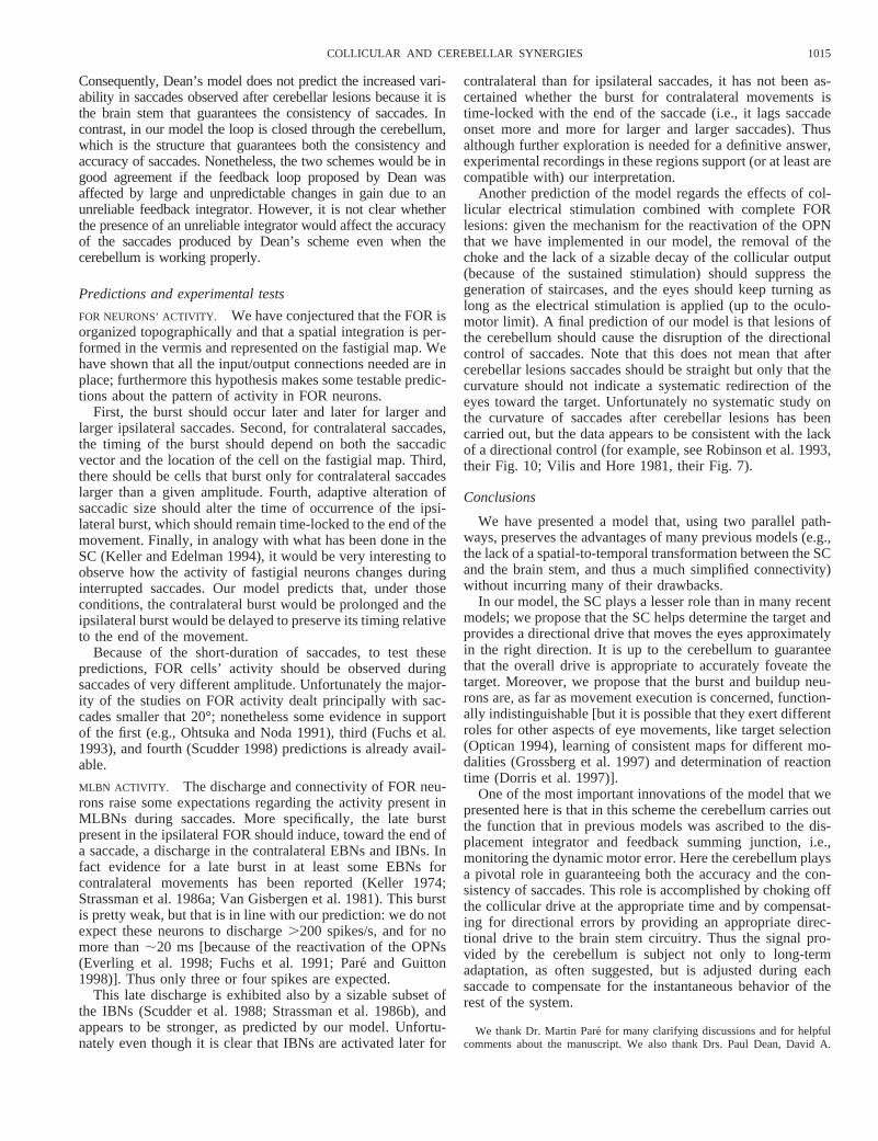
Consequently, Dean’s model does not predict the increased vari-ability in saccades observed after cerebellar lesions because it isthe brain stem that guarantees the consistency of saccades. Incontrast, in our model the loop is closed through the cerebellum,which is the structure that guarantees both the consistency andaccuracy of saccades. Nonetheless, the two schemes would be ingood agreement if the feedback loop proposed by Dean wasaffected by large and unpredictable changes in gain due to anunreliable feedback integrator. However, it is not clear whetherthe presence of an unreliable integrator would affect the accuracyof the saccades produced by Dean’s scheme even when thecerebellum is working properly.
Predictions and experimental tests
FOR NEURONS’ ACTIVITY. We have conjectured that the FOR isorganized topographically and that a spatial integration is per-formed in the vermis and represented on the fastigial map. Wehave shown that all the input/output connections needed are inplace; furthermore this hypothesis makes some testable predic-tions about the pattern of activity in FOR neurons.
First, the burst should occur later and later for larger andlarger ipsilateral saccades. Second, for contralateral saccades,the timing of the burst should depend on both the saccadicvector and the location of the cell on the fastigial map. Third,there should be cells that burst only for contralateral saccadeslarger than a given amplitude. Fourth, adaptive alteration ofsaccadic size should alter the time of occurrence of the ipsi-lateral burst, which should remain time-locked to the end of themovement. Finally, in analogy with what has been done in theSC (Keller and Edelman 1994), it would be very interesting toobserve how the activity of fastigial neurons changes duringinterrupted saccades. Our model predicts that, under thoseconditions, the contralateral burst would be prolonged and theipsilateral burst would be delayed to preserve its timing relativeto the end of the movement.
Because of the short-duration of saccades, to test thesepredictions, FOR cells’ activity should be observed duringsaccades of very different amplitude. Unfortunately the major-ity of the studies on FOR activity dealt principally with sac-cades smaller that 20°; nonetheless some evidence in supportof the first (e.g., Ohtsuka and Noda 1991), third (Fuchs et al.1993), and fourth (Scudder 1998) predictions is already avail-able.
MLBN ACTIVITY. The discharge and connectivity of FOR neu-rons raise some expectations regarding the activity present inMLBNs during saccades. More specifically, the late burstpresent in the ipsilateral FOR should induce, toward the end ofa saccade, a discharge in the contralateral EBNs and IBNs. Infact evidence for a late burst in at least some EBNs forcontralateral movements has been reported (Keller 1974;Strassman et al. 1986a; Van Gisbergen et al. 1981). This burstis pretty weak, but that is in line with our prediction: we do notexpect these neurons to discharge.200 spikes/s, and for nomore than;20 ms [because of the reactivation of the OPNs(Everling et al. 1998; Fuchs et al. 1991; Pare´ and Guitton1998)]. Thus only three or four spikes are expected.
This late discharge is exhibited also by a sizable subset ofthe IBNs (Scudder et al. 1988; Strassman et al. 1986b), andappears to be stronger, as predicted by our model. Unfortu-nately even though it is clear that IBNs are activated later for
contralateral than for ipsilateral saccades, it has not been as-certained whether the burst for contralateral movements istime-locked with the end of the saccade (i.e., it lags saccadeonset more and more for larger and larger saccades). Thusalthough further exploration is needed for a definitive answer,experimental recordings in these regions support (or at least arecompatible with) our interpretation.
Another prediction of the model regards the effects of col-licular electrical stimulation combined with complete FORlesions: given the mechanism for the reactivation of the OPNthat we have implemented in our model, the removal of thechoke and the lack of a sizable decay of the collicular output(because of the sustained stimulation) should suppress thegeneration of staircases, and the eyes should keep turning aslong as the electrical stimulation is applied (up to the oculo-motor limit). A final prediction of our model is that lesions ofthe cerebellum should cause the disruption of the directionalcontrol of saccades. Note that this does not mean that aftercerebellar lesions saccades should be straight but only that thecurvature should not indicate a systematic redirection of theeyes toward the target. Unfortunately no systematic study onthe curvature of saccades after cerebellar lesions has beencarried out, but the data appears to be consistent with the lackof a directional control (for example, see Robinson et al. 1993,their Fig. 10; Vilis and Hore 1981, their Fig. 7).
Conclusions
We have presented a model that, using two parallel path-ways, preserves the advantages of many previous models (e.g.,the lack of a spatial-to-temporal transformation between the SCand the brain stem, and thus a much simplified connectivity)without incurring many of their drawbacks.
In our model, the SC plays a lesser role than in many recentmodels; we propose that the SC helps determine the target andprovides a directional drive that moves the eyes approximatelyin the right direction. It is up to the cerebellum to guaranteethat the overall drive is appropriate to accurately foveate thetarget. Moreover, we propose that the burst and buildup neu-rons are, as far as movement execution is concerned, function-ally indistinguishable [but it is possible that they exert differentroles for other aspects of eye movements, like target selection(Optican 1994), learning of consistent maps for different mo-dalities (Grossberg et al. 1997) and determination of reactiontime (Dorris et al. 1997)].
One of the most important innovations of the model that wepresented here is that in this scheme the cerebellum carries outthe function that in previous models was ascribed to the dis-placement integrator and feedback summing junction, i.e.,monitoring the dynamic motor error. Here the cerebellum playsa pivotal role in guaranteeing both the accuracy and the con-sistency of saccades. This role is accomplished by choking offthe collicular drive at the appropriate time and by compensat-ing for directional errors by providing an appropriate direc-tional drive to the brain stem circuitry. Thus the signal pro-vided by the cerebellum is subject not only to long-termadaptation, as often suggested, but is adjusted during eachsaccade to compensate for the instantaneous behavior of therest of the system.
We thank Dr. Martin Pare´ for many clarifying discussions and for helpfulcomments about the manuscript. We also thank Drs. Paul Dean, David A.
1015COLLICULAR AND CEREBELLAR SYNERGIES
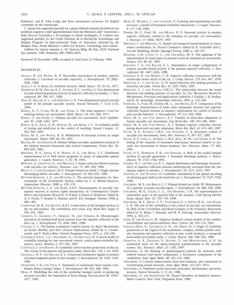
Robinson, and R. John Leigh and three anonymous reviewers for helpfulcomments on the manuscript.
C. Quaia was supported partially by a grant (Sistemi naturali ed artificiali neiproblemi cognitivi e dell’apprendimento) from the Ministero dell’ Universita` edella Ricerca Scientifica e Tecnologica to Paolo Inchingolo. P. Lefe`vre wassupported partially by the Fonds National de la Recherche Scientifique andBelgian Program on Inter-university Poles of Attraction, initiated by theBelgian State, Prime Minister’s Office for Science, Technology and Culture.
Address for reprint requests: L. M. Optican, Bldg. 49, Rm. 2A50, NationalEye Institute, NIH, Bethesda, MD 20892-4435.
Received 20 November 1998; accepted in final form 22 February 1999.
REFERENCES
AIZAWA , H. AND WURTZ, R. H. Reversible inactivation of monkey superiorcolliculus. I. Curvature of saccadic trajectory.J. Neurophysiol.79: 2082–2096, 1998.
ALBUS, J. S. A theory of cerebellar function.Math. Biosci.10: 25–61, 1971.ANDERSON, R. W., KELLER, E. L., GANDHI, N. J.,AND DAS, S. Two-dimensional
saccade-related population activity in superior colliculus in monkey.J. Neu-rophysiol.80: 798–817, 1998.
ARAI, K., KELLER, E. L., AND EDELMAN, J. A. Two-dimensional neural networkmodel of the primate saccadic system.Neural Networks7: 1115–1135,1994.
BAHILL , A. T., CLARK, M. R., AND STARK, L. The main sequence: a tool forstudying human eye movements.Math. Biosci.24: 191–204, 1975.
BAHILL , T. AND STARK, L. Oblique saccadic eye movements.Arch. Ophthal-mol. 95: 1258–1261, 1977.
BARTO, A. G., FAGG, A. H., SITKOFF, N., AND HOUK, J. C. A cerebellar modelof timing and prediction in the control of reaching.Neural Comput.11:565–594, 1999.
BASSO, M. A. AND WURTZ, R. H. Modulation of neuronal activity by targetuncertainty.Nature389: 66–69, 1997.
BECKER, W. AND JURGENS, R. Human oblique saccades: quantitative analysis ofthe relation between horizontal and vertical components.Vision Res.30:893–920, 1990.
BERTHIER, N. E., SINGH, S. P., BARTO, A. G., AND HOUK, J. C. Distributedrepresentation of of limb motor programs in arrays of adjustable patterngenerators.J. Cognit. Neurosci.5: 56–78, 1993.
BERTHOZ, A., GRANTYN, A., AND DROULEZ, J. Some collicular efferent neuronscode saccadic eye velocity.Neurosci. Lett.72: 289–294, 1986.
BRUCE, C. J.AND GOLDBERG, M. E. Primate frontal eye fields. I. Single neuronsdischarging before saccades.J. Neurophysiol.53: 603–635, 1985.
BUTTNER-ENNEVER, J. A. AND BUTTNER, U. The reticular formation. In:Neu-roanatomy of the Oculomotor System,edited by J. A. Buttner-Ennever.Amsterdam: Elsevier, 1988, p. 119–176.
BUTTNER-ENNEVER, J. A. AND HORN, A.K.E. Neuroanatomy of saccadic om-nipause neurons in nucleus raphe interpositus. In:Contemporary OcularMotor and Vestibular Research: A Tribute to David A. Robinson,edited byA. F. Fuchs, T. Brandt, U. Buttner, and D. Zee. Stuttgart: Thieme, 1994, p.488–495.
CARPENTER, M. B. AND BATTON, R.R.I. Connections of the fastigial nucleus inthe cat and monkey. The cerebellum, new vistas.Exp. Brain Res. Suppl.6:250–291, 1982.
CHIMOTO, S., IWAMOTO, Y., SHIMAZU , H., AND YOSHIDA, K. Monosynapticactivation of medium-lead burst neurons from the superior colliculus in thealert cat.J. Neurophysiol.75: 2658–2661, 1996.
COLLINS, C. C. The human oculomotor control system. In:Basic Mechanismsof Ocular Motility and their Clinical Implications,edited by G. Lenner-strand, and P. Bach-y-Rita. Oxford: Pergamon Press, 1975, p. 145–180.
CONTRERAS-VIDAL , J. L., GROSSBERG, S.,AND BULLOCK, D. A neural model ofcerebellar learning for arm movement control: cortico-spino-cerebellar dy-namics.Learn. Memory3: 475–502, 1997.
COURVILLE, J.AND DIAKIW , N. Cerebellar cortico-nuclear projections in the cat.The vermis of the anterior and posterior lobes.Brain Res.110: 1–20, 1976.
CRANDALL , W. F.AND KELLER, E. L. Visual and oculomotor signals in nucleusreticularis tegmenti pontis in alert monkey.J. Neurophysiol.54: 1326–1345,1985.
CRAWFORD, J. D. AND VILIS, T. Symmetry of oculomotor burst neuron coor-dinates about Listing’s plane.J. Neurophysiol.68: 432–448, 1992.
DEAN, P. Modelling the role of the cerebellar fastigial nuclei in producingaccurate saccades: the importance of burst timing.Neuroscience68: 1059–1077, 1995.
DEAN, P., MAJHEW, J., AND LANGDON, P. Learning and maintaining saccadicaccuracy: a model of brainstem-cerebellar interactions.J. Cognit. Neurosci.6: 117–138, 1994.
DORRIS, M. C., PARE, M., AND MUNOZ, D. P. Neuronal activity in monkeysuperior colliculus related to the initiation of saccadic eye movements.J. Neurosci.17: 8566–8579, 1997.
DROULEZ, J. AND BERTHOZ, A. Spatial and temporal transformations in visuo-motor coordination. In:Neural Computers,edited by R. Eckmiller and C.von der Malsburg. Berlin: Springer-Verlag, 1988, p. 345–357.
DUHAMEL, J.-R., COLBY, C. L., AND GOLDBERG, M. E. The updating of therepresentation of visual space in parietal cortex by intended eye movements.Science255: 90–92, 1992.
EDELMAN, J. A. AND KELLER, E. L. Dependence on target configuration ofexpress saccade-related activity in the primate superior colliculus.J. Neu-rophysiol.80: 1407–1426, 1998.
EDWARDS, S. B. AND HENKEL, C. K. Superior colliculus connections with theextraocular motor nuclei in the cat.J. Comp. Neurol.179: 451–467, 1978.
ERKELENS, C. J. AND SLOOT, O. B. Initial direction and landing positions ofbinocular saccades.Vision Res.35: 3297–3303, 1995.
ERKELENS, C. J. AND VOGELS, I.M.L.C. The relationship between the initialdirection and landing position of saccades. In:Eye Movements Research:Mechanisms, Processes and Applications,edited by J. Findlay, J. R. Walker,and R. W. Kentridge. Amsterdam: Elsevier, 1995, p. 133–144.
EVERLING, S., PARE, M., DORRIS, M. C., AND MUNOZ, D. P. Comparison of thedischarge characteristics of brain stem omnipause neurons and superiiorcolliculus fixation neurons in monkey: implications for control of fixationand saccade behavior.J. Neurophysiol.79: 511–528, 1998.
FRENS, M. A. AND VAN OPSTAL, A. J. Transfer of short-term adaptation inhuman saccadic eye movements.Exp Brain Res.100: 293–306, 1994.
FRENS, M. A. AND VAN OPSTAL, A. J. Monkey superior colliculus activityduring short-term saccadic adaptation.Brain Res. Bull.43: 473–483, 1997.
FUCHS, A. F., KANEKO, C.R.S.,AND SCUDDER, C. A. Brainstem control ofsaccadic eye movements.Annu. Rev. Neurosci.8: 307–337, 1985.
FUCHS, A. F., LING, L., KANEKO, C.R.S., KING, W. M., AND USHER, S. D. Thetiming of the response of brainstem omni-pause neurones relative to sac-cadic eye movements in rhesus monkeys.Soc. Neurosci. Abstr.17: 462,1991.
FUCHS, A. F., ROBINSON, F. R.,AND STRAUBE, A. Role of the caudal fastigialnucleus in saccade generation. I. Neuronal discharge patterns.J. Neuro-physiol.70: 1723–1740, 1993.
GANDHI, N. J.AND KELLER, E. L. Spatial distribution and discharge character-istics of superior colliculus neurons antidromically activated from the om-nipause region in monkey.J. Neurophysiol.78: 2221–2225, 1997.
GOFFART, L. AND PELISSON, D. Cerebellar contribution to the spatial encodingof orienting gaze shifts in the head-free cat.J. Neurophysiol.72: 7547–7550,1994.
GOLDBERG, M. E. AND BRUCE, C. J. Primate frontal eye fields. III. Maintenanceof a spatially accurate saccade signal.J. Neurophysiol.64: 489–508, 1990.
GOLDBERG, M. E., COLBY, C. L., AND DUHAMEL, J.-R. The representation ofvisuomotor space in the parietal lobe of the monkey.Cold Spring HarborSymp. Quant. Biol.55: 729–739, 1990.
GOLDBERG, M. E., MUSIL, S. Y., FITZGIBBON, E. J., SMITH, M. K., AND OLSON,C. R. The role of the cerebellum in the control of saccadic eye movements.In: Role of the Cerebellum and Basal Ganglia in the Voluntary Movement,edited by N. Mano, I. Hamada, and M. R. DeLong. Amsterdam: Elsevier,1993, p. 203–211.
GOMI, H. AND KAWATO, M. Adaptive feedback control models of the vestibu-locerebellum and spinocerebellum.Biol. Cybern.68: 105–114, 1992.
GONZALO-RUIZ, A., LEICHNETZ, G. R., AND SMITH, D. J. Origin of cerebellarprojections to the region of the oculomotor complex, medial pontine retic-ular formation and superior colliculus in new world monkeys: a retrogradehorseradish peroxidase study.J. Comp. Neurol.268: 508–526, 1988.
GRANTYN, A. A., DALEZIOS, Y., KITAMA , T., AND MOSCHOVAKIS, A. K. Ananatomical basis for the spatio-temporal transformation in the saccadicsystem.Soc. Neurosci. Abstr.23: 1295, 1997.
GROSSBERG, S. On learning of spatiotemporal patterns by networks withordered sensory and motor components. I. Excitatory components of thecerebellum.Stud. Appl. Math.48: 105–132, 1969.
GROSSBERG, S. Contour enhancement, short term memory, and constancies inreverberating neural networks.Stud. Appl. Math.213–257: 1973.
GROSSBERG, S. Nonlinear neural networks: principles, mechanisms, and archi-tectures.Neural Networks1: 17–61, 1988.
GROSSBERG, S. AND KUPERSTEIN, M. Neural Dynamics of Adaptive Sensory-Motor Control.New York: Pergamon Press, 1989.
1016 C. QUAIA, P. LEFE` VRE, AND L. M. OPTICAN

GROSSBERG, S., ROBERTS, K., AGUILAR, M., AND BULLOCK, D. A neural modelof multimodal adaptive saccadic eye movement control by superior collicu-lus. J. Neurosci.17: 9706–9725, 1997.
GUITTON, D. Control of saccadic eye and gaze movements by the superiorcolliculus and basal ganglia. In:Vision and Visual Disfunction, Eye Move-ments,edited by R.H.S. Carpenter. London: MacMillan, 1991, p. 244–276.
GUITTON, D., MUNOZ, D. P.,AND PELISSON, D. Are gaze shifts controlled by a‘‘moving hill’’ of activity in the superior colliculus [Reply]?Trends Neu-rosci. 16: 216–218, 1993.
GUTHRIE, B. L., PORTER, J. D.,AND SPARKS, D. L. Corollary discharge providesaccurate eye position information to the oculomotor system.Science221:1193–1195, 1983.
HAUSTEIN, W. Considerations on Listing’s law and the primary position bymeans of a matrix description of eye position control.Biol. Cybern.60:411–420, 1989.
HELMCHEN, C. AND BUTTNER, U. Saccade-related Purkinje cell activity in theoculomotor vermis during spontaneous eye movements in light and dark-ness.Exp. Brain Res.103: 198–208, 1995.
HELMCHEN, C., STRAUBE, A., AND BUTTNER, U. Saccade-related activity in thefastigial oculomotor region of the macaque monkey during spontaneous eyemovements in light and darkness.Exp. Brain Res.98: 474–482, 1994.
HEPP, K., HENN, V., VILIS, T., AND COHEN, B. Brainstem regions related tosaccade generation. In:The Neurobiology of Saccadic Eye Movements,Reviews of Oculomotor Research,edited by R. H. Wurtz and M. E. Gold-berg. Amsterdam: Elsevier, 1989, vol. III, p. 105–212.
HIKOSAKA, O. AND WURTZ, R. H. Modification of saccadic eye movements byGABA-related substances. I. Effect of muscimol and bicuculline in monkeysuperior colliculus.J. Neurophysiol.53: 266–291, 1985.
HIKOSAKA, O. AND WURTZ, R. H. Saccadic eye movements following injectionof lidocaine into the superior colliculus.Exp. Brain Res.61: 531–539, 1986.
HOUK, J. C. Cooperative control of limb movements by the motor cortex,brainstem and cerebellum. In:Models of Brain Function,edited by R.M.J.Cotterill. Cambridge: Cambridge, 1989, p. 309–325.
HOUK, J. C., BUCKINGHAM, J. T.,AND BARTO, A. G. Models of the cerebellumand motor learning.Behav. Brain Sci.19: 368–383, 1996.
HOUK, J. C., GALIANA , H. L., AND GUITTON, D. Cooperative control of gaze bythe superior colliculus, brainstem and cerebellum. In:Tutorials in MotorBehavior II,edited by G. E. Stelmach and J. Requin. Amsterdam: Elsevier,1992, p. 443–474.
JORDAN, M. I. AND RUMELHART, D. E. Forward models: supervised learningwith a distal teacher.Cognit. Sci.16: 307–354, 1992.
KASE, M., MILLER, D. C., AND NODA, H. Discharges of Purkinje cells andmossy fibers in the cerebellar vermis of the monkey during saccadic eyemovements and fixation.J. Physiol. (Lond.)300: 539–555, 1980.
KAWATO, M. AND GOMI, H. A computational model of four regions of thecerebellum based on feedback-error learning.Biol. Cybern.68: 95–103,1992.
KELLER, E. L. Participation of medial pontine reticular formation in eyemovement generation in monkey.J. Neurophysiol.37: 316–332, 1974.
KELLER, E. L. AND EDELMAN, J. A. Use of interrupted saccade paradigm tostudy spatial and temporal dynamics of saccadic burst cells in the superiorcolliculus.J. Neurophysiol.72: 2754–2770, 1994.
KELLER, E. L., GANDHI, N. J.,AND WEIR, P. T. Discharge of superior colliculusneurons during saccades made to moving targets.J. Neurophysiol.76:3573–3577, 1996.
KELLER, E. L. AND ROBINSON, D. A. Abducens unit behavior in the monkeyduring vergence movements.Vision Res.12: 369–382, 1972.
KELLER, E. L., SLAKEY , D. P.,AND CRANDALL , W. F. Microstimulation of theprimate cerebellar vermis during saccadic eye movements.Brain Res.288:131–143, 1983.
KING, W. M., LISBERGER, S. G., AND FUCHS, A. F. Oblique saccadic eyemovements of primates.J. Neurophysiol.56: 769–784, 1986.
KLIER, E. M. AND CRAWFORD, J. D. Human oculomotor system accounts for3-D eye orientation in the visual-motor transformation for saccades.J. Neu-rophysiol.80: 2274–2294, 1998.
LEE, C., ROHRER, W. H., AND SPARKS, D. L. Population coding of saccadic eyemovements by neurons in the superior colliculus.Nature 332: 357–360,1988.
LEFEVRE, P. AND GALIANA , H. L. Dynamic feedback to the superior colliculusin a neural network model of the gaze control system.Neural Networks5:871–890, 1992.
LEFEVRE, P., QUAIA , C., AND OPTICAN, L. M. A new model of the saccadicsystem. II. Directional drive by the superior colliculus.Soc. Neurosci. Abstr.22: 1457, 1996.
LEFEVRE, P., QUAIA , C., AND OPTICAN, L. M. Distributed model of control ofsaccades by superior colliculus and cerebellum.Neural Networks11: 1175–1190, 1998.
MARR, D. A theory of cerebellar cortex.J. Physiol. (Lond.)202: 437–470,1969.
MAY, P. J., HARTWICH-YOUNG, R., NELSON, J., SPARKS, D. L., AND PORTER,J. D. Cerebellotectal pathways in the macaque: implications for colliculargeneration of saccades.Neuroscience36: 305–324, 1990.
MIALL , R. C., WEIR, D. J., WOLPERT, D. M., AND STEIN, J. F. Is the cerebelluma Smith predictor?J. Mot. Behav.25: 203–216, 1993.
MIALL , R. C. AND WOLPERT, D. M. Forward models for physiological motorcontrol.Neural Networks9: 1265–1279, 1996.
MOSCHOVAKIS, A. K., SCUDDER, C. A., AND HIGHSTEIN, S. M. Structure of theprimate burst generator. I. Medium-lead burst neurons with upward on-directions.J. Neurophysiol.65: 203–217, 1991.
MUNOZ, D. P., GUITTON, D., AND PELISSON, D. Control of orienting gaze shiftsby the tectoreticulospinal system in the head-free cat. III. Spatiotemporalcharacteristics of phasic motor discharges.J. Neurophysiol.66: 1642–1666,1991a.
MUNOZ, D. P.AND ISTVAN, P. J. Lateral inhibitory interactions in the interme-diate layers of the monkey superior colliculus.J. Neurophysiol.79: 1193–1209, 1998.
MUNOZ, D. P., PELISSON, D., AND GUITTON, D. Movement of neural activity onthe superior colliculus motor map during gaze shifts.Science251: 1358–1360, 1991b.
MUNOZ, D. P. AND WURTZ, R. H. Role of the rostral superior colliculus inactive visual fixation and execution of express saccades.J. Neurophysiol.67: 1000–1002, 1992.
MUNOZ, D. P.AND WURTZ, R. H. Fixation cells in monkey superior colliculus.I. Characteristics of cell discharge.J. Neurophysiol.70: 559–575, 1993a.
MUNOZ, D. P.AND WURTZ, R. H. Fixation cells in monkey superior colliculus.II. Reversible activation and deactivation.J. Neurophysiol.70: 576–589,1993b.
MUNOZ, D. P.AND WURTZ, R. H. Saccade-related activity in monkey superiorcolliculus. I. Characteristics of burst and buildup cells.J. Neurophysiol.73:2313–2333, 1995a.
MUNOZ, D. P.AND WURTZ, R. H. Saccade-related activity in monkey superiorcolliculus. II. Spread of activity during saccades.J. Neurophysiol.73:2334–2348, 1995b.
NODA, H. Cerebellar control of saccadic eye movements: its neural mecha-nisms and pathways.Jpn. J. Physiol.41: 351–368, 1991.
NODA, H. AND FUJIKADO, T. Involvement of Purkinje cells in evoking saccadiceye movements by microstimulation of the posterior cerebellar vermis ofmonkeys.J. Neurophysiol.57: 1247–1261, 1987.
NODA, H., SUGITA, S.,AND IKEIDA, Y. Afferent and efferent connections of theoculomotor region of the fastigial nucleus in the macaque monkey.J. Comp.Neurol.302: 330–348, 1990.
OHTSUKA, K. AND NODA, H. Direction selective saccadic-burst neurons in thefastigial oculomotor region of the macaque.Exp. Brain Res.81: 659–662,1990.
OHTSUKA, K. AND NODA, H. Saccadic burst neurons in the oculomotor regionof the fastigial nucleus of the macaque monkey.J. Neurophysiol.65:1422–1434, 1991.
OHTSUKA, K. AND NODA, H. Burst discharges of mossy fibers in the oculomotorvermis of macaque monkeys during saccadic eye movements.Neurosci. Res.15: 102–114, 1992.
OHTSUKA, K. AND NODA, H. Discharge properties of Purkinje cells in theoculomotor vermis during visually guided saccades in the macaque monkey.J. Neurophysiol.74: 1828–1840, 1995.
OHTSUKA, K., SATO, H., AND NODA, H. Saccadic burst neurons in the fastigialnucleus are not involved in compensating for orbital nonlinearities.J. Neu-rophysiol.71: 1976–1980, 1994.
OPTICAN, L. M. Adaptive control of saccadic and smooth pursuit eye move-ment. In:Adaptive Processes in Visual and Oculomotor Systems,edited byE. L. Keller, and D. S. Zee. Oxford: Pergamon Press, 1986, p. 313–320.
OPTICAN, L. M. Control of saccade trajectory by the superior colliculus. In:Contemporary Ocular Motor and Vestibular Research: A Tribute to DavidA. Robinson,edited by A. F. Fuchs, T. Brandt, U. Buttner, and D. S. Zee.Stuttgart: Thieme, 1994, p. 98–105.
OPTICAN, L. M. A field theory of saccade generation: temporal-to-spatialtransform in the superior colliculus.Vision Res.35: 3313–3320, 1995.
OPTICAN, L. M. AND MILES, F. A. Visually induced adaptive changes in primatesaccadic oculomotor control signals.J. Neurophysiol.54: 940–958, 1985.
1017COLLICULAR AND CEREBELLAR SYNERGIES
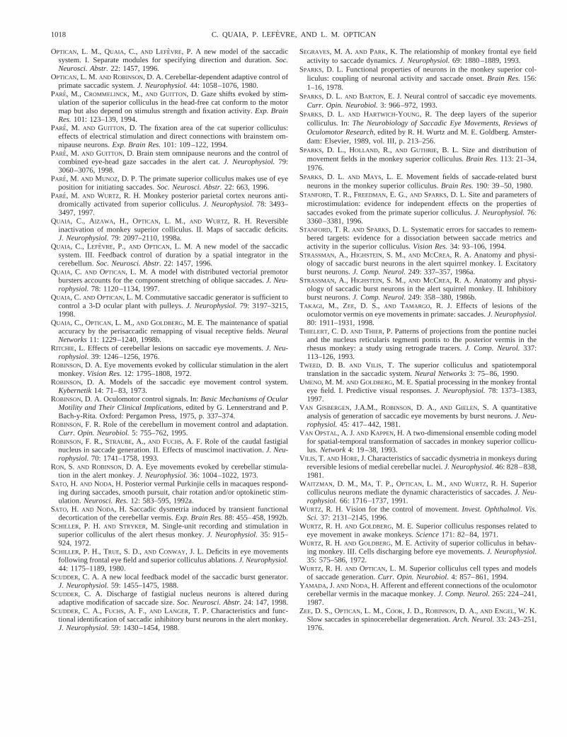
OPTICAN, L. M., QUAIA , C., AND LEFEVRE, P. A new model of the saccadicsystem. I. Separate modules for specifying direction and duration.Soc.Neurosci. Abstr.22: 1457, 1996.
OPTICAN, L. M. AND ROBINSON, D. A. Cerebellar-dependent adaptive control ofprimate saccadic system.J. Neurophysiol.44: 1058–1076, 1980.
PARE, M., CROMMELINCK, M., AND GUITTON, D. Gaze shifts evoked by stim-ulation of the superior colliculus in the head-free cat conform to the motormap but also depend on stimulus strength and fixation activity.Exp. BrainRes.101: 123–139, 1994.
PARE, M. AND GUITTON, D. The fixation area of the cat superior colliculus:effects of electrical stimulation and direct connections with brainstem om-nipause neurons.Exp. Brain Res.101: 109–122, 1994.
PARE, M. AND GUITTON, D. Brain stem omnipause neurons and the control ofcombined eye-head gaze saccades in the alert cat.J. Neurophysiol.79:3060–3076, 1998.
PARE, M. AND MUNOZ, D. P. The primate superior colliculus makes use of eyeposition for initiating saccades.Soc. Neurosci. Abstr.22: 663, 1996.
PARE, M. AND WURTZ, R. H. Monkey posterior parietal cortex neurons anti-dromically activated from superior colliculus.J. Neurophysiol.78: 3493–3497, 1997.
QUAIA , C., AIZAWA , H., OPTICAN, L. M., AND WURTZ, R. H. Reversibleinactivation of monkey superior colliculus. II. Maps of saccadic deficits.J. Neurophysiol.79: 2097–2110, 1998a.
QUAIA , C., LEFEVRE, P., AND OPTICAN, L. M. A new model of the saccadicsystem. III. Feedback control of duration by a spatial integrator in thecerebellum.Soc. Neurosci. Abstr.22: 1457, 1996.
QUAIA , C. AND OPTICAN, L. M. A model with distributed vectorial premotorbursters accounts for the component stretching of oblique saccades.J. Neu-rophysiol.78: 1120–1134, 1997.
QUAIA , C. AND OPTICAN, L. M. Commutative saccadic generator is sufficient tocontrol a 3-D ocular plant with pulleys.J. Neurophysiol.79: 3197–3215,1998.
QUAIA , C., OPTICAN, L. M., AND GOLDBERG, M. E. The maintenance of spatialaccuracy by the perisaccadic remapping of visual receptive fields.NeuralNetworks11: 1229–1240, 1998b.
RITCHIE, L. Effects of cerebellar lesions on saccadic eye movements.J. Neu-rophysiol.39: 1246–1256, 1976.
ROBINSON, D. A. Eye movements evoked by collicular stimulation in the alertmonkey.Vision Res.12: 1795–1808, 1972.
ROBINSON, D. A. Models of the saccadic eye movement control system.Kybernetik14: 71–83, 1973.
ROBINSON, D. A. Oculomotor control signals. In:Basic Mechanisms of OcularMotility and Their Clinical Implications,edited by G. Lennerstrand and P.Bach-y-Rita. Oxford: Pergamon Press, 1975, p. 337–374.
ROBINSON, F. R. Role of the cerebellum in movement control and adaptation.Curr. Opin. Neurobiol.5: 755–762, 1995.
ROBINSON, F. R., STRAUBE, A., AND FUCHS, A. F. Role of the caudal fastigialnucleus in saccade generation. II. Effects of muscimol inactivation.J. Neu-rophysiol.70: 1741–1758, 1993.
RON, S. AND ROBINSON, D. A. Eye movements evoked by cerebellar stimula-tion in the alert monkey.J. Neurophysiol.36: 1004–1022, 1973.
SATO, H. AND NODA, H. Posterior vermal Purkinjie cells in macaques respond-ing during saccades, smooth pursuit, chair rotation and/or optokinetic stim-ulation.Neurosci. Res.12: 583–595, 1992a.
SATO, H. AND NODA, H. Saccadic dysmetria induced by transient functionaldecortication of the cerebellar vermis.Exp. Brain Res.88: 455–458, 1992b.
SCHILLER, P. H. AND STRYKER, M. Single-unit recording and stimulation insuperior colliculus of the alert rhesus monkey.J. Neurophysiol.35: 915–924, 1972.
SCHILLER, P. H., TRUE, S. D.,AND CONWAY, J. L. Deficits in eye movementsfollowing frontal eye field and superior colliculus ablations.J. Neurophysiol.44: 1175–1189, 1980.
SCUDDER, C. A. A new local feedback model of the saccadic burst generator.J. Neurophysiol.59: 1455–1475, 1988.
SCUDDER, C. A. Discharge of fastigial nucleus neurons is altered duringadaptive modification of saccade size.Soc. Neurosci. Abstr.24: 147, 1998.
SCUDDER, C. A., FUCHS, A. F., AND LANGER, T. P. Characteristics and func-tional identification of saccadic inhibitory burst neurons in the alert monkey.J. Neurophysiol.59: 1430–1454, 1988.
SEGRAVES, M. A. AND PARK, K. The relationship of monkey frontal eye fieldactivity to saccade dynamics.J. Neurophysiol.69: 1880–1889, 1993.
SPARKS, D. L. Functional properties of neurons in the monkey superior col-liculus: coupling of neuronal activity and saccade onset.Brain Res.156:1–16, 1978.
SPARKS, D. L. AND BARTON, E. J. Neural control of saccadic eye movements.Curr. Opin. Neurobiol.3: 966–972, 1993.
SPARKS, D. L. AND HARTWICH-YOUNG, R. The deep layers of the superiorcolliculus. In: The Neurobiology of Saccadic Eye Movements, Reviews ofOculomotor Research,edited by R. H. Wurtz and M. E. Goldberg. Amster-dam: Elsevier, 1989, vol. III, p. 213–256.
SPARKS, D. L., HOLLAND, R., AND GUTHRIE, B. L. Size and distribution ofmovement fields in the monkey superior colliculus.Brain Res.113: 21–34,1976.
SPARKS, D. L. AND MAYS, L. E. Movement fields of saccade-related burstneurons in the monkey superior colliculus.Brain Res.190: 39–50, 1980.
STANFORD, T. R., FREEDMAN, E. G.,AND SPARKS, D. L. Site and parameters ofmicrostimulation: evidence for independent effects on the properties ofsaccades evoked from the primate superior colliculus.J. Neurophysiol.76:3360–3381, 1996.
STANFORD, T. R. AND SPARKS, D. L. Systematic errors for saccades to remem-bered targets: evidence for a dissociation between saccade metrics andactivity in the superior colliculus.Vision Res.34: 93–106, 1994.
STRASSMAN, A., HIGHSTEIN, S. M., AND MCCREA, R. A. Anatomy and physi-ology of saccadic burst neurons in the alert squirrel monkey. I. Excitatoryburst neurons.J. Comp. Neurol.249: 337–357, 1986a.
STRASSMAN, A., HIGHSTEIN, S. M., AND MCCREA, R. A. Anatomy and physi-ology of saccadic burst neurons in the alert squirrel monkey. II. Inhibitoryburst neurons.J. Comp. Neurol.249: 358–380, 1986b.
TAKAGI , M., ZEE, D. S., AND TAMARGO, R. J. Effects of lesions of theoculomotor vermis on eye movements in primate: saccades.J. Neurophysiol.80: 1911–1931, 1998.
THIELERT, C. D. AND THIER, P. Patterns of projections from the pontine nucleiand the nucleus reticularis tegmenti pontis to the posterior vermis in therhesus monkey: a study using retrograde tracers.J. Comp. Neurol.337:113–126, 1993.
TWEED, D. B. AND VILIS, T. The superior colliculus and spatiotemporaltranslation in the saccadic system.Neural Networks3: 75–86, 1990.
UMENO, M. M. AND GOLDBERG, M. E. Spatial processing in the monkey frontaleye field. I. Predictive visual responses.J. Neurophysiol.78: 1373–1383,1997.
VAN GISBERGEN, J.A.M., ROBINSON, D. A., AND GIELEN, S. A quantitativeanalysis of generation of saccadic eye movements by burst neurons.J. Neu-rophysiol.45: 417–442, 1981.
VAN OPSTAL, A. J.AND KAPPEN, H. A two-dimensional ensemble coding modelfor spatial-temporal transformation of saccades in monkey superior collicu-lus. Network4: 19–38, 1993.
VILIS, T. AND HORE, J. Characteristics of saccadic dysmetria in monkeys duringreversible lesions of medial cerebellar nuclei.J. Neurophysiol.46: 828–838,1981.
WAITZMAN , D. M., MA, T. P., OPTICAN, L. M., AND WURTZ, R. H. Superiorcolliculus neurons mediate the dynamic characteristics of saccades.J. Neu-rophysiol.66: 1716–1737, 1991.
WURTZ, R. H. Vision for the control of movement.Invest. Ophthalmol. Vis.Sci.37: 2131–2145, 1996.
WURTZ, R. H. AND GOLDBERG, M. E. Superior colliculus responses related toeye movement in awake monkeys.Science171: 82–84, 1971.
WURTZ, R. H. AND GOLDBERG, M. E. Activity of superior colliculus in behav-ing monkey. III. Cells discharging before eye movements.J. Neurophysiol.35: 575–586, 1972.
WURTZ, R. H. AND OPTICAN, L. M. Superior colliculus cell types and modelsof saccade generation.Curr. Opin. Neurobiol.4: 857–861, 1994.
YAMADA , J.AND NODA, H. Afferent and efferent connections of the oculomotorcerebellar vermis in the macaque monkey.J. Comp. Neurol.265: 224–241,1987.
ZEE, D. S., OPTICAN, L. M., COOK, J. D., ROBINSON, D. A., AND ENGEL, W. K.Slow saccades in spinocerebellar degeneration.Arch. Neurol.33: 243–251,1976.
1018 C. QUAIA, P. LEFE` VRE, AND L. M. OPTICAN