Cerebellar ataxia: Quantitative assessment and cybernetic interpretation
Upload
independentCategory
view
4download
0
Journal of Physiology - Paris 106 (2012) 128–136
Contents lists available at SciVerse ScienceDirect
Journal of Physiology - Paris
journal homepage: www.elsevier .com/locate / jphyspar is
Tetrode recordings in the cerebellar cortex
HongYing Gao a,b,c,d,e, Camille de Solages a,b,c,d, Clément Lena a,b,c,d,⇑a Institut de Biologie de l’Ecole Normale Supérieure, IBENS, Paris F-75005, Franceb CNRS, UMR 8197, Paris F-75005, Francec Inserm, U1024, Paris F-75005, Franced Ecole Normale Supérieure, Paris F-75005, Francee Institutes for Advanced Interdisciplinary Research, East China Normal University, Shanghai 200062, China
a r t i c l e i n f o
Article history:Available online 28 October 2011
Keywords:CerebellumTetrodeIn vivo recordingPurkinje cellsSynchrony
0928-4257/$ - see front matter � 2011 Elsevier Ltd. Adoi:10.1016/j.jphysparis.2011.10.005
⇑ Corresponding author at: Institut de Biologie deIBENS, 46 rue d’Ulm, Paris F-75005, France.
E-mail address: [email protected] (C. Lena).
a b s t r a c t
Multi-unit recordings with tetrodes have been used in brain studies for many years, but surprisingly,scarcely in the cerebellum. The cerebellum is subdivided in multiple small functional zones. Understand-ing the proper features of the cerebellar computations requires a characterization of neuronal activitywithin each area. By allowing simultaneous recordings of neighboring cells, tetrodes provide a helpfultechnique to study the dynamics of the cerebellar local networks. Here, we discuss experimental config-urations to optimize such recordings and demonstrate their use in the different layers of the cerebellarcortex. We show that tetrodes can also be used to perform simultaneous recordings from neighboringunits in freely moving rats using a custom-made drive, thus permitting studies of cerebellar networkdynamics in a large variety of behavioral conditions.
� 2011 Elsevier Ltd. All rights reserved.
1. Introduction
The cerebellar cortex consists of three layers and is divided intomany functional zones. Most incoming information reaches thecortex via mossy fibers terminating in the granule layer, and isthen relayed to the output layer formed by the Purkinje cells. Gran-ule cell axons, referred to as parallel fibers, ascend to the molecularlayer and synapse onto Purkinje cell dendrites. This relatively sim-ple organization is repeated throughout the cerebellar cortex, con-cealing the topography of the functional organization of thecerebellum. Indeed, the cerebellar cortex is divided into multiplefunctional zones (Apps and Hawkes, 2009). These zones may beevidenced by molecular markers, the zebrins (Brochu et al.,1990), whose expression is organized in roughly parasagittal bandsperpendicular to the parallel fibers and results from developmentalprocesses (Larouche and Hawkes, 2006). Since parallel fibers prop-agate the information across the boundaries of the zones (e.g. Gaoet al., 2006), the zebrin zonation might not reflect functional divi-sions. However, the bands receive distinct inputs from mossy fibersand specific input from climbing fibers to the Purkinje cells that, inturn, project to specific areas of the deep cerebellar nuclei(Buisseret-Delmas and Angaut, 1993; Sugihara and Shinoda,2004; Voogd and Bigaré, 1980; Voogd and Glickstein, 1998; Voogd
ll rights reserved.
l’Ecole Normale Supérieure,
and Ruigrok, 2004). Studies of sensory receptive fields suggest aneven finer functional organization of the inputs and outputs ofthe cerebellar cortex (Bower and Woolston, 1983; Cohen and Yar-om, 1998; review in Garwicz et al., 1998). The functional impor-tance of the parasagittal zonation of the cerebellar cortex is alsoreflected by the temporal organization of inputs from the inferiorolive. These inputs take the form of unitary climbing fibers contact-ing the Purkinje cells and their activation produces an extremelypowerful excitation that drives complex spikes in these cells. Mul-tiple single-electrode (Lang et al., 1999; Lou and Bloedel, 1992;Sasaki et al., 1989) and optical (Ozden et al., 2008; Schultz et al.,2009) recordings have shown a strong synchrony of activation ofclimbing fibers in narrow parasagittal bands which correspond tozebrin bands (Sugihara et al., 2007). This circumscribed functionaltopography of the cerebellar extrinsic connectivity is comple-mented by the stronger efficiency of local granule cells to exciteneighboring Purkinje cells (Isope and Barbour, 2002) and by thestructure of the intrinsic inhibition, characterized by a relativelylimited spatial extension of the territory innervated by mostinhibitory interneurons (e.g. Dizon and Khodakhah, 2011) and acompartmentalization of local inhibition by the zebrin zones(Gao et al., 2006; Ozden et al., 2009; Sillitoe et al., 2008). Specificcerebellar computations are thus likely to be operated insmall functional zones within the network, and are thus bestanalyzed by studying the dynamics of neuronal activity in localcerebellar network. In vivo recordings of neighboring cerebellarcells with pairs of electrodes are difficult experiments in
H. Gao et al. / Journal of Physiology - Paris 106 (2012) 128–136 129
anesthetized/decerebrate preparations (Bell and Grimm, 1969;Ebner and Bloedel, 1981; Shin and De Schutter, 2006), and evenharder in awake animals. The combination of in vitro and modelingapproaches of the local networks provides an alternate approach,which has brought much insight (e.g. Dugué et al., 2009; Maexand De Schutter, 1998; Medina et al., 2000; Solinas et al., 2007;Vervaeke et al., 2010); however further understanding thecerebellar network dynamics will require studying experimentallyhow neighboring cells, which are likely to collaborate in signalprocessing, may coordinate their activity to collectively encodeinformation (Thier et al., 2000).
In the last two decades, multi-contact electrode recordings haveproved a key technique to study population activity in the brain.These electrodes allow the sampling of extracellular potential atmultiple neighboring points in space. The contacts of the elec-trodes may be spread around an axis (Gray et al., 1995) or arrangedlinearly (e.g. Bragin et al., 2000; Csicsvari et al., 2003), the formerbeing particularly efficient for recording simultaneously multipleneighboring cells. In configurations where the distance betweenthe multiple contacts of the electrodes corresponds roughly tothe distance over which the extracellular spike decays, the extra-cellular spikes of individual cells are recorded by several channelsof the electrode with a unique distribution of amplitudes over thechannels: largest amplitude for the contact closest to the cell andsmallest amplitude for the farthest. The relative amplitude overthe channels thus provides a signature of the physical position ofthe cell relative to the different contacts of the electrode and al-lows discrimination of spikes from different cells. While multi-con-tact electrodes, and particularly tetrodes, have been usedintensively to study the patterns of activity in some brain struc-tures such as the hippocampus (e.g. Wilson and McNaughton,1993) or neocortex (e.g. Jung et al., 1998), other neural structureslike the cerebellum have been scarcely investigated with suchelectrodes.
Linear multi-site electrode (Delescluse and Pouzat, 2006; deSolages et al., 2008; Tahon et al., 2011) and tetrode (de Solageset al., 2008; Halverson et al., 2010; Soteropoulos and Baker,2008) recordings have been recently introduced in the study of cer-ebellar function. Linear electrodes are well suited to identify cur-rent sources and sinks in a laminar structure such as thecerebellum. Tetrode recordings improve the yield of experiments,and allow studying the coordination of discharge between neigh-boring neurons. They have proved helpful in demonstrating thatthe fast cerebellar oscillations, first reported by Adrian (1935),arise from a Purkinje cell population oscillation notably associatedwith short-term (�5 ms) correlations between neighboring Pur-kinje cells (de Solages et al., 2008). Here, we show that tetrodescan be used to record in the other layers of the cerebellar cortex,and that they can be adapted to record neuronal activity fromfreely-moving animals. We discuss the benefits and limitations ofthese electrodes and finally briefly discuss the relevance of suchrecordings for the study of neuronal firing coordination in the Pur-kinje cell layer.
2. Cerebellar recordings with tetrodes
2.1. Methods
2.1.1. Drive and electrodesWe manufactured an implantable, stable and light-weight
tetrode headstage which holds multiple microdrives, holding areference and one to four quartz tetrodes. Each microdrive has amicro-machined backbone holding at its top and bottom athreaded rod that is allowed to rotate freely. A small cubic screwis mounted on the rod and translates when the rod is turned. The
screw carries a stainless steel tube to which the tetrode is attached.A 30 gauge quartz tube is used to guide each electrode to the brainand the upper part of the tube is beveled to facilitate the insertionof the electrode without damaging its tip. The free ends of the te-trodes are connected to the pins of connector with silver paint andsecured with heat shrink tubing. The whole drive is enveloped by aconic piece of cardboard covered with aluminum foil connected tothe ground to reduce the electromagnetic interference during therecordings.
Hand-made tetrodes are obtained by twisting four polymide-insulated 12 lm Nickel-Chrome wires (Goodfellow, Kanthal PalmCoast, Florida, USA). The four wires are fused together by heat-ing, the bundle is cut with sharp fine scissors to ensure goodshape of the tip. Quartz tetrodes are obtained from 4 plati-num/tungsten-cores in a quartz rod, sculpted with a sharp tip(Thomas Recording Gmbh, Germany). Before recording, the tipsof the tetrodes are cleaned (Microelectrode Tip Cleaner, ThomasRecording Gmbh) and gold-plated (gold solution, Sifco) to lowertheir impedance to 200–300 kX and improve the signal-to-noiseratio.
2.1.2. Surgery and recordingsThe Experimental Procedures are conducted in conformity
with institutional guidelines and in compliance with nationaland European laws and policies. After the induction of anesthesiawith a ketamine-xylazine mixture, animals are mounted in a ste-reotaxic frame (David Kopf Instruments, CA) with bars in the earsand mouth and maintained anesthetized during the whole exper-iment with a mixture of isoflurance (0.5–1.5%) and O2. Heart rateand blood O2 concentration are monitored to adjust the level ofanesthesia. A heating device, controlled by rectal temperature,is used to maintain the rat at physiological temperature. Beforethe incision of the scalp, 3% lidocaine is injected subcutaneouslyat the site of incision. The skull and dura over the vermian part oflobule V and VI are removed using a dental drill, a curved syringeneedle, and fine forceps. Subdural meninges are gently removedat the location the electodes are to be lowered. For experimentsin anesthetized animals, a commercial quartz tetrode is insertedin the cerebellar cortex and advanced through different layers.The surface of the cerebellum is maintained moist with a salinesolution. For experiments in awake rodents, a headstage isimplanted and fixed in the skull just above the cerebellar cortexwith dental cement. The wound is carefully sutured and coveredwith fusidic acid cream (bacteriostatic antibiotic). After surgery,animals are allowed to recover several days before recording.Extracellular potentials are acquired with a Tucker DavisTechnologies system 3. During tetrode adjustment and record-ings, the activities of cerebellar neurons are continuouslymonitored through loudspeakers and displayed on a computerscreen monitor.
2.1.3. Data analysisTo isolate spikes, continuous wide-band extracellular record-
ings are first filtered off-line with a 2-pole Butterworth 500 Hzhigh pass filter. Spikes are then extracted by thresholding the fil-tered trace and the main parameters of their wave form extracted(width and amplitude on the 4 channels). The data are hand-clustered by polygon-cutting in 2-dimension projections of theparameter space using Xclust (Matt Wilson, MIT). The quality ofclustering is evaluated by inspecting the auto-correlograms ofthe units. The discrimination of complex spikes is performed asin de Solages et al. (2008) by searching spikes followed by largestereotyped positive wave, and the shared cellular origin ofcomplex spike and simple spikes is verified by inspection of theircross-correlogram.
130 H. Gao et al. / Journal of Physiology - Paris 106 (2012) 128–136
2.2. Results
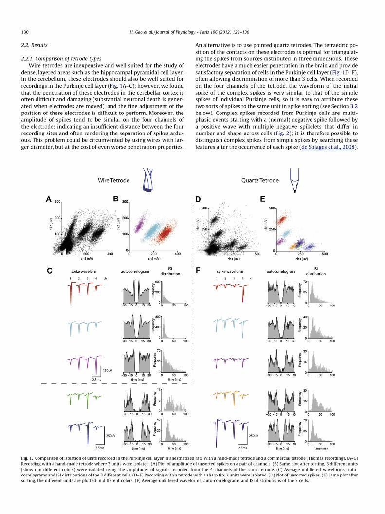
2.2.1. Comparison of tetrode typesWire tetrodes are inexpensive and well suited for the study of
dense, layered areas such as the hippocampal pyramidal cell layer.In the cerebellum, these electrodes should also be well suited forrecordings in the Purkinje cell layer (Fig. 1A–C); however, we foundthat the penetration of these electrodes in the cerebellar cortex isoften difficult and damaging (substantial neuronal death is gener-ated when electrodes are moved), and the fine adjustment of theposition of these electrodes is difficult to perform. Moreover, theamplitude of spikes tend to be similar on the four channels ofthe electrodes indicating an insufficient distance between the fourrecording sites and often rendering the separation of spikes ardu-ous. This problem could be circumvented by using wires with lar-ger diameter, but at the cost of even worse penetration properties.
Fig. 1. Comparison of isolation of units recorded in the Purkinje cell layer in anesthetizedRecording with a hand-made tetrode where 3 units were isolated. (A) Plot of amplitude o(shown in different colors) were isolated using the amplitudes of signals recorded frcorrelograms and ISI distributions of the 3 different cells. (D–F) Recording with a tetrode wsorting, the different units are plotted in different colors. (F) Average unfiltered wavefo
An alternative is to use pointed quartz tetrodes. The tetraedric po-sition of the contacts on these electrodes is optimal for triangulat-ing the spikes from sources distributed in three dimensions. Theseelectrodes have a much easier penetration in the brain and providesatisfactory separation of cells in the Purkinje cell layer (Fig. 1D–F),often allowing discrimination of more than 3 cells. When recordedon the four channels of the tetrode, the waveform of the initialspike of the complex spikes is very similar to that of the simplespikes of individual Purkinje cells, so it is easy to attribute thesetwo sorts of spikes to the same unit in spike sorting (see Section 3.2below). Complex spikes recorded from Purkinje cells are multi-phasic events starting with a (normal) negative spike followed bya positive wave with multiple negative spikelets that differ innumber and shape across cells (Fig. 2); it is therefore possible todistinguish complex spikes from simple spikes by searching thesefeatures after the occurrence of each spike (de Solages et al., 2008).
rats with a hand-made tetrode and a commercial tetrode (Thomas recording). (A–C)f unsorted spikes on a pair of channels. (B) Same plot after sorting, 3 different units
om the 4 channels of the same tetrode. (C) Average unfiltered waveforms, auto-ith a sharp tip. 7 units were isolated. (D) Plot of unsorted spikes. (E) Same plot after
rms, auto-correlograms and ISI distributions of the 7 cells.
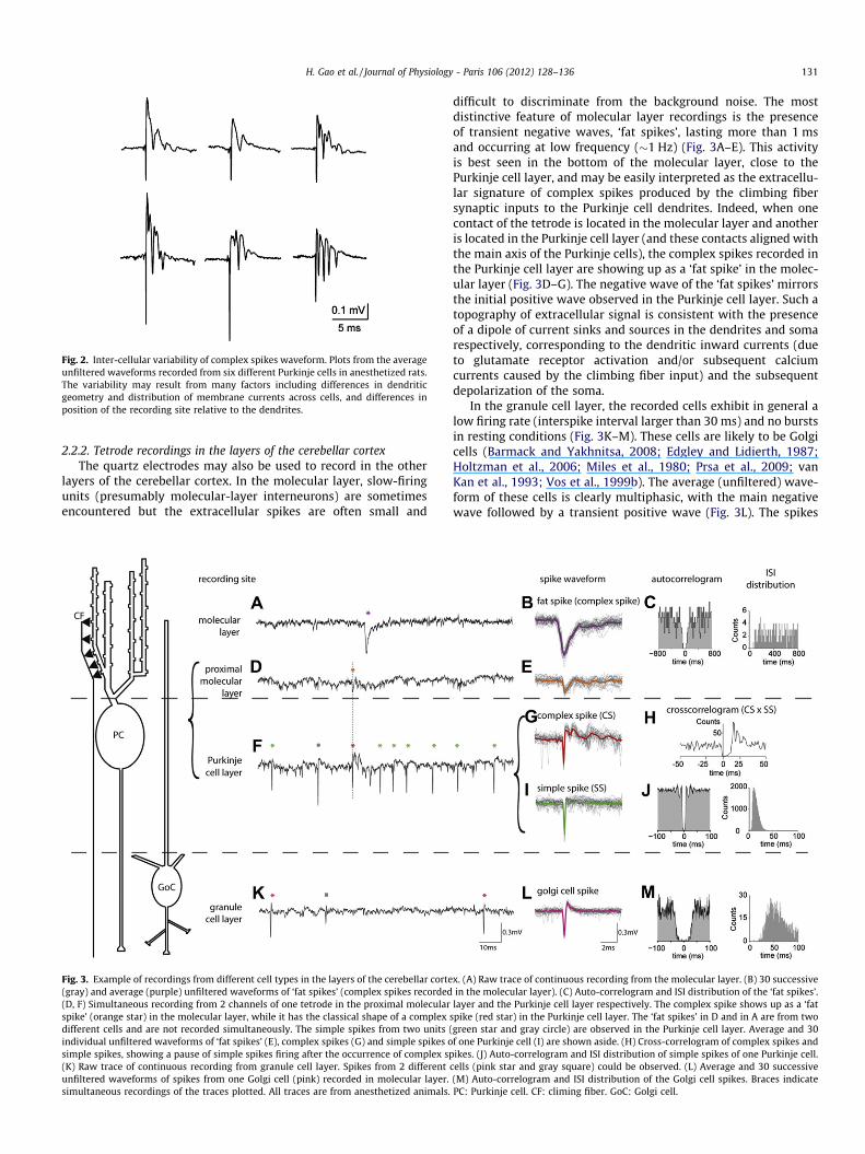
Fig. 2. Inter-cellular variability of complex spikes waveform. Plots from the averageunfiltered waveforms recorded from six different Purkinje cells in anesthetized rats.The variability may result from many factors including differences in dendriticgeometry and distribution of membrane currents across cells, and differences inposition of the recording site relative to the dendrites.
H. Gao et al. / Journal of Physiology - Paris 106 (2012) 128–136 131
2.2.2. Tetrode recordings in the layers of the cerebellar cortexThe quartz electrodes may also be used to record in the other
layers of the cerebellar cortex. In the molecular layer, slow-firingunits (presumably molecular-layer interneurons) are sometimesencountered but the extracellular spikes are often small and
Fig. 3. Example of recordings from different cell types in the layers of the cerebellar corte(gray) and average (purple) unfiltered waveforms of ‘fat spikes’ (complex spikes recorded(D, F) Simultaneous recording from 2 channels of one tetrode in the proximal molecularspike’ (orange star) in the molecular layer, while it has the classical shape of a complexdifferent cells and are not recorded simultaneously. The simple spikes from two units (individual unfiltered waveforms of ‘fat spikes’ (E), complex spikes (G) and simple spikes osimple spikes, showing a pause of simple spikes firing after the occurrence of complex sp(K) Raw trace of continuous recording from granule cell layer. Spikes from 2 differentunfiltered waveforms of spikes from one Golgi cell (pink) recorded in molecular layer.simultaneous recordings of the traces plotted. All traces are from anesthetized animals.
difficult to discriminate from the background noise. The mostdistinctive feature of molecular layer recordings is the presenceof transient negative waves, ‘fat spikes’, lasting more than 1 msand occurring at low frequency (�1 Hz) (Fig. 3A–E). This activityis best seen in the bottom of the molecular layer, close to thePurkinje cell layer, and may be easily interpreted as the extracellu-lar signature of complex spikes produced by the climbing fibersynaptic inputs to the Purkinje cell dendrites. Indeed, when onecontact of the tetrode is located in the molecular layer and anotheris located in the Purkinje cell layer (and these contacts aligned withthe main axis of the Purkinje cells), the complex spikes recorded inthe Purkinje cell layer are showing up as a ‘fat spike’ in the molec-ular layer (Fig. 3D–G). The negative wave of the ‘fat spikes’ mirrorsthe initial positive wave observed in the Purkinje cell layer. Such atopography of extracellular signal is consistent with the presenceof a dipole of current sinks and sources in the dendrites and somarespectively, corresponding to the dendritic inward currents (dueto glutamate receptor activation and/or subsequent calciumcurrents caused by the climbing fiber input) and the subsequentdepolarization of the soma.
In the granule cell layer, the recorded cells exhibit in general alow firing rate (interspike interval larger than 30 ms) and no burstsin resting conditions (Fig. 3K–M). These cells are likely to be Golgicells (Barmack and Yakhnitsa, 2008; Edgley and Lidierth, 1987;Holtzman et al., 2006; Miles et al., 1980; Prsa et al., 2009; vanKan et al., 1993; Vos et al., 1999b). The average (unfiltered) wave-form of these cells is clearly multiphasic, with the main negativewave followed by a transient positive wave (Fig. 3L). The spikes
x. (A) Raw trace of continuous recording from the molecular layer. (B) 30 successivein the molecular layer). (C) Auto-correlogram and ISI distribution of the ‘fat spikes’.layer and the Purkinje cell layer respectively. The complex spike shows up as a ‘fat
spike (red star) in the Purkinje cell layer. The ‘fat spikes’ in D and in A are from twogreen star and gray circle) are observed in the Purkinje cell layer. Average and 30f one Purkinje cell (I) are shown aside. (H) Cross-correlogram of complex spikes andikes. (J) Auto-correlogram and ISI distribution of simple spikes of one Purkinje cell.
cells (pink star and gray square) could be observed. (L) Average and 30 successive(M) Auto-correlogram and ISI distribution of the Golgi cell spikes. Braces indicatePC: Purkinje cell. CF: climing fiber. GoC: Golgi cell.
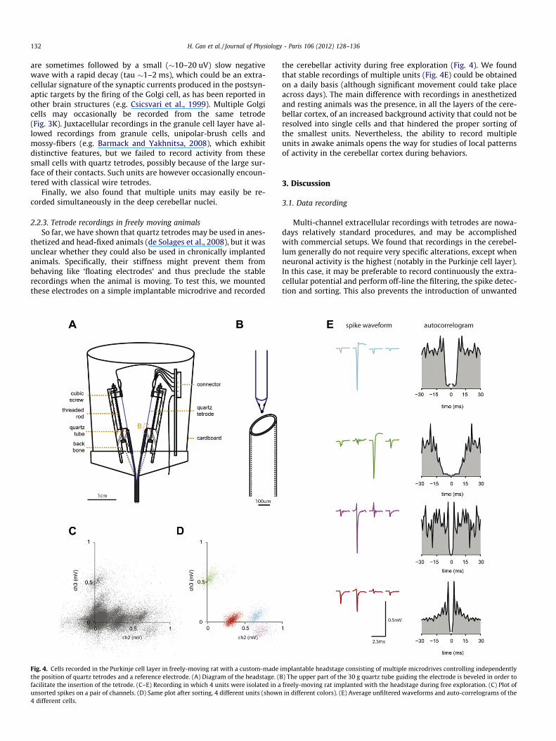
132 H. Gao et al. / Journal of Physiology - Paris 106 (2012) 128–136
are sometimes followed by a small (�10–20 uV) slow negativewave with a rapid decay (tau �1–2 ms), which could be an extra-cellular signature of the synaptic currents produced in the postsyn-aptic targets by the firing of the Golgi cell, as has been reported inother brain structures (e.g. Csicsvari et al., 1999). Multiple Golgicells may occasionally be recorded from the same tetrode(Fig. 3K). Juxtacellular recordings in the granule cell layer have al-lowed recordings from granule cells, unipolar-brush cells andmossy-fibers (e.g. Barmack and Yakhnitsa, 2008), which exhibitdistinctive features, but we failed to record activity from thesesmall cells with quartz tetrodes, possibly because of the large sur-face of their contacts. Such units are however occasionally encoun-tered with classical wire tetrodes.
Finally, we also found that multiple units may easily be re-corded simultaneously in the deep cerebellar nuclei.
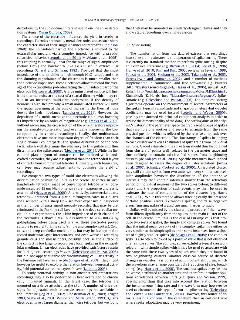
2.2.3. Tetrode recordings in freely moving animalsSo far, we have shown that quartz tetrodes may be used in anes-
thetized and head-fixed animals (de Solages et al., 2008), but it wasunclear whether they could also be used in chronically implantedanimals. Specifically, their stiffness might prevent them frombehaving like ‘floating electrodes’ and thus preclude the stablerecordings when the animal is moving. To test this, we mountedthese electrodes on a simple implantable microdrive and recorded
Fig. 4. Cells recorded in the Purkinje cell layer in freely-moving rat with a custom-madethe position of quartz tetrodes and a reference electrode. (A) Diagram of the headstage. (Bfacilitate the insertion of the tetrode. (C–E) Recording in which 4 units were isolated in aunsorted spikes on a pair of channels. (D) Same plot after sorting, 4 different units (shown4 different cells.
the cerebellar activity during free exploration (Fig. 4). We foundthat stable recordings of multiple units (Fig. 4E) could be obtainedon a daily basis (although significant movement could take placeacross days). The main difference with recordings in anesthetizedand resting animals was the presence, in all the layers of the cere-bellar cortex, of an increased background activity that could not beresolved into single cells and that hindered the proper sorting ofthe smallest units. Nevertheless, the ability to record multipleunits in awake animals opens the way for studies of local patternsof activity in the cerebellar cortex during behaviors.
3. Discussion
3.1. Data recording
Multi-channel extracellular recordings with tetrodes are nowa-days relatively standard procedures, and may be accomplishedwith commercial setups. We found that recordings in the cerebel-lum generally do not require very specific alterations, except whenneuronal activity is the highest (notably in the Purkinje cell layer).In this case, it may be preferable to record continuously the extra-cellular potential and perform off-line the filtering, the spike detec-tion and sorting. This also prevents the introduction of unwanted
implantable headstage consisting of multiple microdrives controlling independently) The upper part of the 30 g quartz tube guiding the electrode is beveled in order tofreely-moving rat implanted with the headstage during free exploration. (C) Plot ofin different colors). (E) Average unfiltered waveforms and auto-correlograms of the
H. Gao et al. / Journal of Physiology - Paris 106 (2012) 128–136 133
distortions by the sub-optimal filters in use in on-line spike detec-tion systems (Quian Quiroga, 2009).
The choice of the electrode influences the yield in cerebellarrecordings. Tetrodes are usually metal electrodes and as such sharethe characteristics of their single-channel counterparts (Robinson,1968): the uninsulated part of the electrode is coupled to theextracellular medium via a complex impedance with a pseudo-capacitive behavior (Lempka et al., 2011; McAdams et al., 1995);this coupling is normally linear for the range of signal amplitudes(below 1 mV) and bandwidth (1–10 kHz) used in extracellularrecordings (Onaral and Schwan, 1982). Provided that the inputimpedance of the amplifier is high enough (G X range), and thatthe shunting capacitance of the electrodes is much smaller thanthe electrode impedance, these electrodes allow to record the aver-age of the extracellular potential facing the uninsulated part of theelectrode (Nelson et al., 2008). A large uninsulated surface will lim-it the thermal noise at the cost of a spatial filtering which may re-sult in an increased multi-unit background if the density ofneurons is high. Reciprocally, a small uninsulated surface will limitthe spatial averaging at the cost of increasing the thermal noisedue to the high impedance (Lempka et al., 2011). However, thedeposition of a noble metal at the electrode tip allows loweringits impedance by an order of magnitude (e.g. Franks et al., 2005)without increasing the cross-section of the wire, therefore improv-ing the signal-to-noise ratio (and eventually improving the bio-compatibility in chronic recordings). Finally, the multicontactelectrodes have one more important degree of freedom than theirsingle channel counterparts: the spatial distribution of the con-tacts, which will determine the efficiency to triangulate and thusdiscriminate the spike sources (Mechler et al., 2011). While the lin-ear and planar layout of contacts can be obtained with hand-crafted electrodes, they are less optimal than the tetrahedral layoutof contacts from commercial tetrodes. Ultimately, each brain area/cell type may require adjustments to optimize the yield ofrecordings.
We compared two types of multi-site electrodes allowing thediscrimination of multiple units in the cerebellar cortex in vivo:hand-made tetrodes (made of conventional tetrode wire: poly-mide-insulated 12 um Nichrome wire) are inexpensive and easilyassembled (Nguyen et al., 2009); Alternatively, commercial quartztetrodes – obtained from 4 platinum/tungsten-cores in a quartzrods, sculpted with a sharp tip – are more expensive but superiorin the number of units simultaneously recorded that may be dis-criminated in the Purkinje cell layer and in the deep cerebellar nu-clei. In our experiments, the 1 kHz impedance of each channel ofthe electrodes is above 1 MX, but is lowered to 200–300 kX bygold-plating before being used in vivo. These electrodes provedsuitable to record Purkinje cells (simple and complex spikes), Golgicells, and deep cerebellar nuclei units, but may be less optimal torecord molecular layer interneurons, and even worse at recordinggranule cells and mossy fibers, possibly because the surface ofthe contact is too large to record very local spikes in the extracel-lular medium. Linear electrodes have provided satisfactory resultsfor Purkinje cell recordings in vitro (Delescluse and Pouzat, 2006),but did not appear suitable for discriminating cellular activity inthe Purkinje cell layer in vivo (de Solages et al., 2008); they mighthowever be useful to explore the relation between neuronal activ-ity/field potential across the layers in vivo (Lu et al., 2005).
To study neuronal activity in non-anesthetized preparations,recordings may also be performed in head-restrained or chroni-cally implanted animals. In the latest case, the tetrodes aremounted on a drive attached to the skull. A number of drive de-signs for adjustable multi-electrode recordings are available inthe literature (Jog et al., 2002; Kloosterman et al., 2009; Krüger,1983; Szabó et al., 2001; Wilson and McNaughton, 1993). Quartzelectrodes have a larger diameter than wire tetrodes, but we found
that they may be mounted in similarly-designed drives and theyallow stable recordings over single sessions.
3.2. Spike sorting
The transformation from raw data of extracellular recordingsinto spike train estimates is the operation of spike sorting. Thereis currently no ‘standard’ method to perform spike sorting, despitean extensive literature (e.g. Brown et al., 2004; Fee et al., 1996;Franke et al., 2010; Kim and Kim, 2003; reviews in Lewicki, 1998;Pouzat et al., 2004; Shoham et al., 2003; Takahashi et al., 2003;Vargas-Irwin and Donoghue, 2007), and a number of methodssupplemented in commercial and free softwares: e.g. klusters(http://klusters.sourceforge.net/, Hazan et al., 2006), mclust (A.D.Redish, http://redishlab.neuroscience.umn.edu/MClust/MClust.html),KlustaKwik (K. Harris, http://klustakwik.sourceforge.net/), Spike-Omatic (e.g. Delescluse and Pouzat, 2006). The simplest sortingalgorithms operate on the measurement of several parameters ofthe spikes (typically amplitude and shape parameters -but waveletcoefficients may be used instead (Letelier and Weber, 2000)-,possibly transformed via principal component analysis in order toreduce the dimensionality of the data). The sorting aims at identify-ing ‘clusters’ in the parameter space that represent groups of spikesthat resemble one another and seem to emanate from the samephysical position, which is reflected by the relative amplitude overthe channels of the electrode. The timestamps of spikes belongingto each cluster are taken as estimates of spike trains from individualneurons. A good estimate of the spike train should thus be obtainedfrom clusters of points well isolated in the parameter space andthus unlikely to contain misclassified spikes from neighboringclusters (de Solages et al., 2008). Specific measures have indeedbeen designed to assess the degree of cluster isolation (Joshuaet al., 2007; Schmitzer-Torbert et al., 2005). An isolated clustermay still contain spikes from two units with very similar extracel-lular amplitude; however the distribution of the inter-spikeintervals may then contain intervals shorter than the refractoryperiod of individual neurons (if the two spikes belong to differentunits), and the proportion of such events may then be used toestimate the rate of contamination of the clusters (e.g. Harriset al., 2000). While this method allows tracking easily the presenceof ‘false positive’ errors (extraneous spikes), the ‘false negative’errors (missing spikes of a unit) are much harder to track.
Spikes will be missed by the clustering procedure if their wave-form differs significantly from the spikes in the main cluster of thecell. In the cerebellum, this is the case of Purkinje cells that pro-duce two sorts of spikes, the simple and complex spikes. We foundthat the initial negative spike of the complex spike may either bevery similar to the simple spikes or, in some instances, form a clus-ter of slightly smaller spikes (de Solages et al., 2008); the complexspike is also often followed by a positive wave that is not observedafter simple spikes. The complex spikes exhibit a typical crosscor-relogram with simple spikes which may be used to associate withthe same unit these two types of spikes when they are found intwo neighboring clusters. Another classical source of discretechanges in waveforms is bursts of action potentials, during whichthe waveform may change considerably (reduction of size, broad-ening) (e.g. Harris et al., 2000). The smallest spikes may be lostor, worse, attributed to another unit and therefore introduce spu-rious correlations between units (e.g. Quirk and Wilson, 1999).Specific algorithms that take into account the relation betweenthe instantaneous firing rate and the waveform may however beused to circumvent this type of error in spike sorting (Delescluseand Pouzat, 2006; Pouzat et al., 2004). However, this source of er-ror is less of a concern in the cerebellum than in cortical tissuewhere spike adaptation may be very prominent.
134 H. Gao et al. / Journal of Physiology - Paris 106 (2012) 128–136
Another important cause of mis-clustering is the overlap ofextracellular spikes due to near-synchronous firing of neighboringunits. In the Purkinje cell layer, overlap is more likely to occur, asmany units fire continuously at a high rate (�50 Hz), and is evenfurther increased by the synchronization of spikes (Bell andGrimm, 1969; de Solages et al., 2008; Ebner and Bloedel, 1981).The overlap of spike waveforms will result in spikes of variousamplitudes and shape depending on the individual waveformsand temporal offset between the spikes. Unless one unit has amuch larger amplitude than the others, this overlap may lead tomis-clustering of the spikes (which will be omitted from the spiketrains of the units that fired together). Since most of the energy ofthe spikes recorded in the cerebellum is concentrated in a shortwindow of 200–300 ls, the overlap of waveforms should mostlytake place for events spaced by this interval or less. The filteringprocedure used to remove the low frequencies from the recordingsmay sensibly increase the temporal window of interaction of extra-cellular waveforms of spikes occurring at short delays. After filter-ing with the IIR (Infinite Impulse Response) high-pass filterscommonly used (Butterworth or Bessel filters), the negative waveof the spike in the filtered extracellular potential will be followedby an exaggerated positive wave whose amplitude and durationwill depend on the filter parameters. Lowering the order and cutoffof the high-pass filter will minimize these effects, so that the inter-action between waveforms separated by more than 1 ms will benegligible (de Solages et al., 2008). Alternatively, wavelet filtersmay be used instead in order to improve the preservation of theextracellular waveforms (Wiltschko et al., 2008). Several algo-rithms already exist to handle overlapping spikes (Takahashiet al., 2003; Vargas-Irwin and Donoghue, 2007) but to our knowl-edge none have yet been applied to cerebellar recordings.
In summary, errors in spike sorting may degrade the quality ofthe estimation (in the statistical sense) of multiple spike trains, andtherefore, the known sources of misclustering should be kept inmind while drawing any scientific conclusion from tetroderecordings.
3.3. Tetrode use for the study of coordinated activity in the cerebellum
The first benefit of tetrode recordings may just be to increasethe yield of experiments, allowing recording more cells with fewerpenetrations. However, since these electrodes allow recordingsimultaneously neighboring neurons, they may also help studyingpatterns of activity involving multiple cells. The most documentedcase of coordinated discharge in the cerebellum is the synchroniza-tion of complex spikes (Welsh et al., 1995, see Introduction above).This coordination is not the result from intra-cerebellar integrationof information, but directly reflects the synchrony between theinferior olive cells that generate the complex spikes in Purkinjecells. Neighboring Purkinje cells are also known to exhibit synchro-nized simple spikes. Purkinje cell synchronization is likely to affectsubstantially the activity in the downstream structures, the deepcerebellar nuclei (Gauck and Jaeger, 2000), allowing to modulatethe deep cerebellar cells at very short time scales. Synchronizationof neighboring Purkinje cells has been noted in early recordings ofpairs of cells in anesthetized and decerebrate animals (Bell andGrimm, 1969; Ebner and Bloedel, 1981). We have recently shown(de Solages et al., 2008) that some of this synchrony may resultfrom a fast cerebellar oscillation (Adrian, 1935) arising from a pop-ulation oscillation of Purkinje cells, associated with millisecond-scale synchrony and 5-ms delayed coordination of discharge (deSolages et al., 2008); such oscillations were indeed visible (butnot noted) in the early studies, as pointed out by Isope et al.(2002). Shin and De Schutter also suggested a synchronizing rolefor local inhibitory neurons, since synchrony appeared associatedwith episodes of slowed firing rate of Purkinje cells (Shin and De
Schutter, 2006). Since inhibitory neurons are known to contactmultiple neighboring Purkinje cells, combined recordings with te-trodes of neighboring interneurons and Purkinje cells should helpto analyze the effect of local inhibition on neighboring cells (deSolages and Léna, in preparation). Shared excitation could also fa-vor synchronization (Jaeger, 2003) at time scales of the order of10 ms. Such slightly broader synchrony is indeed observed be-tween Purkinje cells (Bosman et al., 2010; De Zeeuw et al., 1997;Wise et al., 2010), mostly in cases where the cells also exhibit syn-chronized complex spikes indicating that they belong to a commonfunctional ensemble. Broad coordination is also found betweenGolgi cells, and is likely due to shared excitatory inputs and gapjunctions (Vos et al., 1999a). A much tighter synchrony at thesub-millisecond scale has also been observed along the parallel fi-ber axis in the cerebellar cortex during a reaching task (Heck et al.,2007). But, instead of single-unit spikes, only multi-unit signalswere used in this study. Tetrode recordings in such experimentscould indeed help identifying the units responsible of thesecorrelations.
4. Conclusion
Tetrode recordings are relatively easy to set up in the cerebel-lum and, provided a few caveats are taken into account, they allowstudying patterns of activity in groups of neighboring cells in thecerebellum. Synchronization is an activity pattern commonly ob-served in the cerebellar cortex, but its prominence between neigh-boring units underlines the importance of studying the cerebellarlocal networks. More elaborated forms of patterned activity havealso been observed in the cerebellar cortex, with notably a numberof normal and pathological rhythmic activities (reviews in Cheronet al., 2008; De Zeeuw et al., 2008). Multi-electrodes studies (e.g.Bosman et al., 2010; Cheron et al., 2004; Heck et al., 2007; Mostofiet al., 2010; Vos et al., 1999a; Welsh et al., 1995; Wise et al., 2010)and tetrode recordings (de Solages et al., 2008) have alreadyproven the interest of recordings multiple cells at the same timeto study patterned neuronal discharge. Bringing tetrode recordingsto freely moving animals holds great promise in characterizing thecontribution of these phenomena to cerebellar function and reveal-ing their underlying mechanisms during a variety of behavioralconditions in awake animals.
Acknowledgements
This work was supported by ANR (08-SYSC-005-02 DALTPAC,09-MNPS-038-01 Cecomod, 07-NEURO-025-03 Natacs), the CentreNational de la Recherche Scientifique (CNRS), the Institut Nationalde la Santé et de la Recherche Médicale (INSERM, C.L.), the EcoleNormale Supérieure (ENS), the Ministère des Affaires Etrangères(H. G.).
References
Adrian, E.D., 1935. Discharge frequencies in the cerebral and cerebellar cortex.Journal of Physiology – London 83, 32–33.
Apps, R., Hawkes, R., 2009. Cerebellar cortical organization: a one-map hypothesis.Nature Reviews Neuroscience 10, 670–681.
Barmack, N.H., Yakhnitsa, V., 2008. Functions of interneurons in mouse cerebellum.Journal of Neuroscience 28, 1140–1152.
Bell, C.C., Grimm, R.J., 1969. Discharge properties of Purkinje cells recorded on singleand double microelectrodes. Journal of Neurophysiology 32, 1044–1055.
Bosman, L.W.J., Koekkoek, S.K.E., Shapiro, J., Rijken, B.F.M., Zandstra, F., Van de Ende,B., Owens, C.B., Potters, J.W., De Gruijl, J.R., Ruigrgok, T.J.H., De Zeeuw, C.I., 2010.Encoding of whisker input by cerebellar Purkinje cells. Journal of Physiology588, 3757–3783.
Bower, J.M., Woolston, D.C., 1983. Congruence of spatial organization of tactileprojections to granule cell and Purkinje cell layers of cerebellar hemispheres ofthe albino rat: vertical organization of cerebellar cortex. Journal ofNeurophysiology 49, 745–766.
H. Gao et al. / Journal of Physiology - Paris 106 (2012) 128–136 135
Bragin, A., Hetke, J., Wilson, C.L., Anderson, D.J., Engel, J., Buzsáki, G., 2000. Multiplesite silicon-based probes for chronic recordings in freely moving rats:implantation, recording and histological verification. Journal of NeuroscienceMethods 98, 77–82.
Brochu, G., Maler, L., Hawkes, R., 1990. Zebrin II: a polypeptide antigen expressedselectively by Purkinje cells reveals compartments in rat and fish cerebellum.Journal of Comparative Neurology 291, 538–552.
Brown, E.N., Kass, R.E., Mitra, P.P., 2004. Multiple neural spike train data analysis:state-of-the-art and future challenges. Nature Neuroscience 7, 456–461.
Buisseret-Delmas, C., Angaut, P., 1993. The cerebellar olivo-corticonuclearconnections in the rat. Progress in Neurobiology 40, 63–87.
Cheron, G., Gall, D., Servais, L., Dan, B., Maex, R., Schiffmann, S.N., 2004. Inactivationof calcium-binding protein genes induces 160 Hz oscillations in the cerebellarcortex of alert mice. Journal of Neuroscience 24, 434–441.
Cheron, G., Servais, L., Dan, B., 2008. Cerebellar network plasticity: from genes tofast oscillation. Neuroscience 153, 1–19.
Cohen, D., Yarom, Y., 1998. Patches of synchronized activity in the cerebellar cortexevoked by mossy-fiber stimulation: questioning the role of parallel fibers. In:Proceedings of the National Academy of Sciences of the United States ofAmerica, vol. 95. pp. 15032–15036.
Csicsvari, J., Hirase, H., Czurkó, A., Mamiya, A., Buzsáki, G., 1999. Oscillatory couplingof hippocampal pyramidal cells and interneurons in the behaving rat. Journal ofNeuroscience 19, 274–287.
Csicsvari, J., Henze, D.A., Jamieson, B., Harris, K.D., Sirota, A., Barthó, P., Wise, K.D.,Buzsáki, G., 2003. Massively parallel recording of unit and local fieldpotentials with silicon-based electrodes. Journal of Neurophysiology 90,1314–1323.
de Solages, C., Léna, C., in preparation. Modulation of Purkinje cell firing andcoordination by molecular layer interneurons in the cerebellar cortex in vivo.
de Solages, C., Szapiro, G., Brunel, N., Hakim, V., Isope, P., Buisseret, P., Rousseau, C.,Barbour, B., Léna, C., 2008. High-frequency organization and synchrony ofactivity in the purkinje cell layer of the cerebellum. Neuron 58, 775–788.
De Zeeuw, C.I., Koekkoek, S.K., Wylie, D.R., Simpson, J.I., 1997. Association betweendendritic lamellar bodies and complex spike synchrony in the olivocerebellarsystem. Journal of Neurophysiology 77, 1747–1758.
De Zeeuw, C.I., Hoebeek, F.E., Schonewille, M., 2008. Causes and consequences ofoscillations in the cerebellar cortex. Neuron 58, 655–658.
Delescluse, M., Pouzat, C., 2006. Efficient spike-sorting of multi-state neurons usinginter-spike intervals information. Journal of Neuroscience Methods 150, 16–29.
Dizon, M.J., Khodakhah, K., 2011. The role of interneurons in shaping purkinje cellresponses in the cerebellar cortex. The Journal of neuroscience 31, 10463–10473.
Dugué, G.P., Brunel, N., Hakim, V., Schwartz, E., Chat, M., Lévesque, M.,Courtemanche, R., Léna, C., Dieudonné, S., 2009. Electrical coupling mediatestunable low-frequency oscillations and resonance in the cerebellar Golgi cellnetwork. Neuron 61, 126–139.
Ebner, T.J., Bloedel, J.R., 1981. Correlation between activity of Purkinje cells and itsmodification by natural peripheral stimuli. Journal of Neurophysiology 45, 948–961.
Edgley, S.A., Lidierth, M., 1987. The discharges of cerebellar Golgi cells duringlocomotion in the cat. Journal of Physiology 392, 315–332.
Fee, M.S., Mitra, P.P., Kleinfeld, D., 1996. Automatic sorting of multiple unit neuronalsignals in the presence of anisotropic and non-Gaussian variability. Journal ofNeuroscience Methods 69, 175–188.
Franke, F., Natora, M., Boucsein, C., Munk, M.H.J., Obermayer, K., 2010. An onlinespike detection and spike classification algorithm capable of instantaneousresolution of overlapping spikes. Journal of Computational Neuroscience 29,127–148.
Franks, W., Schenker, I., Schmutz, P., Hierlemann, A., 2005. Impedancecharacterization and modeling of electrodes for biomedial applications. IEEETransactions on Biomedical Engineering 52, 1295–1301.
Gao, W., Chen, G., Reinert, K.C., Ebner, T.J., 2006. Cerebellar cortical molecular layerinhibition is organized in parasagittal zones. Journal of Neuroscience 26, 8377–8387.
Garwicz, M., Ekerot, C.F., Jörntell, H., 1998. Organizational principles of cerebellarneuronal circuitry. News in Physiological Sciences 13, 26–32.
Gauck, V., Jaeger, D., 2000. The control of rate and timing of spikes in the deepcerebellar nuclei by inhibition. Journal of Neuroscience 20, 3006–3016.
Gray, C.M., Maldonado, P.E., Wilson, M., McNaughton, B., 1995. Tetrodes markedlyimprove the reliability and yield of multiple single-unit isolation from multi-unit recordings in cat striate cortex. Journal of Neuroscience Methods 63, 43–54.
Halverson, H.E., Lee, I., Freeman, J.H., 2010. Associative plasticity in the medialauditory thalamus and cerebellar interpositus nucleus during eyeblinkconditioning. Journal of Neuroscience 30, 8787–8796.
Harris, K.D., Henze, D.A., Csicsvari, J., Hirase, H., Buzsáki, G., 2000. Accuracy oftetrode spike separation as determined by simultaneous intracellular andextracellular measurements. Journal of Neurophysiology 84, 401–414.
Hazan, L., Zugaro, M., Buzsáki, G., 2006. Klusters, NeuroScope, NDManager: a freesoftware suite for neurophysiological data processing and visualization. Journalof Neuroscience Methods 155, 207–216.
Heck, D.H., Thach, W.T., Keating, J.G., 2007. On-beam synchrony in the cerebellumas the mechanism for the timing and coordination of movement. In:Proceedings of the National Academy of Sciences of the United States ofAmerica, vol. 104. pp. 7658–7663.
Holtzman, T., Rajapaksa, T., Mostofi, A., Edgley, S.A., 2006. Different responses of ratcerebellar Purkinje cells and Golgi cells evoked by widespread convergentsensory inputs. Journal of Physiology 574, 491–507.
Isope, P., Barbour, B., 2002. Temporal organization of activity in the cerebellarcortex: a manifesto for synchrony. Journal of Neuroscience 22, 9668–9678.
Isope, P., Dieudonné, S., Barbour, B., 2002. Temporal organization of activity in thecerebellar cortex: a manifesto for synchrony. Annals of the New York Academyof Sciences 978, 164–174.
Jaeger, D., 2003. No parallel fiber volleys in the cerebellar cortex: evidence fromcross-correlation analysis between Purkinje cells in a computer model and inrecordings from anesthetized rats. Journal of Computational Neuroscience 14,311–327.
Jog, M.S., Connolly, C.I., Kubota, Y., Iyengar, D.R., Garrido, L., Harlan, R., Graybiel,A.M., 2002. Tetrode technology: advances in implantable hardware,neuroimaging, and data analysis techniques. Journal of Neuroscience Methods117, 141–152.
Joshua, M., Elias, S., Levine, O., Bergman, H., 2007. Quantifying the isolation qualityof extracellularly recorded action potentials. Journal of Neuroscience Methods163, 267–282.
Jung, M.W., Qin, Y., McNaughton, B.L., Barnes, C.A., 1998. Firing characteristics ofdeep layer neurons in prefrontal cortex in rats performing spatial workingmemory tasks. Cerebral Cortex 8, 437–450.
Kan, P.L.V., Gibson, A.R., Houk, J.C., 1993. Movement-related inputs to intermediatecerebellum of the monkey. Journal of Neurophysiology 69, 74–94.
Kim, K.H., Kim, S.J., 2003. Method for unsupervised classification of multiunit neuralsignal recording under low signal-to-noise ratio. IEEE Transactions onBiomedical Engineering 50, 421–431.
Kloosterman, F., Davidson, T.J., Gomperts, S.N., Layton, S.P., Hale, G., Nguyen, D.P.,Wilson, M.A., 2009. Micro-drive array for chronic in vivo recording: drivefabrication. Journal of Visualized Experiments 26, 2–5.
Krüger, J., 1983. Simultaneous individual recordings from many cerebral neurons:techniques and results. Reviews of Physiology, Biochemistry and Pharmacology98, 177–233.
Lang, E.J., Sugihara, I., Welsh, J.P., Llinás, R., 1999. Patterns of spontaneous purkinjecell complex spike activity in the awake rat. Journal of Neuroscience 19, 2728–2739.
Larouche, M., Hawkes, R., 2006. From clusters to stripes: the developmental originsof adult cerebellar compartmentation. Cerebellum 5, 77–88.
Lempka, S.F., Johnson, M.D., Moffit, M.A., Otto, K.J., Ripke, D.R., McIntyre, C.C., 2011.Theoretical analysis of intracortical microelectrode recordings. Journal ofNeural Engineering 8, 045006.
Letelier, J.C., Weber, P.P., 2000. Spike sorting based on discrete wavelet transformcoefficients. Journal of Neuroscience Methods 101, 93–106.
Lewicki, M.S., 1998. A review of methods for spike sorting: the detection andclassification of neural action potentials. Network: Computation in NeuralSystems 9, 53–78.
Lou, J.S., Bloedel, J.R., 1992. Responses of sagittally aligned Purkinje cells duringperturbed locomotion: synchronous activation of climbing fiber inputs. Journalof neurophysiology 68, 570–580.
Lu, H., Hartmann, M.J., Bower, J.M., 2005. Correlations between purkinje cell single-unit activity and simultaneously recorded field potentials in the immediatelyunderlying granule cell layer. Journal of Neurophysiology 94, 1849–1860.
Maex, R., De Schutter, E., 1998. Synchronization of Golgi and granule cell firing in adetailed network model of the cerebellar granule cell layer. Journal ofNeurophysiology 80, 2521–2537.
McAdams, E.T., Lackermeier, A., McLaughlin, J.A., Macken, D., Jossinet, J., 1995. Thelinear and nonlinear electrical-properties of the electrode–electrolyte interface.Biosensors and Bioelectronics 10, 67–74.
Mechler, F., Victor, J.D., Ohiorhenuan, I., Schmid, A.M., Hu, Q., 2011. Three-dimensional localization of neurons in cortical tetrode recordings. Journal ofNeurophysiology 106, 828–848.
Medina, J.F., Garcia, K.S., Nores, W.L., Taylor, N.M., Mauk, M.D., 2000. Timingmechanisms in the cerebellum: testing predictions of a large-scale computersimulation. Journal of Neuroscience 20, 5516–5525.
Miles, F.A., Fuller, J.H., Braitman, D.J., Dow, B.M., 1980. Long-term adaptive changesin primate vestibuloocular reflex. III. Electrophysiological observations inflocculus of normal monkeys. Journal of Neurophysiology 43, 1437–1476.
Mostofi, A., Holtzman, T., Grout, A.S., Yeo, C.H., Edgley, S.A., 2010.Electrophysiological localization of eyeblink-related microzones in rabbitcerebellar cortex. Journal of Neuroscience 30, 8920–8934.
Nelson, M.J., Pouget, P., Nilsen, E.A., Patten, C.D., Schall, J.D., 2008. Review of signaldistortion through metal microelectrode recording circuits and filters. Journalof Neuroscience Methods 169, 141–157.
Nguyen, D.P., Layton, S.P., Hale, G., Gomperts, S.N., Davidson, T.J., Kloosterman, F.,Wilson, M.A., 2009. Micro-drive array for chronic in vivo recording: tetrodeassembly. Journal of Visualized Experiments 26, 7–9.
Onaral, B., Schwan, H.P., 1982. Linear and non-linear properties of platinumelectrode polarization part I. Frequency dependence at very low frequencies.Medical and Biological Engineering and Computing 20, 299–306.
Ozden, I., Lee, H.M., Sullivan, M.R., Wang, S.S.H., 2008. Identification and clusteringof event patterns from in vivo multiphoton optical recordings of neuronalensembles. Journal of Neurophysiology 100, 495–503.
Ozden, I., Sullivan, M.R., Lee, H.M., Wang, S.S.H., 2009. Reliable coding emerges fromcoactivation of climbing fibers in microbands of cerebellar Purkinje neurons.Journal of Neuroscience 29, 10463–10473.

136 H. Gao et al. / Journal of Physiology - Paris 106 (2012) 128–136
Pouzat, C., Delescluse, M., Viot, P., Diebolt, J., 2004. Improved spike-sorting bymodeling firing statistics and burst-dependent spike amplitude attenuation: aMarkov chain Monte Carlo approach. Journal of Neurophysiology 91, 2910–2928.
Prsa, M., Dash, S., Catz, N., Dicke, P.W., Thier, P., 2009. Characteristics of responses ofGolgi cells and mossy fibers to eye saccades and saccadic adaptation recordedfrom the posterior vermis of the cerebellum. Journal of Neuroscience 29, 250–262.
Quian Quiroga, R., 2009. What is the real shape of extracellular spikes? Journal ofNeuroscience Methods 177, 194–198.
Quirk, M.C., Wilson, M.A., 1999. Interaction between spike waveform classificationand temporal sequence detection. Journal of Neuroscience Methods 94, 41–52.
Robinson, D.A., 1968. The electrical properties of metal microelectrodes.Proceedings of IEEE 56, 1065–1071.
Sasaki, K., Bower, J.M., Llinás, R., 1989. Multiple Purkinje cell recording in rodentcerebellar cortex. European Journal of Neuroscience 1, 572–586.
Schmitzer-Torbert, N., Jackson, J., Henze, D., Harris, K., Redish., A.D., 2005.Quantitative measures of cluster quality for use in extracellular recordings.Neuroscience 131, 1–11.
Schultz, S., Kitamura, K., Post-Uiterweer, A., Krupic, J., Häusser, M., 2009. Spatialpattern coding of sensory information by climbing fiber-evoked calcium signalsin networks of neighboring cerebellar Purkinje cells. Journal of Neuroscience 29,8005–8015.
Shin, S.L., De Schutter, E., 2006. Dynamic synchronization of Purkinje cell simplespikes. Journal of Neurophysiology 96, 3485–3491.
Shoham, S., Fellows, M.R., Normann, R.A., 2003. Robust, automatic spike sortingusing mixtures of multivariate t-distributions. Journal of Neuroscience Methods127, 111–122.
Sillitoe, R.V., Chung, S.H., Fritschy, J.M., Hoy, M., Hawkes, R., 2008. Golgi celldendrites are restricted by Purkinje cell stripe boundaries in the adult mousecerebellar cortex. Journal of Neuroscience 28, 2820–2826.
Solinas, S., Forti, L., Cesana, E., Mapelli, J., De Schutter, E., D0Angelo, E., 2007. Fast-reset of pacemaking and theta-frequency resonance patterns in cerebellar Golgicells: simulations of their impact in vivo. Frontiers in Cellular Neuroscience 1, 4.
Soteropoulos, D.S., Baker, S.N., 2008. Bilateral representation in the deep cerebellarnuclei. Journal of Physiology 586, 1117–1136.
Sugihara, I., Shinoda, Y., 2004. Molecular, topographic, and functional organizationof the cerebellar cortex: a study with combined aldolase C and olivocerebellarlabeling. Journal of Neuroscience 24, 8771–8785.
Sugihara, I., Marshall, S.P., Lang, E.J., 2007. Relationship of complex spike synchronybands and climbing fiber projection determined by reference to aldolase Ccompartments in crus IIa of the rat cerebellar cortex. Journal of ComparativeNeurology 501, 13–29.
Szabó, I., Czurkó, A., Csicsvari, J., Hirase, H., Leinekugel, X., Buzsáki, G., 2001. Theapplication of printed circuit board technology for fabrication of multi-channelmicro-drives. Journal of Neuroscience Methods 105, 105–110.
Tahon, K., Wijnants, M., De Schutter, E., Maex, R., 2011. Current source densitycorrelates of cerebellar Golgi and Purkinje cell responses to tactile input. Journalof Neurophysiology 105, 1327–1341.
Takahashi, S., Anzai, Y., Sakurai, Y., 2003. Automatic sorting for multi-neuronalactivity recorded with tetrodes in the presence of overlapping spikes. Journal ofNeurophysiology 89, 2245–2258.
Thier, P., Dicke, P.W., Haas, R., Barash, S., 2000. Encoding of movement time bypopulations of cerebellar Purkinje cells. Nature 405, 72–76.
Vargas-Irwin, C., Donoghue, J.P., 2007. Automated spike sorting using density gridcontour clustering and subtractive waveform decomposition. Journal ofNeuroscience Methods 164, 1–18.
Vervaeke, K., Lorincz, A., Gleeson, P., Farinella, M., Nusser, Z., Silver, R.A., 2010. Rapiddesynchronization of an electrically coupled interneuron network with sparseexcitatory synaptic input. Neuron 67, 435–451.
Voogd, J., Bigaré, F., 1980. Topographical distribution of olivary and cortico-nuclearfibres in the cerebellum: a review. In: Courville, J., de Montigny, C., Lamarre, Y.(Eds.), The inferior olivary nucleus: anatomy and physiology. Raven Press, NewYork, pp. 207–234.
Voogd, J., Glickstein, M., 1998. The anatomy of the cerebellum. Trends inNeurosciences 21, 370–375.
Voogd, J., Ruigrok, T.J.H., 2004. The organization of the corticonuclear andolivocerebellar climbing fiber projections to the rat cerebellar vermis: thecongruence of projection zones and the zebrin pattern. Journal ofNeurocytology 33, 5–21.
Vos, B.P., Maex, R., Volny-Luraghi, A., De Schutter, E., 1999a. Parallel fiberssynchronize spontaneous activity in cerebellar Golgi cells. Journal ofNeuroscience 19, RC6.
Vos, B.P., Volny-Luraghi, A., De Schutter, E., 1999b. Cerebellar Golgi cells in the rat:receptive fields and timing of responses to facial stimulation. European Journalof Neuroscience 11, 2621–2634.
Welsh, J.P., Lang, E.J., Suglhara, I., Llinás, R., 1995. Dynamic organization of motorcontrol within the olivocerebellar system. Nature 374, 453–457.
Wilson, M.A., McNaughton, B.L., 1993. Dynamics of the hippocampal ensemble codefor space. Science 261, 1055–1058.
Wiltschko, A.B., Gage, G.J., Berke, J.D., 2008. Wavelet filtering before spike detectionpreserves waveform shape and enhances single-unit discrimination. Journal ofNeuroscience Methods 173, 34–40.
Wise, A.K., Cerminara, N.L., Marple-Horvat, D.E., Apps, R., 2010. Mechanisms ofsynchronous activity in cerebellar Purkinje cells. Journal of Physiology 588,2373–2390.
Copyright © 2022 FDOKUMEN