Reconstructing the Engram: Simultaneous, Multisite, Many Single Neuron Recordings
9
Neuron, Vol. 18, 529–537, April, 1997, Copyright 1997 by Cell Press Reconstructing the Engram: Neurotechnique Simultaneous, Multisite, Many Single Neuron Recordings Miguel A. L. Nicolelis, Asif A. Ghazanfar, large populations of neurons involved in processing and storing information (Sherrington, 1906; Hebb, 1949; Barbara M. Faggin, Scott Votaw, and Laura M. O. Oliveira Lashley, 1950; Erickson, 1968; Freeman, 1975; Fuster, 1995). Here, we describe the implementation of a tech- Department of Neurobiology Duke University Medical School nique for simultaneous, multisite, many single neuron recordings and argue that it can be applied to the investi- Durham, North Carolina 27710 gation of large-scale processing of information by popu- lations of a few hundred neurons, located at multiple levels of the neuroaxis of behaving animals. Summary Little is known about the physiological principles that Results govern large-scale neuronal interactions in the mam- malian brain. Here, we describe an electrophysiologi- A total of 22 rats were used in the experiments described cal paradigm capable of simultaneously recording the here. In these animals, a total of 883 neurons were iso- extracellular activity of large populations of single neu- lated by electrode configurations like the ones illustrated rons, distributed across multiple cortical and subcorti- in Figure 1A. In these configurations, the ideal microwire cal structures in behaving and anesthetized animals. spacing for cortical and subcortical implants was at Up to 100 neurons were simultaneously recorded after 100–250 mm (Figures 1B and 1C); a maximum of 48 48 microwires were implanted in the brain stem, thala- microwires were implanted per animal. This arrange- mus, and somatosensory cortex of rats. Overall, 86% ment allowed simultaneous recordings from neurons in of the implanted microwires yielded single neurons, the trigeminal ganglion (Vg), principal (PrV) and spinal and an average of 2.3 neurons were discriminated per (SpV) nuclei of the trigeminal brain stem complex, the microwire. Our population recordings remained stable ventral posterior medial (VPM) nucleus of the thalamus, for weeks, demonstrating that this method can be em- and the primary somatosensory (SI) cortex (Figure 1D). ployed to investigate the dynamic and distributed neu- Long-term recordings were obtained by using micro- ronal ensemble interactions that underlie processes wires with blunt tips (Figure 1B), which proved to be such as sensory perception, motor control, and senso- much more suitable for long-term chronic recordings rimotor learning in freely behaving animals. than electrodes with fine tips. Our multielectrode probes proved to be very effective since an average of 2.3 6 0.4 neurons (mean 6 SEM) could be isolated per microwire, and, for each array of 16 microwires, an average of Introduction 13.9 6 2.1 electrodes (or 86% of the implanted micro- wires) yielded at least one discriminable single unit. As It is well known that even the simplest of behaviors depends on the concurrent activation of large popula- an example of a typical recording, Figure 2 illustrates four distinct extracellular action potentials obtained by tions of neurons, distributed at different levels of the neuroaxis. Yet, most of the contemporary neurophysio- recording from a single microwire implanted in the rat SI cortex. Subsequent analysis of interspike interval, logical theories still focus on the individual properties of single neurons without much consideration given for poststimulus time histograms, and principal component analysis revealed that these waveforms corresponded the potential role played by the emergent properties of large neuronal ensembles. In part, this is a direct to four independent cortical neurons. Extracellular ac- tion potentials in these recordings averaged 73.7 6 consequence of the most common electrophysiological approach used to investigate brain function. First intro- 12.1 mV (range: 50 mV–1.5 mV) , while background mV noise remained at 20 mV, indicating that the average duced by Adrian (1926), the single electrode recording technique aims at sampling the activity of one neuron signal-to-noise ratio of our single-unit recordings was around 3.7. at a time. As a result, the experimental investigation focuses on the response properties of single neurons A Many Neuron Acquisition Processor (MNAP, see Experimental Procedures) system (Figure 3) was used and how an individual neuron may encode a given sen- sory percept or generate a particular behavior (Barlow, for all experiments. The configuration of this system was fundamental for the performance of population re- 1995). Despite its fundamental contribution to modern neuroscience, the single neuron recording technique cordings, since our strategy was aimed at maximizing the number of units discriminated in real-time, which severely limits the investigation of the concurrent time- dependent interactions between large neuronal popula- reduced the time spent on off-line analysis. Analog sig- nals (waveforms) could be captured for validation of tions (Vaadia et al., 1995; de Charms and Merzenich, 1996), since large neuronal ensembles can be studied discrimination parameters. With this approach, we could perform simultaneous multisite recordings that allowed only by obtaining sequential unitary samples (Georgo- poulos et al., 1986). Therefore, the lack of adequate us to reconstruct the parallel flow of sensory information across several subcortical and cortical relays of the rat electrophysiological techniques for neuronal ensemble recordings has contributed to the difficulty in character- trigeminal somatosensory system (Figure 4). By using a sequence of 3-D graphs, Figure 4 offers a izing the distributed and dynamic interactions between
Transcript of Reconstructing the Engram: Simultaneous, Multisite, Many Single Neuron Recordings

Neuron, Vol. 18, 529–537, April, 1997, Copyright 1997 by Cell Press
Reconstructing the Engram: NeurotechniqueSimultaneous, Multisite,Many Single Neuron Recordings
Miguel A. L. Nicolelis, Asif A. Ghazanfar, large populations of neurons involved in processing andstoring information (Sherrington, 1906; Hebb, 1949;Barbara M. Faggin, Scott Votaw,
and Laura M. O. Oliveira Lashley, 1950; Erickson, 1968; Freeman, 1975; Fuster,1995). Here, we describe the implementation of a tech-Department of Neurobiology
Duke University Medical School nique for simultaneous, multisite, many single neuronrecordings and argue that it can beapplied to the investi-Durham, North Carolina 27710gation of large-scale processing of information by popu-lations of a few hundred neurons, located at multiplelevels of the neuroaxis of behaving animals.Summary
Little is known about the physiological principles that Resultsgovern large-scale neuronal interactions in the mam-malian brain. Here, we describe an electrophysiologi- A total of 22 rats were used in the experiments describedcal paradigm capable of simultaneously recording the here. In these animals, a total of 883 neurons were iso-extracellular activity of large populationsof single neu- lated by electrode configurations like the ones illustratedrons, distributed across multiple cortical and subcorti- in Figure 1A. In these configurations, the ideal microwirecal structures in behaving and anesthetized animals. spacing for cortical and subcortical implants was atUp to 100 neurons were simultaneously recorded after 100–250 mm (Figures 1B and 1C); a maximum of 4848 microwires were implanted in the brain stem, thala- microwires were implanted per animal. This arrange-mus, and somatosensory cortex of rats. Overall, 86% ment allowed simultaneous recordings from neurons inof the implanted microwires yielded single neurons, the trigeminal ganglion (Vg), principal (PrV) and spinaland an average of 2.3 neurons were discriminated per (SpV) nuclei of the trigeminal brain stem complex, themicrowire. Our population recordings remained stable ventral posterior medial (VPM) nucleus of the thalamus,for weeks, demonstrating that this method can be em- and the primary somatosensory (SI) cortex (Figure 1D).ployed to investigate the dynamic and distributed neu- Long-term recordings were obtained by using micro-ronal ensemble interactions that underlie processes wires with blunt tips (Figure 1B), which proved to besuch as sensory perception, motor control, and senso- much more suitable for long-term chronic recordingsrimotor learning in freely behaving animals. than electrodes with fine tips. Our multielectrode probes
proved to be very effective since an average of 2.3 6 0.4neurons (mean 6 SEM) could be isolated per microwire,and, for each array of 16 microwires, an average ofIntroduction13.9 6 2.1 electrodes (or 86% of the implanted micro-wires) yielded at least one discriminable single unit. AsIt is well known that even the simplest of behaviors
depends on the concurrent activation of large popula- an example of a typical recording, Figure 2 illustratesfour distinct extracellular action potentials obtained bytions of neurons, distributed at different levels of the
neuroaxis. Yet, most of the contemporary neurophysio- recording from a single microwire implanted in the ratSI cortex. Subsequent analysis of interspike interval,logical theories still focus on the individual properties
of single neurons without much consideration given for poststimulus time histograms, and principal componentanalysis revealed that these waveforms correspondedthe potential role played by the emergent properties
of large neuronal ensembles. In part, this is a direct to four independent cortical neurons. Extracellular ac-tion potentials in these recordings averaged 73.7 6consequence of the most common electrophysiological
approach used to investigate brain function. First intro- 12.1 mV (range: 50 mV–1.5 mV) , while background mVnoise remained at 20 mV, indicating that the averageduced by Adrian (1926), the single electrode recording
technique aims at sampling the activity of one neuron signal-to-noise ratio of our single-unit recordings wasaround 3.7.at a time. As a result, the experimental investigation
focuses on the response properties of single neurons A Many Neuron Acquisition Processor (MNAP, seeExperimental Procedures) system (Figure 3) was usedand how an individual neuron may encode a given sen-
sory percept or generate a particular behavior (Barlow, for all experiments. The configuration of this system wasfundamental for the performance of population re-1995). Despite its fundamental contribution to modern
neuroscience, the single neuron recording technique cordings, since our strategy was aimed at maximizingthe number of units discriminated in real-time, whichseverely limits the investigation of the concurrent time-
dependent interactions between large neuronal popula- reduced the time spent on off-line analysis. Analog sig-nals (waveforms) could be captured for validation oftions (Vaadia et al., 1995; de Charms and Merzenich,
1996), since large neuronal ensembles can be studied discrimination parameters. With this approach, we couldperform simultaneous multisite recordings that allowedonly by obtaining sequential unitary samples (Georgo-
poulos et al., 1986). Therefore, the lack of adequate us to reconstruct the parallel flow of sensory informationacross several subcortical and cortical relays of the ratelectrophysiological techniques for neuronal ensemble
recordings has contributed to the difficulty in character- trigeminal somatosensory system (Figure 4).By using a sequence of 3-D graphs, Figure 4 offers aizing the distributed and dynamic interactions between

Neuron530
Figure 1. Electrode Configuration for Multilevel Chronic Implants
(A) Microwire arrays (1) and bundles (2) were designed to maximize the sampling across multiple subregions of the cortical and subcorticalstructures of interest. Arrays (1) were primarily used for cortical and thalamic implants while bundles (2) were used for brain stem implants.Field-effect transistors (arrow) were placed in the head stage to correct a mismatch in impedance and to amplify the extracellular signal.(B) High power view of a microwire array depicting two rows of eight microwires just before a chronic implant.(C) Top view of six microwires forming an array. Notice the Teflon coat (solid arrow) surrounding the stainless steel microwire all the way tothe blunt tip (open arrow). (D) Example of a multisite implant. Scale bars 5 (A) 1 mm per division, (B) 250 mm, and (C) 60 mm.

Simultaneous, Multisite, Many Single Neuron Recordings531
Figure 2. Real-Time Discrimination of Neu-ronal Action Potentials
Multiple waveforms per microwire were dis-criminated in our recording paradigm. Eachneuron’s waveform was identified by using areal-timeprincipalcomponentalgorithm com-bined with a pair of time-voltage boxes.
way to visualize the spatiotemporal spread of sensory were obtained from the same animals for several weeksafter the implantation surgery. In these experiments, ainformation as it ascends from the brain stem to the
neocortex and how most of the somatosensory system maximum of 100 neurons were obtained when 48 mi-crowires were implanted. Since different animals re-is still responding long after the onset (time 0) of a dis-
crete tactile stimulus. Analysis of multisite recordings ceived different numbers of microwires (48, 32, or 16),the average number of recorded neurons in our animalrevealed that, whereas a very restricted sensory re-
sponse is usually observed in the PrV, the sensory re- sample was 41 6 19 (mean 6 SEM). Further quantitativeanalysis of our results revealed that 9, 13, and 21–30sponses in the SpV, VPM, and SI cortex cannot be de-
fined as discrete representations of the cutaneous days postsurgically, the percentage of microwires thatyielded recordable single neurons was 86% 6 11%,periphery. Figure 5 further supports this finding by dem-
onstrating that stimulation of different single whiskers 90% 6 7%, and 85% 6 11%, respectively. During thesame period, the number of neurons isolated per mi-produced unique spatiotemporal patterns of sensory
responses from the same ensemble of 30 neurons in crowire also remained very stable or even increasedslightly: at 9 days postsurgically, 2.14 6 0.47; at 13 days,the SI cortex. Inspection of each of these population
maps reveals that the same cortical neurons contrib- 2.39 6 0.32; and 2.92 6 0.26 neurons per microwire21–30 days postsurgically. Since most animals wereuted, with different response magnitudes and latencies,
to the responses triggered by the stimulation of different sacrificed after a month of recordings, we have not de-termined how much longer viable recordings could bewhiskers (Figure 5). Only by reconstructing the spatial
and temporal domains of the ensemble response can maintained in rats. However, we observed that in a fewanimals, recordings were maintained for 2 months afterone reveal the continuous, distributed representations
that define the location of the tactile stimulus. the initial surgery. Figure 6B provides an example of ourlong-term recordings in rats by demonstrating that thePerhaps the most important result obtained here was
the demonstration that our ensemble recordings can same set of microwires provided viable extracellular re-cordings over a period of 5 weeks in the same animal.remain stable for long periods. Poststimulus time histo-
grams (Figure 6A) were used to demonstrate that the Moreover, by using the same approach to record fromcortical neurons in primates (owl monkeys), we havesensory responses of the same set of cortical neurons
remained extremely constant for several hours. Stable maintained viable recordings of 50–70 single units for8–15 months (Nicolelis et al., 1996). Evidently, the possi-recordings like these demonstrated that our paradigm
can be used for long-term, real-time monitoring of neu- bility of carrying out such long-term recordings, whilesampling from the same set of neurons, allows multipleronal ensemble activity in behaving animals. This is a
fundamental feature of our paradigm, since it allows one experiments to be repeated in the same animal andmultiple paradigms to be tested in the same experimen-to continuously quantitate the modifications induced by
learning on large populations of cortical and subcortical tal sample. In addition, it allows us to investigate theimpact of behavioral training on the properties of neu-neurons during behavioral tasks.
Using our paradigm, reliable ensemble recordings ronal circuits by continuously studying the physiological
Neuron532
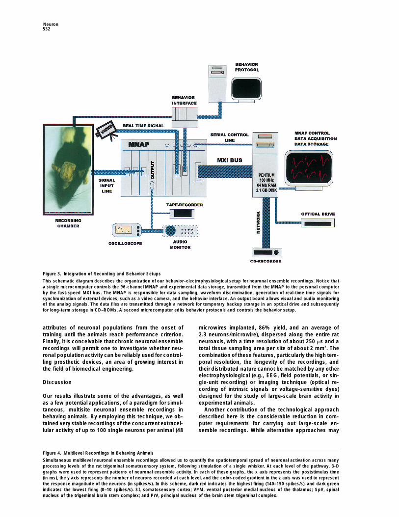
Figure 3. Integration of Recording and Behavior Setups
This schematic diagram describes the organization of our behavior–electrophysiological setup for neuronal ensemble recordings. Notice thata single microcomputer controls the 96-channel MNAP and experimental data storage, transmitted from the MNAP to the personal computerby the fast-speed MXI bus. The MNAP is responsible for data sampling, waveform discrimination, generation of real-time time signals forsynchronization of external devices, such as a video camera, and the behavior interface. An output board allows visual and audio monitoringof the analog signals. The data files are transmitted through a network for temporary backup storage in an optical drive and subsequentlyfor long-term storage in CD–ROMs. A second microcomputer edits behavior protocols and controls the behavior setup.
attributes of neuronal populations from the onset of microwires implanted, 86% yield, and an average of2.3 neurons/microwire), dispersed along the entire rattraining until the animals reach performance criterion.
Finally, it is conceivable that chronic neuronal ensemble neuroaxis, with a time resolution of about 250 ms and atotal tissue sampling area per site of about 2 mm2. Therecordings will permit one to investigate whether neu-
ronal population activity can be reliably used for control- combination of these features, particularly the high tem-poral resolution, the longevity of the recordings, andling prosthetic devices, an area of growing interest in
the field of biomedical engineering. their distributed nature cannot be matched by any otherelectrophysiological (e.g., EEG, field potentials, or sin-gle-unit recording) or imaging technique (optical re-Discussioncording of intrinsic signals or voltage-sensitive dyes)designed for the study of large-scale brain activity inOur results illustrate some of the advantages, as well
as a few potential applications, of a paradigm for simul- experimental animals.Another contribution of the technological approachtaneous, multisite neuronal ensemble recordings in
behaving animals. By employing this technique, we ob- described here is the considerable reduction in com-puter requirements for carrying out large-scale en-tained very stable recordings of the concurrent extracel-
lular activity of up to 100 single neurons per animal (48 semble recordings. While alternative approaches may
Figure 4. Multilevel Recordings in Behaving Animals
Simultaneous multilevel neuronal ensemble recordings allowed us to quantify the spatiotemporal spread of neuronal activation across manyprocessing levels of the rat trigeminal somatosensory system, following stimulation of a single whisker. At each level of the pathway, 3-Dgraphs were used to represent patterns of neuronal ensemble activity. In each of these graphs, the x axis represents the poststimulus time(in ms), the y axis represents the number of neurons recorded at each level, and the color-coded gradient in the z axis was used to representthe response magnitude of the neurons (in spikes/s). In this scheme, dark red indicates the highest firing (140–150 spikes/s), and dark greenindicates the lowest firing (0–10 spikes/s). SI, somatosensory cortex; VPM, ventral posterior medial nucleus of the thalamus; SpV, spinalnucleus of the trigeminal brain stem complex; and PrV, principal nucleus of the brain stem trigeminal complex.

Simultaneous, Multisite, Many Single Neuron Recordings533

Neuron534
Figure 5. Distributed Responses in the Rat Somatosensory Cortex
In each of the 3-D graphs depicted here, simultaneous neuronal ensemble recordings were employed to quantify the spatiotemporal spreadof cortical responses following the stimulation of a single whisker (identified on the top of the graph). The x axis represents poststimulus time,and the z axis represents response magnitude in spikes/s. All neurons were located in the infragranular layers of the primary somatosensorycortex.
require a large number of microcomputers to be syn- As required for any paradigm designed for the studyof neuronal ensembles, our recordings allow large sam-chronized (Wilson and McNaugton, 1994), in our para-
digm, a single personal computer is used to control ples of neurons to be monitored per animal. The use ofmicrowire arrays was fundamental to achieve this goal.the entire electrophysiological setup and store neuronal
spiking data, while a second microcomputer, interfaced While neuronal ensemble recordings have been ob-tained acutely in the past (Gerstein and Clark, 1964;to the MNAP, controls the behavior setup.

Simultaneous, Multisite, Many Single Neuron Recordings535
Figure 6. Recording Stability and Longevity
(A) Poststimulus time histograms depict the sensory responses of a set of six cortical neurons at two distinct times.(B) Microwires provided viable recordings for several weeks.
reviewed by Kruger, 1983), most studies aimed at ob- long-term chronic recordings. In fact, we and othershave recently demonstrated that by using microwiretaining chronic recordings of single units have employed
microwire bundles (Kubie, 1984; Shin and Chapin, 1990). arrays, reliable long-term neuronal ensemble recordingscan be obtained in many mammalian species currentlyThe main reason for this choice is the stability and reli-
able yield of single neurons that can be obtained with used in neurophysiology. Those include adult and youngferrets (Nicolelis et al., unpublished data), owl monkeysmicrowires. Here, data obtained with microwire arrays
further demonstrate that in regions of moderate cell (Nicolelis et al., 1996), and rhesus monkeys (Perepelkinand Schwartz, 1996).density, such as the cortex, thalamus, and trigeminal
brain stem complex, one does not need to employ more Many neuroscientists (Sherrington, 1906; Hebb, 1949;Lashley, 1950; Lilly, 1958; Erickson, 1968; Somjen, 1972;complex electrode configurations, such as tetrodes
(Wilson and McNaugton, 1994; Gray et al., 1995), for Freeman, 1975) foresaw that to understand how sensoryinformation is processed, converted into percepts,successfully carrying out neuronal ensemble record-
ings. The difficulty in manufacturing tetrodes, and the stored into memory, and then used to generate behav-iors, one needed to comprehend the physiological prin-fact that no group has so far provided quantitative data
to demonstrate that these electrodes maintain high neu- ciples that underlie large-scale interactions amongwidely distributed and interconnected populations ofronal yield (15–20 neurons per tetrode) for many weeks,
further justify the use of arrays of isolated microwires neurons. The available experimental evidence has sup-ported this view by indicating that most fundamentalas the most efficient solution for carrying out chronic
neuronal ensemble recordings. New multichannel elec- brain functions involve highly dynamic and distributedneuronal interactions (Freeman, 1983; Georgopoulos ettrode designs have recently appeared in the literature
(Hoogerwerf and Wise, 1994; Nordhausen et al., 1994), al., 1986, 1993; Nicolelis and Chapin, 1994; Wilson andMcNaugton, 1994; Fuster, 1995; Nicolelis et al., 1995;but there is still scant evidence to support their use in
Neuron536
previously implanted in the skull. The craniotomy was then sealedWelsh et al., 1995; Deadwyler et al., 1996). Therefore,with a layer of agar (4% in saline). Once the agar solidified, dentalthe results described here further support the notioncement was applied until it reached the top border of the plasticthat, as neuronal ensemble recordings are incorporatedconnector. This procedure was repeated until all arrays and bundles
into the arsenal of current neurophysiological tech- had been implanted. The end result of one of these implants isniques and explored to their full potential, we will have, illustrated in Figure 1D. To finish the procedure, the skin was loosely
sealed around the probes and the animal was transferred to a recov-at last, a realistic chance of reconstructing the neuronalery cage. The analgesic Buprenorphine (0.1–0.5 mg/kg S.C. everyengram, a task which, as put by Somjen (1972), has8 hr) was administered for 2–3 days while the animal recovered fromeluded us all.the surgery. Electrophysiological recordings started 5–7 days afterthe surgery. Experiments were carried out in a recording chamber
Experimental Procedures using anesthetized and freely behaving animals.
Microelectrode Design Instrumentation for Real-Time Ensemble RecordingsDifferent configurations of microelectrode matrices were specially Simultaneous recordings of large samples of single neurons weredesigned (NB Laboratories, Dennison, TX) to allow simultaneous carried out by an MNAP (Spectrum Scientific, Dallas; see Figure 3).recordings of the extracellular activity of large numbers of single In the most complete configuration, the MNAP allows simultaneousneurons, distributed across up to five distinct neuronal structures sampling from 96 microwires and discrimination of up to four individ-that define the rat somatosensory system. In all experiments de- ual action potential waveforms per wire, for a maximum of 384scribed here, stainless steel, Teflon-coated microwires (50 mm in recorded neurons. In this configuration, head stages (NBLABS, Den-diameter, California Fine wire) were used to build different micro- nison, TX), containing 16 field-effect transistors (FETs, Motorolaelectrode matrices, each of which was designed to maximize the MMBF5459) arranged in two rows of eight at the end of TVC-insu-number of neurons sampled throughout the spatial domain of differ- lated cables, are used to connect the 20-channel plastic connectorsent cortical and subcortical structures. For instance, microelectrode cemented in the animal’s head to the MNAP preamplifiers (Figurearrays, containing two parallel rows of eight microwires each, were 1D). In all recordings, the FETs were set as voltage followers withused for chronic implants in the rat primary (SI) somatosensory unit gain. The MNAP preamplifiers contain differential OP-Ampscortex. In these arrays, the distance between the microwire rows (gain 100; bandpass 100 Hz–16 kHz). Their output signals are trans-varied from 0.5–1 mm, and the distance betweenpairs of microwires mitted, through ribbon cables, to 96 A/C-coupled differential amplifi-in a row varied from 100–250 mm (Figure 1). Another commonly used ers on 6 input boards, each containing 16 amplifiers. Once in theconfiguration was the microelectrode bundle, containing either 8 or input boards, the analog signals pass through the first level of ampli-16 microwires staggered at different lengths, which were primarily fication (jumperable gain of 1, 10, or 20), are filtered (bandpass 400used to record from neurons in deep structures, such as the ventral Hz–8 kHz), and reach the final stage of amplification (programmableposterior medial nucleus of the thalamus and the different subnuclei multiplier stage, ranging from 1–30). These boards also include oneof the trigeminal brain stem complex. Arrays and bundles were built 12-bit analog-to-digital (A/D) converter per channel, which simulta-by first soldering the microwires to a 20-pin plastic connector, then neously digitizes the waveforms defining extracellular action poten-covering the connections with a thin layer of epoxy. Two ground tials at 40 kHz. After A/D conversion, the signals are routed to DSPwires were also soldered to free pins in the plastic connector. Finally, boards, each of which contain four digital signal processors (DSP,polyethylene glycol was used to mold the arrays and bundles and Motorola 5602) running at 40 MHz (instruction read at 20 MHz). Eachprovide them with enough stiffness to be implanted in the brain. DSP handles data from eight input channels and contains 32 K 24
bits of SRAM and 4 K 16-bit words of dual port SRAM memory. Atiming board is responsible for distributing timing and synchroniza-Surgical Procedure
Microwire arrays and bundles were implanted during stereotaxic tion signals to the entire MNAP. It also provides a digital time outputthat is used to synchronize external devices or to drive a video timersurgeries. For these procedures, rats were first anesthetized with a
single injection of pentobarbital sodium (50 mg/kg, I.P.) and then for a professional VCR used to store video records of the animal’sbehavior. The DSP boards also provide inputs for sampling digitaltransferred to a stereotaxic apparatus. Supplementary doses (one
third of the original) of pentobarbital were given when necessary. pulses generated from behavioral cages. A single host Pentiummicrocomputer (100–200 MHz, with 64 MB of RAM and 2.1 GB ofThe head was shaved and then cleaned with a betadine solution.
A single midsagittal incision of the skin was used to access the disk space) running a C11 software (SSCP, Spectrum Scientific,Dallas) in the Windows 3.11 operating system (Microsoft, Seattle)surface of the skull. Following retraction of soft tissue, the perios-
teum of the dorsal surface of the skull was completely removed, controls the MNAP over a serial line. Spike discrimination programsare downloaded from the PC host to the DSPs. Single spikes areand the bone surface was scrubbed with saline until no sign of
blood was left. The surface of the skull was then dried, and a series discriminated by combining a modified version (Nicolelis andChapin, 1994) of a principal component algorithm (Abeles andof small craniotomies were produced with a high speed dental drill
at the stereotaxic coordinates required for the implantation of elec- Goldstein, 1977), running in real-time, and one pair of time-voltagewindows per unit. The size and position of the time-voltage boxestrode arrays in the Vg, PrV, SpV, VPM, and SI cortex. A second
series of four to six craniotomies were open so that stainless steel are defined by the experimenter to isolate the waveforms that be-longed to a given unit. Spikes are only accepted as valid whenscrews could be firmly attached to the skull. These screws were
used for securing probes and for grounding purposes. Just before they pass through both boxes. During the experiment, the time ofoccurrence of each of the valid spikes for all 96 channels is trans-the implantation, microwires forming bundles were cut to the ideal
length, using a sharp pair of scissors, and then soaked in a supersat- ferred to the hard disk of the PC host through a parallel bus (MXI-Bus, National Instruments, CA), which can transfer 2 MB of data/s.urated solution of sucrose. Special care was taken not to allow the
sucrose to cover the microwire tips. After drying, the arrays and Digitized samples of the spike waveforms are also recorded periodi-cally and stored for off-line analysis using a visualization programbundles were mounted in the holder of a hydraulic micropositioner
(Kopf, Tujunga, CA) and subsequently slowly driven (z100 mm/min) (SpikeWorks, Spectrum Scientific, Dallas). Optical drives (1.2–2.4GB cartridges, Pinnacle Inc., CA) are used for temporary backup ofinto the brain. Although the 16-microwire arrays could be driven
through the dura most of the time, in some instances, a small slit the data files. These files are then transferred through an ethernet-based network to a computer server containing a CD-Recorderwas made in the dura overlying the SI cortex to facilitate the implant.
Single and multiunit activity were monitored continuously through- (PinnacleMicro, CA), which produces permanent records of the datain CD–ROMs. The MNAP also supplies options for analog backupout the surgery to help locate the position of each electrode. During
these recordings, the receptive field of each unit was qualitatively using 8–16 channel tape recorders. In all experiments, low thresholdtactile stimulation (step pulses of 100 ms in duration, delivered atcharacterized until the arrays were positioned in the structure of
interest. After reaching the final target, the ground wires of each 1–10 Hz) is provided by a computer-monitored Grass S8800 stimula-tor, which drives a vibromechanical probe. The digital outputs ofconnector were wrapped around one or two of the metal screws
Simultaneous, Multisite, Many Single Neuron Recordings537
the stimulator were also transmitted to the MNAP to be stored with Gray, C.M., Maldonado, P.E., Wilson, M., and McNaughton, B.(1995). Tetrodesmarkedly improve thereliability andyield of multiplethe spike data.single-unit isolation from multi-unit recordings in cat striate cortex.J. Neurosci. Methods 63, 43–54.Population Data Analysis
Neural ensemble data were processed off-line using a series of Hebb, D.O. (1949). The Organization of Behavior: ANeuropsycholog-graphics, statistical, and numerical analysis packages. The first level ical Theory. (New York: John Wiley and Sons, Inc).of analysis involved the production of single cell poststimulus time Hoogerwerf, A.C., and Wise, K.D. (1994). A three-dimensional micro-and cumulative histograms, which were used for defining the spatio- electrode array for chronic neural recording. IEEE Trans. Biomed.temporal receptive fields of single neurons. All of these preliminary Eng. 41, 1136–1146.steps of data analysis were carried out by using a spike train analysis
Kruger, J. (1983). Simultaneous individual recordings from manysoftware (Stranger, Biographics, Winston-Salem, NC) specially de-cerebral neurons: techniques and results. Rev. Physiol. Biochem.signed to handle large neuronal ensemble data sets. SpatiotemporalPharmacol. 98, 177–233.reconstructions of single neuronal receptive fields and populationKubie, J.L. (1984). A drivable bundle of microwires for collectingmaps were carried out by using a graphics interface software, devel-single-unit data from freely moving rats.Physiol. Behav. 32, 115–118.oped in C11, combined to MATLAB (Mathworks, MA). Finally, sta-Lashley, K.S. (1950). In search of the engram. Symp. Soc. Exp. Biol.tistical packages like CSS–Statistica (Statsoft, Tulsa, OK) and SPSS4, 454–482.(SPSS Inc., Chicago) were used for applying multivariate statistical
methods to the analysis of neuronal ensemble data. Lilly, J.C. (1958). Correlations between neurophysiological activityin the cortex and short-term behavior in the monkey. In Biological
Acknowledgments and Biochemical Bases of Behavior, H. F.Harlow, andC. N. Woolsey,eds. (Madison: The University of Wisconsin Press), pp. 83–100.
We thank Suzette Casal, Brett Carswell, and Kevin Tri Nguyen for Nicolelis, M.A.L., and Chapin, J.K. (1994). Spatiotemporal structuretheir invaluable technical support; Larry Andrews (NBLABS, Den- of somatosensory responses of many-neuron ensembles in the ratnison, TX) for manufacturing our microwire arrays, bundles, and ventral posterior medial nucleus of the thalamus. J. Neurosci. 14,head sets; Harvey Wiggins (Spectrum Scientific, Dallas) for the de- 3511–3532.sign and continuous support of our MNAP systems; Alexander Kiri-
Nicolelis, M.A.L., Baccala, L.A., Lin, R.C.S., and Chapin, J.K. (1995).lov (Biographics, Winston-Salem, NC) for the support of Stranger;
Sensorimotor encoding by synchronous neural ensemble activity atand Erika Fanselow for photographic work and comments on the
multiple levels of the somatosensory system. Science 268, 1353–manuscript. We are particularly indebted to John K. Chapin, a pio-
1358.neer in the field of neuronal ensemble recordings, for his continuous
Nicolelis, M.A.L., Carswell, B., Oliveira, L.M.O., Ghazanfar, A.A.,suggestions and advice. M. A. L. N. also thanks professor GyorgyChapin, J.K., Lin, R.C.S., Nelson, R.J., and Kaas, J.H. (1996). Long-M. Bohm for being a continuous source of motivation. This workterm simultaneous recordings of neuronal ensembles across multi-was supported by grants from the National Institute of Dental Re-ple cortical areas in behaving primates. Soc. Neurosci. (Summ.) 3,search (DE-111121–01), the Whitehall Foundation, the McDonnell2023.Pew Foundation, the Klingenstein Foundation, the Duke-SandozNordhausen, C.T., Rousche, P.J., and Normann, R.A. (1994). Opti-program, and a Whitehead Scholar Award to M. A. L. N.mizing recording capabilities of the Utah intracortical electrode.Brain Res. 637, 27–36.Received November 5, 1996; revised February 25, 1997.Perepelkin, P.D., and Schwartz, A.B. (1996). Simultaneous popula-tions of single-cell activity recorded bilaterally in primate motorReferencescortex. Soc. Neurosci. (Summ.) 3, 2022.
Abeles, M., and Goldstein, M. (1977). Multispike train analysis. IEEE Sherrington, C.S. (1906). The Integrative Action of the Nervous Sys-Proc. 65, 762–773. tem. (New Haven, Connecticut: Yale University Press).Adrian, E.D. (1926). The impulses produced by sensory nerve end- Shin, H.-C., and Chapin, J.K. (1990). Modulation of afferent transmis-ings. Part I. J. Physiol. (Lond.) 61, 49–72. sion to single neurons in the ventroposterior thalamus during move-
ments in rats. Neurosci. Lett. 108, 116–120.Barlow, H. (1995). The neuron doctrine in perception. In The Cogni-tive Neurosciences, M. S. Gazzaniga, ed. (Cambridge, Massachu- Somjen, G. (1972). Sensory coding in the mammalian nervous sys-setts: MIT Press), pp. 415–436. tem. (New York: Appleton-Century-Crofts).
Deadwyler, S.A., Bunn, T., and Hampson, R.E. (1996). Hippocampal Vaadia, E., Haalman, I., Abeles, M., Bergman, H., Prut, Y., Slovin,ensemble activity during spatial delayed-nonmatch-to-sample per- H., Aertsen, A. (1995). Dynamics of neuronal interactions in monkeyformance in rats. J. Neurosci. 16, 354–372. cortex in relation to behavioural events. Nature 373, 515–518.
de Charms, R.C., Merzenich, MM. (1996). Primary cortical represen- Welsh, J.P., Lang, E.J., Suglhara, I., and Llinas, R. (1995). Dynamictation of sounds by the coordination of action-potential timing. Na- organization of motor control within the olivocerebellar system. Na-ture 381, 610–613. ture 374, 453–457.
Wilson, M.A., and McNaugton, B.L. (1994). Reactivation of hippo-Erickson, R.P. (1968). Stimulus coding in topographic and non-topo-campal ensemble memories during sleep. Science 265, 676–679.graphic afferent modalities: on the significance of the activity of
individual sensory neurons. Psych. Rev. 75, 447–465.
Freeman, W.J. (1975). Mass action in the nervous system. (NewYork: Academic Press).
Freeman, W.J. (1983). The physiological basis of mental images.Biol. Psych. 18, 1107–1125.
Fuster, J.M. (1995). Memory in the cerebral cortex. (Cambridge,Massachusetts: MIT Press).
Georgopoulos, A.P., Schwartz, A.B., and Ketter, R.E. (1986). Neu-ronal population coding of movement direction. Science 233, 1416–1419.
Georgopoulos, A.P., Taira, M., and Lukashin, A. (1993). Cognitiveneurophysiology of the motor cortex. Science 260, 47–52.
Gerstein, G.L., and Clark W.A. (1964). Simultaneous study of firingpatterns in several neurons. Science 143, 1325–1327.