Molecular, Topographic, and Functional Organization of the Cerebellar Cortex: A Study with Combined...
15
Behavioral/Systems/Cognitive Molecular, Topographic, and Functional Organization of the Cerebellar Cortex: A Study with Combined Aldolase C and Olivocerebellar Labeling Izumi Sugihara and Yoshikazu Shinoda Department of Systems Neurophysiology, Tokyo Medical and Dental University, Graduate School of Medicine, Tokyo 113-8519, Japan Aldolase C (zebrin) expression in Purkinje cells reveals stripe-shaped compartments in the cerebellar cortex. However, it is not clear how these compartments are related to cerebellar functional localization. Therefore, we identified olivocerebellar projections to aldolase C compartments by labeling climbing fibers with biotinylated dextran injected into various small areas within the inferior olive in rats. Specific rostral and caudal aldolase C compartments were linked in an orderly manner by common olivocerebellar projection across the rostrocaudal boundary on lobule VIc-crus Ib. Based on the localization of the olivary origins of projection to similar compartments, the compartments and olivocerebellar projections could be sorted into five groups: group I, positive compartments extending from the posterior lobe to the anterior lobe innervated by the principal olive and some neighboring areas; group II, positive compartments localized within the posterior lobe innervated by several medial subnuclei; group III, vermal and central negative compartments inner- vated by the centrocaudal medial accessory olive; group IV, negative and lightly positive compartments in the hemisphere and the rostral and caudal pars intermedia innervated by the dorsal accessory olive and some neighboring areas; group V, the flocculus and nodulus. The olivocerebellar topography within each group was simple and suggests an “orientation axis” within the concerned parts of the inferior olive. Furthermore, parts of the inferior olive in each group receive specific afferent inputs, indicating a close relationship between aldolase C compartments and functional localization. Thus, the five-group scheme we propose here may integrate the molecular, topo- graphic, and functional organization of the cerebellum. Key words: climbing fiber; inferior olive; cerebellum; biotinylated dextran amine; zebrin; rat Introduction Neuronal activities related to specific behaviors can be recorded from distinct areas in the cerebellar cortex, suggesting the pres- ence of a fine functional map. However, it is not clear how the cerebellar cortex is organized to give such fine functional local- ization. The cerebellar cortex shows both transverse organization based on its lobulation and longitudinal organization in which three subdivisions, the vermis, pars intermedia, and hemisphere, were originally recognized (Brodal, 1981). The longitudinal or- ganization has been further elaborated by the demonstration of zones A–D (Voogd, 1967; Groenewegen and Voogd, 1977; Buisseret-Delmas and Angaut, 1993) and even much narrower “microzones” (Andersson and Oscarsson, 1978), which are based on topographic corticonuclear Purkinje cell projection and olivocortical climbing fiber projection. Recent analyses have shown that the axon of an inferior olivary neuron provides mul- tiple climbing fibers to a single narrow longitudinal band-shaped area comparable in size to a cortical microzone and sends collat- erals to a small area in the cerebellar nucleus (Sugihara et al., 1999, 2001). In contrast, a single lateral reticular nucleus neuron branches to form multiple mossy fibers that project to several longitudinal band-shaped areas (Wu et al., 1999). Although these single-axon projection patterns are likely to be directly related to cerebellar functional localization, their organization across the cerebellum has not been addressed in previous studies. In addition to axonal projections, the fundamental organiza- tion of the cerebellar cortex may be revealed in the stripe-shaped expression patterns of several marker molecules for Purkinje cell populations (Hawkes and Leclerc, 1987; Brochu et al., 1990; Bailly et al., 1995; Herrup and Kuemerle, 1997). Among these molecules is zebrin II (Brochu et al., 1990; Hawkes, 1997), which has been shown to be aldolase C (Ahn et al., 1994). Although several studies have shown parallels between zebrin compart- ments and the climbing and mossy fiber projection patterns in some areas of the cerebellar cortex (Gravel et al., 1987; Matsushita et al., 1991; Voogd and Ruigrok, 1997; Voogd et al., 2003), a systematic comparison throughout the cerebellar cortex has not yet been made. Thus, the significance of zebrin compartments in terms of cerebellar morphology and physiology is not yet clear. In the present study, we first formed a comprehensive two- dimensional map of the aldolase C compartments in the entire rat Received May 20, 2004; revised Aug. 25, 2004; accepted Aug. 27, 2004. This study was supported by the following: the Human Frontier Science Program (Y.S.); Grants-in-Aid for Scien- tific Research from the Japan Society for the Promotion of Science (I.S. and Y.S.); Grants-in-Aid for Scientific Research on Priority Areas from The Ministry of Education, Culture, Sports, Science and Technology, Japan (I.S. and Y.S.); a grant from the Japan Space Forum (Y.S.); and a grant of the 21st Center of Excellence Program. We thank Drs. Y. Kubo and T. Misaka for help in antibody development and Western blotting and Dr. E. Lang for reading this manuscript. Correspondence should be addressed to Dr. Yoshikazu Shinoda, Department of Systems Neurophysiology, Tokyo Medical and Dental University, Graduate School of Medicine, 1-5-45 Yushima, Bunkyo-ku, Tokyo 113-8519, Japan. E-mail: [email protected]. DOI:10.1523/JNEUROSCI.1961-04.2004 Copyright © 2004 Society for Neuroscience 0270-6474/04/248771-15$15.00/0 The Journal of Neuroscience, October 6, 2004 • 24(40):8771– 8785 • 8771
-
Upload
independent -
Category
Documents
-
view
0 -
download
0
Transcript of Molecular, Topographic, and Functional Organization of the Cerebellar Cortex: A Study with Combined...

Behavioral/Systems/Cognitive
Molecular, Topographic, and Functional Organization of theCerebellar Cortex: A Study with Combined Aldolase C andOlivocerebellar Labeling
Izumi Sugihara and Yoshikazu ShinodaDepartment of Systems Neurophysiology, Tokyo Medical and Dental University, Graduate School of Medicine, Tokyo 113-8519, Japan
Aldolase C (zebrin) expression in Purkinje cells reveals stripe-shaped compartments in the cerebellar cortex. However, it is not clear howthese compartments are related to cerebellar functional localization. Therefore, we identified olivocerebellar projections to aldolase Ccompartments by labeling climbing fibers with biotinylated dextran injected into various small areas within the inferior olive in rats.Specific rostral and caudal aldolase C compartments were linked in an orderly manner by common olivocerebellar projection across therostrocaudal boundary on lobule VIc-crus Ib. Based on the localization of the olivary origins of projection to similar compartments,the compartments and olivocerebellar projections could be sorted into five groups: group I, positive compartments extending from theposterior lobe to the anterior lobe innervated by the principal olive and some neighboring areas; group II, positive compartmentslocalized within the posterior lobe innervated by several medial subnuclei; group III, vermal and central negative compartments inner-vated by the centrocaudal medial accessory olive; group IV, negative and lightly positive compartments in the hemisphere and the rostraland caudal pars intermedia innervated by the dorsal accessory olive and some neighboring areas; group V, the flocculus and nodulus. Theolivocerebellar topography within each group was simple and suggests an “orientation axis” within the concerned parts of the inferiorolive. Furthermore, parts of the inferior olive in each group receive specific afferent inputs, indicating a close relationship betweenaldolase C compartments and functional localization. Thus, the five-group scheme we propose here may integrate the molecular, topo-graphic, and functional organization of the cerebellum.
Key words: climbing fiber; inferior olive; cerebellum; biotinylated dextran amine; zebrin; rat
IntroductionNeuronal activities related to specific behaviors can be recordedfrom distinct areas in the cerebellar cortex, suggesting the pres-ence of a fine functional map. However, it is not clear how thecerebellar cortex is organized to give such fine functional local-ization. The cerebellar cortex shows both transverse organizationbased on its lobulation and longitudinal organization in whichthree subdivisions, the vermis, pars intermedia, and hemisphere,were originally recognized (Brodal, 1981). The longitudinal or-ganization has been further elaborated by the demonstration ofzones A–D (Voogd, 1967; Groenewegen and Voogd, 1977;Buisseret-Delmas and Angaut, 1993) and even much narrower“microzones” (Andersson and Oscarsson, 1978), which are basedon topographic corticonuclear Purkinje cell projection andolivocortical climbing fiber projection. Recent analyses have
shown that the axon of an inferior olivary neuron provides mul-tiple climbing fibers to a single narrow longitudinal band-shapedarea comparable in size to a cortical microzone and sends collat-erals to a small area in the cerebellar nucleus (Sugihara et al.,1999, 2001). In contrast, a single lateral reticular nucleus neuronbranches to form multiple mossy fibers that project to severallongitudinal band-shaped areas (Wu et al., 1999). Although thesesingle-axon projection patterns are likely to be directly related tocerebellar functional localization, their organization across thecerebellum has not been addressed in previous studies.
In addition to axonal projections, the fundamental organiza-tion of the cerebellar cortex may be revealed in the stripe-shapedexpression patterns of several marker molecules for Purkinje cellpopulations (Hawkes and Leclerc, 1987; Brochu et al., 1990;Bailly et al., 1995; Herrup and Kuemerle, 1997). Among thesemolecules is zebrin II (Brochu et al., 1990; Hawkes, 1997), whichhas been shown to be aldolase C (Ahn et al., 1994). Althoughseveral studies have shown parallels between zebrin compart-ments and the climbing and mossy fiber projection patterns insome areas of the cerebellar cortex (Gravel et al., 1987; Matsushitaet al., 1991; Voogd and Ruigrok, 1997; Voogd et al., 2003), asystematic comparison throughout the cerebellar cortex has notyet been made. Thus, the significance of zebrin compartments interms of cerebellar morphology and physiology is not yet clear.
In the present study, we first formed a comprehensive two-dimensional map of the aldolase C compartments in the entire rat
Received May 20, 2004; revised Aug. 25, 2004; accepted Aug. 27, 2004.This study was supported by the following: the Human Frontier Science Program (Y.S.); Grants-in-Aid for Scien-
tific Research from the Japan Society for the Promotion of Science (I.S. and Y.S.); Grants-in-Aid for Scientific Researchon Priority Areas from The Ministry of Education, Culture, Sports, Science and Technology, Japan (I.S. and Y.S.); agrant from the Japan Space Forum (Y.S.); and a grant of the 21st Center of Excellence Program. We thank Drs. Y. Kuboand T. Misaka for help in antibody development and Western blotting and Dr. E. Lang for reading this manuscript.
Correspondence should be addressed to Dr. Yoshikazu Shinoda, Department of Systems Neurophysiology, TokyoMedical and Dental University, Graduate School of Medicine, 1-5-45 Yushima, Bunkyo-ku, Tokyo 113-8519, Japan.E-mail: [email protected].
DOI:10.1523/JNEUROSCI.1961-04.2004Copyright © 2004 Society for Neuroscience 0270-6474/04/248771-15$15.00/0
The Journal of Neuroscience, October 6, 2004 • 24(40):8771– 8785 • 8771

cerebellar cortex from serial sections. We then carefully mappedolivocerebellar climbing fibers, labeled by a small injection of ananterograde tracer into various areas of the inferior olive, ontothe aldolase C map generated from the same rat. A small injectionwas expected to label adjacent olivary neurons tightly coupledelectrotonically (Llinas et al., 1974) and functionally related. Thetopographic olivocerebellar projection pattern and aldolase C com-partments were then compared to reveal the fundamental functionaland morphological organization of the cerebellar cortex.
Materials and MethodsPreparation of polyclonal antibody against aldolase C. A peptide (CGAA-TEEFIKRAEMNGLAAQGKYE) that consisted of the amino acid se-quence 322–344 of rat aldolase C (Mukai et al., 1991), plus a cysteineresidue at the N terminus to facilitate conjugation to the carrier proteinKLH, was synthesized and purified. This peptide was used for immuni-zation, an ELISA test, affinity purification of antisera, and a blocking testin immunohistochemistry. Two rabbits (69075 and 69076) were immu-nized by five subcutaneous injections of 0.2 mg of the peptide conjugatedwith KLH at 2, 3, 2, and 2 week intervals. The rabbits were exsanguinated2 weeks after the final boost. Crude antisera of both rabbits were affinitypurified against the immobilized immunizing peptide. Some purifiedantibody of 69076 was conjugated with biotin. The above procedureswere performed by Sawady Technologies (Itabashi, Tokyo, Japan).
Western blot analysis. To test the specificity of the affinity-purifiedantibody, the whole cerebellum was dissected out from a Long–Evansmale adult rat and then homogenized and sonicated in ice-cold PBS plusa protease inhibitor mixture (Complete; Roche, Basel, Switzerland). Pro-tein concentration was determined by the Lowry method. Samples of 30�g of proteins were separated by electrophoresis on 7.5 and 12% SDS-polyacrylamide gels and electrotransferred to polyvinylidene difluoridemembranes (Immobilon-P; Millipore, Billerica, MA). Immunoblotswere probed with purified antibodies against aldolase C (125–250 ng/ml;1 hr at room temperature) in a blocking solution containing 5% nonfatskim milk (Difco, Detroit, MI) in 20 mM Tris HCl, pH 8.0, 150 mM NaCl,and 0.05% Tween 20 and were subsequently treated with donkey anti-rabbit IgG horseradish peroxidase-conjugated secondary antibodyF(ab�)2 fragment (1:2000 dilution; 1 hr at room temperature; NA9340;Amersham Biosciences, Little Chalfont, Buckinghamshire, UK). Detec-tion was performed using chemiluminescence of 4-chloro-1-naphthol(ECL plus RPN 2132; Amersham Biosciences) according to the manu-facturer’s instructions. The membranes were then exposed to instant andpeel-apart film (667; Polaroid, Waltham, MA).
Tracer injection and histological procedure. Fifty-four and 10 Long–Evans adult rats were used for anterograde and retrograde labeling ofolivocerebellar projections by tracer injections into the inferior olive andcerebellar cortex, respectively, followed by secondary labeling of aldolaseC. Twelve Long–Evans adult rats were used only for labeling aldolase C.All of the experimental animals in this study were treated according to theguiding principles for the care and use of animals in the field of physio-logical sciences of the Japanese Physiological Society (2001, 2002), andthe experimental protocols were approved by the Institutional AnimalCare and Use Committee of Tokyo Medical and Dental University (no.0020238). The methods of anesthesia, surgery, and histological proce-dures were the same as those described previously (Sugihara et al., 1999,2001). Biotinylated dextran amine (BDA; D-1956; 10,000 molecularweight; Molecular Probes, Eugene, OR) was pressure injected at variouslocations in the inferior olive (�0.004 �l of 10% solution in saline) foranterograde labeling. Two injections were made in the left and right oliveto facilitate the study by using nearly exclusively contralateral projection.The injected tracer spread to a diameter of 0.1– 0.2 mm. Serial frozensections (80 �m thick) were cut from the entire cerebellum and medullahorizontally in seven rats and coronally in the others. After the sectionswere incubated with biotinylated peroxidase–avidin complex (PK6100Elite ABC kit; Vector Laboratories, Burlingame, CA), BDA was visualizedin black by incubating samples with diaminobenzidine (0.5 mg/ml), glu-cose oxidase (0.01 mg/ml; type II, G-6125; Sigma, St. Louis, MO), nickelammonium sulfate (2 mg/ml), ammonium chloride (4 mg/ml), and
�-D(�)-glucose (2 mg/ml) in Tris buffer (50 mM, pH 7.4) for 30 – 60 minand washed with PBS. For retrograde labeling, larger amounts of BDA(�0.01– 0.02 �l) were injected bilaterally into lobules IX, VII, VIII, andcrus II. Histological procedures for retrograde labeling were the same asthose for anterograde labeling.
Immunohistological labeling of aldolase C. Brain sections either after orwithout BDA labeling were incubated with anti-aldolase C antibody (10ng/ml purified 69076 or 20 ng/ml purified 69075) in 2% normal goatserum and PBS with 0.15% Triton X-100 (PBST) for 48 –72 hr and thenincubated with biotinylated anti-rabbit IgG antibody (BA-1000; VectorLaboratories) in PBST with 2% normal goat serum for 6 –12 hr. In laterexperiments, these two steps were replaced by a single step of incubationwith biotin-conjugated anti-aldolase C antibody (320 ng/ml 69076) inPBST with normal rabbit serum for 48 –72 hr. After being washed, thesections were incubated with biotinylated peroxidase–avidin complex(PK6100 Elite ABC kit; Vector Laboratories) for 4 – 8 hr. When BDAlabeling was performed, the sections were then incubated without heavymetal ions to stain the diaminobenzidine reaction product a brownishcolor in contrast to the black BDA reaction product. For experiments inwhich no BDA injection was made, the incubation solution containednickel ammonium sulfate (200 mg/ml) in Tris buffer (50 mM) to stain thediaminobenzidine reaction product black. Sections were mounted onglass slides. Some sections were counterstained with thionine. Sectionswere coverslipped with Permount (Fisher Scientific, Fair Lawn, NJ).Stained axons and labeled aldolase C zones were photographed using adigital camera (DP-50; Olympus, Tokyo, Japan) attached to a micro-scope (BX41; Olympus). Photographs were assembled using PhotoshopLE software (Adobe, San Jose, CA). The software was used to adjustcontrast and brightness, but no digital enhancements were applied.
Tracing the continuity of aldolase C zones in serial sections. Coronal,horizontal, and parasagittal sections stained with anti-aldolase C anti-body were photographed with a film scanner (Dimage AF-5000; Minolta,Osaka, Japan) at a resolution of 4800 pixels per inch. A graphics file of thefirst section was opened in the bottom layer of graphics software (Illus-trator 10; Adobe). Lines were superimposed in the top layer on the bor-ders of aldolase C compartments and cortical outlines. The graphics fileof the second section was then opened in the intermediate layer. Theposition and orientation of the second section was adjusted to obtain abest fit to the drawing of the first section in the top layer. The continuityof the compartments could be reliably judged by carefully comparingtheir positions in two consecutive sections. This procedure was repeatedthroughout the cerebellum.
Mapping climbing fiber locations on the unfolded cerebellar cortex. Be-cause almost all olivocerebellar axons project contralaterally (Sugihara etal., 1999), projections from the left and right injections were analyzedseparately. Ipsilateral projections, encountered infrequently, were ig-nored. A previous standardized unfolded representation of the Purkinjecell layer of the whole cerebellar cortex (Sugihara et al., 2001) was mod-ified by incorporating the aldolase C labeling pattern. The mediolateraldistance for a given compartment was measured at the apex of eachfolium from the midline along the cerebellar surface. For the sake ofsimplicity, the changes in the mediolateral positions of the compart-ments attributable to foliation of the cerebellum were ignored. To plotlabeled climbing fibers, the aldolase C compartment in which they weredistributed was carefully identified by following it through serial sec-tions. The relative mediolateral position of the labeled climbing fiber in agiven aldolase C compartment and the relative rostrocaudal position ofthe fiber within the folium were then measured to plot the climbing fiberon the unfolded representation of the cerebellar cortex.
The procedure for mapping injection sites in the horizontal represen-tation of the inferior olive from brains cut coronally has been describedpreviously (Sugihara et al., 2001). In brains cut horizontally, the contourof the inferior olive, the cut end of the medulla, the midline, and theinjection spots were drawn using a camera lucida apparatus from each ofthe serial sections. The drawings were superimposed on each other toidentify the contour of the entire inferior olive and the positions of theinjection sites. We used the nomenclature of the subnuclei of the inferiorolive proposed by Ruigrok and Cella (1995). A solid representation of theinferior olive was produced from camera lucida drawings using three-
8772 • J. Neurosci., October 6, 2004 • 24(40):8771– 8785 Sugihara and Shinoda • Aldolase C and Olivocerebellar Projection

dimensional graphics software (Rhinoceros; Robert McNeel & Associ-ates, Seattle, WA).
In our figures, all brain slices and drawings for which injections weremade into the right inferior olive were reversed so that all climbing fiberswere plotted in the right cerebellar cortex.
ResultsAldolase C antibodiesAntibodies were affinity purified from antisera obtained fromtwo rabbits (69075 and 69076). These antisera and purified anti-bodies showed high reactivity in an ELISA test with the peptide.Western blotting of two purified antibodies showed that theyspecifically label a single identical band of protein with a molec-ular mass of 39 kDa (Fig. 1A), similar to that of mouse aldolase C(36 kDa) (Ahn et al., 1994).
Histochemistry with these antisera and antibodies labeled asubpopulation of Purkinje cells located in the same pattern of
multiple longitudinal compartments as with zebrin II antibody(Brochu et al., 1990) in all levels of the cerebellum (Fig. 1D–F).The addition of the antigen peptide to the solution of anti-aldolase C antibodies blocked the labeling in a concentration-dependent manner (Fig. 1B). Under high magnification, labelingwas present in the somata, dendrites (Fig. 1C), axons, and axonalterminals (data not shown) of a population of Purkinje cells. Thislabeling was the same as that with zebrin II antibody (Brochu etal., 1990). These results indicated that the present two anti-aldolase C antibodies label the same compartments as zebrin IIantibody, as expected from the fact that aldolase C is zebrin II(Ahn et al., 1994).
Organization of reconstructed aldolase C compartmentsBecause of the deep foliation of the cerebellar cortex, it is difficultto accurately determine the arrangement of the striped areas
Figure 1. Specificity and labeling properties of anti-aldolase C antibodies. A, Western blots using antibodies against aldolase C. In comparing Coomasie staining of standard proteins (Prestainedprecision protein standards; 162-0372; Bio-Rad, Hercules, CA) and sample proteins of the cerebellum, a single identical band (39 kDa) was recognized in the Western blots of sample proteins bypurified antibodies 69075 (b; 250 ng/ml) and 69076 (c; 125 ng/ml). The results with 12% SDS-polyacrylamide gel are shown here. B, Concentration-dependent blocking by the antigen peptide ofimmunohistological labeling of populations of Purkinje cells located in multiple zones in lobules VIII and IX. Adjacent coronal sections were incubated with one of the primary antibodies againstaldolase C (20 ng/ml 69705, top row; 10 ng/ml 69076, bottom row) and various concentrations (51.2, 12.8, 3.2, 0.8, and 0 �g/ml; from left to right) of the peptide. C, High-magnificationphotomicrograph of a labeled Purkinje cell. The somata and dendrites as well as axons and axonal terminals are labeled by the anti-aldolase C antibody. D–F, Photomicrographs of coronal sectionsof the cerebellum at the caudal ( D), intermediate ( E), and rostral ( F) levels labeled with the antibody 69076. Lines show the continuity of positive compartments across folia. I–X, Lobules I–X; a– c,sublobules a– c; CP, copula pyramidis; Cr I, crus I of ansiform lobule; CrII, crus II of the ansiform lobule; sim, simple lobule.
Sugihara and Shinoda • Aldolase C and Olivocerebellar Projection J. Neurosci., October 6, 2004 • 24(40):8771– 8785 • 8773

(“compartments”) defined by the expression of aldolase C or anyother molecular marker. Thus, complete reconstruction of themolecular compartments was accomplished only for the medialvermis (Dore et al., 1990; Ozol et al., 1999). For other areas of thecerebellum, data are available only for the superficial cortex(Hawkes and Leclerc, 1987; Brochu et al., 1990; Voogd et al.,2003; Voogd and Ruigrok, 2004), leaving the complete compart-ment pattern unidentified especially in the transitional region ofthe rostral and caudal cerebellum in simple lobule and crus I–II.Therefore, we carefully traced all aldolase C compartmentsthrough serial coronal sections (six rats) in the entire cerebellarcortex including the depths of the folia (see Materials and Meth-ods). Because each compartment abruptly shifts laterally in thesimple lobule and in crus Ia, compartments were also traced inhorizontal (five rats) and parasagittal (one rat) sections for accu-racy. Ignoring minor variations, the pattern of the major com-partments was consistent among different individuals. Figure 2shows a representative compartment pattern in the scheme of theunfolded cerebellar cortex.
We basically adopted the original numeric (Hawkes andLeclerc, 1987) and additional alphabetic nomenclatures of aldo-lase C compartments (Voogd et al., 2003; Voogd and Ruigrok,2004). A plus sign was added for positive compartments. A neg-ative compartment was designated by adding a minus sign to thename of the positive compartment located immediately mediallyto that negative compartment. To refer to a compartment, com-partment 1� was abbreviated to 1�, for example. Although thebasic organization of aldolase C (zebrin II) compartments hasbeen reported (Voogd et al., 2003; Voogd and Ruigrok, 2004), theadditional characteristics of the aldolase C compartments re-vealed in the present study are described below.
The aldolase C compartments were not oriented exactly par-allel to the midline, but instead ran in a laterocaudal direction inlobules I–VI and in a laterorostral direction in lobules VII–IX(Fig. 2). A hypothetical transverse line, designated as the “rostro-caudal boundary” (Fig. 2, dotted transverse line), could be drawnat the peak of the lateral expansion of positive compartments fromcaudal lobule VIc through crus Ib. At this line, all aldolase C-negativecompartments disappeared, and the positive compartments mergedinto one transverse compartment. We use this boundary to define a“rostral” and a “caudal” cerebellar cortex. Although the major com-partments in the rostral and caudal cerebellar cortex have beengiven the same numeric names (1�, 2�, and so on) (Hawkes andLeclerc, 1987), our map shows that they do not necessarily belongto the same continuous compartment (Fig. 2).
In the rostral cerebellar cortex, aldolase C-positive compart-ments could be classified into three types. The first type includedthose that were heavily labeled and continuous nearly through-out the rostral cerebellar cortex (1�, 2�, 4�, 5�, and 6�). Thesecond type included those that were also heavily labeled butlocated mainly caudal to the primary fissure (Fig. 2, pf) (a�, 2b�,c�, and d�). The third type included lightly labeled compart-ments (Fig. 1F) located mainly rostral to the primary fissure (b�,3�, and 3b�). Among these positive compartments, 2b� and3b� were newly defined and temporarily designated as such inthis study. The most lateral part of original 2� in lobule VIa-c(2b�) was distinguished from 2� because it was separated from2� by a narrow negative compartment (designated here as 2a�)and 2� and 2b� received different projections, as described laterhere. Compartment 3b� was recognized between 3� and 4� inlobules IV–V (Fig. 1F). An additional positive compartment of-ten seen between d� and 4� was also regarded as d� here (Fig.1E). Although a faintly labeled compartment was seen between
4� and 5� (not specifically designated), all the area betweenrostral 4� and rostral 5� was regarded as rostral 4� in this study.All labeling tended to be slightly weaker in the simple lobule thanin other areas.
In the caudal cerebellar cortex, 4b� and 5a� ended in therostral paramedian lobule, and lightly labeled compartments(e�) (Voogd et al., 2003) were seen in the caudal paramedianlobule and copula pyramidis between 4� and 5�. In this area, wetemporarily divided the original e� into e1�, e1�, and e2�,because the center of e� was slightly more faintly labeled than themedial and lateral parts (Fig. 1D) and the olivocerebellar projec-
Figure 2. Reconstruction of aldolase C compartments throughout the cerebellar cortex. Con-tinuity of the compartments in the rostrocaudal direction was determined by accurate tracing ofindividual compartments on serial sections and then plotted on a map of the unfolded cerebellarcortex (see Materials and Methods). Several new compartments were discovered (marked byasterisks prefixed to their proposed names) beyond those identified previously by Hawkes andLeclerc (1987), Voogd et al. (2003), and Voogd and Ruigrok (2004). A positive compartment andits laterally neighboring negative compartment usually have the same name with differentsuffixes (�, positive compartment; �, negative compartment). The intensity of the gray colorapproximately corresponds to the intensity of labeling. The dotted line on the left indicates therostrocaudal boundary of the compartment pattern (see Results). Arrowheads indicate areas inwhich the molecular layer is absent, one of which in the caudal apex of lobule VIc-crus I approx-imately coincided with the rostrocaudal border. I–X, Lobules I–X; a– c, sublobules a– c; CP,copula pyramidis; Cr I, crus I of ansiform lobule; Cr II, crus II of ansiform lobule; DPFL, dorsalparaflocculus; FL, flocculus; Par, paramedian lobule; pf, primary fissure; Sim, simple lobule;VPFL, ventral paraflocculus.
8774 • J. Neurosci., October 6, 2004 • 24(40):8771– 8785 Sugihara and Shinoda • Aldolase C and Olivocerebellar Projection

tion pattern fit better with these smaller divisions (see below). Alightly labeled narrow compartment (designated f�) was recog-nized immediately lateral to 4� with a narrow gap of a negativearea (4�) in lobule VIII– copula pyramidis (Fig. 1D). The origi-nal compartment 4� in crus I, II, and the rostral paramedianlobule seemed to be distinct from 4� in the caudal paramedianlobule and copula pyramidis in terms of olivocerebellar projec-tion (see below). Therefore, the former was temporarily desig-nated as 4a�, because it was medial to 4b�.
A specific pair of aldolase C compartments in the rostral andcaudal cerebellum is linked througholivocerebellar projectionTo compare the olivocerebellar projection patterns with aldolaseC compartments, we labeled climbing fibers by small injections of
BDA into individual subnuclei of the inferior olive (unilateralinferior olive injections in 23 animals and bilateral injections in31 animals) and mapped the labeled climbing fibers to the iden-tified aldolase C compartments. The diameter of spread from aninjection site was �0.2 mm (Fig. 3B,C) and usually resulted inlabeled climbing fibers being distributed to one or more segmentsof a single narrow longitudinal band with no (n � 31 of 85 injec-tions) or �10% (n � 22) of labeled climbing fibers located out-side the main band. We initially used the results from these ex-periments while simply ignoring the “outside” climbing fibers.For the remaining injections (n � 32), �10% of labeled climbingfibers were located outside the main termination band. However,the banding patterns from these outside distributions could usu-ally be explained by spread of the tracer into adjacent but distinctareas in the inferior olive and correspondence of the aldolase C
Figure 3. Narrow band-shaped longitudinal olivocerebellar projection connects corresponding rostral and caudal aldolase C compartments. A, Photomicrographs of several coronal sectionsshowing that the narrow band of the distribution of labeled climbing fibers (asterisks) is parallel with and belongs to an aldolase C compartment. The brackets in each panel indicate positivecompartments. All labeled climbing fibers were located in the medial portion of rostral compartment 1� or caudal compartment 1�, hereafter classified as 1�(med)//1� (see Results). B, C,Photomicrograph ( B) and plot of the injection site in the inferior olive (C; horizontal representation of the left inferior olive) (the same experiment as shown in A). D, Distribution of all labeled climbingfibers in the experiment shown in A (red) and three other experiments (green, blue, violet) plotted in an unfolded representation of the cerebellar cortex. The colored squares indicate locations ofindividual labeled climbing fibers. The colored circles indicate the approximate areas of the photographs in A, E, F, and G. The dotted transverse line indicates the rostrocaudal boundary. E–G,Photomicrographs of double staining of aldolase C and climbing fibers in the three experiments plotted in green, blue, and violet in D. Labeled climbing fibers are indicated by circles, the colors ofwhich correspond to those in D. The injection sites of these experiments were located in the lateral subnucleus b of the caudal part of the MAO (c-MAO; E), the rostral subnucleus b of the c-MAO ( F),and the rostral part of the MAO (r-MAO; G). I–X, Lobules I–X; a– c, sublobules a– c; CP, copula pyramidis; Sim, simple lobule.
Sugihara and Shinoda • Aldolase C and Olivocerebellar Projection J. Neurosci., October 6, 2004 • 24(40):8771– 8785 • 8775

compartment– olivocerebellar band as determined from themore-limited injections.
In general, the bands of the labeled climbing fiber distributionwere much narrower than a single aldolase C-positive or-negative compartment (Fig. 3A). Only in very narrow compart-ments, such as 2a�, did the width of a band of labeled climbingfibers coincide with the width of the aldolase C compartment.However, the lateral borders of the aldolase C compartment andthe band of the climbing fiber distribution were completely par-allel (Fig. 3A,D).
Although the aldolase C compartment pattern was inter-rupted at the rostrocaudal boundary (above), a longitudinal bandof labeled climbing fibers arising from a single injection site wasdistributed in both the rostral and caudal cerebellar cortex inalmost all injections (n � 82 of 85) (Fig. 3D–G), as reportedpreviously (Sugihara et al., 2001). Figure 3 shows typical exam-ples of individual longitudinal bands extending in both the ros-tral and caudal cerebellar cortex. Climbing fibers were distrib-uted in the medial portion of rostral 1� in lobules I–VI andcaudal 1� in lobule VII (Fig. 3D, red), the lateral portion ofrostral 1� and caudal 2� (Fig. 3D, green; E), (rostral) 3b� and(caudal) e2� (Fig. 3D, blue; F), and rostral 4� and caudal 5�(Fig. 3D, violet; G). Each band was distributed in a specific pair ofrostral and caudal compartments positioned approximately atsimilar mediolateral distances from the midline. In some cases,the band of labeled climbing fibers was continuous from therostral to caudal cerebellum across the rostrocaudal boundary,indicating the continuity of the specific rostral- and caudal-positive compartments.
Single olivocerebellar axons often innervate the rostral andcaudal cerebellum by their axon collaterals (Sugihara et al., 2001).Taking advantage of this property, we linked a specific pair ofrostral and caudal compartments based on common olivocer-ebellar projection. Here, we designate each pair of rostrocaudallycorresponding compartments by using a format in which thenames of the rostral and caudal compartments linked by thecommon olivocerebellar projection are separated by a doubleslash (i.e., rostral compartment//caudal compartment) (Table 1).Every pair of linked rostral and caudal compartments will bedescribed below in relation to the common topographic olivocer-ebellar projection to the pair of compartments.
Whereas previous studies have presumed that rostral and cau-dal compartments with the same numbers corresponded to eachother (Hawkes and Leclerc, 1987; Brochu et al., 1990), this rela-tionship does not always hold. Compartment a� (Voogd et al.,2003; Voogd and Ruigrok, 2004), located lateral to 1�(med)//1�and caudal to the primary fissure, has been ignored in the originalnumeral nomenclature (Hawkes and Leclerc, 1987). This compart-ment was linked with caudal 2� (a�//2�). Accordingly, each of theother rostral numeric compartments was basically linked with a cor-responding caudal compartment that was one digit higher (2�//3�,4�//5�, 5�//6�, and 6�//7�). Thus, the results in the presentstudy can explain why the most lateral compartments were des-ignated differently [6� and 7� for the rostral and caudal com-partments, respectively (Hawkes and Leclerc, 1987)].
Relationship between aldolase C compartments andolivocerebellar topographyWe tried to identify the topographic relationship between subnu-clei of the inferior olive and aldolase C compartments by exam-ining the mapped distribution of climbing fibers labeled by aninjection localized in a subnucleus. Additionally, to confirm theorigin of the projection to some compartments in which not
many climbing fibers happened to be labeled, retrograde tracerwas injected into these compartments (n � 10) and the locationsof retrogradely labeled neurons within the inferior olive weredetermined. In this study, we used the conventional nomencla-ture of the olivary subnuclei in which the inferior olive is subdi-vided into three major subnuclei [principal olive (PO), dorsalaccessory olive (DAO), and medial accessory olive (MAO)] andfour small subnuclei [subnucleus �, dorsomedial cell column(DMCC), dorsal cap (DC), and ventrolateral outgrowth (VLO)].The PO is further subdivided into the ventral and dorsal lamellas(v-PO, d-PO) and the dorsomedial group (DM), the DAO isdivided into dorsal and ventral folds (d-DAO, v-DAO), and theMAO is divided into rostral and caudal parts (r-MAO, c-MAO)(Ruigrok and Cella, 1995). The c-MAO was further subdividedcytologically into lateral, intermediate, and medial parts (subnu-cleus a, b, and c, respectively) (Gwyn et al., 1977).
We noticed that origins of the projections to the negative andlightly labeled positive compartments are clustered only in twoareas of the inferior olive: (1) the subnucleus b of the c-MAO; and(2) neighboring dorsal subnuclei including the d-DAO, v-DAO,and central and rostral DM. All the rest of the inferior olive pro-jected to the darkly labeled positive compartments. Furthermore,concerning the projections to the darkly labeled positive com-partments, mainly lateral areas of the inferior olive (subnucleus a,r-MAO, most of the v-PO, and d-PO) projected to the compart-ments that extend beyond the primary fissure to the anterior lobe(designated as group I), whereas mainly medial areas of the infe-rior olive (subnucleus c, subnucleus �, DMCC, caudal DM, andcaudomedial v-PO) projected to the compartments that do notextend beyond the primary fissure but are located in the posteriorlobe (group II). In addition, the projection from the DC and VLOseemed distinct from the others (group V). Concerning the pro-
Table 1. Link between rostral and caudal aldolase C compartments determined bythe major olivocerebellar projection pattern
Rostral compartment Caudal compartment Designation for the paira
1� 1� 1�//1�Medial 1� 1� 1�(med)//1�a� 2� a�//2�Lateral 1� 2� 1�(lat)/a�//2�2� 3� 2�//3�2a� 3� 2a�//3�2�b 4�c 2�//4�B�b f�c b�//f�b�b f�c b�//f�3�b e1�c 3�//e1�3�b e1�c 3�//e1�3b�b e2�c 3b�//e2�2b� 4� 2b�//4�2b� 4a� 2b�//4a�c� 4b� c�//4b�c� 4b� c�//4b�d� 5a� d�//5a�d� 5a� d�//5a�3b� e2� 3b�//e2�4� 5� 4�//5�4� 5� 4�//5�5� 6� 5�//6�5� 6� 5�//6�6� 7� 6�//7�aThis format is used to indicate not only the linked compartments but also the olivocerebellar projection pattern tothe compartments in this study.bCompartments in lobules I–V.cCompartments in the caudal paramedian lobule and copula pyramidis.
8776 • J. Neurosci., October 6, 2004 • 24(40):8771– 8785 Sugihara and Shinoda • Aldolase C and Olivocerebellar Projection

jections to the negative and lightly labeled positive compart-ments, the subnucleus b (group III) and the neighboring dorsalsubnuclei (group IV) mostly projected to distinct compartmentsin the cerebellar cortex (medial and lateral, respectively). Theabove indicated that the olivocerebellar projection to aldolase Ccompartments could be classified into five groups based on the la-beling and geometric similarity of the target compartments and theadjacency of the olivary subnuclei of origin. Therefore, we describethe projection patterns by sorting them into these five groups in thefollowing sections. Group V, which consists of the projections to theflocculus and nodulus from the DC and VLO of the inferior olive(Sugihara et al., 2004), was not addressed in this study.
In previous studies, olivocerebellar projection patterns usedto be sorted based on the subdivisions of the inferior olive. In-
deed, relatively simple projections were seen in a few subnuclei inwhich one subnucleus exclusively innervated only one pair ofaldolase C compartments: the r-MAO innervated 4�//5 andd-PO innervated 6�//7�. However, olivocerebellar projectionswere more complicated in other subnuclei. For example, somesubnuclei (e.g., d-DAO and v-DAO) innervated multiple pairs ofneighboring aldolase C compartments, and some other subnuclei(e.g., c-MAO, v-PO, and DM) innervated multiple pairs of noncon-tiguous aldolase C compartments, suggesting that these subnucleiwere composed of parts with distinct projection patterns. Further-more, parts of some different subnuclei innervated the same pair ofcompartments. Therefore, we did not use not the conventional sub-divisions of the inferior olive but used the five groups that we pro-posed above to sort the results in the present study.
Figure 4. Mapping of labeled climbing fibers and summarized topographic scheme of the olivocerebellar projection belonging to group I. This group consisted of projections from caudalsubnucleus a and the intermediate and rostral subnucleus a of the caudal part of the MAO (c-MAO) to 1�//1� and 2�//3�, respectively, from the rostral part of the MAO (r-MAO) to 4�//5�;from the ventral lamella of the PO (v-PO), except for the caudomedial part, to 5�//6�; and from the dorsal lamella of the PO (d-PO) to 6�//7�. A–C, Plots of labeled climbing fibers in 19 injectionsterminating in 1�//1� and 2�//3� ( A), 4�//5� ( B), and 5�//6� and 6�//7� ( C). Labeled climbing fibers in all of these experiments ( A–C) were nearly aligned in a single narrow bandin a pair of linked rostral and caudal aldolase C compartments, except for one case (B; red), in which some outside climbing fibers were seen. D, Putative olivocerebellar topography within group I.
Sugihara and Shinoda • Aldolase C and Olivocerebellar Projection J. Neurosci., October 6, 2004 • 24(40):8771– 8785 • 8777

Group IGroup I consisted of heavily labeled aldolase C-positive compart-ments in the anterior lobe (rostral 1�, 2�, 4�, 5�, and 6�) andtheir corresponding positive caudal compartments (caudal 1�,3�, 5�, 6�, and 7�). The compartments in this group wereinnervated by neighboring central and lateral parts of subdivi-sions of the inferior olive.
The caudal pole of the most lateral part of the c-MAO (sub-nucleus a) projected to rostral 1� from lobules I to VIc andcaudal 1� from VIII to IXc (Fig. 4A, red). This projection patternwas simply designated as 1�//1� because it covers most of thelinked compartments 1�//1�. Although 1� is located on themidline, the labeled climbing fibers were distributed mostly on
the side contralateral to the injection site, agreeing with the gen-eral contralateral olivocerebellar projection. The rest of subnu-cleus a, elongated in the rostrocaudal direction, projected to ros-tral 2� in lobules I through VIb and to caudal 3� in lobules VIIand VIII (2�//3�) (Fig. 4A; yellow, green, and purple). Therostral and central subnucleus a projected laterally and mediallywithin compartments 2�//3�, respectively.
The r-MAO projected to rostral 4�, caudal 5�, and its puta-tive extension in the paraflocculus and flocculus and the mostmedial portion of the caudal 6� in the paramedian lobule (Fig.4B). This projection pattern was simply designated as 4�//5�because the involvement of caudal 6� was relatively small. Thev-PO, except for its caudomedial portion (see below), projected
Figure 5. Mapping of labeled climbing fibers and summarized topographic scheme of the olivocerebellar projection belonging to group II. This group consisted of projections from subnucleus cof the caudal part of the MAO (c-MAO) to a�//2�, c�//4b�, and d�//5a�; from subnucleus � (BETA) to a�//2�; and from the DMCC, caudal dorsomedial subnucleus of the PO (DM), andcaudomedial ventral lamella of the PO (v-PO) to 2b�//4�. A, Plots of climbing fibers in eight injections terminating in a�//2� labeled with injections into the subnucleus � and caudalsubnucleus c of the c-MAO. Two injections (red and light green) were spread to both the c-MAO and subnucleus �. B, Plots of climbing fibers in five injections terminating in a�//2�, c�//4b�,and d�//5a� and retrogradely labeled olivary neurons in three experiments (yellow green, green, and purple). Plots of injection sites and retrogradely labeled neurons in the inferior olive wereclassified into medial and lateral ones and drawn separately to improve visibility. The yellow injection was spread to the subnucleus �, presumably explaining the labeled climbing fibers in lobulesVIII–IX in this case. C, Plots of climbing fibers in five injections terminating in 2b�//4� and retrogradely labeled olivary neurons in two experiments (light blue and purple circles). Labeled climbingfibers in each anterograde labeling experiment (A, B) were nearly aligned in a single narrow band in a pair of linked rostral and caudal aldolase C compartments with few, if any, outside climbingfibers, except for a few cases (A, blue and light blue; B, blue) in which climbing fibers were distributed in multiple compartments. D, Putative olivocerebellar topography within group II.
8778 • J. Neurosci., October 6, 2004 • 24(40):8771– 8785 Sugihara and Shinoda • Aldolase C and Olivocerebellar Projection

to 5�//6� (Fig. 4C; red, orange, yellow, and pink). The d-POprojected to 6�//7� (Fig. 4C; light green, green, light blue, blue,and purple). The topographic projection pattern of group I issummarized in Figure 4D based on the data obtained in 20 injec-tions including the above.
Group IIGroup II consisted of heavily labeled aldolase C-positive com-partments that do not extend rostrally beyond the primary fissure(a�, 2b�, c�, and d�) and their corresponding caudal com-partments (2�, 4�, 4b�, and 5a�). These compartments were
Figure 6. Mapping of labeled climbing fibers and summarized topographic scheme of the olivocerebellar projection belonging to group III. This group consisted of projections from subnucleusb of the caudal part of the MAO (c-MAO) to 1�(med)//1�, 1�(lat)/a�//2�, 2a�//3�, 2b�//4a�, c�//4b�, d�//5a�, and 3b�//e2�. A, Plots of labeled climbing fibers in seveninjections terminating in 1�(med)//1�, 1�(lat)/a�//2�, and 2a�//3�. B, Plots of labeled climbing fibers in seven injections terminating in 2b�//4a�, c�//5a�, d�//5a�, and3b�//e2�. Labeled climbing fibers in each anterograde injection in A and B were nearly aligned in a single narrow band with few, if any, outside climbing fibers. C, Putative olivocerebellartopography within group III determined from the results shown in A and B and 13 other cases. b, Subnucleus b.
Sugihara and Shinoda • Aldolase C and Olivocerebellar Projection J. Neurosci., October 6, 2004 • 24(40):8771– 8785 • 8779
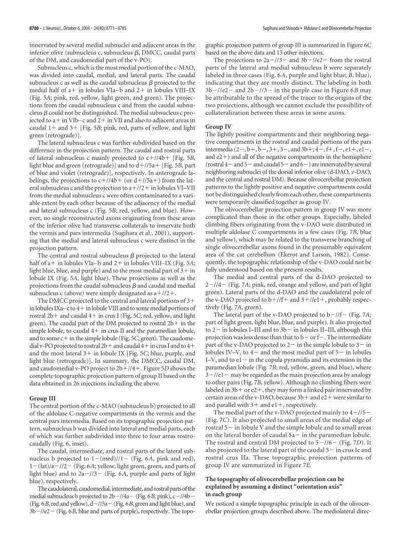
innervated by several medial subnuclei and adjacent areas in theinferior olive (subnucleus c, subnucleus �, DMCC, caudal partsof the DM, and caudomedial part of the v-PO).
Subnucleus c, which is the most medial portion of the c-MAO,was divided into caudal, medial, and lateral parts. The caudalsubnucleus c as well as the caudal subnucleus � projected to themedial half of a� in lobules VIa– b and 2� in lobules VIII–IX(Fig. 5A; pink, red, yellow, light green, and green). The projec-tions from the caudal subnucleus c and from the caudal subnu-cleus � could not be distinguished. The medial subnucleus c pro-jected to a� in VIb– c and 2� in VII and also to adjacent areas incaudal 1� and 3� [Fig. 5B; pink, red, parts of yellow, and lightgreen (retrograde)].
The lateral subnucleus c was further subdivided based on thedifference in the projection pattern. The caudal and rostral partsof lateral subnucleus c mainly projected to c�//4b� [Fig. 5B,light blue and green (retrograde)] and to d�//5a� [Fig. 5B, partof blue and violet (retrograde)], respectively. In anterograde la-belings, the projections to c�//4b� (or d�//5a�) from the lat-eral subnucleus c and the projection to a�//2� in lobules VI–VIIfrom the medial subnucleus c were often contaminated to a vari-able extent by each other because of the adjacency of the medialand lateral subnucleus c (Fig. 5B; red, yellow, and blue). How-ever, no single reconstructed axons originating from these areasof the inferior olive had transverse collaterals to innervate boththe vermis and pars intermedia (Sugihara et al., 2001), support-ing that the medial and lateral subnucleus c were distinct in theprojection pattern.
The central and rostral subnucleus � projected to the lateralhalf of a� in lobules VIa– b and 2� in lobules VIII–IX (Fig. 5A;light blue, blue, and purple) and to the most medial part of 3� inlobule IX (Fig. 5A, light blue). These projections as well as theprojections from the caudal subnucleus � and caudal and medialsubnucleus c (above) were simply designated as a�//2�.
The DMCC projected to the central and lateral portions of 3�in lobules IXa–c to 4� in lobule VIII and to some medial portions ofrostral 2b� and caudal 4� in crus I (Fig. 5C; red, yellow, and lightgreen). The caudal part of the DM projected to rostral 2b� in thesimple lobule, to caudal 4� in crus II and the paramedian lobule,and to some c� in the simple lobule (Fig. 5C, green). The caudome-dial v-PO projected to rostral 2b� and caudal 4� in crus I and to 4�and the most lateral 3� in lobule IX [Fig. 5C; blue, purple, andlight blue (retrograde)]. In summary, the DMCC, caudal DM,and caudomedial v-PO project to 2b�//4�. Figure 5D shows thecomplete topographic projection pattern of group II based on thedata obtained in 26 injections including the above.
Group IIIThe central portion of the c-MAO (subnucleus b) projected to allof the aldolase C-negative compartments in the vermis and thecentral pars intermedia. Based on its topographic projection pat-tern, subnucleus b was divided into lateral and medial parts, eachof which was further subdivided into three to four areas rostro-caudally (Fig. 6, inset).
The caudal, intermediate, and rostral parts of the lateral sub-nucleus b projected to 1�(med)//1� (Fig. 6A, pink and red),1�(lat)/a�//2� (Fig. 6A; yellow, light green, green, and parts oflight blue) and to 2a�//3� (Fig. 6A, purple and parts of lightblue), respectively.
Thecaudolateral,caudomedial, intermediate,androstralpartsofthemedial subnucleus b projected to 2b�//4a� (Fig. 6B, pink), c�//4b�(Fig. 6B, red and yellow), d�//5a� (Fig. 6B, green and light blue), and3b�//e2� (Fig. 6B, blue and parts of purple), respectively. The topo-
graphic projection pattern of group III is summarized in Figure 6Cbased on the above data and 13 other injections.
The projections to 2a�//3� and 3b�//e2� from the rostralparts of the lateral and medial subnucleus b were separatelylabeled in three cases (Fig. 6 A, purple and light blue; B, blue),indicating that they are mostly distinct. The labeling in both3b�//e2� and 2b�//3� in the purple case in Figure 6 B maybe attributable to the spread of the tracer to the origins of thetwo projections, although we cannot exclude the possibility ofcollateralization between these areas in some axons.
Group IVThe lightly positive compartments and their neighboring nega-tive compartments in the rostral and caudal portions of the parsintermedia (2�, b�, b�, 3�, 3�, and 3b�; 4�, f�, f�, e1�, e1�,and e2�) and all of the negative compartments in the hemisphere(rostral 4�and 5�and caudal 5�and 6�) are innervated by severalneighboring subnuclei of the dorsal inferior olive (d-DAO, v-DAO,and the central and rostral DM). Because olivocerebellar projectionpatterns to the lightly positive and negative compartments couldnot be distinguished clearly from each other, these compartmentswere temporarily classified together as group IV.
The olivocerebellar projection pattern in group IV was morecomplicated than those in the other groups. Especially, labeledclimbing fibers originating from the v-DAO were distributed inmultiple aldolase C compartments in a few cases (Fig. 7B, blueand yellow), which may be related to the transverse branching ofsingle olivocerebellar axons found in the presumably equivalentarea of the cat cerebellum (Ekerot and Larson, 1982). Conse-quently, the topographic relationship of the v-DAO could not befully understood based on the present results.
The medial and central parts of the d-DAO projected to2�//4� (Fig. 7A; pink, red, orange and yellow, and part of lightgreen). Lateral parts of the d-DAO and the caudolateral pole ofthe v-DAO projected to b�//f� and 3�//e1�, probably respec-tively (Fig. 7A, green).
The lateral part of the v-DAO projected to b�//f� (Fig. 7A;part of light green, light blue, blue, and purple). It also projectedto 2� in lobules I–III and to 3b� in lobules II–III, although thisprojection was less dense than that to b� or f�. The intermediatepart of the v-DAO projected to 2� in the simple lobule to 3� inlobules IV–V, to 4� and the most medial part of 5� in lobulesI–V, and to e1� in the copula pyramidis and its extension in theparamedian lobule (Fig. 7B; red, yellow, green, and blue), where3�//e1� may be regarded as the main projection area by analogyto other pairs (Fig. 7B, yellow). Although no climbing fibers werelabeled in 3b� or e2�, they may form a linked pair innervated bycertain areas of the v-DAO, because 3b� and e2� were similar toand parallel with 3� and e1�, respectively.
The medial part of the v-DAO projected mainly to 4�//5�(Fig. 7C). It also projected to small areas of the medial edge ofrostral 5� in lobule V and the simple lobule and to small areason the lateral border of caudal 5a� in the paramedian lobule.The rostral and central DM projected to 5�//6� (Fig. 7D). Italso projected to the lateral part of the caudal 5� in crus Ic androstral crus IIa. These topographic projection patterns ofgroup IV are summarized in Figure 7E.
The topography of olivocerebellar projection can beexplained by assuming a distinct “orientation axis”in each group
We noticed a simple topographic principle in each of the olivocer-ebellar projection groups described above. The mediolateral direc-
8780 • J. Neurosci., October 6, 2004 • 24(40):8771– 8785 Sugihara and Shinoda • Aldolase C and Olivocerebellar Projection

tion in the cerebellar cortex within each of the five groups could bemapped to an orientation axis defined in the subnuclei of the inferiorolive (Fig. 8). These axes may be related to the conjunctive relation-ship among different subnuclei and the convoluted organizationof the entire inferior olive, although the significance of these axesin the evolution and development has to be clarified.
In the part of the inferior olive that belongs to group I (Fig. 4),the orientation axis ran from subnucleus a of the MAO (caudal torostral), through the r-MAO (caudal to rostral) and v-PO (me-dial to lateral), to the d-PO (lateral to medial) (Fig. 8A, greenarrows). As shown in the three-dimensional display of olivarysubnuclei, these areas of the inferior olive were located close toeach other along the axis.
Group II (Fig. 5) had a slightly more complex topographybut could be reasonably explained by assuming two axes. Thefirst orientation axis ran from the caudal subnucleus � andcaudal subnucleus c, which are cytologically indistinguishablefrom each other (Ruigrok and Cella, 1995), through theDMCC and the caudal DM, to the caudomedial v-PO (Fig. 8 A,blue arrows). Although these subnuclei are separated from
each other by the white matter, they are still located close toeach other along the axis. The second axis ran caudorostrallythrough the medial and lateral subnucleus c in succession (Fig.8 A, violet arrows). Within the compartments that belonged togroup II in the cerebellar cortex, the first and second axescorresponded to the rostral and caudal parts (a�//2� in lob-ules V–VIa and VIII–X and 2b�//4�) (Fig. 8 A, left panel, bluearrow) and the central parts (a�//2� in lobules VIb– c and VIIand c�//4b� and d�//5a�) (Fig. 8 A, left panel, violet arrow),respectively.
In group III (Fig. 6), the topography could be explained rathersimply because only subnucleus b of the MAO was involved. Theorientation axis first ran in the lateral subnucleus b (caudal torostral), corresponding to the projection to 1�(med)//1�,1�(lat)/a�//2�, and 2a�//3�. The axis then ran in the medialsubnucleus b (again caudal to rostral), corresponding to theprojection to 2b�//4a�, c�//4b�, and d�//5a� (Fig. 8 A,ocher arrows).
In group IV (Fig. 7), a single orientation axis running from thed-DAO (medial to lateral), through the v-DAO (caudolateral to
Figure 7. Mapping of labeled climbing fibers and summarized topographic scheme of the olivocerebellar projection belonging to group IV. This group consisted of projections from the dorsal foldof the DAO (d-DAO), ventral fold of the DAO (v-DAO), and dorsomedial subnucleus of the PO (DM) to 2�//4�, b�//f�, b�//f�, 3�//e�, 3�//e1�, 4�//5�, and 5�//6�, and possiblyto 3b�//e2�. A, Plots of labeled climbing fibers in nine injections terminating mainly in compartments 2�//4�, b�//f�, b�//f�, and 3�//e�. Two injections (green and light green) werespread to the d-DAO and v-DAO. B, Plots of labeled climbing fibers in four injections in the intermediate v-DAO, terminating in several compartments, including 3�//e1� (yellow). C, Plots of labeledclimbing fibers in five injections in the medial v-DAO, terminating mainly in 4�//5� and also in caudal 5a�. D, Plots of labeled climbing fibers in four injections in the DM, terminating in 5�//6�and in caudal 5�. Labeled climbing fibers in 9 of 22 experiments were virtually aligned in a single narrow band, if a small number of outside climbing fibers can be ignored. E, Putative olivocerebellartopography within group III determined from the results shown in A and B.
Sugihara and Shinoda • Aldolase C and Olivocerebellar Projection J. Neurosci., October 6, 2004 • 24(40):8771– 8785 • 8781

Figure 8. Organization of aldolase C compartments and topographic olivocerebellar projection based on the five groups in the present study. A, Topographic organization explained withorientation axes. Areas belonging to each of the five groups (green, blue, yellow, red, and gray for groups I–V, respectively) were mapped on the aldolase C compartment pattern of the cerebellarcortex (left) and on the solid schemes of the inferior olive (right). Arrows with dark colors indicate the mediolateral direction in each group in the cerebellar cortex and the orientation axes in theinferior olive. The orientation axes correspond to the medial to lateral direction in the cerebellar cortex in terms of the topography of the projection. Group II has two orientation axes, correspondingto the projection to the central cortical area from the rostral and lateral subnucleus c (violet) and to the projection to the rostral and caudal cortical areas from the caudal subnucleus c, subnucleus�, DMCC, caudal dorsomedial subnucleus of the PO (DM), and caudomedial ventral lamella of the PO (v-PO) (blue). This panel is based on the data shown in Figures 4 –7. Group V (gray) consists ofthe flocculus and nodulus and a few areas in the hemisphere innervated by the DC and VLO (Sugihara et al., 2004). B, A schematic of major inputs to the inferior olive reported so far sorted accordingto the groups classified in the present study. Question mark indicates that the specific inputs in those areas have not been clarified. See Discussion for details. Origins of major afferents to the inferiorolive are arbitrarily colored. APP, Area parafascicularis prerubralis; DCN, dorsal column nucleus; IO, inferior olive; ND, nucleus of Darkschewitsch; NOT, nucleus of the optic tract; RN, red nucleus; SC,superior colliculus; VN, vestibular nucleus. C, Correspondence between the aldolase C-based five-group olivocerebellar organization in the present study and conventional olivocerebellar zones(zones A–D). Colors indicate the same groups as in panels A and B. I–X, Lobules I–X; a– c, sublobules a– c or subnuclei a– c; BETA, subnucleus �; c-MAO, caudal part of the MAO; CP, copula pyramidis;Cr I, crus I of ansiform lobule; Cr II, crus II of ansiform lobule; d-DAO, dorsal fold of the DAO; d-PO, dorsal lamella of the PO; DC, DC of Kooy; DM, dorsomedial subnucleus of the PO; DMCC, DMCCsubnucleus; DPFL, dorsal paraflocculus; FL, flocculus; Param, paramedian lobule; r-MAO, rostral part of the MAO; Sim, simple lobule; v-DAO, ventral fold of the DAO; v-PO, ventral lamella of the PO;VPFL, ventral paraflocculus.
8782 • J. Neurosci., October 6, 2004 • 24(40):8771– 8785 Sugihara and Shinoda • Aldolase C and Olivocerebellar Projection

rostromedial), to the DM could mostly account for the topo-graphical projection to all compartments (medial to lateral) ingroup IV (Fig. 8A, red arrows). These subnuclei of the inferiorolive are located close to each other along the axis.
Concerning group V, we examined the detailed projectionpattern from the DC and VLO to the flocculus (Sugihara et al.,2004). However, it was not easy to determine an orientation axisin this group.
DiscussionThis study showed that the organization of the olivocerebellartopography and aldolase C compartment pattern is rather simplyexplained in the five-group scheme, although the significance ofaldolase C expression itself is not clear (cf. Welsh et al., 2002).Whether the corticonuclear and olivonuclear topographies areexplained according to this five-group organization will be thenext question.
Organization of the inferior olive: five groups related todistinct functionsThe divisions of the inferior olive in the present five-groupscheme do not fully correspond to the conventional olivary sub-nuclei. Histochemical and immunohistological observationshave recognized molecular patterns in the olive that are not nec-essarily correlated with these subdivisions [acetylcholinesterase(Marani et al., 1977); calbindin calcitonin gene-related peptideand parvalbumin (Wassef et al., 1992)]. The present five groupsand the olivary molecular patterns may agree in the c-MAO(Wassef et al., 1992) but not in other areas of the olive. In con-
trast, the present grouping in the olive seems to correlate wellwith the difference in input to the olive.
To consider the functional aspects of the groups defined here,major inputs to the parts of the inferior olive were comparedusing previous reports (Fig. 8B). In group I, the v-PO, d-PO,r-MAO, and subnucleus a receive input from the cerebral cortexrelayed by nuclei in the mesodiencephalic junction (Swenson andCastro, 1983; Onodera, 1984; Holstege and Tan, 1988; de Zeeuw,1989) (Fig. 8B, green arrows), although the subnucleus a alsoreceive input from the spinal cord (Matsushita et al., 1991).
Concerning group II, the superior colliculus projects to whatseems to be the medial and lateral subnucleus c of the MAO, andthe vestibular nuclei project to the DMCC, subnucleus �, andsubnucleus c (Brown et al., 1977; Swenson and Castro, 1983;Gerrits et al., 1985; Akaike, 1992) (Fig. 8B, violet and blue ar-rows). The projections from the spinal cord and dorsal columnnucleus, which are predominant in the subnucleus b, lack in thesubnucleus c and � (Boesten and Voogd, 1975). The major inputsto the caudal DM and caudomedial v-PO are unknown.
Concerning group III, the subnucleus b of the MAO receivesheterogeneous input from the spinal cord and dorsal columnnuclei with crude somatotopy (Boesten and Voogd, 1975; Berkleyand Hand, 1978; Matsushita et al., 1992; Molinari et al., 1996) andfrom the vestibular nuclei and the mesodiencephalic junction(Swenson and Castro, 1983; Onodera, 1984; Gerrits et al., 1985)(Fig. 8B, ocher arrow).
The d-DAO and v-DAO in group IV receive inputs from thespinal cord and the dorsal column nuclei with refined cutaneous
Table 2. Major olivocerebellar topographic projection pattern in terms of aldolase C compartments and their putative correspondence to classic olivocerebellar zones
Group
Origin in the inferior olive
Aldolase C compartmentsa Conventional olivocerebellar zonesbSubnucleus Subareas
I Subnucleus a (c-MAO) Caudal 1�//1� AIntermediate 2�//3� (medial) A (AX)c
Rostral 2�//3� (lateral) A (AX)c
r-MAO 4�//5� C2c
v-PO 5�//6� D1c
d-PO 6�//7� D2II Subnucleus � a�//2� A
Subnucleus c (c-MAO) Caudal and medial a�//2� ALateral Caudal c�//4b� Lateral A
Rostral d�//5a� Lateral ADMCC 2b�//4� X-CXDM Caudal 2b�//4� X-CXv-PO Caudomedial 2b�//4� X-CX
III Subnucleus b (c-MAO) Lateral Caudal 1�(med)//1� AIntermediate 1�(lat)/a�//2� ARostral 2a�//3� Xc
Medial Caudolateral 2b�//4a� Lateral ACaudomedial c�//4b� Lateral AIntermediate d�//5a� Lateral ARostral 3b�//e2� CXc
IV d-DAO Medial and central 2�//4� Bc
Lateral b�//f� Bv-DAO Caudolateral b�//f� and 3�//e1� C1c
Central 3�//e1� C1c
Rostromedial 4�//5� C3c
DM Central and rostral 5�//6� D0V Dorsal cap and ventrolateraloutgrowth Flocculus and nodulusd
I–X, Lobules I-X; a– c, sublobules a– c or subnuclei a– c; c-MAO, caudal part of the MAO; r-MAO, rostral part of the MAO; v-PO, ventral lamella of the PO; d-PO, dorsal lamella of the PO; d-DAO, dorsal fold of the DAO; v-DAO, ventral fold ofthe DAO.aOnly major termination compartments are shown. See Results for details.bData are from Voogd and Bigaré (1980), Buisseret-Delmas and Angaut (1993), Voogd et al. (2003), and Voogd and Ruigrok (2004).cZones in which the correspondence to zebrin compartments has been shown recently (Voogd et al., 2003; Voogd and Ruigrok, 2004) and confirmed in the present study.dData are from Sugihara et al. (2004).
Sugihara and Shinoda • Aldolase C and Olivocerebellar Projection J. Neurosci., October 6, 2004 • 24(40):8771– 8785 • 8783

somatotopy (v-DAO) and with preponderance of deep modality(d-DAO) (Berkley and Hand, 1978; Gellman et al., 1983; Swen-son and Castro, 1983; Matsushita et al., 1992; Molinari et al.,1996) (Fig. 8B, red arrows). Input to the DM has not been re-ported clearly. However, some figures in the reports by Swensonand Castro (1983, their Fig. 5-3), Huerta et al. (1985, their Fig. 2),and Van Ham and Yeo (1992, their Figs. 4, 5) suggest the trigem-inal projection to the DM (Fig. 8B, hashed red arrow).
The VLO and DC subnuclei, which belong to group V, receiveinput from nuclei of the accessory optic system (Leonard et al.,1988) (Fig. 8B, black arrows).
Thus, groups I–V primarily receive mesodiencephalic, vestib-ular and collicular, somatosensory plus other (mesodiencephalicand vestibular), somatosensory, and retinal inputs, respectively.This finding indicates that the aldolase C labeling pattern isclosely related to the functional compartmentalization of the cer-ebellar cortex. However, it does not mean each aldolase C com-partment simply circumscribes functionally homogeneousclimbing fibers, because climbing fibers originating from a smallgroup of inferior olive neurons are distributed in a longitudinalband-shaped area that is much narrower than a single aldolase Ccompartment (Fig. 3A). Therefore, the relationship between thealdolase C compartments and the electrophysiologically identi-fied longitudinal structure in the cerebellar cortex regardingclimbing fiber activity (Ekerot et al., 1991; Lang et al., 1999; Han-son et al., 2000) is to be examined (cf. Voogd et al., 2003).
Interpretation of the conventional cerebellar zones with thepresent resultsPrevious retrograde and anterograde mass labeling studies andelectrophysiological studies have identified so-called conven-tional olivocerebellar zones (A, B, X, C1, CX, X-CX, C2, C3, D0,D1, and D2) that are innervated topographically from differentsubnuclei of the inferior olive (Groenewegen and Voogd, 1977;Brodal, 1981; Azizi and Woodward, 1987; Buisseret-Delmas andAngaut, 1993). A relationship between conventional olivocer-ebellar zones and zebrin (aldolase C) compartments has beenshown recently in several areas that mainly belong to groups I andIV in our designation (Voogd et al., 2003, 2004), which mostlyagrees with the present results (see the last note of Table 2). How-ever, concerning compartments belonging to groups II and III,which are rather complicated than other compartments, theirrostrocaudal continuity or the olivocerebellar projections tothem could not be properly analyzed in previous studies. Therelationship between conventional olivocerebellar zones and al-dolase C compartments can be determined throughout the cere-bellar cortex in this study by comparing the present results with ascheme of the conventional olivocerebellar topography in termsof the positions of zones and olivary origins, as listed in Table 2and depicted in Figure 8C.
Conventional zone A occupies nearly the entire vermis, andlateral zone A extends to the central pars intermedia. Althoughthese zones presumably include multiple aldolase C compart-ments, its subdivisions have not been much clarified, except forthe most lateral part of zone A (recently designated as AX) thatcorresponds to 2�//3� (Voogd and Ruigrok, 2004). The presentresults show clear subdivisions corresponding to all aldolase Ccompartments within conventional zone A and lateral zone A.These subdivisions belong to groups I, II, and III. The separateentity of lateral zone A proposed on the basis of the cortico-nuclear projection pattern (Goodman et al., 1963) was sup-ported, because the lateral subnucleus c and the neighboring me-dial subnucleus b project to it (Fig. 8C).
Conventional zones B, C1, C3, and D0 belong to group IVaccording to the present results. Zones B and C1 appear to cor-respond to multiple negative and lightly positive aldolase C com-partments in the rostral and caudal cerebellar cortex (Table 2, Fig.8C). Zone C3 mostly corresponds to 4�//5�. Zone C2 mostlycorresponds to 4�//5� and extends into the paraflocculus andthe most caudal portion of the flocculus (Ruigrok et al., 1992;Sugihara et al., 2004). These results concerning zones B, C1, C2,and C3 were similar to the recent observations of Voogd et al.(2003) and Voogd and Ruigrok (2004). Zone C2 in lobule IX(Sato and Barmack, 1985; Voogd and Ruigrok, 1997) was notconfirmed in the present study.
Based on the present results, zone D1 corresponds to 5�//6�innervated by the v-PO, and lateral zone D2 corresponds to6�//7� innervated by the d-PO. This projection is the same asthat postulated in the cat (Voogd and Bigare, 1980; Rosina andProvini, 1982) and inferred in the rat (Voogd et al., 2004) but isnot consistent with previous reports in the rat (Azizi and Wood-ward, 1987; Buisseret-Delmas and Angaut, 1993), in which thev-PO has been thought to innervate a more lateral area than thed-PO. Zone D0 has been reported to be located medial to D1(Buisseret-Delmas and Angaut, 1993) or between D1 and D2(Voogd et al., 2003). The present results suggest that D0 is locatednot only in the entire 5�//6� compartment but also in a smalllateral part of the 4�//5� compartment in crus II, indicating thatboth reports are correct, although the nuclear projections of theseareas have to be determined to confirm it.
Among zones added later (X, CX, X-CX) (Buisseret-Delmas etal., 1993; Yatim et al., 1995), zone X likely corresponds to 2a�//3�, and zone CX corresponds to 3b�//e2� (parts of group III).Zone X-CX remains enigmatic but appears to correspond to2b�//4� innervated by the DMCC. No zones previously re-ported appear to correspond to the projection of the caudal DMand caudomedial v-PO to the rest of 2�//4�. However, thisprojection may also be regarded as part of zone X-CX because itresembles the projection of the DMCC.
Besides zones A–D, more classical aspects of the cerebellarorganization can be related to the present results. Group II islocated only caudal to the primary fissure, and group IV occupiesa substantial portion of the area rostral to the primary fissure.This may help explain the apparent difference in the gross func-tion of the cerebellum rostral and caudal to the primary fissure(Brodal, 1981), although mossy fiber projection must also beconsidered.
Functional implications of the link between the specific rostraland caudal aldolase C compartments and the translobularbranching of single olivocerebellar axons (Sugihara et al., 2001)as the basis for this link are not known. However, the rostrocau-dal boundary defined here seems a fundamental landmark at leastfor the morphological organization of the cerebellar cortex.
ReferencesAhn AH, Dziennis S, Hawkes R, Herrup K (1994) The cloning of zebrin II
reveals its identity with aldolase C. Development 120:2081–2090.Akaike T (1992) The tectorecipient zone in the inferior olivary nucleus in
the rat. J Comp Neurol 320:398 – 414.Andersson G, Oscarsson O (1978) Climbing fiber microzones in cerebellar
vermis and their projection to different groups of cells in the lateral ves-tibular nucleus. Exp Brain Res 32:565–579.
Azizi SA, Woodward DJ (1987) Inferior olivary nuclear complex of the rat:morphology and comments on the principles of organization within theolivocerebellar system. J Comp Neurol 263:467– 484.
Bailly Y, Schoen SW, Delhaye-Bouchaud N, Kreutzberg GW, Mariani J(1995) 5�-Nucleotidase activity as a synaptic marker of parasagittal com-partmentation in the mouse cerebellum. J Neurocytol 24:879 – 890.
8784 • J. Neurosci., October 6, 2004 • 24(40):8771– 8785 Sugihara and Shinoda • Aldolase C and Olivocerebellar Projection

Berkley KJ, Hand PJ (1978) Projections to the inferior olive of the cat. II.Comparisons of input from the gracile, cuneate and the spinal trigeminalnuclei. J Comp Neurol 180:253–264.
Boesten AJ, Voogd J (1975) Projections of the dorsal column nuclei and thespinal cord on the inferior olive in the cat. J Comp Neurol 161:215–237.
Brochu G, Maler L, Hawkes R (1990) Zebrin II: a polypeptide antigen ex-pressed selectively by Purkinje cells reveals compartments in rat and fishcerebellum. J Comp Neurol 291:538 –552.
Brodal A (1981) Neurological anatomy in relation to clinical medicine, Ed 3.New York: Oxford UP.
Brown JT, Chan-Palay V, Palay SL (1977) A study of afferent input to theinferior olivary complex in the rat by retrograde axonal transport ofhorseradish peroxidase. J Comp Neurol 176:1–22.
Buisseret-Delmas C, Angaut P (1993) The cerebellar olivocorticonuclearconnections in the rat. Prog Neurobiol 40:63– 87.
Buisseret-Delmas C, Yatim N, Buisseret P, Angaut P (1993) The X zone andCX subzone of the cerebellum in the rat. Neurosci Res 16:195–207.
de Zeeuw CI, Holstege JC, Ruigrok TJ, Voogd J (1989) Ultrastructural studyof the GABAergic, cerebellar, and mesodiencephalic innervation of the catmedial accessory olive: anterograde tracing combined with immunocyto-chemistry. J Comp Neurol 284:12–35.
Dore L, Jacobson CD, Hawkes R (1990) Organization and postnatal develop-ment of zebrin II antigenic compartmentation in the cerebellar vermis of thegrey opossum, Monodelphis domestica. J Comp Neurol 291:431–449.
Ekerot C-F, Larson B (1982) Branching of olivary axons to innervate pairs ofsagittal zones in the cerebellar anterior lobe in the cat. Exp Brain Res48:185–198.
Ekerot C-F, Garwicz M, Schouenborg J (1991) Topography and nociceptivereceptive fields of climbing fibres projecting to the cerebellar anterior lobein the cat. J Physiol (Lond) 441:257–274.
Gellman R, Houk JC, Gibson AR (1983) Somatosensory properties of theinferior olive of the cat. J Comp Neurol 215:228 –243.
Gerrits NM, Voogd J, Magras IN (1985) Vestibular afferents of the inferiorolive and the vestibulo-olivo-cerebellar climbing fiber pathway to theflocculus in the cat. Brain Res 332:325–336.
Goodman DC, Hallett RE, Welch RB (1963) Patterns of localization in the cer-ebellar corticonuclear projections of albino rat. J Comp Neurol 121:51–67.
Gravel C, Eisenman LM, Sasseville R, Hawkes R (1987) Parasagittal organi-zation of the rat cerebellar cortex: direct correlation between antigenicPurkinje cell bands revealed by mabQ113 and the organization of theolivocerebellar projection. J Comp Neurol 265:294 –310.
Groenewegen HJ, Voogd J (1977) The parasagittal zonation within theolivocerebellar projection. I. Climbing fiber distribution in the vermis ofthe cat cerebellum. J Comp Neurol 174:417– 488.
Gwyn DG, Nicholson GP, Flumerfelt BA (1977) The inferior olivary nucleus ofthe rat: a light and electron microscopic study. J Comp Neurol 174:489–520.
Hanson CL, Chen G, Ebner TJ (2000) Role of climbing fibers in determiningthe spatial patterns of activation in the cerebellar cortex to peripheralstimulation: an optical imaging study. Neuroscience 96:317–331.
Hawkes R (1997) An anatomical model of cerebellar modules. Prog BrainRes 114:39 –52.
Hawkes R, Leclerc N (1987) Antigenic map of the rat cerebellar cortex: thedistribution of parasagittal bands as revealed by monoclonal anti-Purkinje cell antibody mobA113. J Comp Neurol 256:29 – 41.
Herrup K, Kuemerle B (1997) The compartmentalization of the cerebellum.Annu Rev Neurosci 20:61–90.
Holstege G, Tan J (1988) Projections from the red nucleus and surroundingareas to the brainstem and spinal cord in the cat. An HRP and autoradio-graphical tracing study. Behav Brain Res 28:33–57.
Huerta MF, Hashikawa T, Gayoso MJ, Harting JK (1985) The trigemino-olivaryprojection in the cat: contributions of individual subnuclei. 241:180–190.
Lang EJ, Sugihara I, Welsh JP, Llinas R (1999) Patterns of spontaneous purkinjecell complex spike activity in the awake rat. J Neurosci 19:2728–2739.
Leonard CS, Simpson JI, Graf W (1988) Spatial organization of visual mes-sages of the rabbit’s cerebellar flocculus. I. Topology of inferior oliveneurons of the dorsal cap of Kooy. J Neurophysiol 60:2073–2090.
Llinas R, Baker R, Sotelo C (1974) Electrotonic coupling between neuronsin cat inferior olive. J Neurophysiol 37:560 –571.
Marani E, Voogd J, Boekee A (1977) Acetylcholinesterase staining in subdi-visions of the cat’s inferior olive. J Comp Neurol 174:209 –226.
Matsushita M, Ragnarson B, Grant G (1991) Topographic relationship be-
tween sagittal Purkinje cell bands revealed by monoclonal antibody tozebrin I and spinocerebellar projections arising from the central cervicalnucleus in the rat. Exp Brain Res 84:133–141.
Matsushita M, Yaginuma H, Tanami T (1992) Somatotopic termination ofthe spino-olivary fibers in the cat, studied with the wheat germ agglutinin-horseradish peroxidase technique. Exp Brain Res 89:397– 407.
Molinari HH, Schultze KE, Strominger NL (1996) Gracile, cuneate, and spi-nal trigeminal projections to inferior olive in rat and monkey. J CompNeurol 375:467– 480.
Mukai T, Yatsuki H, Masuko S, Arai Y, Joh K, Hori K (1991) The structureof the brain-specific rat aldolase C gene and its regional expression. Bio-chem Biophys Res Commun 174:1035–1042.
Onodera S (1984) Olivary projections from the mesodiencephalic struc-tures in the cat studied by means of axonal transport of horseradish per-oxidase and tritiated amino acids. J Comp Neurol 227:37– 49.
Ozol K, Hayden JM, Oberdick J, Hawkes R (1999) Transverse zones in thevermis of the mouse cerebellum. J Comp Neurol 412:95–111.
Rosina A, Provini L (1982) Longitudinal and topographical organization of theolivary projection to the cat ansiform lobule. Neuroscience 7:2657–2676.
Ruigrok TJH, Cella F (1995) Precerebellar nuclei and red nucleus. In: The ratnervous system, Ed 2 (Paxinos G, ed), pp 277–308. San Diego: Academic.
Ruigrok TJH, Osse RJ, Voogd J (1992) Organization of inferior olivary pro-jections to the flocculus and ventral paraflocculus of the rat cerebellum.J Comp Neurol 316:129 –150.
Sato Y, Barmack NH (1985) Zonal organization of olivocerebellar projec-tions to the uvula in rabbits. Brain Res 359:281–292.
Sugihara I, Wu H-S, Shinoda Y (1999) Morphology of single olivocerebellaraxons labeled with biotinylated dextran amine in the rat. J Comp Neurol414:131–148.
Sugihara I, Wu H-S, Shinoda Y (2001) The entire trajectories of single olivo-cerebellar axons in the cerebellar cortex and their contribution to cerebel-lar compartmentalization. J Neurosci 21:7715–7723.
Sugihara I, Ebata S, Shinoda Y (2004) Functional compartmentalization in theflocculus and the ventral dentate and dorsal group y nuclei: An analysis ofsingle olivocerebellar axonal morphology. J Comp Neurol 470:113–133.
Swenson RS, Castro AJ (1983) The afferent connections of the inferior oli-vary complex in rats. An anterograde study using autoradiographic andaxonal degeneration techniques. Neuroscience 8:259 –275.
Van Ham JJ, Yeo CH (1992) Somatosensory trigeminal projections to theinferior olive, cerebellum and other precerebellar nuclei in rabbits. EurJ Neurosci 4:302–317.
Voogd J (1967) Comparative aspects of the structure and fibre connexionsof the mammalian cerebellum. In: Progress in brain research, Vol. 25: Thecerebellum (Fox CA, Snider RS, eds), pp 94 –135. Amsterdam: Elsevier.
Voogd J, Bigare F (1980) Topographical distribution of olivary and corticonuclear fibers in the cerebellum: a review. In: The inferior olivary nucleus:anatomy and physiology (Courville J, de Montigny C, Lamarre Y, eds), pp207–234. New York: Raven.
Voogd J, Ruigrok TJH (1997) Transverse and longitudinal patterns in themammalian cerebellum. Prog Brain Res 114:21–37.
Voogd J, Ruigrok TJH (2004) The organization of the corticonuclear and olivo-cerebellar climbing fiber projections to the rat cerebellar vermis: the congru-ence of projection zones and the zebrin pattern. J Neurocytol 33:5–21.
Voogd J, Pardoe J, Ruigrok TJH, Apps R (2003) The distribution of climbingand mossy fiber collateral branches from the copula pyramidis and the para-median lobule: congruence of climbing fiber cortical zones and the pattern ofzebrin banding within the rat cerebellum. J Neurosci 23:4645–4656.
Wassef M, Cholley B, Heizmann CW, Sotelo C (1992) Development of theolivocerebellar projection in the rat: II. Matching of the developmentalcompartmentations of the cerebellum and inferior olive through the pro-jection map. J Comp Neurol 323:537–550.
Welsh JP, Yuen G, Placantonakis DG, Vu TQ, Haiss F, O’Hearn E, MolliverME, Aicher SA (2002) Why do Purkinje cells die so easily after globalbrain ischemia? Aldolase C, EAAT4, and the cerebellar contribution toposthypoxic myoclonus. Adv Neurol 89:331–359.
Wu H-S, Sugihara I, Shinoda Y (1999) Projection patterns of single mossyfibers originating from the lateral reticular nucleus in the rat cerebellarcortex and nuclei. J Comp Neurol 411:97–118.
Yatim N, Compoint C, Buisseret P, Angaut P, Buisseret-Delmas C (1995)On the caudal extension of the X zone in the cerebellar cortex of the rat.Neurosci Res 23:223–227.
Sugihara and Shinoda • Aldolase C and Olivocerebellar Projection J. Neurosci., October 6, 2004 • 24(40):8771– 8785 • 8785