Dose-dependent activation of putative oncogene SBSN by BORIS
Upload
independentCategory
view
6download
0
EMBOopen
Cancer induction by restriction of oncogeneexpression to the stem cell compartment
This is an open-access article distributed under the terms of the Creative Commons Attribution License, which permitsdistribution, andreproduction inanymedium,provided theoriginalauthorandsourcearecredited.This licensedoesnotpermit commercial exploitation without specific permission.
Marıa Perez-Caro1, Cesar Cobaleda2,Ines Gonzalez-Herrero1, Carolina Vicente-Duenas1, Camino Bermejo-Rodrıguez1,Margarita Sanchez-Beato3, Alberto Orfao4,Belen Pintado5, Teresa Flores6,Manuel Sanchez-Martın7, Rafael Jimenez2,Miguel A Piris3 and Isidro Sanchez-Garcıa1,*1Experimental Therapeutics and Translational Oncology Program,Instituto de Biologıa Molecular y Celular del Cancer, CSIC/Universidadde Salamanca, Salamanca, Spain, 2Departamento de Fisiologıa yFarmacologıa, Universidad de Salamanca, Salamanca, Spain, 3MolecularPathology Program, Centro Nacional de Investigaciones Oncologicas,Madrid, Spain, 4Servicio de Citometrıa and Departamento de Medicina,Universidad de Salamanca, Salamanca, Spain, 5Genetically EngineeredMouse Facility, CNB-CSIC, Madrid, Spain, 6Departamento de AnatomıaPatologica, Universidad de Salamanca, Salamanca, Spain and7Departamento de Medicina, Genetically Engineered Mouse Facility,SEA, University of Salamanca, Salamanca, Spain
In human cancers, all cancerous cells carry the oncogenic
genetic lesions. However, to elucidate whether cancer is a
stem cell-driven tissue, we have developed a strategy to
limit oncogene expression to the stem cell compartment
in a transgenic mouse setting. Here, we focus on the effects
of the BCR-ABLp210 oncogene, associated with chronic
myeloid leukaemia (CML) in humans. We show that
CML phenotype and biology can be established in mice
by restricting BCR-ABLp210 expression to stem cell anti-
gen 1 (Sca1)þ cells. The course of the disease in Sca1-BCR-
ABLp210 mice was not modified on STI571 treatment.
However, BCR-ABLp210-induced CML is reversible
through the unique elimination of the cancer stem cells
(CSCs). Overall, our data show that oncogene expression
in Sca1þ cells is all that is required to fully reprogramme
it, giving rise to a full-blown, oncogene-specified tumour
with all its mature cellular diversity, and that elimination
of the CSCs is enough to eradicate the whole tumour.
The EMBO Journal (2009) 28, 8–20. doi:10.1038/
emboj.2008.253; Published online 27 November 2008
Subject Categories: signal transduction; molecular biology
of disease
Keywords: cancer; cancer stem cells (CSCs); CSC inhibitors;
drug discovery; mouse models
Introduction
An axiom in the treatment of tumours is that the remission is,
in general, more difficult to achieve with each relapse.
Despite a better understanding of the biology of tumour
cells, the treatment of most cancers has not significantly
changed for the past three decades and the decreasing
mortality has been mostly the result of early detection and
prevention rather than the consequence of effective thera-
peutics (Etzioni et al, 2003; Chabner and Roberts, 2005; Huff
et al, 2006).
A new hypothesis in cancer biology postulates that
tumours are hierarchically structured as abnormal tissues,
which are maintained by cancer stem cells (CSCs) (Reya et al,
2001; Perez-Caro and Sanchez-Garcia, 2006; Dalerba et al,
2007a; Sanchez-Garcıa et al, 2007). This theory provides
a rationale to explain the failure of many currently used
antitumoral strategies, because just a small population of
CSCs resistant to the therapy would be enough to maintain
the whole tumour mass (Etzioni et al, 2003; Chabner and
Roberts, 2005; Huff et al, 2006). Evidences supporting this
model have been recently published for human leukaemias
(Bonnet and Dick, 1997; Cobaleda et al, 2000; Cox et al, 2004,
2007; Hope et al, 2004) and solid tumours (Al-Hajj et al, 2003;
Singh et al, 2004; Collins et al, 2005; Kim et al, 2005; Xin et al,
2005; Bao et al, 2006a, b; Dalerba et al, 2007b; Li et al, 2007;
O’Brien et al, 2007; Prince et al, 2007; Ricci-Vitiani et al,
2007). However, this emerging concept has not been fully
validated yet in an experimental model system. If validated,
the CSC hypothesis will have far-reaching consequences for
our understanding of cancer biology and for the development
of new strategies to improve cancer treatment. A major
obstacle to elucidate the contribution that CSCs make to the
development and maintenance of cancer and their suitability
as a target is the lack of a system to limit oncogene expression
to the CSC compartment.
Since the discovery of oncogenes in human tumours, many
efforts have been made to elucidate the causal role that these
oncogenes have in cancer development. These previous
studies have shown that oncogene expression is not only
required for initiation of cancer but also for the maintenance
of the disease (Chin et al, 1999; Huettner et al, 2000; Boxer
et al, 2004). In mouse models where oncogene expression is
driven by tissue-specific promoters, tumours arise at high
frequency, but disappear again when the inducing stimulus is
switched off (Chin et al, 1999; Huettner et al, 2000; Boxer
et al, 2004). However, it has not been biologically proven that
cancer is a hierarchically organized tissue that can be created
and maintained similar to a normal stem cell-based tissue.
If that is indeed the case, then cancer should be created and
maintained similarly to any other normal stem cell-driven
tissue, similar to the haematopoietic system. In a normalReceived: 24 June 2008; accepted: 7 November 2008; publishedonline: 27 November 2008
*Corresponding author. Experimental Therapeutics and TranslationalOncology Program, Instituto de Biologıa Molecular y Celular del Cancer(IBMCC), CSIC/Universidad de Salamanca, Campus Unamuno, S/N,37007 Salamanca, Spain. Tel.: þ 34 923 238403; þ 34 923 294813;E-mail: [email protected]
The EMBO Journal (2009) 28, 8–20 | & 2009 European Molecular Biology Organization | Some Rights Reserved 0261-4189/09
www.embojournal.org
The EMBO Journal VOL 28 | NO 1 | 2009 &2009 European Molecular Biology Organization
EMBO
THE
EMBOJOURNAL
THE
EMBOJOURNAL
8
stem cell-driven tissue, genetic programming of stem cells is
all that is required to (re)constitute all differentiated cells
forming the tissue and the genetic information responsible for
the stem cell programming is not present anymore within the
differentiated cells that form the tissue. Thus, we reasoned
that a similar organization could be happening for cancer
formation.
To address these biological questions, we have used the
BCR-ABLp210 oncogene associated with chronic myeloid
leukaemia (CML) in humans as a model (Koeffler and
Golde, 1981; Melo and Barnes, 2007). CML is a paradigmatic
stem cell disorder that begins as a protracted chronic phase,
characterized by high leukocyte counts and enlarged spleen
and liver. In all patients, the chronic phase of CML eventually
converts to a blast crisis that is indistinguishable from acute
leukaemia. The specific BCR-ABL inhibitor STI571 can pro-
long the remission times of CML patients because it is able to
eliminate the BCR-ABL-expressing differentiated cells that
constitute the bulk of the tumour (Druker et al, 2001).
However, it cannot eliminate BCR-ABL-expressing CSCs,
even though it can penetrate into the cells, and these CSCs
eventually repopulate the tumour with STI571-resistant ma-
ture cells (Graham et al, 2002; Hu et al, 2006; Primo et al,
2006; Jiang et al, 2007).
Here, we show that CML phenotype and biology can
be established in mice in which we limited BCR-ABLp210
expression to stem cell antigen 1 (Sca1)þ cells. These model
mice mimic CML human pathology. The course of the CML
disease in the Sca1-BCR-ABLp210 mice was not modified on
STI571 treatment. However, we show that BCR-ABLp210-
induced CML is reversible through the unique elimination
of the CSCs. To our knowledge, this study demonstrates for
the first time that limited oncogene expression in CSCs is all
that is required to fully reprogramme it, giving rise to a full-
blown, oncogene-specified tumour with all its mature cellular
diversity, and that CSC elimination is enough to eradicate the
whole tumour.
Results
Derivation of Sca1-BCR-ABLp210 mice
The mouse Ly-6E.1 promoter (Miles et al, 1997) was used to
drive Sca1-directed expression of a human BCR-ABLp210
(Sca1-BCR-ABLp210) or TK-IRES-BCR-ABLp210 transgene
(Sca1-TK-IRES-BCR-ABLp210) in C57BL/6�CBA mice
(Figure 1). This promoter is known to have heterologous
expression in haematopoietic stem cells (HSCs) (Miles et al,
1997), hence transgene expression is restricted to a limited
percentage of Sca1-expressing cells. The BCR-ABLp210-over-
expressing animals had normal gestation and were viable.
Three independent lines were obtained and used to examine
the phenotype further (Table I). As c-kit is known from earlier
studies to be downregulated in leukaemia stem cells (Blair
and Sutherland, 2000; Neering et al, 2007), our functional
definition of stem cell in this study does not include c-kit as a
surface marker. Quantitative RT–PCR of BCR-ABLp210 and/or
TK messenger mRNA confirmed that expression was largely
confined to Sca1þ cells with little or no ectopic expression in
Sca1� cells (Figure 1). In line with these results, neither BCR-
ABL protein nor downstream signalling was detected in
Sca1�Linþ cells of Sca1-BCR-ABLp210 mice (Supplementary
Figure 1 online). As reported earlier in transgenic reporter
lines with the same promoter (Miles et al, 1997; Ma et al,
2002a, b), BCR-ABLp210 was expressed in a limited percen-
tage of Sca1þ cells (25–50%) as measured by single-cell
RT–PCR (Supplementary Figure 2 online). This enables
cell transformation to occur in a similar manner as it happens
in humans, where cells expressing the oncogene are present
along with non-expressing cells. The phenotype described
here is therefore due primarily to the expression of BCR-
ABLp210 in Sca1þ cells.
CML development in Sca1-BCR-ABLp210 mice
The expression of BCR-ABLp210 in Sca1þ cells led to sig-
nificant increases in the numbers of white blood cells (WBCs)
and in the percentages of neutrophils (Table I) in the periph-
eral blood (PB) of Sca1-BCR-ABLp210 animals (Figure 2A).
Flow cytometry of bone marrow (BM) cells demonstrated
significantly increased numbers of cells staining positively for
the myeloid markers Gr1 and Mac-1, comprising up to
75–90% of the cells in the BM of transgenic animals
(Figure 2A). However, the percentage of Sca1þLin� cells in
BM did not increase (data not shown), similarly as it happens
in human CML (Jamieson et al, 2004). Macroscopic analysis
of these animals showed splenomegaly (Table I) and hepato-
megaly (79% of the animals). The histologic examination of
the spleen and liver demonstrated the presence of megakar-
yocytes as markers of myeloid metaplasia (Figure 2B) and
expansion of the splenic red pulp by predominantly granulo-
cytic myeloid cells (Figure 2C). This was confirmed by flow
cytometric analysis (Figure 2A). In addition, infiltration of
myeloid cells was also seen in the liver, lymph nodes and
lung (Figure 2C). The major cause of death in animals that
had progressive disease was related to myeloid infiltration of
extramedullary organs and kidney failure (30%), or the
evidence for progression to blast crisis (70%). In addition,
targeting Sca1þ cells in non-haematopoietic tissues gave rise
to carcinoma development (lung adenocarcinoma 10%, he-
patocarcinoma 3%, GIST 2%, osteogenic sarcoma 2% and
Sertoli cell tumour 2%; data not shown). Altogether, the
survival time of mice expressing BCR-ABLp210 in Sca1þ
cells ranged from 4 to 18 months (Figure 1E; Table I). Mice
surviving myeloid infiltration of extramedullary organs pro-
gressed spontaneously to a blast crisis characterized by the
appearance of blasts (myeloid or lymphoid) in PB, BM,
spleen and liver (Figure 2D and E). Similar to the human
disease, this secondary disorder appeared in the context of a
myeloproliferative disease (Figure 2F). The time of transition
to blast crisis has been described to be reduced with in-
creased levels of BCR-ABL in human CML cells (Melo and
Barnes, 2007; Modi et al, 2007). Similarly, when compound
Sca1-BCR-ABLp210 transgenic mice were generated
(Figure 1), leukaemia was detectable at 1–2 months of age
(Figure 1; Table I). Thus, the limited expression of BCR-
ABLp210 to Sca1þ cells is able to mimic human CML,
characterized also by a progression from chronic towards
an acute phase (blast crisis), which is also invariably fatal in
Sca1-BCR-ABLp210 mice and that is dependent on the dose of
the oncogene expression in the Sca1þ cells.
The fusion protein responsible for CML has been shown to
cause genome instability in humans (Giehl et al, 2005; Melo
and Barnes, 2007). We examined Sca1-BCR-ABLp210 mice for
evidence of secondary genetic changes. First, we examined
the overall DNA methylation status in tumour cells of
Stem cell-driven cancerM Perez-Caro et al
&2009 European Molecular Biology Organization The EMBO Journal VOL 28 | NO 1 | 2009 9
Sca1-BCR-ABLp210 mice by immunolocalization of 5-methyl-
cytosine to give qualitative information on nuclear distribu-
tion. We performed similar analyses in human CD34þ CML
cells. Although control cells present unmethylated DNA, an
aberrant DNA methylation pattern was observed in tumour
cells of human CML (n¼ 3) and Sca1-BCR-ABLp210 mice
(n¼ 8) (see Supplementary Figure 3 online). As genetic
instability and centrosome defects are common and early
detectable features in CML (Melo and Barnes, 2007), we
sought to investigate whether centrosome aberrations occur
in Sca1-BCR-ABLp210 mice. We examined 14 CML samples
including Sca1þ cells of 9 newly diagnosed mice (chronic
phase) and 5 blast crisis specimens by using a centrosome-
specific antibody to pericentrin. Centrosome abnormalities
were detected in 23.6±5.1% of chronic phase cells and in
61.6±5.3% of blast crisis cells, but in only 3.1±0.9% of
controls (see Supplementary Figure 3 online). The similarities
between the secondary genetic changes observed in humans
and Sca1-BCR-ABLp210 mice suggest that similar mechan-
isms contribute to CML progression in both species.
Mar
ker
(kb)
IS1A
+ IS
1B
Probe:ABL
wt
IS1B
IS1B
IS1A
Probe:ABL
Con
trol
IS1B
IS1A
wt
IS1A
0.4
0.3
0.2
0.1
0
0.4
0.3
0.2
0.1
0
IS1B
IS1B
23
9.1
6.4
4.3
B C
Per
cent
age
ofB
CR
-AB
Lp21
0tr
ansc
ripts
Sca
1+ L
in–
Sca
1+ L
in–
Sca
1+ L
in–
Sca
1– Li
n+
Sca
1– Li
n+
Sca
1– Li
n+
Sca1-BCR-ABLp210 mice
Per
cent
age
oftk
anB
CR
-AB
Lp21
0tr
ansc
ripts
TK BCR-ABLp210
Sca1-TK-IRES-BCR-ABLp210 mice
A
Stem cell antigen 1 gene
5′ 3′
ATG
Eco
RI Eco
RI
Eco
RI
1 kb
Not
I
Not
I
Human BCR-ABLp210
TK-IRES-BCR-ABLp210
Per
cent
age
oB
CR
-AB
Lp21
0tr
ansc
ripts
IS1A
IS1B
Sca1+ Lin– cells
IS1A
+IS
1B
D
E
0
20
40
60
80
100
0 1 2 3 4 5 6 7 8 9 10 11 12 13 14 15 16 17 18
Time (months)
Control (n= 25)
IS1A+IS1B (n= 10; P value<0.00001)
IS1A (n= 34; P value<0.001)
IS1B (n= 25; P value<0.001)
IS9A (n = 23; P value<0.001)
Su
rviv
al (
%)
0.4
0.3
0.2
0.1
0
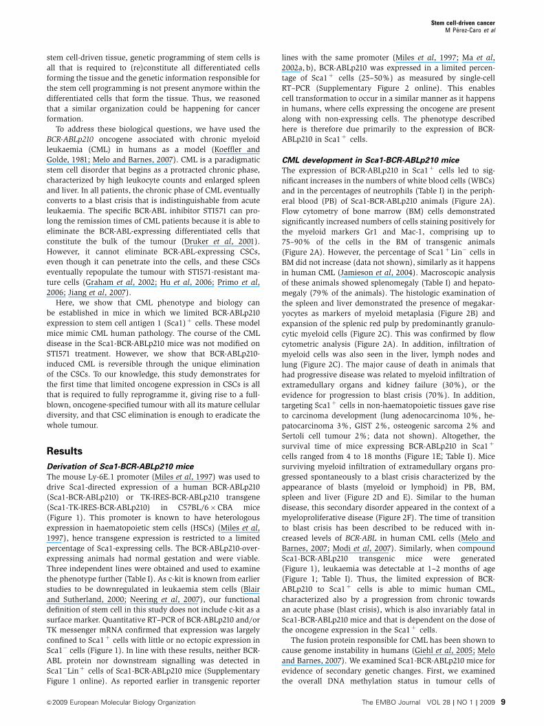
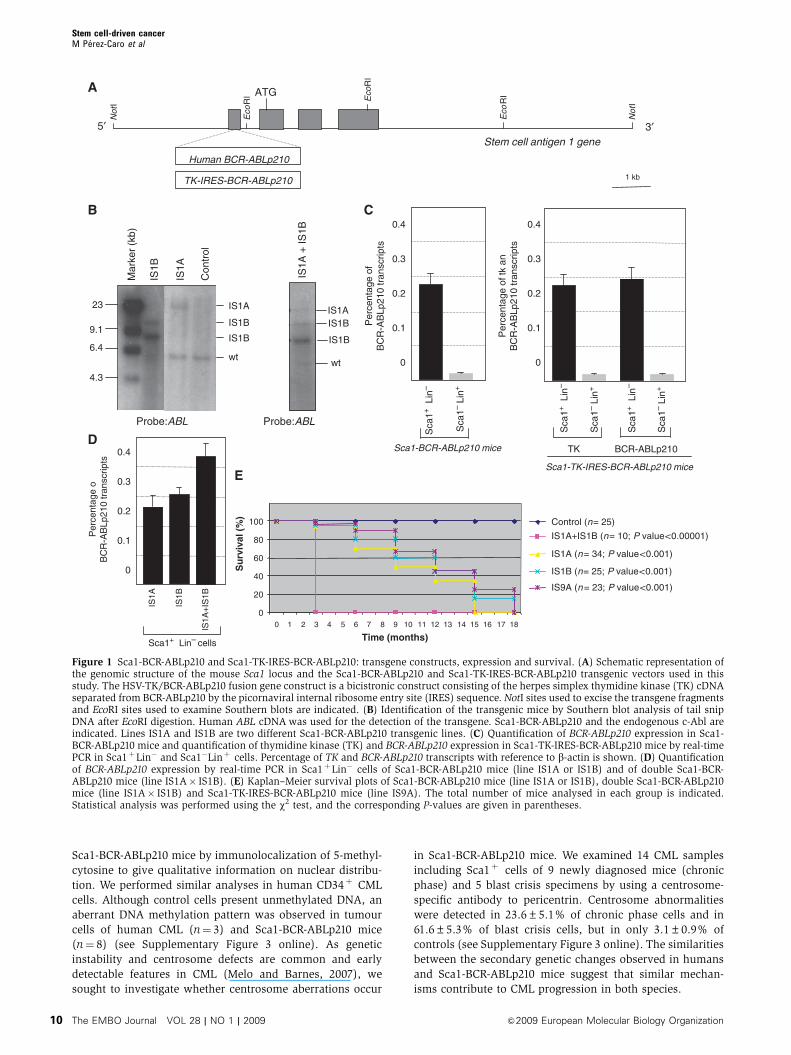
Figure 1 Sca1-BCR-ABLp210 and Sca1-TK-IRES-BCR-ABLp210: transgene constructs, expression and survival. (A) Schematic representation ofthe genomic structure of the mouse Sca1 locus and the Sca1-BCR-ABLp210 and Sca1-TK-IRES-BCR-ABLp210 transgenic vectors used in thisstudy. The HSV-TK/BCR-ABLp210 fusion gene construct is a bicistronic construct consisting of the herpes simplex thymidine kinase (TK) cDNAseparated from BCR-ABLp210 by the picornaviral internal ribosome entry site (IRES) sequence. NotI sites used to excise the transgene fragmentsand EcoRI sites used to examine Southern blots are indicated. (B) Identification of the transgenic mice by Southern blot analysis of tail snipDNA after EcoRI digestion. Human ABL cDNA was used for the detection of the transgene. Sca1-BCR-ABLp210 and the endogenous c-Abl areindicated. Lines IS1A and IS1B are two different Sca1-BCR-ABLp210 transgenic lines. (C) Quantification of BCR-ABLp210 expression in Sca1-BCR-ABLp210 mice and quantification of thymidine kinase (TK) and BCR-ABLp210 expression in Sca1-TK-IRES-BCR-ABLp210 mice by real-timePCR in Sca1þLin� and Sca1�Linþ cells. Percentage of TK and BCR-ABLp210 transcripts with reference to b-actin is shown. (D) Quantificationof BCR-ABLp210 expression by real-time PCR in Sca1þLin� cells of Sca1-BCR-ABLp210 mice (line IS1A or IS1B) and of double Sca1-BCR-ABLp210 mice (line IS1A� IS1B). (E) Kaplan–Meier survival plots of Sca1-BCR-ABLp210 mice (line IS1A or IS1B), double Sca1-BCR-ABLp210mice (line IS1A� IS1B) and Sca1-TK-IRES-BCR-ABLp210 mice (line IS9A). The total number of mice analysed in each group is indicated.Statistical analysis was performed using the w2 test, and the corresponding P-values are given in parentheses.
Stem cell-driven cancerM Perez-Caro et al
The EMBO Journal VOL 28 | NO 1 | 2009 &2009 European Molecular Biology Organization10
Nature of the leukaemogenic cell in the
Sca1-BCR-ABLp210 model
We next examined the nature of the leukaemogenic cell in the
Sca1-BCR-ABLp210 model. To determine whether the
Sca1þLin� population contains CSC, we sorted Sca1þLin�
and Sca1�Linþ cells from mice that developed CML.
Transplantation of purified fractions of cells into sublethally
irradiated syngeneic recipient mice was used to assess leu-
kaemogenesis in vivo. Each of the mice transplanted with
Sca1þ Lin� cells developed CML that was phenotypically
identical to the primary disease (Table II). Importantly,
Sca1�Linþ cells did not show leukaemic engraftment into
secondary recipients or induced leukaemia, even at 10 times
higher concentrations. Overall, these data indicate that the
CSCs reside in the Sca1þ cell compartment. All these facts
indicate that restricting oncogene expression to the stem cell
compartment is sufficient to generate all the CML cell types,
including the most differentiated ones, which do not express
the oncogene and are unable to propagate the disease.
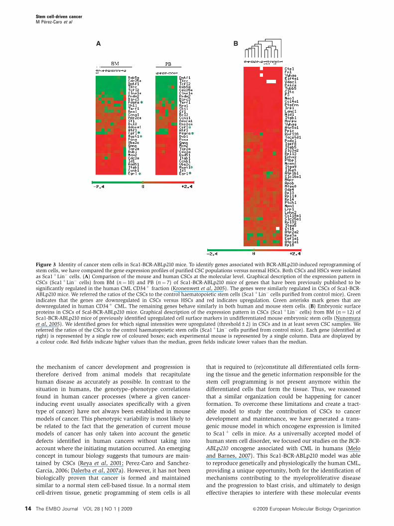
Characterization of CSCs in Sca1-BCR-ABLp210 mice
Having prospectively purified a population highly enriched
for CSC, we used gene expression to identify the genes that
are associated with BCR-ABLp210 reprogramming of stem
cells (Supplementary Table I online; Figure 3). We performed
a supervised analysis of the transcriptional profiles of CSCs
purified from Sca1-BCR-ABLp210 versus those from control
mice. The data showed that a total of 293 genes are repro-
ducibly regulated in Sca1-BCR-ABLp210 versus control HSC
with a false discovery rate (FDR) p0.07% (Supplementary
Table I online). Human CML has been characterized earlier
according to gene expression profile (Kronenwett et al, 2005).
Remarkably, the gene expression patterns of Sca1þLin� cells
from Sca1-BCR-ABLp210 mice were similar to those from the
human CML CD34þ cells (Figure 3A), thereby further vali-
dating the model by reflecting the similarities in the tran-
scriptome between the two populations. Thus, BCR-ABLp210-
dependent, stem cell-driven murine CML shares the molecu-
lar features of human CML.
The CSC hypothesis represents a modern-day interpreta-
tion of the proposal made by Rudolph Virchow and Julius
Cohnheim that cancer results from the activation of dormant
embryonal-rest cells (Virchow, 1855; Cohnheim, 1867).
Accordingly, we next proceeded to examine, in CSCs from
Scal-BCR-ABLp210 mice, the expression of embryonic surface
markers, the presence of which has been identified earlier in
undifferentiated mouse embryonic stem cells (Nunomura
et al, 2005) (Figure 3B). We could identify reproducible
upregulation of gene signals corresponding to 55 embryonic
stem cell surface proteins in the CSCs from Sca1-BCR-
ABLp210 mice (FDR p1%). These results show that CSCs
in Sca1-BCR-ABLp210 present embryonic figures that could
represent attractive targets for selective CSC removal.
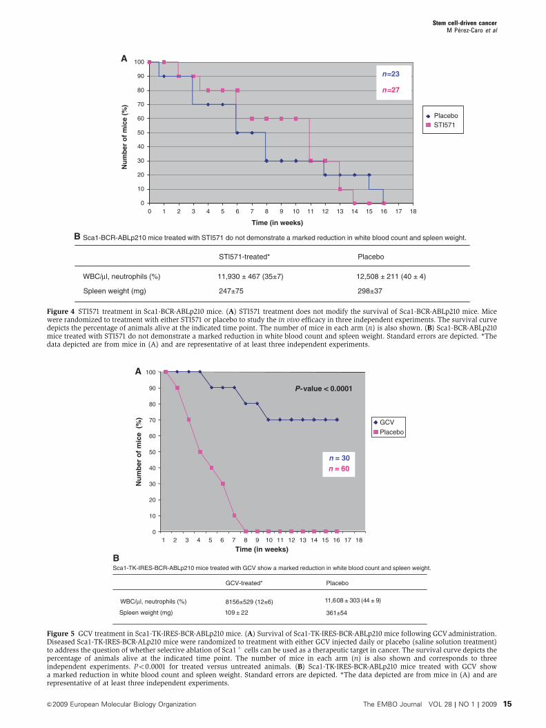
STI571 treatment does not modify the survival of
Sca1-BCR-ABLp210 mice
We also examined the effect of STI571 treatment in Sca1-BCR-
ABLp210 mice. STI571 treatment began 1 day after leukaemia
was confirmed by PB analysis. Mice were monitored clini-
cally and by serial PB count for evidence of leukaemia.
STI571 did not prolong the survival of these mice
(Figure 4A) and Sca1-BCR-ABLp210 mice treated with
STI571 did not demonstrate a marked reduction in WBC
and spleen weight (Figure 4B). This idea is in agreement
with the insensitivity of the human leukaemic stem cells to
STI571 (Graham et al, 2002; Hu et al, 2006; Primo et al, 2006;
Jiang et al, 2007). Thus, we next examined whether CSCs
from STI571-treated Sca1-BCR-ABLp210 mice propagates
CML disease to secondary recipients. Equal numbers of
Sca1þLin� cells (5�103) from the BM of STI571-treated
animals failing therapy were used to reconstitute sublethally
irradiated recipients. All reconstituted animals (n¼ 5) devel-
oped CML disease with an average latency of only 24 days,
confirming that persistent BCR-ABLp210 expression is not
required to maintain the malignant phenotype of CML CSCs.
CSC ablation eradicates CML in Sca1-BCR-ABLp210
mice
We next examined whether CSC ablation implies elimination
of CML in vivo following tumour formation in a whole
animal. For this purpose, we generated Sca1-TK-IRES-BCR-
ABLp210 mice. These animals developed disease similarly to
the Sca1-BCR-ABLp210 ones (Figure 1; Table I). Diseased
Sca1-TK-IRES-BCR-ABLp210 mice were injected daily with
ganciclovir (GCV) as described in the Materials and methods
section. During this period, mice were monitored for evi-
dence of leukaemia. GCV treatment clearly reduced the
number of mice developing tumours (Figure 5A), with the
majority of animals demonstrating a marked reduction in
splenomegaly and, in some cases, complete normalization of
their peripheral WBC (Figure 5B) and lack of solid tumours
(although we could not confirm their presence before treat-
Table I Incidence and age of CML onset in Sca1-BCR-ABLp210 and Sca1-TK-IRES-BCR-ABLp210 mice
Transgenic line Miceautopsieda
Mice withtumour(%)b
Age in monthsat tumour
onset
Haematopoietictumour type
(%)
WBC/ml,neutrophils (%)
Hgb(g/100 ml)
Spleenweight(mg)
IS1A (Sca1-BCR-ABLp210) 34 34 (100) 8–12 CML–BC (100) 12 900±1750 (43±4) 11.5±1.3 285±36IS1B (Sca1-BCR-ABLp210) 25 25 (100) 4–10 CML–BC (100) 11 850±1650 (38±5) 12.7±1.1 217±29IS1A+IS1B 10 10 (100) 1–2 Leukaemia ND 13.2±1.3 NDIS9A (Sca1-TK-IRES-BCR-ABLp210) 23 23 (100) 6–10 CML–BC (100) 13 300±230 (41±8) 11.1±1.2 261±23
ND, not determined.aNumber of mice during or after the period of cancer.bNumber of mice with CML and percentage of tumour incidence. Normal range of WBC counts was 4000–10 000/ml. Mean neutrophilpercentage of control animals was 10±4% WBCs, and the neutrophil percentage in control animals did not exceed 20%. Thus, neutrophilia isdefined as more than 20% neutrophils. Hgb indicates haemoglobin (normal range, 12–16 g/100 ml). Normal spleen weight in control animals is78–92 mg (n¼ 25). The WCC estimations were made when the mice were premorbid. CML–BC indicates mice spontaneously progress to blastcrisis. Po0.001 for Sca1-BCR-ABLp210 mice versus control mice.
Stem cell-driven cancerM Perez-Caro et al
&2009 European Molecular Biology Organization The EMBO Journal VOL 28 | NO 1 | 2009 11
ment). So GCV treatment cures CML in previously sick
transgenic animals. Although all animals exhibited a pro-
longed survival with GCV treatment, approximately 23% of
the animals died of leukaemia during the treatment with the
drug. To test the possibility that CSCs would remain within
Sca1-TK-IRES-BCR-ABLp210 mice after GCV treatment, we
highly purified the Sca1þLin� sub-population from BM and
transplanted it into sublethally irradiated syngeneic recipi-
ents (n¼ 5). Sca1þLin� cells did not show leukaemic en-
graftment into secondary recipients or induced leukaemia,
indicating that leukaemogenic cells had been removed from
this population by the GCV treatment. To our knowledge, this
study demonstrates for the first time that killing CSCs is an
effective therapeutic strategy for cancer treatment.
LiverSpleenBA
SCA1 -> Gr 1 ->
76.4%mature granules
PBPB
Gr
1
TR
AN
SF
OR
ME
D S
SC
->
SCA1 ->
Gr
1
9%
Control Sca1-BCR-ABLp210
B220 FITC ->
spleen
Mac
1
Mac 1 ->
BM
Gr
1
Sca1-BCR-ABLp210
Sca1-BCR-ABLp210
PB X 20
X 40
X 20
X 40100
100
101
101
102
102
103
103
104
100 101 102 103 104 100 101 102 103 104
104
100
101
102
103
104
100
101
102
103
104
100
101
102
103
104 1024
768
512
256
0
100 101 102 103 104 100 101 102 103 104
X20
X40
X40
X20
X40
X20
20X
X40
X20
Sp
leen
Control
X20
Liv
er
X20
No
de
Sca1-BCR-ABLp210C
Sca1-BCR-ABLp210
X40Lung
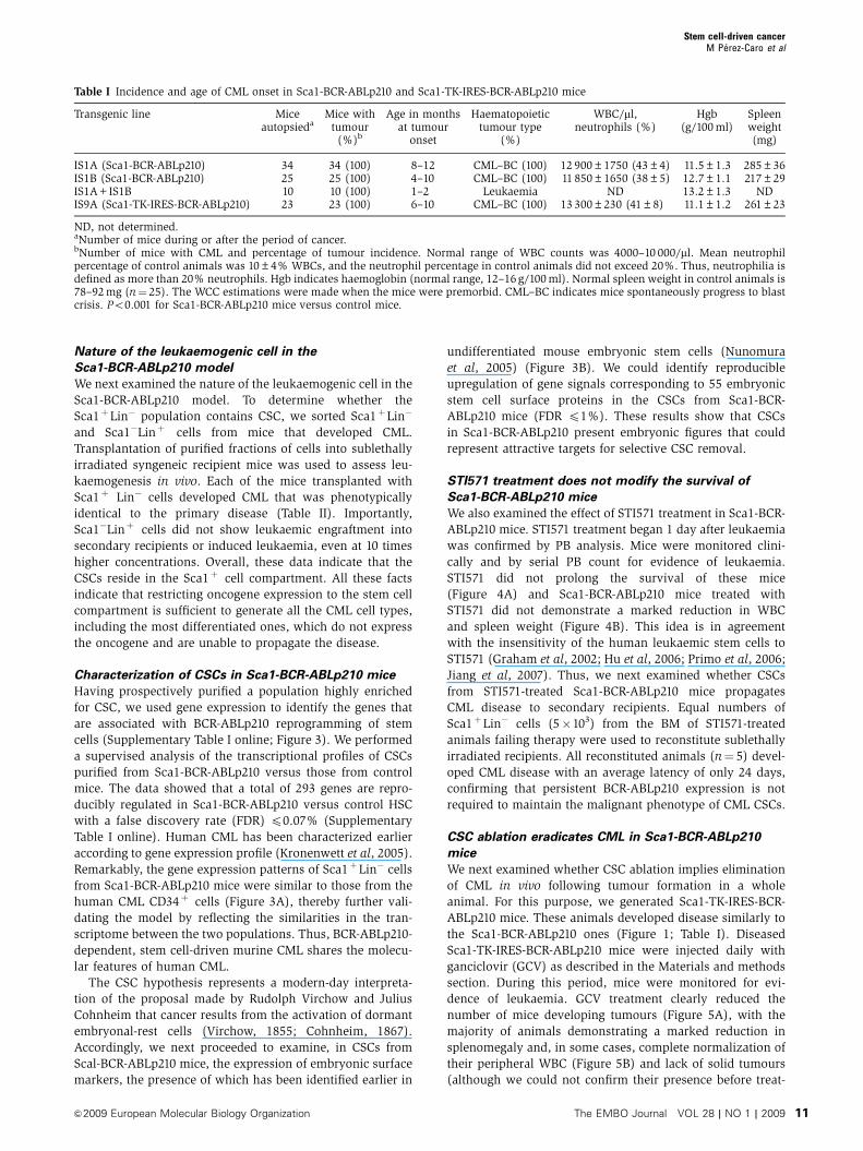
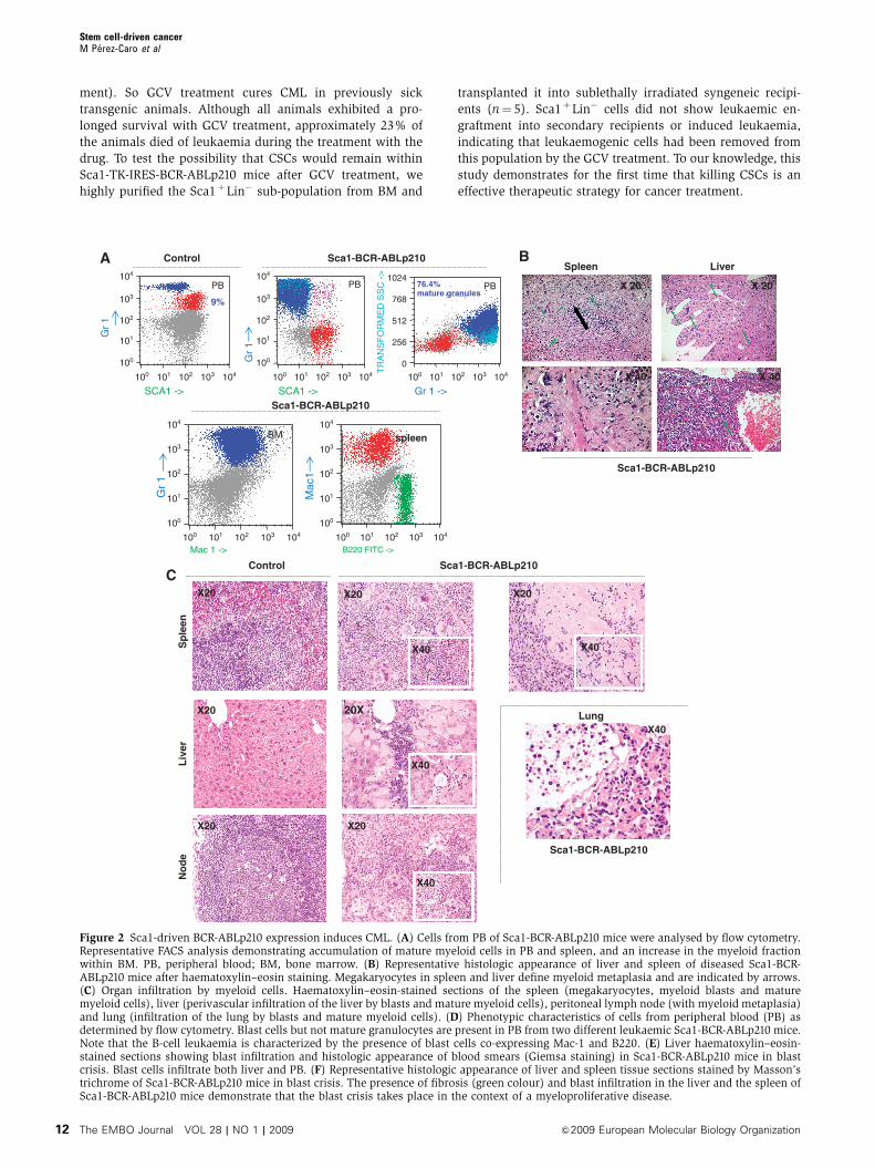
Figure 2 Sca1-driven BCR-ABLp210 expression induces CML. (A) Cells from PB of Sca1-BCR-ABLp210 mice were analysed by flow cytometry.Representative FACS analysis demonstrating accumulation of mature myeloid cells in PB and spleen, and an increase in the myeloid fractionwithin BM. PB, peripheral blood; BM, bone marrow. (B) Representative histologic appearance of liver and spleen of diseased Sca1-BCR-ABLp210 mice after haematoxylin–eosin staining. Megakaryocytes in spleen and liver define myeloid metaplasia and are indicated by arrows.(C) Organ infiltration by myeloid cells. Haematoxylin–eosin-stained sections of the spleen (megakaryocytes, myeloid blasts and maturemyeloid cells), liver (perivascular infiltration of the liver by blasts and mature myeloid cells), peritoneal lymph node (with myeloid metaplasia)and lung (infiltration of the lung by blasts and mature myeloid cells). (D) Phenotypic characteristics of cells from peripheral blood (PB) asdetermined by flow cytometry. Blast cells but not mature granulocytes are present in PB from two different leukaemic Sca1-BCR-ABLp210 mice.Note that the B-cell leukaemia is characterized by the presence of blast cells co-expressing Mac-1 and B220. (E) Liver haematoxylin–eosin-stained sections showing blast infiltration and histologic appearance of blood smears (Giemsa staining) in Sca1-BCR-ABLp210 mice in blastcrisis. Blast cells infiltrate both liver and PB. (F) Representative histologic appearance of liver and spleen tissue sections stained by Masson’strichrome of Sca1-BCR-ABLp210 mice in blast crisis. The presence of fibrosis (green colour) and blast infiltration in the liver and the spleen ofSca1-BCR-ABLp210 mice demonstrate that the blast crisis takes place in the context of a myeloproliferative disease.
Stem cell-driven cancerM Perez-Caro et al
The EMBO Journal VOL 28 | NO 1 | 2009 &2009 European Molecular Biology Organization12
Discussion
The elucidation of the molecular mechanisms that underlie
tumour development not only remains a tremendous chal-
lenge for basic science but also represents an essential step in
the development of new and more potent drugs, in particular
with the emergence of cancer-specific, targeted therapies. The
origin of human cancer within a particular tissue is often
impossible to determine, due to the advanced stages of many
tumours when patients enter the clinic. Our knowledge about
Table II CML disease is readily transplantable to secondary recipients
Sorted cells Number oftransplanted cells
Transplantedanimals
Incidence ofCML (%)
Latency of disease(days±s.d.)
Sca1+Lin� (BM, Sca1-BCR-ABLp210 mice) 10.000 8 100 69±141.000 8 100 85±11
Sca1+Lin� (BM, control mice) 10.000 8 0 NA1.000 8 0 NA
Sca1�Lin+ (BM, Sca1-BCR-ABLp210 mice) 1�105 8 0 NA1�106 8 0 NA
Sca1�Lin+ (BM, control mice) 1�105 8 0 NA1�106 8 0 NA
NA, not applicable.Eight irradiated syngenic recipients mice per cohort were transplanted with the indicated number of cells. The diagnosis of CML was confirmedby histological and immunophenotypic examination of the ill recipient mice.
Liver
Blood smear
E
X20
X40MAC-1 ->
B-cell leukaemia
D
Mac 1 ->
Sca1-BCR-ABLp210 Control
79, 7%
B220 FITC ->
50.63%17.89%
Myeloid leukaemia
Sp
leen
Liv
er
ControlF
Sca1-BCR-ABLp210
X20
X40X20 X20
X40X20
100
100
101
101
102
102
103
103
B22
0 ->
Mac
1 P
E -
>
B22
0->
104
104
100
100
101
101
102
102
103
103
104
104
100
100
101
101
102
102
103
103
104
104
Figure 2 Continued.
Stem cell-driven cancerM Perez-Caro et al
&2009 European Molecular Biology Organization The EMBO Journal VOL 28 | NO 1 | 2009 13
the mechanism of cancer development and progression is
therefore derived from animal models that recapitulate
human disease as accurately as possible. In contrast to the
situation in humans, the genotype–phenotype correlations
found in human cancer processes (where a given cancer-
inducing event usually associates specifically with a given
type of cancer) have not always been established in mouse
models of cancer. This phenotypic variability is most likely to
be related to the fact that the generation of current mouse
models of cancer has only taken into account the genetic
defects identified in human cancers without taking into
account where the initiating mutation occurred. An emerging
concept in tumour biology suggests that tumours are main-
tained by CSCs (Reya et al, 2001; Perez-Caro and Sanchez-
Garcia, 2006; Dalerba et al, 2007a). However, it has not been
biologically proven that cancer is formed and maintained
similar to a normal stem cell-based tissue. In a normal stem
cell-driven tissue, genetic programming of stem cells is all
that is required to (re)constitute all differentiated cells form-
ing the tissue and the genetic information responsible for the
stem cell programming is not present anymore within the
differentiated cells that form the tissue. Thus, we reasoned
that a similar organization could be happening for cancer
formation. To overcome these limitations and create a tract-
able model to study the contribution of CSCs to cancer
development and maintenance, we have generated a trans-
genic mouse model in which oncogene expression is limited
to Sca1þ cells in mice. As a universally accepted model of
human stem cell disorder, we focused our studies on the BCR-
ABLp210 oncogene associated with CML in humans (Melo
and Barnes, 2007). This Sca1-BCR-ABLp210 model was able
to reproduce genetically and physiologically the human CML,
providing a unique opportunity, both for the identification of
mechanisms contributing to the myeloproliferative disease
and the progression to blast crisis, and ultimately to design
effective therapies to interfere with these molecular events
A B
Figure 3 Identity of cancer stem cells in Sca1-BCR-ABLp210 mice. To identify genes associated with BCR-ABLp210-induced reprogramming ofstem cells, we have compared the gene expression profiles of purified CSC populations versus normal HSCs. Both CSCs and HSCs were isolatedas Sca1þLin� cells. (A) Comparison of the mouse and human CSCs at the molecular level. Graphical description of the expression pattern inCSCs (Sca1þLin� cells) from BM (n¼ 10) and PB (n¼ 7) of Sca1-BCR-ABLp210 mice of genes that have been previously published to besignificantly regulated in the human CML CD34þ fraction (Kronenwett et al, 2005). The genes were similarly regulated in CSCs of Sca1-BCR-ABLp210 mice. We referred the ratios of the CSCs to the control haematopoietic stem cells (Sca1þLin� cells purified from control mice). Greenindicates that the genes are downregulated in CSCs versus HSCs and red indicates upregulation. Green asterisks mark genes that aredownregulated in human CD34þ CML. The remaining genes behave similarly in both human and mouse stem cells. (B) Embryonic surfaceproteins in CSCs of Scal-BCR-ABLp210 mice. Graphical description of the expression pattern in CSCs (Sca1þLin� cells) from BM (n¼ 12) ofSca1-BCR-ABLp210 mice of previously identified upregulated cell surface markers in undifferentiated mouse embryonic stem cells (Nunomuraet al, 2005). We identified genes for which signal intensities were upregulated (threshold±2) in CSCs and in at least seven CSC samples. Wereferred the ratios of the CSCs to the control haematopoietic stem cells (Sca1þLin� cells purified from control mice). Each gene (identified atright) is represented by a single row of coloured boxes; each experimental mouse is represented by a single column. Data are displayed bya colour code. Red fields indicate higher values than the median, green fields indicate lower values than the median.
Stem cell-driven cancerM Perez-Caro et al
The EMBO Journal VOL 28 | NO 1 | 2009 &2009 European Molecular Biology Organization14
PlaceboSTI571
Nu
mb
ero
fm
ice
(%)
0
10
20
30
40
50
60
70
80
90
100
0 1 2 3 4 5 6 7 8 9 10 11 12 13 14 15 16 17 18
Time (in weeks)
n=23
n=27
A
B Sca1-BCR-ABLp210 mice treated with STI571 do not demonstrate a marked reduction in white blood count and spleen weight.
PlaceboSTl571-treated*
WBC/μl, neutrophils (%) 11,930 ± 467 (35±7) 12,508 ± 211 (40 ± 4)
247±75 298±37Spleen weight (mg)
Figure 4 STI571 treatment in Sca1-BCR-ABLp210 mice. (A) STI571 treatment does not modify the survival of Sca1-BCR-ABLp210 mice. Micewere randomized to treatment with either STI571 or placebo to study the in vivo efficacy in three independent experiments. The survival curvedepicts the percentage of animals alive at the indicated time point. The number of mice in each arm (n) is also shown. (B) Sca1-BCR-ABLp210mice treated with STI571 do not demonstrate a marked reduction in white blood count and spleen weight. Standard errors are depicted. *Thedata depicted are from mice in (A) and are representative of at least three independent experiments.
BSca1-TK-IRES-BCR-ABLp210 mice treated with GCV show a marked reduction in white blood count and spleen weight.
GCV-treated* Placebo
)9± 44( 303± 8061,18156±529 (12±6)
361±5422± 901Spleen weight (mg)
WBC/μl, neutrophils (%)
0
10
20
30
40
50
60
70
80
90
100
1 2 3 4 5 6 7 8 9 10 11 12 13 14 15 16 17 18
Nu
mb
ero
fm
ice
(%)
Time (in weeks)
n = 30n = 60
P-value < 0.0001
A
GCV
Placebo
Figure 5 GCV treatment in Sca1-TK-IRES-BCR-ABLp210 mice. (A) Survival of Sca1-TK-IRES-BCR-ABLp210 mice following GCV administration.Diseased Sca1-TK-IRES-BCR-ALp210 mice were randomized to treatment with either GCV injected daily or placebo (saline solution treatment)to address the question of whether selective ablation of Sca1þ cells can be used as a therapeutic target in cancer. The survival curve depicts thepercentage of animals alive at the indicated time point. The number of mice in each arm (n) is also shown and corresponds to threeindependent experiments. Po0.0001 for treated versus untreated animals. (B) Sca1-TK-IRES-BCR-ABLp210 mice treated with GCV showa marked reduction in white blood count and spleen weight. Standard errors are depicted. *The data depicted are from mice in (A) and arerepresentative of at least three independent experiments.
Stem cell-driven cancerM Perez-Caro et al
&2009 European Molecular Biology Organization The EMBO Journal VOL 28 | NO 1 | 2009 15
and to study the contribution of CSCs to disease development
and maintenance.
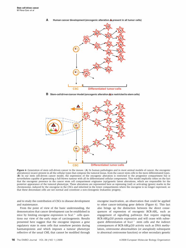
From the point of view of the basic understanding, the
demonstration that cancer development can be established in
mice by limiting oncogene expression to Sca1þ cells ques-
tions our view of the early steps of carcinogenesis. Results
presented here suggest that the oncogene imposes a gene
regulatory state in stem cells that somehow persists during
haematopoiesis and which imposes a tumour phenotype
reflective of the usual CML that cannot be modified through
oncogene inactivation, an observation that could be applied
to other cancer-initiating gene defects (Figure 6). This fact
also brings up the distinction between the direct conse-
quences of expression of oncogenic BCR-ABL, such as
engagement of signalling pathways that require ongoing
BCR-ABLp210 protein expression and will cease with subse-
quent differentiation of Sca1þ stem cells and the indirect
consequences of BCR-ABLp210 activity such as DNA methy-
lation, centrosome abnormalities (or aneuploidy subsequent
to abnormal centrosome function) or other secondary genetic
A
B
d
d c m o
t c
t c
Figure 6 Generation of stem cell-driven cancer in the mouse. (A) In human pathologies and in most animal models of cancer, the oncogenicalteration(s) is(are) present in all the cellular types that compose the tumoral tissue, from the cancer stem cells to the more differentiated types.(B) In our stem cell-driven cancer model, the expression of the oncogenic alteration is restricted to the progenitor compartment but isnevertheless capable of generating a full-blown tumour with all its differentiated cellular components. This model implicitly relies on the factthat the oncogenic presence in the cancer stem cell compartment originates (epi)genetic latent alterations, which are responsible for theposterior appearance of the tumoral phenotype. These alterations are represented here as repressing (red) or activating (green) marks in thechromosome, induced by the oncogene in the CSCs and inherited in the lower compartments where the oncogene is no longer expressed, sothat these descendant cells are not normal and constitute a non-clonogenic leukaemic progeny.
Stem cell-driven cancerM Perez-Caro et al
The EMBO Journal VOL 28 | NO 1 | 2009 &2009 European Molecular Biology Organization16
changes that may be inherited by subsequent generations
regardless of continued BCR-ABLp210 expression. Our find-
ings show that BCR-ABL appears to cause epigenetic and/or
genetic changes in tumour-maintaining cells that could render
them insensitive to BCR-ABL inactivation. Thus, BCR-ABL
oncogene inactivation cannot change this epigenetic/genetic
context in differentiated tumour cells in agreement with the
common occurrence of tumour relapse by which tumours
evolve to escape oncogene dependence. Therefore, we hy-
pothesize that BCR-ABL mediates tumorigenesis through epi-
genetic/genetic modification of target genes that remain in
this modified state in the mature tumour even in the absence
of BCR-ABL. As an added level of complexity, this effect of
BCR-ABL in stem cells could be mediated by a kinase-inde-
pendent mechanism and therefore it would be Gleevec in-
sensitive. The model provides evidence that the oncogenic
proteins expressed in stem/progenitor cells can have selective
impacts that depend on their intrinsic molecular properties.
This result in turn provides a rationale for the striking
associations between different chromosome translocations,
unique fusion genes and cancer phenotypes. Importantly, a
small subset of mice developed additional solid tumours,
indicating that BCR-ABL stem cell-driven oncogenesis is not
only specific to haematopoietic tissues but also represents a
broader mechanism for deregulation of stem cell differentia-
tion, providing a paradigm that can be applied to solid-organ
cancers. Thus, these results support the view of cancer as a
disease of cell differentiation rather than multiplication, ask-
ing for a redefinition of the role of oncogenes.
Overall, our findings indicate that BCR-ABLp210 appears to
reprogramme stem cells rendering them insensitive to poster-
ior BCR-ABLp210 inactivation. This evidence that BCR-
ABLp210 inactivation could not stop tumour growth is in
apparent conflict with earlier studies showing that BCR-
ABLp210 is required for the persistence of tumoral character-
istics in cellular systems (Szczylik et al, 1991; Choo et al,
1994; Skorski et al, 1994; Cobaleda and Sanchez-Garcia,
2000; Huettner et al, 2000). However, we should consider
that the effects of BCR-ABLp210 inactivation will depend on
the mechanisms by which BCR-ABLp210 is actually contribut-
ing to the tumorigenic phenotype, which are likely to vary
according to the genetic and cellular context. When BCR-
ABLp210 causes CML in vivo, as described here and observed
in humans, its inactivation cannot eliminate CML cell pro-
genitors. By contrast, when BCR-ABLp210 is just transforming
a cell line into tumorigenic, its inactivation would result in
tumour regression (Szczylik et al, 1991; Choo et al, 1994;
Skorski et al, 1994; Cobaleda and Sanchez-Garcia, 2000;
Huettner et al, 2000). So, as mentioned before, it is very
possible that the molecular mechanisms of action of BCR-
ABLp210 at the CSC level are different from those acting at
later stages of tumoral cell differentiation (i.e. direct versus
indirect BCR-ABL effects as discussed above).
The data presented here further show for the first time the
in vivo physiological relevance of the CSC suppression using
a model system representing in vivo biology of the human
CML disease. Specific CSC ablation is able to eradicate CML
in our model, although these cells were insensitive to
Gleevec-mediated BCR-ABLp210 inactivation, suggesting
that the reprogramming imposed by BCR-ABLp210 renders
the cells oncogene independent. We conclude that limited
oncogene expression in a CSC is all that is required to fully
reprogramme it, giving rise to a full-blown, oncogene-speci-
fied tumour with all its mature cellular diversity, and that
BCR-ABLp210-induced CML is reversible through the unique
elimination of the CSC. Further experiments should confirm
that this observation could be applied to other cancer-initiat-
ing gene defects. However, are there targets that can be
exploited to eradicate CSCs without affecting healthy stem
cells? The characterization of the CSCs in the Sca1-BCR-
ABLp210 mice showed that CSCs were different from normal
stem cells exhibiting cell surface embryonic figures that could
be used to target CSCs. Thus, in agreement with earlier
studies (Yilmaz et al, 2006; Liu et al, 2007; Ito et al, 2008),
these data provide a rationale for the development of diag-
nostic and therapeutic strategies targeting CSCs without
affecting normal stem cells. The hope is to translate this
discovery into new tools for a better diagnostic and thera-
peutic management of the human disease.
Materials and methods
Generation of transgenic mice and BM transplantationThe Ly-6E.1 gene has been shown earlier to direct heterologousgene expression in HSCs of the adult BM and to specify copynumber-dependent and integration site-independent expression(Miles et al, 1997). In this study, we have exploited the fact thatthis transgene is not expressed in all Sca1-positive cells for the studyof cancer. The Sca1-BCR-ABLp210 and Sca1-TK-IRES-BCR-ABLp210vectors were generated as follows. The 6 kb EcoRI–EcoRI fragment,containing the human BCR-ABLp210 cDNA, and the 9 kb EcoRI–EcoRI TK-IRES-BCR-ABLp210 cassette were inserted into the ClaIsite of the pLy6 vector (Miles et al, 1997), resulting in Sca1-BCR-ABLp210 and Sca1-TK-IRES-BCR-ABLp210 vectors, respectively. Thetransgene fragments (Figure 1A) were excised from its vectors byrestriction digestion with NotI, purified for injection (2 ng/ml) andinjected into CBA�C57BL/6J fertilized eggs. Transgenic mice wereidentified by Southern blot analysis of tail snip DNA after EcoRIdigestion. Human ABL cDNA was used for detection of thetransgene. A total of 82 transgenic animals and 25 control animalswere used to define the phenotype. Three independent transgeniclines were generated and analysed and similar phenotypic featureswere seen in both Sca1-BCR-ABLp210 lines and Sca1-TK-IRES-BCR-ABLp210 mice. IS1A line was crossed with IS1B transgenic mice togenerate compound heterozygotes and increase BCR-ABL dosage(Figure 1B and D).
To determine the nature of the leukaemogenic cell, Sca1þLin�
and Sca1�Linþ cells were isolated and highly purified from the BMof a leukaemic primary mouse before or after different treatments orfrom control mice. The sorting purity of these cells was re-analysedwith the fluorescence-activated cell sorting (FACS) and determinedto be over 98%. In each cohort, these cells were injectedintravenously into sublethally irradiated (4 Gy) recipient mice.Diseased mice were killed and assessed for leukaemia development.Mice that did not develop disease within 20 weeks post-transplantation were killed and tested for engraftment.
Histological analysisAll mice included in this study were subjected to standard necropsy.All major organs were examined under the dissecting microscope,and samples of each organ were processed into paraffin, sectionedand examined histologically. All tissue samples were taken fromhomogenous and viable portions of the resected sample by thepathologist and fixed within 2–5 min of excision. Haematoxylin–eosin-stained sections of each tissue were reviewed by a singlepathologist (TF). Demonstration of fibrosis in spleen and liver ofdiseased Sca1-BCR-ABLp210 mice (green colour) was carried out byMasson’s trichrome staining. For comparative studies, age-matchedmice were used.
Analysis and monitoring of diseasePB was collected from retro-orbital plexus with a heparinizedcapillary tube, and total WBC and differential counts wereperformed twice a week. The number of WBCs was determined
Stem cell-driven cancerM Perez-Caro et al
&2009 European Molecular Biology Organization The EMBO Journal VOL 28 | NO 1 | 2009 17
with a haemocytometer after lysis of enucleated red blood cells withRCLB lysis buffer (0.15 M NH4Cl; 1 mM KHCO3; 0.1 mM Na2-EDTA,pH 7.4).
Flow cytometryNucleated cells were obtained from total BM (flushing from the longbones), PB, thymus, liver and spleen. To prepare the cells for flowcytometry, contaminating red blood cells were lysed with RCLBlysis buffer and the remaining cells were then washed in PBS with2% fetal calf serum (FCS). After staining, all cells were washed oncein PBS with 2% FCS containing 2mg/ml propidium iodide (PI) toallow dead cells to be excluded from both analyses and sortingprocedures. Monoclonal antibodies were obtained from Pharmin-gen and included: lineage markers (CD45R/B220, for B lineagestaining; CD4, CD8 and CD3 for T-cell lineage; CD11b and Gr1 formyeloid lineage and TER119 for erythroid lineage) and Sca1 (E13-161.7) for stem cells. Single cell suspensions from the differenttissue samples obtained by routine techniques were incubated firstwith purified anti-mouse CD32/CD16 (Pharmingen) prior to theaddition of other antibodies, to block binding through Fc receptorsand then with an appropriate dilution of the different antibodies atroom temperature or 41C, respectively. The samples and the datawere analysed in a FACSCalibur using CellQuest software (BectonDickinson). Specific fluorescence of FITC and PE excited at 488 nm(0.4 W) and 633 nm (30 mW), respectively, as well as knownforward and orthogonal light scattering properties of mouse cellswere used to establish gates. For each analysis, a total of at least5.000 viable (PI�) cells were assessed.
Cell purificationFor cell sorter separation, BM cells were incubated with anti-Sca1and anti-lineage marker antibodies (CD3, CD4, CD8, B220, TER119,Gr1 and Mac-1). Sca1þLin� and Sca1�Linþ cells were isolated andhighly purified from the BM of leukaemic primary mice or controlmice by FACS (FACSVANTAGE; Becton Dickinson). c-kit (CD117)was not used for stem cell isolation, as earlier studies of human andmouse specimens have described downregulation of c-kit as afeature of leukaemia stem cells (Blair and Sutherland, 2000;Neering et al, 2007). Sorted cells were then re-analysed for puritywith the FACS and determined to be over 98%.
RNA extractionTotal RNA was isolated in two steps using TRIzol (Life TechnologiesInc., Grand Island, NY) followed by RNeasy Mini-kit (Qiagen Inc.,Valencia, CA) purification following the manufacturer’s RNA clean-up protocol with the optional On-column DNase treatment. Theintegrity and the quality of RNA were verified by electrophoresisand its concentration was measured.
Lineal RNA amplificationT-7-based RNA amplifications and preparations of cDNA probeswere performed. Briefly, a maximum amount of 5 mg of total RNAwas converted to double-stranded cDNA using the superscriptchoice system (Life Technologies Inc.) using oligo-dT primercontaining a T7 RNA polymerase promoter. The double-strandedcDNA was cleaned up, and T7 in vitro transcription was performedusing Megascript T7 in vitro transcription kit (Ambion, Austin, TX)following the manufacturers’ instructions.
Microarray proceduresSecond round amplified RNA (2.5mg) from each sample wasdirectly labelled with cyanine 3 (Cy3)-conjugated dUTP, whereas2.5mg of second round amplified RNA from the Universal MouseReference RNA (Stratagene) was labelled with cyanine 5 (Cy5)-conjugated dUTP as a reference. For all the microarray studies, theCNIO MouseChip was used and hybridizations were performed asdescribed (Bermejo-Rodriguez et al, 2006). After washing, the slideswere scanned using a Scanarray 5000 XL (GSI Lumonics Kanata,Ontario, Canada) and images were analysed with the GenePix 4.0program (Axon Instruments Inc., Union City, CA).
Data analysisData obtained from each hybridization were stored in a database foranalysis. The Cy3/Cy5 ratios were normalized to the median ratiovalue of all of the spots in the array. After normalization, spots withintensities for both channels (sum of medians) less than that of thelocal background were discarded. The ratios of the remaining spots
were log transformed (base 2), and duplicated spots on theMouseChip were averaged to the median. To obtain the expressionprofile of CSCs (Sca1þLin� cells purified from Sca1-BCR-ABLp210mice), we referred the ratios of the CSCs to the control HSCs(Sca1þLin� cells purified from control mice).
Real-time PCR quantificationcDNA for use in quantitative PCR studies was synthesized usingreverse transcriptase (Access RT–PCR System; Promega, Madison,WI). Second round amplified RNA (2 ml) was transcribed. Primersand probes used for quantitative PCR are commercially available(TaqMan Assays-on-Demand Gene Expression Products; AppliedBiosystems, Foster City, CA). In addition, the probes were designedso that genomic DNA would not be detected during the PCR. Thesequences of the specific primers and probes were as follows: BCR-ABLp210, sense primer 50-TTCTGAATGTCATCGTCCACTCA-30, anti-sense primer 50-AGATGCTACTGGCCGCTGA-30 and probe 50-CCACTGGATTTAAGCAGAGTTCAAAAGCCC-30; c-Abl, sense primer50-CACTCTCAGCATCACTAAAGGTGAA-30, antisense primer 50-CGTTTGGGCTTCACACCATT-30 and probe 50-CCGGGTCTTGGGTTATAATCACAATG-30.
Immunolocalization of 5mC and pericentrinHuman and mouse cells grown on coverslips were stained withDAPI (blue) and mouse monoclonal antibodies against 5mC (red)(kindly provided by Alain Niveleau, Universite Claude Bernard,Lyon, France) as described earlier (Habib et al, 1999). Images of thenuclear immunolocalization of 5 mC were obtained in a LeicaDMRA fluorescence microscope coupled to a Leica DC200 digitalcamera and captured with the Adobe Photoshop software. Controland Sca1-BCR-ABLp210 Sca1þ cells were stained with antibodiesagainst pericentrin (green; Berkeley ref. PRB-432C) to labelcentrosomes and with DAPI (blue) to label nuclei.
STI571 (Gleevec)STI571 treatment regimens were based on earlier pharmacokineticstudies of STI in BCR-ABL tumour-bearing mice (le Coutre et al,1999; Druker et al, 2001; Perez-Caro et al, 2007). Effectivity of BCR-ABLp210 suppression by STI571 was confirmed by assaying thesurvival of Ba/F3 cells expressing BCR-ABLp210 24 h after STI571treatment and by the treatment of BCR-ABL transgenic miceaccording to previously published studies (Perez-Caro et al, 2007)(data not shown). For the animal studies, stock solutions of 5 and10 mg/ml were prepared fresh in water, sterile filtered andadministered to mice in a volume of 250ml by gavage twice a day.Mice were started on STI571 or placebo (the same volume of diluentwater) beginning 1 day after leukaemia was confirmed (day 0) bymeans of an STI571 regimen of 50 mg/kg every morning and100 mg/kg every evening by gavage. STI571 was administered in avolume of 250ml sterile water by means of straight or curved animalfeeding needles. Mice tolerated the therapy well and no interruptionof therapy was necessary. Mice were followed clinically three timesa week, and periodic PB counts were obtained by tail vein blooddraw as indicated. For the survival analysis portion of this study,the death end point was determined either by spontaneous death ofthe animal or by elective killing of the animal because of signs ofpain or suffering according to established criteria.
GCV treatmentSca1-TK-IRES-BCR-ABLp210 mice were used to test the effectivenessof GCV-induced cell in CSCs that express BCR-ABL. Expression ofTK induces conversion of the prodrug nucleoside GCV to its drugform as a phosphorylated base analogue. The phosphorylated GCVis incorporated into the DNA of replicating cells causing irreversiblearrest at the G2/M checkpoint followed by apoptosis (Rubsam et al,1998). GCV, after preliminary testing, was administered at a dose of100 mg/kg/day by i.p. injection for 14 days. This dose has beenreported to kill cells expressing TK in transgenic mice (Bush et al,1998). Dosing started when the mice were leukaemic. A controlgroup was given injections of normal saline.
Statistical analysisThe w2 test was used to compare leukaemia incidence in Sca1-BCR-ABLp210 mice versus control mice. Significance analysis ofmicroarrays proposed by Tusher et al (2001) was used to assessstatistical significance of our differentially expressed genes.
Stem cell-driven cancerM Perez-Caro et al
The EMBO Journal VOL 28 | NO 1 | 2009 &2009 European Molecular Biology Organization18
Western blot analysisWestern blot analysis of BCR-ABLp210 expression and presence ofphospho-CrkL (Tyr207) was carried out in Sca1�Linþ cells ofcontrol and Sca1-BCR-ABLp210 mice. Tyr207 in CrkL is the BCR-ABLphosphorylation site. As positive controls, we used the followingcell lines: Ba/F3, a murine haematopoietic precursor IL-3-depen-dent cell line (Palacios and Steinmetz, 1985), Ba/F3þBCR-ABLp210, a Ba/F3 cell line expressing human BCR-ABLp210(Sanchez-Garcıa and Grutz, 1995), and K562 cells that were thefirst human immortalized myelogenous cell line to be establishedderived from a 53-year-old female CML patient in blast crisis(Lozzio and Lozzio, 1975). All cells were maintained in DMEM with10% FCS. When required, 5% of WEHI-3B-conditioned mediumwas included as a source of IL-3. Extracts were normalized forprotein content by Bradford analysis (Bio-Rad Laboratories Inc.,Melville, NY, USA) and Coommasie blue gel staining. Lysates wererun on a 10% SDS–PAGE gel and transferred to a PVDF membrane.After blocking, the membrane was probed with the followingprimary antibodies: BCR (Ab-2) (Oncogene Science), Phospho-CrkL(Tyr207) (Cell Signaling) and actin (I-19; Santa Cruz Biotechnol-ogy). Reactive bands were detected with an ECL system (Amer-sham).
Supplementary dataSupplementary data are available at The EMBO Journal Online(http://www.embojournal.org).
Acknowledgements
We thank all members of both lab 13 at IBMCC and MolecularPathology at CNIO for their helpful comments and constructivediscussions on this project. We also thank the anonymous reviewerfor many helpful comments. We are grateful to Dr Pedro Soria forcontinuous and generous help with the mice irradiation,Dr E Dzierzak for the Sca1 promoter and Dr Alain Niveleau formouse monoclonal antibody against 5mC. Research in our group issupported partially by FEDER and by MEC (SAF2006-03726), Juntade Castilla y Leon (CSI13A08 and GR15), FIS (PI050087),Federacion de Cajas de Ahorro Castilla y Leon (I Convocatoria deAyudas para Proyectos de Investigacion Biosanitaria con CelulasMadre), CDTEAM project (CENIT-Ingenio 2010) and MEC OncoBIOConsolider-Ingenio 2010 (ref. CSD2007-0017). CC is a Spanish‘Ramon y Cajal’ investigator from the Spanish Ministerio deEducacion y Ciencia. Research at CC’s lab is partially supportedby Fondo de Investigaciones Sanitarias (PI04/0261; PI080164),Junta de Castilla y Leon (SA087A06) and Fundacion deInvestigacion Medica MM. MS-M is a Spanish ‘Ramon y Cajal’investigator from the Spanish Ministerio de Educacion y Ciencia.Research at MS-M’s lab is supported by FIS (grant no. PI041271) andJunta de Castilla y Leon (SA085A06). Research by AO is supportedby a grant from the Instituto de Salud Carlos III, Ministerio deSanidad y Consumo, Madrid, Spain (IISCIII-RTICC RD06/0020/0035-FEDER).
References
Al-Hajj M, Wicha MS, Benito-Hernandez A, Morrison SJ, Clarke MF(2003) Prospective identification of tumorigenic breast cancercells. Proc Natl Acad Sci USA 100: 3983–3988
Bao S, Wu Q, McLendon RE, Hao Y, Shi Q, Hjelmeland AB, DewhirstMW, Bigner DD, Rich JN (2006a) Glioma stem cells promoteradioresistance by preferential activation of the DNA damageresponse. Nature 444: 756–760
Bao S, Wu Q, Sathornsumetee S, Hao Y, Li Z, Hjelmeland AB, Shi Q,McLendon RE, Bigner DD, Rich JN (2006b) Stem cell-like gliomacells promote tumor angiogenesis through vascular endothelialgrowth factor. Cancer Res 66: 7843–7848
Bermejo-Rodriguez C, Perez-Caro M, Perez-Mancera PA, Sanchez-Beato M, Piris MA, Sanchez-Garcia I (2006) Mouse cDNA micro-array analysis uncovers Slug targets in mouse embryonic fibro-blasts. Genomics 87: 113–118
Blair A, Sutherland HJ (2000) Primitive acute myeloid leukemiacells with long-term proliferative ability in vitro and in vivolack surface expression of c-kit (CD117). Exp Hematol 28:660–671
Bonnet D, Dick JE (1997) Human acute myeloid leukemia isorganized as a hierarchy that originates from a primitive hema-topoietic cell. Nat Med 3: 730–737
Boxer RB, Jang JW, Sintasath L, Chodosh LA (2004) Lack ofsustained regression of c-MYC-induced mammary adenocarcino-mas following brief or prolonged MYC inactivation. Cancer Cell 6:577–586
Bush TG, Savidge TC, Freeman TC, Cox HJ, Campbell EA, Mucke L,Johnson MH, Sofroniew MV (1998) Fulminant jejuno-ileitis fol-lowing ablation of enteric glia in adult transgenic mice. Cell 93:189–201
Chabner BA, Roberts Jr TG (2005) Timeline: chemotherapy and thewar on cancer. Nat Rev Cancer 5: 65–72
Chin L, Tam A, Pomerantz J, Wong M, Holash J, Bardeesy N, ShenQ, O’Hagan R, Pantginis J, Zhou H, Horner JW, Cordon-Cardo C,Yancopoulos GD, DePinho RA (1999) Essential role for oncogenicRas in tumour maintenance. Nature 400: 468–472
Choo Y, Sanchez-Garcia I, Klug A (1994) In vivo repression by a site-specific DNA-binding protein designed against an oncogenicsequence. Nature 372: 642–645
Cobaleda C, Gutierrez-Cianca N, Perez-Losada J, Flores T, Garcia-Sanz R, Gonzalez M, Sanchez-Garcia I (2000) A primitive hema-topoietic cell is the target for the leukemic transformation inhuman Philadelphia-positive acute lymphoblastic leukemia.Blood 95: 1007–1013
Cobaleda C, Sanchez-Garcia I (2000) In vivo inhibition by a site-specific catalytic RNA subunit of RNase P designed against the
BCR-ABL oncogenic products: a novel approach for cancer treat-ment. Blood 95: 731–737
Cohnheim J (1867) Ueber entzundung und eiterung. Path AnatPhysiol Klin Med 40: 1–79
Collins AT, Berry PA, Hyde C, Stower MJ, Maitland NJ (2005)Prospective identification of tumorigenic prostate cancer stemcells. Cancer Res 65: 10946–10951
Cox CV, Evely RS, Oakhill A, Pamphilon DH, Goulden NJ, Blair A(2004) Characterization of acute lymphoblastic leukemia progeni-tor cells. Blood 104: 2919–2925
Cox CV, Martin HM, Kearns PR, Virgo P, Evely RS, Blair A (2007)Characterization of a progenitor cell population in childhoodT-cell acute lymphoblastic leukemia. Blood 109: 674–682
Dalerba P, Cho RW, Clarke MF (2007a) Cancer stem cells: modelsand concepts. Annu Rev Med 58: 267–284
Dalerba P, Dylla SJ, Park IK, Liu R, Wang X, Cho RW, Hoey T,Gurney A, Huang EH, Simeone DM, Shelton AA, Parmiani G,Castelli C, Clarke MF (2007b) Phenotypic characterization ofhuman colorectal cancer stem cells. Proc Natl Acad Sci USA104: 10158–10163
Druker BJ, Talpaz M, Resta DJ, Peng B, Buchdunger E, Ford JM,Lydon NB, Kantarjian H, Capdeville R, Ohno-Jones S, Sawyers CL(2001) Efficacy and safety of a specific inhibitor of the BCR-ABLtyrosine kinase in chronic myeloid leukemia. N Engl J Med 344:1031–1037
Etzioni R, Urban N, Ramsey S, McIntosh M, Schwartz S, Reid B,Radich J, Anderson G, Hartwell L (2003) The case for earlydetection. Nat Rev Cancer 3: 243–252
Giehl M, Fabarius A, Frank O, Hochhaus A, Hafner M, Hehlmann R,Seifarth W (2005) Centrosome aberrations in chronic myeloidleukemia correlate with stage of disease and chromosomal in-stability. Leukemia 19: 1192–1197
Graham SM, Jorgensen HG, Allan E, Pearson C, Alcorn MJ,Richmond L, Holyoake TL (2002) Primitive, quiescent,Philadelphia-positive stem cells from patients with chronicmyeloid leukemia are insensitive to STI571 in vitro. Blood 99:319–325
Habib M, Fares F, Bourgeois CA, Bella C, Bernardino J, Hernandez-Blazquez F, de Capoa A, Niveleau A (1999) DNA global hypo-methylation in EBV-transformed interphase nuclei. Exp Cell Res249: 46–53
Hope KJ, Jin L, Dick JE (2004) Acute myeloid leukemia originatesfrom a hierarchy of leukemic stem cell classes that differ in self-renewal capacity. Nat Immunol 5: 738–743
Hu Y, Swerdlow S, Duffy TM, Weinmann R, Lee FY, Li S (2006)Targeting multiple kinase pathways in leukemic progenitors and
Stem cell-driven cancerM Perez-Caro et al
&2009 European Molecular Biology Organization The EMBO Journal VOL 28 | NO 1 | 2009 19
stem cells is essential for improved treatment of Ph+ leukemia inmice. Proc Natl Acad Sci USA 103: 16870–16875
Huettner CS, Zhang P, Van Etten RA, Tenen DG (2000) Reversibilityof acute B-cell leukaemia induced by BCR-ABL1. Nat Genet 24:57–60
Huff CA, Matsui W, Smith BD, Jones RJ (2006) The paradoxof response and survival in cancer therapeutics. Blood 107:431–434
Ito K, Bernardi R, Morotti A, Matsuoka S, Saglio G, Ikeda Y,Rosenblatt J, Avigan DE, Teruya-Feldstein J, Pandolfi PP (2008)PML targeting eradicates quiescent leukaemia-initiating cells.Nature 453: 1072–1078
Jamieson CH, Ailles LE, Dylla SJ, Muijtjens M, Jones C, Zehnder JL,Gotlib J, Li K, Manz MG, Keating A, Sawyers CL, Weissman IL(2004) Granulocyte-macrophage progenitors as candidateleukemic stem cells in blast-crisis CML. N Engl J Med 351:657–667
Jiang X, Zhao Y, Smith C, Gasparetto M, Turhan A, Eaves A, Eaves C(2007) Chronic myeloid leukemia stem cells possess multipleunique features of resistance to BCR-ABL targeted therapies.Leukemia 21: 926–935
Kim CF, Jackson EL, Woolfenden AE, Lawrence S, Babar I, Vogel S,Crowley D, Bronson RT, Jacks T (2005) Identification of bronch-ioalveolar stem cells in normal lung and lung cancer. Cell 121:823–835
Koeffler HP, Golde DW (1981) Chronic myelogenous leukemia—new concepts (first of two parts). N Engl J Med 304: 1201–1209
Kronenwett R, Butterweck U, Steidl U, Kliszewski S, Neumann F,Bork S, Blanco ED, Roes N, Graf T, Brors B, Eils R, Maercker C,Kobbe G, Gattermann N, Haas R (2005) Distinct molecularphenotype of malignant CD34(+) hematopoietic stem and pro-genitor cells in chronic myelogenous leukemia. Oncogene 24:5313–5324
le Coutre P, Mologni L, Cleris L, Marchesi E, Buchdunger E, GiardiniR, Formelli F, Gambacorti-Passerini C (1999) In vivo eradicationof human BCR/ABL-positive leukemia cells with an ABL kinaseinhibitor. J Natl Cancer Inst 91: 163–168
Li C, Heidt DG, Dalerba P, Burant CF, Zhang L, Adsay V, Wicha M,Clarke MF, Simeone DM (2007) Identification of pancreatic cancerstem cells. Cancer Res 67: 1030–1037
Liu R, Wang X, Chen GY, Dalerba P, Gurney A, Hoey T, Sherlock G,Lewicki J, Shedden K, Clarke MF (2007) The prognostic role of agene signature from tumorigenic breast-cancer cells. N Engl J Med356: 217–226
Lozzio CB, Lozzio BB (1975) Human chronic myelogenous leuke-mia cell-line with positive Philadelphia chromosome. Blood 45:321–334
Ma X, de Bruijn M, Robin C, Peeters M, Kong-A-San J, de Wit T,Snoijs C, Dzierzak E (2002a) Expression of the Ly-6A (Sca-1) lacZtransgene in mouse haematopoietic stem cells and embryos. Br JHaematol 116: 401–408
Ma X, Robin C, Ottersbach K, Dzierzak E (2002b) The Ly-6A (Sca-1)GFP transgene is expressed in all adult mouse hematopoietic stemcells. Stem Cells 20: 514–521
Melo JV, Barnes DJ (2007) Chronic myeloid leukaemia as amodel of disease evolution in human cancer. Nat Rev Cancer 7:441–453
Miles C, Sanchez MJ, Sinclair A, Dzierzak E (1997) Expression ofthe Ly-6E.1 (Sca-1) transgene in adult hematopoietic stemcells and the developing mouse embryo. Development 124:537–547
Modi H, McDonald T, Chu S, Yee JK, Forman SJ, Bhatia R (2007)Role of BCR/ABL gene-expression levels in determining thephenotype and imatinib sensitivity of transformed human hema-topoietic cells. Blood 109: 5411–5421
Neering SJ, Bushnell T, Sozer S, Ashton J, Rossi RM, Wang PY, BellDR, Heinrich D, Bottaro A, Jordan CT (2007) Leukemia stem cellsin a genetically defined murine model of blast crisis CML. Blood110: 2578–2585
Nunomura K, Nagano K, Itagaki C, Taoka M, Okamura N, YamauchiY, Sugano S, Takahashi N, Izumi T, Isobe T (2005) Cell surfacelabeling and mass spectrometry reveal diversity of cell surfacemarkers and signaling molecules expressed in undifferentiatedmouse embryonic stem cells. Mol Cell Proteomics 4: 1968–1976
O’Brien CA, Pollett A, Gallinger S, Dick JE (2007) A human coloncancer cell capable of initiating tumour growth in immunodefi-cient mice. Nature 445: 106–110
Palacios R, Steinmetz M (1985) Il-3-dependent mouse clones thatexpress B-220 surface antigen, contain Ig genes in germ-lineconfiguration, and generate B lymphocytes in vivo. Cell 41:727–734
Perez-Caro M, Gutierrez-Cianca N, Gonzalez-Herrero I, Lopez-Hernandez I, Flores T, Orfao A, Sanchez-Martin M, Gutierrez-Adan A, Pintado B, Sanchez-Garcia I (2007) Sustained leukaemicphenotype after inactivation of BCR-ABLp190 in mice. Oncogene26: 1702–1713
Perez-Caro M, Sanchez-Garcia I (2006) Killing time for cancer stemcells (CSC): discovery and development of selective CSC inhibi-tors. Curr Med Chem 13: 1719–1725
Primo D, Flores J, Quijano S, Sanchez ML, Sarasquete ME, del Pino-Montes J, Gaarder PI, Gonzalez M, Orfao A (2006) Impact ofBCR/ABL gene expression on the proliferative rate of differentsubpopulations of haematopoietic cells in chronic myeloid leu-kaemia. Br J Haematol 135: 43–51
Prince ME, Sivanandan R, Kaczorowski A, Wolf GT, Kaplan MJ,Dalerba P, Weissman IL, Clarke MF, Ailles LE (2007)Identification of a subpopulation of cells with cancer stem cellproperties in head and neck squamous cell carcinoma. Proc NatlAcad Sci USA 104: 973–978
Reya T, Morrison SJ, Clarke MF, Weissman IL (2001) Stem cells,cancer, and cancer stem cells. Nature 414: 105–111
Ricci-Vitiani L, Lombardi DG, Pilozzi E, Biffoni M, Todaro M,Peschle C, De Maria R (2007) Identification and expansion ofhuman colon-cancer-initiating cells. Nature 445: 111–115
Rubsam LZ, Davidson BL, Shewach DS (1998) Superior cytotoxicitywith ganciclovir compared with acyclovir and 1-beta-D-arabino-furanosylthymine in herpes simplex virus-thymidine kinase-ex-pressing cells: a novel paradigm for cell killing. Cancer Res 58:3873–3882
Sanchez-Garcıa I, Grutz G (1995) Tumorigenic activity of the BCR-ABL oncogenes is mediated by BCL2. Proc Natl Acad Sci USA 92:5287–5291
Sanchez-Garcıa I, Vicente-Duenas C, Cobaleda C (2007) The theo-retical basis of cancer-stem-cell-based therapeutics of cancer: canit be put into practice? Bioessays 12: 1269–1280
Singh SK, Hawkins C, Clarke ID, Squire JA, Bayani J, Hide T,Henkelman RM, Cusimano MD, Dirks PB (2004) Identificationof human brain tumour initiating cells. Nature 432: 396–401
Skorski T, Nieborowska-Skorska M, Nicolaides NC, Szczylik C,Iversen P, Iozzo RV, Zon G, Calabretta B (1994) Suppression ofPhiladelphia1 leukemia cell growth in mice by BCR-ABLantisense oligodeoxynucleotide. Proc Natl Acad Sci USA 91:4504–4508
Szczylik C, Skorski T, Nicolaides NC, Manzella L, Malaguarnera L,Venturelli D, Gewirtz AM, Calabretta B (1991) Selective inhibitionof leukemia cell proliferation by BCR-ABL antisense oligodeox-ynucleotides. Science 253: 562–565
Tusher VG, Tibshirani R, Chu G (2001) Significance analysis ofmicroarrays applied to the ionizing radiation response. Proc NatlAcad Sci USA 98: 5116–5121
Virchow R (1855) Editorial. Virchows Arch Pathol Anat Physiol Med3: 23.
Xin L, Lawson DA, Witte ON (2005) The Sca-1 cell surface markerenriches for a prostate-regenerating cell subpopulation thatcan initiate prostate tumorigenesis. Proc Natl Acad Sci USA 102:6942–6947
Yilmaz OH, Valdez R, Theisen BK, Guo W, Ferguson DO,Wu H, Morrison SJ (2006) Pten dependence distinguishes hae-matopoietic stem cells from leukaemia-initiating cells. Nature441: 475–482
The EMBO Journal is published by NaturePublishing Group on behalf of European
Molecular Biology Organization. This article is licensedunder a Creative Commons Attribution-Noncommercial-Share Alike 3.0 Licence. [http://creativecommons.org/licenses/by-nc-sa/3.0/]
Stem cell-driven cancerM Perez-Caro et al
The EMBO Journal VOL 28 | NO 1 | 2009 &2009 European Molecular Biology Organization20
Copyright © 2022 FDOKUMEN