Diagnosis and management of fetal growth restriction
16
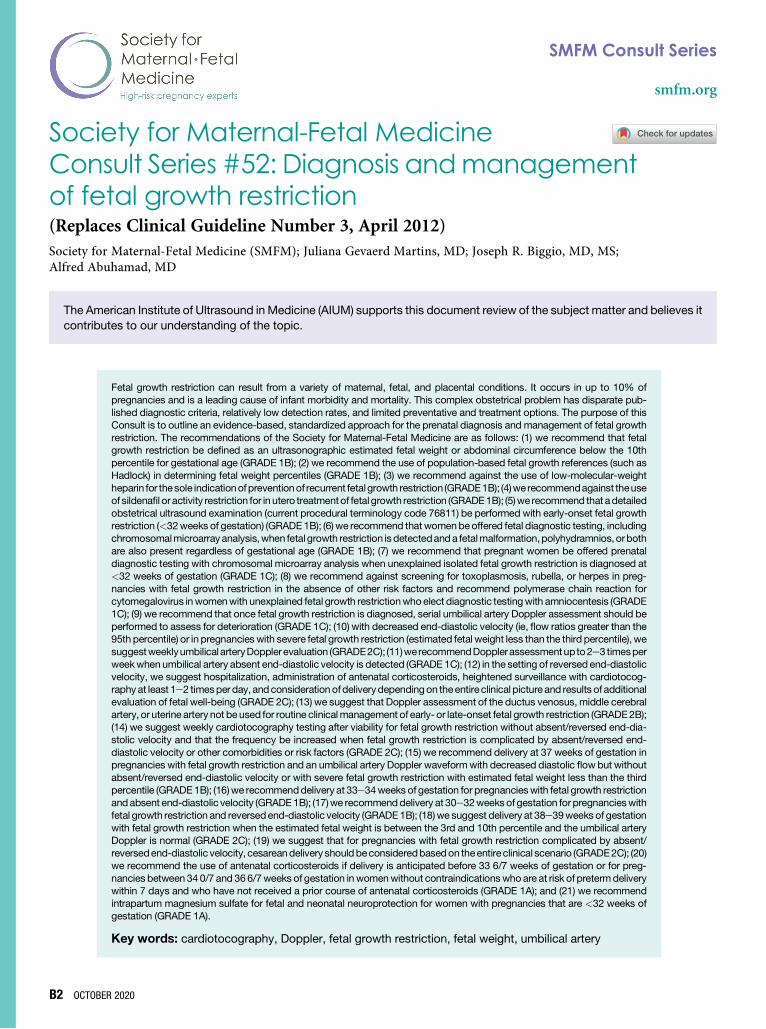
Society for Maternal-Fetal Medicine Consult Series #52: Diagnosis and management of fetal growth restriction (Replaces Clinical Guideline Number 3, April 2012) Society for Maternal-Fetal Medicine (SMFM); Juliana Gevaerd Martins, MD; Joseph R. Biggio, MD, MS; Alfred Abuhamad, MD The American Institute of Ultrasound in Medicine (AIUM) supports this document review of the subject matter and believes it contributes to our understanding of the topic. Fetal growth restriction can result from a variety of maternal, fetal, and placental conditions. It occurs in up to 10% of pregnancies and is a leading cause of infant morbidity and mortality. This complex obstetrical problem has disparate pub- lished diagnostic criteria, relatively low detection rates, and limited preventative and treatment options. The purpose of this Consult is to outline an evidence-based, standardized approach for the prenatal diagnosis and management of fetal growth restriction. The recommendations of the Society for Maternal-Fetal Medicine are as follows: (1) we recommend that fetal growth restriction be defined as an ultrasonographic estimated fetal weight or abdominal circumference below the 10th percentile for gestational age (GRADE 1B); (2) we recommend the use of population-based fetal growth references (such as Hadlock) in determining fetal weight percentiles (GRADE 1B); (3) we recommend against the use of low-molecular-weight heparin for the sole indication of prevention of recurrent fetal growth restriction (GRADE 1B); (4) we recommend against the use of sildenafil or activity restriction for in utero treatment of fetal growth restriction (GRADE 1B); (5) we recommend that a detailed obstetrical ultrasound examination (current procedural terminology code 76811) be performed with early-onset fetal growth restriction (<32 weeks of gestation) (GRADE 1B); (6) we recommend that women be offered fetal diagnostic testing, including chromosomal microarray analysis, when fetal growth restriction is detected and a fetal malformation, polyhydramnios, or both are also present regardless of gestational age (GRADE 1B); (7) we recommend that pregnant women be offered prenatal diagnostic testing with chromosomal microarray analysis when unexplained isolated fetal growth restriction is diagnosed at <32 weeks of gestation (GRADE 1C); (8) we recommend against screening for toxoplasmosis, rubella, or herpes in preg- nancies with fetal growth restriction in the absence of other risk factors and recommend polymerase chain reaction for cytomegalovirus in women with unexplained fetal growth restriction who elect diagnostic testing with amniocentesis (GRADE 1C); (9) we recommend that once fetal growth restriction is diagnosed, serial umbilical artery Doppler assessment should be performed to assess for deterioration (GRADE 1C); (10) with decreased end-diastolic velocity (ie, flow ratios greater than the 95th percentile) or in pregnancies with severe fetal growth restriction (estimated fetal weight less than the third percentile), we suggest weekly umbilical artery Doppler evaluation (GRADE 2C); (11) we recommend Doppler assessment up to 2e3 times per week when umbilical artery absent end-diastolic velocity is detected (GRADE 1C); (12) in the setting of reversed end-diastolic velocity, we suggest hospitalization, administration of antenatal corticosteroids, heightened surveillance with cardiotocog- raphy at least 1e2 times per day, and consideration of delivery depending on the entire clinical picture and results of additional evaluation of fetal well-being (GRADE 2C); (13) we suggest that Doppler assessment of the ductus venosus, middle cerebral artery, or uterine artery not be used for routine clinical management of early- or late-onset fetal growth restriction (GRADE 2B); (14) we suggest weekly cardiotocography testing after viability for fetal growth restriction without absent/reversed end-dia- stolic velocity and that the frequency be increased when fetal growth restriction is complicated by absent/reversed end- diastolic velocity or other comorbidities or risk factors (GRADE 2C); (15) we recommend delivery at 37 weeks of gestation in pregnancies with fetal growth restriction and an umbilical artery Doppler waveform with decreased diastolic flow but without absent/reversed end-diastolic velocity or with severe fetal growth restriction with estimated fetal weight less than the third percentile (GRADE 1B); (16) we recommend delivery at 33e34 weeks of gestation for pregnancies with fetal growth restriction and absent end-diastolic velocity (GRADE 1B); (17) we recommend delivery at 30e32 weeks of gestation for pregnancies with fetal growth restriction and reversed end-diastolic velocity (GRADE 1B); (18) we suggest delivery at 38e39 weeks of gestation with fetal growth restriction when the estimated fetal weight is between the 3rd and 10th percentile and the umbilical artery Doppler is normal (GRADE 2C); (19) we suggest that for pregnancies with fetal growth restriction complicated by absent/ reversed end-diastolic velocity, cesarean delivery should be considered based on the entire clinical scenario (GRADE 2C); (20) we recommend the use of antenatal corticosteroids if delivery is anticipated before 33 6/7 weeks of gestation or for preg- nancies between 34 0/7 and 36 6/7 weeks of gestation in women without contraindications who are at risk of preterm delivery within 7 days and who have not received a prior course of antenatal corticosteroids (GRADE 1A); and (21) we recommend intrapartum magnesium sulfate for fetal and neonatal neuroprotection for women with pregnancies that are <32 weeks of gestation (GRADE 1A). Key words: cardiotocography, Doppler, fetal growth restriction, fetal weight, umbilical artery B2 OCTOBER 2020 SMFM Consult Series smfm.org
-
Upload
khangminh22 -
Category
Documents
-
view
3 -
download
0
Transcript of Diagnosis and management of fetal growth restriction
SMFM Consult Series
smfm.org
Society for Maternal-Fetal Medicine
Consult Series #52: Diagnosis andmanagementof fetal growth restriction (Replaces Clinical Guideline Number 3, April 2012)Society for Maternal-Fetal Medicine (SMFM); Juliana Gevaerd Martins, MD; Joseph R. Biggio, MD, MS;Alfred Abuhamad, MDB2
The American Institute of Ultrasound in Medicine (AIUM) supports this document review of the subject matter and believes itcontributes to our understanding of the topic.
OCTOBER
Fetal growth restriction can result from a variety of maternal, fetal, and placental conditions. It occurs in up to 10% ofpregnancies and is a leading cause of infant morbidity and mortality. This complex obstetrical problem has disparate pub-
lished diagnostic criteria, relatively low detection rates, and limited preventative and treatment options. The purpose of thisConsult is to outline an evidence-based, standardized approach for the prenatal diagnosis and management of fetal growthrestriction. The recommendations of the Society for Maternal-Fetal Medicine are as follows: (1) we recommend that fetalgrowth restriction be defined as an ultrasonographic estimated fetal weight or abdominal circumference below the 10thpercentile for gestational age (GRADE 1B); (2) we recommend the use of population-based fetal growth references (such asHadlock) in determining fetal weight percentiles (GRADE 1B); (3) we recommend against the use of low-molecular-weightheparin for thesole indicationofpreventionof recurrent fetalgrowth restriction (GRADE1B); (4)we recommendagainst theuseof sildenafil or activity restriction for in utero treatmentof fetal growth restriction (GRADE1B); (5)we recommend that adetailedobstetrical ultrasound examination (current procedural terminology code 76811) be performed with early-onset fetal growthrestriction (<32weeks of gestation) (GRADE1B); (6) we recommend thatwomenbeoffered fetal diagnostic testing, includingchromosomalmicroarrayanalysis,when fetal growth restriction is detectedanda fetalmalformation,polyhydramnios, orbothare also present regardless of gestational age (GRADE 1B); (7) we recommend that pregnant women be offered prenataldiagnostic testing with chromosomal microarray analysis when unexplained isolated fetal growth restriction is diagnosed at<32 weeks of gestation (GRADE 1C); (8) we recommend against screening for toxoplasmosis, rubella, or herpes in preg-nancies with fetal growth restriction in the absence of other risk factors and recommend polymerase chain reaction forcytomegalovirus inwomenwith unexplained fetal growth restrictionwhoelect diagnostic testingwith amniocentesis (GRADE1C); (9) we recommend that once fetal growth restriction is diagnosed, serial umbilical artery Doppler assessment should beperformed to assess for deterioration (GRADE 1C); (10) with decreased end-diastolic velocity (ie, flow ratios greater than the95th percentile) or in pregnancies with severe fetal growth restriction (estimated fetal weight less than the third percentile), wesuggestweeklyumbilical arteryDoppler evaluation (GRADE2C); (11)werecommendDopplerassessmentup to2e3 timesperweekwhenumbilical artery absent end-diastolic velocity is detected (GRADE 1C); (12) in the setting of reversed end-diastolicvelocity, we suggest hospitalization, administration of antenatal corticosteroids, heightened surveillance with cardiotocog-raphy at least 1e2 timesper day, andconsiderationofdeliverydependingon theentire clinical picture and results of additionalevaluation of fetal well-being (GRADE 2C); (13) we suggest that Doppler assessment of the ductus venosus, middle cerebralartery, or uterine artery not beused for routine clinicalmanagement of early- or late-onset fetal growth restriction (GRADE2B);(14) we suggest weekly cardiotocography testing after viability for fetal growth restriction without absent/reversed end-dia-stolic velocity and that the frequency be increased when fetal growth restriction is complicated by absent/reversed end-diastolic velocity or other comorbidities or risk factors (GRADE 2C); (15) we recommend delivery at 37 weeks of gestation inpregnancies with fetal growth restriction and an umbilical artery Doppler waveformwith decreased diastolic flow but withoutabsent/reversed end-diastolic velocity or with severe fetal growth restriction with estimated fetal weight less than the thirdpercentile (GRADE1B); (16)we recommenddelivery at 33e34weeks of gestation for pregnancieswith fetal growth restrictionand absent end-diastolic velocity (GRADE1B); (17)we recommenddelivery at 30e32weeks of gestation for pregnancieswithfetal growth restriction and reversedend-diastolic velocity (GRADE1B); (18)we suggest delivery at 38e39weeks of gestationwith fetal growth restriction when the estimated fetal weight is between the 3rd and 10th percentile and the umbilical arteryDoppler is normal (GRADE 2C); (19) we suggest that for pregnancies with fetal growth restriction complicated by absent/reversedend-diastolic velocity, cesareandelivery shouldbeconsideredbasedon theentire clinical scenario (GRADE2C); (20)we recommend the use of antenatal corticosteroids if delivery is anticipated before 33 6/7 weeks of gestation or for preg-nancies between 34 0/7 and 366/7weeks of gestation inwomenwithout contraindicationswho are at risk of pretermdeliverywithin 7 days and who have not received a prior course of antenatal corticosteroids (GRADE 1A); and (21) we recommendintrapartum magnesium sulfate for fetal and neonatal neuroprotection for women with pregnancies that are <32 weeks ofgestation (GRADE 1A).Key words: cardiotocography, Doppler, fetal growth restriction, fetal weight, umbilical artery
2020

smfm.org SMFM Consult Series
Introduction practice. The term FGR has been used to describe a fetus
Fetal growth restriction (FGR) can result from a variety ofmaternal, fetal, and placental conditions.1 Although theprimary underlying mechanisms for FGR are varied, theyoften share the same final common pathway of suboptimalfetal nutrition and uteroplacental perfusion.1,2 Chromo-somal disorders and congenital malformations are respon-sible for approximately 20% of FGR cases.2,3 Suboptimalperfusion of the maternal placental circulation is the mostcommon cause of FGR and accounts for 25e30% of allcases.2,3
FGR occurs in up to 10% of pregnancies and is a leadingcause of infant morbidity and mortality.1,4,5 In fetuses at allgestational ages with weights below the 10th percentile, thestillbirth rate is approximately 1.5%, which is twice the rate infetuses with normal growth. With fetal weights below the fifthpercentile, the stillbirth rate can be as high as 2.5%.6,7
Furthermore, infants with birthweights below the 10thpercentile aremore likely to have severe acidosis at birth, low5-minute Apgar scores, and neonatal intensive care unit ad-missions.8 Prematurity further compounds the risk of adverseoutcomes inFGR.9Studies report a 2- to5-fold increased rateof perinatal death among preterm FGR fetuses comparedwith term FGR fetuses.9 Perinatal outcomes are largelydependent on the severity of FGR, with the worst outcomesnoted in fetuses with estimated fetal weights (EFWs) at lessthan the third percentile or in association with fetal Dopplerabnormalities.5,10
In addition to its significant perinatal impact, FGR also hasan impact on long-term health outcomes. It has beenassociated with metabolic programming that increases therisk of future development of metabolic syndrome andconsequent cardiovascular and endocrine diseases.11,12 Italso can contribute to cardiac remodeling, leading to car-diovascular dysfunction that can persist into childhood andadolescence.13,14 In addition, studies have shown an as-sociation between FGR and long-term neurologicimpairment,15e20 with rates of cognitive and learning dis-abilities as high as 20%e40% by school age.21
FGR remains a complex obstetrical problem with dispa-rate published diagnostic criteria, relatively low detectionrates, and limited preventative and treatment options.22e25
Antenatal care of FGR is often complicated by the presenceof maternal disease, such as hypertension, and optimalmanagement involves balancing maternal, fetal, andneonatal risks. The purpose of this document is to outline anevidence-based, standardized approach for the prenataldiagnosis and management of FGR.
Terminology and diagnostic criteriaFGR and small for gestational age (SGA) are terms some-times used interchangeably in the literature and clinical
Corresponding author: Society for Maternal-Fetal Medicine: PublicationsCommittee. [email protected]
with an EFW below the 10th percentile and SGA to describea newbornwhosebirthweight is less than the 10th percentilefor gestational age.26 The use of the term intrauterine growthrestriction (IUGR) should be abandoned in favor of FGR.Fetuses with FGR are not always SGA at birth, and SGA
neonates have often not been diagnosed as growthrestricted on prenatal ultrasound.27 Of fetuses diagnosedwith FGR, approximately 18%e22% will be constitutionallysmall but healthy at birth with a normal outcome.24 A sig-nificant challenge in the prenatal management of FGR isdifferentiating the constitutionally small fetus from one whois pathologically growth restricted and at risk for postnatalcomplications.FGR is commonly defined as an ultrasonographic EFW
below the 10th percentile for gestational age. A review ofnational guidelines for the diagnostic criteria for FGR from 6countries (United States, United Kingdom, France, Ireland,Canada, and New Zealand) reveals a broad consensus onthis definition of FGR.24 However, there is significant vari-ation in the diagnostic criteria used for FGR. Some diag-nostic criteria are limited to fetal biometric measurements,whereas others incorporate abnormal Doppler findings.28
Moreover, the biometric component of the FGR diagnosticcriteria differs according to the choice of population vscustomized reference growth standards, whether EFW isused alone or together with abdominal circumference (AC),and which cutoff is used to define abnormal growth.24,29,30
For example, 3 of the 6 countries also include AC as adiagnostic criterion, with the United Kingdom and Canadausing an AC cutoff of less than the 10th percentile and NewZealand using an AC cutoff of less than the 5th percentile.24
Evidence supports the use of AC as a diagnostic criterionfor FGR. In a prospective study in 1000 low-risk pregnan-cies, an AC of less than the 10th percentile was found tohave diagnostic accuracy similar to EFW less than the 10thpercentile for the prediction of SGA.31 In a meta-analysispublished in 2017, an AC of less than the 10th percentilepredicted SGA as well as ultrasonographic EFW less thanthe 10th percentile, with comparable sensitivity and speci-ficity. Compared with other cutoffs, an AC of less than thefifth percentile has significantly lower sensitivity but higherspecificity in predicting SGA.32 Another systematic reviewand meta-analysis reported that AC and EFW performedsimilarly, and for a 10% fixed false-positive rate, AC hadhigher sensitivity.33
An alternative approach to the diagnosis of FGR includesthe determination of fetal growth trajectory, generated frommultiple ultrasound examinations, and the identification ofthe fetus that drops off its own growth trajectory. Theoreti-cally, this approach takes into consideration the dynamicaspect of growth and the individualized growth potential ofeach fetus.34 However, this approach requires multiple ul-trasound examinations, and prospective studies fail todemonstrate the superiority of this approach in improvingclinical outcomes.35 We recommend that FGR be defined as an
OCTOBER 2020 B3

SMFM Consult Series smfm.org
ultrasonographic EFW or AC below the 10th percentile for gesta-tional age (GRADE 1B).
Ultrasonographic estimation of fetal weightAccurate pregnancy dating is an important prerequisite fordiagnosing FGR. Parameters for assigning gestational ageby ultrasound have been recently updated.36 Pregnancydating is best established when first-trimester crown-rumplength is used to either confirm menstrual dates or assignnew dates.36 Ultrasonographic fetal weight estimation isgenerated by the use of regression equations that combinebiometric measurements of the fetal biparietal diameter,head circumference (HC), AC, and femur length; a multi-society task force has recently standardized criteria forthese images obtained for fetal biometry.37 The ultrasono-graphic EFW is then compared with a reference chart togenerate a weight percentile.The first ultrasonographic equation used to estimate fetal
weight was published by Warsof et al in 1977, and sincethen, many others have been developed.38 Considerablevariation in accuracy was noted in a retrospective review of26 formulas for ultrasonographic fetal weight estimation.For birthweights in the range of 1000e4500 g, formulasbased on 3 or 4 fetal biometric indices were significantlymore accurate in estimating fetal weights than formulasbased on 1 or 2 indices.39 In a review of the literaturerelating to methods and sources of inaccuracies in theestimation of fetal weight, the authors concluded thataveraging of multiple measurements, improvements in im-age quality, uniform calibration of equipment, and regularaudits may help to improve fetal weight estimation andreduce errors.40
Fetal growth nomograms generally represent either un-adjusted population standards or customized standardsthat adjust for constitutional or physiological variations offetal size based on sex and race.35,41e44 The most widelyused method for estimating fetal weight and calculatingweight percentile in the United States is based on theHadlock formula, which was generated from a studyinvolving 392 pregnancies in predominantly white, middle-class women conducted at a single institution in Texas.41 Insome studies, the use of customized growth standards hasbeen shown to improve the ability to distinguish growth-restricted fetuses from constitutionally small fetuses.45e47
Whether the use of customized growth standards trans-lates to improved pregnancy outcomes was the subject ofseveral recent studies: the INTERGROWTH-21st stan-dard,44 the Eunice Kennedy Shriver National Institute ofChild Health and Human Development (NICHD) stan-dards,48 and the World Health Organization (WHO) stan-dard.49,50 The INTERGROWTH-21st study included healthypregnant womenwith nomaternal or fetal risk factors from 8countries and created a single universal standard for fetalgrowth without adjusting for ethnic variation.44 The NICHDstudy, performed at 12 sites in the United States, developedracial/ethnic-specific standards of fetal growth.48 Finally,
B4 OCTOBER 2020
the WHO study developed an overall growth standardbased on data collected from 10 countries.49,50
Although both the NICHD and WHO studies identifiedracial/ethnic differences in fetal growth, evidence to dateindicates that the use of these new formulas in clinicalpractice does not improve the detection and outcome ofFGR.51e53 In a preterm population in France, the INTER-GROWTH-21st formula was associated with a higher meanpercentage error and a higher underestimation of birth-weight at >28 weeks of gestation when compared withHadlock. The Hadlock formula classifiedmore infants within10% of actual birthweight and was more accurate than theINTERGROWTH-21st in the overall estimation of weight forfetuses delivered between 22 and 34 weeks of gestation.53
The diagnostic accuracy for estimating fetal weight and theprediction of neonatal morbidity was compared using theNICHD standard and Hadlock in 1514 pregnant womenwithdifferent ethnicities. The Hadlock formula better predictedSGA and composite neonatal morbidity at birth and had alower ultrasound-to-birthweight percentile discrepancythan the NICHD growth standard. Fetuses classified asgrowth restricted by Hadlock, but not by the NICHD growthstandard, had significantly higher composite morbidity thanfetuses of normal growth.51 In view of these findings,we recommend the use of population-based fetal growth refer-ences (such as Hadlock) in determining fetal weight percentiles(GRADE 1B).
Classification of fetal growth restriction
Timing of diagnosis FGR has been categorized as early or late onset based ongestational age at prenatal ultrasound diagnosis, with early-onset FGR diagnosed before 32 weeks of gestation andlate-onset FGR diagnosed at or after 32 weeks of gestation.In a cohort of 656 pregnancies with FGR, a gestational ageof 32weeks at diagnosis was identified as the optimal cutofftomaximize the differences in associated comorbidities andpregnancy outcomes between early- and late-onset FGR.54The clinical spectrum of early- and late-onset FGR alsodiffers; early-onset FGR is typically more severe, tends tofollow an established Doppler pattern of fetal deterioration,is more commonly associated with maternal hypertensivedisorders of pregnancy, and shows more significantplacental dysfunction than late-onset FGR.23,28,54e56 Fe-tuseswith genetic abnormalities can also present with early-onset FGR, commonly in association with fetal and amnioticfluid abnormalities.3 Late-onset FGR represents approxi-mately 70%e80% of FGR cases and is typically milder inpresentation.55,56 Unlike early-onset FGR, late-onset FGR isless likely to be associated with maternal hypertensivedisorders and typically has less extensive placental histo-pathologic findings of underperfusion.57e59 In early-onsetFGR, the pattern of Doppler deterioration progresses fromabnormalities in the umbilical arteries and the ductusvenosus to abnormal biophysical parameters.55,56 Incontrast, cardiovascular adaptation of late-onset FGR is

smfm.org SMFM Consult Series
typically limited to the cerebral circulation and is commonlyassociated with normal Doppler of the umbilicalarteries.57,60,61
Severity of fetal growth restriction
Studies have reviewed various ultrasonographic parame-ters to better identify growth-restricted fetuses at increasedrisk for perinatal morbidity and mortality.28 The presence ofabnormal umbilical artery Doppler indices has been found topredict adverse perinatal outcomes.62 An EFW below thethird percentile has also been associated with an increasedrisk of adverse perinatal outcome irrespective of umbilicaland middle cerebral artery Doppler indices.10 In a largeretrospective cohort of more than 3 million singleton preg-nancies, the risk of stillbirth at birthweights of less than the3rd percentile was increased approximately 3-fold over the3rd to 5th percentile group at nearly all gestational ages, andthere was an increased risk of 4-fold to 7-fold over the 5th to10th percentile group.63 These results are consistent withneonatal data showing a significantly increased risk ofmorbidity and mortality in infants born at term with birth-weights below the third percentile.64 Therefore, an EFWbelow the third percentile has been found to represent amore severe form of FGR.Symmetric and asymmetric fetal growthrestriction
FGR has been classified as symmetric or asymmetric basedon the ratio between the head circumference and theabdominal circumference (HC/AC). In the past, such clas-sification was thought to provide valuable information aboutthe timing of pregnancy insult and the etiology and prog-nosis of FGR.65 More recently, growth and developmentaldelay have been evaluated from birth to the age of 4 yearsand shown to be similar in symmetric and asymmetricgrowth-restricted preterm newborns.66 Furthermore,HC/AC was not found to be an independent predictor ofadverse pregnancy outcomes.67Management of fetal growth restriction
General considerations There are currently no preventative strategies or treat-ments for FGR that have been proven to be effective. Thereis no consistent evidence that nutritional and dietary sup-plements or bed rest prevents FGR or reduces the inci-dence of SGA births.68e71 The use of prophylactic low-dose aspirin was shown to provide a modest risk reductionin FGR and SGA in 2 meta-analyses.72,73 However, thisfinding was not confirmed in the Aspirin for Evidence-Based Preeclampsia Prevention (ASPRE) trial, which wasprimarily designed for preterm preeclampsia preven-tion.74,75 Due to the conflicting evidence on the role of low-dose aspirin in the prevention of recurrent FGR in other-wise low-risk women, the American College of Obstetri-cians and Gynecologists recommends against the use oflow-dose aspirin for the sole indication of FGRprevention.76 Furthermore, the use of low-molecular-weight heparin has not been shown to reduce the risk ofrecurrent placenta-mediated pregnancy complications inat-risk women.75,77,78 At present, there is no evidence thattherapeutic interventions, including sildenafil to augmentuteroplacental perfusion through vasodilation, improveplacental perfusion and outcome in pregnancies withFGR.75,79 We recommend against the use of low-molecular-weight heparin for the sole indication of prevention of recurrentFGR (GRADE 1B). We also recommend against sildenafil or ac-tivity restriction for in utero treatment of FGR (GRADE 1B).Management of FGR is based on early diagnosis, optimal
fetal surveillance, and timely delivery that reduces perinatalmortality and minimizes short- and long-term morbidity. Inpregnancies with FGR, delivery decisions require balancingthe risk of prematurity against that of stillbirth. The decisionto deliver is typically guided bymaternal factors, such as thepresence of maternal hypertension, and by fetal comor-bidities, such as the degree of growth restriction and theseverity of abnormal fetal surveillance results. There iscurrently no consensus on the best approach to the man-agement of FGR, despite a large body of literature on thesubject. This lack of agreement is primarily due to thepaucity of randomized trials and the heterogeneity of studypopulations.Despite these limitations, accumulating evidence sug-
gests a benefit to the use of umbilical artery Doppler in thesurveillance of FGR. Furthermore, the presence of a stan-dardized protocol for diagnosis and management appearsto be associated with more favorable outcomes, as evi-denced in the better-than-expected perinatal morbidity andmortality in the Trial of RandomizedUmbilical and Fetal Flowin Europe (TRUFFLE).80 Results of this trial, which stan-dardized the approach to care and criteria for delivery, are incontrast to those of the Growth Restriction Intervention Trial(GRIT),81,82 which left management to the discretion of themanaging providers. The single most important prognosticfactor in preterm fetuses with growth restriction is thegestational age at delivery.80,83 A large longitudinal cohortstudy on FGR showed an increase of 1%e2% in intactsurvival for every additional day spent in utero up until 32weeks of gestation.83 An algorithm for the diagnosis andmanagement of FGR is provided in Figure 1.Maternal hypertensive disease is common in early-onset
FGR and plays an important role in pregnancy outcomes. InTRUFFLE, maternal hypertension was present in 50% ofpregnancies during the study and 70%of pregnancies at thetime of delivery. The presence of maternal hypertensionwasone of the most important independent determinants ofpoor outcomes.16,80 Pregnant women with hypertensionhad a significantly shorter median interval from studyenrollment to delivery, and newborns of mothers with hy-pertension were delivered at an earlier gestational age andhad lower birthweights.80 Women with early-onset FGRshould be closely monitored for the development of hy-pertensive disorders of pregnancy.
OCTOBER 2020 B5

Summary of recommendations
Number Recommendations Grade
1 We recommend that FGR be defined as an ultrasonographic EFW or AC belowthe 10th percentile for gestational age.
1BStrong recommendation, moderate-quality evidence
2 We recommend the use of population-based fetal growth references (such asHadlock) in determining fetal weight percentiles.
1BStrong recommendation, moderate-quality evidence
3 We recommend against the use of low-molecular-weight heparin for the soleindication of prevention of recurrent FGR.
1BStrong recommendation, moderate-quality evidence
4 We recommend against the use of sildenafil or activity restriction for in uterotreatment of FGR.
1BStrong recommendation, moderate-quality evidence
5 We recommend that a detailed obstetrical ultrasound examination (CPT code76811) be performed with early-onset FGR (<32 weeks of gestation) becauseup to 20% of cases are associated with fetal or chromosomal abnormalities.
1BStrong recommendation, moderate-quality evidence
6 We recommend that women be offered fetal diagnostic testing, including CMA,when FGR is detected and a fetal malformation, polyhydramnios, or both arealso present regardless of gestational age.
1BStrong recommendation, moderate-quality evidence
7 We recommend that pregnant women be offered prenatal diagnostic testingwith CMA when unexplained isolated FGR is diagnosed at <32 weeks ofgestation.
1CStrong recommendation, low-quality evidence
8 We recommend against screening for toxoplasmosis, rubella, or herpes inpregnancies with FGR in the absence of other risk factors and recommend PCRfor CMV in women with unexplained FGR who elect diagnostic testing withamniocentesis.
1CStrong recommendation, low-quality evidence
9 We recommend that once FGR is diagnosed, serial umbilical artery Dopplerassessment should be performed to assess for deterioration.
1CStrong recommendation, low-quality evidence
10 With decreased end-diastolic velocity (ie, flow ratios greater than the 95thpercentile) or in pregnancies with severe FGR (EFW less than the 3rdpercentile), we suggest weekly umbilical artery Doppler evaluation.
2CWeak recommendation, low-quality evidence
11 We recommend Doppler assessment up to 2e3 times per week whenumbilical AEDV is detected because of the potential for deterioration anddevelopment of REDV.
1CStrong recommendation, low-quality evidence
12 In the setting of REDV, we suggest hospitalization, administration of antenatalcorticosteroids, heightened surveillance with CTG at least 1e2 times per day,and consideration of delivery depending on the entire clinical picture andresults of additional evaluation of fetal well-being.
2CWeak recommendation, low-quality evidence
13 We suggest that Doppler assessment of the ductus venosus, middle cerebralartery, or uterine artery not be used for routine clinical management of early- orlate-onset FGR.
2BWeak recommendation, moderate-quality evidence
14 We suggest weekly CTG testing after viability for FGR without AEDV/REDV andthat the frequency be increased when FGR is complicated by AEDV/REDV orother comorbidities or risk factors.
2CWeak recommendation, low-quality evidence
15 We recommend delivery at 37 weeks of gestation in pregnancies with FGR andan umbilical artery Doppler waveform with decreased diastolic flow but withoutAEDV/REDV or with severe FGR with EFW less than the third percentile.
1BStrong recommendation, moderate-quality evidence
16 We recommend delivery at 33e34 weeks of gestation for pregnancies withFGR and AEDV.
1BStrong recommendation, moderate-quality evidence
17 We recommend delivery at 30e32 weeks of gestation for pregnancies withFGR and REDV.
1BStrong recommendation, moderate-quality evidence
18 We suggest delivery at 38e39 weeks of gestation with FGR when the EFW isbetween the 3rd and 10th percentile and the umbilical artery Doppler isnormal.
2CWeak recommendation, low-quality evidence
Society for Maternal-Fetal Medicine. SMFM Consult Series #52: Diagnosis and management of fetal growth restriction. Am J Obstet Gynecol 2020. (continued)
SMFM Consult Series smfm.org
B6 OCTOBER 2020

Summary of recommendations (continued)
Number Recommendations Grade
19 We suggest that for pregnancies with FGR complicated by AEDV/REDV,cesarean delivery should be considered based on the entire clinical scenario.
2CWeak recommendation, low-quality evidence
20 We recommend the use of antenatal corticosteroids if delivery is anticipatedbefore 33 6/7 weeks of gestation or for pregnancies between 34 0/7 and36 6/7 weeks of gestation in women without contraindications who are at riskof preterm delivery within 7 days and who have not received a previous courseof antenatal corticosteroids.
1AStrong recommendation, high-quality evidence
21 We recommend intrapartum magnesium sulfate for fetal and neonatalneuroprotection for women with pregnancies that are<32 weeks of gestation.
1AStrong recommendation, high-quality evidence
AC, abdominal circumference; AEDV, artery absent end-diastolic velocity; CMA, chromosomal microarray analysis; CMV, cytomegalovirus; CPT, current procedural terminology; CTG, car-diotocography; EFW, estimated fetal weight; FGR, fetal growth restriction; PCR, polymerase chain reaction; REDV, reversed end-diastolic velocity.
Society for Maternal-Fetal Medicine. SMFM Consult Series #52: Diagnosis and management of fetal growth restriction. Am J Obstet Gynecol 2020.
smfm.org SMFM Consult Series
Initial diagnosis
With the initial diagnosis of FGR and if not previously per-formed, we recommend that a detailed obstetrical ultrasoundexamination (current procedural terminology code 76811) beperformed with early-onset FGR because up to 20% of cases areassociated with fetal or chromosomal abnormalities2,3,84,85(GRADE 1B). The combination of FGR with a fetal malforma-tion or polyhydramnios should prompt genetic counselingand consideration of prenatal diagnostic testing.86 Werecommend that women be offered fetal diagnostic testing,including chromosomal microarray analysis (CMA), when FGR isdetected and a fetal malformation, polyhydramnios, or both arealso present regardless of gestational age (GRADE 1B).Although chromosome abnormalities aremore frequent in
pregnancies with structural anomalies and FGR, in a sys-tematic review that included fetuses with no structuralmalformations, the mean rate of chromosomal abnormal-ities was 6.4%. Only a fraction of the studies includedwomen in the third trimester with apparently isolated FGR,but no karyotype abnormalities were identified in thosewomen. Due to substantial heterogeneity of the selectedstudies in the systemic review, meta-analytic methods,such as calculating the effect estimates, could not beapplied.87 More recent studies have evaluated the role ofCMA in fetuses with early-onset growth restriction andno structural malformations; such studies have identified a4%e10% incremental yield of CMA over karyotype.88e90
We recommend that pregnant women be offered prenatal diag-nostic testing with CMA when unexplained isolated FGR is diag-nosed at <32 weeks of gestation (GRADE 1C).The association of maternal infections with FGR was
recently evaluated in a study that included 319 pregnancies.No cases of maternal or congenital infection with toxo-plasma, rubella, or herpes were found, whereas 6 (1.8%)fetuses were diagnosed as having congenital cytomegalo-virus (CMV). Two (0.6%) of the fetuses with congenital CMVhad no ultrasonographic findings other than FGR.91 In
another prospective cohort study of 48 pregnancies withFGR, 1 newborn (2.1%) was diagnosed with congenitalCMV.92 We recommend against screening for toxoplasmosis,rubella, or herpes in pregnancies with FGR in the absence of otherrisk factors and recommend polymerase chain reaction (PCR) forCMV in women with unexplained FGR who elect diagnostic testingwith amniocentesis (GRADE 1C). However, given the low inci-dence of CMV in cases of FGR, the lack of effective ante-natal interventions, and the limited utility of serologic testingfor CMV in the third trimester, routine infectious serologiesmay not be warranted in the absence of risk factors or ul-trasonographic markers of fetal infection.91e94 PCR is thepreferred testing approach for CMV and should be per-formed in women with unexplained FGR who undergodiagnostic testing with amniocentesis.
Umbilical artery Doppler
Umbilical artery Doppler assesses the impedance to bloodflow along the fetal component of the placental unit. As earlyas 14 weeks of gestation, low impedance of the fetalplacental circulation permits continuous forward flow in theumbilical artery throughout the cardiac cycle.95 Dopplerwaveforms of the umbilical artery can be obtained from anysegment along the umbilical cord. Waveforms obtainednear the placental end of the cord reflect downstreamimpedance and show higher end-diastolic blood flow ve-locity than waveforms obtained near the fetal cord inser-tion.95 In general, this variation in umbilical artery Dopplerend-diastolic flow along the umbilical cord is minimal andnot significant enough to affect clinical decision-making.The pulsatility index (PI), resistance index (RI), or systolic-to-diastolic (S/D) ratio can be used for quantification of theDoppler waveform in the umbilical artery, although recentstudies have generally used either the PI or RI.5,16,28,30,80,83
An abnormal umbilical artery Doppler is defined as a PI, RI,or S/D ratio greater than the 95th percentile for gestationalage or an absent or reversed end-diastolic velocity (AEDV or
OCTOBER 2020 B7

FIGURE 1Algorithm for the diagnosis and management of fetal growth restriction
DiagnosisEFW < 10th %ile
and / orAC < 10th %ile
Classifica�onEarly FGR: < 32 weeks at ini�al diagnosisLate FGR: ≥ 32 weeks at ini�al diagnosis
Severe FGR: EFW < 3rd %ile
Work-up• Detailed obstetrical ultrasound (76811)• Diagnos�c gene�c tes�ng (CMA) for:• Early-onset FGR• Sonographic abnormali�es• Polyhydramnios
• PCR CMV on amnio�c fluid if pa�ent has amniocentesis
Fetal Surveillance• UA Doppler• CTG
Deliver for repe��ve late decelera�ons a�er fetal viability
Normal UA:S/D, PI, RI ≤ 95%
UA Decreased EDV:S/D, PI, RI > 95%
UA Doppler weeklyCTG 1-2x per week
Consider EFW q 2 weeks
UA Absent EDV:
Consider inpa�ent admissionUA Doppler 2-3x per week
Cor�costeroids for FLMCTG 2x per week if managed
as outpa�entConsider EFW q 2 weeks
UA Reversed EDV:
Inpa�ent admission Cor�costeroids for FLM
CTG 1-2x per dayConsider EFW q 2 weeks
Deliver at 33-34 weeks Deliver at 30-32 weeksDeliver at 37 weeks
Deliver at 38-39 weeks
EFW ≥ 3rd - 9th %ile
UA Doppler q 1-2 weeks for 1-2 weeks. If stable findings,
UA Doppler q 2-4 weeksCTG 1x per weekEFW q 3-4 weeks
Deliver at 37 weeks
EFW < 3rd %ile
UA Doppler weeklyCTG 1x per week
Consider EFW q 2 weeks
SMFM Consult Series smfm.org
B8 OCTOBER 2020

=
smfm.org SMFM Consult Series
REDV). The progression from an abnormal umbilical arteryDoppler with a decreased diastolic flow to AEDV/REDV cantake several days to weeks, especially in the absence ofmaternal disease. In a large study on FGR, the mean time-to-delivery interval for umbilical artery PI greater than the95th percentile, AEDV, and REDV was 26, 13, and 4 days,respectively.62
An abnormal umbilical artery Doppler waveform reflectsthe presence of placental insufficiency and can help differ-entiate the growth-restricted fetus from the constitutionallysmall fetus. Incorporation of umbilical artery Doppler eval-uation in the management of high-risk pregnancies hasbeen shown to significantly reduce the risk of perinataldeath, induction of labor, and cesarean delivery. As such, itis an essential component of fetal surveillance in FGR.96,97
In contrast, a systematic review of 5 trials found no evidenceof maternal or neonatal benefit from the routine use of um-bilical artery Doppler in low-risk pregnancies.98
AEDV/REDV in the umbilical artery reflects the presenceof significant placental deterioration and is associated withhigh perinatal mortality. The finding of AEDV/REDV of theumbilical artery can be intermittent; this likely represents thecontinuum of Doppler deterioration that occurs before theabsent or reversed flow becomes persistent.99 A meta-analysis of 31 studies on the risk of fetal death in FGR before34 weeks of gestation reported odds ratios for fetal death of3.59 (95% confidence interval [CI], 2.3e5.6) and 7.27 (95%CI, 4.6e11.4) for AEDV andREDV, respectively. Pooled datafrom this meta-analysis also revealed a risk of stillbirth of6.8% for AEDV and 19% for REDV in the umbilical artery orductus venosus.100 These risks of stillbirth are higher thanthe risk of infant mortality or severe morbidity at 33e34weeks for AEDV and at 30e32 weeks for REDV as reportedin TRUFFLE.80
Evidence suggests that umbilical artery Doppler does notreliably predict adverse pregnancy outcome in late-onsetFGR.101 This result is probably related to the lower fre-quency of placental pathologic findings in late-onset FGRwhen compared with early-onset FGR.102e104 Experimentalmodeling suggests that a threshold of placental vascularobliteration is required before umbilical artery Doppler ab-normalities are seen; therefore, the presence of a normalumbilical artery Doppler in late-onset FGR does not rule outplacental disease.105,106
There are currently no randomized trials with adequatesample size to inform recommendations regarding theoptimal frequency of umbilical artery Doppler for FGR sur-veillance.107 Protocols vary from weekly umbilical arteryDoppler to a 2- to 4-week interval.24,108 A prospectiveobservational study of the progression of Doppler
AC, abdominal circumference; CMA, chromosomal microarray analysis; CMV, cytomegalovirus; CTG, carFLM, fetal lung maturity; PCR, polymerase chain reaction; PI, pulsatility index; RI, resistance index; S/D
Society for Maternal-Fetal Medicine. SMFM Consult Series #52: Diagnosis and management of
abnormalities in FGR suggests that rapid progression, if it isgoing to occur, is typically noted within the first 2 weeksafter diagnosis.24,108 We recommend that once FGR is diag-nosed, serial umbilical artery Doppler assessment should beperformed to assess for deterioration (GRADE 1C). This assess-ment should initially occur every 1-2 weeks. If the umbilicalartery Doppler remains normal after this initial assessment, aless frequent interval of umbilical artery Doppler testing (eg,every 2e4 weeks) may be considered.108
With decreased end-diastolic velocity (ie, flow ratios greaterthan the 95th percentile) or in pregnancies with severe FGR (EFWless than the 3rd percentile), we suggest weekly umbilical arteryDoppler evaluation24,95 (GRADE 2C). We recommend Dopplerassessment up to 2e3 times per week when umbilical artery AEDVis detected due to the potential for deterioration and developmentof REDV (GRADE 1C). In the setting of REDV, we suggest hospi-talization, administration of antenatal corticosteroids, heightenedsurveillance with cardiotocography (CTG) at least 1e2 times perday, and consideration of delivery depending on the entire clinicalpicture and results of additional evaluation of fetal well-being(GRADE 2C). Hospital admission should be considered if fetalsurveillance of more often than 3 times per week is deemednecessary. Once FGR is diagnosed, assessment of fetalgrowth and weight should be performed at least every 3e4weeks; consideration can be given for a 2-week interval incases of severe FGR or with abnormal umbilical arteryDoppler.109
Ductus venosus Doppler
Longitudinal studies have shown that Doppler abnormalitiesof the ductus venosus in FGR reflect an advanced stage offetal compromise, associated with increased perinatalmorbidity and mortality.2,23,55,110e117 A meta-analysis ofFGR at <34 weeks of gestation reported odds ratios forstillbirth of 11.16 (95% CI, 6.31e19.73) for absent orreversed A-wave of the ductus venosus and a frequency ofstillbirth of 20%; the risk of stillbirth with a reversed A-wavewas 46%.100 In FGR, Doppler abnormalities of the ductusvenosus primarily reflect increased central venous pressure,resulting from increased right ventricular end-diastolicpressure and decreased cardiac muscle compliance.110,118Reversed A-wave of the ductus venosus in FGR signifiesmore significant fetal cardiac compromise.119 Doppler ab-normalities of the ductus venosus in the setting of a normalumbilical artery Doppler indicate an alternative pathophys-iological etiology, possibly related to the presence of fetalcardiac, vascular, or genetic abnormalities, and thus aremost often not reflective of significant placental disease.TRUFFLE compared ductus venosus Doppler and com-
puter-generated short-term fetal heart rate variability (cSTV)
diotocography; EDV, end-diastolic velocity; EFW, estimated fetal weight; FGR, fetal growth restriction;, systolic-to-diastolic ratio; UA, umbilical artery.
fetal growth restriction. Am J Obstet Gynecol 2020
OCTOBER 2020 B9

SMFM Consult Series smfm.org
in the monitoring and timing of delivery in early-onset FGR.After correction for prematurity, survival without neurologicimpairment was found to be significantly higher in the groupdelivered according to late ductus venosus changes (95%)compared with cSTV (85%).16 However, caution is urgedwhen extrapolating the findings of TRUFFLE to practice inthe United States. TRUFFLE compared cSTV with ductusvenosus Doppler, and results cannot be generalized to thevisual interpretation of CTG. Furthermore, absent orreversed A-wave of the ductus venosus represents anadvanced stage of fetal compromise and is uncommon.Even in pregnancies with AEDV/REDV of the umbilical ar-tery, late Doppler abnormalities of the ductus venosus arenoted in only about 41% of fetuses.117 After 32 weeks ofgestation, abnormal CTG findings will almost invariablyprecede Doppler abnormalities of the ductus venosus.111 InTRUFFLE, delivery decisions guided by ductus venosusDoppler findings only accounted for about 11% of preg-nancies allocated to the late ductus venosus findings groupbecause most delivered due to other fetal or maternal in-dications.115,120,121 Prospective research is needed tofurther elucidate the role of ductus venosus Doppler inpregnancies with early-onset FGR before its use in routinesurveillance of pregnancies with FGR can berecommended.
Middle cerebral artery Doppler
The middle cerebral artery is the largest vessel of the fetalcerebral circulation and carries about 80%of cerebral bloodflow.122 Fetal hypoxemia associated with growth restrictionresults in cerebral vasodilation, an early adaptive mecha-nism termed the brain-sparing effect. Measurement of flowthrough the middle cerebral artery using Doppler can iden-tify cerebral vasodilation, which can bequantified usingPI orthe cerebroplacental ratio (CPR). CPR is calculated bydividing the middle cerebral artery PI by the umbilical arteryPI.123e126 The role of middle cerebral artery Doppler in themanagement of early-onset FGR has been evaluated inseveral studies.127e129 In a meta-analysis of 35 studies,abnormal middle cerebral artery Doppler had a low likeli-hood ratio (LR) for prediction of perinatal mortality (LR 1.36[1.10e1.67]) and adverse perinatal outcome (LR 2.77[1.93e3.96]).130 Similarly, in a secondary analysis of datafrom TRUFFLE, middle cerebral artery Doppler did not adduseful information beyond umbilical artery and ductusvenosus Doppler assessments for optimizing the timing ofdelivery.131Studies have found that 15%e20% of late-onset growth-restricted fetuses with normal umbilical blood flow havemiddle cerebral artery Doppler findings of cerebral vasodi-lation, and CPR has also been studied for its utility in pre-dicting adverse outcomes and guiding the timing of deliveryin late-onset cases.101,115,132e137 The Prospective Obser-vational Trial to Optimize Pediatric Health in IUGR (PORTO)study evaluated the optimal management of fetuses withFGR at 24 0/7 to 36 6/7 weeks of gestation, including
B10 OCTOBER 2020
multivessel Doppler measurement and CPR. Data from thisstudy showed that CPR evaluation had a sensitivity of 66%and specificity of 85% for the prediction of adverse out-comes.138 However, a large systematic review and meta-analysis on the prognostic accuracy of CPR and middlecerebral artery Doppler for adverse perinatal outcomes inFGR revealed few high-quality studies and reported largevariations in sensitivity and specificity.139 The available ev-idence does not indicate improved accuracy of CPR overumbilical artery Doppler, and clinical trials are needed toevaluate the effectiveness of CPR in guiding clinical man-agement, especially in late-onset FGR, before its use inroutine surveillance of pregnancies with FGR can berecommended.139
Uterine artery Doppler
Uterine artery Doppler assesses the maternal component ofplacental blood flow and is a marker of remodeling of thespiral arteries by trophoblastic cellular invasion. In normalpregnancies, spiral artery remodeling results in a low-impedance circulation, which is reflected in the uterine ar-teries by the presence of high velocity and continuous for-ward flow in diastole.140 This pregnancy adaptationoptimizes the intervillous placental blood flow and deliveryof oxygen and nutrients to the fetus. Severe early-onsetFGR is characterized by failure of trophoblastic invasion ofthe myometrial spiral arteries, resulting in reduced utero-placental perfusion.140Abnormal uterine artery Doppler, defined as a PI greaterthan the 95th percentile for gestational age or the presence ofa diastolic notch, has been associated with adverse preg-nancy outcomes, including preeclampsia, FGR, and perinatalmortality.137,141e147 However, uterine artery Doppler haslimited diagnostic accuracy and clinical utility in predictingFGR, SGA birth, and perinatal mortality.148,149 Although FGRdetection rates >90% have been reported in first- and sec-ond-trimester prediction models that combine maternal fac-tors, biochemical markers, and uterine artery Doppler, lack ofexternal validation or demonstration of improved pregnancyoutcomes limits their clinical applicability.145,150,151 Based onthe available evidence, uterine artery Doppler does not addclinically valuable information for diagnosis or management.We suggest that Doppler assessment of the ductus venosus, middlecerebral artery, or uterine artery not be used for routine clinicalmanagement of early- or late-onset FGR (GRADE 2B).
Cardiotocography
CTG is currently accepted as the primary method for fetalsurveillance in high-risk pregnancies in the United States.Despite the absence of large prospective studies on the roleof CTG in the management of FGR, a normal CTG in preg-nancies with FGR is more likely to be associated with anormal perinatal outcome, and the presence of sponta-neous repetitive late decelerations is accepted as an indi-cation for delivery in viable pregnancies with FGR,irrespective of Doppler findings.121 Although there is limited
smfm.org SMFM Consult Series
evidence to support the frequency of CTG in pregnancieswith FGR, it is reasonable to initiate testing at diagnosis afterviability, or at a gestational age at which an abnormal findingwould trigger intervention.24 We suggest weekly CTG testingafter viability for FGR without AEDV/REDV and that the frequencybe increased when FGR is complicated by AEDV/REDV or othercomorbidities or risk factors (GRADE 2C).
Biophysical profile
Observational studies have indicated that an abnormalbiophysical profile (BPP) is a late manifestation of placentaldisease that appears to become abnormal 48e72 hoursafter ductus venosus Doppler abnormalities in 90% ofcases.152 More recent studies have questioned the value ofBPP in fetal surveillance of high-risk pregnancies, includingearly-onset severe FGR, because of a high prevalence offalse-positive and false-negative results. ACochrane reviewconcluded that available evidence from randomizedcontrolled trials does not support the use of BPP as a test offetal well-being in high-risk pregnancies.153,154 Althoughfetal deterioration has been reported to be independentlyreflected by Doppler and BPP testing, further studies arerequired to prove the usefulness of BPP or of combiningthese testing modalities.155Amniotic fluid volume
Oligohydramnios is defined as a single deepest verticalpocket of amniotic fluid of less than 2 cm. The PORTOstudy, which included more than 1100 pregnancies withFGR, noted that amniotic fluid volume abnormalities did notindependently increase the risk for adverse outcomes inFGR.30 There is currently a paucity of data on the role ofamniotic fluid volume measurement in FGR managementand delivery.30 However, current guidelines on medicallyindicated late-preterm and early-term deliveries suggestdelivery at 34 0/7 to 37 6/7 weeks of gestation for FGRassociated with oligohydramnios.156Neonatal outcomes and delivery timing
The decision for delivery in FGR is driven by fetal andmaternal factors. Fetal factors include EFW, gestationalage, and findings on fetal surveillance. Maternal factorsinclude the presence of comorbidities, such as hyperten-sion. In the periviable period, the decision for delivery maybe challenging because the rates of perinatal death, neu-rodevelopmental impairment, and other adverse outcomesare high in this gestational age window.157e159Survival of very preterm neonates gradually decreaseswith decreasing weight percentiles.160e163 Neonatal mor-tality in SGA infants born between 24 and 29 weeks ofgestation is increased 2-fold to 4-fold when compared withappropriately grown neonates.4,164e166 In a large Europeanstudy, birthweights between the 10th and 25th percentileswere associated with a 2-fold increase in mortality whencompared with the 50th to 75th percentile weight group.167
In early-onset FGR associated with abnormal Doppler
studies, neonatal survival increased from 13% at 24 weeksto 43%at 25weeks and 58%-76%at 26weeks of gestation.Intact survival was 0% at 24 weeks, 13% at 25 weeks, and6%e31%at 26weeks of gestation.159 Given the high rate ofadverse outcomes, thresholds of 26weeks of gestation, 500g, or both have been suggested for the delivery of preg-nancies with severe early-onset FGR.55,80,83,159 With recentadvances in neonatal care and survival of fetuses at thelimits of viability, the decision for delivery before 26weeks ofgestation or at 500 g should include coordination of carebetweenmaternal-fetal medicine and neonatology services,along with comprehensive patient counseling on neonatalmorbidity and mortality and shared decision-makingregarding pregnancy management.The evidence supporting the timing of delivery in preg-
nancieswith FGR and abnormal umbilical artery Doppler butwithout AEDV/REDV is limited.168 In a retrospective cohortstudy of pregnancies with FGR, no difference in compositeneonatal outcome was seen between delivery at 39 weeksof gestation in fetuses with normal umbilical artery Dopplerand delivery at 37 weeks of gestation in fetuses withelevated umbilical artery S/D ratio.168 A large US cohortstudy reported that delivery at 37 weeks of gestation resultsin a decrease in the stillbirth rate in the presence of riskfactors, such as FGR.169We recommend delivery at 37 weeks ofgestation in pregnancies with FGR and an umbilical artery Dopplerwaveformwith decreased diastolic flow (S/D, RI, or PI greater thanthe 95th percentile) but without AEDV/REDV or with severe FGRwith EFW less than the 3rd percentile (GRADE 1B).As discussed previously, neonatal morbidity andmortality
rates associated with AEDV are higher than rates of com-plications of prematurity at 33e34 weeks of gestation.100
Therefore, we recommend delivery at 33e34 weeks of gestationfor pregnancies with FGR and AEDV (GRADE 1B). In the presenceof REDV, neonatal morbidity and mortality rates are higherthan complications of prematurity at 30e32 weeks ofgestation.100 Therefore, we recommend delivery at 30e32 weeksof gestation for pregnancies with FGR and REDV (GRADE 1B). Wesuggest delivery at 38e39 weeks of gestation with FGR when theEFW is between the 3rd and 10th percentile and the umbilicalartery Doppler is normal (GRADE 2C).There are limited data to inform recommendations
regarding the mode of delivery in pregnancies complicatedby FGR. Growth-restricted fetuses, particularly those withAEDV/REDV, are at an increased risk for decelerations inlabor, emergency cesarean delivery, and metabolic acid-emia at delivery.170,171 Older studies reported rates ofintrapartum fetal heart rate decelerations requiring cesareandelivery in 75%e95% of pregnancies with FGR andAEDV/REDV.172,173 National guidelines from 4 countriesrecommend cesarean delivery when FGR is complicated byAEDV/REDV of the umbilical artery.24 In recent studies thatreported outcomes of pregnancies complicated by FGRwith AEDV/REDV, the mode of delivery was primarily bycesarean, thus rendering it impossible to determine thelikelihood of adverse outcomes associated with
OCTOBER 2020 B11

The Society for Maternal-Fetal Medicine grading system: grading of recommendations assessment,development, and evaluation176
Grade ofrecommendation Clarity of risk and benefit Quality of supporting evidence Implications
1A. Strongrecommendation,high-qualityevidence
Benefits clearly outweigh risksand burdens or vice versa.
Consistent evidence from well-performed,randomized controlled trials oroverwhelming evidence of some otherform. Further research is unlikely tochange confidence in the estimate ofbenefit and risk.
Strong recommendation that can applyto most patients in most circumstanceswithout reservation. Clinicians shouldfollow a strong recommendation unlessa clear and compelling rationale for analternative approach is present.
1B. Strongrecommendation,moderate-qualityevidence
Benefits clearly outweigh risksand burdens or vice versa.
Evidence from randomized controlledtrials with important limitations(inconsistent results, methodologicflaws, indirect or imprecise) or verystrong evidence of some other researchdesign. Further research (if performed)is likely to have an impact on confidencein the estimate of benefit and risk andmay change the estimate.
Strong recommendation that applies tomost patients. Clinicians should follow astrong recommendation unless a clearand compelling rationale for analternative approach is present.
1C. Strongrecommendation,low-qualityevidence
Benefits seem to outweigh risksand burdens or vice versa.
Evidence from observational studies,unsystematic clinical experience, orrandomized controlled trials with seriousflaws. Any estimate of effect is uncertain.
Strong recommendation that applies tomost patients. Some of the evidencebase supporting the recommendationis, however, of low quality.
2A. Weakrecommendation,high-qualityevidence
Benefits closely balanced withrisks and burdens.
Consistent evidence from well-performedrandomized controlled trials oroverwhelming evidence of some otherform. Further research is unlikely tochange confidence in the estimate ofbenefit and risk.
Weak recommendation; best actionmay differ depending on circumstancesor patients or societal values.
2B. Weakrecommendation,moderate-qualityevidence
Benefits closely balanced withrisks and burdens; some uncertaintyin the estimates of benefits, risks,and burdens.
Evidence from randomized controlledtrials with important limitations(inconsistent results, methodologicflaws, indirect or imprecise), or verystrong evidence of some other researchdesign. Further research (if performed)is likely to have an effect on confidencein the estimate of benefit and risk andmay change the estimate.
Weak recommendation; alternativeapproaches likely to be better for somepatients under some circumstances.
2C. Weakrecommendation,low-qualityevidence
Uncertainty in the estimates ofbenefits, risks, and burdens; benefitsmay be closely balanced with risksand burdens.
Evidence from observational studies,unsystematic clinical experience, orrandomized controlled trials with seriousflaws. Any estimate of effect is uncertain.
Very weak recommendation; otheralternatives may be equally reasonable.
Best practice Recommendation in which either(1) there is an enormous amount ofindirect evidence that clearly justifiesstrong recommendation (direct evidencewould be challenging, and inefficientuse of time and resources, to bringtogether and carefully summarize)or (2) recommendation to the contrarywould be unethical.
— —
Adapted from Guyatt et al.177
Society for Maternal-Fetal Medicine. SMFM Consult Series #52: Diagnosis and management of fetal growth restriction. Am J Obstet Gynecol 2020.
SMFM Consult Series smfm.org
spontaneous or induced vaginal delivery.86 Given thesedata and outcomes, we suggest that for pregnancies with FGRcomplicated by AEDV/REDV, cesarean delivery should be consid-ered based on the entire clinical scenario (GRADE 2C).
B12 OCTOBER 2020
In accordance with other guidelines,174 we recommend theuse of antenatal corticosteroids if delivery is anticipated before 336/7 weeks of gestation or for pregnancies between 34 0/7 and 366/7 weeks of gestation in women without contraindications who

smfm.org SMFM Consult Series
are at risk of preterm delivery within 7 days and who have notreceived a previous course of antenatal corticosteroids175
(GRADE 1A). We also recommend intrapartum magnesium sulfatefor fetal and neonatal neuroprotection for women with pregnan-cies that are less than 32 weeks of gestation (GRADE 1A). n
REFERENCES
1. Swanson AM, David AL. Animal models of fetal growth restriction:Considerations for translational medicine. Placenta 2015;36:623–30.2. Bamfo JE, Odibo AO. Diagnosis and management of fetal growth re-striction. J Pregnancy 2011;2011:640715.3. Resnik R. Intrauterine growth restriction. Obstet Gynecol 2002;99:490–6.4. Bernstein IM, Horbar JD, Badger GJ, Ohlsson A, Golan A. Morbidity andmortality among very-low-birth-weight neonates with intrauterine growthrestriction. The Vermont Oxford Network. Am JObstet Gynecol 2000;182:198–206.5. Unterscheider J, O’Donoghue K, Daly S, et al. Fetal growth restrictionand the risk of perinatal mortality-case studies from themulticentre PORTOstudy. BMC Pregnancy Childbirth 2014;14:63.6. Ego A, Subtil D, Grange G, et al. Customized versus population-basedbirth weight standards for identifying growth restricted infants: a Frenchmulticenter study. Am J Obstet Gynecol 2006;194:1042–9.7. Getahun D, Ananth CV, Kinzler WL. Risk factors for antepartum andintrapartum stillbirth: a population-based study. Am J Obstet Gynecol2007;196:499–507.8. Madden JV, Flatley CJ, Kumar S. Term small-for-gestational-age infantsfrom low-risk women are at significantly greater risk of adverse neonataloutcomes. Am J Obstet Gynecol 2018;218:525.e1–9.9. Pallotto EK, Kilbride HW. Perinatal outcome and later implications ofintrauterine growth restriction. Clin Obstet Gynecol 2006;49:257–69.10. Savchev S, Figueras F, Cruz-Martinez R, Illa M, Botet F, Gratacos E.Estimated weight centile as a predictor of perinatal outcome in small-for-gestational-age pregnancies with normal fetal and maternal Dopplerindices. Ultrasound Obstet Gynecol 2012;39:299–303.11. Crispi F, Figueras F, Cruz-Lemini M, Bartrons J, Bijnens B, Gratacos E.Cardiovascular programming in children born small for gestational age andrelationship with prenatal signs of severity. Am J Obstet Gynecol2012;207:121.e1–9.12. Crispi F, Miranda J, Gratacos E. Long-term cardiovascular conse-quences of fetal growth restriction: biology, clinical implications, and op-portunities for prevention of adult disease. AmJObstet Gynecol 2018;218:S869–79.13. Cruz-Lemini M, Crispi F, Valenzuela-Alcaraz B, et al. Fetal cardiovas-cular remodeling persists at 6 months in infants with intrauterine growthrestriction. Ultrasound Obstet Gynecol 2016;48:349–56.14. Crispi F, Bijnens B, Figueras F, et al. Fetal growth restriction results inremodeled and less efficient hearts in children. Circulation 2010;121:2427–36.15. Egana-Ugrinovic G, Sanz-Cortes M, Couve-Perez C, Figueras F,Gratacos E. Corpus callosum differences assessed by fetal MRI in late-onset intrauterine growth restriction and its association with neuro-behavior. Prenat Diagn 2014;34:843–9.16. Lees CC, Marlow N, van Wassenaer-Leemhuis A, et al. 2 year neu-rodevelopmental and intermediate perinatal outcomes in infants with verypreterm fetal growth restriction (TRUFFLE): a randomised trial. Lancet2015;385:2162–72.17. Sanz-Cortes M, Egana-Ugrinovic G, Zupan R, Figueras F, Gratacos E.Brainstem and cerebellar differences and their association with neuro-behavior in term small-for-gestational-age fetuses assessed by fetal MRI.Am J Obstet Gynecol 2014;210:452.e1–8.18. Sanz-Cortes M, Figueras F, Bargallo N, Padilla N, Amat-Roldan I,Gratacos E. Abnormal brain microstructure and metabolism in small-for-gestational-age term fetuses with normal umbilical artery Doppler. Ultra-sound Obstet Gynecol 2010;36:159–65.
19. Story L, Damodaram MS, Allsop JM, et al. Brain metabolism in fetalintrauterine growth restriction: a protonmagnetic resonance spectroscopystudy. Am J Obstet Gynecol 2011;205:483.e1–8.20. Tolsa CB, Zimine S, Warfield SK, et al. Early alteration of structural andfunctional brain development in premature infants born with intrauterinegrowth restriction. Pediatr Res 2004;56:132–8.21. Leitner Y, Fattal-Valevski A, Geva R, et al. Neurodevelopmentaloutcome of childrenwith intrauterine growth retardation: a longitudinal, 10-year prospective study. J Child Neurol 2007;22:580–7.22. Copel JA, BahtiyarMO. A practical approach to fetal growth restriction.Obstet Gynecol 2014;123:1057–69.23. Figueras F, Gratacos E. Stage-based approach to the management offetal growth restriction. Prenat Diagn 2014;34:655–9.24. McCowan LM, Figueras F, Anderson NH. Evidence-based nationalguidelines for the management of suspected fetal growth restriction:comparison, consensus, and controversy. Am J Obstet Gynecol2018;218:S855–68.25. Unterscheider J, Daly S,GearyMP, et al. Definition andmanagement offetal growth restriction: a survey of contemporary attitudes. Eur J ObstetGynecol Reprod Biol 2014;174:41–5.26. Platz E, Newman R. Diagnosis of IUGR: traditional biometry. SeminPerinatol 2008;32:140–7.27. Monier I, Ancel PY, Ego A, et al. Fetal and neonatal outcomes of pre-term infants born before 32 weeks of gestation according to antenatal vspostnatal assessments of restricted growth. Am J Obstet Gynecol2017;216:516.e1–10.28. Gordijn SJ, Beune IM, Thilaganathan B, et al. Consensus definition offetal growth restriction: a Delphi procedure. Ultrasound Obstet Gynecol2016;48:333–9.29. Chauhan SP, Gupta LM, Hendrix NW, Berghella V. Intrauterine growthrestriction: comparison of American College of Obstetricians and Gyne-cologists practice bulletin with other national guidelines. Am J ObstetGynecol 2009;200:409.e1–6.30. Unterscheider J, Daly S, Geary MP, et al. Optimizing the definition ofintrauterine growth restriction: the multicenter prospective PORTO Study.Am J Obstet Gynecol 2013;208:290.e1–6.31. David C, Tagliavini G, Pilu G, Rudenholz A, Bovicelli L. Receiver-operator characteristic curves for the ultrasonographic prediction of small-for-gestational-age fetuses in low-risk pregnancies. Am J Obstet Gynecol1996;174:1037–42.32. Blue NR, Yordan JMP, Holbrook BD, Nirgudkar PA, Mozurkewich EL.Abdominal Circumference Alone versus Estimated Fetal Weight after 24Weeks to Predict Small or Large for Gestational Age at Birth: A Meta-Analysis. Am J Perinatol 2017;34:1115–24.33. Caradeux J, Martinez-Portilla RJ, Peguero A, Sotiriadis A, Figueras F.Diagnostic performance of third-trimester ultrasound for the prediction oflate-onset fetal growth restriction: a systematic review and meta-analysis.Am J Obstet Gynecol 2019;220:449–59.e19.34. Deter RL, Lee W, Yeo L, et al. Individualized growth assessment:conceptual framework and practical implementation for the evaluation offetal growth and neonatal growth outcome. Am J Obstet Gynecol2018;218:S656–78.35. Grantz KL, Hediger ML, Liu D, Buck Louis GM. Fetal growthstandards: the NICHD fetal growth study approach in contextwith INTERGROWTH-21st and the World Health Organization Multi-centre Growth Reference Study. Am J Obstet Gynecol 2018;218:S641–55.36. American College of Obstetricians and Gynecologists. CommitteeOpinion No. 700: Methods for estimating the due date. Obstet Gynecol2017;129:e150–4.37. AbuhamadA, Minton KK, Benson CB, et al. Obstetric and gynecologicultrasound curriculum and competency assessment in residencytraining programs: consensus report. Am J Obstet Gynecol 2018;218:29–67.38.Warsof SL, Gohari P, Berkowitz RL, Hobbins JC. The estimation offetal weight by computer-assisted analysis. Am J Obstet Gynecol1977;128:881–92.
OCTOBER 2020 B13

SMFM Consult Series smfm.org
39. Melamed N, Yogev Y, Meizner I, Mashiach R, Bardin R, Ben-Haroush A. Sonographic fetal weight estimation: which model should beused? J Ultrasound Med 2009;28:617–29.40. Dudley NJ. A systematic review of the ultrasound estimation of fetalweight. Ultrasound Obstet Gynecol 2005;25:80–9.41. Hadlock FP, Harrist RB, Martinez-Poyer J. In utero analysis of fetalgrowth: a sonographic weight standard. Radiology 1991;181:129–33.42. Gardosi J, Francis A, Turner S, WilliamsM. Customized growth charts:rationale, validation and clinical benefits. Am J Obstet Gynecol 2018;218:S609–18.43. Papageorghiou AT, Kennedy SH, Salomon LJ, et al. The INTER-GROWTH-21(st) fetal growth standards: toward the global integrationof pregnancy and pediatric care. Am J Obstet Gynecol 2018;218:S630–40.44. Papageorghiou AT, Ohuma EO, Altman DG, et al. International stan-dards for fetal growth based on serial ultrasound measurements: the FetalGrowth Longitudinal Study of the INTERGROWTH-21st Project. Lancet2014;384:869–79.45. Villar J, Cheikh Ismail L, Staines Urias E, et al. The satisfactory growthand development at 2 years of age of the INTERGROWTH-21(st) FetalGrowth Standards cohort support its appropriateness for constructinginternational standards. Am J Obstet Gynecol 2018;218:S841-54.e2.46. Hanley GE, Janssen PA. Ethnicity-specific birthweight distributionsimprove identification of term newborns at risk for short-term morbidity.Am J Obstet Gynecol 2013;209:428.e1–6.47. Anderson NH, Sadler LC, McKinlay CJD, McCowan LME. INTER-GROWTH-21st vs customized birthweight standards for identificationof perinatal mortality and morbidity. Am J Obstet Gynecol 2016;214:509.e1–7.48. Buck Louis GM, Grewal J, Albert PS, et al. Racial/ethnic standards forfetal growth: the NICHD Fetal Growth Studies. Am J Obstet Gynecol2015;213:449.e1–41.49. Kiserud T, Piaggio G, Carroli G, et al. The World Health OrganizationFetal Growth Charts: A Multinational Longitudinal Study of UltrasoundBiometric Measurements and Estimated Fetal Weight. PLoS Med2017;14:e1002220.50. Kiserud T, Benachi A, Hecher K, et al. The World Health Organizationfetal growth charts: concept, findings, interpretation, and application. Am JObstet Gynecol 2018;218:S619–29.51. Blue NR, Beddow ME, Savabi M, Katukuri VR, Chao CR. Comparingthe Hadlock fetal growth standard to the Eunice Kennedy Shriver NationalInstitute of ChildHealth andHumanDevelopment racial/ethnic standard forthe prediction of neonatal morbidity and small for gestational age. Am JObstet Gynecol 2018;219:474.e1–12.52. Blue NR, Savabi M, Beddow ME, et al. The Hadlock Method Is Su-perior to NewerMethods for the Prediction of the BirthWeight Percentile. JUltrasound Med 2019;38:587–96.53. Monier I, Ego A, Benachi A, Ancel PY, Goffinet F, Zeitlin J. Comparisonof the Hadlock and INTERGROWTH formulas for calculating estimatedfetal weight in a preterm population in France. Am J Obstet Gynecol2018;219:476.e1–12.54. Savchev S, Figueras F, Sanz-Cortes M, et al. Evaluation of an optimalgestational age cut-off for the definition of early- and late-onset fetal growthrestriction. Fetal Diagn Ther 2014;36:99–105.55. Dall’Asta A, Brunelli V, Prefumo F, Frusca T, Lees CC. Early onset fetalgrowth restriction. Matern Health Neonatol Perinatol 2017;3:2.56. Figueras F, Caradeux J, Crispi F, Eixarch E, Peguero A, Gratacos E.Diagnosis and surveillance of late-onset fetal growth restriction. Am JObstet Gynecol 2018;218:S790–802.e1.57. Parra-Saavedra M, Crovetto F, Triunfo S, et al. Neurodevelopmentaloutcomes of near-term small-for-gestational-age infants with and withoutsigns of placental underperfusion. Placenta 2014;35:269–74.58. Burton GJ, Jauniaux E. Pathophysiology of placental-derived fetalgrowth restriction. Am J Obstet Gynecol 2018;218:S745–61.59. Sultana Z, Maiti K, Dedman L, Smith R. Is there a role for placentalsenescence in the genesis of obstetric complications and fetal growthrestriction? Am J Obstet Gynecol 2018;218:S762–73.
B14 OCTOBER 2020
60. Figueras F, Eixarch E, Meler E, et al. Small-for-gestational-age fetuseswith normal umbilical artery Doppler have suboptimal perinatal and neu-rodevelopmental outcome. Eur J Obstet Gynecol Reprod Biol 2008;136:34–8.61. Severi FM, Bocchi C, Visentin A, et al. Uterine and fetal cerebralDoppler predict the outcome of third-trimester small-for-gestational agefetuses with normal umbilical artery Doppler. Ultrasound Obstet Gynecol2002;19:225–8.62. Unterscheider J, Daly S, Geary MP, et al. Predictable progressiveDoppler deterioration in IUGR: does it really exist? Am J Obstet Gynecol2013;209:539.e1–7.63. PilliodRA,ChengYW,Snowden JM,DossAE,CaugheyAB. The risk ofintrauterine fetal death in the small-for-gestational-age fetus. Am J ObstetGynecol 2012;207:318.e1–6.64. McIntire DD, Bloom SL, Casey BM, Leveno KJ. Birth weight in relationto morbidity and mortality among newborn infants. N Engl J Med1999;340:1234–8.65. Hiersch L,MelamedN. Fetal growth velocity and body proportion in theassessment of growth. Am J Obstet Gynecol 2018;218:S700–11.e1.66. Bocca-Tjeertes I, Bos A, Kerstjens J, de Winter A, Reijneveld S.Symmetrical and asymmetrical growth restriction in preterm-born children.Pediatrics 2014;133:e650–6.67. David C, Gabrielli S, Pilu G, Bovicelli L. The head-to-abdomencircumference ratio: a reappraisal. Ultrasound Obstet Gynecol 1995;5:256–9.68. Gülmezoglu AM, Hofmeyr GJ. Bed rest in hospital for suspectedimpaired fetal growth. Cochrane Database Syst Rev 2000;1996:Cd000034.69. Khoury J, Henriksen T, Christophersen B, Tonstad S. Effect of acholesterol-lowering diet on maternal, cord, and neonatal lipids, andpregnancy outcome: a randomized clinical trial. Am J Obstet Gynecol2005;193:1292–301.70. Mori R, Ota E, Middleton P, Tobe-Gai R, Mahomed K, Bhutta ZA. Zincsupplementation for improving pregnancy and infant outcome. CochraneDatabase Syst Rev 2012:Cd000230.71. Peña-Rosas JP, De-Regil LM,Garcia-Casal MN, Dowswell T. Daily oraliron supplementation during pregnancy. Cochrane Database Syst Rev2015:Cd004736.72. Roberge S, Nicolaides K, Demers S, Hyett J, Chaillet N, Bujold E. Therole of aspirin dose on the prevention of preeclampsia and fetal growthrestriction: systematic review and meta-analysis. Am J Obstet Gynecol2017;216:110–20.e6.73. Meher S, Duley L, Hunter K, Askie L. Antiplatelet therapy before or after16 weeks’ gestation for preventing preeclampsia: an individual participantdata meta-analysis. Am J Obstet Gynecol 2017;216:121–8.e2.74. Rolnik DL, Wright D, Poon LC, et al. Aspirin versus Placebo in Preg-nancies at High Risk for Preterm Preeclampsia. N Engl J Med 2017;377:613–22.75. Groom KM, David AL. The role of aspirin, heparin, and other in-terventions in the prevention and treatment of fetal growth restriction. Am JObstet Gynecol 2018;218:S829–40.76. American College of Obstetricians and Gynecologists. ACOG Com-mittee Opinion No. 743: Low-Dose Aspirin Use During Pregnancy. ObstetGynecol 2018;132:e44–52.77. Haddad B, Winer N, Chitrit Y, et al. Enoxaparin and Aspirin ComparedWith Aspirin Alone to Prevent Placenta-Mediated Pregnancy Complica-tions: A RandomizedControlled Trial. Obstet Gynecol 2016;128:1053–63.78. Groom KM, McCowan LM, Mackay LK, et al. Enoxaparin for the pre-vention of preeclampsia and intrauterine growth restriction inwomenwith ahistory: a randomized trial. Am J Obstet Gynecol 2017;216:296.e1–14.79. Groom KM, McCowan LM, Mackay LK, et al. STRIDER NZAus: amulticentre randomised controlled trial of sildenafil therapy in early-onsetfetal growth restriction. Bjog 2019;126:997–1006.80. Lees C, Marlow N, Arabin B, et al. Perinatal morbidity and mortality inearly-onset fetal growth restriction: cohort outcomes of the trial of ran-domized umbilical and fetal flow in Europe (TRUFFLE). Ultrasound ObstetGynecol 2013;42:400–8.

smfm.org SMFM Consult Series
81. Thornton JG, Hornbuckle J, Vail A, Spiegelhalter DJ, Levene M. Arandomised trial of timed delivery for the compromised preterm fetus: shortterm outcomes and Bayesian interpretation. Bjog 2003;110:27–32.82. Thornton JG, Hornbuckle J, Vail A, Spiegelhalter DJ, Levene M. Infantwellbeing at 2 years of age in the Growth Restriction Intervention Trial(GRIT): multicentred randomised controlled trial. Lancet 2004;364:513–20.83. Baschat AA, Cosmi E, Bilardo CM, et al. Predictors of neonataloutcome in early-onset placental dysfunction. Obstet Gynecol 2007;109:253–61.84. Hendrix N, Berghella V. Non-placental causes of intrauterine growthrestriction. Semin Perinatol 2008;32:161–5.85. Maulik D. Fetal growth restriction: the etiology. Clin Obstet Gynecol2006;49:228–35.86. Royal College of Obstetricians and Gynaecologists. The investigationand management of the small-for-gestational age fetus. RCOGGreen-topGuideline No. 31. 2014. Accessed April 24, 2020. https://www.rcog.org.uk/globalassets/documents/guidelines/gtg_31.pdf.87. Sagi-Dain L, Peleg A, Sagi S. Risk for chromosomal aberrations inapparently isolated intrauterine growth restriction: A systematic review.Prenat Diagn 2017;37:1061–6.88. Borrell A, Grande M, Pauta M, Rodriguez-Revenga L, Figueras F.Chromosomal Microarray Analysis in Fetuses with Growth Restriction andNormal Karyotype: A Systematic Review and Meta-Analysis. Fetal DiagnTher 2018;44:1–9.89. Borrell A, GrandeM,Meler E, et al. GenomicMicroarray in FetuseswithEarly Growth Restriction: A Multicenter Study. Fetal Diagn Ther 2017;42:174–80.90. An G, Lin Y, Xu LP, et al. Application of chromosomal microarray toinvestigate genetic causes of isolated fetal growth restriction. Mol Cyto-genet 2018;11:33.91. Yamamoto R, Ishii K, Shimada M, et al. Significance of maternalscreening for toxoplasmosis, rubella, cytomegalovirus and herpes simplexvirus infection in cases of fetal growth restriction. J Obstet Gynaecol Res2013;39:653–7.92. Tsuge M, Hida AI, Minematsu T, et al. Prospective Cohort Study ofCongenital Cytomegalovirus Infection during Pregnancy with Fetal GrowthRestriction: Serologic Analysis and Placental Pathology. J Pediatr2019;206:42–8.e2.93. Sukenik-Halevy R, Katz A, Regev RH, et al. The yield of the prenatalwork-up in intrauterine growth restriction and the spectrum of fetal ab-normalities detected postnatally (dagger). J Matern Fetal Neonatal Med2019;32:753–9.94. Chung MH, Shin CO, Lee J. TORCH (toxoplasmosis, rubella, cyto-megalovirus, and herpes simplex virus) screening of small for gestationalage and intrauterine growth restricted neonates: efficacy study in a singleinstitute in Korea. Korean J Pediatr 2018;61:114–20.95. Trudinger BJ. Doppler ultrasonography and fetal well-being. In:Reece EA, Hobbins JC, Mahoney M, Petrie RH, eds. Medicine of the Fetusand Mother. Philadelphia, PA: JB Lippincott Co; 1992.96. McCowan LM, Harding JE, Roberts AB, Barker SE, Ford C,Stewart AW. A pilot randomized controlled trial of two regimens of fetalsurveillance for small-for-gestational-age fetuses with normal results ofumbilical artery doppler velocimetry. Am J Obstet Gynecol 2000;182:81–6.97. Alfirevic Z, Stampalija T, Dowswell T. Fetal and umbilical Doppler ul-trasound in high-risk pregnancies. Cochrane Database Syst Rev 2017;6:Cd007529.98. Alfirevic Z, Stampalija T, Medley N. Fetal and umbilical Doppler ultra-sound in normal pregnancy. Cochrane Database Syst Rev 2015;2015:Cd001450.99. Rosner J, Rochelson B, Rosen L, Roman A, Vohra N, Tam Tam H.Intermittent absent end diastolic velocity of the umbilical artery: antenataland neonatal characteristics and indications for delivery. J Matern FetalNeonatal Med 2014;27:94–7.100. Caradeux J, Martinez-Portilla RJ, Basuki TR, Kiserud T, Figueras F.Risk of fetal death in growth-restricted fetuseswith umbilical and/or ductus
venosus absent or reversed end-diastolic velocities before 34 weeks ofgestation: a systematic review and meta-analysis. Am J Obstet Gynecol2018;218:S774–82.e21.101. Oros D, Figueras F, Cruz-Martinez R, Meler E, Munmany M,Gratacos E. Longitudinal changes in uterine, umbilical and fetal cerebralDoppler indices in late-onset small-for-gestational age fetuses. UltrasoundObstet Gynecol 2011;37:191–5.102. Apel-Sarid L, Levy A, Holcberg G, Sheiner E. Term and preterm (<34and <37 weeks gestation) placental pathologies associated with fetalgrowth restriction. Arch Gynecol Obstet 2010;282:487–92.103. Stanek J. Comparison of placental pathology in preterm, late-pre-term, near-term, and term births. Am J Obstet Gynecol 2014;210:234.e1–6.104. Salafia CM, Vintzileos AM, Silberman L, Bantham KF, Vogel CA.Placental pathology of idiopathic intrauterine growth retardation at term.Am J Perinatol 1992;9:179–84.105.Morrow RJ, Adamson SL, Bull SB, Ritchie JW. Effect of placentalembolization on the umbilical arterial velocity waveform in fetal sheep. Am JObstet Gynecol 1989;161:1055–60.106. Thompson RS, Stevens RJ. Mathematical model for interpretation ofDoppler velocity waveform indices. Med Biol Eng Comput 1989;27:269–76.107. Grivell RM, Wong L, Bhatia V. Regimens of fetal surveillancefor impaired fetal growth. Cochrane Database Syst Rev 2012:Cd007113.108. Turan OM, Turan S, Gungor S, et al. Progression of Doppler abnor-malities in intrauterine growth restriction. Ultrasound Obstet Gynecol2008;32:160–7.109.Mongelli M, Ek S, Tambyrajia R. Screening for fetal growth restriction:a mathematical model of the effect of time interval and ultrasound error.Obstet Gynecol 1998;92:908–12.110. Baschat AA. Arterial and venous Doppler in the diagnosis and man-agement of early onset fetal growth restriction. Early Hum Dev 2005;81:877–87.111. Baschat AA, Gembruch U, Weiner CP, Harman CR. Qualitativevenous Doppler waveform analysis improves prediction of critical perinataloutcomes in premature growth-restricted fetuses. Ultrasound ObstetGynecol 2003;22:240–5.112. Bellotti M, Pennati G, De Gasperi C, Bozzo M, Battaglia FC,Ferrazzi E. Simultaneous measurements of umbilical venous, fetal hepatic,and ductus venosus blood flow in growth-restricted human fetuses. Am JObstet Gynecol 2004;190:1347–58.113. Ferrazzi E, BozzoM, Rigano S, et al. Temporal sequence of abnormalDoppler changes in the peripheral and central circulatory systems of theseverely growth-restricted fetus. Ultrasound Obstet Gynecol 2002;19:140–6.114. Frauenschuh I, Frambach T, Karl S, Dietl J, Muller T. [Ductus venosusblood flow prior to intrauterine foetal death in severe placental insufficiencycan be unaffected as shown by doppler sonography]. Z GeburtshilfeNeonatol 2014;218:218–22.115. GanzevoortW,Mensing VanCharante N, Thilaganathan B, et al. Howtomonitor pregnancies complicated by fetal growth restriction and deliverybefore 32 weeks: post-hoc analysis of TRUFFLE study. Ultrasound ObstetGynecol 2017;49:769–77.116. Hecher K, Bilardo CM, Stigter RH, et al. Monitoring of fetuses withintrauterine growth restriction: a longitudinal study. Ultrasound ObstetGynecol 2001;18:564–70.117. Schwarze A, Gembruch U, Krapp M, Katalinic A, Germer U, Axt-Fliedner R. Qualitative venous Doppler flow waveform analysis in pretermintrauterine growth-restricted fetuses with ARED flow in the umbilical ar-tery–correlation with short-term outcome. Ultrasound Obstet Gynecol2005;25:573–9.118. Abuhamad A, Chaoui R. A Practical Guide to Fetal Echocardiogra-phy: Normal and Abnormal Hearts, 3rd ed. Philadelphia: Wolters Kluwer;2016.119. Yagel S, Kivilevitch Z, Cohen SM, et al. The fetal venous system, PartII: ultrasound evaluation of the fetus with congenital venous system
OCTOBER 2020 B15

SMFM Consult Series smfm.org
malformation or developing circulatory compromise. Ultrasound ObstetGynecol 2010;36:93–111.120. Bilardo CM, Hecher K, Visser GHA, et al. Severe fetal growth re-striction at 26-32 weeks: key messages from the TRUFFLE study. Ultra-sound Obstet Gynecol 2017;50:285–90.121. Visser GHA, Bilardo CM, Derks JB, et al. Fetal monitoring indicationsfor delivery and 2-year outcome in 310 infants with fetal growth restrictiondelivered before 32 weeks’ gestation in the TRUFFLE study. UltrasoundObstet Gynecol 2017;50:347–52.122. Veille JC, Hanson R, Tatum K. Longitudinal quantitation of middlecerebral artery blood flow in normal human fetuses. Am J Obstet Gynecol1993;169:1393–8.123.Mari G, Abuhamad AZ, Cosmi E, Segata M, Altaye M, Akiyama M.Middle cerebral artery peak systolic velocity: technique and variability. JUltrasound Med 2005;24:425–30.124. Baschat AA, Gembruch U. The cerebroplacental Doppler ratiorevisited. Ultrasound Obstet Gynecol 2003;21:124–7.125. Gramellini D, Folli MC, Raboni S, Vadora E, Merialdi A. Cerebral-umbilical Doppler ratio as a predictor of adverse perinatal outcome. ObstetGynecol 1992;79:416–20.126. Odibo AO, Riddick C, Pare E, Stamilio DM, Macones GA. Cere-broplacental Doppler ratio and adverse perinatal outcomes in intrauterinegrowth restriction: evaluating the impact of using gestational age-specificreference values. J Ultrasound Med 2005;24:1223–8.127. DeVore GR. The importance of the cerebroplacental ratio in theevaluation of fetal well-being in SGA and AGA fetuses. Am J ObstetGynecol 2015;213:5–15.128.Meher S, Hernandez-Andrade E, Basheer SN, Lees C. Impact ofcerebral redistribution on neurodevelopmental outcome in small-for-gestational-age or growth-restricted babies: a systematic review. Ultra-sound Obstet Gynecol 2015;46:398–404.129. Hernandez-Andrade E, Stampalija T, Figueras F. Cerebral blood flowstudies in the diagnosis andmanagement of intrauterine growth restriction.Curr Opin Obstet Gynecol 2013;25:138–44.130.Morris RK, Say R, Robson SC, Kleijnen J, Khan KS. Systematic re-view and meta-analysis of middle cerebral artery Doppler to predict peri-natal wellbeing. Eur J Obstet Gynecol Reprod Biol 2012;165:141–55.131. Stampalija T, Arabin B,Wolf H, BilardoCM, Lees C. Is middle cerebralartery Doppler related to neonatal and 2-year infant outcome in early fetalgrowth restriction? Am J Obstet Gynecol 2017;216:521.e1–13.132. Cruz-Martinez R, Figueras F, Hernandez-Andrade E, Oros D,Gratacos E. Fetal brain Doppler to predict cesarean delivery for non-reassuring fetal status in term small-for-gestational-age fetuses. ObstetGynecol 2011;117:618–26.133. Cruz-Martinez R, Figueras F, Hernandez-Andrade E, Puerto B,Gratacos E. Longitudinal brain perfusion changes in near-term small-for-gestational-age fetuses as measured by spectral Doppler indices or byfractional moving blood volume. Am J Obstet Gynecol 2010;203:42.e1–6.134. Cruz-Martinez R, Figueras F, Oros D, et al. Cerebral blood perfusionand neurobehavioral performance in full-term small-for-gestational-agefetuses. Am J Obstet Gynecol 2009;201:474.e1–7.135. Hernandez-Andrade E, Figueroa-Diesel H, Jansson T, Rangel-Nava H, Gratacos E. Changes in regional fetal cerebral blood flow perfu-sion in relation to hemodynamic deterioration in severely growth-restrictedfetuses. Ultrasound Obstet Gynecol 2008;32:71–6.136. Hershkovitz R, Kingdom JC, Geary M, Rodeck CH. Fetal cerebralblood flow redistribution in late gestation: identification of compromise insmall fetuses with normal umbilical artery Doppler. Ultrasound ObstetGynecol 2000;15:209–12.137. Khalil A, Morales-Rosello J, Townsend R, et al. Value of third-trimester cerebroplacental ratio and uterine artery Doppler indices aspredictors of stillbirth and perinatal loss. Ultrasound Obstet Gynecol2016;47:74–80.138. Flood K, Unterscheider J, Daly S, et al. The role of brain sparing in theprediction of adverse outcomes in intrauterine growth restriction: results ofthe multicenter PORTO Study. Am J Obstet Gynecol 2014;211:288.e1–5.
B16 OCTOBER 2020
139. Vollgraff Heidweiller-Schreurs CA, De Boer MA, Heymans MW, et al.Prognostic accuracy of cerebroplacental ratio and middle cerebral arteryDoppler for adverse perinatal outcome: systematic review and meta-analysis. Ultrasound Obstet Gynecol 2018;51:313–22.140. Sciscione AC, Hayes EJ. Uterine artery Doppler flow studies in ob-stetric practice. Am J Obstet Gynecol 2009;201:121–6.141. Conde-Agudelo A, Bird S, Kennedy SH, Villar J, Papageorghiou AT.First- and second-trimester tests to predict stillbirth in unselected pregnantwomen: a systematic review and meta-analysis. Bjog 2015;122:41–55.142. Papageorghiou AT, Roberts N. Uterine artery Doppler screening foradverse pregnancy outcome. Curr Opin Obstet Gynecol 2005;17:584–90.143. Papageorghiou AT, Yu CK, Cicero S, Bower S, Nicolaides KH.Second-trimester uterine artery Doppler screening in unselected pop-ulations: a review. J Matern Fetal Neonatal Med 2002;12:78–88.144. Papageorghiou AT, Yu CK, Bindra R, Pandis G, Nicolaides KH.Multicenter screening for pre-eclampsia and fetal growth restriction bytransvaginal uterine artery Doppler at 23 weeks of gestation. UltrasoundObstet Gynecol 2001;18:441–9.145. Poon LC, LesmesC, Gallo DM, Akolekar R, Nicolaides KH. Predictionof small-for-gestational-age neonates: screening by biophysical andbiochemical markers at 19-24 weeks. Ultrasound Obstet Gynecol2015;46:437–45.146. Levytska K, Higgins M, Keating S, et al. Placental Pathology inRelation to Uterine Artery Doppler Findings in Pregnancies with SevereIntrauterine Growth Restriction and Abnormal Umbilical Artery DopplerChanges. Am J Perinatol 2017;34:451–7.147. Roberts LA, LingHZ, Poon LC, NicolaidesKH, KametasNA.Maternalhemodynamics, fetal biometry and Doppler indices in pregnancies fol-lowed up for suspected fetal growth restriction. Ultrasound ObstetGynecol 2018;52:507–14.148. Chien PF, Arnott N, Gordon A, Owen P, Khan KS. How useful isuterine artery Doppler flow velocimetry in the prediction of pre-eclampsia,intrauterine growth retardation and perinatal death? An overview. Bjog2000;107:196–208.149. Parry S, Sciscione A, Haas DM, et al. Role of early second-trimesteruterine artery Doppler screening to predict small-for-gestational-age ba-bies in nulliparous women. Am J Obstet Gynecol 2017;217:594.e1–10.150. Karagiannis G, Akolekar R, Sarquis R, Wright D, Nicolaides KH.Prediction of small-for-gestation neonates from biophysical andbiochemical markers at 11-13 weeks. Fetal Diagn Ther 2011;29:148–54.151. Cruz-Martinez R, Savchev S, Cruz-Lemini M, Mendez A, Gratacos E,Figueras F. Clinical utility of third-trimester uterine artery Doppler in theprediction of brain hemodynamic deterioration and adverse perinataloutcome in small-for-gestational-age fetuses. Ultrasound Obstet Gynecol2015;45:273–8.152. Baschat AA, Gembruch U, Harman CR. The sequence of changes inDoppler and biophysical parameters as severe fetal growth restrictionworsens. Ultrasound Obstet Gynecol 2001;18:571–7.153. Lalor JG, Fawole B, Alfirevic Z, Devane D. Biophysical profile for fetalassessment in high risk pregnancies. Cochrane Database Syst Rev 2008:Cd000038.154. Kaur S, Picconi JL, ChadhaR, KrugerM,Mari G. Biophysical profile inthe treatment of intrauterine growth-restricted fetuses who weigh <1000g. Am J Obstet Gynecol 2008;199:264.e1–4.155. Figueras F, Gardosi J. Intrauterine growth restriction: new concepts inantenatal surveillance, diagnosis, and management. Am J Obstet Gynecol2011;204:288–300.156. SpongCY,Mercer BM,D’AltonM, Kilpatrick S, Blackwell S, SaadeG.Timing of indicated late-preterm and early-term birth. Obstet Gynecol2011;118:323–33.157. Raju TN, Mercer BM, Burchfield DJ, Joseph GF. Periviable birth:executive summary of a Joint Workshop by the Eunice Kennedy ShriverNational Institute of Child Health and Human Development, Society forMaternal-Fetal Medicine, American Academy of Pediatrics, and AmericanCollege of Obstetricians and Gynecologists. J Perinatol. 2014;34(5):333–42. https://doi.org/10.1038/jp.2014.70.

smfm.org SMFM Consult Series
158. The American College of Obstetricians and Gynecologists and theSociety for Maternal-Fetal Medicine. Obstetric Care consensus No. 6:Periviable Birth. Obstet Gynecol 2017;130:e187–99.159. Visser GH, Bilardo CM, Lees C. Fetal growth restriction at the limits ofviability. Fetal Diagn Ther 2014;36:162–5.160. Story L, Sankaran S, Mullins E, et al. Survival of pregnancies withsmall for gestational age detected before 24weeks gestation. Eur J ObstetGynecol Reprod Biol 2015;188:100–3.161. Lawin-O’Brien AR, Dall’Asta A, Knight C, et al. Short-term outcome ofperiviable small-for-gestational-age babies: is our counseling up to date?Ultrasound Obstet Gynecol 2016;48:636–41.162. Cole TJ, Hey E, Richmond S. The PREM score: a graphical tool forpredicting survival in very preterm births. Arch Dis Child Fetal Neonatal Ed2010;95:F14–9.163. Bader D, Kugelman A, Boyko V, et al. Risk factors and estimation toolfor death among extremely premature infants: a national study. Pediatrics2010;125:696–703.164. Regev RH, Lusky A, Dolfin T, Litmanovitz I, Arnon S, Reichman B.Excess mortality and morbidity among small-for-gestational-age prema-ture infants: a population-based study. J Pediatr 2003;143:186–91.165.WestbyWold SH, Sommerfelt K, Reigstad H, et al. Neonatal mortalityand morbidity in extremely preterm small for gestational age infants: apopulation based study. Arch Dis Child Fetal Neonatal Ed 2009;94:F363–7.166. De Jesus LC, Pappas A, Shankaran S, et al. Outcomes of small forgestational age infants born at<27 weeks’ gestation. J Pediatr 2013;163:55–60.e1-3.167. Zeitlin J, El Ayoubi M, Jarreau PH, et al. Impact of fetal growth re-striction on mortality and morbidity in a very preterm birth cohort. J Pediatr2010;157:733–9.e1.168.Maggio L, Dahlke JD, Mendez-Figueroa H, Albright CM,Chauhan SP, Wenstrom KD. Perinatal outcomes with normal comparedwith elevated umbilical artery systolic-to-diastolic ratios in fetal growthrestriction. Obstet Gynecol 2015;125:863–9.169. Trudell AS, Cahill AG, Tuuli MG, Macones GA, Odibo AO. Risk ofstillbirth after 37 weeks in pregnancies complicated by small-for-gesta-tional-age fetuses. Am J Obstet Gynecol 2013;208:376.e1–7.170. Lin CC,MoawadAH, RosenowPJ, River P. Acid-base characteristicsof fetuses with intrauterine growth retardation during labor and delivery.Am J Obstet Gynecol 1980;137:553–9.171. Baschat AA, Weiner CP. Umbilical artery doppler screening fordetection of the small fetus in need of antepartum surveillance. Am JObstet Gynecol 2000;182:154–8.172. Karsdorp VH, van Vugt JM, van Geijn HP, et al. Clinical significance ofabsent or reversed end diastolic velocity waveforms in umbilical artery.Lancet 1994;344:1664–8.173. Forouzan I. Absence of end-diastolic flow velocity in the umbilicalartery: a review. Obstet Gynecol Surv 1995;50:219–27.
ª 2020 Published
174. American College of Obstetricians and Gynecologists. CommitteeOpinion No. 713: Antenatal Corticosteroid Therapy for Fetal Maturation.Obstet Gynecol 2017;130:e102–9.175. Gyamfi-Bannerman C, Thom EA, Blackwell SC, et al. AntenatalBetamethasone forWomen at Risk for Late PretermDelivery. N Engl JMed2016;374:1311–20.176. Chauhan SP, Blackwell SC. SMFM adopts GRADE (Grading ofRecommendations Assessment, Development, and Evaluation) for clinicalguidelines. Am J Obstet Gynecol 2013;209:163–5.177. Guyatt GH, Oxman AD, Vist GE, et al. GRADE: an emergingconsensus on rating quality of evidence and strength of recommendations.Bmj 2008;336:924–6.
All authors and committee members have filed a conflict of interestdisclosure delineating personal, professional, and/or business intereststhat might be perceived as a real or potential conflict of interest inrelation to this publication. Any conflicts have been resolved through aprocess approved by the executive board. The Society for Maternal-Fetal Medicine (SMFM) has neither solicited nor accepted any com-mercial involvement in the development of the content of thispublication.
This document has undergone an internal peer review through amultilevel committee process within SMFM. This review involvescritique and feedback from the SMFM Publications and DocumentReview Committees and final approval by the SMFM Executive Com-mittee. SMFM accepts sole responsibility for document content.SMFM publications do not undergo editorial and peer review by theAmerican Journal of Obstetrics & Gynecology. The SMFMPublicationsCommittee reviews publications every 18e24 months and issues up-dates as needed. Further details regarding SMFM Publications can befound at www.smfm.org/publications.
SMFM has adopted the use of the word “woman” (and the pronouns“she” and “her”) to apply to individuals who are assigned female sex atbirth, including individuals who identify as men as well as nonbinaryindividuals who identify as both genders or neither gender. As genderneutral language continues to evolve in the scientific and medicalcommunities, SMFM will reassess this usage and make appropriateadjustments as necessary.
All questions or comments regarding the document should be referredto the SMFM Publications Committee at [email protected].
by Elsevier Inc. https://doi.org/10.1016/j.ajog.2020.05.010 OCTOBER 2020 B17