Diagnosis and management of suspected fetal macrosomia
17
Diagnosis and management of suspected fetal macrosomia (C-Obs 65) Page 1 of 17 CATEGORY: CLINICAL GUIDANCE STATEMENT Diagnosis and management of suspected fetal macrosomia This statement has been developed and reviewed by the Women’s Health Committee and approved by the RANZCOG Council. A list of Women’s Health Committee Members can be found in Appendix A. Disclosure statements have been received from all members of this committee. Disclaimer This information is intended to provide general advice to practitioners. This information should not be relied on as a substitute for proper assessment with respect to the particular circumstances of each case and the needs of any patient. This document reflects emerging clinical and scientific advances as of the date issued and is subject to change. The document has been prepared having regard to general circumstances. First endorsed by RANZCOG: November 2021 Current: November 2021 Review due: November 2026
-
Upload
khangminh22 -
Category
Documents
-
view
0 -
download
0
Transcript of Diagnosis and management of suspected fetal macrosomia
Diagnosis and management of suspected fetal macrosomia (C-Obs 65) Page 1 of 17
CATEGORY: CLINICAL GUIDANCE STATEMENT
Diagnosis and management of suspected fetal macrosomia
This statement has been developed and reviewed by the Women’s Health Committee and approved by the RANZCOG Council. A list of Women’s Health Committee Members can be found in Appendix A. Disclosure statements have been received from all members of this committee. Disclaimer This information is intended to provide general advice to practitioners. This information should not be relied on as a substitute for proper assessment with respect to the particular circumstances of each case and the needs of any patient. This document reflects emerging clinical and scientific advances as of the date issued and is subject to change. The document has been prepared having regard to general circumstances. First endorsed by RANZCOG: November 2021 Current: November 2021 Review due: November 2026

Diagnosis and management of suspected fetal macrosomia (C-Obs 65) Page 2 of 17
Contents
1. Plain language summary ............................................................................................................................... 3
2. Introduction .................................................................................................................................................. 3
2.1 Definition of macrosomia ........................................................................................................................................... 3
2.2 Risk factors for fetal macrosomia ............................................................................................................................... 4
3. Summary of recommendations .................................................................................................................... 4
4. Discussion and recommendation .................................................................................................................. 5
4.1 Diagnosis of macrosomia ............................................................................................................................................ 5
4.2 Risks associated with macrosomia ............................................................................................................................. 6
4.2.1 Maternal Morbidity: ............................................................................................................................................... 6
4.2.2 Fetal Morbidity and Mortality: ............................................................................................................................... 6
4.3 Management of term patients with suspected macrosomia ................................................................................... 7
4.3.1 Timing of birth ........................................................................................................................................................ 7
4.3.2 Mode of birth .......................................................................................................................................................... 8
4.3.3 Management of labour and birth .......................................................................................................................... 9
4.4 Prevention of macrosomia ....................................................................................................................................... 10
5. References ..................................................................................................................................................11
Appendices .........................................................................................................................................................14
Appendix A Women’s Health Committee Membership ....................................................................................................... 14
Appendix B Overview of the development and review process for this statement ........................................................... 14
Appendix C Full Disclaimer ..................................................................................................................................................... 15

Diagnosis and management of suspected fetal macrosomia (C-Obs 65) Page 3 of 17
1. Plain language summary
The term ‘macrosomia’ is interchangeably used with ‘large for gestational age’ to describe a ‘big baby’. This implies growth beyond an absolute birth weight, which is defined either as birth weight over 4000g or over 4500g. The risk of complications for both mother and baby increase with increasing birth weight, especially when the birth weight is over 4500g. The prediction of birth weight using either clinical examination or ultrasonography is imprecise. There is some evidence to suggest that inducing labour prior to 39 weeks gestation for a suspected large baby can reduce the risk of shoulder dystocia. It is also reasonable to offer elective caesarean birth to women with an estimated birth weight of 4500g in the presence of diabetes (or 5000g in the absence of diabetes). Decisions regarding timing and mode of birth should be made taking into consideration the woman’s wishes and the full clinical picture. Reducing the likelihood of macrosomia is possible by optimising pre-pregnancy weight, encouraging pregnant women (with no contra-indications) to participate in regular exercise and maintaining near-normal blood sugar levels in women who have diabetes.
2. Introduction
The term ‘macrosomia’ implies growth beyond an absolute birth weight but establishing a universally accepted definition for macrosomia is challenging. It is variably defined as a birthweight over 4000g, over 4500g or above the 90th centile of weight for gestation. Suspected macrosomia is encountered commonly in obstetric practice. The risk of morbidity for women and infants when birth weight is over 4000g is more than that of the general obstetric population. The risks associated with increasing birth weight increase on a continuum with a sharp increase when the birthweight is more than 4500g1, 2. The purpose of this document is to provide clinicians with guidance regarding accuracy and limitations of methods for estimating fetal weight, quantifying the risks of macrosomia and suggesting evidence based clinical management principles for a pregnancy with suspected macrosomia.
2.1 Definition of macrosomia Two terms are applied commonly to excessive fetal growth. Large for gestational age (LGA) - implies a birthweight equal to or more than the 90th centile for a given gestational age. Macrosomia - implies growth beyond an absolute birth weight, usually 4000g or 4500g, regardless of gestational age.
Australian and New Zealand birthweight data for singleton pregnancies at 40 weeks gestation shows the following birthweight centiles3:
AUSTRALIA
NEW ZEALAND
MALE (n = 409976)
FEMALE (n = 398257)
MALE (n = 81946)
FEMALE (n = 81214)
90% 4195 4030 4260 4120 95% 4370 4200 4450 4300 99% 4708 4525 4810 4670
*All weight in grams
Diagnosis and management of suspected fetal macrosomia (C-Obs 65) Page 4 of 17
For the purposes of this guideline, the definitions used are: • Suspected Fetal Macrosomia - Ultrasound Estimated Fetal Weight (EFW) and/ or Abdominal
Circumference (AC) >/= 95th centile for gestation
2.2 Risk factors for fetal macrosomia Maternal4
• Pre-existing diabetes or gestational diabetes • Race • Pre-pregnancy body mass index (BMI)/ maternal obesity • Prior history of LGA/ macrosomia • Maternal age > 30yr • High parity • Post term pregnancy • Excessive maternal weight gain.
Fetal2
• Male infant
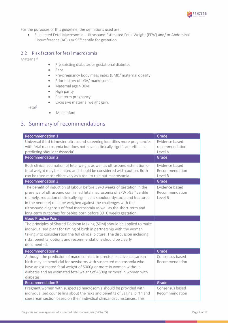
3. Summary of recommendations
Recommendation 1 Grade Universal third trimester ultrasound screening identifies more pregnancies with fetal macrosomia but does not have a clinically significant effect at predicting shoulder dystocia5.
Evidence based recommendation Level A
Recommendation 2 Grade
Both clinical estimation of fetal weight as well as ultrasound estimation of fetal weight may be limited and should be considered with caution. Both can be used most effectively as a tool to rule out macrosomia.
Evidence based Recommendation Level B
Recommendation 3 Grade The benefit of induction of labour before 39+0 weeks of gestation in the presence of ultrasound confirmed fetal macrosomia of EFW >95th centile (namely, reduction of clinically significant shoulder dystocia and fractures in the neonate) must be weighed against the challenges with the ultrasound diagnosis of fetal macrosomia as well as the short-term and long-term outcomes for babies born before 39+0 weeks gestation.
Evidence based Recommendation Level B
Good Practice Point The principles of Shared Decision Making (SDM) should be applied to make individualised plans for timing of birth in partnership with the woman taking into consideration the full clinical picture. The discussion including risks, benefits, options and recommendations should be clearly documented.
Recommendation 4 Grade Although the prediction of macrosomia is imprecise, elective caesarean birth may be beneficial for newborns with suspected macrosomia who have an estimated fetal weight of 5000g or more in women without diabetes and an estimated fetal weight of 4500g or more in women with diabetes.
Consensus based Recommendation
Recommendation 5 Grade Pregnant women with suspected macrosomia should be provided with individualised counselling about the risks and benefits of vaginal birth and caesarean section based on their individual clinical circumstances. This
Consensus based Recommendation
Diagnosis and management of suspected fetal macrosomia (C-Obs 65) Page 5 of 17
4. Discussion and recommendation
4.1 Diagnosis of macrosomia
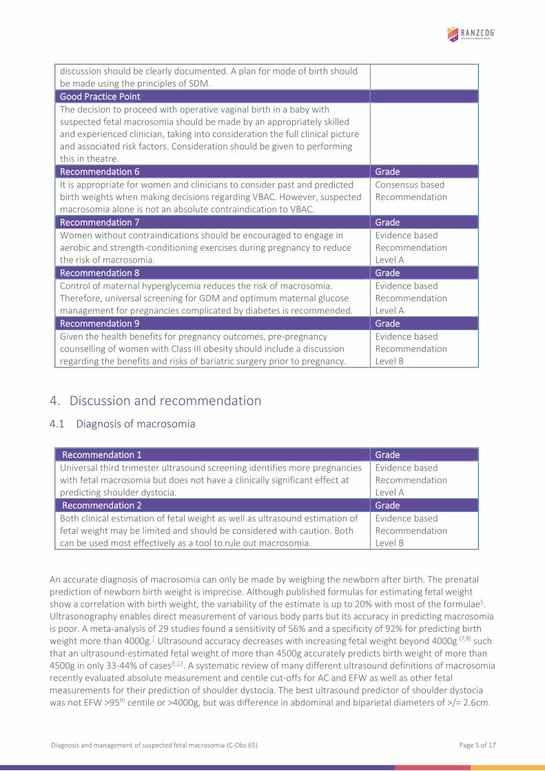
An accurate diagnosis of macrosomia can only be made by weighing the newborn after birth. The prenatal prediction of newborn birth weight is imprecise. Although published formulas for estimating fetal weight show a correlation with birth weight, the variability of the estimate is up to 20% with most of the formulae6. Ultrasonography enables direct measurement of various body parts but its accuracy in predicting macrosomia is poor. A meta-analysis of 29 studies found a sensitivity of 56% and a specificity of 92% for predicting birth weight more than 4000g.7 Ultrasound accuracy decreases with increasing fetal weight beyond 4000g (7,8) such that an ultrasound-estimated fetal weight of more than 4500g accurately predicts birth weight of more than 4500g in only 33-44% of cases8-13. A systematic review of many different ultrasound definitions of macrosomia recently evaluated absolute measurement and centile cut-offs for AC and EFW as well as other fetal measurements for their prediction of shoulder dystocia. The best ultrasound predictor of shoulder dystocia was not EFW >95th centile or >4000g, but was difference in abdominal and biparietal diameters of >/= 2.6cm.
discussion should be clearly documented. A plan for mode of birth should be made using the principles of SDM. Good Practice Point The decision to proceed with operative vaginal birth in a baby with suspected fetal macrosomia should be made by an appropriately skilled and experienced clinician, taking into consideration the full clinical picture and associated risk factors. Consideration should be given to performing this in theatre.
Recommendation 6 Grade It is appropriate for women and clinicians to consider past and predicted birth weights when making decisions regarding VBAC. However, suspected macrosomia alone is not an absolute contraindication to VBAC.
Consensus based Recommendation
Recommendation 7 Grade Women without contraindications should be encouraged to engage in aerobic and strength-conditioning exercises during pregnancy to reduce the risk of macrosomia.
Evidence based Recommendation Level A
Recommendation 8 Grade Control of maternal hyperglycemia reduces the risk of macrosomia. Therefore, universal screening for GDM and optimum maternal glucose management for pregnancies complicated by diabetes is recommended.
Evidence based Recommendation Level A
Recommendation 9 Grade Given the health benefits for pregnancy outcomes, pre-pregnancy counselling of women with Class III obesity should include a discussion regarding the benefits and risks of bariatric surgery prior to pregnancy.
Evidence based Recommendation Level B
Recommendation 1 Grade Universal third trimester ultrasound screening identifies more pregnancies with fetal macrosomia but does not have a clinically significant effect at predicting shoulder dystocia.
Evidence based Recommendation Level A
Recommendation 2 Grade Both clinical estimation of fetal weight as well as ultrasound estimation of fetal weight may be limited and should be considered with caution. Both can be used most effectively as a tool to rule out macrosomia.
Evidence based Recommendation Level B
Diagnosis and management of suspected fetal macrosomia (C-Obs 65) Page 6 of 17
A variety of other techniques and formulae have also proven to be unhelpful at accurately predicting macrosomia. Neither longitudinal ultrasound examinations nor individual growth-curve modelling improves prediction of macrosomia14. Using height and weight customised growth curves has proved to be no better than using population-based growth curves. However, there is evidence to show that height customised charts improve the prediction of LGA babies that are at increased risk of intrapartum caesarean section15. Magnetic resonance imaging has been shown to have higher sensitivity and specificity than ultrasonography8,
16 but, given its cost and discomfort, further study is needed to determine its appropriate use in this setting.
Measurement of symphysis to fundal height alone is a poor predictor of macrosomia. Retrospective studies suggest that the sensitivity of fundal height measurement alone for detection of macrosomia is 20-70% depending on the thresholds used17, 18, although specificity is more than 90% indicating that it is more effective for ruling out macrosomia than ruling it in.
Studies comparing the accuracy of ultrasound with that of physical examination for detection of macrosomia have had inconsistent findings. Universal third trimester ultrasound screening identifies more pregnancies with fetal macrosomia but does not have a clinically significant effect at predicting shoulder dystocia. Parous women appear to be able to predict the weight of their newborns as well as clinicians who use ultrasonography or clinical palpation19.
4.2 Risks associated with macrosomia
4.2.1 Maternal Morbidity: Increased risk of
• Caesarean birth: This is due to labour abnormalities (prolonged labour and arrest of dilatation and or descent). Studies show that with birth weights of more than 4500g, the risk of caesarean birth for women attempting a vaginal birth is at least double that of controls2, 20-22 .
• Postpartum haemorrhage (OR 3.1)23 • Chorioamnionitis (OR 2.4)23 • Third-degree or fourth-degree tears (OR 1.7)24. This risk is especially higher when the birth is
complicated by shoulder dystocia25.
4.2.2 Fetal Morbidity and Mortality: Increased risk of
• Shoulder dystocia (OR 7.1)23 • Injuries associated with shoulder dystocia - fracture of the clavicle26 (10-fold increase in the presence
of macrosomia), damage to the nerves of the brachial plexus leading to Erb-Duchenne paralysis27 (18-fold to 21-fold increase in the presence of macrosomia with absolute rates between 2.6% and 7%28). Between 80-90% of cases of brachial plexus palsy resolve by age 1 year25. Persistent injury is more common with higher birth weights and, in particular, birth weights over 4500g29.
• Low 5-minute Apgar scores20, 23 • Hypoglycemia30 • Rates of admission to neonatal intensive care unit30 • Overweight, obesity and metabolic syndrome later in life31
Diagnosis and management of suspected fetal macrosomia (C-Obs 65) Page 7 of 17
4.3 Management of term patients with suspected macrosomia
4.3.1 Timing of birth
Two randomised clinical trials have examined the effect of a policy of induction of labour at term for ultrasonography-derived estimated fetal weight more than the 90th percentile. In the first trial (1997), a total of 273 women at 38 weeks of gestation or later with ultrasonographically-derived estimated fetal weights between 4000g and 4500g were randomised to either planned induction of labour or expectant management32. The caesarean birth rates were similar: 19.4% for the induction group and 21.6% for the expectant group. There were 5 cases of shoulder dystocia in the induction group and 6 in the expectant group. All were managed without brachial plexus palsy or other trauma. However, it is important to note that this data is from 1997.
In a second trial (2015), a total of 822 women with a clinical suspicion of macrosomia and an estimated fetal weights (EFW) on follow up US to be above the 95th percentile for gestational age between 37+0 to 38+6 weeks of gestation were randomised to induction of labour within 3 days or to expectant management33. With induction of labour, the risk of shoulder dystocia was reduced from 4% to 1% (RR 0,32; 95% CI 0.12 to 0.85). Importantly, there were no instances of brachial plexus palsy in either group, and the caesarean birth rates were similar: 28% in the induction group and 32% in the expectant management group (RR 0.89; 95% CI 0.72 to 1.09). The only significant differences in newborn outcomes were a decrease in clinically significant shoulder dystocia (from 4% to 1%) and neonatal fractures (from 1% to 0.8%). There was an increase in neonatal hyperbilirubinemia and the need for phototherapy, especially in the group that gave birth before 38+6 weeks of gestation. The results of this trial should be interpreted noting that the definition of macrosomia used was specifically EFW > 95th centile at 36-38 weeks of gestation (AC was not used to define macrosomia) and that the trial had 409 women in the IOL arm and 413 women in the expectant management arm. Larger trials are needed to confirm these findings.
Two meta-analyses involving a total of 1190 women with suspected macrosomia have been published34, 35. Compared with expectant management, induction of labour for suspected macrosomia reduced the risk of shoulder dystocia (RR 0.60; 95% CI, 0.37 to 0.98) and any type of fracture (RR 0.20; 95% CI, 0.05 to 0.79) with no change in the risk of caesarean birth (RR 0.91; 95% CI, 0.76 to 1.09) or instrumental birth (RR 0.86; 95% CI, 0.65 to 1.13)34. However, there were no significant differences between the groups for brachial plexus palsy, although this outcome was infrequent (RR 0.21; 95% CI, 0.01 to 4.28). Findings from trials included in the review suggest that to prevent one fracture it would be necessary to induce labour in 60 women.
Whether intervention is better than expectant management for fetuses with suspected macrosomia and the gestational age at which delivery should be performed are unclear36. Meta-analysis of available trials suggests that IOL for suspected macrosomia can reduce the risks of shoulder dystocia and birth trauma but this benefit
Recommendation 3 Grade The benefit of induction of labour before 39+0 weeks of gestation in the presence of ultrasound confirmed fetal macrosomia of EFW >95th centile (namely, reduction of clinically significant shoulder dystocia and fractures in the neonate) must be weighed against the challenges with the ultrasound diagnosis of fetal macrosomia as well as the short-term and long-term outcomes for babies born before 39+0 weeks gestation.
Evidence based Recommendation Level B
Good Practice Point The principles of Shared Decision Making (SDM) should be applied to make individualised plans for timing of birth in partnership with the woman taking into consideration the full clinical picture. The discussion including risks, benefits, options and recommendations should be clearly documented.
Diagnosis and management of suspected fetal macrosomia (C-Obs 65) Page 8 of 17
is best realized when IOL is undertaken shortly after the diagnosis of macrosomia at 37-38 weeks’ gestation. The benefit of IOL solely for the indication of suspected macrosomia later in pregnancy (>39 weeks) is not established. Observational data suggests that planned birth after 39+0 weeks results in better short term and long term neonatal, infant and childhood outcomes37. A large before and after study showed that a policy that restricts both IOL and elective caesarean section before 39+0 weeks gestation is associated with reduced risk of admission to neonatal intensive care unit38. There is insufficient evidence to compare IOL above 39 weeks to expectant management where the benefit of reducing shoulder dystocia does not outweigh the harm of early birth.
The benefits of ‘routine’ IOL at 39 weeks for nulliparous women with a normally grown baby are increasingly being recognized39, 40 and can form an additional consideration when counselling women regarding their options for timing of birth.
To enable women to make an informed choice based on their individual circumstances and options, the principles of Shared Decision Making (SDM) should be applied. SDM is defined as ‘an approach where clinicians and patients share the best available evidence when faced with the task of making decisions, and where patients are supported to consider options, to achieve informed preferences.
4.3.2 Mode of birth
Suspected fetal macrosomia is not a contraindication to vaginal birth. Although fetal and maternal morbidity increase with birth weights more than 4000g, most births of macrosomic newborns are uncomplicated23, 41. Elective caesarean section reduces, but does not eliminate, the risk of birth trauma and neonatal brachial plexus palsy associated with macrosomia27, 42. Although the prediction of macrosomia is imprecise, elective caesarean section may be beneficial for newborns who have an estimated fetal weight of at least 4500g in women with diabetes and an estimated fetal weight of 5000g in women without diabetes. However, in the absence of randomized clinical trials, elective caesarean section for suspected macrosomia is based on expert opinion.
Studies using estimates of the prevalence of permanent brachial plexus palsy at birth found that between 155 to 1026 caesarean births would need to be performed to prevent one occurrence of permanent brachial plexus palsy for newborns with a birth weight of 4500g and between 79 to 373 caesarean births for newborns with a birth weight of 5000g27, 42. If the imperfect predictive values of ultrasonography for macrosomia are accounted for, the number of caesarean births is much higher. A policy of elective caesarean birth for suspected macrosomia in newborns weighing less than 5000g in women without diabetes would result in significant maternal morbidity42. Despite the lack of evidence supporting caesarean birth, most, but not all, researchers agree that consideration should be given to caesarean birth when the estimated fetal weight is 5000g or more because of increased risk of stillbirth and higher rates of other morbidities in women and newborns27, 42.
Recommendation 4 Grade Although the prediction of macrosomia is imprecise, elective caesarean birth may be beneficial for newborns with suspected macrosomia who have an estimated fetal weight of 5000g or more in women without diabetes and an estimated fetal weight of 4500g or more in women with diabetes.
Consensus based Recommendation
Recommendation 5 Grade Pregnant women with suspected macrosomia should be provided with individualised counselling about the risks and benefits of vaginal birth and caesarean section based on their individual clinical circumstances. This discussion should be clearly documented. A plan for mode of birth should be made using the principles of SDM.
Consensus based Recommendation
Diagnosis and management of suspected fetal macrosomia (C-Obs 65) Page 9 of 17
For women with diabetes (pre-existing or gestational), the risks of fetal and maternal morbidity increase when newborns weigh more than 4500g23, 27 and the number of caesarean births to prevent one occurrence of permanent brachial plexus palsy is more favourable than it is for women without diabetes27, 42. Hence, there is more evidence for recommending caesarean birth in women with diabetes with an estimated fetal weight of more than 4500g.
Pregnant women with suspected macrosomia should be provided with individualised counselling about the risks of vaginal birth and caesarean birth accounting for their relevant clinical considerations. Discussion should include the difficulty to predict birth weight accurately, the low incidence of brachial plexus palsy and shoulder dystocia even in macrosomic fetuses and the fact that the risk of brachial plexus palsy is not eliminated by caesarean birth. The principles of Shared Decision Making should be followed when counselling about mode of birth.
4.3.3 Management of labour and birth
Prolonged first and second stages of labour are common when macrosomia is present20, 23. Labour abnormalities have been associated with shoulder dystocia in some, but not all, studies, and these abnormalities occur too frequently to be useful predictors of shoulder dystocia41, 43. Some studies have however, shown that the combination of an estimated fetal weight of more than 4500g and arrest of labour is significantly associated with shoulder dystocia44. Therefore, when the fetal weight is estimated to be more than 4500g, a prolonged second stage of labour or arrest of descent in second stage above +2 (out of +5) station should prompt senior obstetric review. This assessment should include both abdominal palpation of the fetal head and vaginal examination to determine position of the fetal vertex and the presence of asynclitism, caput and molding.
Whether to conduct an operative vaginal birth in cases of suspected macrosomia is another important consideration. Observational studies consistently demonstrate an increased risk of shoulder dystocia when a macrosomic fetus is delivered using forceps or vacuum extraction. Two large population-based studies found a threefold to fivefold increase in the odds of shoulder dystocia with vacuum extraction41, 45. A meta-analysis of four observational studies calculated a summary OR of 2.98 (95% CI, 2.3 to 3.9) (40). Studies and meta-analysis have found no increase in risk of shoulder dystocia with forceps birth (OR 1.1) compared with vacuum extraction41, 46.
The risk of shoulder dystocia at the time of operative vaginal birth increases when more than one risk factor is present. Thus, the clinician should have a heightened awareness for shoulder dystocia in these situations. Judicious use of operative vaginal birth conducted by an appropriately skilled and experienced clinician is reasonable even in the presence of risk factors. The woman should be counselled regarding the risks and preparation should be made for the possibility of encountering shoulder dystocia. It may be prudent to perform operative vaginal birth in an operating theatre, but this decision should be made based on individual circumstances by the most senior clinician at the time.
Good Practice Point The decision to proceed with operative vaginal birth in a baby with suspected fetal macrosomia should be made by an appropriately skilled and experienced clinician, taking into consideration the full clinical picture and associated risk factors. Consideration should be given to performing this in theatre.
Recommendation 6 Grade It is appropriate for women and clinicians to consider past and predicted birth weights when making decisions regarding VBAC. However, suspected macrosomia alone is not an absolute contraindication to VBAC.
Consensus based Recommendation
Diagnosis and management of suspected fetal macrosomia (C-Obs 65) Page 10 of 17
Suspected macrosomia alone is not an absolute contraindication to attempting vaginal birth after caesarean section (VBAC). Although success rates of VBAC decrease as newborn weight increases to 4000g or more, this effect does not decrease absolute VBAC success rate to less than 50% in women who have had a previous vaginal birth or previous VBAC 42, 47, 48. There may be a higher risk of uterine rupture during labour after caesarean section with neonatal birth weights more than 4000g. The rates of rupture are highest in women with no prior history of vaginal birth and with increasing birth weight42, 48. However, the studies used actual birth weight as opposed to estimated fetal weight and hence, have limited applicability in making decisions regarding mode of delivery before labour42. Once again, the principles of SDM should be used when counselling women about their options for VBAC.
There is insufficient evidence to guide decision making for external cephalic version (ECV) in women with suspected macrosomia. There are no reported studies on the relationship between suspected macrosomia and success or failure of ECV. The principles of SDM should be used to guide discussion taking into consideration the full clinical picture.
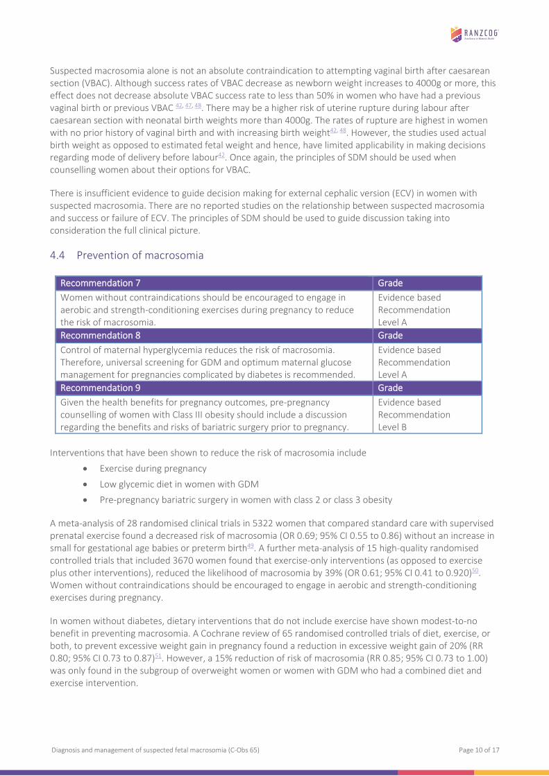
4.4 Prevention of macrosomia
Interventions that have been shown to reduce the risk of macrosomia include
• Exercise during pregnancy
• Low glycemic diet in women with GDM
• Pre-pregnancy bariatric surgery in women with class 2 or class 3 obesity
A meta-analysis of 28 randomised clinical trials in 5322 women that compared standard care with supervised prenatal exercise found a decreased risk of macrosomia (OR 0.69; 95% CI 0.55 to 0.86) without an increase in small for gestational age babies or preterm birth49. A further meta-analysis of 15 high-quality randomised controlled trials that included 3670 women found that exercise-only interventions (as opposed to exercise plus other interventions), reduced the likelihood of macrosomia by 39% (OR 0.61; 95% CI 0.41 to 0.920)50. Women without contraindications should be encouraged to engage in aerobic and strength-conditioning exercises during pregnancy.
In women without diabetes, dietary interventions that do not include exercise have shown modest-to-no benefit in preventing macrosomia. A Cochrane review of 65 randomised controlled trials of diet, exercise, or both, to prevent excessive weight gain in pregnancy found a reduction in excessive weight gain of 20% (RR 0.80; 95% CI 0.73 to 0.87)51. However, a 15% reduction of risk of macrosomia (RR 0.85; 95% CI 0.73 to 1.00) was only found in the subgroup of overweight women or women with GDM who had a combined diet and exercise intervention.
Recommendation 7 Grade Women without contraindications should be encouraged to engage in aerobic and strength-conditioning exercises during pregnancy to reduce the risk of macrosomia.
Evidence based Recommendation Level A
Recommendation 8 Grade Control of maternal hyperglycemia reduces the risk of macrosomia. Therefore, universal screening for GDM and optimum maternal glucose management for pregnancies complicated by diabetes is recommended.
Evidence based Recommendation Level A
Recommendation 9 Grade Given the health benefits for pregnancy outcomes, pre-pregnancy counselling of women with Class III obesity should include a discussion regarding the benefits and risks of bariatric surgery prior to pregnancy.
Evidence based Recommendation Level B
Diagnosis and management of suspected fetal macrosomia (C-Obs 65) Page 11 of 17
Control of maternal hyperglycemia reduces the risk of macrosomia. In the Australian Carbohydrate Intolerance Study in Pregnant Women (ACHOIS) trial, the risk of birth weight more than 4000g was reduced from 21% to 10% (RR 0.47; 95% CI 0.34 to 0.64; P=.001)52.
Specific diets for reduction in macrosomia have also been investigated. Women with GDM on a low glycemic diet were found to have 73% reduction in macrosomia (OR 0.27; 95% CI 0.10 to 0.71) compared with usual diet53. Diets that included additional dietary fibre further decreased the risk.
For women with class 2 or class 3 obesity, having had bariatric surgery before pregnancy is associated with decreased odds of GDM (OR 0.31 and 0.47, respectively) and LGA newborns (OR 0.40 and 0.46 respectively) in meta-analyses of observational studies54, 55. However, previous bariatric surgery is also associated with an increase in small for gestational age babies. Preterm births were not statistically different. Given the health benefits, particularly for pregnancy outcomes, pre-pregnancy counselling women with Class III obesity regarding benefits and risks of bariatric surgery is recommended.
5. References 1. Doty MS, Chen HY, Sibai BM, Chauhan SP. Maternal and Neonatal Morbidity Associated With Early Term Delivery of Large-for-Gestational-Age But Nonmacrosomic Neonates. Obstetrics and gynecology. 2019;133(6):1160-6. 2. Zhang X, Decker A, Platt RW, Kramer MS. How big is too big? The perinatal consequences of fetal macrosomia. American journal of obstetrics and gynecology. 2008;198(5):517 e1-6. 3. Dobbins TA, Sullivan EA, Roberts CL, Simpson JM. Australian national birthweight percentiles by sex and gestational age, 1998-2007. Med J Aust. 2012;197(5):291-4. 4. Orskou J, Henriksen TB, Kesmodel U, Secher NJ. Maternal characteristics and lifestyle factors and the risk of delivering high birth weight infants. Obstetrics and gynecology. 2003;102(1):115-20. 5. Moraitis AA, Shreeve N, Sovio U, Brocklehurst P, Heazell AEP, Thornton JG, et al. Universal third-trimester ultrasonic screening using fetal macrosomia in the prediction of adverse perinatal outcome: A systematic review and meta-analysis of diagnostic test accuracy. PLoS Med. 2020;17(10):e1003190. 6. Hadlock FP, Deter RL, Harrist RB, Park SK. Estimating fetal age: computer-assisted analysis of multiple fetal growth parameters. Radiology. 1984;152(2):497-501. 7. Robinson R, Walker KF, White VA, Bugg GJ, Snell KIE, Jones NW. The test accuracy of antenatal ultrasound definitions of fetal macrosomia to predict birth injury: A systematic review. European Journal of Obstetrics and Gynecology and Reproductive Biology. 2020;246:79-85. 8. Malin GL, Bugg GJ, Takwoingi Y, Thornton JG, Jones NW. Antenatal magnetic resonance imaging versus ultrasound for predicting neonatal macrosomia: a systematic review and meta-analysis. BJOG : an international journal of obstetrics and gynaecology. 2016;123(1):77-88. 9. Scioscia M, Vimercati A, Ceci O, Vicino M, Selvaggi LE. Estimation of birth weight by two-dimensional ultrasonography: a critical appraisal of its accuracy. Obstetrics and gynecology. 2008;111(1):57-65. 10. Zafman KB, Bergh E, Fox NS. Accuracy of sonographic estimated fetal weight in suspected macrosomia: the likelihood of overestimating and underestimating the true birthweight. J Matern Fetal Neonatal Med. 2020;33(6):967-72. 11. Sandmire HF. Whither ultrasonic prediction of fetal macrosomia? Obstetrics and gynecology. 1993;82(5):860-2. 12. Chauhan SP, Hendrix NW, Magann EF, Morrison JC, Kenney SP, Devoe LD. Limitations of clinical and sonographic estimates of birth weight: experience with 1034 parturients. Obstetrics and gynecology. 1998;91(1):72-7. 13. Aviram A, Yogev Y, Ashwal E, Hiersch L, Danon D, Hadar E, et al. Different formulas, different thresholds and different performance-the prediction of macrosomia by ultrasound. J Perinatol. 2017;37(12):1285-91.
Diagnosis and management of suspected fetal macrosomia (C-Obs 65) Page 12 of 17
14. Zhang J, Kim S, Grewal J, Albert PS. Predicting large fetuses at birth: do multiple ultrasound examinations and longitudinal statistical modelling improve prediction? Paediatr Perinat Epidemiol. 2012;26(3):199-207. 15. Pritchard N, Lindquist A, Hiscock R, Diksha P, Walker SP, Permezel M. Customised growth charts in large-for-gestational-age infants and the association with emergency caesarean section rate. Aust N Z J Obstet Gynaecol. 2019;59(3):380-6. 16. Kadji C, Cannie MM, Resta S, Guez D, Abi-Khalil F, De Angelis R, et al. Magnetic resonance imaging for prenatal estimation of birthweight in pregnancy: review of available data, techniques, and future perspectives. American journal of obstetrics and gynecology. 2019;220(5):428-39. 17. Goetzinger KR, Tuuli MG, Odibo AO, Roehl KA, Macones GA, Cahill AG. Screening for fetal growth disorders by clinical exam in the era of obesity. J Perinatol. 2013;33(5):352-7. 18. Sparks TN, Cheng YW, McLaughlin B, Esakoff TF, Caughey AB. Fundal height: a useful screening tool for fetal growth? J Matern Fetal Neonatal Med. 2011;24(5):708-12. 19. Harlev A, Walfisch A, Bar-David J, Hershkovitz R, Friger M, Hallak M. Maternal estimation of fetal weight as a complementary method of fetal weight assessment: a prospective clinical trial. J Reprod Med. 2006;51(7):515-20. 20. Boulet SL, Alexander GR, Salihu HM, Pass M. Macrosomic births in the united states: determinants, outcomes, and proposed grades of risk. American journal of obstetrics and gynecology. 2003;188(5):1372-8. 21. Rossi AC, Mullin P, Prefumo F. Prevention, management, and outcomes of macrosomia: a systematic review of literature and meta-analysis. Obstet Gynecol Surv. 2013;68(10):702-9. 22. Beta J, Khan N, Khalil A, Fiolna M, Ramadan G, Akolekar R. Maternal and neonatal complications of fetal macrosomia: systematic review and meta-analysis. Ultrasound Obstet Gynecol. 2019;54(3):308-18. 23. King JR, Korst LM, Miller DA, Ouzounian JG. Increased composite maternal and neonatal morbidity associated with ultrasonographically suspected fetal macrosomia. J Matern Fetal Neonatal Med. 2012;25(10):1953-9. 24. Gupta N, Kiran TU, Mulik V, Bethel J, Bhal K. The incidence, risk factors and obstetric outcome in primigravid women sustaining anal sphincter tears. Acta Obstet Gynecol Scand. 2003;82(8):736-43. 25. Gauthaman N, Walters S, Tribe IA, Goldsmith L, Doumouchtsis SK. Shoulder dystocia and associated manoeuvres as risk factors for perineal trauma. International urogynecology journal. 2016;27(4):571-7. 26. Ahn ES, Jung MS, Lee YK, Ko SY, Shin SM, Hahn MH. Neonatal clavicular fracture: recent 10 year study. Pediatr Int. 2015;57(1):60-3. 27. Ahn JSAJT. Fetal Macrosomia UpToDate2019. Available from: https://www.uptodate.com/contents/fetal-macrosomia. 28. Esakoff TF, Cheng YW, Sparks TN, Caughey AB. The association between birthweight 4000 g or greater and perinatal outcomes in patients with and without gestational diabetes mellitus. American journal of obstetrics and gynecology. 2009;200(6):672 e1-4. 29. Gherman RB, Ouzounian JG, Satin AJ, Goodwin TM, Phelan JP. A comparison of shoulder dystocia-associated transient and permanent brachial plexus palsies. Obstetrics and gynecology. 2003;102(3):544-8. 30. Gillean JR, Coonrod DV, Russ R, Bay RC. Big infants in the neonatal intensive care unit. American journal of obstetrics and gynecology. 2005;192(6):1948-53; discussion 53-5. 31. Cnattingius S, Villamor E, Lagerros YT, Wikström AK, Granath F. High birth weight and obesity--a vicious circle across generations. International journal of obesity (2005). 2012;36(10):1320-4. 32. Gonen O, Rosen DJ, Dolfin Z, Tepper R, Markov S, Fejgin MD. Induction of labor versus expectant management in macrosomia: a randomized study. Obstetrics and gynecology. 1997;89(6):913-7. 33. Boulvain M, Senat MV, Perrotin F, Winer N, Beucher G, Subtil D, et al. Induction of labour versus expectant management for large-for-date fetuses: a randomised controlled trial. Lancet. 2015;385(9987):2600-5. 34. Boulvain M, Irion O, Dowswell T, Thornton JG. Induction of labour at or near term for suspected fetal macrosomia. The Cochrane database of systematic reviews. 2016(5):CD000938. 35. Magro-Malosso ER, Saccone G, Chen M, Navathe R, Di Tommaso M, Berghella V. Induction of labour for suspected macrosomia at term in non-diabetic women: a systematic review and meta-analysis of randomized controlled trials. BJOG : an international journal of obstetrics and gynaecology. 2017;124(3):414-21.
Diagnosis and management of suspected fetal macrosomia (C-Obs 65) Page 13 of 17
36. Caughey AB. Should pregnancies be induced for impending macrosomia? Lancet. 2015;385(9987):2557-9. 37. Chan E, Leong P, Malouf R, Quigley MA. Long-term cognitive and school outcomes of late-preterm and early-term births: a systematic review. Child Care Health Dev. 2016;42(3):297-312. 38. Ehrenthal DB, Hoffman MK, Jiang X, Ostrum G. Neonatal outcomes after implementation of guidelines limiting elective delivery before 39 weeks of gestation. Obstetrics and gynecology. 2011;118(5):1047-55. 39. Grobman WA, Rice MM, Reddy UM, Tita ATN, Silver RM, Mallett G, et al. Labor Induction versus Expectant Management in Low-Risk Nulliparous Women. N Engl J Med. 2018;379(6):513-23. 40. Wagner SM, Sandoval G, Grobman WA, Bailit JL, Wapner RJ, Varner MW, et al. Labor Induction at 39 Weeks Compared with Expectant Management in Low-Risk Parous Women. Am J Perinatol. 2020. 41. Overland EA, Vatten LJ, Eskild A. Risk of shoulder dystocia: associations with parity and offspring birthweight. A population study of 1 914 544 deliveries. Acta Obstet Gynecol Scand. 2012;91(4):483-8. 42. Macrosomia: ACOG Practice Bulletin, Number 216. Obstetrics and gynecology. 2020;135(1):e18-e35. 43. Laughon SK, Berghella V, Reddy UM, Sundaram R, Lu Z, Hoffman MK. Neonatal and maternal outcomes with prolonged second stage of labor. Obstetrics and gynecology. 2014;124(1):57-67. 44. Palatnik A, Grobman WA, Hellendag MG, Janetos TM, Gossett DR, Miller ES. Predictors of shoulder dystocia at the time of operative vaginal delivery. American journal of obstetrics and gynecology. 2016;215(5):624 e1- e5. 45. Sheiner E, Levy A, Hershkovitz R, Hallak M, Hammel RD, Katz M, et al. Determining factors associated with shoulder dystocia: a population-based study. European journal of obstetrics, gynecology, and reproductive biology. 2006;126(1):11-5. 46. Dall'Asta A, Ghi T, Pedrazzi G, Frusca T. Does vacuum delivery carry a higher risk of shoulder dystocia? Review and meta-analysis of the literature. European journal of obstetrics, gynecology, and reproductive biology. 2016;204:62-8. 47. Jastrow N, Roberge S, Gauthier RJ, Laroche L, Duperron L, Brassard N, et al. Effect of birth weight on adverse obstetric outcomes in vaginal birth after cesarean delivery. Obstetrics and gynecology. 2010;115(2 Pt 1):338-43. 48. Elkousy MA, Sammel M, Stevens E, Peipert JF, Macones G. The effect of birth weight on vaginal birth after cesarean delivery success rates. American journal of obstetrics and gynecology. 2003;188(3):824-30. 49. Wiebe HW, Boule NG, Chari R, Davenport MH. The effect of supervised prenatal exercise on fetal growth: a meta-analysis. Obstetrics and gynecology. 2015;125(5):1185-94. 50. Davenport MH, Meah VL, Ruchat SM, Davies GA, Skow RJ, Barrowman N, et al. Impact of prenatal exercise on neonatal and childhood outcomes: a systematic review and meta-analysis. Br J Sports Med. 2018;52(21):1386-96. 51. Muktabhant B, Lawrie TA, Lumbiganon P, Laopaiboon M. Diet or exercise, or both, for preventing excessive weight gain in pregnancy. The Cochrane database of systematic reviews. 2015(6):CD007145. 52. Crowther CA, Hiller JE, Moss JR, McPhee AJ, Jeffries WS, Robinson JS, et al. Effect of treatment of gestational diabetes mellitus on pregnancy outcomes. N Engl J Med. 2005;352(24):2477-86. 53. Wei J, Heng W, Gao J. Effects of Low Glycemic Index Diets on Gestational Diabetes Mellitus: A Meta-Analysis of Randomized Controlled Clinical Trials. Medicine (Baltimore). 2016;95(22):e3792. 54. Yi XY, Li QF, Zhang J, Wang ZH. A meta-analysis of maternal and fetal outcomes of pregnancy after bariatric surgery. Int J Gynaecol Obstet. 2015;130(1):3-9. 55. Galazis N, Docheva N, Simillis C, Nicolaides KH. Maternal and neonatal outcomes in women undergoing bariatric surgery: a systematic review and meta-analysis. European journal of obstetrics, gynecology, and reproductive biology. 2014;181:45-53.
Diagnosis and management of suspected fetal macrosomia (C-Obs 65) Page 14 of 17
Appendices
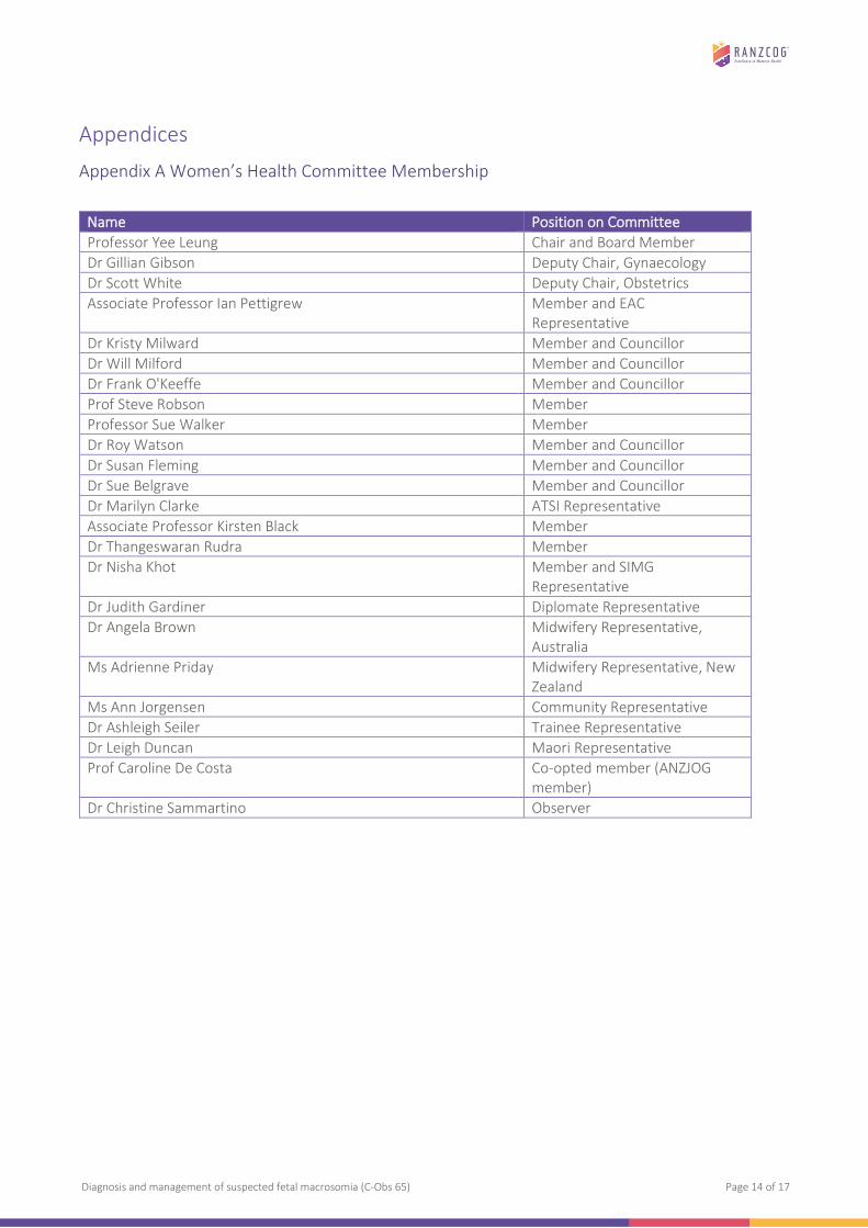
Appendix A Women’s Health Committee Membership
Name Position on Committee Professor Yee Leung Chair and Board Member Dr Gillian Gibson Deputy Chair, Gynaecology Dr Scott White Deputy Chair, Obstetrics Associate Professor Ian Pettigrew Member and EAC
Representative Dr Kristy Milward Member and Councillor Dr Will Milford Member and Councillor Dr Frank O'Keeffe Member and Councillor Prof Steve Robson Member Professor Sue Walker Member Dr Roy Watson Member and Councillor Dr Susan Fleming Member and Councillor Dr Sue Belgrave Member and Councillor Dr Marilyn Clarke ATSI Representative Associate Professor Kirsten Black Member Dr Thangeswaran Rudra Member Dr Nisha Khot Member and SIMG
Representative Dr Judith Gardiner Diplomate Representative Dr Angela Brown Midwifery Representative,
Australia Ms Adrienne Priday Midwifery Representative, New
Zealand Ms Ann Jorgensen Community Representative Dr Ashleigh Seiler Trainee Representative Dr Leigh Duncan Maori Representative Prof Caroline De Costa Co-opted member (ANZJOG
member) Dr Christine Sammartino Observer
Diagnosis and management of suspected fetal macrosomia (C-Obs 65) Page 15 of 17
Appendix B Overview of the development and review process for this statement i. Steps in developing and updating this statement This statement was developed in November 2021. The Women’s Health Committee carried out the following steps in reviewing this statement: • Declarations of interest were sought from all members prior to reviewing this statement. • Structured clinical questions were developed and agreed upon. • An updated literature search to answer the clinical questions was undertaken. • At the November 2021 Committee meeting, the recommendations were reviewed and updated (where
appropriate) based on the available body of evidence and clinical expertise. Recommendations were graded as set out below in Appendix B part iii)
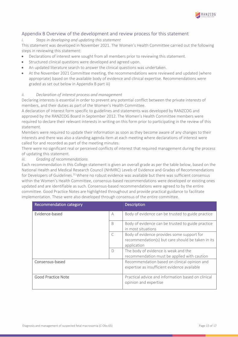
ii. Declaration of interest process and management Declaring interests is essential in order to prevent any potential conflict between the private interests of members, and their duties as part of the Women’s Health Committee. A declaration of interest form specific to guidelines and statements was developed by RANZCOG and approved by the RANZCOG Board in September 2012. The Women’s Health Committee members were required to declare their relevant interests in writing on this form prior to participating in the review of this statement. Members were required to update their information as soon as they become aware of any changes to their interests and there was also a standing agenda item at each meeting where declarations of interest were called for and recorded as part of the meeting minutes. There were no significant real or perceived conflicts of interest that required management during the process of updating this statement. iii. Grading of recommendations Each recommendation in this College statement is given an overall grade as per the table below, based on the National Health and Medical Research Council (NHMRC) Levels of Evidence and Grades of Recommendations for Developers of Guidelines.31 Where no robust evidence was available but there was sufficient consensus within the Women’s Health Committee, consensus-based recommendations were developed or existing ones updated and are identifiable as such. Consensus-based recommendations were agreed to by the entire committee. Good Practice Notes are highlighted throughout and provide practical guidance to facilitate implementation. These were also developed through consensus of the entire committee.
Recommendation category Description
Evidence-based A Body of evidence can be trusted to guide practice
B Body of evidence can be trusted to guide practice in most situations
C Body of evidence provides some support for recommendation(s) but care should be taken in its application
D The body of evidence is weak and the recommendation must be applied with caution
Consensus-based Recommendation based on clinical opinion and expertise as insufficient evidence available
Good Practice Note Practical advice and information based on clinical opinion and expertise
Diagnosis and management of suspected fetal macrosomia (C-Obs 65) Page 16 of 17
Appendix C Full Disclaimer Purpose This Statement has been developed to provide general advice to practitioners about women’s health issues concerning Diagnosis and management of suspected fetal macrosomia and should not be relied on as a substitute for proper assessment with respect to the particular circumstances of each case and the needs of any person with suspected fetal macrosomia. It is the responsibility of each practitioner to have regard to the particular circumstances of each case. Clinical management should be responsive to the needs of the individual person with suspected fetal macrosomia and the particular circumstances of each case. Quality of information The information available in diagnosis and management of fetal macrosomia (C-Obs 65) is intended as a guide and provided for information purposes only. The information is based on the Australian/New Zealand context using the best available evidence and information at the time of preparation. While the Royal Australian and New Zealand College of Obstetricians and Gynaecologists (RANZCOG) had endeavoured to ensure that information is accurate and current at the time of preparation, it takes no responsibility for matters arising from changed circumstances or information or material that may have become subsequently available. The use of this information is entirely at your own risk and responsibility. For the avoidance of doubt, the materials were not developed for use by patients, and patients must seek medical advice in relation to any treatment. The material includes the views or recommendations of third parties and does not necessarily reflect the views of RANZCOG or indicate a commitment to a particular course of action. Third-party sites Any information linked in this Statement is provided for the user’s convenience and does not constitute an endorsement or a recommendation or indicate a commitment to a particular course of action of this information, material, or content unless specifically stated otherwise. RANZCOG disclaims, to the maximum extent permitted by law any responsibility and all liability (including without limitation, liability in negligence) to you or any third party for inaccurate, out of context, incomplete or unavailable information contained on the third-party website, or for whether the information contained on those websites is suitable for your needs or the needs of any third party for all expenses, losses, damages and costs incurred. Exclusion of liability The College disclaims, to the maximum extent permitted by law, all responsibility and all liability (including without limitation, liability in negligence) to you or any third party for any loss or damage which may result from your or any third party’s use of or reliance of this guideline, including the materials within or referred to throughout this document being in any way inaccurate, out of context, incomplete or unavailable for all expenses, losses, damages, and costs incurred. Exclusion of warranties To the maximum extent permitted by law, RANZCOG makes no representation, endorsement or warranty of any kind, expressed or implied in relation to the materials within or referred to throughout this guideline being in any way inaccurate, out of context, incomplete or unavailable for all expenses, losses, damages and costs incurred. These terms and conditions will be constructed according to and are governed by the laws of Victoria, Australia.

Diagnosis and management of suspected fetal macrosomia (C-Obs 65) Page 17 of 17
Version Date of Version Pages revised / Brief Explanation of Revision
v1.1 Nov / 2021 WHC
Policy Version: Version 1.1
Policy Owner: Women’s Health Committee
Policy Approved by: RANZCOG Council/Board
Review of Policy: March / 2027


![[Colorectal Carcinoma with Suspected Lynch Syndrome: A Multidisciplinary Algorithm.]](https://static.fdokumen.com/doc/165x107/6335f98064d291d2a302b343/colorectal-carcinoma-with-suspected-lynch-syndrome-a-multidisciplinary-algorithm.jpg)