Deep Learning-Based Computer-Aided Fetal Echocardiography
20
sensors Article Deep Learning-Based Computer-Aided Fetal Echocardiography: Application to Heart Standard View Segmentation for Congenital Heart Defects Detection Siti Nurmaini 1, * , Muhammad Naufal Rachmatullah 1 , Ade Iriani Sapitri 1 , Annisa Darmawahyuni 1 , Bambang Tutuko 1 , Firdaus Firdaus 1 , Radiyati Umi Partan 2 and Nuswil Bernolian 3 Citation: Nurmaini, S.; Rachmatullah, M.N.; Sapitri, A.I.; Darmawahyuni, A.; Tutuko, B.; Firdaus, F.; Partan, R.U.; Bernolian, N. Deep Learning-Based Computer-Aided Fetal Echocardiography: Application to Heart Standard View Segmentation for Congenital Heart Defects Detection. Sensors 2021, 21, 8007. https://doi.org/10.3390/s21238007 Academic Editors: Steven Su, Qian Peter Su and Ahmadreza Argha Received: 15 November 2021 Accepted: 29 November 2021 Published: 30 November 2021 Publisher’s Note: MDPI stays neutral with regard to jurisdictional claims in published maps and institutional affil- iations. Copyright: © 2021 by the authors. Licensee MDPI, Basel, Switzerland. This article is an open access article distributed under the terms and conditions of the Creative Commons Attribution (CC BY) license (https:// creativecommons.org/licenses/by/ 4.0/). 1 Intelligent System Research Group, Faculty of Computer Science, Universitas Sriwijaya, Palembang 30139, Indonesia; [email protected] (M.N.R.); [email protected] (A.I.S.); [email protected] (A.D.); [email protected] (B.T.) [email protected] (F.F.) 2 Faculty of Medicine, Universitas Sriwijaya, Palembang 30139, Indonesia; [email protected] 3 Division of Maternal-Fetal Medicine, Department of Obstetrics and Gynecology, Mohammad Hoesin General Hospital, Palembang 30126, Indonesia; [email protected] * Correspondence: [email protected]; Tel.: +62-852-6804-8092 Abstract: Accurate segmentation of fetal heart in echocardiography images is essential for detecting the structural abnormalities such as congenital heart defects (CHDs). Due to the wide variations attributed to different factors, such as maternal obesity, abdominal scars, amniotic fluid volume, and great vessel connections, this process is still a challenging problem. CHDs detection with expertise in general are substandard; the accuracy of measurements remains highly dependent on humans’ training, skills, and experience. To make such a process automatic, this study proposes deep learning-based computer-aided fetal heart echocardiography examinations with an instance segmentation approach, which inherently segments the four standard heart views and detects the defect simultaneously. We conducted several experiments with 1149 fetal heart images for predicting 24 objects, including four shapes of fetal heart standard views, 17 objects of heart-chambers in each view, and three cases of congenital heart defect. The result showed that the proposed model performed satisfactory performance for standard views segmentation, with a 79.97% intersection over union and 89.70% Dice coefficient similarity. It also performed well in the CHDs detection, with mean average precision around 98.30% for intra-patient variation and 82.42% for inter-patient variation. We believe that automatic segmentation and detection techniques could make an important contribution toward improving congenital heart disease diagnosis rates. Keywords: fetal echocardiography; deep learning; fetal heart standard view; heart defect; instance segmentation 1. Introduction Fetal echocardiography examination is widely applied in clinical settings due to its non-invasive nature, reduced cost, and real-time acquisition [1]. Such examination is usually assessed by ultrasound after an approximate gestational (menstrual) age of 18 weeks to find the heart structural abnormalities [2]. Assessment and evaluation of fetal heart abnormalities provide crucial information to families prior to the anticipated birth of their children about diagnosis, underlying etiology, and potential treatment options, which can greatly improve the survival rates of fetuses. One of the most common structural heart diseases is congenital heart defects (CHDs), which affect 5–9 out of 1000 births; CHDs cause 5% of all childhood deaths [2,3] and 18% of liveborn infants with CHDs die within the first year [4]. The process of CHDs examination begins with determining the location of the fetal heart based on four standard views, i.e., four-chamber (4CH), three-vessel and three-vessel Sensors 2021, 21, 8007. https://doi.org/10.3390/s21238007 https://www.mdpi.com/journal/sensors
-
Upload
khangminh22 -
Category
Documents
-
view
7 -
download
0
Transcript of Deep Learning-Based Computer-Aided Fetal Echocardiography

sensors
Article
Deep Learning-Based Computer-Aided Fetal Echocardiography:Application to Heart Standard View Segmentation forCongenital Heart Defects Detection
Siti Nurmaini 1,* , Muhammad Naufal Rachmatullah 1, Ade Iriani Sapitri 1, Annisa Darmawahyuni 1 ,Bambang Tutuko 1, Firdaus Firdaus 1 , Radiyati Umi Partan 2 and Nuswil Bernolian 3
�����������������
Citation: Nurmaini, S.;
Rachmatullah, M.N.; Sapitri, A.I.;
Darmawahyuni, A.; Tutuko, B.;
Firdaus, F.; Partan, R.U.; Bernolian, N.
Deep Learning-Based
Computer-Aided Fetal
Echocardiography: Application to
Heart Standard View Segmentation
for Congenital Heart Defects
Detection. Sensors 2021, 21, 8007.
https://doi.org/10.3390/s21238007
Academic Editors: Steven Su, Qian
Peter Su and Ahmadreza Argha
Received: 15 November 2021
Accepted: 29 November 2021
Published: 30 November 2021
Publisher’s Note: MDPI stays neutral
with regard to jurisdictional claims in
published maps and institutional affil-
iations.
Copyright: © 2021 by the authors.
Licensee MDPI, Basel, Switzerland.
This article is an open access article
distributed under the terms and
conditions of the Creative Commons
Attribution (CC BY) license (https://
creativecommons.org/licenses/by/
4.0/).
1 Intelligent System Research Group, Faculty of Computer Science, Universitas Sriwijaya,Palembang 30139, Indonesia; [email protected] (M.N.R.); [email protected] (A.I.S.);[email protected] (A.D.); [email protected] (B.T.) [email protected] (F.F.)
2 Faculty of Medicine, Universitas Sriwijaya, Palembang 30139, Indonesia; [email protected] Division of Maternal-Fetal Medicine, Department of Obstetrics and Gynecology, Mohammad Hoesin General
Hospital, Palembang 30126, Indonesia; [email protected]* Correspondence: [email protected]; Tel.: +62-852-6804-8092
Abstract: Accurate segmentation of fetal heart in echocardiography images is essential for detectingthe structural abnormalities such as congenital heart defects (CHDs). Due to the wide variationsattributed to different factors, such as maternal obesity, abdominal scars, amniotic fluid volume,and great vessel connections, this process is still a challenging problem. CHDs detection withexpertise in general are substandard; the accuracy of measurements remains highly dependent onhumans’ training, skills, and experience. To make such a process automatic, this study proposesdeep learning-based computer-aided fetal heart echocardiography examinations with an instancesegmentation approach, which inherently segments the four standard heart views and detects thedefect simultaneously. We conducted several experiments with 1149 fetal heart images for predicting24 objects, including four shapes of fetal heart standard views, 17 objects of heart-chambers ineach view, and three cases of congenital heart defect. The result showed that the proposed modelperformed satisfactory performance for standard views segmentation, with a 79.97% intersectionover union and 89.70% Dice coefficient similarity. It also performed well in the CHDs detection,with mean average precision around 98.30% for intra-patient variation and 82.42% for inter-patientvariation. We believe that automatic segmentation and detection techniques could make an importantcontribution toward improving congenital heart disease diagnosis rates.
Keywords: fetal echocardiography; deep learning; fetal heart standard view; heart defect; instancesegmentation
1. Introduction
Fetal echocardiography examination is widely applied in clinical settings due toits non-invasive nature, reduced cost, and real-time acquisition [1]. Such examinationis usually assessed by ultrasound after an approximate gestational (menstrual) age of18 weeks to find the heart structural abnormalities [2]. Assessment and evaluation of fetalheart abnormalities provide crucial information to families prior to the anticipated birth oftheir children about diagnosis, underlying etiology, and potential treatment options, whichcan greatly improve the survival rates of fetuses. One of the most common structural heartdiseases is congenital heart defects (CHDs), which affect 5–9 out of 1000 births; CHDscause 5% of all childhood deaths [2,3] and 18% of liveborn infants with CHDs die withinthe first year [4].
The process of CHDs examination begins with determining the location of the fetalheart based on four standard views, i.e., four-chamber (4CH), three-vessel and three-vessel
Sensors 2021, 21, 8007. https://doi.org/10.3390/s21238007 https://www.mdpi.com/journal/sensors

Sensors 2021, 21, 8007 2 of 20
trachea (3VV/3VT), and left and right ventricular outflow tract (LVOT/RVOT) [4]. The4CH view is a basic standard fetal heart scan, whereas LVOT, RVOT, and 3VT views arecomplex fetal heart scans [3]. By using such views, the fetal heart anatomy abnormalitiesor CHDs can be detected. The previous result indicates that CHDs detection has improvedfrom 55–65% in 4CH view evaluation only, and increased to 80–84% with combination viewof LVOT, RVOT, and 3VT assessment [3,5]. However, physiological assessment to obtainfetal heart anatomy abnormalities utilizing such standard views requires well-trained andexperienced maternal–fetal clinicians.
CHDs detection with expertise in general are substandard, the detection rates of only30–50% [5]. Although a detailed quality control guideline has been developed to evaluatefetal heart standard planes, the accuracy of measurements remains highly dependent onhumans’ training, skills, and experience [5]. Intra-observer and inter-observer variabilitiesexist in routine practice produce inconsistencies in image quality [6], which can leadto variances in the reading of specific heart anatomic structures [5,7]. In most casesof missed CHDs, either the fetal heart view is not correctly obtained, or the defect isclearly demonstrated but not recognized by the clinicians and operator [6]. Furthermore,there is a lack of well-trained clinicians in areas with poor medical conditions, makingfetal echocardiography examinations impossible to perform. Previous work has shown apositive impact of increasing the operator experience and clinicians training programs onrecognition of fetal heart anatomy [8–12]. Unfortunately, such programs are labor and timeintensive and need to be repeated with staff turnover. To this end, automatic approachesto fetal echocardiography image quality assessment are needed to ensure that imagesare captured as required by guidelines and provide accurate and reproducible fetal heartbiometric measurements.
Computer-aided diagnosis (CAD) with artificial intelligence (AI) can be used in fetalechocardiography image assessment to automatically segment and classify the fetal heartorgan [11–13] and detect defects in the heart septum [7]. In the anatomically structure,CHDs condition commonly is recognized by a hole in atria, ventricle, or both, named atrialseptal defect (ASD), ventricular septal defect (VSD), and atrioventricular septal defect(AVSD) [3,5], respectively. These conditions are very dangerous, as they allow shunt ofblood flow from the right heart chambers to the left, and vice versa [3]. The deep learning(DL)-based convolutional neural networks (CNNs) architecture is an AI approach that canapply to fetal object diagnosis [6,7,9–18].
Several studies have reported powerful results regarding CNNs’ ability in segmen-tation, classification, and detection based on medical imaging [15–21]. CNNs, which areapplications that perform adaptation functions without being specifically programmed,learn from data and make accurate predictions or decisions based on past data [6–10].However, in the fetal echocardiography study based on CNNs leaks through missingboundaries caused by intra-chamber walls remain unresolved [11]. The previous stud-ies proposed extracting heart structure patterns by selecting suitable regions of interest(RoIs) [10]. However, in the experiment, the explored object detection methods work forone candidate region only; they are hard to implement for detecting multiple candidates.This issue has been solved using a classification approach [20]. However, in this case,the CNNs is applied for only single task learning at a time, the process of segmentation,classification, or object detection is conducted separately in other words, these processesare not conducted simultaneously.
Multi-task learning in DL is essential for fetal heart imaging, as with the use of sucha combination task, a model can segment multiple regions, select multiple candidates,classify multiple RoI, and detect multiple medical objects [11,12]. In this study, multi-tasklearning in terms of segmentation, classification, and detection processes are performedsimultaneously for accurate fetal heart diagnosis. The contributions of this study areas follows:
• Propose a methodology for automatic segmentation with two-dimensional fetal heartechocardiography ultrasound images in normal and abnormal anatomic structure;
Sensors 2021, 21, 8007 3 of 20
• Develops an instance segmentation approach for multi-task learning;• Implements the proposed model by conducting the experiment with 24 objects, in-
cluding 4 shapes of basic fetal standard views, 3 structural of congenital heart defect,and 17 objects of fetal heart chambers in 4 views;
• Validates the robustness of the proposed model with intra- and inter-patient scenario.
The remainder of this paper is organized as follows: Section 2 presents the detailsof the materials and methods. The experimental results and discussion are provided inSection 3. Finally, Section 4 concludes the study.
2. Materials and Methods
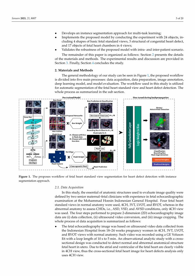
The general methodology of our study can be seen in Figure 1, the proposed workflowis divided into five main processes: data acquisition, data preparation, image annotation,deep learning model, and model evaluation. The workflow used in this study is utilizedfor automatic segmentation of the fetal heart standard view and heart defect detection. Thewhole process as summarized in the sub section.
Figure 1. The proposes workflow of fetal heart standard view segmentation for heart defect detection with instancesegmentation approach.
2.1. Data Acquisition
In this study, the essential of anatomic structures used to evaluate image quality weredefined by two senior maternal–fetal clinicians with experience in fetal echocardiographicexamination at the Mohammad Hoesin Indonesian General Hospital. Four fetal heartstandard views in normal anatomy were used, 4CH, 3VT, LVOT, and RVOT, whereas in theabnormal anatomy to assess CHDs, i.e., ASD, VSD, and AVSD conditions, only 4CH viewwas used. The four steps performed to prepare 2-dimension (2D) echocardiography imagedata are (i) data collection, (ii) ultrasound video conversion, and (iii) image cropping. Thewhole process of data acquisition is summarized as follows:
• The fetal echocardiography image was based on ultrasound video data collected fromthe Indonesian Hospital from 18–24 weeks pregnancy women in 4CH, 3VT, LVOT,and RVOT views with normal anatomy. Such video was recorded using a GE VolusonE6 with a loop length of 10 s to 5 min. An observational analytic study with a cross-sectional design was conducted to detect normal and abnormal anatomical structurefetal heart in utero. Due to the atrial and ventricular of the fetal heart are clearly visiblein 4CH view, thus the cross-sectional fetal heart image for heart defects analysis onlyuses 4CH view.

Sensors 2021, 21, 8007 4 of 20
• The ultrasound video was taken with several size variations of 1.02 megabytes to331 kilobytes. All ultrasound videos should be transformed into frames and then re-sized to a resolution of 400 × 300 pixels. All fetal heart images were retrieved for retro-spective analysis using the digital imaging and communications in medicine (DICOM)format. The framing process from video to 2D images utilize the cv2.VideoCapture()function. The ultrasound video was read frame by frame into the new size, and all thegenerate frames were stored in frame storage using the cv2.imwrite() code to createground truth images.
• The whole fetal heart images generated by software are verified by maternal–fetal clin-icians in the department of obstetrics and gynecology, General Hospital MohammadHoesin Palembang, Indonesia. By using the cropping process, unnecessary informa-tion of the raw images was removed. The outputs were coded after cropping from theechocardiogram video using output_movie.release(). All processes were run on thePython OpenCV library.
2.2. Data Preparation
The fetal heart ultrasound datasets collection comprised four views of imaging planesof normal and defective fetal hearts. All images were labeled in accordance with widelyused fetal heart anatomical planes by a maternal–fetal clinician. The dataset represented areal clinical setting and the ultrasound video data were acquired during standard clinicalpractice in one year (between 2020 and 2021). However, due to the pandemic situation,only about 100 pregnant women attending for routinary pregnancy screening during theirsecond and third trimesters (18–24 weeks) were included in this study. From the wholedata, the CHDs condition is hard to find, therefore only 20 pregnant women included in thisstudy exhibited abnormal anatomy, alongside 30 pregnant women with normal anatomy.
Each ultrasound video for one patient produces 40 images; thus, 50 pregnant womenproduce about 2000 images in normal and abnormal anatomy. The maternal–fetal clinicianselected images belonging to the four anatomical planes most widely used in routinematernal–fetal screening. The clinician selected only images complying with the minimumquality requirements, only a clear cross-sectional scan image was included to processfurther. It consists of 332 images for fetal standard view segmentation and 917 images forheart defect detection (refer to Table 1). The training process randomly split the collectedcases into a training set and a validation set, and the model established by the training setdata was tested against the validation set in order to ensure the accuracy and stability ofthe model.
Table 1. Data distribution of fetal heart view and CHDs images. All data are extracted fromultrasound video from normal and abnormal anatomy.
Class Training Validation Testing Total
3VT view 58 8 6 72
4CV view 75 8 11 94
LVOT view 29 4 3 36
RVOT view 23 4 3 30
ASD 200 9 24 233
VSD 115 10 16 141
AVSD 168 9 18 195
Normal 303 10 35 348
Total 1149
Images with inappropriate anatomical planes (cropped or badly captured) and thosewith calipers were excluded. The dataset composition was clearly imbalanced (some classeswere more frequent than others), as is usually the case in real clinical scenarios. The sample

Sensors 2021, 21, 8007 5 of 20
of the raw ultrasound image was based on four views in normal anatomy, as depicted inFigure 2. In such sample, there are the left atrium (LA), left ventricle (LV), right atrium (RA),right ventricle (RV), ductus arteriosus (DUCT), superior vena cava (SVC), aorta ascendens(AoA), aorta descendens (Ao), and main pulmonary artery (MPA), whereas the sample ofthe raw ultrasound image of abnormal anatomy structure, with the three heart defects suchas ASD, VSD, and AVSD condition, is compared to normal anatomy structure in Figure 3.In the abnormal structure, there are hole (H) as heart defect in each condition. Each defecthas the variation of hole size; such hole size indicates the disease severity. However, in thisstudy, we only detected the hole, without measuring the hole size.
Figure 2. Fetal heart scan in four standard views of normal anatomy: (a) 4CH; (b) LVOT; (c) RVOT; and (d) 3VT.
2.3. Image Annotation
The anatomical heart structures are critical for the segmentation process. The maternal–fetal clinician as the image annotator should drew precise boundaries around the heartimages manually with data annotation tool (LabelMe) [7]. LabelMe to provide an onlineannotation tool to build image databases for computer vision research. The significantvariations in image quality, shapes, sizes, and orientations between the pregnant womenwere used to create a database of ground truths. In the fetal echocardiography with normalanatomy, each standard view has a different structure of heart chamber; therefore, theannotation should be conduct for all standard views with their respective chamber suchas, 4CH standard view consists of five heart chambers, i.e., Ao, LA, LV, RA, and RV; 3VTstandard view consists of three heart chambers, i.e., DUCT, SVC, and AoA; LVOT standardview consists of five heart chambers, i.e., AoA, LA, LV, RA, and RV; and RVOT standardview consists of four heart chambers, i.e., DUCT, SVC, AoA, and MPA.

Sensors 2021, 21, 8007 6 of 20
Figure 3. Fetal heart scan in 4CH view for CHDs detection: (a) ASD; (b) VSD; (c) AVSD; and (d) Normal.
Especially for heart defect detection, only 4CH view was used to analyze ASD, VSD,and AVSD images. Annotated images indicate the position of defect in the atrium, ventricle,or both of them. Figure 4 depicts the sample of annotated images for a standard view of4CH, 3VT, LVOT, and RVOT, and Figure 5 shows the sample annotated images of defectposition in ASD, VSD, and AVSD. Finally, the whole annotated images are labelled as theground truth database, and it was saved in the JSON file format (json).
Figure 4. The sample of annotated images by maternal–fetal clinician for standard fetal heart view segmentation in (a) 4CH(orange: view, cyan: AoA, red: LA, grey: RA, green: LV, and red: RV); (b) LVOT (orange: view, cyan: LA, purple: RV, andblue: LV); (c) RVOT (orange: view, green: MPA, red: DUCT, and yellow: SVC); and (d) 3VT (purple: view, yellow: AoA,green: SVC, and red: DUCT); based on normal anatomy.

Sensors 2021, 21, 8007 7 of 20
Figure 5. The sample of annotated image by maternal–fetal clinician for heart defect detection in case: (a) ASD; (b) VSD;and (c) AVSD. In the annotation, the green line is RA, the red line is LA, the purple line is RV, the blue line is LV, and theyellow line is defect.
2.4. Deep Learning Model
In this study, the instance segmentation approach is developed based on Mask-RCNNarchitecture (refer to Figure 6) [18,22]. The Mask-RCNN structure has two main processes,region proposal networks (RPNs) as feature extraction and fully convolutional networks(FCNs) as multi-task learning process in terms of simultaneous classification, detection,and segmentation.
Figure 6. Instance segmentation approach.

Sensors 2021, 21, 8007 8 of 20
2.4.1. Region Proposal Networks
The input of the region proposal networks (RPNs) is 2D ultrasound images; all thefetal hearts have the same size of resolution around 400 × 300 pixels. The ResNet50architecture was applied as the backbone in the RPNs for the feature extraction mechanism.It can represent more complex functions and learn features from different network levels,from edges (shallower layers) to very complex features (deeper layers). The RPNs use togenerate RoIs, which will be used to predict classes and generate masks. Each RPNs hadfive convolutional layers, which were used to process high-level feature inputs to low-leveloutputs. The ResNet 50 structure as seen in Table 2, and the example feature map fromResNet 50 as seen in Figure 7.
Table 2. ResNet50 Structure.
Layer Name Output Size Output Shape
Conv 1 112 × 112 7 × 7, 64 Stride 2
Conv 2 56 × 56
3 × 3 max pool, stride 2 1× 1; 643× 3; 64
1× 1; 256
× 3
Conv 3 28 × 28
1× 1; 1283× 3; 1281× 1; 512
× 4
Conv 4 14 × 14
1× 1; 2563× 3; 2561× 1; 1024
× 6
Conv 5 7 × 7
1× 1; 5123× 3; 5121× 1; 2048
× 3
- 1 × 1 Average pool, 1000-d FC, Softmax
FLOPs 3.8 × 10⁹
Figure 7. The example of feature map extracted from ResNet50 architecture in the RPNs back bone.
Mask-RCNN adds learning process for segmentation masks in each RoI. The segmen-tation process is simultaneous with each of the other processes (bounding box regressionand class generation), using convolution arrays (feature map) from RPNs. The RPNsclassify the feature and tighten bounding boxes with region of interest (RoI) alignment(RoIAlign). The hyperparameters used in the RPNs’ structure are batch size for each imageof 256; learning rate of 0.001; momentum of 0.9; non-maximum suppression threshold ofabout 0.7; intersection over union (IoU) baseline of 0.5; and anchor sizes of 32, 64, 128, 256,and 512. RoI alignment was performed to pool all RoIs remaining on the feature maps

Sensors 2021, 21, 8007 9 of 20
to a fixed size. As the regression model produced the RoI position, it was generally afloating-point number; however, the pooled feature map required a fixed size.
2.4.2. Fully Convolutional Networks
Fixed-size RoIs were sent to the fully convolutional networks (FCNs) for object classi-fication, detection, and segmentation. The mask branch module is a small FCN appliedto each RoI, and it predicts a segmentation mask for each pixel. In this study, fetal heartarea segmentation was a parallel branch to the wall-chamber classification and boundingbox regression of the heart position. The FCNs utilize stride 2 and 3 × 3 max pooling,with the Softmax as the objective function. The general FCNs structure use in fetal heartsegmentation as seen in Table 3.
Table 3. FCNs architecture.
Layer Kernel Size. Feature Map Stride Output Shape
Input Image - - 256 × 256 × 1
Convolution Layer 1 28 × 28 × 256 2 3 × 3
Pooling Layer 1 2 × 2 14 × 14 × 256
Convolution Layer 2 14 × 14× 256 2 3 × 3
Pooling layer 1 2 × 2 7 × 7 × 256
Convolution Layer 3 7 × 7 × 7 × 256 × 7 × 256 2 -
Deconvolution 2 × 2 14 × 14 × 256
Convolution Layer 4 14 × 14 × 256 2 3 × 3
Deconvolution 2 × 2 28 × 28 × 256
Convolution Layer 5 28 × 28 × 256 3 × 3
Convolution 28 × 28 × C 28 × 28 × 1
Output Layer - - 1
2.4.3. Training and Validation Performance
By using Mask-RCNN approach, the system can recognize the objects’ classes, lo-cations (the bounding box), and shapes [22]. The proposed model utilizes a multi-tasklearning with the loss function that incorporates losses from predictions in classification,detection, and segmentation for each instance [22]. The first term in the loss function isLcls, which measures the error in the predicted class label. The class prediction branch usesa SoftMax layer to output the final class predictions for each instance. For instance, i, theclass prediction, is a vector denoted by p→i. Each element p→i
j exists in the interval (0, 1)and is interpreted as the predicted probability that instance i belongs to class j. If the trueclass of an instance i is u, then Lcls is given by the log loss function (Equation (1)).
Lcls(p→i , u) = −logp→i
u (1)
The second term is Lbbox, which measures the error predicted bounding boxes. Theground truth for the bounding box for an instance of class u is given by the vector→v = (vx, vy, vw, vh), where the four indices indicate the x and y coordinates of the centerof the box, w width of the box, and h height of the box. Detailed information about theformat of the bounding boxes is given in [22]. The predicted bounding box is denoted by→t and has the same form as
→v . Lbbox is given by following equation:
Lbbox = ∑i∈x,y,w,h
smoothL1(tui −vi) (2)

Sensors 2021, 21, 8007 10 of 20
smoothL1(x) ={
0.5x2
|x| − 0.5, if |x|<1
, else(3)
The third term is Lmask, which measures the error predicted segmentation masks foreach instance. In the mask prediction branch, a sigmoid activation is applied to everypixel in the final feature map. The sigmoid value bounds at 0 to 1 and is interpreted as theprobability that a given pixel is included in the proposed segmentation mask. Then, Lmaskisgiven by the binary cross-entropy between the predicted and ground truth masks. Let Yi
and_p i correspond to the ground truth pixel label (0 or 1) and the predicted probabilities
for pixel i, respectively. For ground truth and predicted masks with N total pixel Lmask ispresented in Equation (4).
Lmask= −1N
N
∑i=1
Yi log>pi + (1− Y i) log(1−>pi) (4)
If the prediction is given by the categorical cross-entropy, then SoftMax activationfunction is applied, and Lmask is presented as Equations (5) and (6) denotes the total loss ofthe model.
Lmask= −1N
>Σi
∑i=1
log (5)
L = Lcls+Lbbox+Lmask (6)
2.5. Model Evaluation
In order to validate and evaluate the performance of the instance segmentation model,the outputs of Mask-RCNN are validated by using six metrics, i.e., loss in classification, lossin segmentation, loss in detection, overlapping between the annotated and predicted inputsof each class in IoU, Jaccard index or Dice coefficient similarity (DCS) for segmentation,and mean average precision (mAP) for object detection [7,17,19].
The DCS is a Jaccard similarity coefficient used for gauging the similarity and diversityof sample sets. In this case, to measure the performance of predictive images with detailedtruth labels. The DCS is illustrated in Equation (7).
DCS (X, Y) =2 ∑N
i XiYi
∑Ni X2
i + ∑Ni Y2
i(7)
where N is the number of runs of the predicted results, Xi is the prediction result, and yi isthe truth label. The pixel index value of the DCS, which is in the interval [0, 1], measuresthe match probability between the predicted and ground truth images.
The performance of the trained/validated Mask R-CNN model was quantitativelyevaluated by mAP. The mAP score is a widely adopted metric for assessing object detectionmodels. The mAP values of the various groups were computed, and their average wasobtained. Although the model would detect various objects, the classes to be assigned tothese objects were not always certain. However, even if the expected class for an object orinstance were correct, the output criterion must still look at how well the model locates itspatially in the picture. Equation (8) depicts the commonly used mAP.
mAP =1
ncl
∑i nii
ti(8)
where ncl is the total of all the different classes and ti = ∑j nij is the total number of pixelsof class i.
3. Experimental Results and Discussion
In ultrasound examination, the position of fetal heart is difficult to predict due tosmall size and unpredictable shape and orientation. Fetal–maternal clinicians conduct

Sensors 2021, 21, 8007 11 of 20
examinations to determine a fetus’ condition in the womb (whether it has a congenitalheart defect) before birth. In this study, we propose the comprehensive computer-assistedechocardiographic interpretation is determining whether computers can learn to recognizesuch condition. To ensure the performance of the learning process, all the networks aretrained in the computer specifications as follows: the processor was an Intel® Core™ i9-9920X CPU @ 3.50GHz and 490191 MB RAM, the GPU was a GeForce 2080 RTX Ti, byNVIDIA Corporation GV102 (rev a1); the operating system was Windows 10 Pro 64-bit(10.0, Build 18363).
3.1. Fetal Heart Standard View Segmentation
We benchmarked widely used state-of-the-art CNNs-based Mask-RCNNs with threedifferent backbone architectures: ResNet50, ResNet101, and MobileNetV1. The networks’original architecture of Mask-RCNN was maintained in all cases. All networks were firstpre-trained using the Microsoft common objects in context (COCO) dataset, then fullyretrained using our training data to produce the probability scores for each class. Weconduct the fetal heart standard view segmentation with normal anatomy of the 4CHview, the expected normal appearance of the LVOT/RVOT view and the additional viewsrequired for the complex ultrasound obstetric images with 3VT view. Whereas fetal heartabnormality anatomy examination by using only the 4CH view.
The performance of Mask-RCNN in fetal heart standard view segmentation can beseen in Table 2, which shows that ResNet50 outperformed ResNet101 and MobileNetV1 interms of the mAP, IoU, and DCS. ResNet50 produced average mAP, IoU, and DCS values of96.59%, 79.97%, and 89.70%, respectively. All values exceeded 50%, given that the baselineof IoU was 50%, and those of mAP and DCS were over 70%. Therefore, the Mask-RCNNmodel with the ResNet50 architecture could detect all heart chamber in the four views.
Table 4 shows the performance of fetal heart standard view. The experimental resultshowed that the heart chambers in the LVOT view were the most difficult to detect basedon three architectures. There were several ambiguities between the 4CH and LVOT cases,as the appearance of the fetal heart is similar between these views. 4CH has four chambers,whereas LVOT has five chambers with ascending aorta. However, ascending aorta looksfaint, as it is close to the valves in the fetal heart. It is differentiated only by subtle, indistinctstructures, such as heart valves, which varied significantly in the ultrasound image artefactsand the relative movement between probe and fetus. It is a long-axis view of the heart,highlighting the path from the left ventricle to the ascending aorta with five-part of the heartchamber. The detection result produced a 60% IoU, but the DCS value reached 86.55%.
Table 4. The performance of fetal heart standard view.
CNNsArchitecture
Performance (%)
View mAP IoU DCS
ResNet 50
3VT
96.59
81.76 90.58
4CH 87.17 90.93
LVOT 66.29 86.55
RVOT 84.64 90.73
ResNet 101
3VT
91.85
84.85 81.76
4CH 79.63 87.17
LVOT 46.80 66.29
RVOT 83.55 84.64
Mobilenetv1
3VT
94.87
79.69 89.88
4CH 87.40 82.35
LVOT 68.59 80.78
RVOT 79.31 86.42

Sensors 2021, 21, 8007 12 of 20
Generally, maternal–fetal clinicians use their judgement to determine whether certainheart substructures are in the correct anatomical localizations by comparing normal andabnormal fetal heart images. Four standard views in the ultrasound images are used inexaminations to perform fetal heart diagnoses. In this study, the fetal heart view wassegmented automatically using the proposed model. Fetal heart chamber as an objectshould be detected and segmented in the four fetal heart standard views namely, AoA,AoD, LA, RA, LV, RV, DUCT, SVC, and MPA. Figures 8 and 9 presented the heart chamberprediction performance for a standard fetal heart scan in terms of the IoU and DCS (Jaccardindex) performance. A total of 17 heart chambers are needed to be segmented and detected:five objects for 4CH view, three objects for 3VT view, five objects for LVOT view, and fourobjects for RVOT view.
Figure 8. The IoU performance in heart chamber segmentation in four fetal heart standard views.
Figure 9. The DCS performance in heart chamber segmentation in four fetal heart standard views.
The IoU and DCS performance shows that the instance segmentation with the ResNet50architecture as the backbone produced excellent predictions for all chambers in each view.Therefore, the Mask-RCNN with the ResNet50 architecture as the backbone of RPNs couldsegmented and detected the object based on the annotated RoI. In Figure 10a, the sampleof segmentation result of fetal standard heart view is provided, and Figure 10b shows the

Sensors 2021, 21, 8007 13 of 20
heart chamber segmentation is presented separately. The standard view segmentation, tomark the shape of the cross sectional of the fetal heart, and the heart chambers segmenta-tion, to show the part of each cross-sectional, belong here, whereas in Figure 11a–d, weexperimented on two combinations, in such process a fetal heart view and heart chamberis merged, with about 17 heart chamber objects and four heart standard views to predict.Figure 11a,d shows the sample of segmentation results with different colors, but each objecthas the same description as Figure 10a,b. Based on the proposed model, all objects canbe predicted with satisfactory performance (about 96.59% mAP, 79.97% IoU, and 89.70%DCS). The high mAP shows that the object detection process based on the proposed modelobtained the overlapping area between the annotated and predicted RoIs of each boundingbox close to 100%. The proposed Mask-RCNN model with ResNet50 yielded a 3.41% errorin prediction between the annotated and predicted RoIs.
Figure 10. The sample segmentation result of standard view and heart chamber for normal heart anatomy structure: (a) redcolor contour denotes the fetal heart boundary segmentation in each view, from left to right are 4CH, 3VT, LVOT, and RVOT;(b) heart chamber segmentation in each view from left to right are 4CH (red: RA, purple: LA, yellow: RV, and blue: LV),3VT (green: DUCT, blue: AoA, and red: SVC), LVOT (green: LV, red: AoA, blue: RA, and yellow: RV), and RVOT (green:DUCT, cyan: MPA, red: AoA, and purple: SVC).
3.2. Heart Defect Segmentation and Detection in 4CH View
The fetal heart anatomy in 4CH view showed the expected normal appearance [23].As apical 4CH is the original gold standard view in fetal echocardiography, inability toimage this should alert the scanner about a potential problem [24]. This view shouldnot be mistaken for a simple chamber count as it involves a careful evaluation of specificcriteria [24]. Based on such criteria, the detection of the fetal heart abnormality was screenedonly by 4CH view [8]. Three CHD conditions (with defects in atria, ventricles, and both)were measured with the IoU and DCS values. The minimum IoU value for detecting thedefect object in each fetal heart was 0.5. High IoU and DCS values indicated that the defectprediction overlapped with the proposed architectural model, which is almost similar tothe ground truth.
Two scenarios for the learning processes were conducted in this study based on intra-and inter-patient variation data. Intra-patient variations meant that a fetal heart imagecoming from the same patient was split for the testing process. Inter-patient variationsmeant that the tested fetal heart images were from different patients. In the intra-patientdata for ASD, VSD, and AVSD, the proposed model produced IoU and DCS values exceed-

Sensors 2021, 21, 8007 14 of 20
ing 50%. However, for the inter-patient data, although a 55.99% IoU was obtained for ASD,the IoU values of VSD and AVSD were under 50%. The DCS value exceeded 50% for ASDand AVSD, but that of VSD was only close to 50% (refer to Table 5).
Figure 11. Fetal heart view with heart chamber segmentation in (a) 4CH, (b) 3VT, (c) LVOT, and (d) RVOT for normal heartanatomy structure. Fetal heart view boundary and heart chamber part as the same description with Figure 10.
Table 5. The IoU and the DCS performance for heart defect segmentation.
Position of Heart DefectIntra-Patient Inter-Patient Intra-Patient Inter-Patient
IoU (%) DCS (%)
Hole in atria 62.72 55.99 77.74 67.69
Hole in ventricle 54.83 42.07 68.26 48.89
Hole in atria and ventricle 58.36 40.54 60.20 52.63
Overall, the defect detection performance reached over 50% in IoU and DCS for intra-patient data. The inter-patient data were hard to detect due to large variations in fetal heartimages, size of defect, and image quality, especially in VSD and AVSD condition. The resultwas under 50% IoU; all measurement decreased about 13 to 15% if the proposed modelwas tested with unseen images.
The sample image of heart defect segmentation and detection is depicted in Figure 12.In the 18–21 weeks of pregnancy, the fetal heart has size around 24 mm [23], thus the hole(defect) size in the heart septum will have a size <24 mm. At this stage of development,therefore, it remains difficult to visualize with precision the details of cardiac anatomy asseen during fetal echocardiography. By using our proposed model, it can be segmentedand detected with IoU and DCS about 59% and 69%, respectively, in intra-patient, andabout 47% IoU and 57% DCS in inter-patient scenarios. This means our model has theability to segment and detect until a 50% overlap with the ground truth.
This study determined the mAP value for each defect condition (ASD, VSD, andAVSD) in addition to the IoU and DCS values. A high mAP value indicated that the defectprediction from the model was similar to the ground truth generated by the maternal–fetalconsultant. Table 6 shows the object detection results with mAP performance; the highestmAP value (98.30%) was obtained from the intra-patient data; however, the mAP decreasedto 82.42% in the inter-patient data. CNN-based instance segmentation works using a simplelinear iterative clustering algorithm, which takes an image as input and outputs its divisioninto super-pixels. The proposed model measures the overlap between the annotated inputand predicted target, but it does not label all the image pixels, as it segments only the RoI.

Sensors 2021, 21, 8007 15 of 20
Therefore, if the input image is new (from inter-patient), the detection performance willdecrease, but its performance still satisfactory due to the reduction only 16% with the mAPvalue over 80%.
Figure 12. Sample image result of CHDs detection with 4CH view. The white arrow indicates the defect, whereas red andblue colors are the defect position in the heart septum.
Table 6. The mAP performance for heart defect detection.
Intra-Patient Inter-Patient
98.30% 82.42%
We also conducted heart chamber segmentation and detection in 4CH view withabnormal anatomy image. The fetal heart chamber prediction with the proposed modelis presented in Figure 13. This experimental result differed from the IoU and DCS perfor-mance in Figure 11, as the fetal heart images are taken from the patients with CHDs. Theexperiment was conducted based on intra- and inter-patient data. The RoI was a segmentof four object classes, namely, LA, LV, RA, and RV. With the use of the Mask-RCNN model,all classes can be segmented and classified in the three conditions. Overall performancesshow that intra-patient data allowed better IoU and DCS performance compared withinter-patient data.
As shown in Figure 13a,d, the proposed model produced satisfactory results, with alarge overlap between the ground truth and the predicted image. All IoU values exceededthe baseline of 0.5, which is the gold standard value for ensuring that all processes canbe run with good performance. The IoU and DCS performances with the intra-patientdata were better than those with the inter-patient data, with scores of above 66.37% and79.60%, respectively. The performance with the inter-patient data was poorer than thatwith the intra-patient data. Due to the inherent differences in appearance across differentimaging modalities, it is challenging to construct accurate image similarity measures. Asthe underlying anatomical components vary between patients, inter-patient registrationmight be difficult. In future work, the detection performance for the inter-patient scenarioshould be enhanced. The image sample for the heart chamber with a heart defect canbe seen in Figure 14 with the defect position marked in red and blue. In this detectionprocess, the defect can be small or large, depending on its severity. However, in this study,the defect size parameter was not taken into account; for further research, it will be veryimportant to diagnose the severity of the condition.

Sensors 2021, 21, 8007 16 of 20
Figure 13. The performance in fetal heart chamber segmentation in 4CH view based on intra- and inter-patient scenario:(a) IoU and (b) DCS.
Figure 14. The sample result of wall-chamber segmentation with 4 CH view in ASD, VSD, and AVSD condition based onabnormal anatomy structure.

Sensors 2021, 21, 8007 17 of 20
The proposed instance segmentation model in RPNs and FCNs block can simulta-neously perform three processes: classification, detection, and segmentation. Therefore,three losses can be achieved, namely, object detection loss or bbox loss, classification orclass loss, and segmentation or mask loss. During the training phase, we added an earlystopping mechanism in order to prevent the model becoming overfit to the training data.We monitor the change of validation loss in order to stop the training. As result, the losscurve can be seen in Figure 15, all the response in RPNs and FCNs in the training andvalidation processes decreased to the stability (zero) point; the gap between the two curvesof training/validation was relatively small. The RPNs’ response reaches around 0.1 to 0.25detection loss in training and validation, and around 0.003 classification loss in trainingand validation. In the FCNs’ response, produce detection loss in training and validationwas around 0.05 to 0.12, classification loss in training and validation was around 0.02,segmentation loss in training and validation was around 0.07 to 0.1, and finally total loss inthe proposed model in training and validation was around 0.3 to 0.8. Hence, we concludedthat the proposed model did not experience overfitting during the training process, despitethe limitations in the training data.
Figure 15. Loss curve of heart defect detection with proposed instance segmentation model. RPNs and FCN loss in trainingand validation set.
3.3. Benchmarking Our Model with Existing Studies
For benchmarking, we compared the proposed model with those from other authorsin medical cases, as presented in Table 7. For making a fair comparison, all selectedmethods are based on the segmentation and object detection approach with Mask-RCNNarchitecture, and the mAP was used. The mAP is a metric to measure the sensitivity of themodel, the high of mAP performance indicates a model that is stable and consistent acrossdifference confidence threshold.
Table 7 provides several existing segmentation studies for psoriasis skin [19], en-doscopy disease [17], exudates and microaneurysms [25], brain tumor [26,27], lung nod-ule [28], breast tumor [29], and nuclei [30]. Mask-RCNNs for medical imaging utilize theinstance segmentation models use super-pixels as the base for segmentation process, asa graphical preparatory clustering method. It clusters pixels in the vicinity in geometricand color spaces prior to object segmentation using a simple linear iterative clusteringalgorithm [17,18]. However, it does not label all of the image pixels, as it segments only theRoIs. From previous studies [17,25,31], the segmentation rate was unsatisfactory, producingmAP values around 0.5. This happens as RGB images differ with a large pixel variation;thus, they cannot follow a distribution in [17]. In Shenavarmasouleh et al. [25], exudatesand microaneurysms segmentation has minimum prediction confidence hyperparameter of0.35 as standard threshold (normally about 0.50), whereas a completely correct prediction

Sensors 2021, 21, 8007 18 of 20
will result in 1.0. In Vuola et al. [30] nuclei have a variety of cells acquired under variousconditions produce the shift in different RGB datasets are significantly large. To improvethe performance, they ensemble Mask-RCNN with U-Net architecture, however morepixel-wise processes are involved, which increases the time consumption and computationcost. In addition, the masks from the models do not exactly fit the object image, and notevery image pixel is marked separately. From all studies, the masks that they had fromthe datasets were only associated with one type of object and, for the most part, minimumoverlap between the two datasets.
Table 7. Research benchmarks with other medical object detection with CNNs techniques.
Author Method Object mAP
Rezvy et al. [17] Modified Mask-RCNN Endoscopy disease 0.51
Lin et al. [19] CNNs-based YOLACT Psoriasis skin 0.85
Shenavarmasouleh et al. [25] Mask-RCNN Exudates andmicroaneurysms 0.43
Pai et al. [26] VGG16 andMask-RCNN Brain tumor 0.90
Masood et al. Mask-RCNN Brain tumor 0.94
Cai et al. [28] Mask-RCNN Pulmonary nodule 0.88
Chiao et al. [29] Mask-RCNN Breast tumor 0.75
Vuola et al. [30] Mask-RCNNU-Net Ensemble Nuclei 0.52
Long et al. Probability-Mask-RCNN Pulmonary embolism 0.81
Our proposed model ResNet50 and Mask-RCNN Fetal heart defect 0.98
A different study produced a mAP of over 0.75; the super pixels of each object imagewere similar between the ground truth and the prediction for the pixel-level instanceperformed the same process [19,26–29,31]. However, in [19,26], they added data augmen-tation and other preprocessing techniques; data augmentation arises from the data bias,as the augmented data distribution can be quite different from the original one. This databias leads to a suboptimal performance. Study [27] produced satisfactory results withhigher mAP with only two classes, tumor and non-tumor. Similarly, with other studiesin [28,29,31] the instance segmentation approach can segment the object with best mAPperformance. However, they only use two classes, healthy and non-healthy lesion, whereasthe instance segmentation is prepared for multi-classes and multi object segmentation.
Our proposed model, the Mask-RCNN based on ResNet 50 backbone, performedwell with intra- and inter-patient data for two objects fetal heart views and fetal heardefects. We conducted the experiment with multi-object and multi-class segmentationfor 24 medical objects. The heterogeneity of data types from the various modalities andclinical challenges caused by variations in the local textures was not an obstacle to producesatisfactory performance in identifying pathologies about 0.98 mAP. The RoI regions wereautomatically delineated, and all features were extracted from raw images by CNNswith ResNet 50 architecture, layer by layer, without previously giving the features. Asa result, the proposed method has the advantage of observation anatomical structurecomprehensively, not only by analyzing single features. This means the proposed modelcan segment four fetal heart views and has the ability to classify the heart chamber andaorta in each view, also detecting the hole as a defect in heart septum.
To our knowledge, no study has been conducted on fetal heart view segmentationand heart defect detection using an instance segmentation technique. Although the resultsare promising, this study has some limitations, (i) only fetal heart on 4CH view was usedin this study for the CHDs detection case, (ii) the defect size was not taken into accountto diagnose the CHDs severity, and (iii) the number of fetal heart image data population

Sensors 2021, 21, 8007 19 of 20
with normal and abnormal fetal heart structure should be added, in order to increasethe inter-patient performance result. Furthermore, many other methods could have beenbenchmarked, and computational models could have benefited from the application ofprevious steps, such as image segmentation, instead of analyzing images as a whole.
4. Conclusions
Deep learning is a data-hungry method, but we showed a surprisingly small numberof fetal echocardiography images can be used to significantly boost diagnosis from whatis commonly found in practice. We conducted the experiment by selecting the input dataaccording to clinical recommendations for only four fetal heart standard views rather thanthe entire ultrasound. This strategy allowed us to reduce the size of the input data to ourdiagnostic model and thereby achieve computational efficiency. Due to this, efficiencyin prediction is key to translating this study toward real-world, resource-poor settings.Quantitative measures of fetal structure and function approximated clinical metrics andfollowed patterns found in normal and abnormal structure. A straightforward model, oursis an effective method of segmenting four fetal heart views, classifying the heart chamberand aorta in each view, and detecting the hole as a defect in heart septum in intra-inter-patient scenario. As a result, our proposed model achieved a remarkable performance, with98.3% and 82.42% in intra-patient and inter-patient scenarios, respectively. We look forwardto testing and refining these models in larger populations in an effort to democratize theexpertise of fetal cardiology experts to providers and patients worldwide.
Author Contributions: S.N. Funding acquisition, Formal analysis, resources, and writing—originaldraft; M.N.R. Software Analyst, and review and editing; A.I.S. Software Analyst; A.D. Writing—review and editing; B.T. Data Analyst; F.F. Data Analyst; R.U.P. Medical Data Verification; N.B. Medi-cal Data Verification. All authors have read and agreed to the published version of the manuscript.
Funding: This work was supported by Applied Grants No. 096/SP2H/LT/DRPM/2020 fromthe Ministry of Research, and Technology, Indonesia and Competitive Profession Grant 2021 fromUniversitas Sriwijaya, Indonesia.
Institutional Review Board Statement: Not applicable.
Informed Consent Statement: Not applicable.
Data Availability Statement: Not Applicable.
Acknowledgments: We thank to Intelligent System Research Group (ISysRG) Universitas Sriwijaya,Indonesia for full support in our research infrastructure.
Conflicts of Interest: The authors declare no conflict of interest.
References1. Bensemlali, M.; Bajolle, F.; Laux, D.; Parisot, P.; Ladouceur, M.; Fermont, L.; Lévy, M.; Le Bidois, J.; Raimondi, F.; Ville, Y.; et al.
Neonatal management and outcomes of prenatally diagnosed CHDs. Cardiol. Young 2017, 27, 344–353. [CrossRef]2. Yoon, S.A.; Hong, W.H.; Cho, H.J. Congenital heart disease diagnosed with echocardiogram in newborns with asymptomatic
cardiac murmurs: A systematic review. BMC Pediatr. 2020, 20, 1–10. [CrossRef] [PubMed]3. Mcleod, G.; Shum, K.; Gupta, T.; Chakravorty, S.; Kachur, S.; Bienvenu, L.; Shah, S.B. Echocardiography in Congenital Heart
Disease. Prog. Cardiovasc. Dis. 2018, 61, 468–475. [CrossRef]4. Patel, N.; Narasimhan, E.; Kennedy, A. Fetal Cardiac US: Techniques and Normal Anatomy Correlated with Adult CT and MR
Imaging. RadioGraphics 2017, 37, 1290–1303. [CrossRef] [PubMed]5. Puri, K.; Allen, H.D.; Qureshi, A.M. Congenital Heart Disease. Pediatr. Rev. 2017, 38, 471–486. [CrossRef] [PubMed]6. Sobhaninia, Z.; Rafiei, S.; Emami, A.; Karimi, N.; Najarian, K.; Samavi, S.; Soroushmehr, S.M.R. Fetal Ultrasound Image
Segmentation for Measuring Biometric Parameters Using Multi-Task Deep Learning. In Proceedings of the 2019 41st AnnualInternational Conference of the IEEE Engineering in Medicine and Biology Society (EMBC), Berlin, Germany, 23–27 July 2019;Volume 2019, pp. 6545–6548.
7. Nurmaini, S.; Rachmatullah, M.N.; Sapitri, A.I.; Darmawahyuni, A.; Jovandy, A.; Firdaus, F.; Tutuko, B.; Passarella, R. AccurateDetection of Septal Defects with Fetal Ultrasonography Images Using Deep Learning-based Multiclass Instance Segmentation.IEEE Access 2020, 8, 1. [CrossRef]

Sensors 2021, 21, 8007 20 of 20
8. Sánchez-Quintana, D.; Picazo-Angelin, B.; Zabala-Argüelles, J.I.; Anderson, R.H. Anatomy of the normal fetal heart: The basis forunderstanding fetal echocardiography. Ann. Pediatr. Cardiol. 2018, 11, 164–173. [CrossRef]
9. Vullings, R. Fetal Electrocardiography and Deep Learning for Prenatal Detection of Congenital Heart Disease. In Proceedings ofthe 2019 Computing in Cardiology Conference (CinC), Singapore, 8–11 September 2019; p. 1.
10. Arnaout, R.; Curran, L.; Zhao, Y.; Levine, J.; Chinn, E.; Moon-Grady, A. Expert-level prenatal detection of complex congenitalheart disease from screening ultrasound using deep learning. medRxiv 2020. [CrossRef]
11. Torrents-Barrena, J.; Piella, G.; Masoller, N.; Gratacós, E.; Eixarch, E.; Ceresa, M.; Ballester, M. Ángel, G. Segmentation andclassification in MRI and US fetal imaging: Recent trends and future prospects. Med. Image Anal. 2019, 51, 61–88. [CrossRef][PubMed]
12. Zhang, B.; Liu, H.; Luo, H.; Li, K. Automatic quality assessment for 2D fetal sonographic standard plane based on multitasklearning. Medicine 2021, 100, e24427. [CrossRef]
13. Gandhi, S.; Mosleh, W.; Shen, J.; Chow, C.-M. Automation, machine learning, and artificial intelligence in echocardiography: Abrave new world. Echocardiography 2018, 35, 1402–1418. [CrossRef] [PubMed]
14. Ghorbani, A.; Ouyang, D.; Abid, A.; He, B.; Chen, J.H.; Harrington, R.A.; Liang, D.H.; Ashley, E.A.; Zou, J.Y. Deep learninginterpretation of echocardiograms. Npj Digit. Med. 2020, 3, 1–10. [CrossRef]
15. Girshick, R.; Donahue, J.; Darrell, T.; Malik, J. Rich Feature Hierarchies for Accurate Object Detection and Semantic Segmentation.In IEEE Conference on Computer Vision and Pattern Recognition, 1st ed.; IEEE: New York, NY, USA, 2014; pp. 580–587.
16. Ravi, D.; Wong, C.; Deligianni, F.; Berthelot, M.; Andreu-Perez, J.; Lo, B.; Yang, G.-Z. Deep Learning for Health Informatics. IEEEJ. Biomed. Health Inform. 2017, 21, 4–21. [CrossRef]
17. Rezvy, S.; Zebin, T.; Braden, B.; Pang, W.; Taylor, S.; Gao, X.W. Transfer Learning For Endoscopy Disease Detection & Segmentationwith Mask-RCNN Benchmark Architecture. In Proceedings of the 2nd International Workshop and Challenge on ComputerVision in Endoscopy, EndoCV@ISBI 2020, Iowa City, IA, USA; 2020; Volume 2595, pp. 68–72.
18. Vo, K.; Le, T.; Rahmani, A.M.; Dutt, N.; Cao, H. An Efficient and Robust Deep Learning Method with 1-D Octave Convolution toExtract Fetal Electrocardiogram. Sensors 2020, 20, 3757. [CrossRef]
19. Lin, G.-S.; Lai, K.-T.; Syu, J.-M.; Lin, J.-Y.; Chai, S.-K. Instance Segmentation Based on Deep Convolutional Neural Networks andTransfer Learning for Unconstrained Psoriasis Skin Images. Appl. Sci. 2021, 11, 3155. [CrossRef]
20. Shin, H.-C.; Roth, H.R.; Gao, M.; Lu, L.; Xu, Z.; Nogues, I.; Yao, J.; Mollura, D.; Summers, R.M. Deep Convolutional NeuralNetworks for Computer-Aided Detection: CNN Architectures, Dataset Characteristics and Transfer Learning. IEEE Trans. Med.Imaging 2016, 35, 1285–1298. [CrossRef] [PubMed]
21. Wang, X.; Peng, Y.; Lu, L.; Lu, Z.; Bagheri, M.; Summers, R.M. ChestX-ray8: Hospital-scale Chest X-ray Database and Benchmarkson Weakly-Supervised Classification and Localization of Common Thorax Diseases. In Proceedings of the IEEE Conference onComputer Vision and Pattern Recognition, Honolulu, HI, USA, 21–26 July 2017.
22. He, K.; Gkioxari, G.; Dollár, P.; Girshick, R. Mask R-CNN. IEEE Trans. Pattern Anal. Mach. Intell. 2020, 42, 386–397. [CrossRef]23. Komatsu, M.; Sakai, A.; Komatsu, R.; Matsuoka, R.; Yasutomi, S.; Shozu, K.; Dozen, A.; Machino, H.; Hidaka, H.; Arakaki, T.;
et al. Detection of Cardiac Structural Abnormalities in Fetal Ultrasound Videos Using Deep Learning. Appl. Sci. 2021, 11, 371.[CrossRef]
24. Gonçalves, L.F.; Lee, W.; Chaiworapongsa, T.; Espinoza, J.; Schoen, M.L.; Falkensammer, P.; Treadwell, M.; Romero, R. Four-dimensional ultrasonography of the fetal heart with spatiotemporal image correlation. Am. J. Obstet. Gynecol. 2003, 189, 1792–1802.[CrossRef]
25. Shenavarmasouleh, F.; Arabnia, H.R. DRDr: Automatic Masking of Exudates and Microaneurysms Caused by Diabetic Retinopa-thy Using Mask R-CNN and Transfer Learning. In Advances in Parallel & Distributed Processing, and Applications; Springer:Berlin/Heidelberg, Germany, 2021; pp. 307–318.
26. Pai, K.R.; Prajwal, N.P.M. Brain Tumor Detection and Segmentation Using VGG16 and Mask R-CNN with Transfer Learning.Solid State Technol. 2020, 63, 9887–9893.
27. Masood, M.; Nazir, T.; Nawaz, M.; Javed, A.; Iqbal, M.; Mehmood, A. Brain tumor localization and segmentation using maskRCNN. Front. Comput. Sci. 2021, 15, 1–3. [CrossRef]
28. Cai, L.; Long, T.; Dai, Y.; Huang, Y. Mask R-CNN-Based Detection and Segmentation for Pulmonary Nodule 3D VisualizationDiagnosis. IEEE Access 2020, 8, 44400–44409. [CrossRef]
29. Chiao, J.-Y.; Chen, K.-Y.; Liao, K.Y.-K.; Hsieh, P.-H.; Zhang, G.; Huang, T.-C. Detection and classification the breast tumors usingmask R-CNN on sonograms. Medicine 2019, 98, e15200. [CrossRef]
30. Vuola, A.O.; Akram, S.U.; Kannala, J. Mask-RCNN and U-Net Ensembled for Nuclei Segmentation. In Proceedings of the 2019IEEE 16th International Symposium on Biomedical Imaging (ISBI 2019), Venice, Italy, 8–11 April 2019; pp. 208–212. [CrossRef]
31. Long, K.; Tang, L.; Pu, X.; Ren, Y.; Zheng, M.; Gao, L.; Song, C.; Han, S.; Zhou, M.; Deng, F. Probability-based Mask R-CNN forpulmonary embolism detection. Neurocomputing 2021, 422, 345–353. [CrossRef]