Respiratory maneuvers in echocardiography: a review of clinical applications
13
BioMed Central Page 1 of 13 (page number not for citation purposes) Cardiovascular Ultrasound Open Access Review Respiratory maneuvers in echocardiography: a review of clinical applications Carmen Ginghina 1,2 , Carmen C Beladan 1,2 , Madalina Iancu 2 , Andreea Calin 1,2 and Bogdan A Popescu* 1,2 Address: 1 "Carol Davila" University of Medicine, Bucharest, Romania and 2 "Prof. Dr. C. C. Iliescu" Institute of Cardiovascular Diseases, Bucharest, Romania Email: Carmen Ginghina - [email protected]; Carmen C Beladan - [email protected]; Madalina Iancu - [email protected]; Andreea Calin - [email protected]; Bogdan A Popescu* - [email protected] * Corresponding author Abstract During echocardiographic examination, respiration induces cyclic physiological changes of intracardiac haemodynamics, causing normal variations of the right and left ventricle Doppler inflows and outflows and physiological variation of extracardiac flows. The respiration related hemodynamic variation in intra and extracardiac flows may be utilized in the echocardiography laboratory to aid diagnosis in different pathological states. Nevertheless, physiologic respiratory phases can cause excessive translational motion of cardiac structures, lowering 2D image quality and interfering with optimal Doppler interrogation of flows or tissue motion. This review focuses on the impact of normal respiratory cycle and provocative respiratory maneuvers in echocardiographic examination, both in physiological and pathological states, emphasizing their applications in specific clinical situations. Respiration induces cyclic physiological modification of intracardiac haemodynamics. Changes are related to vari- ations in intrathoracic and intraabdominal pressure, sys- temic and pulmonary venous return, intrapericardial pressure, pericardial constraint and interdependence between the four cardiac chambers. With inspiration intrathoracic and intrapericardial pressures decrease. This results in augmented right ventricular filling and stroke volume and, as the total pericardial space is limited, a compensatory decrease in left ventricular stroke volume occurs in early inspiration. With expiration, intrathoracic and intrapericardial pressures increase, resulting in mild decrease in right ventricular diastolic filling and a subse- quent increase in left ventricular filling. Because of the constraining effect of the pericardium on the combined volume of the four cardiac chambers, respi- ratory variation in intrapericardial pressure results in reciprocal variation in the filling of both ventricles [1]. Influence of normal respiration on echocardiographic parameters • 2D echocardiography Image quality With inspiration the anteroposterior diameter of the chest increases, and the lungs inflate and expand particularly anteriorly, partly filling the space between the heart and the thoracic cage. Inspiratory movements do not only decrease the amount of cardiac tissues that lies close to the Published: 26 August 2009 Cardiovascular Ultrasound 2009, 7:42 doi:10.1186/1476-7120-7-42 Received: 20 July 2009 Accepted: 26 August 2009 This article is available from: http://www.cardiovascularultrasound.com/content/7/1/42 © 2009 Ginghina et al; licensee BioMed Central Ltd. This is an Open Access article distributed under the terms of the Creative Commons Attribution License (http://creativecommons.org/licenses/by/2.0 ), which permits unrestricted use, distribution, and reproduction in any medium, provided the original work is properly cited.
-
Upload
independent -
Category
Documents
-
view
0 -
download
0
Transcript of Respiratory maneuvers in echocardiography: a review of clinical applications

BioMed CentralCardiovascular Ultrasound
ss
Open AcceReviewRespiratory maneuvers in echocardiography: a review of clinical applicationsCarmen Ginghina1,2, Carmen C Beladan1,2, Madalina Iancu2, Andreea Calin1,2 and Bogdan A Popescu*1,2Address: 1"Carol Davila" University of Medicine, Bucharest, Romania and 2"Prof. Dr. C. C. Iliescu" Institute of Cardiovascular Diseases, Bucharest, Romania
Email: Carmen Ginghina - [email protected]; Carmen C Beladan - [email protected]; Madalina Iancu - [email protected]; Andreea Calin - [email protected]; Bogdan A Popescu* - [email protected]
* Corresponding author
AbstractDuring echocardiographic examination, respiration induces cyclic physiological changes ofintracardiac haemodynamics, causing normal variations of the right and left ventricle Dopplerinflows and outflows and physiological variation of extracardiac flows. The respiration relatedhemodynamic variation in intra and extracardiac flows may be utilized in the echocardiographylaboratory to aid diagnosis in different pathological states. Nevertheless, physiologic respiratoryphases can cause excessive translational motion of cardiac structures, lowering 2D image qualityand interfering with optimal Doppler interrogation of flows or tissue motion.
This review focuses on the impact of normal respiratory cycle and provocative respiratorymaneuvers in echocardiographic examination, both in physiological and pathological states,emphasizing their applications in specific clinical situations.
Respiration induces cyclic physiological modification ofintracardiac haemodynamics. Changes are related to vari-ations in intrathoracic and intraabdominal pressure, sys-temic and pulmonary venous return, intrapericardialpressure, pericardial constraint and interdependencebetween the four cardiac chambers. With inspirationintrathoracic and intrapericardial pressures decrease. Thisresults in augmented right ventricular filling and strokevolume and, as the total pericardial space is limited, acompensatory decrease in left ventricular stroke volumeoccurs in early inspiration. With expiration, intrathoracicand intrapericardial pressures increase, resulting in milddecrease in right ventricular diastolic filling and a subse-quent increase in left ventricular filling.
Because of the constraining effect of the pericardium onthe combined volume of the four cardiac chambers, respi-ratory variation in intrapericardial pressure results inreciprocal variation in the filling of both ventricles [1].
Influence of normal respiration on echocardiographic parameters• 2D echocardiographyImage qualityWith inspiration the anteroposterior diameter of the chestincreases, and the lungs inflate and expand particularlyanteriorly, partly filling the space between the heart andthe thoracic cage. Inspiratory movements do not onlydecrease the amount of cardiac tissues that lies close to the
Published: 26 August 2009
Cardiovascular Ultrasound 2009, 7:42 doi:10.1186/1476-7120-7-42
Received: 20 July 2009Accepted: 26 August 2009
This article is available from: http://www.cardiovascularultrasound.com/content/7/1/42
© 2009 Ginghina et al; licensee BioMed Central Ltd. This is an Open Access article distributed under the terms of the Creative Commons Attribution License (http://creativecommons.org/licenses/by/2.0), which permits unrestricted use, distribution, and reproduction in any medium, provided the original work is properly cited.
Page 1 of 13(page number not for citation purposes)

Cardiovascular Ultrasound 2009, 7:42 http://www.cardiovascularultrasound.com/content/7/1/42
sternum through anterior pulmonary expansion, but alsoproduce a posterior displacement and rotation of theheart relative to a fixed echo beam [2].
Expiration will give a better parasternal and often apicalaccess to the heart by lungs deflation [see Additional files1, 2, 3 and 4]. In contrast, in the subxiphoid area inspira-tion will bring the diaphragm down improving access tothe heart [see Additional file 5]. Sometimes half a breathis preferred, as with deep inspiration the abdomenbecomes too tense to allow the transducer to be placedunder the sternum.
Optimal views are usually obtained with the patient in thesteep left lateral decubitus position with the left armraised to spread the ribs. Rotation of the patient to the leftwill tend to deflate the left lung avoiding the pulmonaryinterference [1,3].
MeasurementsSpontaneous respiration has been shown to causechanges in echocardiographic measurements of left ven-tricular (LV) dimensions. An inspiratory reduction in LVend-diastolic dimension assessed by M-mode echocardi-ography was found in normal subjects [4]. As the heartwas found to move medially in the short-axis view duringinspiration, one possible explanation for this findingcould be an inspiratory tangential cut by the M-mode cur-sor when measured through the center of the left ventricle.Thus the inspiratory dimensions are smaller along the cur-sor than through the center of the short-axis area [5].Another possible explanation could be an inspiratorydecrease in LV end-diastolic volume and stroke volume asa consequence of preload reduction (decreased diastolicfilling) and increased afterload (increased impedance toLV emptying).
Excessive translational motion can be avoided by acquir-ing images during quiet respiration and in this case severalcycles must be recorded. [see Additional files 6, 7] Ifimages are obtained during held end-expiration, caremust be taken to avoid a Valsalva maneuver, which candegrade image quality. Image acquisition during quiet orsuspended respiration (at end-expiration) is currently rec-ommended for two-dimensional quantitation [3].
• Doppler echocardiographyPhysiological respiratory variations of intra and extracardiac flowDuring inspiration, intrathoracic and intrapericardialpressures decrease, resulting in augmented flow into theright atrium and right ventricle, with decreased flow out ofthe pulmonary veins into the left atrium and left ventricle.Reciprocal changes occur during expiration, and can bedocumented by Doppler echocardiographic changes inmitral and tricuspid inflow as well as pulmonary and sys-
temic outflow. Under normal circumstances, peak veloc-ity of mitral inflow varies by 15% or less with respirationand tricuspid inflow by 25% or less. Variation in peakvelocity and time velocity integral of aortic and pulmo-nary flow profiles typically is less than 10%. [1.6](Figure1)
Marked respiratory changes in Doppler velocity curves areobserved for hepatic veins. With inspiration there is a sig-nificant increase in both normal forward systolic (S) anddiastolic (D) flow velocities as well as retrograde A veloc-ity compared to apnea. (Figure 2A).
The normal forward biphasic flow velocity pattern in thesuperior vena cava (SVC) with systolic flow velocitygreater than diastolic flow velocity, shows inspiratorychanges similar to those of hepatic vein flow.
Respiratory variations are minimal for pulmonary veinflow (Figure 2B)
Doppler measurementsDiaphragmatic traction associated with respirationchanges the position of intracardiac structures in relationto a fixed Doppler sample volume causing errors in veloc-ities measurement.
The Doppler sound beam should be oriented as parallel aspossible to the flow, guided both by the 2D image (some-times assisted by color flow imaging). Small (<20 degrees)deviations in angle produce mild (<10%) errors in veloc-ity measurements. Although these errors may be accepta-ble for low-velocity flows, when Doppler is used to derivepressure gradients even a small error in velocity measure-ment can lead to significant underestimation of the gradi-ent because of the quadratic relation between velocity andpressure gradient. The effects of respiration can be mini-mized by taking measurements during short periods ofapnea or by averaging multiple consecutive beats [7,8].
The phase of respiration affects Doppler Tissue Imaging(DTI) recordings in the same manner because the shift inheart position during respiration over a fixed Dopplersample volume leads to inaccurate recording of the annu-lar velocities (Figure 3A).
Whenever possible, the echocardiographer should obtainannular DTI recordings during end-expiratory apnea (Fig-ure 3B). Once the Doppler cursor is aligned optimally andthe Doppler tissue preset activated the patient should beasked to breathe in, breathe out, and then hold theirbreath at the end of expiration. The sample volume will becarefully repositioned directly into the selected portion ofthe annulus and pulsed wave tissue Doppler activated.Peak DTI waveform velocities should be uniform, consist-
Page 2 of 13(page number not for citation purposes)

Cardiovascular Ultrasound 2009, 7:42 http://www.cardiovascularultrasound.com/content/7/1/42
ent, with little or no beat-to-beat variation of the peakvelocities [9].
Respiration and its relationship to cardiac haemodynamic parameters• Estimation of right atrial (RA) pressureEvaluation of the inferior vena cava (IVC) from the sub-costal view is part of the routine TTE examination. Thediameter of the IVC decreases in response to inspirationwith minimal size observed at end inspiration when thenegative intrathoracic pressure leads to an increase in RVfilling from the systemic veins. IVC size is significantlyinfluenced by patient position, being largest in the rightlateral position, intermediate in the supine position, andsmallest in the left lateral position [10]. The diameter ofthe IVC, measured with the patient in the left decubitusposition at 1.0 to 2.0 cm from the junction with the RA,
using the long axis view, and the percent decrease in itsdiameter during inspiration (collapsibility index) corre-late with RA pressure. A brief sniff is often required [seeAdditional files 8, 9] as normal inspiration may not elicitthe inspiratory response [3]:
• The normal IVC diameter is less than 1.7 cm andthere is a 50% decrease in the diameter when the RApressure is normal (0–5 mm Hg).
• A dilated IVC (>1.7 cm) with normal inspiratory col-lapse (>50%) is suggestive of a mildly elevated RApressure (6–10 mm Hg).
• When the inspiratory collapse is less than 50%, theRA pressure is usually between 10 and 15 mm Hg.
Physiological variations of the velocity across the tricuspid valve (A), mitral valve (B), pulmonary valve (C) and aortic valve (D) during quiet respiration in a normal heartFigure 1Physiological variations of the velocity across the tricuspid valve (A), mitral valve (B), pulmonary valve (C) and aortic valve (D) during quiet respiration in a normal heart.
Page 3 of 13(page number not for citation purposes)

Cardiovascular Ultrasound 2009, 7:42 http://www.cardiovascularultrasound.com/content/7/1/42
• Finally, a dilated IVC without any collapse suggests amarkedly increased RA pressure of greater than 15 mmHg. (Figure 4).
A small IVC (usually < 1.2 cm) with spontaneous collapseis often seen in the presence of intravascular volumedepletion [11].
In a recent paper, Brennan et al evaluated the IVC diame-ter in patients undergoing right heart catheterization [12].
The measurements were made from the subcostal viewwith the patient in supine position. The authors ques-tioned the traditional classification of RAP into narrow 5-mm Hg ranges based on IVC size and collapsibility. Theyfound that the IVC size cutoff with optimum predictiveuse for RAP above or below 10 mm Hg was 2.0 cm (sensi-tivity 73% and specificity 85%). They also found that theoptimal IVC collapsibility cutoff was 40% (sensitivity73% and specificity 84%). Defining collapsibility as high(>55%), low (<35%), or normal (35%–50%) and the
Physiological variations of the velocity across suprahepatic veins (A) and pulmonary veins (B) during quiet respiration in a nor-mal heartFigure 2Physiological variations of the velocity across suprahepatic veins (A) and pulmonary veins (B) during quiet res-piration in a normal heart.
Septal annular velocities measured by tissue Doppler echocardiography during quiet respiration (A), and during end-expiratory apnea (B)Figure 3Septal annular velocities measured by tissue Doppler echocardiography during quiet respiration (A), and dur-ing end-expiratory apnea (B).
Page 4 of 13(page number not for citation purposes)

Cardiovascular Ultrasound 2009, 7:42 http://www.cardiovascularultrasound.com/content/7/1/42
inferior vena cava as small (<1.7 cm), normal (1.7–2.1cm), or large (>2.1 cm) the authors suggest a new classifi-cation of RAP:
(1) High collapsibility with a small or normal-sizedIVC; RAP is very likely low (<5 mm Hg).
(2) High collapsibility with a large IVC or normal col-lapsibility with a small/normal-sized IVC; RAP isprobably between 0 and 10 mm Hg
(3) Normal collapsibility with large IVC; RAP is 10 to15 mm Hg
(4) Low collapsibility with a large IVC; RAP is clearlyhigh (10–20 mm Hg)
(5) RAP in patients with low collapsibility and a nor-mal-sized or small IVC should be interpreted as inde-terminate.
There are several additional conditions to be consideredwhen evaluating the IVC:
• Athletes have been shown to have dilated IVCs withnormal collapsibility index
• In mechanically ventilated patients, the relationshipbetween IVC and RAP is controversial; it was shownthat a dilated IVC did not always indicate a high RApressure Nonetheless a small IVC (< 1.2 cm) had a
100% specificity (with a low sensitivity) for a RA pres-sure of less than 10 mm Hg. It was also suggested thatthe IVC diameter measured at end expiration and enddiastole using M-mode echocardiography correlatesbetter with RA pressure than IVC diameter at end-expi-ration without ECG synchronization [13].
•Evaluation of systolic pulmonary artery pressure (PASP)In patients with chronic obstructive pulmonary disease(COPD) pulmonary hypertension (PH) is sometimes dif-ficult to assess by direct evaluation using the tricuspidregurgitant Doppler velocity because of emphysema ormediastinal deviation. An alternative method of respira-tion-aided evaluation of PH was proposed by Kunichikaet al [14]. Authors found that the expiratory SVC peaksystolic forward flow velocity (an index of the RA flowreserve during expiration) is increased in COPD patientswith PH, owing to enhancement of the RA flow reservethrough the respiratory cycle that compensates for theimpaired right ventricular (RV) filling caused by PH.Therefore, respiratory variation in SVC systolic forwardflow may be a useful Doppler flow index for the assess-ment of severity of PH in patients with stable, chronicCOPD that cannot been assessed by conventional TTE
• Assessment of left ventricular diastolic functionTransmitral Doppler flow parameters are widely used inthe assessment of LV diastolic function. It is well knownthat with spontaneous respiration small changes (<15%)in transmitral peak flow velocities occur in healthy sub-jects. While some authors found no alteration in the ratioof early-to-late peak flow velocity (E/A) [15] other studiesreported a significant decrease in E/A ratio with inspira-tion in normal subjects [16].
The potential effects of spontaneous respiration on mitralinflow Doppler patterns in patients with abnormal LVdiastolic function are poorly understood. Tsai et al. [17]observed that during inspiration, the early diastolic peakflow velocity and E/A ratio were reduced in patients withcoronary artery disease and abnormal LV relaxation pat-tern. More recently, Yuan et al. [18] evaluated 51 hyper-tensive patients presenting with different LV diastolicpatterns and observed in 10 of them a characteristic tras-mitral Doppler flow phenomenon that displayed E/Aratio > 1 on expiration and < 1 on inspiration. As 8 of 10patients revealed an early-to-late diastolic tissue velocityratio <1 authors suggested that this phenomenon couldcharacterize either a new type of diastolic impairment orjust pseudo-normal filling unmasked by inspiration. Theyalso suggested that reversed E/A ratio at end-inspirationrather than at end-expiration might be a more sensitiveand accurate indicator for abnormal LV diastolic function.Further studies using simultaneous invasive measures areneeded to confirm these findings. As the potential effects
Patient with dilative cardiomyopathy: M-mode at the inferior vena cava (IVC) level from subcostal view, during respiratory phasesFigure 4Patient with dilative cardiomyopathy: M-mode at the inferior vena cava (IVC) level from subcostal view, during respiratory phases. There is no inspiratory varia-tion of the IVC diameter, indicating increased right atrial pressure.
Page 5 of 13(page number not for citation purposes)

Cardiovascular Ultrasound 2009, 7:42 http://www.cardiovascularultrasound.com/content/7/1/42
of respiration on mitral inflow Doppler pattern is stillunder debate it seems reasonable to take measurementseither with simultaneous recording of respiration or dur-ing short periods of apnea/by averaging multiple consec-utive beats to minimize the effects of respiration.
In diseased ventricles, progressive shortening of the trans-mitral DT and increasing E/A ratio can be seen withdecreasing ventricular compliance and increasing leftatrial pressure. Impaired relaxation is frequently maskedby elevated filling pressures resulting in a pseudonormalflow pattern (E/A ratio >1). The Valsalva maneuver hasbeen found to effectively unload the heart and unmask animpaired relaxation pattern and high filling pressures inpatients with a baseline pseudonormal flow pattern. Thismaneuver is accomplished first by cessation of breathingat any point in the respiratory cycle when assessment ofmitral inflow is optimized. Next, the participant exerts afirm contraction of the abdominal muscles to force airagainst the closed glottis for approximately 10 secondsthereby increasing the intrathoracic pressure. In order tobe properly performed, the Valsalva maneuver should bestandardized. This can be done by attaching a mouthpiece(e.g. the tube of a syringe from which the plunger hasbeen detached) to a sphygmomanometer and asking thepatient to blow into it in order to raise the column of Hgto 40 mm and keep it stable at this value for 10 seconds[19]. This results in a decrease in both right ventricularand LV filling as a result of an increased thoracic-to-extrathoracic pressure gradient. The mitral inflow patternis observed for changes during live 2D monitoring of theplacement of the Doppler sample. An absolute decrease inthe mitral E/A ratio of 0.5 or more can predict an elevatedLV filling pressure with a specificity of 100% [20].
Changes in transmitral E/A ratio during the Valsalvamaneuver across different stages of diastolic dysfunctionwere recently described by Reagan et al [21].
Because the E/A ratio increases as filling pressures rise it isgenerally assumed that an E/A ratio <1 (impaired relaxa-tion pattern) indicates lower or even normal filling pres-sures when compared with patients who have apseudonormal or restrictive filling pattern. However,Schwammenthal et al demonstrated that patients with anE/A ratio of <1 can nonetheless have severely elevated fill-ing pressures. In these patients LV relaxation is so severelyimpaired that E/A ratio remains <1 despite increased fill-ing pressures. The standardized Valsalva maneuver canunmask the presence of elevated filling pressures inpatients with a baseline impaired relaxation pattern. TheA wave will increase during the maneuver in patients withelevated filling pressures in proportion to the level of LVend-diastolic pressure, and independent of the baselinetransmitral flow pattern [19].(Figure 5)
Pericardial diseases and respiration• Cardiac tamponadeThe increased pericardial pressure in cardiac tamponadeproduces reciprocal respiration-related changes in rightand left ventricular volumes, diastolic filling, and systolicemptying that can be documented by echocardiography.The normal effects of respiration are accentuated such thatvenous return and right-sided filling occur during inspira-tion as intrathoracic pressures fall, providing a pressuregradient from the systemic veins to the RA. Because thetotal intrapericardial volume is fixed by the pressurizedeffusion, this increased inspiratory RV filling causes theinterventricular septum to shift to the left, exaggerating
55 year-old woman, with LV hypertrophy and moderately dilated left atriumFigure 555 year-old woman, with LV hypertrophy and moderately dilated left atrium. TTE, apical 4C view, mitral inflow pulse Doppler patterns. A. Mitral inflow pattern appears "normal" at rest. B. During Valsalva maneuver the E/A ratio decreased from 1.3 to 0.6 and peak A velocity increased from 60 to 75 cm/s, unmasking the impaired relaxation pattern.
Page 6 of 13(page number not for citation purposes)

Cardiovascular Ultrasound 2009, 7:42 http://www.cardiovascularultrasound.com/content/7/1/42
the normal decrease in LV filling volume and, hence,stroke volume (ventricular interdependence). Thus, intamponade, left heart filling occurs preferentially duringexpiration when there is less filling of the right heart. Thesmall normal respiratory variation in left ventricular (LV)stroke volume and systolic arterial pressure (<10 mm Hg)is markedly accentuated in cardiac tamponade, resultingin the clinical finding of "paradoxical pulse" [1,22-24].
Echocardiography is particularly useful in demonstratingthe exaggerated phasic variation in cardiac volumes andflows caused by tamponade [23]. The diastolic collapse ofthe free walls of the right atrium and/or right ventricle,due to compression of these relatively low-pressure cham-bers by the higher-pressure pericardial effusion, is exag-gerated during expiration when right heart filling isreduced. The intermittent collapse of these structures isbest documented with M-mode echo. [see Additional files10, 11] Right ventricular collapse may not be seen inpatients with PH and RV hypertrophy [25]
Respiratory variation in tricuspid and pulmonary flow ismore dramatic than mitral and aortic flow, but there isprogressive impairment in all intracardiac flows as thedegree of tamponade worsens: with inspiration, the RVearly diastolic filling is augmented (>25%), while LVdiastolic filling diminishes (>15%). The flow velocityintegral in the pulmonary artery increases with inspira-tion, while the aortic flow velocity integral decreases(>10%) [1].
The hepatic vein flow pattern may also reflect the exagger-ated respiratory phase dependency of right ventricular fill-ing. The loss of forward flow in the hepatic veins duringthe expiration phase of the respiratory cycle with flow outof the hepatic veins confined to the early inspiratory phasecan be observed in patients with hemodynamically signif-icant pericardial effusion [1]. Superior vena cava flowshows a reduction in diastolic flow velocities (<0.15 m/sec) initially in expiration, later in inspiration as well(systolic dominant and monophasic) [26]
• Constrictive pericarditis (CP)In constrictive pericarditis, the rigid pericardium impedesthe transmission of intrathoracic pressures to the cardiacchambers. During inspiration there is a lower drivingforce from the lungs into the left side of the heart and theLV becomes underfilled. The constricting pericardiumalso results in an increase interventricular interaction, sothat with LV volume decrease, there is a correspondingincrease in right ventricular volume [27] [see Additionalfile 12]
In Doppler echocardiographic studies the dissociation ofintrathoracic and intracardiac pressures is manifested by
an inspiratory increase in peak tricuspid flow velocity anda simultaneous decrease in mitral flow velocity, withopposite changes occurring in expiration [24].
The Doppler findings reflecting abnormal haemodynamics during respiration in constrictive pericarditis are [1,24,28,29]
• PW Doppler mitral inflow: high E velocity, E/A ratio> 2, short E wave deceleration time (EdT < 160 ms);inspiration: decrease E velocity >25%, prolonged IVRT>25%; expiration: opposite changes (Figure 6A)
• PW Doppler tricuspid inflow: E>A; inspiration:increased tricuspid E velocity >35%, characteristicphenomenon increased TR velocity (Figure 6B)
• PW Doppler recordings of hepatic vein flow: inspira-tion-minimally increased S and D; expiration:decreased diastolic flow/exaggerated atrial reversalwaves (Figure 7)
• PW Doppler recordings of pulmonary vein flow: S/Dratio = 1, inspiration: decreased PV S and D waves,expiration: opposite changes
• SVC Doppler usually shows a diastolic dominantpattern, minimal respiratory variation as right atrialpressure is constantly elevated throughout the respira-tory cycle by the thickened, constraining pericardium
• Inspiration: aortic velocity decreases (-14 ± 5%), pul-monary artery velocity increases (16 ± 4%)
• Dilated IVC with reduced inspiratory change indiameter
• Respiratory changes in the proximal aortic waveformin constrictive pericarditis, similar to those describedin the LV outflow tract and transmitral pattern, arereported by some authors [30]: an inspiratory decreaseand an expiratory increase in forward systolic flow.
There are some clinical and hemodynamic situations thatcause difficulties in revealing the significant respiratoryDoppler findings for constrictive pericarditis as follows:
False negative respiratory variations• In patients with notably elevated left atrial pressure,respiratory variation of the Doppler inflows may notbe present unless preload is reduced by head-up tilt ordiuretics [31]
• Not all patients with surgically proven constrictionactually have increased respiratory variation of Dopplerinflows [32].
Page 7 of 13(page number not for citation purposes)

Cardiovascular Ultrasound 2009, 7:42 http://www.cardiovascularultrasound.com/content/7/1/42
• In patients with atrial fibrillation some authors [33]reported that evaluation of respiratory variation inpulmonary vein flow could be more helpful in diag-nosing CP than mitral valve inflow, because there isgreater respiratory variation in the D wave velocity var-iables than in the E velocity variables.
False positive respiratory variations• The same pattern of constriction, with respiratoryvariations, can persist for variables time periods afterpericardiectomy
• Patients with chronic obstructive pulmonary disease(COPD) [34] without constriction may have increasedrespiratory variation in mitral and tricuspid inflowvelocities due to increased change in intrathoracicpressure with respiration. In contrast to constriction,variation is not typically maximal on the first inspira-tory beat. In addition, mitral inflow is less restrictivewith lower E/A ratios and longer deceleration timesthan in patients with constriction. Analysis of the SVCDoppler signal may be useful in this setting. SVC Dop-pler shows pronounced increase in forward flow withinspiration in patients with COPD which is not usu-ally seen in constriction. In addition, patients withCOPD usually have systolic dominant SVC Dopplerflow rather than diastolic dominance seen in constric-tion.
Respiratory maneuvers are very useful in echocardio-graphical differential diagnosis constrictive pericarditis(CP) – RCM (restrictive cardiomyopathy) as follows(Table 1) [28]
Mixed pathology: restriction and constriction [35]• High respiratory variation in tricuspid and mitralflow velocities, one of the major criteria to confirmconstrictive physiology are decreased in patients withmixed pathology (CP and RCM) because ventricularfilling is limited mainly by a noncompliant restrictivemyocardium rather than a constrictive pericardium. Inaddition, these patients usually have markedlyincreased left atrial and pulmonary venous pressure.Thus, the normal inspiratory intrathoracic pressure
Constrictive pericarditisFigure 6Constrictive pericarditis. A (left): respiratory variations of transmitral Doppler inflow: a decrease of peak flow velocity with > 25% (from 2.1 to 1.5 m/s) during inspiration. B (right): respiratory variations of tricuspid inflow (increased flow velocity dur-ing inspiration from 1.6 to 1.9 m/s).
Constrictive pericarditisFigure 7Constrictive pericarditis. Respiratory variation of Dop-pler curve of hepatic vein flow (minimal increase in S and D peak velocities during inspiration, increased A wave during expiration).
Page 8 of 13(page number not for citation purposes)
Cardiovascular Ultrasound 2009, 7:42 http://www.cardiovascularultrasound.com/content/7/1/42
decline may cause minimal change in pulmonaryvenous and left atrial pressure.
Differential diagnosis for the respiratory variations of diastolic pattern
• Similar respiratory variation in Doppler mitral Ewave velocity are caused, as shown, by cardiac tampon-ade, constrictive pericarditis, chronic obstructive pulmo-nary disease, but also by acute right ventriculardilatation due to right ventricular infarction or pulmo-nary embolism. Most of these conditions can be distin-guished by clinical and morphological
echocardiographic features (i.e., presence of a pericar-dial effusion or markedly dilated right ventricle.
• Valsalva maneuver in the diagnosis of atrial shunt and its functional significanceA patent foramen ovale (PFO) can be reliably detectedwith contrast echocardiography, using agitated saline; thetransthoracic and transesophageal echocardiography eval-uation for detecting a patent foramen ovale is performedduring normal respiration and during Valsalva maneuverDuring Valsalva, atrial shunting from right to left willbegin during the release phase (phase III) [36]. The Val-
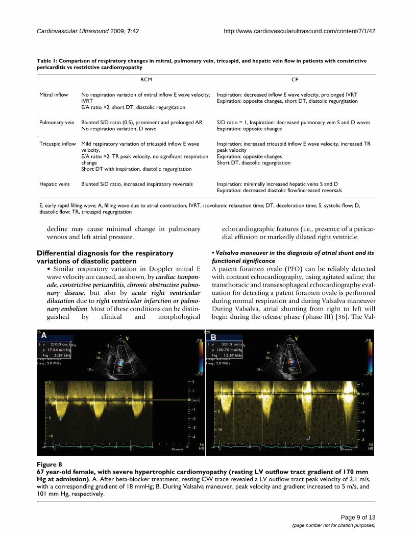
67 year-old female, with severe hypertrophic cardiomyopathy (resting LV outflow tract gradient of 170 mm Hg at admission)Figure 867 year-old female, with severe hypertrophic cardiomyopathy (resting LV outflow tract gradient of 170 mm Hg at admission). A. After beta-blocker treatment, resting CW trace revealed a LV outflow tract peak velocity of 2.1 m/s, with a corresponding gradient of 18 mmHg; B. During Valsalva maneuver, peak velocity and gradient increased to 5 m/s, and 101 mm Hg, respectively.
Table 1: Comparison of respiratory changes in mitral, pulmonary vein, tricuspid, and hepatic vein flow in patients with constrictive pericarditis vs restrictive cardiomyopathy
RCM CP
Mitral inflow No respiration variation of mitral inflow E wave velocity, IVRTE/A ratio >2, short DT, diastolic regurgitation
Inspiration: decreased inflow E wave velocity, prolonged IVRTExpiration: opposite changes, short DT, diastolic regurgitation
Pulmonary vein Blunted S/D ratio (0.5), prominent and prolonged ARNo respiration variation, D wave
S/D ratio = 1, Inspiration: decreased pulmonary vein S and D wavesExpiration: opposite changes
Tricuspid inflow Mild respiratory variation of tricuspid inflow E wave velocity,E/A ratio >2, TR peak velocity, no significant respiration changeShort DT with inspiration, diastolic regurgitation
Inspiration: increased tricuspid inflow E wave velocity, increased TR peak velocityExpiration: opposite changesShort DT, diastolic regurgitation
Hepatic veins Blunted S/D ratio, increased inspiratory reversals Inspiration: minimally increased hepatic veins S and DExpiration: decreased diastolic flow/increased reversals
E, early rapid filling wave; A, filling wave due to atrial contraction; IVRT, isovolumic relaxation time; DT, deceleration time; S, systolic flow; D, diastolic flow; TR, tricuspid regurgitation
Page 9 of 13(page number not for citation purposes)

Cardiovascular Ultrasound 2009, 7:42 http://www.cardiovascularultrasound.com/content/7/1/42
salva maneuver may enhance atrial level shunting becauseof increase in right heart pressure, making the shuntdetectable by either transthoracic echocardiography (TTE)or, with a better sensitivity, by transesophageal echocardi-
ography (TEE). From a clinical point of view, it appearsthat a resting PFO may have a higher risk for clinicalevents than a PFO with shunt only during a provocativemaneuver.
Table 2: When, why and how to use normal respiration or Valsalva maneuver during an echo study
WHEN WHY HOW
Poor 2D image quality To optimize the quality of the echo view Expiration:better parasternal and often apical access to the heartInspiration:brings the diaphragm down improving access to the heart
M-mode measurements of the LV or 2D-quantitation
To avoid measurement errors due to excessive translational motion of the heart
Quiet respiration/held end-expiration
Doppler measurements (flow/tissue velocities)
To avoid measurement errors due to excessive translational motion of the heart
End-expiratory apnea
Estimation of right atrial (RA) pressure To elicit the inspiratory response of the inferior vena cava in order to assess the collapsibility index
Inspiration/brief sniff
Evaluation of systolic pulmonary artery pressure in patients with stable, chronic COPD that cannot been assessed by conventional TTE
To assess the respiratory variation in superior vena cava (SVC) systolic forward flow
Normal respiration
Assessment of left ventricular diastolic function
To unmask elevated LV filling pressure in patients with normal or reduced LV systolic function and
Standardized Valsalva maneuver
pseudo-normal filling pattern at baseline decrease in the mitral E/A ratio of 0.5 or more during Valsalva
impaired relaxation pattern at baseline increase in peak A wave velocity during Valsalva
Cardiac tamponade To assess respiratory variation in cardiac volumes and flow (see text)
Normal respiration
Constrictive pericarditis To assess respiratory variation in mitral, tricuspid, pulmonary and hepatic vein flow(see table 1)
Normal respiration
Restrictive cardiomyopathy To assess respiratory variation in mitral, tricuspid, pulmonary and hepatic vein flow(see table 1)
Normal respiration
Detection of Patent Foramen Ovale by TTE or TEE
To assess the appearence of contrast in the LA shortly after injection of saline contrast into an upper extremity vein, with good opacification of the RA
Normal respirationwithin 3 cardiac cycles after injection of saline contrast
Valsalva maneuverduring the release phase
Hypertrophic cardiomyopathy with mild or absent resting obstruction
To unmask latent gradients/to increase LVOT gradient
Valsalva maneuverduring the strain phase
LV, left ventricle; RA, right atrium; SVC, superior vena cava; COPD, chronic obstructive pulmonary disease; TTE, transthoracic echocardiography; E, peak early diastolic velocity oF mitral inflow; A, peak late diastolic velocity of mitral inflow; TEE, transesophageal echocardiography; LA, left atrium; LVOT, left ventricular outflow tract.
Page 10 of 13(page number not for citation purposes)

Cardiovascular Ultrasound 2009, 7:42 http://www.cardiovascularultrasound.com/content/7/1/42
The right-to-left shunt of a large atrial septal defect may benearly continuous, whereas for smaller atrial septaldefects the appearance of contrast in the left atrium maybe phasic, coordinated with the respiratory cycle; duringinspiration, right heart filling increases, thus increasingthe flow of contrast into the right atrium.
Dynamic obstruction of hypertrophic cardiomyopathy during ValsalvaIn patients with obstructive hypertrophic cardiomyopathythe obstruction of the LV outflow tract is dynamic andmay be mild or nonexistent at rest. Echocardiography dur-ing Valsalva maneuver can unmask latent gradients inpatients without resting obstruction and reveal the basisfor drug treatment choices. During the strain phase of Val-salva maneuver, due to the decrease in preload, end-diastolic left ventricular volume and afterload, SAMoccurs earlier in systole, mitral-septal contact lasts longerand left ventricular outflow tract gradient increases (Fig-ure 8).
• Valsalva for assessing mitral regurgitation in mitral valve prolapseDue to left ventricular volume decrease during Valsalvamaneuver in patients with mitral valve prolapse mitralregurgitation starts earlier in systole and severity of mitralregurgitation increases.
• Műler maneuverThe Mûller maneuver, the opposite of Valsalva, less oftenused in echo examinations, is performed by forciblyinspiring while the nose is held closed and the mouth issealed for about 10 seconds. Because the maneuver exag-gerates inspiratory effort, right-sided filling is augmentedand can be use for the augmentation of tricuspid regurgi-tation.
Limitations• In the absence of normal sinus rhythm, respiratoryvariation of Doppler flow cannot be assessed. An irreg-ular heart rhythm does not allow a meaningful inter-pretation of the influence of respiration ontransvalvular Doppler flow.
• The Valsalva maneuver should only be performed ina standardized manner. Lack of standardization of theValsalva maneuver can limit its feasibility and, moreimportantly, can lead to wrong conclusions.
• The use of Valsalva maneuver is limited in certainconditions: lack of patient' cooperation; patient's ina-bility to adequately increase intrathoracic pressurebecause of medical illness, recent operation, sedation(eg during TEE).
• The decrease in image quality sometimes encoun-tered during Valsalva maneuver or with normal respi-
ration may interfere with the interpretation of thefindings. However, appropriate training and regularuse of respiratory maneuvers help in this regard.
ConclusionUsing normal respiratory phases and provocative respira-tory maneuvers (e.g. Valsalva) can be very useful in char-acterizing normal cardiac function parameters as well asvarious cardiac disorders (Table 2). The extra timerequired to perform these maneuvers is worthwhile andwell compensated by the significant increase in examina-tion quality and diagnostic accuracy.
Competing interests declarationThe authors declare that they have no competing interests.
Authors' contributionsCG conceived of the review, participated in its coordina-tion and revised the final draft of the manuscript. CCBrevised the literature, prepared the final draft of the man-uscript and provided echocardiographic illustrations. MIrevised the literature and prepared the first draft of themanuscript. AC revised the manuscript critically forimportant intellectual content. BAP participated in thecoordination of this review and revised the manuscriptcritically for important intellectual content. All authorsread and approved the final manuscript.
Additional material
Additional file 1Changes in the quality of the echocardiographic image with inspira-tion. Transthoracic parasternal long axis view of the left ventricle recorded with normal respiration revealing a decrease in the quality of the echocar-diographic image with inspiration.Click here for file[http://www.biomedcentral.com/content/supplementary/1476-7120-7-42-S1.mpg]
Additional file 2Improvement in the quality of the echocardiographic image recorded during held end expiration. Transthoracic parasternal long axis view of the left ventricle recorded during held end expiration showing a significant improvement in the quality of the echocardiographic image.Click here for file[http://www.biomedcentral.com/content/supplementary/1476-7120-7-42-S2.mpg]
Additional file 3Influence of pulmonary interference on echocardiographic visualiza-tion of cardiac structures during normal respiration. Transthoracic api-cal 4 chamber view recorded during normal respiration. During inspiration pulmonary interference does not allow the visualization of car-diac structures.Click here for file[http://www.biomedcentral.com/content/supplementary/1476-7120-7-42-S3.mpg]
Page 11 of 13(page number not for citation purposes)

Cardiovascular Ultrasound 2009, 7:42 http://www.cardiovascularultrasound.com/content/7/1/42
AcknowledgementsThis work was partially supported by a Romanian National Research Pro-gramme II grant: [grant number ID_222, contract 199/2007], IDEI 2007.
References1. Feigenbaum H, Armstrong WF, Ryan T: Feigenbaum's Echocardiography
6th edition. Lippincott, Williams and Wilkins; 2005. 2. Fenichel NM, Arora J, Khan R, Antoniou C, Ahuja S, Thompson EJ:
The effect of respiratory motion on the echocardiogram.Chest 1976, 69:655-659.
3. Lang RM, Bierig M, Devereux RB, Flachskampf FA, Foster E, PellikkaPA, Picard MH, Roman MJ, Seward J, Shanewise JS, Solomon SD, Spen-cer KT, Sutton MSJ, Stewart WJ: Recommendations for Cham-ber Quantification: A Report from the American Society ofEchocardiography's Guidelines and Standards Committeeand the Chamber Quantification Writing Group, Developedin Conjunction with the European Association of Echocardi-ography, a Branch of the European Society of Cardiology, aBranch of the European Society of Cardiology. J Am SocEchocardiogr 2005, 18:1440-1463.
4. JI Brenner, Waugh RA: Effect of phasic respiration on left ven-tricular dimension and performance in a normal population.An echocardiographic study. Circulation 1978, 57:122-127.
5. Andersen K, Vik-Mo H: Effects of spontaneous respiration onleft ventricular function assessed by echocardiography. Circu-lation 1984, 69:874-9.
6. Yuan L, Cao T, Duan Y, Yang G, Wang Z, Ruan L: Noninvasiveassessment of Influence of Resistant Respiration on BloodFlow across the Cardiac valves in Humans – A QuantificationStudy by Echocardiography. Echocardiography 2004, 21:391-398.
7. Appleton CP, Jensen JL, Hatle LK, Oh JK: Doppler Evaluation ofLeft and Right Ventricular Diastolic Function: A TechnicalGuide for Obtaining Optimal Flow Velocity Recordings. J AmSoc Echocardiogr 1997, 10(3):271-291.
8. Zoghbi WA, Habib GB, Quinones MA: Doppler Assessment ofRight Ventricular Filling in a Normal Population Compari-son With Left Ventricular Filling Dynamics. Circulation 1990,82:1316-1324.
9. Hill JC, Palma RA: Doppler Tissue Imaging for the Assessmentof Left Ventricular Diastolic Function: A SystematicApproach for the Sonographer. J Am Soc Echocardiogr 2005,18:80-90.
10. Lee KS, Abbas AE, Khandheria BK, Lester SJ: EchocardiographicAssessment of Right Heart Hemodynamic Parameters. J AmSoc Echocardiogr 2007, 20:773-782.
Additional file 4Influence of expiration on echocardiographic visualization of cardiac structures. Transthoracic apical 4 chamber view. Expiration allows apical access to the heart by lung deflation.Click here for file[http://www.biomedcentral.com/content/supplementary/1476-7120-7-42-S4.mpg]
Additional file 5Influence of inspiration on echocardiographic image in subcostal view. Subcostal 4 chamber view. Inspiration brings the diaphragm down, improving imaging of the heart.Click here for file[http://www.biomedcentral.com/content/supplementary/1476-7120-7-42-S5.mpg]
Additional file 6Excessive translational motion of the heart with normal respiration. Transthoracic short axis view recorded during normal respiration. The heart moves medially with inspiration.Click here for file[http://www.biomedcentral.com/content/supplementary/1476-7120-7-42-S6.mpg]
Additional file 7Image acquisition during suspended respiration (held end-expiration) in a patient with excessive translational motion of the heart with nor-mal respiration. Transthoracic short axis view. Excessive translational motion can be avoided by acquiring images during held end-expiration.Click here for file[http://www.biomedcentral.com/content/supplementary/1476-7120-7-42-S7.mpg]
Additional file 8Lack of variation of the inferior vena cava diameter during deep inspi-ration in a patient with dilated cardiomyopathy and severe pulmonary hypertension. The subcostal view adjusted to demonstrate the long axis of the inferior vena cava (IVC) in a patient with dilated cardiomyopathy and severe pulmonary hypertension. There is no inspiratory variation of the IVC diameter during deep inspiration.Click here for file[http://www.biomedcentral.com/content/supplementary/1476-7120-7-42-S8.mpg]
Additional file 9Lack of variation of the inferior vena cava diameter during brief sniff in a patient with dilated cardiomyopathy and severe pulmonary hyper-tension. Evaluation of the inferior vena cava (IVC) from the subcostal view during brief sniff in a patient with dilated cardiomyopathy and severe pulmonary hypertension demonstrates no variation of the IVC diameter.Click here for file[http://www.biomedcentral.com/content/supplementary/1476-7120-7-42-S9.mpg]
Additional file 10Phasic variation in cardiac volumes visualized in transthoracic basal short axis view caused by cardiac tamponade. Transthoracic basal short axis view in a patient with cardiac tamponade demonstrates diastolic col-lapse of the free walls of the right atrium and right ventricle.Click here for file[http://www.biomedcentral.com/content/supplementary/1476-7120-7-42-S10.mpg]
Additional file 11Phasic variation in cardiac volumes caused by cardiac tamponade, vis-ualized in transthoracic apical 4 chamber view. The intermittent col-lapse of the free walls of the right atrium and right ventricle visualized in the transthoracic apical 4 chamber view in a patient with cardiac tampon-ade.Click here for file[http://www.biomedcentral.com/content/supplementary/1476-7120-7-42-S11.mpg]
Additional file 12Increased interventricular interaction in a patient with constrictive pericarditis. Transthoracic apical 4 chamber view in a patient with con-strictive pericarditis reveals the increased interventricular interaction (with left ventricular volume decrease there is a corresponding increase in right ventricular volume).Click here for file[http://www.biomedcentral.com/content/supplementary/1476-7120-7-42-S12.mpg]
Page 12 of 13(page number not for citation purposes)
http://www.ncbi.nlm.nih.gov/entrez/query.fcgi?cmd=Retrieve&db=PubMed&dopt=Abstract&list_uids=1269274
http://www.ncbi.nlm.nih.gov/entrez/query.fcgi?cmd=Retrieve&db=PubMed&dopt=Abstract&list_uids=1269274
http://www.ncbi.nlm.nih.gov/entrez/query.fcgi?cmd=Retrieve&db=PubMed&dopt=Abstract&list_uids=6705162
http://www.ncbi.nlm.nih.gov/entrez/query.fcgi?cmd=Retrieve&db=PubMed&dopt=Abstract&list_uids=6705162
http://www.ncbi.nlm.nih.gov/entrez/query.fcgi?cmd=Retrieve&db=PubMed&dopt=Abstract&list_uids=9109692
http://www.ncbi.nlm.nih.gov/entrez/query.fcgi?cmd=Retrieve&db=PubMed&dopt=Abstract&list_uids=9109692
http://www.ncbi.nlm.nih.gov/entrez/query.fcgi?cmd=Retrieve&db=PubMed&dopt=Abstract&list_uids=9109692
http://www.ncbi.nlm.nih.gov/entrez/query.fcgi?cmd=Retrieve&db=PubMed&dopt=Abstract&list_uids=2401065
http://www.ncbi.nlm.nih.gov/entrez/query.fcgi?cmd=Retrieve&db=PubMed&dopt=Abstract&list_uids=2401065

Cardiovascular Ultrasound 2009, 7:42 http://www.cardiovascularultrasound.com/content/7/1/42
Publish with BioMed Central and every scientist can read your work free of charge
"BioMed Central will be the most significant development for disseminating the results of biomedical research in our lifetime."
Sir Paul Nurse, Cancer Research UK
Your research papers will be:
available free of charge to the entire biomedical community
peer reviewed and published immediately upon acceptance
cited in PubMed and archived on PubMed Central
yours — you keep the copyright
Submit your manuscript here:http://www.biomedcentral.com/info/publishing_adv.asp
BioMedcentral
11. Kircher BJ, Himelman RB, Schiller NB: Noninvasive estimation ofright atrial pressure from the inspiratory collapse of the infe-rior vena cava. Am J Cardiol 1990, 66:493-6.
12. Brennan JM, Blair JE, Goonewardena S, Ronan A, Shah D, Vasaiwala S,Kirkpatrick JN: Reappraisal of the Use of Inferior Vena Cavafor Estimating Right Atrial Pressure. J Am Soc Echocardiogr 2007,20:857-861.
13. Bendjelid K, Romand JA, Walder B, Suter PM, Fournier G: Correla-tion Between Measured Inferior Vena Cava Diameter andRight Atrial Pressure Depends on the EchocardiographicMethod Used in Patients Who Are Mechanically Ventilated.J Am Soc Echocardiogr 2002, 15:944-9.
14. Kunichika N, Miyahara N, Harada M, Tanimoto M: Respiratory Var-iation in Superior Vena Cava Flow in Patients with ChronicObstructive Pulmonary Disease: Estimation of PulmonaryHypertension Using Doppler Flow Index. J Am Soc Echocardiogr2002, 15:1165-9.
15. Dabestani A, Takenaka K, Allen B, Gardin JM, Fischer S, Russell D,Henry WL: Effects of spontaneous respiration on diastolic leftventricular filling assessed by pulsed Doppler echocardiogra-phy. Am J Cardiol 1988, 61:1356-8.
16. Alehan FK, Özkutlu S, Alehan D: Effects of respiration on leftventricular diastolic function in healthy children. Eur Heart J1996, 17:453-6.
17. Tsai LM, Kuo KJ, Chen JH: Effects of spontaneous respiration ontransmitral Doppler flow patterns in normal subjects andpatients with coronary artery disease. Am Heart J 1998,136:99-102.
18. Yuan L, Cao T, Zang Y, Pei J, Duan Y, Gao F: Reversal E/A Valueat End-Inspiration Might Be More Sensitive and Accurate forDiagnosing Abnormal Left Ventricular Diastolic Function.Echocardiography 2007, 24:472-477.
19. Schwammenthal E, Popescu BA, Popescu AC, Di Segni E, Kaplinsky E,Rabinowitz B, Guetta V, Rath S, Feinberg MS: Noninvasive assess-ment of left ventricular end-diastolic pressure by theresponse of the transmitral a-wave velocity to a standard-ized Valsalva maneuver. Am J Cardiol 2000, 86:169-174.
20. Nagueh SF, Appleton CP, Gillebert TC, Marino PN, Oh JK, SmisethOA, Vaggoner AD, Flachskampf FA, Pellikka PA, Evangelista A: Rec-ommendations for the Evaluation of Left Ventricular Diasto-lic Function by Echocardiography. Eur J Echocardiogr 2009,10:165-93.
21. Reagan BW, Helmcke F, Kerut EK: Commonly used respiratoryand pharmacologic interventions in the Echocardiographylaboratory. Echocardiography 2004, 22:455-59.
22. Little WC, Freeman GL: Pericardial Disease. Circulation 2006,113:1622-1632.
23. Wann S, Passen E: Echocardiography in pericardial disease. JAm Soc Echocardiogr 2008, 21:7-13.
24. Ivens EL, Munt BI, Moss RR: Pericardial disease: what the gen-eral cardiologist needs to know. Heart 2007, 93:993-1000.
25. Singh S, Wann LS, Schuchard GH: Right ventricular and rightatrial collapse in patients with cardiac tamponade – a com-bined echocardiographic and hemodynamic study. Circulation1984, 70:966-72.
26. Byrd BF 3rd, Linden RW: Superior vena cava Doppler flowvelocity pattern in pericardial disease. Am J Cardiol 1990,65:1464-70.
27. Nishimura RA: Constrictive pericarditis in the modern era: adiagnostic dilemma. Heart 2001, 86:619-23.
28. Maisch B, Seferović PM, Ristić AD, Erbel R, Rienmüller R, Adler Y,Tomkowski WZ, Thiene G, Yacoub MH: Guidelines on the diag-nosis and management of pericardial diseases. Eur Heart J2004, 25:587-610.
29. Hatle LK, Appleton CP, Papp RL: Differentiation of constrictivepericarditis and restrictive cardiomyopathy by Dopplerechocardiography. Circulation 1989, 79:357-70.
30. Tom CW, Oh JK, Spittell PC: The abdominal aorta and constric-tive pericarditis: abdominal aortic respiratory variation as anechocardiographic finding in constrictive pericarditis. J AmSoc Echocardiogr 2005, 18:282-4.
31. Oh JK, Tajik AJ, Appleton CP, Hatle LK, Nishimura RA, Seward JB:Preload reduction to unmask the characteristic Doppler fea-tures of constrictive pericarditis. A new observation. Circula-tion 1997, 95:796-9.
32. Ha JW, Oh JK, Ommen SR, Ling LH, Tajik AJ: Diagnostic value ofmitral annular velocity for constrictive pericarditis in theabsence of respiratory variation in mitral inflow velocity. JAm Soc Echocardiogr 2002, 15:1468-71.
33. Tabata T, Kabbani SS, Murray RD, Thomas JD, Abdalla I, Klein AL:Difference in the Respiratory Variation Between PulmonaryVenous and Mitral Inflow Doppler Velocities in PatientsWith Constrictive Pericarditis With and Without AtrialFibrillation. J Am Coll Cardiol 2001, 37:1936-42.
34. Boonyaratavej S, Oh JK, Tajik J, Appleton CP, Seward JB: Compari-son of Mitral Inflow and Superior Vena Cava Doppler Veloc-ities in Chronic Obstructive Pulmonary Disease andConstrictive Pericarditis. J Am Coll Cardiol 1998, 32:2043-8.
35. Yamada H, Tabata T, Jaffer SJ, Drinko JK, Jaspre SE, Lauer MS, ThomasJD, Klein AL: Clinical features of mixed physiology of constric-tion and restriction: Echocardiographic characteristics andclinical outcome. Eur J Echocardiography 2007, 8:185-94.
36. Kerut EK, Lee S, Fox E: Diagnosis of an Anatomically and Phys-iologically Significant Patent Foramen Ovale. Echocardiography2006, 23:810-15.
Page 13 of 13(page number not for citation purposes)
http://www.ncbi.nlm.nih.gov/entrez/query.fcgi?cmd=Retrieve&db=PubMed&dopt=Abstract&list_uids=2386120
http://www.ncbi.nlm.nih.gov/entrez/query.fcgi?cmd=Retrieve&db=PubMed&dopt=Abstract&list_uids=2386120
http://www.ncbi.nlm.nih.gov/entrez/query.fcgi?cmd=Retrieve&db=PubMed&dopt=Abstract&list_uids=2386120
http://www.ncbi.nlm.nih.gov/entrez/query.fcgi?cmd=Retrieve&db=PubMed&dopt=Abstract&list_uids=3376898
http://www.ncbi.nlm.nih.gov/entrez/query.fcgi?cmd=Retrieve&db=PubMed&dopt=Abstract&list_uids=3376898
http://www.ncbi.nlm.nih.gov/entrez/query.fcgi?cmd=Retrieve&db=PubMed&dopt=Abstract&list_uids=3376898
http://www.ncbi.nlm.nih.gov/entrez/query.fcgi?cmd=Retrieve&db=PubMed&dopt=Abstract&list_uids=8737221
http://www.ncbi.nlm.nih.gov/entrez/query.fcgi?cmd=Retrieve&db=PubMed&dopt=Abstract&list_uids=8737221
http://www.ncbi.nlm.nih.gov/entrez/query.fcgi?cmd=Retrieve&db=PubMed&dopt=Abstract&list_uids=9665225
http://www.ncbi.nlm.nih.gov/entrez/query.fcgi?cmd=Retrieve&db=PubMed&dopt=Abstract&list_uids=9665225
http://www.ncbi.nlm.nih.gov/entrez/query.fcgi?cmd=Retrieve&db=PubMed&dopt=Abstract&list_uids=9665225
http://www.ncbi.nlm.nih.gov/entrez/query.fcgi?cmd=Retrieve&db=PubMed&dopt=Abstract&list_uids=6499153
http://www.ncbi.nlm.nih.gov/entrez/query.fcgi?cmd=Retrieve&db=PubMed&dopt=Abstract&list_uids=6499153
http://www.ncbi.nlm.nih.gov/entrez/query.fcgi?cmd=Retrieve&db=PubMed&dopt=Abstract&list_uids=6499153
http://www.ncbi.nlm.nih.gov/entrez/query.fcgi?cmd=Retrieve&db=PubMed&dopt=Abstract&list_uids=2191583
http://www.ncbi.nlm.nih.gov/entrez/query.fcgi?cmd=Retrieve&db=PubMed&dopt=Abstract&list_uids=2191583
http://www.ncbi.nlm.nih.gov/entrez/query.fcgi?cmd=Retrieve&db=PubMed&dopt=Abstract&list_uids=2914352
http://www.ncbi.nlm.nih.gov/entrez/query.fcgi?cmd=Retrieve&db=PubMed&dopt=Abstract&list_uids=2914352
http://www.ncbi.nlm.nih.gov/entrez/query.fcgi?cmd=Retrieve&db=PubMed&dopt=Abstract&list_uids=2914352
http://www.ncbi.nlm.nih.gov/entrez/query.fcgi?cmd=Retrieve&db=PubMed&dopt=Abstract&list_uids=9054732
http://www.ncbi.nlm.nih.gov/entrez/query.fcgi?cmd=Retrieve&db=PubMed&dopt=Abstract&list_uids=9054732
http://www.ncbi.nlm.nih.gov/entrez/query.fcgi?cmd=Retrieve&db=PubMed&dopt=Abstract&list_uids=9054732
http://www.ncbi.nlm.nih.gov/entrez/query.fcgi?cmd=Retrieve&db=PubMed&dopt=Abstract&list_uids=9857891
http://www.ncbi.nlm.nih.gov/entrez/query.fcgi?cmd=Retrieve&db=PubMed&dopt=Abstract&list_uids=9857891