Human Respiratory Syncytial Virus
36
Transcript of Human Respiratory Syncytial Virus


Acute respiratory tract infections
Health and economic impactHuman respiratory tract is prone to infection by different pathogens such as viruses, bacteria, and fungi.
Lower ARTIs: First cause of death in low-income countries.
Health impact:
US$ 600 million annually in 42 low-income countries WHO, 2011.
Fourth cause of death in middle-income countries.Fifth cause of death in high-income countries.Deaths (3.9 millions annually)
WHO, 2008
Hospitalization (HRSV responsible for 100,000 cases annually)
Deaths, hospitalizations, medications, and vaccination. Temporal loss of activity, sleepless nights, work / school absence.
Economic impact:

Viruses are considered the major cause of ARTIs.Viruses are responsible for one-third of the deaths caused by ARTIs in the developing countries. More than 200 viruses belonging to different families are incriminated as possible causes of ARTIs.
RhinovirusesCoronavirusesAdenoviruses
Human Respiratory Syncytial VirusInfluenzaPara-Influenza Viruses
URTI LRTI
Acute respiratory tract infections
Impact of viruses
HRSV is the primary cause of lower ARTIs in children.
HRSV is responsible for:64 million cases; 100,000 hospitalization; up to 1 million deaths every year worldwide
HRSV infects 50% of children during the first year of life. All children become infected by the end of second year.
HRSV is highly contagious, nosocomial pathogen, spread by close contact with infectious secretions.
Two major strain groups of HRSV, designated A and B, were found to circulate concurrently during epidemics.
Human Respiratory Syncytial Virus
HRSV

HRSV Classification
Fusion (F) protein
Attachment )G (protein
Small hydrophobic
)SH (protein-ss RNA
M2-1 & M2-2
Phosphoprotein
Polymerase
Matrix protein 3´ 5´
NS1 and NS2
HRSV Structure

HRSV in Saudi Arabia

Detection and typing of HRSV in clinical samples collected from KKUH, Riyadh during two winters (2007/08 and 2008/09).
Preparation and evaluation of polyclonal antibodies to HRSV for the development of in-house diagnostic kits.Sequence and phylogenetic analysis of the most variable and immunogenic protein genes (G and F) of HRSV Saudi strains.
Isolation and characterization of HRSV Saudi strains.
Aim of the Study

Nasopharyngeal Aspirate samples
HRSV detection(RT-PCR)
Positive Samples
Sequence analysis
HRSV Isolation(Cell
culture)
Typing(RT-PCR)
HRSV Characterization (RT-PCR, IFA, VNT
and ELISA).
Production of Hyper-immune
serum
HRSV Concentration(PEG-6000) and
titration
Phylogenetic
analysis.Part one
Part four
Part two
Part three

Part 1
Detection and
Typing of HRSV in
Clinical Samples

100 NPAs were collected from hospitalized children at KKUH during two winters
Detection and Typing of HRSV in Clinical Samples
Viral RNA was extracted from NPAs and used for detection and typing of HRSV by one-step RT-PCR assays.
Sex Age
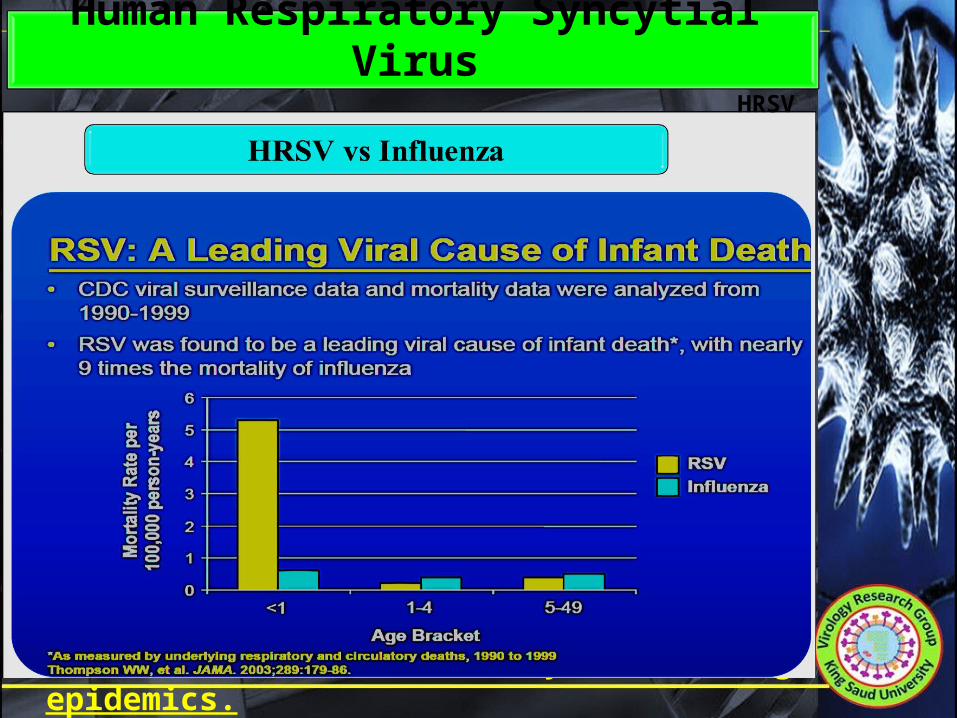
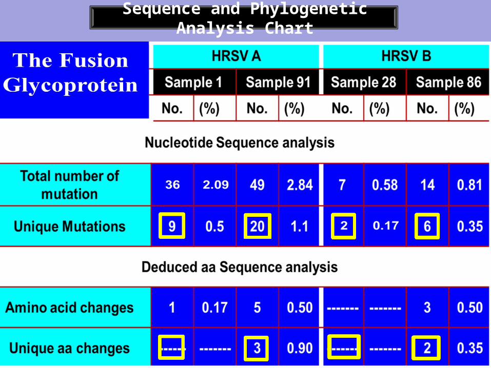
HRSV Incidence and Risk Factors

The two types of HRSV were found to circulate concurrently with slight predominance of type A (16%) more than type B (10%).
Infants younger than 6 months of age (31.1%) are highly infected than any other age group.
Males (31.1%) are more prone to HRSV infection thanfemales (17.9%).
26 out of 100 NAPs were HRSV-positive (26%).
Conclusion (part 1)

Part 2Isolation and
Characterization of HRSV Saudi strains ?

Three passages (7-days each) were applied for each positive sample.
No CPE was developed on VERO cells (All samples).
Two types of permissive cell lines were used (VERO, HEp2).
Two samples (Code 38 and 91 ) developed HRSV characteristic CPE (Syncytia) on HEp2 cells – Both are type A viruses.
The CPE produced by the isolate 38 is more rapid than that of isolate 91.
HRSV Isolation

A B
C D
Progress of CPE on HEp2 cells (10X):

The recovered isolates were titrated using infectivity titration assay:
Isolate 38 = 108.5
TCID50/ml Isolate 91= 105.5
TCID50/ml
The recovered isolates were concentrated using PEG-6000 and were kept at -80oC for further use.
HRSV Titration and Concentration

Characterization of HRSV Saudi isolates

Two Saudi HRSV type A strains were isolated using cell culture for the first time in Saudi Arabia.The two isolates varied in their behavior on cell culture.
Both isolates were able to produce a significant titer of infective virus particles.
HRSV Saudi isolates share the common antigenic and genetic characteristics of international HRSV strains as confirmed by: RT-PCR, ELIZA, IFA and VNT.
Conclusion (part 2)

Part 3Production and Evaluation
of polyclonal antibodies
in Rats against Saudi HRSV strains
?

1 2 3 4Hyper immune serum Preparation
Chart
PEG-Concentrated antigens
(virus+Hep-2)
Preparation of antigen
and adjuvant mixture
Rat injection (3 Doses)
2 weeks each
Rat Bleeding and serum collection
5Evaluation of Serum
6Serum will be processed for diagnostic
uses.

Part 4Sequence and Phylogenetic
Analysis of G and F genes
?

Why G and F genes?
G is the most variable gene among HRSV strains.
HRSV is classified into two distinct groups based on the reactivity of the G protein with monoclonal antibodies.
Due to its highly conserved nature, the fusion protein induces protective immunity against different HRSV strains.
The molecular changes of G and F genes affect the functional and antigenic stability of HRSV.

Cytoplasmic tail (37 aa)Hydrophobic
domain
NH2COOH
Strain specific epitopes
Conserved epitopes
Group specific epitopes
Structure of G protein

Structure of the F protein
SــــــS69ــــــ212
Antigenic Site IAntigenic Site IV, V, VI
Antigenic Site II
F1F2
Cleavage site I Cleavage site II

1 2 3 4Sequence and Phylogenetic
Analysis Chart
Selection of strain
candidatesSequencing Strategy and
primer design
Amplification of fragments cover the G
gene(RT-PCR)
Sequencing of amplified fragments(Gene Art, Germany)
5Sequence
editing and assembly(Bioedit)
6Retrieval of complete gene sequence and
GenBank submission
7Sequence Analysis(MegAlign)
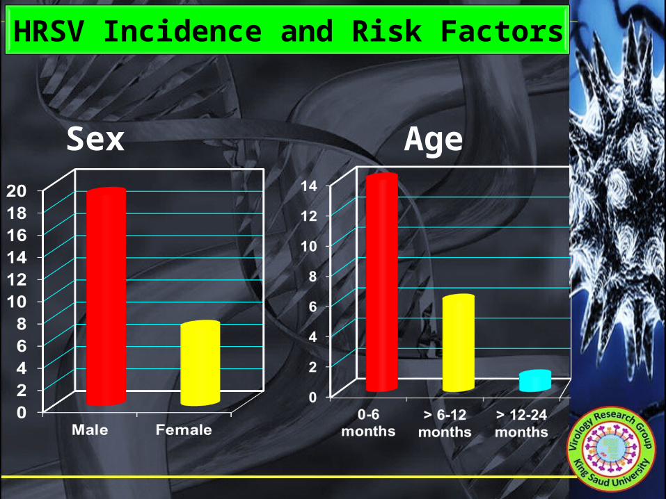
Sequence and Phylogenetic Analysis Chart
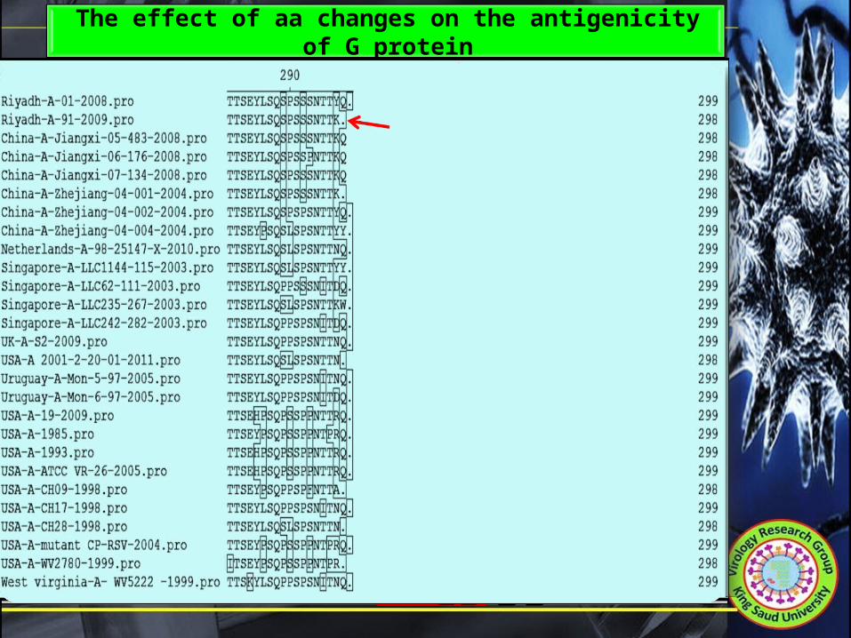
Premature stop codon was formed in Riyadh strain 91/2009.
Replacement of serine by threonine or phenylalanine was reported to abrogate antibody binding. Amino acids at positions 269 and 270 were changed from serine to threonine and phenylalanine, respectively (Thr269Ser and Phe270Ser) .
Replacement of a hydrophilic amino acid by a hydrophobic one was reported to abrogate antibody binding. Amino acids at positions 106 and 133 were changed from hydrophilic to hydrophobic (Gly106Glu and Ile133Thr).
No changes were recorded in the conserved region.
COOHNH2
The effect of aa changes on the antigenicity of G protein

The effect of glycosylation on the antigenicity of G protein

1 2 3 4Sequence and Phylogenetic
Analysis Chart
Selection of strain
candidatesSequencing Strategy and
primer design
Amplification of fragments cover the G
gene(RT-PCR)
Sequencing of amplified fragments(Gene Art, Germany)
5Sequence
editing and assembly(Bioedit)
6Retrieval of complete gene sequence and
GenBank submission
7Sequence Analysis(MegAlign)
8Phylogenetic analysis
(MegAlign)

Phylogenetic Analysis

Mutation rate in G gene of HRSV type A strains is higher than type B strains. This may explain the predominance of type A over type B.
Conclusion (part 4)
In G protein, -FOUR amino acid changes were recorded to have a possible impact on epitope integrity.- SIX amino acid changes, FOUR in type A and
TWO in type B were predicted as highly potential sites for O-glycosylation.
- TWO amino acid changes in type B were predicted as highly potential sites for N-glycosylation.
American and Argentinian strains appear to be the most relevant to Saudi HRSV type B strains.
Asian strains are the most relevant to Saudi HRSV type A strains. They may be originated from the same ancestor.

A set of objective and comprehensive studies is required for better understanding of HRSV circulation in Saudi Arabia.HRSV Saudi isolates, antigens and antibodies produced in this study will be fundamental for:
Recommendations
Sequence analysis of more HRSV strains on temporal and spatial basis will be helpful for understanding HRSV circulation pattern in Saudi Arabia. Complete genome sequencing of Saudi HRSV strains will be helpful to determine their ancestral origin and evolutionary pathway.
- Further basic and applied studies on HRSV.- Development of improved diagnostic tools.
- Vaccine production.

Milestone Achievements
Isolation, characterization and sequence analysis of two HRSV Saudi isolates.
Eight genes were deposited at GenBank Database.
“Polyclonal Antibodies against Saudi strains of Human Respiratory Syncytial Viruses”. A Patent was submitted to (Intellectual Property and
Technology Licensing Program, KSU, KSA)
Two papers in preparation for submission.