Microbial Natural Products with Antiviral Activities, Including ...
Upload
independentCategory
view
3download
0
Innate Immune Signals Modulate Antiviral andPolyreactive Antibody Responses during SevereRespiratory Syncytial Virus Infection
Jennifer L. Reed,1 Timothy P. Welliver,2,a Gary P. Sims,2 LuAnn McKinney,2 Luis Velozo,4 Luis Avendano,5
Karen Hintz,3 Jayson Luma,3 Anthony J. Coyle,2 and Robert C. Welliver, Sr.3
1Center for Biologics Evaluation and Research, Food and Drug Administration, Rockville, and 2Respiratory, Inflammation, and Autoimmunity Group,MedImmune, Gaithersburg, Maryland; 3Department of Pediatrics, Women and Children’s Hospital, State University of New York at Buffalo,Buffalo, New York; and 4Unidad de Anatomía Patológica, Hospital Roberto del Río, and 5Programa de Virología, Universidad de Chile, Santiago,Chile
Antiviral antibody production during respiratory syncytial virus (RSV) infection in infants is poorly understood.To characterize local B lymphocyte responses, lung tissue and secretions from infants with RSV bronchiolitis wereanalyzed for innate B cell–stimulating factors and antiviral antibodies. In lung tissues of infants with fatal RSVbronchiolitis, CD20� lymphocytes and IgM-positive, IgG-positive, and IgA-positive plasma cells were prominentbut CD4� T lymphocytes were not. Type I interferon–induced proteins and B cell tropic factors, including Bcell–activating factor (BAFF) and a proliferation-inducing ligand (APRIL), were colocalized in infected epithe-lium. In nasopharyngeal secretions from infants who survived RSV infection, class-switched antiviral and anti-nucleosomal antibodies were detected at presentation and correlated with BAFF and APRIL levels. Expression ofAPRIL and antiviral antibodies of IgA and IgM but not IgG isotype predicted better oxygen saturation. We con-clude that B lymphocyte–stimulating factors derived from infected epithelium are primary determinants of themucosal antibody response in infant RSV bronchiolitis.
Respiratory syncytial virus (RSV) is the most important
cause of severe lower respiratory tract infection (LRI) in
infants. Virtually all infants have encountered RSV be-
fore the age of 2 years, resulting in �100,000 infant hos-
pitalizations and 400 infant deaths in the United States
each year and 1 million deaths worldwide annually
[1–3]. RSV LRI in infancy has been associated with
chronic wheezing and asthma later in childhood [4]. Be-
cause there is currently no vaccination strategy to pre-
vent infection in infants, RSV continues to pose a signif-
icant health problem.
B lymphocyte responses to RSV are thought to con-
tribute to protection and immunopathologic activities
associated with primary infection [5]. Low levels of se-
rum IgM, IgG, and IgA are detected in most infants 1
month after exposure and persist for months after infec-
tion [6]. Antiviral antibodies are also found in nasopha-
ryngeal secretions from patients with acute infection as
early as 1 week after infection onset [7, 8]. The relation-
ship between antiviral antibody responses and protec-
tion is unclear. Serum RSV-directed antibody titers are
low in the youngest infants, possibly predisposing for
more severe disease in this age group [9]. High levels
of maternal antibody are associated with protection
against acute infection, and boosted antibody levels with
reexposure coincide with milder disease in older chil-
dren [10, 11]. However, reinfections with RSV are com-
mon even in adults who have high anti-RSV antibody
titers, suggesting that antiviral antibody is not com-
pletely protective [12]. In rodent models of RSV infec-
tion, T helper type 2 (Th2) lymphocyte responses, which
Received 2 May 2008; accepted 9 July 2008; electronically published 24 March2009.
Potential conflicts of interest: G.P.S., L.M., and A.J.C. are current employees of,J.L.R. is a former employee of, T.P.W. served as a research intern at, and R.C.W.serves as a scientific advisor to MedImmune. All other authors: none reported.
Presented in part: 6th International Respiratory Syncytial Virus Symposium,Marco Island, FL, October 2007.
Financial support: MedImmune.a Present affiliation: Department of Immunology, University of Michigan, Ann
Arbor, MI.Reprints or correspondence: Dr. Jennifer L. Reed, FDA/CBER, 1401 Rockville Pk.,
HFM-345, Rockville, MD 20852-1448 ([email protected]).
The Journal of Infectious Diseases 2009; 199:1128 –38© 2009 by the Infectious Diseases Society of America. All rights reserved.0022-1899/2009/19908-0007$15.00DOI: 10.1086/597386
M A J O R A R T I C L E
1128 ● JID 2009:199 (15 April) ● Reed et al.
by guest on July 3, 2016http://jid.oxfordjournals.org/
Dow
nloaded from
favor antibody production over cytotoxic responses, were asso-
ciated with RSV persistence and immunopathogenesis [13].
Clinical experience with a formalin-inactivated RSV vaccine also
unfortunately demonstrated eosinophil and lymphocyte activa-
tion in blood and lung tissue after infection with wild-type RSV,
consistent with a Th2-skewed response. Antibodies elicited with
formalin-inactivated RSV vaccine were not protective and pos-
sibly contributed to immunopathogenesis when recipients were
exposed to natural infection [14].
Recently, we had the opportunity to examine secretions from
a cohort of infants with upper respiratory tract infection or LRI
with RSV, in addition to lung tissue obtained from infants with
acutely fatal RSV LRI [15]. Although our goal was to confirm T
lymphocyte contributions to severe disease, we instead observed
that both CD4� and CD8� lymphocytes and their associated
cytokines were nearly absent in RSV LRI. Our observations sug-
gested that an inadequate adaptive immune response, rather
than an exaggerated one, underlies severe RSV LRI in humans.
However, weak T helper lymphocyte responses seemed at odds
with reports of prominent mucosal antiviral antibody detection
within days after initial RSV exposure [7, 8]. For this reason, we
have extended our studies to characterize B lymphocyte re-
sponses during primary RSV disease in infants. Here, we report
massive B lymphocyte recruitment to lung tissue that occurred
immediately at the time of presentation of RSV infection.
Marked expression of B cell activation and survival factors in
infected epithelium occurs early after virus exposure and is cor-
related with mucosal production of RSV-directed immunoglob-
ulin.
PATIENTS, MATERIALS, AND METHODS
Study populations. Postmortem lung tissues from 9 infants
with RSV LRI were provided by Hospital Roberto del Rio (San-
tiago, Chile) (table 1, which appears only in the electronic edi-
tion of the Journal) [15]. Lung tissue from 3 age-matched infants
who died from other causes were provided by the National In-
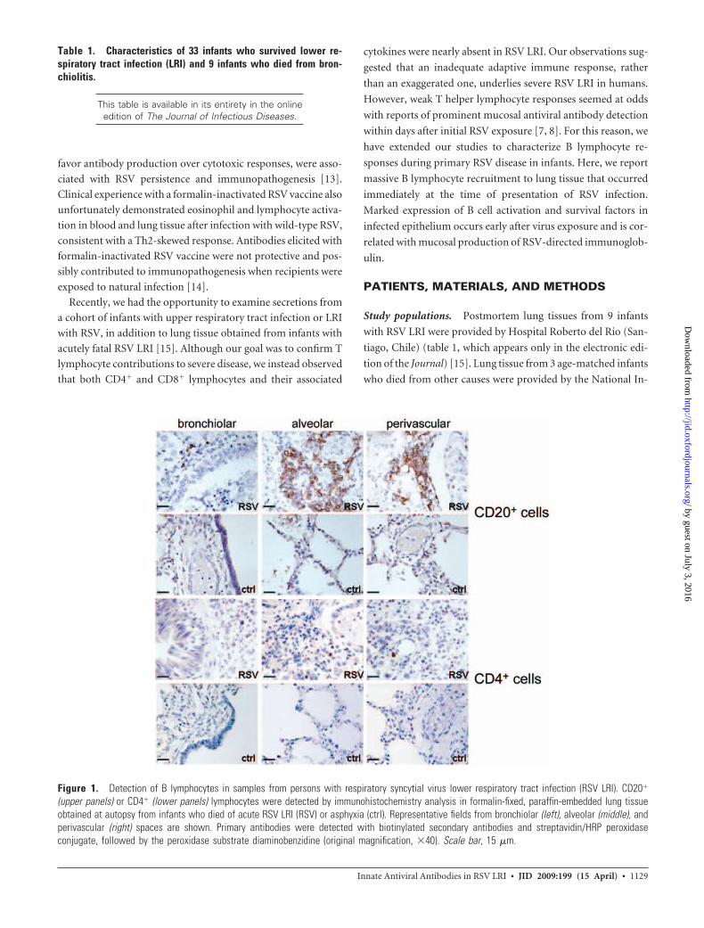
Figure 1. Detection of B lymphocytes in samples from persons with respiratory syncytial virus lower respiratory tract infection (RSV LRI). CD20�
(upper panels) or CD4� (lower panels) lymphocytes were detected by immunohistochemistry analysis in formalin-fixed, paraffin-embedded lung tissueobtained at autopsy from infants who died of acute RSV LRI (RSV) or asphyxia (ctrl). Representative fields from bronchiolar (left), alveolar (middle), andperivascular (right) spaces are shown. Primary antibodies were detected with biotinylated secondary antibodies and streptavidin/HRP peroxidaseconjugate, followed by the peroxidase substrate diaminobenzidine (original magnification, �40). Scale bar, 15 �m.
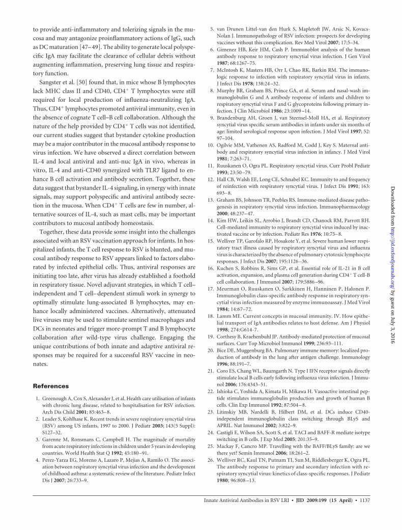
Table 1. Characteristics of 33 infants who survived lower re-spiratory tract infection (LRI) and 9 infants who died from bron-chiolitis.
This table is available in its entirety in the onlineedition of The Journal of Infectious Diseases.
Innate Antiviral Antibodies in RSV LRI ● JID 2009:199 (15 April) ● 1129
by guest on July 3, 2016http://jid.oxfordjournals.org/
Dow
nloaded from
stitute of Child Health and Development (Baltimore, MD). Na-
sopharyngeal secretions from surviving infants with acute bron-
chiolitis were collected at Women and Children’s Hospital
(Buffalo, NY), as previously described (table 1) [15]. No subject
had received RSV antibody preparations, corticosteroids, or an-
tiviral agents. Secretions were obtained �24 h after hospitaliza-
tion and �5 days after the onset of respiratory symptoms in all
cases. Values of oxygen saturation were determined by pulse
oximetry at the time secretions were obtained.
Immunohistochemistry analysis. Formalin-fixed, paraffin-
embedded lung tissues were sectioned and processed through xy-
lene to remove paraffin. Heat-induced epitope retrieval was per-
formed before immunohistochemistry (IHC) analysis. Primary
antibodies reactive with CD20 (Lab Vision), CD4, IgM, IgG, IgA
(Millipore), myxovirus resistance protein A, OAS (Santa Cruz Bio-
technology), vasoactive intestinal peptide (VIP [US Biological]), B
cell–activating factor (BAFF), a proliferation-inducing ligand
(APRIL), B cell maturation antigen (BCMA), transmembrane acti-
vator calcium modulator and cyclophylin ligand interactor (TACI),
and BAFF-R (ProSci) were used according to the manufacturers’
recommendations. Primary antibodies were detected with biotin-
ylated secondary antibodies (Jackson Immunoresearch) and strep-
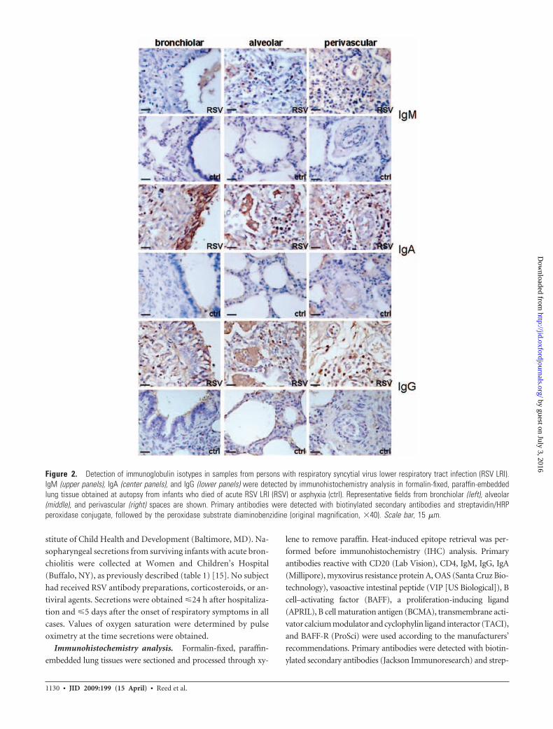
Figure 2. Detection of immunoglobulin isotypes in samples from persons with respiratory syncytial virus lower respiratory tract infection (RSV LRI).IgM (upper panels), IgA (center panels), and IgG (lower panels) were detected by immunohistochemistry analysis in formalin-fixed, paraffin-embeddedlung tissue obtained at autopsy from infants who died of acute RSV LRI (RSV) or asphyxia (ctrl). Representative fields from bronchiolar (left), alveolar(middle), and perivascular (right) spaces are shown. Primary antibodies were detected with biotinylated secondary antibodies and streptavidin/HRPperoxidase conjugate, followed by the peroxidase substrate diaminobenzidine (original magnification, �40). Scale bar, 15 �m.
1130 ● JID 2009:199 (15 April) ● Reed et al.
by guest on July 3, 2016http://jid.oxfordjournals.org/
Dow
nloaded from
tavidin/horseradish peroxidase conjugate (GE Healthcare) with
peroxidase substrate (Sigma Aldrich).
Nasopharyngeal secretions analysis. VIP, BAFF, and APRIL
were measured in secretions by ELISA (Phoenix Pharmaceuticals;
R&D Systems; Bender MedSystems) according to the manufactur-
ers’ instructions. Cytokines were assessed using the BioPlex human
cytokine 17Plex panel (BioRad Laboratories). The lower limit of
detection of mediators was 7.5 pg/mL. Total IgG, IgM, and IgA
levels were measured using a capture-detect ELISA system (Bethyl
Laboratories) according to the manufacturers’ recommendations.
For detection of antibody reactivity against RSV antigens, Hep-2
lysate, BSA, and nucleosomes, flat-well ELISA plates (Maxisorp
[Nalge Nunc International]) were coated with RSV-A2 virus stock
(104 pfu/well [MedImmune]), BSA (1 �g/mL), Hep-2 lysate (1 �g/
mL), or a 1:500 dilution of nuclear extract prepared from
campothecin-induced U937 cells (cell-death detection ELISA kit
[Roche]). Nonspecific protein interactions were blocked by incu-
bation in saline containing 3% BSA (Sigma). Nasopharyngeal se-
cretions were added to wells at a 1:10 dilution in blocking buffer.
Bound antibodies were detected with biotinylated, isotype-specific
polyclonal antisera (Jackson Immunoresearch) and then strep-
tavidin-HRP conjugate (GE Healthcare) and HRP substrate (Sure-
Blue [KPL]). The optical density was quantified at 410 nmol/L, us-
ing a 96-well plate reader (Bio-Tek Instruments).
B cell stimulation. Purified B cells were cultured for 7 days
at 1 � 106 cells/mL in either 1 mL in 24-well culture plates or
100 �L in 96-well round-bottom culture plates. The cells were
incubated with combinations of the Toll-like receptor 7 (TLR7)
agonist imiquimod or the TLR9 agonist CpG-B(2006) (0.1–10
�g/mL [InvivoGen]), leukocyte-derived type I interferon (IFN;
100 IU/mL [PBL Interferon Source]), human IL-4 (100 ng/mL
[R&D Systems]), BAFF (100 ng/mL [R&D Systems]), antihu-
man CD40 (1 �g/mL [R&D Systems]), and anti-IgM (5 �g/mL
[Jackson ImmunoResearch]). Secreted IL-6 and IL-10 in the cul-
ture supernatant was measured using commercially available
ELISA kits (R&D Systems) according to the manufacturer’s in-
structions. Secreted immunoglobulin in the culture supernatant
was quantitated by ELISA as previously described [16, 17].
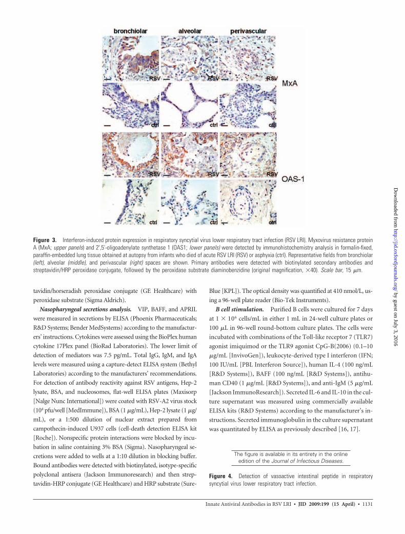
Figure 3. Interferon-induced protein expression in respiratory syncytial virus lower respiratory tract infection (RSV LRI). Myxovirus resistance proteinA (MxA; upper panels) and 2',5'-oligoadenylate synthetase 1 (OAS1; lower panels) were detected by immunohistochemistry analysis in formalin-fixed,paraffin-embedded lung tissue obtained at autopsy from infants who died of acute RSV LRI (RSV) or asphyxia (ctrl). Representative fields from bronchiolar(left), alveolar (middle), and perivascular (right) spaces are shown. Primary antibodies were detected with biotinylated secondary antibodies andstreptavidin/HRP peroxidase conjugate, followed by the peroxidase substrate diaminobenzidine (original magnification, �40). Scale bar, 15 �m.
The figure is available in its entirety in the onlineedition of the Journal of Infectious Diseases.
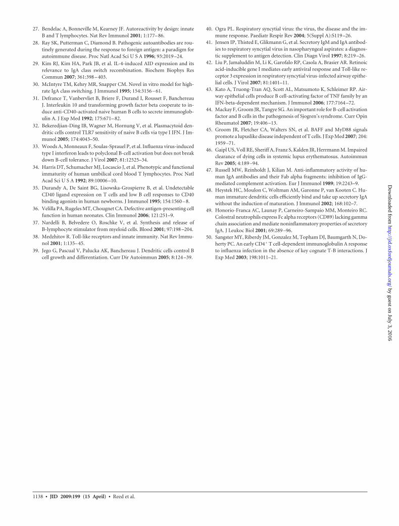
Figure 4. Detection of vasoactive intestinal peptide in respiratorysyncytial virus lower respiratory tract infection.
Innate Antiviral Antibodies in RSV LRI ● JID 2009:199 (15 April) ● 1131
by guest on July 3, 2016http://jid.oxfordjournals.org/
Dow
nloaded from
Statistical analysis. For analysis of nasopharyngeal secre-
tions, data were log-transformed to obtain a normal distribu-
tion. Pearson analysis of covariation was performed using
GraphPad Prism software. Statistical significance was defined as
a P value of �.05.
RESULTS
B cells are prominent in lung tissue following primary RSV
LRI. Mucosal antibodies may be transported from the serum to
the lung through transudation and/or active transport [18, 19], or
they are locally produced by B lymphocytes in tissue [20]. To deter-
mine the source of antibody in RSV-exposed infants, we identified
B cells by IHC analysis in a panel of lung tissues obtained from 9
infants with acutely fatal RSV LRI. CD20� B lymphocytes were
abundant in lung tissue of infants with RSV LRI and localized to
alveolar and perivascular spaces (figure 1). CD20� cells were nearly
absent in control lung tissues obtained from 3 infants who died of
asphyxia (figure 1). CD4� cells, in contrast, were rarely identified in
lung tissue from patients with RSV LRI or from control patients
(figure 1) [15]. In RSV LRI, the lung-associated B lymphocyte pop-
ulation included plasma cells, based on detection of substantial cy-
toplasmic levels of IgA, IgG, and IgM, whereas almost no IgA, IgG,
or IgM was detected in lung tissue from age-matched control pa-
tients (figure 2). IgM-positive, IgG-positive, and IgA-positive cells
were prominent in perivascular and alveolar spaces. IgA was also
strongly deposited on bronchiolar epithelium in specimens from
patients with RSV LRI.
Innate immune factors in RSV-infected lung tissue. Because a
vigorous B lymphocyte response was apparent in lung tissue from
infants with acute RSV LRI, whereas T lymphocytes were rare, we
considered the possibility of T cell–independent B lymphocyte
antibody production. Type I IFN has been implicated in early antiviral
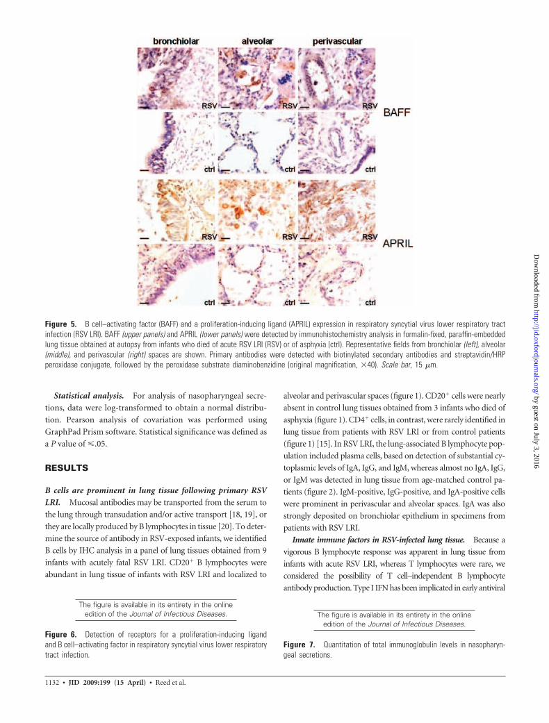
Figure 5. B cell–activating factor (BAFF) and a proliferation-inducing ligand (APRIL) expression in respiratory syncytial virus lower respiratory tractinfection (RSV LRI). BAFF (upper panels) and APRIL (lower panels) were detected by immunohistochemistry analysis in formalin-fixed, paraffin-embeddedlung tissue obtained at autopsy from infants who died of acute RSV LRI (RSV) or of asphyxia (ctrl). Representative fields from bronchiolar (left), alveolar(middle), and perivascular (right) spaces are shown. Primary antibodies were detected with biotinylated secondary antibodies and streptavidin/HRPperoxidase conjugate, followed by the peroxidase substrate diaminobenzidine (original magnification, �40). Scale bar, 15 �m.
The figure is available in its entirety in the onlineedition of the Journal of Infectious Diseases.
Figure 6. Detection of receptors for a proliferation-inducing ligandand B cell–activating factor in respiratory syncytial virus lower respiratorytract infection.
The figure is available in its entirety in the onlineedition of the Journal of Infectious Diseases.
Figure 7. Quantitation of total immunoglobulin levels in nasopharyn-geal secretions.
1132 ● JID 2009:199 (15 April) ● Reed et al.
by guest on July 3, 2016http://jid.oxfordjournals.org/
Dow
nloaded from
B cell responses [21]. Reagents for detection of IFN-� or IFN-�
by IHC analysis were not available. However, we did detect
type I IFN–induced proteins, myxovirus resistance protein A,
and 2',5'-oligoadenylate synthetase 1 at high levels in bron-
chiolar and alveolar epithelium of lung tissue from patients
with RSV LRI but not in tissue from control patients (figure
3). VIP promotes T cell–independent B cell activation [22],
and the CD40L-related factors BAFF and APRIL have been
more recently implicated in CD40-independent immuno-
globulin production and class-switch recombination [23, 24].
We were able to confirm strong expression of VIP (unpub-
lished data) (figure 4, which appears only in the electronic
edition of the Journal) and BAFF and APRIL (figure 5) in
alveolar and bronchiolar epithelium of infants presenting
with acutely fatal RSV infection. VIP and APRIL were also
detected in perivascular immune cells. We detected minimal
expression of VIP, BAFF, and APRIL in lung tissue from age-
matched control patients (figures 4 and 5). Also, in RSV LRI
tissues we detected expression of BAFF-R, TACI, and BCMA,
the identified receptors for BAFF and APRIL [25], in a subset
of perialveolar lymphocytes, some with paradigmatic plasma
cell morphologic characteristics (figure 6, which appears only
in the electronic edition of the Journal).
Antiviral and polyreactive antibodies in infant nasopha-
ryngeal secretions. Next, we assessed immunoglobulin levels
in nasopharyngeal secretions obtained from 33 infants with
acute LRI (RSV was detected in 22, influenza virus in 10, and
adenovirus in 1). The characteristics of this patient population
are detailed in table 1. Total IgG, IgM, and IgA levels were similar
in all nasopharyngeal secretions analyzed (figure 7, which ap-
pears only in the electronic edition of the Journal). As has been
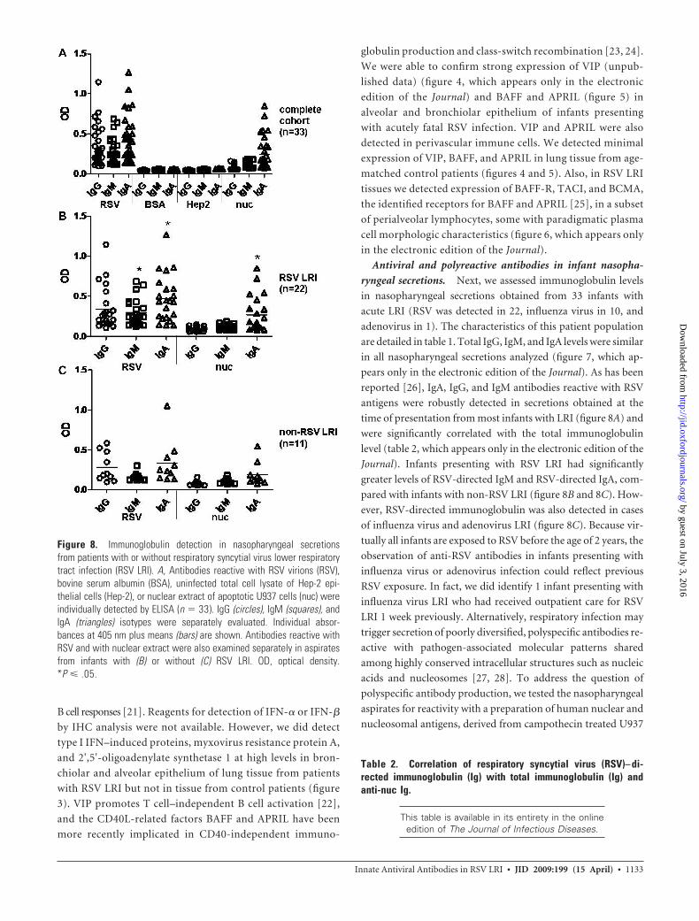
reported [26], IgA, IgG, and IgM antibodies reactive with RSV
antigens were robustly detected in secretions obtained at the
time of presentation from most infants with LRI (figure 8A) and
were significantly correlated with the total immunoglobulin
level (table 2, which appears only in the electronic edition of the
Journal). Infants presenting with RSV LRI had significantly
greater levels of RSV-directed IgM and RSV-directed IgA, com-
pared with infants with non-RSV LRI (figure 8B and 8C). How-
ever, RSV-directed immunoglobulin was also detected in cases
of influenza virus and adenovirus LRI (figure 8C). Because vir-
tually all infants are exposed to RSV before the age of 2 years, the
observation of anti-RSV antibodies in infants presenting with
influenza virus or adenovirus infection could reflect previous
RSV exposure. In fact, we did identify 1 infant presenting with
influenza virus LRI who had received outpatient care for RSV
LRI 1 week previously. Alternatively, respiratory infection may
trigger secretion of poorly diversified, polyspecific antibodies re-
active with pathogen-associated molecular patterns shared
among highly conserved intracellular structures such as nucleic
acids and nucleosomes [27, 28]. To address the question of
polyspecific antibody production, we tested the nasopharyngeal
aspirates for reactivity with a preparation of human nuclear and
nucleosomal antigens, derived from campothecin treated U937
Figure 8. Immunoglobulin detection in nasopharyngeal secretionsfrom patients with or without respiratory syncytial virus lower respiratorytract infection (RSV LRI). A, Antibodies reactive with RSV virions (RSV),bovine serum albumin (BSA), uninfected total cell lysate of Hep-2 epi-thelial cells (Hep-2), or nuclear extract of apoptotic U937 cells (nuc) wereindividually detected by ELISA (n � 33). IgG (circles), IgM (squares), andIgA (triangles) isotypes were separately evaluated. Individual absor-bances at 405 nm plus means (bars) are shown. Antibodies reactive withRSV and with nuclear extract were also examined separately in aspiratesfrom infants with (B) or without (C) RSV LRI. OD, optical density.*P � .05.
Table 2. Correlation of respiratory syncytial virus (RSV)– di-rected immunoglobulin (Ig) with total immunoglobulin (Ig) andanti-nuc Ig.
This table is available in its entirety in the onlineedition of The Journal of Infectious Diseases.
Innate Antiviral Antibodies in RSV LRI ● JID 2009:199 (15 April) ● 1133
by guest on July 3, 2016http://jid.oxfordjournals.org/
Dow
nloaded from
cells. Immunoglobulin reactive with nuclear and nucleosomal
antigens (anti-nuc) was present in aspirates, with detection of
greater IgA levels, compared with IgG or IgM levels (figure 8A).
However, no reactivity was observed against BSA or a lysate pre-
pared from uninfected Hep-2 cells, the cell line in which the RSV
antigens were prepared (figure 8A). Infants presenting with RSV
LRI had significantly greater antinuclear and nucleosomal (anti-
nuc) IgA than those presenting with infection due to influenza
virus or adenovirus (figure 8B and 8C). Recovery of RSV-
directed IgM and IgA was significantly correlated with recovery
of anti-nuc IgM and IgA (table 2). Thus, the recovery of immu-
noglobulin reactive with apoptotic cell epitopes appeared to be
linked to antiviral immunoglobulin recovery, raising the possi-
bility that factors present in infected lung tissue might be re-
sponsible for promoting both immunoglobulin types.
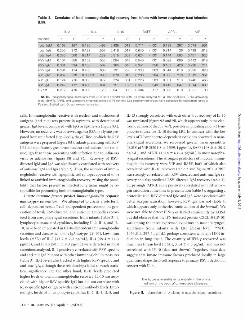
Innate immune factors predict immunoglobulin response
and oxygen saturation. We attempted to clarify a role for T
cell– dependent versus T cell–independent processes in the gen-
eration of total, RSV-directed, and anti-nuc antibodies recov-
ered from nasopharyngeal secretions from infants (table 3). T
lymphocyte–associated cytokines, including IL-2, IL-4, and IL-
10, have been implicated in CD40-dependent immunoglobulin
secretion and class switch to the IgA isotype [29 –31]. Low mean
levels (�SD) of IL-2 (33.7 � 7.2 pg/mL), IL-4 (59.4 � 11.3
pg/mL), and IL-10 (50.5 � 9.3 pg/mL) were detected in most
secretions analyzed. IL-4 positively correlated with RSV-specific
and anti-nuc IgA but not with other immunoglobulin measures
(table 3). IL-2 levels also tracked with higher RSV-specific and
anti-nuc IgA, although these relationships failed to reach statis-
tical significance. On the other hand, IL-10 levels predicted
higher levels of total immunoglobulin recovery. IL-10 was asso-
ciated with higher RSV-specific IgG but did not correlate with
RSV-specific IgM or IgA or with anti-nuc antibody levels. Inter-
estingly, levels of T lymphocyte cytokines IL-2, IL-4, IL-5, and
IL-13 strongly correlated with each other, but recovery of IL-10
was unrelated (figure 9A and 9B, which appears only in the elec-
tronic edition of the Journal), possibly implicating a non-T lym-
phocyte source for IL-10 during LRI. In contrast with the low
levels of T lymphocyte– dependent cytokines observed in naso-
pharyngeal secretions, we recovered greater mean quantities
(�SD) of VIP (5201.8 � 1559.4 pg/mL), BAFF (108.0 � 20.9
pg/mL), and APRIL (1518 � 635.4 pg/mL) in most nasopha-
ryngeal secretions. The strongest predictors of mucosal immu-
noglobulin recovery were VIP and BAFF, both of which also
correlated with IL-10 recovery (table 3 and figure 9C). APRIL
was strongly correlated with RSV-directed and anti-nuc IgA re-
covery and also predicted RSV-directed IgM recovery (table 3).
Surprisingly, APRIL alone positively correlated with better oxy-
gen saturation at the time of presentation (table 3), suggesting a
protective role. RSV-directed IgM and IgA were associated with
better oxygen saturation; however, RSV IgG was not (table 4,
which appears only in the electronic edition of the Journal). We
were not able to detect IFN-� or IFN-� consistently by ELISA
but did observe that the IFN-induced protein CXCL10 (IP-10)
was among the most expressed cytokines in nasopharyngeal
secretions from infants with LRI (mean level [�SD],
2053.8 � 297.5 pg/mL), perhaps consistent with type I IFN in-
duction in lung tissue. The quantity of IFN-� recovered was
much less (mean level [�SD], 31.4 � 6.0 pg/mL) and was not
correlated with IP-10 (data not shown). Together, these data
suggest that innate immune factors produced locally in large
quantities shape the B cell response to primary RSV infection in
concert with IL-4.
The figure is available in its entirety in the onlineedition of the Journal of Infectious Diseases.
Figure 9. Correlation of cytokines in nasopharyngeal secretions.
Table 3. Correlates of local immunoglobulin (Ig) recovery from infants with lower respiratory tract infection(LRI).
IL-2 IL-4 IL-10 BAFF APRIL VIP
Variable r P r P r P r P r P r P
Total IgM 0.163 .167 0.126 .493 0.435 .013 0.711 �.001 0.193 .367 0.574 .001Total IgG 0.250 .372 0.122 .507 0.418 .017 0.640 �.001 0.314 .136 0.438 .012Total IgA 0.336 .060 0.214 .239 0.515 .003 0.624 �.001 0.144 .503 0.401 .023RSV IgM 0.104 .568 0.109 .555 0.084 .648 0.540 .001 0.522 .009 0.412 .019RSV IgG 0.301 .094 0.100 .592 0.365 .040 0.341 .056 0.169 .429 0.226 .214RSV IgA 0.284 .114 0.460 .008 0.191 .296 0.333 .063 0.514 .010 0.386 .029nuc IgM 0.067 .820 0.00063 .998 0.070 .813 0.296 .304 0.386 .270 0.019 .951nuc IgG 0.124 .716 0.055 .872 0.324 .331 0.226 .503 0.051 .913 0.246 .466nuc IgA 0.247 .172 0.498 .004 0.251 .166 0.351 .049 0.410 .047 0.314 .080O2 sat 0.212 .420 0.393 .120 0.041 .880 0.384 .117 0.698 .010 0.331 .190
NOTE. Nasopharyngeal secretions from 33 infants hospitalized with LRI were analyzed for Ig, Th2 cytokines, B cell–activatingfactor (BAFF), APRIL, and vasoactive intestinal peptide (VIP) content. Log-transformed values were assessed for correlation, using aPearson 2-tailed test. O2 sat, oxygen saturation.
1134 ● JID 2009:199 (15 April) ● Reed et al.
by guest on July 3, 2016http://jid.oxfordjournals.org/
Dow
nloaded from
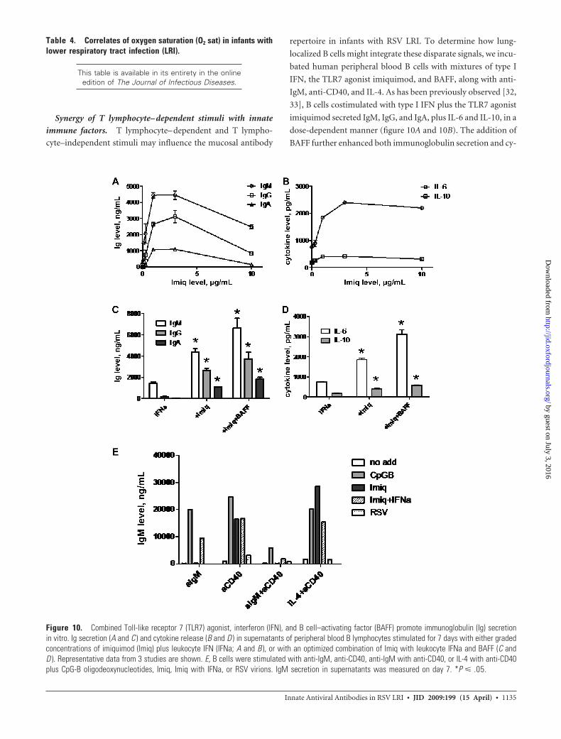
Synergy of T lymphocyte– dependent stimuli with innate
immune factors. T lymphocyte– dependent and T lympho-
cyte–independent stimuli may influence the mucosal antibody
repertoire in infants with RSV LRI. To determine how lung-
localized B cells might integrate these disparate signals, we incu-
bated human peripheral blood B cells with mixtures of type I
IFN, the TLR7 agonist imiquimod, and BAFF, along with anti-
IgM, anti-CD40, and IL-4. As has been previously observed [32,
33], B cells costimulated with type I IFN plus the TLR7 agonist
imiquimod secreted IgM, IgG, and IgA, plus IL-6 and IL-10, in a
dose-dependent manner (figure 10A and 10B). The addition of
BAFF further enhanced both immunoglobulin secretion and cy-
Figure 10. Combined Toll-like receptor 7 (TLR7) agonist, interferon (IFN), and B cell–activating factor (BAFF) promote immunoglobulin (Ig) secretionin vitro. Ig secretion (A and C ) and cytokine release (B and D ) in supernatants of peripheral blood B lymphocytes stimulated for 7 days with either gradedconcentrations of imiquimod (Imiq) plus leukocyte IFN (IFNa; A and B ), or with an optimized combination of Imiq with leukocyte IFNa and BAFF (C andD ). Representative data from 3 studies are shown. E, B cells were stimulated with anti-IgM, anti-CD40, anti-IgM with anti-CD40, or IL-4 with anti-CD40plus CpG-B oligodeoxynucleotides, Imiq, Imiq with IFNa, or RSV virions. IgM secretion in supernatants was measured on day 7. *P � .05.
Table 4. Correlates of oxygen saturation (O2 sat) in infants withlower respiratory tract infection (LRI).
This table is available in its entirety in the onlineedition of The Journal of Infectious Diseases.
Innate Antiviral Antibodies in RSV LRI ● JID 2009:199 (15 April) ● 1135
by guest on July 3, 2016http://jid.oxfordjournals.org/
Dow
nloaded from
tokine release (figure 10C and 10D). We found that the combi-
nation of TLR7 agonist plus type I IFN could synergize with
anti-IgM, anti-CD40, and IL-4 to augment secretion of IgM (fig-
ure 10E), IgG, and IgA in the absence of T cell help (figure 11A
and 11B, which appears only in the electronic edition of the
Journal). The magnitude of synergy was comparable to that ob-
served with CpG-B oligodeoxynucleotides (figures 10E, 11A,
and 11B), although CpG stimulated IgG and IgA release more
efficiently. Immunoglobulin secretion in the presence of anti-
CD40 or anti-CD40 plus IL-4 was augmented by TLR7 agonist,
whereas type I IFN provided no synergy. As has been previously
observed with influenza virus [34, 35], incubation of B lympho-
cytes with RSV virions, either alone or in combination with anti-
IgM, anti-CD40, or IL-4, had minimal impact on immunoglob-
ulin release (figures 10E, 11A, and 11B). These data indicate that
innate stimuli associated with acute RSV infected cells, such as
type I IFN, BAFF, and TLR7 ligand, may synergize to induce
robust B cell responses in the absence of T cell help.
DISCUSSION
T lymphocyte– dependent antibody production is inefficient in
infants. Neonatal T lymphocytes deficits include low expression
of TCR, adhesion molecules, and CD40L, which result in
blunted T cell help for B lymphocytes [36, 37]. Nevertheless, an
antiviral antibody response in nasopharyngeal secretions of in-
fants is present early during primary RSV LRI. In this study, we
demonstrate that B lymphocytes, including plasma cells, are
heavily recruited to infant lung at the peak of RSV illness,
whereas CD4� T lymphocytes are not abundant. We also show
that B cell detection in tissue coincides with local expression of B
cell tropic factors, including BAFF, APRIL, and VIP, localized
primarily to infected respiratory epithelium. Similarly, specific
and total immunoglobulin levels in secretions from surviving
infants with RSV LRI coincide and correlate with BAFF, APRIL,
and VIP recovery, suggesting a causative relationship. Assuming
similar disease pathogenesis in Chilean and US infants, these
data implicate T lymphocyte–independent processes in shaping
the primary antiviral antibody response in a setting where cog-
nate T cell help is suboptimal.
Previous studies have focused on the dendritic cell (DC) as the
prime regulator of antiviral immunoglobulin responses in adult
systems, either indirectly through T cell stimulation or through
IFN-dependent release of IL-6 and BAFF [38 – 40]. DCs ob-
tained from infants exhibit multiple deficiencies [38], including
limited responses to in vitro stimuli and low expression of major
histocompatibility complex (MHC) class II and costimulatory
molecules. Thus, although mature DCs can clearly make enor-
mous contributions to B cell activation and immunoglobulin
production, it is unclear whether immature DCs in infants can
play this role. Most studies that implicate DCs in antibody pro-
duction measure serum levels of immunoglobulin. In fact, in
infants �6 months old, serum IgM and IgA responses to RSV are
almost undetectable during the first month [7, 40], whereas the
antibody response in secretions appears within days and is sim-
ilar qualitatively and quantitatively in infants and older children
with acute RSV LRI [41]. This apparent dissociation between
mucosal and systemic immunoglobulin responses in infants
may highlight a differential role for DCs in these 2 anatomic
sites. In the context of inefficient DC function, the local epithe-
lium may provide an alternative source of B lymphocyte– di-
rected stimuli to maintain mucosal antibody levels. In support of
this point, recent in vitro studies show that RSV infection of
respiratory epithelial cells results in TLR3 activation, IFN-� re-
lease, and IFN-�– dependent BAFF and APRIL expression in
cultured respiratory epithelium [42, 43]. Our data confirm
prominent epithelial expression of VIP, BAFF, and APRIL in
vivo during RSV LRI. Colocalization of IFN-induced proteins
such as MxA and OAS in RSV-infected alveolar epithelium and
high levels of CXCL10 in nasopharyngeal secretions imply that
type I IFN is probably also locally expressed. Together, these data
point to RSV-infected epithelium as an important source of in-
nate immune factors, particularly during uncontrolled respira-
tory infection, and suggest that these cytokines may be sufficient
to induce T lymphocyte–independent antibody production if
DC functions are immature.
BAFF and APRIL overexpression have been previously impli-
cated in the generation of self-reactive antibodies in patients
with autoimmune disease [44, 45]. Here, we report antibodies
reactive with apoptotic cell epitopes in nasopharyngeal secre-
tions during primary RSV LRI. Antinucleosomal antibodies in
nasopharyngeal secretions were strongly correlated to BAFF and
APRIL, suggesting that similar mechanisms of B lymphocyte ac-
tivation could be present in systemic lupus erythematosus and
infant respiratory infection. In acute RSV LRI, airways are oc-
cluded with dense apoptotic cellular debris arising from massive
infection of respiratory epithelium [15]. Accumulation of apo-
ptotic cellular debris is also a cardinal feature of systemic lupus
erythematosus, in which a failure in macrophage clearance of
debris is implicated in pathogenesis [46]. The generation of an-
tibodies recognizing cellular debris may be a conserved, protec-
tive response. Autoreactive antibodies are proposed to have a
beneficial role in removing immune-stimulating components of
dying cells, thus avoiding inflammatory cell recruitment and tis-
sue destruction [27]. Supporting this idea, APRIL and anti-nuc
antibodies of IgA isotype were associated with better oxygen sat-
uration values in this cohort of infants with bronchiolitis, sug-
gesting a role for APRIL and/or APRIL-induced IgA in providing
some degree of protection in air-exchange tissue. IgA is thought
The figure is available in its entirety in the onlineedition of the Journal of Infectious Diseases.
Figure 11. Combined Toll-like receptor 7 agonist, interferon, and Bcell–activating factor promote immunoglobulin secretion in vitro.
1136 ● JID 2009:199 (15 April) ● Reed et al.
by guest on July 3, 2016http://jid.oxfordjournals.org/
Dow
nloaded from
to provide anti-inflammatory and tolerizing signals in the mu-
cosa and may antagonize proinflammatory actions of IgG, such
as DC maturation [47– 49]. The ability to generate local polyspe-
cific IgA may facilitate the clearance of cellular debris without
augmenting inflammation, preserving lung tissue and respira-
tory function.
Sangster et al. [50] found that, in mice whose B lymphocytes
lack MHC class II and CD40, CD4� T lymphocytes were still
required for local production of influenza-neutralizing IgA.
Thus, CD4� lymphocytes promoted antiviral immunity, even in
the absence of cognate T cell–B cell collaboration. Although the
nature of the help provided by CD4� T cells was not identified,
our current studies suggest that bystander cytokine production
may be a major contributor in the mucosal antibody response to
virus infection. We have observed a direct correlation between
IL-4 and local antiviral and anti-nuc IgA in vivo, whereas in
vitro, IL-4 and anti-CD40 synergized with TLR7 ligand to en-
hance B cell activation and antibody secretion. Together, these
data suggest that bystander IL-4 signaling, in synergy with innate
signals, may support polyspecific and antiviral antibody secre-
tion in the mucosa. When CD4� T cells are few in number, al-
ternative sources of IL-4, such as mast cells, may be important
contributors to mucosal antibody homeostasis.
Together, these data provide some insight into the challenges
associated with an RSV vaccination approach for infants. In hos-
pitalized infants, the T cell response to RSV is blunted, and mu-
cosal antibody response to RSV appears linked to factors elabo-
rated by infected epithelial cells. Thus, antiviral responses are
initiating too late, after virus has already established a foothold
in respiratory tissue. Novel adjuvant strategies, in which T cell–
independent and T cell– dependent stimuli work in synergy to
optimally stimulate lung-associated B lymphocytes, may en-
hance locally administered vaccines. Alternatively, attenuated
live viruses may be used to stimulate sentinel macrophages and
DCs in neonates and trigger more-prompt T and B lymphocyte
collaboration after wild-type virus challenge. Engaging the
unique contributions of both innate and adaptive antiviral re-
sponses may be required for a successful RSV vaccine in neo-
nates.
References
1. Greenough A, Cox S, Alexander J, et al. Health care utilisation of infantswith chronic lung disease, related to hospitalisation for RSV infection.Arch Dis Child 2001; 85:463– 8.
2. Leader S, Kohlhase K. Recent trends in severe respiratory syncytial virus(RSV) among US infants, 1997 to 2000. J Pediatr 2003; 143(5 Suppl):S127–32.
3. Garenne M, Ronsmans C, Campbell H. The magnitude of mortalityfrom acute respiratory infections in children under 5 years in developingcountries. World Health Stat Q 1992; 45:180 –91.
4. Perez-Yarza EG, Moreno A, Lazaro P, Mejias A, Ramilo O. The associ-ation between respiratory syncytial virus infection and the developmentof childhood asthma: a systematic review of the literature. Pediatr InfectDis J 2007; 26:733–9.
5. van Drunen Littel-van den Hurk S, Mapletoft JW, Arsic N, Kovacs-Nolan J. Immunopathology of RSV infection: prospects for developingvaccines without this complication. Rev Med Virol 2007; 17:5–34.
6. Gimenez HB, Keir HM, Cash P. Immunoblot analysis of the humanantibody response to respiratory syncytial virus infection. J Gen Virol1987; 68:1267–75.
7. McIntosh K, Masters HB, Orr I, Chao RK, Barkin RM. The immuno-logic response to infection with respiratory syncytial virus in infants.J Infect Dis 1978; 138:24 –32.
8. Murphy BR, Graham BS, Prince GA, et al. Serum and nasal-wash im-munoglobulin G and A antibody response of infants and children torespiratory syncytial virus F and G glycoproteins following primary in-fection. J Clin Microbiol 1986; 23:1009 –14.
9. Brandenburg AH, Groen J, van Steensel-Moll HA, et al. Respiratorysyncytial virus specific serum antibodies in infants under six months ofage: limited serological response upon infection. J Med Virol 1997; 52:97–104.
10. Ogilvie MM, Vathenen AS, Radford M, Codd J, Key S. Maternal anti-body and respiratory syncytial virus infection in infancy. J Med Virol1981; 7:263–71.
11. Ruuskanen O, Ogra PL. Respiratory syncytial virus. Curr Probl Pediatr1993; 23:50 –79.
12. Hall CB, Walsh EE, Long CE, Schnabel KC. Immunity to and frequencyof reinfection with respiratory syncytial virus. J Infect Dis 1991; 163:693– 8.
13. Graham BS, Johnson TR, Peebles RS. Immune-mediated disease patho-genesis in respiratory syncytial virus infection. Immunopharmacology2000; 48:237– 47.
14. Kim HW, Leikin SL, Arrobio J, Brandt CD, Chanock RM, Parrott RH.Cell-mediated immunity to respiratory syncytial virus induced by inac-tivated vaccine or by infection. Pediatr Res 1976; 10:75– 8.
15. Welliver TP, Garofalo RP, Hosakote Y, et al. Severe human lower respi-ratory tract illness caused by respiratory syncytial virus and influenzavirus is characterized by the absence of pulmonary cytotoxic lymphocyteresponses. J Infect Dis 2007; 195:1126 –36.
16. Kuchen S, Robbins R, Sims GP, et al. Essential role of IL-21 in B cellactivation, expansion, and plasma cell generation during CD4� T cell-Bcell collaboration. J Immunol 2007; 179:5886 –96.
17. Meurman O, Ruuskanen O, Sarkkinen H, Hanninen P, Halonen P.Immunoglobulin class-specific antibody response in respiratory syn-cytial virus infection measured by enzyme immunoassay. J Med Virol1984; 14:67–72.
18. Lamm ME. Current concepts in mucosal immunity. IV. How epithe-lial transport of IgA antibodies relates to host defense. Am J Physiol1998; 274:G614-7.
19. Corthesy B, Kraehenbuhl JP. Antibody-mediated protection of mucosalsurfaces. Curr Top Microbiol Immunol 1999; 236:93–111.
20. Bice DE, Muggenburg BA. Pulmonary immune memory: localized pro-duction of antibody in the lung after antigen challenge. Immunology1996; 88:191–7.
21. Coro ES, Chang WL, Baumgarth N. Type I IFN receptor signals directlystimulate local B cells early following influenza virus infection. J Immu-nol 2006; 176:4343–51.
22. Ishioka C, Yoshida A, Kimata H, Mikawa H. Vasoactive intestinal pep-tide stimulates immunoglobulin production and growth of human Bcells. Clin Exp Immunol 1992; 87:504 – 8.
23. Litinskiy MB, Nardelli B, Hilbert DM, et al. DCs induce CD40-independent immunoglobulin class switching through BLyS andAPRIL. Nat Immunol 2002; 3:822–9.
24. Castigli E, Wilson SA, Scott S, et al. TACI and BAFF-R mediate isotypeswitching in B cells. J Exp Med 2005; 201:35–9.
25. Mackay F, Cancro MP. Travelling with the BAFF/BLyS family: are wethere yet? Semin Immunol 2006; 18:261–2.
26. Welliver RC, Kaul TN, Putnam TI, Sun M, Riddlesberger K, Ogra PL.The antibody response to primary and secondary infection with re-spiratory syncytial virus: kinetics of class-specific responses. J Pediatr1980; 96:808 –13.
Innate Antiviral Antibodies in RSV LRI ● JID 2009:199 (15 April) ● 1137
by guest on July 3, 2016http://jid.oxfordjournals.org/
Dow
nloaded from
27. Bendelac A, Bonneville M, Kearney JF. Autoreactivity by design: innateB and T lymphocytes. Nat Rev Immunol 2001; 1:177– 86.
28. Ray SK, Putterman C, Diamond B. Pathogenic autoantibodies are rou-tinely generated during the response to foreign antigen: a paradigm forautoimmune disease. Proc Natl Acad Sci U S A 1996; 93:2019 –24.
29. Kim RJ, Kim HA, Park JB, et al. IL-4-induced AID expression and itsrelevance to IgA class switch recombination. Biochem Biophys ResCommun 2007; 361:398 – 403.
30. McIntyre TM, Kehry MR, Snapper CM. Novel in vitro model for high-rate IgA class switching. J Immunol 1995; 154:3156 – 61.
31. Defrance T, Vanbervliet B, Briere F, Durand I, Rousset F, BanchereauJ. Interleukin 10 and transforming growth factor beta cooperate to in-duce anti-CD40-activated naive human B cells to secrete immunoglob-ulin A. J Exp Med 1992; 175:671– 82.
32. Bekeredjian-Ding IB, Wagner M, Hornung V, et al. Plasmacytoid den-dritic cells control TLR7 sensitivity of naive B cells via type I IFN. J Im-munol 2005; 174:4043–50.
33. Woods A, Monneaux F, Soulas-Sprauel P, et al. Influenza virus-inducedtype I interferon leads to polyclonal B-cell activation but does not breakdown B-cell tolerance. J Virol 2007; 81:12525–34.
34. Harris DT, Schumacher MJ, Locascio J, et al. Phenotypic and functionalimmaturity of human umbilical cord blood T lymphocytes. Proc NatlAcad Sci U S A 1992; 89:10006 –10.
35. Durandy A, De Saint BG, Lisowska-Grospierre B, et al. UndetectableCD40 ligand expression on T cells and low B cell responses to CD40binding agonists in human newborns. J Immunol 1995; 154:1560 – 8.
36. Velilla PA, Rugeles MT, Chougnet CA. Defective antigen-presenting cellfunction in human neonates. Clin Immunol 2006; 121:251–9.
37. Nardelli B, Belvedere O, Roschke V, et al. Synthesis and release ofB-lymphocyte stimulator from myeloid cells. Blood 2001; 97:198 –204.
38. Medzhitov R. Toll-like receptors and innate immunity. Nat Rev Immu-nol 2001; 1:135– 45.
39. Jego G, Pascual V, Palucka AK, Banchereau J. Dendritic cells control Bcell growth and differentiation. Curr Dir Autoimmun 2005; 8:124 –39.
40. Ogra PL. Respiratory syncytial virus: the virus, the disease and the im-mune response. Paediatr Respir Rev 2004; 5(Suppl A):S119 –26.
41. Jensen IP, Thisted E, Glikmann G, et al. Secretory IgM and IgA antibod-ies to respiratory syncytial virus in nasopharyngeal aspirates: a diagnos-tic supplement to antigen detection. Clin Diagn Virol 1997; 8:219 –26.
42. Liu P, Jamaluddin M, Li K, Garofalo RP, Casola A, Brasier AR. Retinoicacid-inducible gene I mediates early antiviral response and Toll-like re-ceptor 3 expression in respiratory syncytial virus-infected airway epithe-lial cells. J Virol 2007; 81:1401–11.
43. Kato A, Truong-Tran AQ, Scott AL, Matsumoto K, Schleimer RP. Air-way epithelial cells produce B cell-activating factor of TNF family by anIFN-beta-dependent mechanism. J Immunol 2006; 177:7164 –72.
44. Mackay F, Groom JR, Tangye SG. An important role for B-cell activationfactor and B cells in the pathogenesis of Sjogren’s syndrome. Curr OpinRheumatol 2007; 19:406 –13.
45. Groom JR, Fletcher CA, Walters SN, et al. BAFF and MyD88 signalspromote a lupuslike disease independent of T cells. J Exp Med 2007; 204:1959 –71.
46. Gaipl US, Voll RE, Sheriff A, Franz S, Kalden JR, Herrmann M. Impairedclearance of dying cells in systemic lupus erythematosus. AutoimmunRev 2005; 4:189 –94.
47. Russell MW, Reinholdt J, Kilian M. Anti-inflammatory activity of hu-man IgA antibodies and their Fab alpha fragments: inhibition of IgG-mediated complement activation. Eur J Immunol 1989; 19:2243–9.
48. Heystek HC, Moulon C, Woltman AM, Garonne P, van Kooten C. Hu-man immature dendritic cells efficiently bind and take up secretory IgAwithout the induction of maturation. J Immunol 2002; 168:102–7.
49. Honorio-Franca AC, Launay P, Carneiro-Sampaio MM, Monteiro RC.Colostral neutrophils express Fc alpha receptors (CD89) lacking gammachain association and mediate noninflammatory properties of secretoryIgA. J Leukoc Biol 2001; 69:289 –96.
50. Sangster MY, Riberdy JM, Gonzalez M, Topham DJ, Baumgarth N, Do-herty PC. An early CD4� T cell-dependent immunoglobulin A responseto influenza infection in the absence of key cognate T-B interactions. JExp Med 2003; 198:1011–21.
1138 ● JID 2009:199 (15 April) ● Reed et al.
by guest on July 3, 2016http://jid.oxfordjournals.org/
Dow
nloaded from
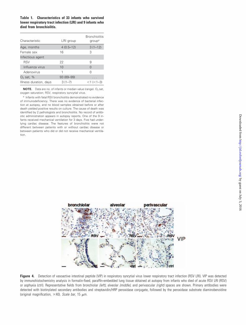
Figure 4. Detection of vasoactive intestinal peptide (VIP) in respiratory syncytial virus lower respiratory tract infection (RSV LRI). VIP was detectedby immunohistochemistry analysis in formalin-fixed, paraffin-embedded lung tissue obtained at autopsy from infants who died of acute RSV LRI (RSV)or asphyxia (ctrl). Representative fields from bronchiolar (left), alveolar (middle), and perivascular (right) spaces are shown. Primary antibodies weredetected with biotinylated secondary antibodies and streptavidin/HRP peroxidase conjugate, followed by the peroxidase substrate diaminobenzidine(original magnification, �40). Scale bar, 15 �m.
Table 1. Characteristics of 33 infants who survivedlower respiratory tract infection (LRI) and 9 infants whodied from bronchiolitis.
Characteristic LRI groupBronchiolitis
groupa
Age, months 4 (0.5–12) 3 (1–12)Female sex 16 3Infectious agent
RSV 22 9Influenza virus 10 0Adenovirus 1 0
O2 sat, % 93 (89–99) . . .Illness duration, days 3 (1–7) �1 (�1–3)
NOTE. Data are no. of infants or median value (range). O2 sat,oxygen saturation; RSV, respiratory syncytial virus.
a Infants with fatal RSV bronchiolitis demonstrated no evidenceof immunodeficiency. There was no evidence of bacterial infec-tion at autopsy, and no blood samples obtained before or afterdeath yielded positive results on culture. The cause of death wasidentified by 2 pathologists and bronchiolitis. No record of antibi-otic administration appears in autopsy reports. One of the 9 in-fants received mechanical ventilation for 3 days. Five had under-lying cardiac disease. The features of bronchiolitis were notdifferent between patients with or without cardiac disease orbetween patients who did or did not receive mechanical ventila-tion.
by guest on July 3, 2016http://jid.oxfordjournals.org/
Dow
nloaded from
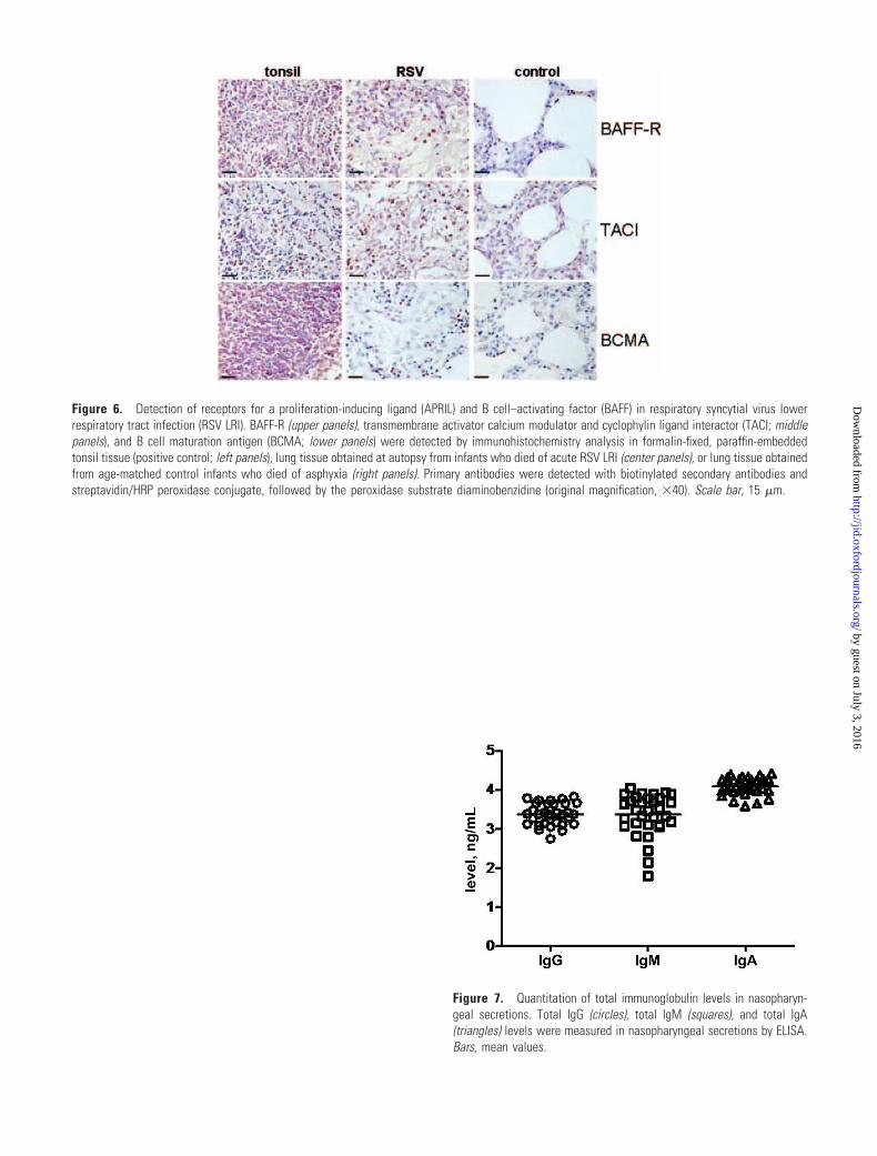
Figure 6. Detection of receptors for a proliferation-inducing ligand (APRIL) and B cell–activating factor (BAFF) in respiratory syncytial virus lowerrespiratory tract infection (RSV LRI). BAFF-R (upper panels), transmembrane activator calcium modulator and cyclophylin ligand interactor (TACI; middlepanels), and B cell maturation antigen (BCMA; lower panels) were detected by immunohistochemistry analysis in formalin-fixed, paraffin-embeddedtonsil tissue (positive control; left panels), lung tissue obtained at autopsy from infants who died of acute RSV LRI (center panels), or lung tissue obtainedfrom age-matched control infants who died of asphyxia (right panels). Primary antibodies were detected with biotinylated secondary antibodies andstreptavidin/HRP peroxidase conjugate, followed by the peroxidase substrate diaminobenzidine (original magnification, �40). Scale bar, 15 �m.
Figure 7. Quantitation of total immunoglobulin levels in nasopharyn-geal secretions. Total IgG (circles), total IgM (squares), and total IgA(triangles) levels were measured in nasopharyngeal secretions by ELISA.Bars, mean values.
by guest on July 3, 2016http://jid.oxfordjournals.org/
Dow
nloaded from
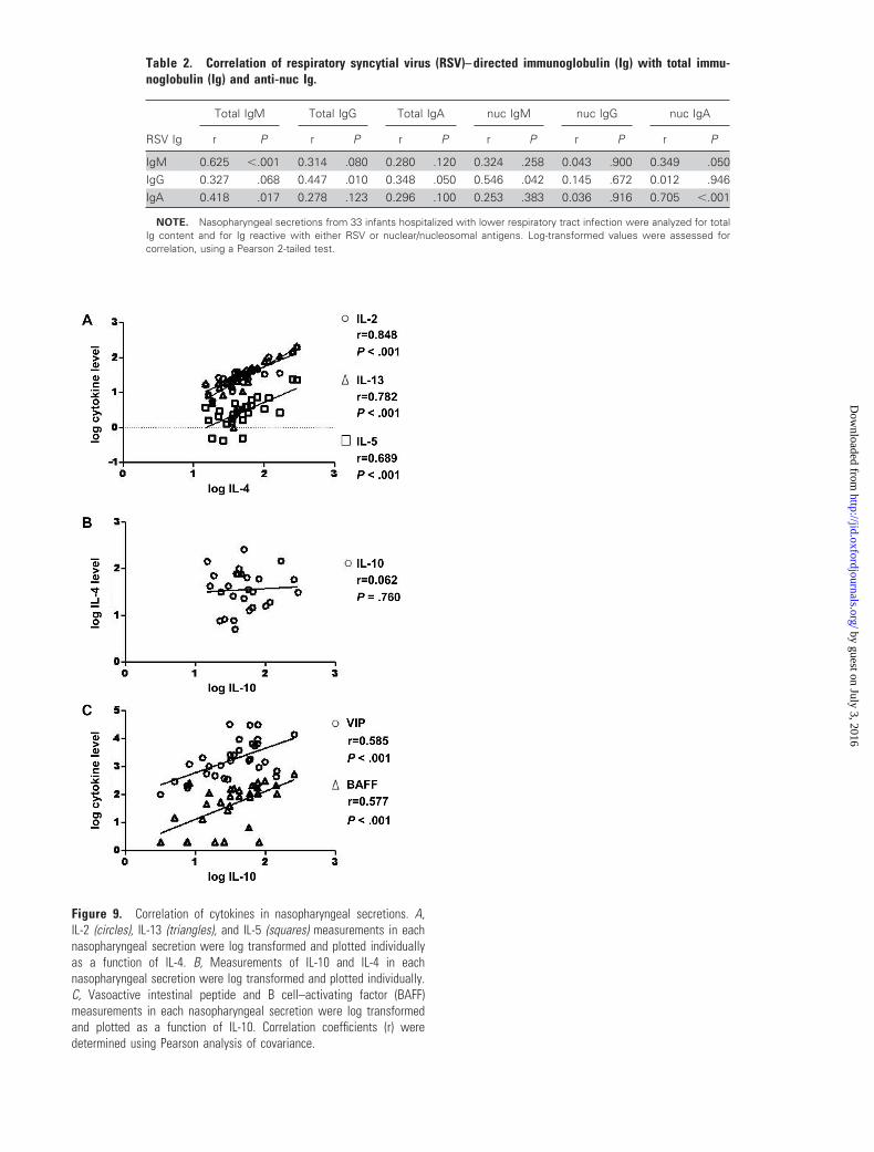
Figure 9. Correlation of cytokines in nasopharyngeal secretions. A,IL-2 (circles), IL-13 (triangles), and IL-5 (squares) measurements in eachnasopharyngeal secretion were log transformed and plotted individuallyas a function of IL-4. B, Measurements of IL-10 and IL-4 in eachnasopharyngeal secretion were log transformed and plotted individually.C, Vasoactive intestinal peptide and B cell–activating factor (BAFF)measurements in each nasopharyngeal secretion were log transformedand plotted as a function of IL-10. Correlation coefficients (r) weredetermined using Pearson analysis of covariance.
Table 2. Correlation of respiratory syncytial virus (RSV)– directed immunoglobulin (Ig) with total immu-noglobulin (Ig) and anti-nuc Ig.
Total IgM Total IgG Total IgA nuc IgM nuc IgG nuc IgA
RSV Ig r P r P r P r P r P r P
IgM 0.625 �.001 0.314 .080 0.280 .120 0.324 .258 0.043 .900 0.349 .050IgG 0.327 .068 0.447 .010 0.348 .050 0.546 .042 0.145 .672 0.012 .946IgA 0.418 .017 0.278 .123 0.296 .100 0.253 .383 0.036 .916 0.705 �.001
NOTE. Nasopharyngeal secretions from 33 infants hospitalized with lower respiratory tract infection were analyzed for totalIg content and for Ig reactive with either RSV or nuclear/nucleosomal antigens. Log-transformed values were assessed forcorrelation, using a Pearson 2-tailed test.
by guest on July 3, 2016http://jid.oxfordjournals.org/
Dow
nloaded from
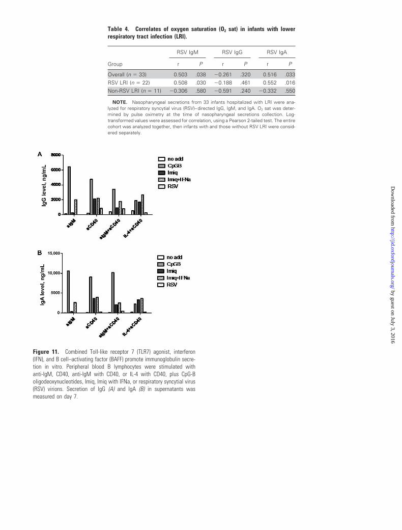
Figure 11. Combined Toll-like receptor 7 (TLR7) agonist, interferon(IFN), and B cell–activating factor (BAFF) promote immunoglobulin secre-tion in vitro. Peripheral blood B lymphocytes were stimulated withanti-IgM, CD40, anti-IgM with CD40, or IL-4 with CD40, plus CpG-Boligodeoxynucleotides, Imiq, Imiq with IFNa, or respiratory syncytial virus(RSV) virions. Secretion of IgG (A) and IgA (B) in supernatants wasmeasured on day 7.
Table 4. Correlates of oxygen saturation (O2 sat) in infants with lowerrespiratory tract infection (LRI).
RSV IgM RSV IgG RSV IgA
Group r P r P r P
Overall (n � 33) 0.503 .038 �0.261 .320 0.516 .033RSV LRI (n � 22) 0.508 .030 �0.188 .461 0.552 .016Non-RSV LRI (n � 11) �0.306 .580 �0.591 .240 �0.332 .550
NOTE. Nasopharyngeal secretions from 33 infants hospitalized with LRI were ana-lyzed for respiratory syncytial virus (RSV)–directed IgG, IgM, and IgA. O2 sat was deter-mined by pulse oximetry at the time of nasopharyngeal secretions collection. Log-transformed values were assessed for correlation, using a Pearson 2-tailed test. The entirecohort was analyzed together, then infants with and those without RSV LRI were consid-ered separately.
by guest on July 3, 2016http://jid.oxfordjournals.org/
Dow
nloaded from
Copyright © 2022 FDOKUMEN