Cricket paralysis virus antagonizes Argonaute 2 to modulate antiviral defense in Drosophila
9
ARTICLES NATURE STRUCTURAL & MOLECULAR BIOLOGY VOLUME 17 NUMBER 5 MAY 2010 547 RNA interference (RNAi) is a highly conserved, post-transcriptional gene-expression control mechanism involved in a number of criti- cal cellular processes 1 . In insects and plants, RNAi functions as a major antiviral defense mechanism 2 . In Drosophila, Dicer2, a class III double-stranded RNA (dsRNA) endonuclease, processes viral dsRNA structures into small interfering RNAs (siRNAs). siRNAs assemble into RNA-induced silencing complexes (RISCs) to serve as guides for the endonuclease Ago2, which targets the viral RNA for degradation. As a countermeasure to RNAi antiviral defense, viruses encode suppressors of RNA silencing (VSR) to modulate host antiviral response. The study of viral RNAi suppressors has provided insights into the mechanistic details of siRNA and miRNA production as well as RISC assembly. A number of factors, including helicases, dsRNA-binding proteins and endonucleases, are required for the proper assembly of functional RISCs 3,4 . Accordingly, viral suppressors can potentially target any of these factors to circumvent the host RNAi defense. Intriguingly, most RNAi suppressors are RNA-binding proteins that interact with dsRNA precursors and/or siRNAs to inhibit dsRNA processing by Dicer and assembly of RISC 5 . However, some plant viral RNAi suppressors also target components of the RNAi machin- ery itself. For instance, P0 suppressor protein of the plant polerovirus inhibits downstream events in RISC assembly by promoting Ago1 degradation 6–8 . Another plant virus RNAi suppressor, cucumber mosaic virus (CMV) 2b, inhibits both the siRNA and miRNA path- ways by blocking Ago1 cleavage activity 9 . Furthermore, inhibition of RNA silencing by most plant viral suppressors also affects miRNA function, causing developmental abnormalities that may contribute to the pathogenic consequences of infection 9,10 . Several insect viruses also encode RNAi suppressors 2,11 . Drosophila C virus (DCV) is a positive-strand RNA virus within the Dicistroviridae family that infects many strains of D. melanogaster and in nature establishes a nonlethal persistent infection 12–17 . A closely related dicistrovirus, cricket paralysis virus (CrPV), however, produces lethal infections in field crickets 18 and fruit flies 19 . The viral RNA genomes are comprised of two distinct open reading frames, termed ORF1 and ORF2, and the expression of both is determined by internal ribosome entry site (IRES)-mediated translational initiation 20 . ORF1 encodes the nonstructural replication proteins, whereas ORF2 encodes the structural proteins that form the viral capsid. Experimental infection by DCV and CrPV is greatly exacerbated in flies with genetic defects abrogating the RNAi effector proteins Dicer2 and Ago2, indicating that RNAi is a bona fide antiviral defense mechanism in insects 21–23 . The different outcomes of DCV and CrPV infections in nature led us to examine whether these closely related viruses use distinct strategies to control the host antiviral response. We previously reported that DCV encodes a dsRNA-binding protein, DCV-1A, that suppresses RNA silencing by specifically blocking Dicer2 processing of virus dsRNA into siRNAs 22 . In contrast, the mechanism used by CrPV to counteract the Drosophila immune system has not been established. Here we show that CrPV encodes a potent VSR, CrPV-1A, that inter- acts with Ago2 and inhibits RISC activity. Furthermore, unlike plant virus RNAi suppressors, this hitherto undescribed RNAi inhibitor neither affects the miRNA pathway nor alters the normal development 1 Department of Microbiology and Immunology, University of California, San Francisco, California, USA. 2 Institut Pasteur, Drosophila Genetics and Epigenetics, Centre National de la Recherche Scientifique, URA 2578, Paris, France. 3 Department of Pharmaceutical Chemistry, University of California, San Francisco, California, USA. Correspondence should be addressed to R.A. ([email protected]). Received 8 May 2009; accepted 19 March 2010; published online 18 April 2010; doi:10.1038/nsmb.1810 Cricket paralysis virus antagonizes Argonaute 2 to modulate antiviral defense in Drosophila Arabinda Nayak 1 , Bassam Berry 2 , Michel Tassetto 1 , Mark Kunitomi 1 , Ashley Acevedo 1 , Changhui Deng 3 , Andrew Krutchinsky 3 , John Gross 3 , Christophe Antoniewski 2 & Raul Andino 1 Insect viruses have evolved strategies to control the host RNAi antiviral defense mechanism. In nature, Drosophila melanogaster C virus (DCV) infection causes low mortality and persistent infection, whereas the closely related cricket paralysis virus (CrPV) causes a lethal infection. We show that these viruses use different strategies to modulate the host RNAi defense machinery. The DCV RNAi suppressor (DCV-1A) binds to long double-stranded RNA and prevents processing by Dicer2. In contrast, the CrPV suppressor (CrPV-1A) interacts with the endonuclease Argonaute 2 (Ago2) and inhibits its activity without affecting the microRNA (miRNA)-Ago1–mediated silencing. We examined the link between viral RNAi suppressors and the outcome of infection using recombinant Sindbis viruses encoding either CrPV-1A or DCV-1A. Flies infected with Sindbis virus expressing CrPV-1A showed a marked increase in virus production, spread and mortality. In contrast, Sindbis pathogenesis was only modestly increased by expression of DCV- 1A. We conclude that RNAi suppressors function as virulence factors in insects and can target the Drosophila RNAi pathway at different points. © 2010 Nature America, Inc. All rights reserved.
Transcript of Cricket paralysis virus antagonizes Argonaute 2 to modulate antiviral defense in Drosophila
A r t i c l e s
nAture structurAl & moleculAr biology VOLUME 17 NUMBER 5 MAY 2010 547
RNA interference (RNAi) is a highly conserved, post-transcriptional gene-expression control mechanism involved in a number of criti-cal cellular processes1. In insects and plants, RNAi functions as a major antiviral defense mechanism2. In Drosophila, Dicer2, a class III double-stranded RNA (dsRNA) endonuclease, processes viral dsRNA structures into small interfering RNAs (siRNAs). siRNAs assemble into RNA-induced silencing complexes (RISCs) to serve as guides for the endonuclease Ago2, which targets the viral RNA for degradation. As a countermeasure to RNAi antiviral defense, viruses encode suppressors of RNA silencing (VSR) to modulate host antiviral response.
The study of viral RNAi suppressors has provided insights into the mechanistic details of siRNA and miRNA production as well as RISC assembly. A number of factors, including helicases, dsRNA-binding proteins and endonucleases, are required for the proper assembly of functional RISCs3,4. Accordingly, viral suppressors can potentially target any of these factors to circumvent the host RNAi defense. Intriguingly, most RNAi suppressors are RNA-binding proteins that interact with dsRNA precursors and/or siRNAs to inhibit dsRNA processing by Dicer and assembly of RISC5. However, some plant viral RNAi suppressors also target components of the RNAi machin-ery itself. For instance, P0 suppressor protein of the plant polerovirus inhibits downstream events in RISC assembly by promoting Ago1 degradation6–8. Another plant virus RNAi suppressor, cucumber mosaic virus (CMV) 2b, inhibits both the siRNA and miRNA path-ways by blocking Ago1 cleavage activity9. Furthermore, inhibition of RNA silencing by most plant viral suppressors also affects miRNA
function, causing developmental abnormalities that may contribute to the pathogenic consequences of infection9,10.
Several insect viruses also encode RNAi suppressors2,11. Drosophila C virus (DCV) is a positive-strand RNA virus within the Dicistroviridae family that infects many strains of D. melanogaster and in nature establishes a nonlethal persistent infection12–17. A closely related dicistrovirus, cricket paralysis virus (CrPV), however, produces lethal infections in field crickets18 and fruit flies19. The viral RNA genomes are comprised of two distinct open reading frames, termed ORF1 and ORF2, and the expression of both is determined by internal ribosome entry site (IRES)-mediated translational initiation20. ORF1 encodes the nonstructural replication proteins, whereas ORF2 encodes the structural proteins that form the viral capsid. Experimental infection by DCV and CrPV is greatly exacerbated in flies with genetic defects abrogating the RNAi effector proteins Dicer2 and Ago2, indicating that RNAi is a bona fide antiviral defense mechanism in insects21–23. The different outcomes of DCV and CrPV infections in nature led us to examine whether these closely related viruses use distinct strategies to control the host antiviral response. We previously reported that DCV encodes a dsRNA-binding protein, DCV-1A, that suppresses RNA silencing by specifically blocking Dicer2 processing of virus dsRNA into siRNAs22. In contrast, the mechanism used by CrPV to counteract the Drosophila immune system has not been established.
Here we show that CrPV encodes a potent VSR, CrPV-1A, that inter-acts with Ago2 and inhibits RISC activity. Furthermore, unlike plant virus RNAi suppressors, this hitherto undescribed RNAi inhibitor neither affects the miRNA pathway nor alters the normal development
1Department of Microbiology and Immunology, University of California, San Francisco, California, USA. 2Institut Pasteur, Drosophila Genetics and Epigenetics, Centre National de la Recherche Scientifique, URA 2578, Paris, France. 3Department of Pharmaceutical Chemistry, University of California, San Francisco, California, USA. Correspondence should be addressed to R.A. ([email protected]).
Received 8 May 2009; accepted 19 March 2010; published online 18 April 2010; doi:10.1038/nsmb.1810
Cricket paralysis virus antagonizes Argonaute 2 to modulate antiviral defense in DrosophilaArabinda Nayak1, Bassam Berry2, Michel Tassetto1, Mark Kunitomi1, Ashley Acevedo1, Changhui Deng3, Andrew Krutchinsky3, John Gross3, Christophe Antoniewski2 & Raul Andino1
Insect viruses have evolved strategies to control the host RNAi antiviral defense mechanism. In nature, Drosophila melanogaster C virus (DCV) infection causes low mortality and persistent infection, whereas the closely related cricket paralysis virus (CrPV) causes a lethal infection. We show that these viruses use different strategies to modulate the host RNAi defense machinery. The DCV RNAi suppressor (DCV-1A) binds to long double-stranded RNA and prevents processing by Dicer2. In contrast, the CrPV suppressor (CrPV-1A) interacts with the endonuclease Argonaute 2 (Ago2) and inhibits its activity without affecting the microRNA (miRNA)-Ago1–mediated silencing. We examined the link between viral RNAi suppressors and the outcome of infection using recombinant Sindbis viruses encoding either CrPV-1A or DCV-1A. Flies infected with Sindbis virus expressing CrPV-1A showed a marked increase in virus production, spread and mortality. In contrast, Sindbis pathogenesis was only modestly increased by expression of DCV- 1A. We conclude that RNAi suppressors function as virulence factors in insects and can target the Drosophila RNAi pathway at different points.
© 2
010
Nat
ure
Am
eric
a, In
c. A
ll ri
gh
ts r
eser
ved
.

548 VOLUME 17 NUMBER 5 MAY 2010 nAture structurAl & moleculAr biology
A r t i c l e s
and physiology of the animal. Given the distinct pathogenic outcomes observed in DCV and CrPV infections in nature, we hypothesized that RNAi suppressors may function as virulence factors. Indeed, we found that CrPV and DCV RNAi suppressors can modulate the outcome of Sindbis virus infection in flies. Recombinant Sindbis virus express-ing CrPV-1A increased virus production, resulting in high mortality, whereas DCV-1A expression resulted in only a modest enhancement of infection. We propose that insect virus RNAi suppressors are key modulators of the host immune response, fine tuning the outcome of infection in line with the evolutionary viral strategy for successful host transmission, and that the suppressors target multiple components of the RNAi pathway in insects as in plants, including Argonaute.
RESULTSCrPV infection blocks RNAi in S2 cellsDrosophila deficient in the key RNAi endonucleases Dicer2 and Ago2 are highly susceptible to CrPV infection, suggesting that the fly RNAi machinery plays an essential role in antiviral defense21–23. Therefore, we examined the possibility that CrPV may encode a suppressor of RNAi to control the RNA-silencing machinery. We analyzed RNAi suppression in S2 cells using a dual luciferase reporter system con-sisting of a firefly luciferase (FLuc)-expressing plasmid and a specific 200-nucleotide (nt) dsRNA targeting the firefly luciferase mRNA or an enhanced green fluorescent protein (eGFP) dsRNA control (Ctrl)22 (Fig. 1a). Drosophila S2 cells were either CrPV- or mock-infected for 24 h before co-transfection with the reporter system for RNAi-silencing activity. We also included an internal control using Renilla reniformis luciferase (RLuc) in each experiment. In uninfected control cells, we observed efficient silencing of firefly luciferase (suppression by a factor of 380) as compared to a control dsRNA. In contrast, silencing was completely suppressed in CrPV infected cells (Fig. 1a), indicating that CrPV encodes a potent suppressor of RNAi.
CrPV ORF1 encodes an RNAi suppressorDCV and CrPV are closely related species within the Cripavirus genus of the Dicistroviridae family. Residue sequence alignment between DCV and CrPV indicated a high degree (~55%) of identity within ORF1. A dsRNA-binding motif (DSRM) was previously identified within the N-terminal 99-residue region of DCV ORF1, spanning residues 25–88, and was shown to encode a potent suppressor of RNAi
(DCV-1A)22. Based on these observations, we set out to determine whether the suppression of RNAi by CrPV could be attributed to a DCV-1A–like RNAi suppressor that mapped to the N terminus of CrPV ORF1.
Alignment of DCV and CrPV in this region revealed no sub-stantial homology compared to homologous downstream resi-due sequences, a group that includes a highly conserved octamer sequence DVEXNPGP (Fig. 1b). This octamer sequence serves as a signal for co-translational protein cleavage between the glycine and proline residues in several picornaviruses24. We hypothesized that this DVEXNPGP sequence may have a similar function during DCV and CrPV polyprotein processing. We previously reported a DSRM within sequences upstream of the DVEXNPGP site that functions as a potent RNAi suppressor protein in DCV22. We detected a protein with a molecular weight consistent with cleavage at the DVEXNPGP site in CrPV-infected cells by western blot analysis using a polyclonal antibody raised against the first 148 N-terminal residues of CrPV ORF1 (Fig. 1c).
Next, we evaluated the ability of the CrPV sequence upstream of the DVEXNPGP motif to suppress RNAi in S2 cells. Using the dual-reporter assay, we co-transfected luciferase (Fluc and Rluc)-expression plasmids with dsRNA that specifically targets firefly luciferase and plasmids encoding C-terminal deletions of the putative CrPV RNAi suppressor protein (N-terminal 168, 148, 128 or 108 residues). Both CrPV-1A168 and CrPV-1A148 efficiently blocked RNA silencing (Fig. 1d). In contrast, CrPV-1A128 and CrPV-1A108 expression did not suppress RNAi (Fig. 1d). Hence, the N-terminal 148 residues of CrPV ORF1 are sufficient to block RNAi in S2 cells. To examine the effects of CrPV-1A suppressor in vivo, we generated transgenic flies expressing the CrPV-1A148 protein, the inactive CrPV-1A108 fragment or GFP. To monitor the effects of the CrPV-1A148 suppressor in vivo, we crossed transgenic flies with flies expressing a hairpin dsRNA (inverted repeat) targeting the white gene (IR[white]). The expres-sion of two mini-white genes would be suppressed by expression of this hairpin dsRNA, thus providing a colorimetric readout for RNA silencing. The white gene was efficiently silenced in flies expressing
aS2 cells± CrPV
TransfectionreportersdsRNA
Induction Lucassay
24 h 24 h 16 h
400
300
200
Sile
ncin
g (f
old
tods
RN
A c
ontr
ol)
100
0dsRNA Luc
Uninfected
IRES 1 IRES 2CrPV genome
Nonstructural StructuralVSR
DCV:CrPV:
5360
DCV:CrPV:
DCV:CrPV:
1A128
20 kDa
15 kDa
CrPV
Unif. 48 h
α–CrPV-1A
24 h
1A148
800d
c
1A168
1A108111180
73120
b
CrPV infected
Ctrl Luc Ctrl
600
400
Sile
ncin
g (f
old
tods
RN
A c
ontr
ol)
200
0
1A10
8
1A12
8
1A14
8
1A16
8
Figure 1 CrPV antagonizes RNAi in S2 cells. (a) CrPV-infected S2 cells or uninfected S2 cells were co-transfected with reporter plasmids encoding firefly and Renilla luciferase and either dsRNA targeting the firefly luciferase (Luc) or eGFP dsRNA control (Ctrl). Silencing efficiency in S2 cells was monitored by comparing the ratio of firefly to Renilla luciferase expression. (b) CrPV genome encodes both nonstructural and structural proteins whose translation is regulated by IRES1 and IRES2, respectively. The extended broken line (−) represents alignment of the VSR domains that include the N-terminal 180 residues of the CrPV ORF1 and the N-terminal 111 residues of the DCV ORF1. - indicates gap; *, complete homology; red open box, the conserved octameric sequence; ↓, nonproteolytic cleavage site; ∆, sites of deletion in the VSR region. (c) S2 cells were either mock (uninfected, Unif.) or CrPV infected (at a multiplicity of infection (MOI) of 1) for 24 h and 48 h. Harvested samples were analyzed for 1A expression by western blot analysis using rabbit polyclonal antibody raised against the suppressor protein (α–CrPV-1A). (d) Plasmids encoding C-terminal residue deletions of the putative CrPV suppressor domain (1A168, 1A148, 1A128 and 1A108) were co-transfected with a dual-luciferase reporter system along with Luc dsRNA targeting firefly luciferase or eGFP dsRNA control as described in a. Silencing was represented as fold silencing compared to the control dsRNA. Data in a and d indicate averages and s.d. of four independent experiments.
© 2
010
Nat
ure
Am
eric
a, In
c. A
ll ri
gh
ts r
eser
ved
.
nAture structurAl & moleculAr biology VOLUME 17 NUMBER 5 MAY 2010 549
A r t i c l e s
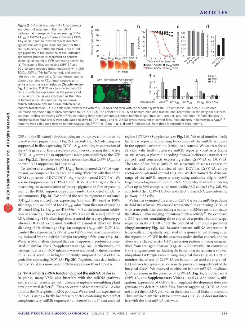
GFP and the IR[white] hairpin, causing an orange eye color due to the loss of red eye pigmentation (Fig. 2a). In contrast, RNA silencing was suppressed in flies expressing CrPV-1A148, resulting in expression of the white gene and, thus, a red eye color. Flies expressing the inactive CrPV-1A108 were able to suppress the white gene similarly to the GFP flies (Fig. 2a). Therefore, our observations show that CrPV-1A148 is a potent RNAi suppressor in Drosophila.
To further characterize CrPV-1A148 (herein named CrPV-1A) sup-pressor, we compared its RNAi-suppressing efficiency with that of the RNAi suppressor of DCV, DCV-1A99 (herein named DCV-1A). We assessed suppression by CrPV-1A and DCV-1A in transgenic flies by measuring the accumulation of red eye pigments in flies expressing each of the RNAi suppressor proteins under the control of identi-cal promoters (Fig. 2a). We defined the red eye pigment absorbance (OD480) from control flies expressing GFP and IR[white] as 100% silencing, and we defined the OD480 value from flies not expressing IR[white] (Fig. 2a, 1A148, no IR [white] (−)) as the maximum inhibi-tion of silencing. Flies expressing CrPV-1A and IR[white] inhibited RNA silencing (14% silencing) thus restored the red eye phenotype, whereas DCV-1A expression resulted in a modest effect on RNA silencing (50% silencing) (Fig. 2a; compare 1A148 with DCV-1A). Control flies expressing CrPV-1A108 or GFP showed maximum silenc-ing induced by the dsRNA hairpin targeting white gene (Fig. 2a). Western blot analysis showed that each suppressor protein accumu-lated to similar levels (Supplementary Fig. 1a). Furthermore, the pathogenic effect of DCV infection was exacerbated by the expression of CrPV-1A, resulting in higher mortality compared to that of trans-genic flies expressing DCV-1A (Fig. 2b). Together, these data indicate that CrPV-1A is a more potent RNAi suppressor than DCV-1A.
CrPV-1A inhibits siRNA function but not the miRNA pathwayIn plants, many VSRs also interfere with the miRNA pathway and are often associated with disease symptoms resembling plant developmental defects25. Thus, we examined whether CrPV-1A also inhibits the Drosophila miRNA pathway. We carried out experiments in S2 cells using a firefly luciferase reporter containing two perfect complementary miR2b sequences (antisense) in its 3′ untranslated
region (UTR)22 (Supplementary Fig. 1b). We used another firefly luciferase reporter containing two copies of the miR2b sequence in the opposite orientation (sense) as a control. We co-transfected S2 cells with firefly luciferase miR2b reporter constructs (sense or antisense), a plasmid encoding Renilla luciferase (transfection control) and constructs expressing either CrPV-1A or DCV-1A. The ratio of luciferase (miR2b antisense/miR2b sense) expression was identical in cells transfected with DCV-1A, CrPV-1A, empty vector or no-plasmid control (Fig. 2c). We determined the dynamic range of the miR2b reporter assay using antisense oligos (ASO) targeting endogenous miR2b miRNA. ASO reduced miR2b silencing effect up to 50% compared to nonspecific ASO control (Fig. 2d). We concluded that CrPV-1A does not affect the miRNA gene silencing pathway in S2 cells.
We further examined the effect of CrPV-1A on the miRNA pathway in third instar larvae. We crossed transgenic flies expressing CrPV-1A with transgenic flies containing a bantam miRNA sensor construct that allows in vivo imaging of bantam miRNA activity26. We expressed a GFP reporter containing three copies of a perfect bantam target sequence in its 3′ UTR under the control of the tubulin promoter (Supplementary Fig. 1c). Because bantam miRNA expression is temporally and spatially regulated in response to patterning cues, the expression of GFP in this case was under similar control, and we observed a characteristic GFP expression pattern in wing imaginal discs from transgenic larvae (Fig. 2e, GFP/bantam). In contrast, a GFP transgene construct lacking the bantam target sequences showed ubiquitous GFP expression in wing imaginal discs (Fig. 2e, GFP). To monitor the effects of CrPV-1A on bantam, we used an engrailed-GAL4 driver to express CrPV-1A in the posterior compartment of the imaginal discs27. We observed no effect on bantam miRNA–mediated GFP repression in the presence of CrPV-1A (Fig. 2e, GFP/bantam + CrPV-1A, and Supplementary Videos 1 and 2). Additionally, ubi-quitous expression of CrPV-1A throughout development does not generate any defect in adult flies, further suggesting CrPV-1A does not affect the miRNA pathway in the intact animal (data not shown). Thus, unlike plant virus RNAi suppressors, CrPV-1A does not inter-fere with the host miRNA pathway.
Figure 2 CrPV-1A is a potent RNAi suppressor and does not interfere in the microRNA pathway. (a) Transgenic flies expressing CrPV-1A148 or CrPV-1A108 or those expressing DCV-1A99or GFP and an inverted repeat directed against the white gene were analyzed for their ability to carry out efficient RNAi. Loss of red eye pigments in the presence of the indicated suppressor proteins is expressed as percent silencing compared to GFP expressing control fly. (b) Transgenic flies expressing CrPV-1A and DCV-1A were injected intrathoracically with 100 TCID50 DCV or Tris buffer control, and survival rate was monitored daily. (c) Luciferase reporter plasmid carrying miR2b target sequences in sense and antisense orientation (Supplementary Fig. 1b) in the 3′ UTR was transfected into S2 cells. Luciferase expression in the presence of CrPV-1A or DCV-1A was expressed as the ratio of luciferase counts produced by luciferase miR2b antisense over luciferase miR2b sense reporter transfection. (d) S2 cells were transfected with miR-2b ASO and then with the reporter system (miR2b antisense). miR-2b ASO restored luciferase expression up to 50% compared to Ctrl ASO. (e) The effect of CrPV-1A on bantam-mediated translational repression in the imaginal disc was analyzed in flies expressing GFP mRNA containing three complementary bantam miRNA target sites. Ant, anterior; pos, posterior. (f) Fold changes in retrotransposon RNA levels were calculated relative to 297, mdg1 and 412 RNA levels measured in control flies. Fold changes in homozygous Ago2414 mutants were calculated relative to heterozygous Ago2414 flies. Data in a, c, d and f indicate s.d. from three independent experiments.
100a
80
40
60
Sile
ncin
g (%
to c
ontr
ol)
20
01A148 1A148 1A108
+IR [white]–
GFP DCV-1A
100
b
eGFP
i ii iiiAnt
–CrPV148
+CrPV148
Pos
GFP/bantam GFP/bantam+ CrPV-1A
80
40
60
Sur
viva
l (%
)
20
00 2 4
Time after inoculation (d)DCV-1A/bufferDCV-1A/DCV
1A148/buffer1A148/DCV
6 8 10 12
0.6
c
0.5
0.3
0.4
Ant
isen
se/s
ense
rat
io
0.2
0
Vecto
r
CrPV-
1A
DCV-1A –
0.1
1.0
d
0.8
0.4
0.6
Ant
isen
se/s
ense
rat
io
0.2
0
Ctrl A
SO
miR
2b A
SO
f 8
4
6
Fol
d ch
ange
2
0Transgenic fly Ago2414 CrPV148 CrPV108 DCV99 DCV K73A
297
412mdg1
© 2
010
Nat
ure
Am
eric
a, In
c. A
ll ri
gh
ts r
eser
ved
.

550 VOLUME 17 NUMBER 5 MAY 2010 nAture structurAl & moleculAr biology
A r t i c l e s
S2 extracts± suppressors
S2 extracts± suppressors
± siRNA
10 mina b
c
3 hEmbryo extract± suppressors
10 min
Urea-PAGE
S2 extract
CrPV-1A
BSA
Ctrl siRNALuc siRNA
5′ product
mRNAsubstrate
Slicing(Ago2)
DCV-1A
S2 extract/CrPvS2 extract/DCV
+
–
––
––
+
–
––
––
+
–
––
––
–
–
––
+–
–
–
––
++–
+
+
––
––
+
++
––
––
+
+++
––
––
+
–
+++–
––
+
–
––
–+
–
–
–
– + – – – – – – – – –– – + + + + + + + + +
+++
––
3 h10 min
20 min
EMSAUrea-PAGE32P-dsRNA
32P-dsRNA
32 P-siR
NA
dsRNA
siRNA
1
1
1
2
2
2
3
3
3
4
4
4
5
5
6
6 7 8 10 119
R2
R1*
R3
Embryo extract
Assembly
32P-siRNA
32P-mRNA
S2 extractCrPV-1A CrPV-1ADCV-1A
DCV-1A
BSA
––––
+–––
++––
+––+
+–+– BSA
+
––
–
+
–+
–
+
+–
–
+
––
+
Dicing(Dicer2)
Recently a new class of small RNA called endogenous small inter-fering RNA (esiRNAs) has been described in Drosophila28–30. The esiRNAs derive from repetitive sequence elements such as retrotrans-posons and control their replication in somatic cells. To examine whether CrPV-1A perturbs the esiRNA pathway, we analyzed steady-state levels of three distinct retrotransposons, 297, mdg1 and 412 (refs. 28,29), in the heads of adult female transgenic flies expressing functional RNAi suppressor CrPV-1A148 or DCV-1A99 as well as those of control flies expressing nonfunctional suppressor CrPV-1A108 or dsRNA binding mutant DCV-1A K73A (ref. 22). We used homozygous Ago2414 mutant flies as positive control (Fig. 2f). Expression of sup-pressor DCV-1A and CrPV-1A but not suppressor mutant controls substantially increased retrotransposon expression levels, indicating that the virus suppressors also inhibit the esiRNA pathway.
CrPV-1A blocks Ago2 cleavage activityWe next examined the mechanism by which CrPV-1A inhibits RNAi. Because most RNAi suppressors bind dsRNA to inhibit silencing5, we first determined whether CrPV-1A binds dsRNA. We performed electrophoretic mobility-shift experiments using radiolabeled dsRNA (200 base pairs (bp)) or siRNA (21 bp) probes and purified recom-binant CrPV-1A or DCV-1A (Supplementary Fig. 2a). Consistent with our previous findings22, DCV-1A efficiently bound long dsRNA (Supplementary Fig. 2a, lane 1, left) and, to a lesser extent, siRNA (Supplementary Fig. 2a, lane 1, right). In contrast, CrPV-1A did not bind dsRNA or siRNA (Supplementary Fig. 2a, lanes 2–7 and 9–14). Next, we examined the effect of CrPV-1A on Dicer2 activity using an in vitro assay. In S2 cell extracts, dsRNA was efficiently processed to 21-bp siRNAs (Fig. 3a, lane 2). Production of siRNA was inhibited by the addition of recombinant DCV-1A (Fig. 3a, lane 5) but not recom-binant CrPV-1A or bovine serum albumin (BSA) control (Fig. 3a, lanes 3 and 4). These experiments indicated that CrPV-1A inhibits RNAi at a different step than does DCV-1A, functioning downstream of siRNA production. Indeed, CrPV-1A, unlike DCV-1A, effectively blocked silencing induced by transfection of both long dsRNA in S2 cells as well as 21-bp siRNA (Supplementary Fig. 2b), supporting a role for CrPV-1A downstream of siRNA production.
siRNAs produced by Dicer2 are incorporated into the RISC to guide mRNA cleavage. Three siRNA nucleoprotein (siRNP) com-plexes occurring during this assembly process can be resolved by native gel electrophoresis, including an initiator R1 (R2D2–Dcr2
initiator complex (RDI)), an intermediate R2 (RISC loading complex (RLC)) and an effector R3 (holo-RISC) complex31–33. We thus used native gel analysis to examine whether CrPV-1A affects RISC assem-bly. Incubation of radiolabeled siRNA duplex in Drosophila embryo extracts resulted in formation of the R1, R2 and R3 complexes (Fig. 3b, lane 1). The presence of CrPV-1A did not affect the assembly of these three complexes (Fig. 3b, lane 3). In contrast, addition of DCV-1A inhibited formation of R3 (Fig. 3b, lane 2), yielding two inter-mediate complexes (Fig. 3b, denoted by asterisk). These experiments indicate that CrPV-1A does not affect siRNA loading or R3 assembly. Furthermore, we find that DCV-1A has a hitherto undescribed func-tion affecting RISC assembly in addition to its previously described function of binding long dsRNA, suggesting that DCV-1A acts at several levels to inhibit RNAi function.
Next, we tested whether CrPV-1A affects RISC-mediated mRNA cleavage using an in vitro target-cleavage assay34 (Fig. 3c). We incu-bated S2 extracts, programmed with a FLuc siRNA or a nonspecific control siRNA, with a 592-nt radiolabeled cap FLuc mRNA substrate. In control reactions, the FLuc mRNA target was efficiently cleaved (5′ product) in the presence of a FLuc specific siRNA, as expected (Fig. 3c; compare lane 3 with lanes 1 and 2). Whereas addition of puri-fied DCV-1A had little effect on target cleavage, FLuc mRNA cleavage was inhibited upon addition of purified recombinant CrPV-1A (Fig. 3c, lanes 6, 7 and 8 for CrPV and lane 11 for DCV). Similarly, cleavage was also inhibited in extracts from CrPV-infected S2 cells (Fig. 3c, lanes 4 and 5) but not in DCV-infected S2 cells (Fig. 3c, lane 10). These experiments show that CrPV-1A suppresses RNAi by inhibiting RISC activity without affecting Dicer processing or RISC assembly.
CrPV-1A interacts with Ago2We hypothesized that CrPV-1A inhibits RISC activity by associating with one or more components of holo-RISC. To examine this pos-sibility, we immunoisolated CrPV-1A carrying a C-terminal tandem
Figure 3 CrPV-1A inhibits RISC activity downstream of Dicer processing. (a) Radiolabeled dsRNA substrates (32P-dsRNA) were incubated in S2 buffer control (lane 1) or in S2 extracts (lane 2). Processing of long dsRNA to siRNA by Dicer2 in the presence of recombinant CrPV-1A (0.35 µM, lane 3), DCV-1A (0.35 µM, lane 5) or BSA (0.35 µM, lane 4) was monitored using 12% denaturing gel with an end-labeled 21-bp synthetic siRNA marker. (b) Recombinant suppressor proteins DCV-1A (0.35 µM, lane 2), CrPV-1A (0.35 µM, lane 3) or BSA (0.35 µM, lane 4) were incubated in Drosophila embryo extract for 10 min. 32P-labeled siRNAs duplex were subsequently added to the reaction for another 20 min. siRNP complexes (R1, R2 and R3) were analyzed by electro-phoretic mobility shift assay (EMSA) using 4% native PAGE. (c) Target mRNA cleavage assay was analyzed in the absence of siRNA (lane 1) and in the presence of GAPDH (Ctrl) siRNA (lane 2) or Luc siRNA (lanes 3–11). To analyze the effects of CrPV-1A, assays were carried out using CrPV infected S2 extracts (MOI 1 in lane 4; MOI 2 in lane 5) or by incubating recombinant CrPV-1A with increasing concentration (0.15 µM, lane 6; 0.25 µM, lane 7; 0.35 µM, lane 8) in uninfected S2 extracts. The activity of the DCV-1A was analyzed using infected DCV S2 extracts (MOI 1, lane 10) or by incubating recombinant DCV-1A (0.35 µM, lane 11) in uninfected S2 extracts. The 5′-cleaved products were analyzed by denaturing polyacrylamide gel.
© 2
010
Nat
ure
Am
eric
a, In
c. A
ll ri
gh
ts r
eser
ved
.
nAture structurAl & moleculAr biology VOLUME 17 NUMBER 5 MAY 2010 551
A r t i c l e s
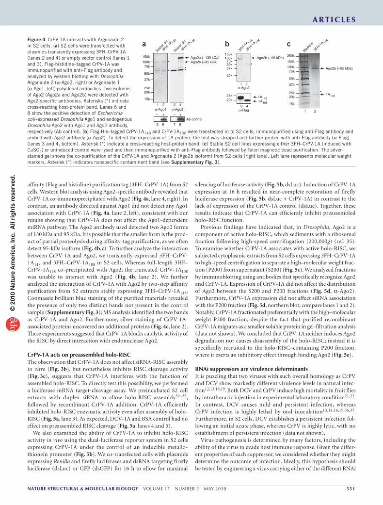
affinity (Flag and histidine) purification tag (3FH–CrPV-1A) from S2 cells. Western blot analysis using Ago2-specific antibody revealed that CrPV-1A co-immunoprecipitated with Ago2 (Fig. 4a, lane 4, right). In contrast, an antibody directed against Ago1 did not detect any Ago1 association with CrPV-1A (Fig. 4a. lane 2, left), consistent with our results showing that CrPV-1A does not affect the Ago1-dependent miRNA pathway. The Ago2 antibody used detected two Ago2 forms of 130 kDa and 95 kDa. It is possible that the smaller form is the prod-uct of partial proteolysis during affinity-tag purification, as we often detect 95-kDa isoform (Fig. 4b,c). To further analyze the interaction between CrPV-1A and Ago2, we transiently expressed 3FH–CrPV-1A148 and 3FH–CrPV-1A108 in S2 cells. Whereas full-length 3HF–CrPV-1A148 co-precipitated with Ago2, the truncated CrPV-1A108 was unable to interact with Ago2 (Fig. 4b, lane 2). We further analyzed the interaction of CrPV-1A with Ago2 by two-step affinity purification from S2 extracts stably expressing 3FH–CrPV-1A148. Coomassie brilliant blue staining of the purified materials revealed the presence of only two distinct bands not present in the control sample (Supplementary Fig. 3); MS analysis identified the two bands as CrPV-1A and Ago2. Furthermore, silver staining of CrPV-1A- associated proteins uncovered no additional proteins (Fig. 4c, lane 2). These experiments suggested that CrPV-1A blocks catalytic activity of the RISC by direct interaction with endonuclease Ago2.
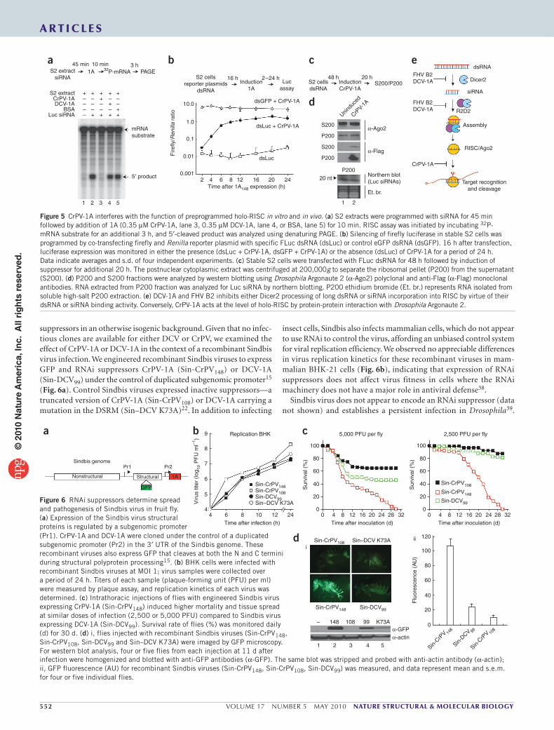
CrPV-1A acts on preassembled holo-RISCThe observation that CrPV-1A does not affect siRNA-RISC assembly in vitro (Fig. 3b), but nonetheless inhibits RISC cleavage activity (Fig. 3c), suggests that CrPV-1A interferes with the function of assembled holo-RISC. To directly test this possibility, we performed a luciferase mRNA target-cleavage assay. We preincubated S2 cell extracts with duplex siRNA to allow holo-RISC assembly31–33, followed by recombinant CrPV-1A addition. CrPV-1A efficiently inhibited holo-RISC enzymatic activity even after assembly of holo-RISC (Fig. 5a, lane 3). As expected, DCV-1A and BSA control had no effect on preassembled RISC cleavage (Fig. 5a, lanes 4 and 5).
We also examined the ability of CrPV-1A to inhibit holo-RISC activity in vivo using the dual-luciferase reporter system in S2 cells expressing CrPV-1A under the control of an inducible metallo-thionein promoter (Fig. 5b). We co-transfected cells with plasmids expressing Renilla and firefly luciferases and dsRNA targeting firefly luciferase (dsLuc) or GFP (dsGFP) for 16 h to allow for maximal
silencing of luciferase activity (Fig. 5b, dsLuc). Induction of CrPV-1A expression at 16 h resulted in near-complete restoration of firefly luciferase expression (Fig. 5b, dsLuc + CrPV-1A) in contrast to the lack of expression of the CrPV-1A control (dsLuc). Together, these results indicate that CrPV-1A can efficiently inhibit preassembled holo-RISC function.
Previous findings have indicated that, in Drosophila, Ago2 is a component of active holo-RISC, which sediments with a ribosomal fraction following high-speed centrifugation (200,000g) (ref. 35). To examine whether CrPV-1A associates with active holo-RISC, we subjected cytoplasmic extracts from S2 cells expressing 3FH–CrPV-1A to high-speed centrifugation to separate a high–molecular weight frac-tion (P200) from supernatant (S200) (Fig. 5c). We analyzed fractions by immunoblotting using antibodies that specifically recognize Ago2 and CrPV-1A. Expression of CrPV-1A did not affect the distribution of Ago2 between the S200 and P200 fractions (Fig. 5d, α-Ago2). Furthermore, CrPV-1A expression did not affect siRNA association with the P200 fraction (Fig. 5d, northern blot; compare lanes 1 and 2). Notably, CrPV-1A fractionated preferentially with the high–molecular weight P200 fraction, despite the fact that purified recombinant CrPV-1A migrates as a smaller soluble protein in gel-filtration analysis (data not shown). We concluded that CrPV-1A neither induces Ago2 degradation nor causes disassembly of the holo-RISC; instead it is specifically recruited to the holo-RISC–containing P200 fraction, where it exerts an inhibitory effect through binding Ago2 (Fig. 5e).
RNAi suppressors are virulence determinantsIt is puzzling that two viruses with such overall homology as CrPV and DCV show markedly different virulence levels in natural infec-tion12,13,18,19. Both DCV and CrPV induce high mortality in fruit flies by intrathoracic injection in experimental laboratory condition21,22. In contrast, DCV causes mild and persistent infection, whereas CrPV infection is highly lethal by oral inoculation13,14,16,19,36,37. Furthermore, in S2 cells, DCV establishes a persistent infection fol-lowing an initial acute phase, whereas CrPV is highly lytic, with no establishment of persistent infection (data not shown).
Virus pathogenesis is determined by many factors, including the ability of the virus to evade host immune response. Given the differ-ent properties of each suppressor, we considered whether they might determine the outcome of infection. Ideally, this hypothesis should be tested by engineering a virus carrying either of the different RNAi
Figure 4 CrPV-1A interacts with Argonaute 2 in S2 cells. (a) S2 cells were transfected with plasmids transiently expressing 3FH–CrPV-1A (lanes 2 and 4) or empty vector control (lanes 1 and 3). Flag-histidine–tagged CrPV-1A was immunopurified with anti-Flag antibody and analyzed by western blotting with Drosophila Argonaute 2 (α-Ago2, right) or Argonaute 1 (α-Ago1, left) polyclonal antibodies. Two isoforms of Ago2 (Ago2a and Ago2b) were detected with Ago2-specific antibodies. Asterisks (*) indicate cross-reacting host-protein band. Lanes 6 and 8 show the positive detection of Escherichia coli–expressed Drosophila Ago1 and endogenous Drosophila Ago2 with Ago1 and Ago2 antibody, respectively (Ab control). (b) Flag-His–tagged CrPV-1A148 and CrPV-1A108 were transfected in to S2 cells, immunopurified using anti-Flag antibody and probed with Ago2 antibody (α-Ago2). To detect the expression of 1A protein, the blot was stripped and further probed with anti-Flag antibody (α-Flag) (lanes 3 and 4, bottom). Asterisk (*) indicate a cross-reacting host-protein band. (c) Stable S2 cell lines expressing either 3FH–CrPV-1A (induced with CuSO4) or uninduced control were lysed and then immunopurified with anti-Flag antibody followed by Talon magnetic bead purification. The silver-stained gel shows the co-purification of the CrPV-1A and Argonaute 2 (Ago2b isoform) from S2 cells (right lane). Left lane represents molecular weight markers. Asterisk (*) indicates nonspecific contaminant band (see Supplementary Fig. 3).
**
*
**
*
150k150k
150k
250kAgo2a (~130 kDa)
Vecto
r ctrl
Vecto
r ctrl
3FH-1
A 148
3FH-1
A 148
3FH-1
A 148
3FH-1
A 148
3FH-1
A 108
Mar
kers
100k100k
100k75k
75k
75k50k
50k
50k37k
37k
37k25k
25k
25k25k20k
15k
20k20k
15k
Ago2b (~95 kDa)Ago2b (~95 kDa)
Ago2b (~95 kDa)
1 2
1 2
1 2– + +–
α-Ago1 α-Ago2
α-Ago2
α-Flag
3
5 6 7 8
4 3 4
Ab control
1A1481A148
1A108
a b c
© 2
010
Nat
ure
Am
eric
a, In
c. A
ll ri
gh
ts r
eser
ved
.
552 VOLUME 17 NUMBER 5 MAY 2010 nAture structurAl & moleculAr biology
A r t i c l e s
100
a c
d
b 9
8
7
6
5
4
Sindbis genomePr1 Pr2
Nonstructural Structural 1A
GFP Sur
viva
l (%
)
Viru
s tit
er (
log 10
PF
U m
l–1)
0 4 8 12Time after inoculation (d)Time after infection (h)
Replication BHK 5,000 PFU per fly
Sin-CrPV108 Sin–DCV K73A
Sin-CrPV148
148
i
ii
1 2 3 4 5
α-GFPα-actin
– 108 99 K73A
Sin-DCV99
16 20 244 6 8 1210 24
Sin-CrPV148Sin-CrPV108Sin-DCV99
Sin-CrPV108
Sin-CrPV148
Sin-DCV99Sin–DCV K73A
28 32 0 4 8 12Time after inoculation (d)
2,500 PFU per fly
Sin-CrP
V 148
Sin-DCV 99
Sin-CrP
V 108
16 20 24 28 32
80
60
40
20
0
100
120
100
80
60
40
20
0
Sur
viva
l (%
)F
luor
esce
nce
(AU
)
80
60
40
20
0
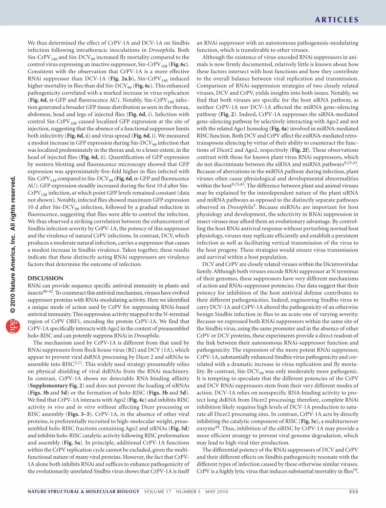
Figure 6 RNAi suppressors determine spread and pathogenesis of Sindbis virus in fruit fly. (a) Expression of the Sindbis virus structural proteins is regulated by a subgenomic promoter
suppressors in an otherwise isogenic background. Given that no infec-tious clones are available for either DCV or CrPV, we examined the effect of CrPV-1A or DCV-1A in the context of a recombinant Sindbis virus infection. We engineered recombinant Sindbis viruses to express GFP and RNAi suppressors CrPV-1A (Sin-CrPV148) or DCV-1A (Sin-DCV99) under the control of duplicated subgenomic promoter15 (Fig. 6a). Control Sindbis viruses expressed inactive suppressors—a truncated version of CrPV-1A (Sin-CrPV108) or DCV-1A carrying a mutation in the DSRM (Sin–DCV K73A)22. In addition to infecting
insect cells, Sindbis also infects mammalian cells, which do not appear to use RNAi to control the virus, affording an unbiased control system for viral replication efficiency. We observed no appreciable differences in virus replication kinetics for these recombinant viruses in mam-malian BHK-21 cells (Fig. 6b), indicating that expression of RNAi suppressors does not affect virus fitness in cells where the RNAi machinery does not have a major role in antiviral defense38.
Sindbis virus does not appear to encode an RNAi suppressor (data not shown) and establishes a persistent infection in Drosophila39.
S2 extract
a b c e
d+ +
+ +
++
++++
+– –
––––
–– –
––––
+CrPV-1ADCV-1A
BSA
mRNAsubstrate
S200
10.0
Fire
fly/Ren
illa
ratio
1.0
0.1
0.01
0.0012 4 6 8 12
Time after 1A148 expression (h)16
dsLuc
Lucassay
dsLuc + CrPV-1A
dsGFP + CrPV-1A
FHV B2DCV-1A
FHV B2DCV-1A
dsRNA
Dicer2
siRNA
R2D2
Assembly
RISC/Ago2
Target recognitionand cleavage
CrPV-1A
20 24
α-Ago2
Unindu
ced
CrPV-
1A
α-Flag
Northern blot(Luc siRNAs)
Et. br.
P200
S200
P200
P200
1 2
20 nt
S2 extractsiRNA
S2 cellsdsRNA
InductionCrPV-1A
S200/P20048 h 20 hS2 cells
reporter plasmidsdsRNA
Induction1A
16 h 2–24 h
45 min 10 min1A 32P-mRNA PAGE
3 h
5′ product
Luc siRNA
1 2 3 4 5
Figure 5 CrPV-1A interferes with the function of preprogrammed holo-RISC in vitro and in vivo. (a) S2 extracts were programmed with siRNA for 45 min followed by addition of 1A (0.35 µM CrPV-1A, lane 3, 0.35 µM DCV-1A, lane 4, or BSA, lane 5) for 10 min. RISC assay was initiated by incubating 32P-mRNA substrate for an additional 3 h, and 5′-cleaved product was analyzed using denaturing PAGE. (b) Silencing of firefly luciferase in stable S2 cells was programmed by co-transfecting firefly and Renilla reporter plasmid with specific FLuc dsRNA (dsLuc) or control eGFP dsRNA (dsGFP). 16 h after transfection, luciferase expression was monitored in either the presence (dsLuc + CrPV-1A, dsGFP + CrPV-1A) or the absence (dsLuc) of CrPV-1A for a period of 24 h. Data indicate averages and s.d. of four independent experiments. (c) Stable S2 cells were transfected with FLuc dsRNA for 48 h followed by induction of suppressor for additional 20 h. The postnuclear cytoplasmic extract was centrifuged at 200,000g to separate the ribosomal pellet (P200) from the supernatant (S200). (d) P200 and S200 fractions were analyzed by western blotting using Drosophila Argonaute 2 (α-Ago2) polyclonal and anti-Flag (α-Flag) monoclonal antibodies. RNA extracted from P200 fraction was analyzed for Luc siRNA by northern blotting. P200 ethidium bromide (Et. br.) represents RNA isolated from soluble high-salt P200 extraction. (e) DCV-1A and FHV B2 inhibits either Dicer2 processing of long dsRNA or siRNA incorporation into RISC by virtue of their dsRNA or siRNA binding activity. Conversely, CrPV-1A acts at the level of holo-RISC by protein-protein interaction with Drosophila Argonaute 2.
(Pr1). CrPV-1A and DCV-1A were cloned under the control of a duplicated subgenomic promoter (Pr2) in the 3′ UTR of the Sindbis genome. These recombinant viruses also express GFP that cleaves at both the N and C termini during structural polyprotein processing15. (b) BHK cells were infected with recombinant Sindbis viruses at MOI 1; virus samples were collected over a period of 24 h. Titers of each sample (plaque-forming unit (PFU) per ml) were measured by plaque assay, and replication kinetics of each virus was determined. (c) Intrathoracic injections of flies with engineered Sindbis virus expressing CrPV-1A (Sin-CrPV148) induced higher mortality and tissue spread at similar doses of infection (2,500 or 5,000 PFU) compared to Sindbis virus expressing DCV-1A (Sin-DCV99). Survival rate of flies (%) was monitored daily (d) for 30 d. (d) i, flies injected with recombinant Sindbis viruses (Sin-CrPV148, Sin-CrPV108, Sin-DCV99 and Sin–DCV K73A) were imaged by GFP microscopy. For western blot analysis, four or five flies from each injection at 11 d after infection were homogenized and blotted with anti-GFP antibodies (α-GFP). The same blot was stripped and probed with anti-actin antibody (α-actin); ii, GFP fluorescence (AU) for recombinant Sindbis viruses (Sin-CrPV148, Sin-CrPV108, Sin-DCV99) was measured, and data represent mean and s.e.m. for four or five individual flies.
© 2
010
Nat
ure
Am
eric
a, In
c. A
ll ri
gh
ts r
eser
ved
.
nAture structurAl & moleculAr biology VOLUME 17 NUMBER 5 MAY 2010 553
A r t i c l e s
We thus determined the effect of CrPV-1A and DCV-1A on Sindbis infection following intrathoracic inoculations in Drosophila. Both Sin-CrPV148 and Sin-DCV99 increased fly mortality compared to the control virus expressing an inactive suppressor, Sin-CrPV108 (Fig. 6c). Consistent with the observation that CrPV-1A is a more effective RNAi suppressor than DCV-1A (Fig. 2a,b), Sin-CrPV148 induced higher mortality in flies than did Sin-DCV99 (Fig. 6c). This enhanced pathogenicity correlated with a marked increase in virus replication (Fig. 6d, α-GFP and fluorescence AU). Notably, Sin-CrPV148 infec-tion generated a broader GFP tissue distribution as seen in the thorax, abdomen, head and legs of injected flies (Fig. 6d, i). Infection with control Sin-CrPV108 caused localized GFP expression at the site of injection, suggesting that the absence of a functional suppressor limits both infectivity (Fig. 6d, ii) and virus spread (Fig. 6d, i). We measured a modest increase in GFP expression during Sin-DCV99 infection that was localized predominately in the thorax and, to a lesser extent, in the head of injected flies (Fig. 6d, ii). Quantification of GFP expression by western blotting and fluorescence microscopy showed that GFP expression was approximately five-fold higher in flies infected with Sin-CrPV148 compared to Sin-DCV99 (Fig. 6d, α-GFP and fluorescence AU). GFP expression steadily increased during the first 10 d after Sin-CrPV148 infection, at which point GFP levels remained constant (data not shown). Notably, infected flies showed maximum GFP expression 10 d after Sin-DCV99 infection, followed by a gradual reduction in fluorescence, suggesting that flies were able to control the infection. We thus observed a striking correlation between the enhancement of Sindbis infection severity by CrPV-1A, the potency of this suppressor and the virulence of natural CrPV infections. In contrast, DCV, which produces a moderate natural infection, carries a suppressor that causes a modest increase in Sindbis virulence. Taken together, these results indicate that these distinctly acting RNAi suppressors are virulence factors that determine the outcome of infection.
DISCUSSIONRNAi can provide sequence specific antiviral immunity in plants and insects40–42. To counteract this antiviral mechanism, viruses have evolved suppressor proteins with RNAi-modulating activity. Here we identified a unique mode of action used by CrPV for suppressing RNAi-based antiviral immunity. This suppression activity mapped to the N-terminal region of CrPV ORF1, encoding the protein CrPV-1A. We find that CrPV-1A specifically interacts with Ago2 in the context of preassembled holo-RISC and can potently suppress RNAi in Drosophila.
The mechanism used by CrPV-1A is different from that used by RNAi suppressors from flock house virus (B2) and DCV (1A), which appear to prevent viral dsRNA processing by Dicer 2 and siRNAs to assemble into RISC2,11. This widely used strategy presumably relies on physical shielding of viral dsRNAs from the RNAi machinery. In contrast, CrPV-1A shows no detectable RNA-binding affinity (Supplementary Fig. 2) and does not prevent the loading of siRNAs (Figs. 3b and 5d) or the formation of holo-RISC (Figs. 3b and 5d). We find that CrPV-1A interacts with Ago2 (Fig. 4c) and inhibits RISC activity in vivo and in vitro without affecting Dicer processing or RISC assembly (Figs. 3–5). CrPV-1A, in the absence of other viral proteins, is preferentially recruited to high–molecular weight, preas-sembled holo-RISC fractions containing Ago2 and siRNAs (Fig. 5d) and inhibits holo-RISC catalytic activity following RISC preformation and assembly (Fig. 5a). In principle, additional CrPV-1A functions within the CrPV replication cycle cannot be excluded, given the multi-functional nature of many viral proteins. However, the fact that CrPV-1A alone both inhibits RNAi and suffices to enhance pathogenicity of the evolutionarily unrelated Sindbis virus shows that CrPV-1A is itself
an RNAi suppressor with an autonomous pathogenesis-modulating function, which is transferable to other viruses.
Although the existence of virus-encoded RNAi suppressors in ani-mals is now firmly documented, relatively little is known about how these factors intersect with host functions and how they contribute to the overall balance between viral replication and transmission. Comparison of RNAi-suppression strategies of two closely related viruses, DCV and CrPV, yields insights into both issues. Notably, we find that both viruses are specific for the host siRNA pathway, as neither CrPV-1A nor DCV-1A affected the miRNA gene-silencing pathway (Fig. 2). Indeed, CrPV-1A suppresses the siRNA-mediated gene-silencing pathway by selectively interacting with Ago2 and not with the related Ago1 homolog (Fig. 4a) involved in miRNA-mediated RISC function. Both DCV and CrPV affect the esiRNA-mediated retro-transposon silencing by virtue of their ability to counteract the func-tions of Dicer2 and Ago2, respectively (Fig. 2f). These observations contrast with those for known plant virus RNAi suppressors, which do not discriminate between the siRNA and miRNA pathways9,25,43. Because of aberrations in the miRNA pathway during infection, plant viruses often cause physiological and developmental abnormalities within the host9,25,43. The difference between plant and animal viruses may be explained by the interdependent nature of the plant siRNA and miRNA pathways as opposed to the distinctly separate pathways observed in Drosophila3. Because miRNAs are important for host physiology and development, the selectivity in RNAi suppression in insect viruses may afford them an evolutionary advantage. By control-ling the host RNAi antiviral response without perturbing normal host physiology, viruses may replicate efficiently and establish a persistent infection as well as facilitating vertical transmission of the virus to the host progeny. These strategies would ensure virus transmission and survival within a host population.
DCV and CrPV are closely related viruses within the Dicistroviridae family. Although both viruses encode RNAi suppressor at N terminus of their genomes, these suppressors have very different mechanisms of action and RNAi-suppressor potencies. Our data suggest that their potency for inhibition of the host antiviral defense contributes to their different pathogenicities. Indeed, engineering Sindbis virus to carry DCV-1A and CrPV-1A altered the pathogenicity of an otherwise benign Sindbis infection in flies to an acute one of varying severity. Because we expressed both RNAi suppressors within the same site of the Sindbis virus, using the same promoter and in the absence of other CrPV or DCV proteins, these experiments provide a direct readout of the link between their autonomous RNAi-suppressor function and pathogenicity. The expression of the more potent RNAi suppressor, CrPV-1A, substantially enhanced Sindbis virus pathogenicity and cor-related with a dramatic increase in virus replication and fly morta-lity. By contrast, Sin-DCV99 was only moderately more pathogenic. It is tempting to speculate that the different potencies of the CrPV and DCV RNAi suppressors stem from their very different modes of action. DCV-1A relies on nonspecific RNA-binding activity to pro-tect long dsRNA from Dicer2 processing; therefore, complete RNAi inhibition likely requires high levels of DCV-1A production to satu-rate all Dicer2 processing sites. In contrast, CrPV-1A acts by directly inhibiting the catalytic component of RISC (Fig. 5e), a multiturnover enzyme44. Thus, inhibition of the siRISC by CrPV-1A may provide a more efficient strategy to prevent viral genome degradation, which may lead to high viral titer production.
The differential potency of the RNAi suppressors of DCV and CrPV and their different effects on Sindbis pathogenicity resonate with the different types of infection caused by these otherwise similar viruses. CrPV is a highly lytic virus that induces substantial mortality in flies19,
© 2
010
Nat
ure
Am
eric
a, In
c. A
ll ri
gh
ts r
eser
ved
.
554 VOLUME 17 NUMBER 5 MAY 2010 nAture structurAl & moleculAr biology
whereas DCV, in nature, establishes a symbiotic, chronic infection in Drosophila without causing appreciable mortality12,13,16,45. Indeed, DCV was first found in field populations by random screening of the flies and not as a result of any particular syndrome13. Given that the pathogenic characteristics of DCV and CrPV infection are replicated by simply transferring the respective RNAi suppressors to Sindbis virus, we argue that these proteins are attuned with the natural virus infection strategy. For instance, the chronic, persistent infection of Drosophila characterized by DCV may be attributed to the modest RNAi-suppressing activity shown by DCV-1A. Our results point to an exquisite equilibrium between the host antiviral defense machinery and the viral RNAi suppressors that serves as an evolutionary fine tuner of the host-virus interactions and determines the pathogenic outcome of infection, virus survival, and virus spread.
METhODSMethods and any associated references are available in the online version of the paper at http://www.nature.com/nsmb/.
Note: Supplementary information is available on the Nature Structural & Molecular Biology website.
ACKNowleDGMeNTsWe thank J. Frydman and Andino laboratory members L. Gitlin, D. Barnes, M. Flenniken and A. Lauring for useful discussion for preparing the manuscript, R. van Rij and C. Saleh for their help, G. Belsham for critical reading of the manuscript and useful comments, P. Zamore (Univ. Massachusetts Medical School), Mikiko Siomi and Keita Miyoshi (Keio Univ. School of Medicine, Japan) for providing antibody directed against Drosophila Argonaute 2, H. Heidner (Univ. Texas, San Antonio) for providing Sindbis virus plasmid and M. Moritz (Univ. California, San Francisco) for Drosophila embryo extract preparation. This work was financially supported by Pasteur Institute, by the Centre National de la Recherche Scientifique, by grants from the Agence Nationale de la Recherche (AKROSS) and by fellowships from the Lebanese Centre National de la Recherche Scientifique to B.B. as well as by US National Institutes of Health grants AI40085 and AI064738 to R.A.
AUTHoR CoNTRIBUTIoNsA.N. and R.A. designed and interpreted most of the experiments; A.N. performed most of the experiments; B.B. and C.A. designed and performed the IR[white], GFP/bantam sensor and retrotransposon experiments in flies; M.T. and M.K. performed the fly injections; A.A. made some plasmid constructs; C.D. and A.K. carried out the MS analysis; J.G. advised on the project; A.N., R.A., A.A. and C.A. prepared the manuscript.
CoMPeTING FINANCIAl INTeResTsThe authors declare no competing financial interests.
Published online at http://www.nature.com/nsmb/. Reprints and permissions information is available online at http://npg.nature.com/reprintsandpermissions/.
1. Siomi, H. & Siomi, M.C. On the road to reading the RNA-interference code. Nature 457, 396–404 (2009).
2. van Rij, R.P. & Andino, R. The silent treatment: RNAi as a defense against virus infection in mammals. Trends Biotechnol. 24, 186–193 (2006).
3. Forstemann, K., Horwich, M.D., Wee, L., Tomari, Y. & Zamore, P.D. Drosophila microRNAs are sorted into functionally distinct argonaute complexes after production by dicer-1. Cell 130, 287–297 (2007).
4. Tomari, Y., Du, T. & Zamore, P.D. Sorting of Drosophila small silencing RNAs. Cell 130, 299–308 (2007).
5. Ding, S.W. & Voinnet, O. Antiviral immunity directed by small RNAs. Cell 130, 413–426 (2007).
6. Baumberger, N., Tsai, C.H., Lie, M., Havecker, E. & Baulcombe, D.C. The Polerovirus silencing suppressor P0 targets ARGONAUTE proteins for degradation. Curr. Biol. 17, 1609–1614 (2007).
7. Bortolamiol, D., Pazhouhandeh, M., Marrocco, K., Genschik, P. & Ziegler-Graff, V. The Polerovirus F box protein P0 targets ARGONAUTE1 to suppress RNA silencing. Curr. Biol. 17, 1615–1621 (2007).
8. Pazhouhandeh, M. et al. F-box-like domain in the polerovirus protein P0 is required for silencing suppressor function. Proc. Natl. Acad. Sci. USA 103, 1994–1999 (2006).
9. Zhang, X. et al. Cucumber mosaic virus-encoded 2b suppressor inhibits Arabidopsis Argonaute1 cleavage activity to counter plant defense. Genes Dev. 20, 3255–3268 (2006).
10. Kasschau, K.D. et al. P1/HC-Pro, a viral suppressor of RNA silencing, interferes with Arabidopsis development and miRNA unction. Dev. Cell 4, 205–217 (2003).
11. Chao, J.A. et al. Dual modes of RNA-silencing suppression by flock house virus protein B2. Nat. Struct. Mol. Biol. 12, 952–957 (2005).
12. Gomariz-Zilber, E. & Thomas-Orillard, M. Drosophila C virus and Drosophila hosts: a good association in various environments. J. Evol. Biol. 6, 677–689 (1993).
13. Brun, P. & Plus, N. The viruses of Drosophila. in The Genetics and Biology of Drosophila Vol. 2, 625–702 (Academic Press, New York, 1980).
14. Jousset, F.-X. & Plus, N. Etude de la transmission horizontale et de la transmis- sion verticale de picornavirus de Drosophila melanogaster et de Drosophila immigrans. Ann. Microbiol. 126, 231–249 (1975).
15. Thomas, J.M., Klimstra, W.B., Ryman, K.D. & Heidner, H.W. Sindbis virus vectors designed to express a foreign protein as a cleavable component of the viral structural polyprotein. J. Virol. 77, 5598–5606 (2003).
16. Aravin, A.A. et al. The small RNA profile during Drosophila melanogaster development. Dev. Cell 5, 337–350 (2003).
17. Gomariz-Zilber, E., Poras, M. & Thomas-Orillard, M. Drosophila C virus: experimental study of infectious yields and underlying pathology in Drosophila melanogaster laboratory populations. J. Invertebr. Pathol. 65, 243–247 (1995).
18. Reinganum, C. The isolation of cricket paralysis virus from the emperor gum moth, Antheraea eucalypti Scott, and its infectivity towards a range of insect species. Intervirology 5, 97–102 (1975).
19. Manousis, T. & Moore, N.F. Cricket paralysis virus, a potential control agent for the olive fruit fly, Dacus oleae Gmel. Appl. Environ. Microbiol. 53, 142–148 (1987).
20. Wilson, J.E., Powell, M.J., Hoover, S.E. & Sarnow, P. Naturally occurring dicistronic cricket paralysis virus RNA is regulated by two internal ribosome entry sites. Mol. Cell. Biol. 20, 4990–4999 (2000).
21. Galiana-Arnoux, D., Dostert, C., Schneemann, A., Hoffmann, J.A. & Imler, J.L. Essential function in vivo for Dicer-2 in host defense against RNA viruses in Drosophila. Nat. Immunol. 7, 590–597 (2006).
22. van Rij, R.P. et al. The RNA silencing endonuclease Argonaute 2 mediates specific antiviral immunity in Drosophila melanogaster. Genes Dev. 20, 2985–2995 (2006).
23. Wang, X.H. et al. RNA interference directs innate immunity against viruses in adult Drosophila. Science 312, 452–454 (2006).
24. Hahn, H. & Palmenberg, A.C. Mutational analysis of the encephalomyocarditis virus primary cleavage. J. Virol. 70, 6870–6875 (1996).
25. Chapman, E.J., Prokhnevsky, A.I., Gopinath, K., Dolja, V.V. & Carrington, J.C. Viral RNA silencing suppressors inhibit the microRNA pathway at an intermediate step. Genes Dev. 18, 1179–1186 (2004).
26. Brennecke, J., Hipfner, D.R., Stark, A., Russell, R.B. & Cohen, S.M. bantam encodes a developmentally regulated microRNA that controls cell proliferation and regulates the proapoptotic gene hid in Drosophila. Cell 113, 25–36 (2003).
27. Roignant, J.Y. et al. Absence of transitive and systemic pathways allows cell-specific and isoform-specific RNAi in Drosophila. RNA 9, 299–308 (2003).
28. Czech, B. et al. An endogenous small interfering RNA pathway in Drosophila. Nature 453, 798–802 (2008).
29. Ghildiyal, M. et al. Endogenous siRNAs derived from transposons and mRNAs in Drosophila somatic cells. Science 320, 1077–1081 (2008).
30. Kawamura, Y. et al. Drosophila endogenous small RNAs bind to Argonaute 2 in somatic cells. Nature 453, 793–797 (2008).
31. Miyoshi, K., Tsukumo, H., Nagami, T., Siomi, H. & Siomi, M.C. Slicer function of Drosophila Argonautes and its involvement in RISC formation. Genes Dev. 19, 2837–2848 (2005).
32. Pham, J.W. & Sontheimer, E.J. Molecular requirements for RNA-induced silencing complex assembly in the Drosophila RNA interference pathway. J. Biol. Chem. 280, 39278–39283 (2005).
33. Sontheimer, E.J. Assembly and function of RNA silencing complexes. Nat. Rev. Mol. Cell Biol. 6, 127–138 (2005).
34. Haley, B., Tang, G. & Zamore, P.D. In vitro analysis of RNA interference in Drosophila melanogaster. Methods 30, 330–336 (2003).
35. Hammond, S.M., Bernstein, E., Beach, D. & Hannon, G.J. An RNA-directed nuclease mediates post-transcriptional gene silencing in Drosophila cells. Nature 404, 293–296 (2000).
36. Thomas-Orillard, M. & Legendre, S. C virus of Drosophila and dynamics of host population. C.R. Acad. Sci. III 319, 615–621 (1996).
37. Plus, N., Croizier, G., Jousset, F.X. & David, J. Picornaviruses of laboratory and wild Drosophila melanogaster: geographical distribution and serotypic composition. Ann. Microbiol. 126, 107–117 (1975).
38. Cullen, B.R. Is RNA interference involved in intrinsic antiviral immunity in mammals? Nat. Immunol. 7, 563–567 (2006).
39. Mudiganti, U., Hernandez, R., Ferreira, D. & Brown, D.T. Sindbis virus infection of two model insect cell systems—a comparative study. Virus Res. 122, 28–34 (2006).
40. Hannon, G.J. RNA interference. Nature 418, 244–251 (2002).41. Voinnet, O. RNA silencing as a plant immune system against viruses. Trends Genet.
17, 449–459 (2001).42. Zamore, P.D. RNA interference: listening to the sound of silence. Nat. Struct. Biol.
8, 746–750 (2001).43. Merai, Z. et al. Double-stranded RNA binding may be a general plant RNA viral
strategy to suppress RNA silencing. J. Virol. 80, 5747–5756 (2006).44. Haley, B. & Zamore, P.D. Kinetic analysis of the RNAi enzyme complex. Nat. Struct.
Mol. Biol. 11, 599–606 (2004).45. Filipe, D. & Thomas-Orillard, M. Experimental study of a Drosophila melanogaster
laboratory population infected by food contamination. Endocytobiosis and Cell Res. 12, 163–176 (1998).
A r t i c l e s©
201
0 N
atu
re A
mer
ica,
Inc.
All
rig
hts
res
erve
d.

nAture structurAl & moleculAr biologydoi:10.1038/nsmb.1810
ONLINE METhODSRNAi reporter assay. We cultured Drosophila S2 cells (Supplementary Fig. 4) on 96-well plates. We transfected S2 cells with plasmids encoding CrPV-1A or infected with CrPV (MOI 1) and carried out RNAi reporter assay as described22. We tested the effect of CrPV-1A on microRNA (miRNA) miR2b function as described22. We designed antisense oligonucleotides (ASO) against endo-genous miR2b46 and transfected them into S2 cells using Dharmafect 4 reagent (Dharmacon) for 48 h followed by co-transfection of the reporter systems. We synthesized 2′-O-Me-3′ cholesterol–modified miR-2b ASO (5′-CAAAUGCUCCUCAAAGCUGGCUGUGAUAAUUCU-3′-Chl) from Dharmacon.
Dicer assay. We performed Dicer assay in S2 cell extracts as described22,34. We incubated recombinant DCV-1A, CrPV-1A or BSA in the reaction for 10 min and then added uniformly labeled 200-bp GL3 dsRNA (105 counts per min) for 3 h at 25 °C.
Slicer assay. We performed RISC cleavage assays as described33 using a 5′-capped FLuc mRNA target (592 nt) with a specific siRNA that generates a 100-nt 5′ cleaved product. We used either CrPV- or DCV-infected S2 extract or sup-plemented recombinant CrPV-1A and DCV-1A in uninfected S2 extract. We performed a 35-µl reaction (40% (v/v) S2 extracts) for 3 h at 25 °C.
Assembly assay. We performed RISC assembly experiments as described31,32. We incubated recombinant suppressor protein in embryo extract for 10 min and subsequently added 32P-radiolabeled siRNA duplex to the reaction for another 20 min. The 5-µl reactions contained 40% (v/v) embryo lysates.
Generation of stable S2 cell lines. We cloned CrPV-1A in frame with C-terminal 3×Flag and hexahistidine tags in vector pMT/V5-HisA (Invitrogen). We gener-ated stable cell lines according to the manufacturer’s instruction (Invitrogen). We induced the expression of the protein with 500 µM CuSO4 for 20 h.
Immunoprecipitation. We transfected plasmids 3FH–pAc-CrPV148,3FH–pAc-CrPV108, pAc5.1-V5/HisA vector (Invitrogen) into S2 cells (1 × 10⁷ cells) for 24 h, resuspended cells in IP buffer (30 mM HEPES, pH 7.4, 150 mM potassium acetate, 2 mM magnesium acetate, 5 mM DTT, 0.1% (v/v) Nonidet P-40, pro-tease inhibitor tablet (Roche)) and sonicated them on ice. We centrifuged the mixture (14,000g) and added the supernatant to magnetic beads (Dynal) con-jugated with anti-Flag M2 antibody. We washed the beads and boiled them in SDS sample buffer at 95 °C for 5 min. We ran eluted sample in 4–20% gradient SDS-PAGE gel, transferred it to PVDF membrane and probed it with Drosophila Ago1 or Ago2 antibodies. We followed the purification profile by western blot-ting (Supplementary Fig. 5).
Suppressor–Argonaute 2 complex purification. We resuspended 2 g of either induced or uninduced stable S2 cells in 1 ml IP buffer and ground them into powder in a mortar and pestle under liquid nitrogen. We prepared crude extracts by adding 9 ml IP buffer to ground S2 cells and homogenized the extracts for 2 min on ice. We centrifuged the beads (14,000g) and added the supernatant to anti-Flag M2 antibody–conjugated magnetic beads (Invitrogen), washed them with IP buffer and eluted them using IP buffer with 3×Flag pep-tide (400 µg ml−1) (Sigma). We added the eluted sample to Talon magnetic beads (Invitrogen), washed it with IP buffer and eluted it in IP buffer contain-ing 250 mM imidazole. We stained the eluted material with sliver according to the manufacturer’s instruction (Invitrogen). To determine the expression profile of Ago2 and CrPV-1A in stable S2 cells, we followed the one-step (Flag IP) purification protocol and analyzed the profiles by western blot analysis (Supplementary Fig. 5).
Protein identification. We resolved eluted sample on a 4–20% gradient gel and stained it using Coomassie brilliant blue (Sigma). We excised protein bands and destained them followed by in-gel trypsin digestion. We analyzed the trypsin-generated peptide fragments using a modular MS tool that includes several MALDI-MS and MALDI-MS/MS mass spectrometers as well as an HPLC-MS/MS mass spectrometer47.
Expression and purification of recombinant proteins. We cloned CrPV-1A into the pHis–Gb1-II vector48, transformed it into BL21DE3 cells and purified His-tag fusion protein using nickel-nitrilotriacetic acid resin (Qiagen). We purified GST–DCV-1A as described22. Polyclonal antibody against CrPV-1A was raised in rabbits immunized with purified recombinant proteins. Expression of full-length Drosophila Argonaute 1 in E. coli was carried out as described for CrPV-1A.
RNAi in fly. We established transgenic flies expressing CrPV-1A in developing eye as described22. We performed eye pigment determination assays on 3-day-old vir-gin females49. We established microRNA sensor lines expressing CrPV-1A as below. We recombined P[Tub–bantam sensor]26 transgenic stocks by genetic crosses with the P[engrailed-Gal4] line27 to obtain the homozygous w1118; II P[engrailed-Gal4]; III P[Tub-bantam sensor] miRNA bantam sensor line. We crossed homozygous P[pUAS-CrPV-148], P[pUAS-CrPV108] and w1118 stocks to the sensor line. We dissected wing imaginal discs from third-instar larvae fixed in 4% (v/v) form-aldehyde and stained them with mouse monoclonal anti-GFP (Roche) to visual-ize sensor expression using Apotome microscopy. To observe the effect of RNAi suppressors on retrotransposons expression, we made transgenic flies express-ing CrPV-1A as described50. We measured the expression levels of 412, mdg1 and 297 transposon in double heterozygous UASp>VSR, da>GAL4 and control heterozygous da>GAL4 2-day-old females. We manually separated 50 heads and processed them for total RNA extraction and reverse transcription. We analyzed transposon levels by real-time PCR using primers as described28,29,50.
Holo-RISC fractionation. We transfected 20 µg dsRNA (200-bp FLuc dsRNA) into stable S2 cells (1 × 10⁷) using Effectene reagent (Qiagen). After 48 h, we induced the cells with CuSO4 for additional 20 h. We performed holo-RISC puri-fication as described35. We resuspended cells in hypotonic buffer (10 mM HEPES, pH 7.4, 6 mM β-mercaptoethanol), lysed them by passing them through insulin syringe and spun them down at 14,000g for 25 min at 4 °C. We centrifuged the postnuclear, cytoplasmic extract at 200,000g for 3 h at 4 °C to separate the ribo-somal pellet (P200) from supernatant (S200). We probed P200 and S200 fraction for Argonaute 2 and CrPV-1A by western blotting.
Northern blotting. We treated the ribosomal pellet (P200) with 1 mM MgCl2 and 400 mM potassium acetate. We centrifuged extracted soluble material at 100,000g for 1 h at 4 °C. We treated the supernatant with SDS and proteinase K and extracted RNA with phenol. We resolved small RNA on 12% (v/v) urea gel, transferred it onto Hybond-N+ nylon membrane (Amersham Biosciences) and hybridized it with Fluc sense strand riboprobe.
Recombinant Sindbis virus production and Fly injection. We generated Sindbis virus–expressing RNAi suppressor CrPV-1A and DCV-1A as described15. We determined growth kinetics of viruses in BHK 21 cells. We injected 5-day-old female flies in the thorax with 50 nl of virus (2,500–5,000 PFU) using a nano-injector (Nanoject II, Drummond Scientific). We observed GFP expression in flies by GFP microscopy and western blot analysis. We incorporated GFP images per group of flies (4 or 5 each) into the Volocity software (Improvision, UK) under the measurement module. We used noninjected flies as background for further calculation. To determine the pathogenicity of DCV in transgenic flies express-ing CrPV-1A and DCV-1A, we injected 100 TCID50 of DCV by intrathoracic injection as described22.
46. Horwich, M.D. & Zamore, P.D. Design and delivery of antisense oligonucleotides to block microRNA function in cultured Drosophila and human cells. Nat. Protoc. 3, 1537–1549 (2008).
47. Blethrow, J.D., Tang, C., Deng, C. & Krutchinsky, A.N. Modular mass spectrometric tool for analysis of composition and phosphorylation of protein complexes. PLoS One 2, e358 (2007).
48. Harper, S.M., Neil, L.C. & Gardner, K.H. Structural basis of a phototropin light switch. Science 301, 1541–1544 (2003).
49. Ephrussi, B. & Herold, J.L. Studies of eye pigments of Drosophila. I. Methods of extraction and quantitative estimation of the pigment components. Genetics 29, 148–175 (1944).
50. Berry, B., Deddouche, S., Kirschner, D., Imler, J.L. & Antoniewski, C. Viral suppressors of RNA silencing hinder exogenous and endogenous small RNA pathways in Drosophila. PLoS One 4, e5866 (2009).
© 2
010
Nat
ure
Am
eric
a, In
c. A
ll ri
gh
ts r
eser
ved
.