
ARGONAUTE PIWI domain and microRNA duplex structure regulate small RNA sorting in Arabidopsis
11
ARTICLE Received 19 Apr 2014 | Accepted 3 Oct 2014 | Published 19 Nov 2014 ARGONAUTE PIWI domain and microRNA duplex structure regulate small RNA sorting in Arabidopsis Xiaoming Zhang 1,2 , DongDong Niu 1,3 , Alberto Carbonell 4 , Airong Wang 1,5 , Angel Lee 1 , Vinnary Tun 1 , Zonghua Wang 5 , James C. Carrington 4 , Chia-en A. Chang 6 & Hailing Jin 1 Small RNAs (sRNAs) are loaded into ARGONAUTE (AGO) proteins to induce gene silencing. In plants, the 5 0 -terminal nucleotide is important for sRNA sorting into different AGOs. Here we show that microRNA (miRNA) duplex structure also contributes to miRNA sorting. Base pairing at the 15th nucleotide of a miRNA duplex is important for miRNA sorting in both Arabidopsis AGO1 and AGO2. AGO2 favours miRNA duplexes with no middle mismatches, whereas AGO1 tolerates, or prefers, duplexes with central mismatches. AGO structure modelling and mutational analyses reveal that the QF-V motif within the conserved PIWI domain contributes to recognition of base pairing at the 15th nucleotide of a duplex, while the DDDE catalytic core of AtAGO2 is important for recognition of the central nucleotides. Finally, we rescued the adaxialized phenotype of ago1-12, which is largely due to miR165 loss-of-function, by changing miR165 duplex structure which we predict redirects it to AGO2. DOI: 10.1038/ncomms6468 1 Department of Plant Pathology and Microbiology, Center for Plant Cell Biology and Institute for Integrative Genome Biology, University of California, Riverside, California 92521, USA. 2 State Key Laboratory of Integrated Management of Pest Insects and Rodents, Institute of Zoology, Chinese Academy of Sciences, Beijing 100101, China. 3 Department of Plant Protection, Nanjing Agriculture University, Nanjing 210095, China. 4 Donald Danforth Plant Science Center, St Louis, Missouri 63132, USA. 5 Key Laboratory of Bio-pesticide and Chemistry Biology, Ministry of Education, Fujian Agricultural and Forestry University, Fuzhou 350002, China. 6 Department of Chemistry, University of California Riverside, Riverside, California 92521, USA. Correspondence and requests for materials should be addressed to H.J. (email: [email protected]). NATURE COMMUNICATIONS | 5:5468 | DOI: 10.1038/ncomms6468 | www.nature.com/naturecommunications 1 & 2014 Macmillan Publishers Limited. All rights reserved.
Transcript of ARGONAUTE PIWI domain and microRNA duplex structure regulate small RNA sorting in Arabidopsis

ARTICLE
Received 19 Apr 2014 | Accepted 3 Oct 2014 | Published 19 Nov 2014
ARGONAUTE PIWI domain and microRNA duplexstructure regulate small RNA sorting in ArabidopsisXiaoming Zhang1,2, DongDong Niu1,3, Alberto Carbonell4, Airong Wang1,5, Angel Lee1, Vinnary Tun1,
Zonghua Wang5, James C. Carrington4, Chia-en A. Chang6 & Hailing Jin1
Small RNAs (sRNAs) are loaded into ARGONAUTE (AGO) proteins to induce gene silencing.
In plants, the 50-terminal nucleotide is important for sRNA sorting into different AGOs. Here
we show that microRNA (miRNA) duplex structure also contributes to miRNA sorting. Base
pairing at the 15th nucleotide of a miRNA duplex is important for miRNA sorting in both
Arabidopsis AGO1 and AGO2. AGO2 favours miRNA duplexes with no middle mismatches,
whereas AGO1 tolerates, or prefers, duplexes with central mismatches. AGO structure
modelling and mutational analyses reveal that the QF-V motif within the conserved PIWI
domain contributes to recognition of base pairing at the 15th nucleotide of a duplex, while the
DDDE catalytic core of AtAGO2 is important for recognition of the central nucleotides.
Finally, we rescued the adaxialized phenotype of ago1-12, which is largely due to miR165
loss-of-function, by changing miR165 duplex structure which we predict redirects it to AGO2.
DOI: 10.1038/ncomms6468
1 Department of Plant Pathology and Microbiology, Center for Plant Cell Biology and Institute for Integrative Genome Biology, University of California,Riverside, California 92521, USA. 2 State Key Laboratory of Integrated Management of Pest Insects and Rodents, Institute of Zoology, Chinese Academy ofSciences, Beijing 100101, China. 3 Department of Plant Protection, Nanjing Agriculture University, Nanjing 210095, China. 4 Donald Danforth Plant ScienceCenter, St Louis, Missouri 63132, USA. 5 Key Laboratory of Bio-pesticide and Chemistry Biology, Ministry of Education, Fujian Agricultural and ForestryUniversity, Fuzhou 350002, China. 6 Department of Chemistry, University of California Riverside, Riverside, California 92521, USA. Correspondence andrequests for materials should be addressed to H.J. (email: [email protected]).
NATURE COMMUNICATIONS | 5:5468 | DOI: 10.1038/ncomms6468 | www.nature.com/naturecommunications 1
& 2014 Macmillan Publishers Limited. All rights reserved.
RNA silencing is a conserved regulatory mechanismmediated by small RNAs (sRNAs) in most eukaryoticorganisms1,2. sRNAs are generated by Dicer (DCR) or
DCR-like proteins and then sorted into distinct ARGONAUTES(AGOs), the core components of the RNA-induced silencingcomplex, to induce silencing of target RNAs or DNAs withcomplementary sequences. sRNA-based silencing can occurthrough transcriptional gene silencing by guiding DNAmethylation or histone modification, or through post-transcriptional gene silencing by direct cleavage (slicing),destabilization or translational inhibition of target transcripts2,3.
A typical AGO protein consists of four functional domains—N,PAZ, MID and PIWI4,5—which form a groove structure with thePAZ domain on the top and a crescent structure formed by theMID, PIWI and N domains at the bottom6–8. The MID–PIWIinterface houses a binding pocket for the 50 end of the functionalguide strand of the sRNA duplex9. The PAZ domain contains abinding pocket for the 30 end of the guide strand, and the PIWIdomain harbours a conserved Asp–Asp–His (DDH)/Asp–Asp–Asp (DDD) catalytic core that cleaves the passenger strand of theduplex between nucleotides 10 and 11 of the sRNAs4,10. Thecrystal structure of budding yeast Kluyveromyces polysporus AGOshows that an invariant glutamic acid (E) residue is inserted intothe catalytic pocket to form the conserved DDHE/DDDE catalytictetrad for slicing11. Studies on AGO structures have focusedmainly on yeasts, animals or humans7,8,12,13; so far, the structuresof plant AGO proteins are still enigmatic.
sRNA sorting into the appropriate AGO is a key step thatdetermines the function of a sRNA. AGOs from differentorganisms exhibit distinct, yet overlapping, sorting mechanisms.In animals, sRNA sorting is determined by the thermodynamicstability of the duplex termini, the specific protein–proteininteractions between the AGO and the DCR complexes, thestructure of the sRNA duplex, the sRNA 50-terminal nucleotide(nt) and the complementarity of the sRNA to its target14–18. Thestrand with less stability at its 50 terminus is incorporated as theguide strand, while the other strand (called the passenger or starstrand) is degraded14,15. In Drosophila melanogaster, miRNAguide strand/miRNA star strand duplexes (miRNA/miRNA*) aregenerated by the DCR1/Loquacious complex and aresubsequently sorted into AGO1 (DmAGO1), whereas siRNAduplexes are processed by the DCR2/R2D2 complex and aresorted into AGO2 (DmAGO2)16,19. miRNA/miRNA* duplexesoften contain central mismatches at nucleotide positions 8–12,whereas siRNA duplexes have perfect or near-perfectcomplementary sequences. In general, DmAGO1 shows a biasfor a 50-terminal (50) U, whereas DmAGO2 favours a 50C(refs 20–22). Sequence complementarity between targets andsRNAs also affect sRNA stability and sorting18,23,24. AlthoughsRNA sorting is not well-understood in Caenorhabditis elegans,miRNA duplexes with central mismatches are preferentiallysorted into two AGO proteins, Argonaute Like Gene-1(CeALG-1) and CeALG-2, while siRNA duplexes with perfectbase pairing are mainly sorted into another AGO protein, RNAinterference-Defective 1 (RDE-1; ref. 25). Human AGO2(HsAGO2) contains a rigid loop in the MID domain thatpreferentially recognizes 50U’s or A’s, which reduces the bindingefficiency of sRNAs with a 50C or G (ref. 26). Interestingly, thisnucleotide specificity loop is conserved in all four HsAGOs. Theyalso have similar sorting preferences for duplexes with centralmismatches, which suggests that animal AGOs may not have astrict sorting systems or their sorting mechanism is still not fullyunderstood27,28.
In plants, the 50-terminal nt of sRNAs is more critical for sRNAsorting into the appropriate AGO than in animals29,30.Arabidopsis AGO1 (AtAGO1) prefers 21-nt sRNAs with a 50U,
AtAGO2 favours 21-nt sRNAs with a 50A, AtAGO5 has a biastowards sRNAs with a 50C but without an obvious size preference,and AtAGO4 preferentially loads 24-nt sRNAs with a 50A (refs31,32). However, the 50-terminal nt-directed sorting model is notthe only mechanism for sRNA sorting in plants. For example,some sRNAs sorted into AtAGO2 do not have a 50A, such asmiR396*, miR165*, miR398*, miR166* and miR156*, while somesRNAs containing a 50A are sorted into AGOs other thanAtAGO2 and AtAGO4, such as miR172 and miR783 (sorted intoAtAGO1) or miR390 (sorted into AtAGO7). Some sRNAscontaining a 50C, such as miR169 and miR395, are sorted intoAtAGO1. Additional mechanisms for sRNA sorting in plants arestill largely unknown, although the importance of sRNA duplexstructures has recently been recognized. For example, the specificsorting of miR166 and miR390 into Arabidopsis AtAGO10 andAtAGO7, respectively, is determined by the secondary structureof the miRNA/miRNA* duplex and by the identity of the50-terminal nt of the miRNA33,34. Moreover, miR408 thatpossesses a 50A is sorted into both AtAGO1 and AtAGO2 toco-regulate the expression of Plantacyanin35.
Our previous study has shown that AtAGO2 plays animportant role in antibacterial defence through its associationwith miR393* (ref. 36). MiR393* and miR393 originate from thesame miRNA duplex and are selectively sorted into AtAGO2 andAtAGO1, respectively. Although the differential sorting ofmiRNA and miRNA* of the same duplex into different AGOshas been observed in multiple plant and animal species, little isknown about the specific features of the miRNAs and/or theAGOs that dictate miRNA strand selection and sorting into theappropriate AGO21,22,30,37. In this study, we unveil the importantroles of both the secondary structure of miRNA duplexes and theAGO PIWI domain in miRNA sorting in Arabidopsis. We havefound that base pairing at the 15th position (#15) of the miRNAduplex is essential for effective sorting into both AtAGO1 andAtAGO2. AtAGO2 favours miRNA duplexes without a mismatchat #11, whereas AtAGO1 tolerates or prefers miRNA duplexeswith central mismatches. Structure modelling and mutationalanalyses of AGO proteins identify the conserved Gln–Phe–Val(QF-V) motif and the catalytic tetrad within the PIWI domain,which contribute to the recognition of nucleotides at #15 and #11of the miRNA duplex. Finally, the stable expression of anAtAGO2-favoured artificial miR165 duplex with no mismatch at#15 and #11 suppresses the adaxialized phenotype of the ago1-12mutant that largely resembles the miR165 loss-of-functionphenotype.
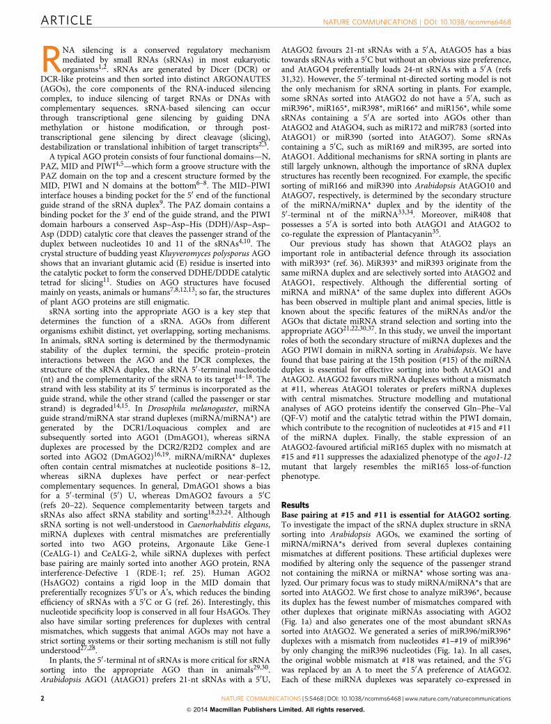
ResultsBase pairing at #15 and #11 is essential for AtAGO2 sorting.To investigate the impact of the sRNA duplex structure in sRNAsorting into Arabidopsis AGOs, we examined the sorting ofmiRNA/miRNA*s derived from several duplexes containingmismatches at different positions. These artificial duplexes weremodified by altering only the sequence of the passenger strandnot containing the miRNA or miRNA* whose sorting was ana-lyzed. Our primary focus was to study miRNA/miRNA*s that aresorted into AtAGO2. We first chose to analyze miR396*, becauseits duplex has the fewest number of mismatches compared withother duplexes that originate miRNAs associating with AGO2(Fig. 1a) and also generates one of the most abundant sRNAssorted into AtAGO2. We generated a series of miR396/miR396*duplexes with a mismatch from nucleotides #1–#19 of miR396*by only changing the miR396 nucleotides (Fig. 1a). In all cases,the original wobble mismatch at #18 was retained, and the 50Gwas replaced by an A to meet the 50A preference of AtAGO2.Each of these miRNA duplexes was separately co-expressed in
ARTICLE NATURE COMMUNICATIONS | DOI: 10.1038/ncomms6468
2 NATURE COMMUNICATIONS | 5:5468 | DOI: 10.1038/ncomms6468 | www.nature.com/naturecommunications
& 2014 Macmillan Publishers Limited. All rights reserved.
Nicotiana benthamiana leaves together with an AtAGO2 proteintagged at its N-terminal region with three tandem repeats of thehaemaglutinin (HA) epitope. MiR396* association with AtAGO2was assessed by co-immunoprecipitation (Co-IP) with HA anti-body followed by sRNA Northern blot analysis. As shown inFig. 1b, a mismatch at #1, #11 or #15 of the miR396/miR396*duplex had the strongest effect on reducing miR396* sortingefficiency into AtAGO2. A mismatch at nucleotides #10 or #19also reduced miR396* sorting, although to a lesser extent. Asnucleotides #1 and #19 are terminal nucleotides of the miRNAduplex, their mismatches would likely interrupt terminal bindingin the AGO binding pockets. Thus, we mainly focused onnucleotides #15 and #11 of the miRNA duplex for furthercharacterization.
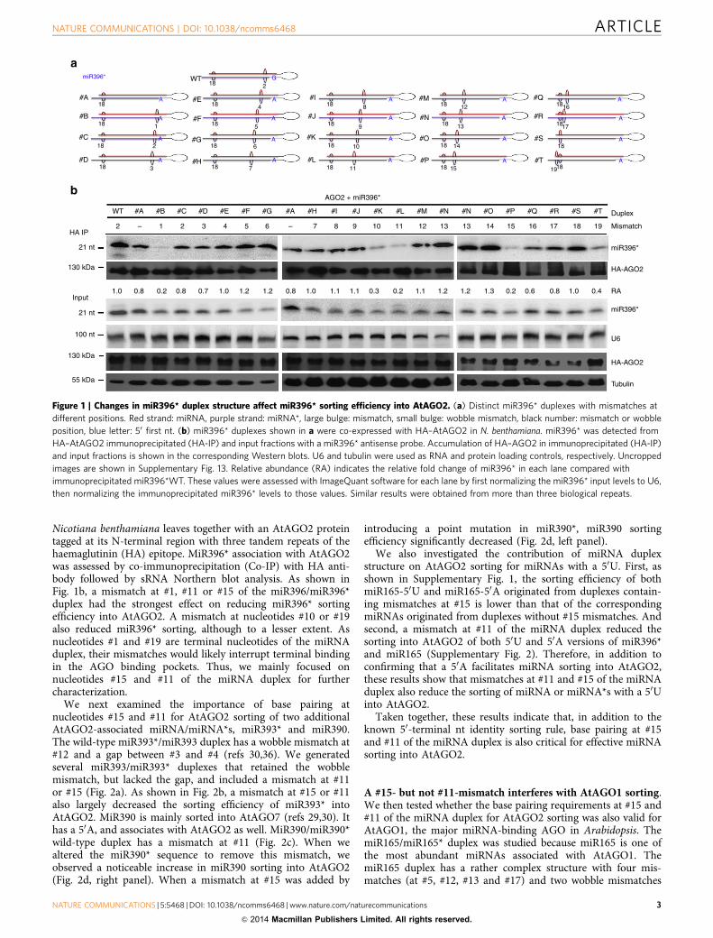
We next examined the importance of base pairing atnucleotides #15 and #11 for AtAGO2 sorting of two additionalAtAGO2-associated miRNA/miRNA*s, miR393* and miR390.The wild-type miR393*/miR393 duplex has a wobble mismatch at#12 and a gap between #3 and #4 (refs 30,36). We generatedseveral miR393/miR393* duplexes that retained the wobblemismatch, but lacked the gap, and included a mismatch at #11or #15 (Fig. 2a). As shown in Fig. 2b, a mismatch at #15 or #11also largely decreased the sorting efficiency of miR393* intoAtAGO2. MiR390 is mainly sorted into AtAGO7 (refs 29,30). Ithas a 50A, and associates with AtAGO2 as well. MiR390/miR390*wild-type duplex has a mismatch at #11 (Fig. 2c). When wealtered the miR390* sequence to remove this mismatch, weobserved a noticeable increase in miR390 sorting into AtAGO2(Fig. 2d, right panel). When a mismatch at #15 was added by
introducing a point mutation in miR390*, miR390 sortingefficiency significantly decreased (Fig. 2d, left panel).
We also investigated the contribution of miRNA duplexstructure on AtAGO2 sorting for miRNAs with a 50U. First, asshown in Supplementary Fig. 1, the sorting efficiency of bothmiR165-50U and miR165-50A originated from duplexes contain-ing mismatches at #15 is lower than that of the correspondingmiRNAs originated from duplexes without #15 mismatches. Andsecond, a mismatch at #11 of the miRNA duplex reduced thesorting into AtAGO2 of both 50U and 50A versions of miR396*and miR165 (Supplementary Fig. 2). Therefore, in addition toconfirming that a 50A facilitates miRNA sorting into AtAGO2,these results show that mismatches at #11 and #15 of the miRNAduplex also reduce the sorting of miRNA or miRNA*s with a 50Uinto AtAGO2.
Taken together, these results indicate that, in addition to theknown 50-terminal nt identity sorting rule, base pairing at #15and #11 of the miRNA duplex is also critical for effective miRNAsorting into AtAGO2.
A #15- but not #11-mismatch interferes with AtAGO1 sorting.We then tested whether the base pairing requirements at #15 and#11 of the miRNA duplex for AtAGO2 sorting was also valid forAtAGO1, the major miRNA-binding AGO in Arabidopsis. ThemiR165/miR165* duplex was studied because miR165 is one ofthe most abundant miRNAs associated with AtAGO1. ThemiR165 duplex has a rather complex structure with four mis-matches (at #5, #12, #13 and #17) and two wobble mismatches
#A18
18
18
18
18
18
18
18
18
2
4
5
62
1
3
18
18
18
18
18 18
18
18
18
16
17
19
18
18
18
12
13
14
1511
10
9
8
7
A
miR396*
A
A
A
A
G
A
A
A
A
A
A
A
A
A
A
A
A
A
A
A
#B
#C
#D
HA IP2 21 3 4 5 6 7 8 9 10 11 12 13 13 14 15 16 17 18 19
RA0.41.00.80.60.21.31.21.21.10.20.31.11.11.00.81.21.21.00.70.80.8 0.21.0
AGO2 + miR396*
– –
WT #A #B #C #D #E #F #G #A #H #I #J #K #L #M #N #N #O #P #Q #R #S #T Duplex
Mismatch
miR396*
miR396*
HA-AGO2
HA-AGO2
U6
Tubulin
21 nt
130 kDa
130 kDa
100 nt
55 kDa
21 nt
Input
#H
#G
#F
#E #I
#J
#K
#L
#M
#N
#O
#P
#Q
#R
#S
#T
WT
Figure 1 | Changes in miR396* duplex structure affect miR396* sorting efficiency into AtAGO2. (a) Distinct miR396* duplexes with mismatches at
different positions. Red strand: miRNA, purple strand: miRNA*, large bulge: mismatch, small bulge: wobble mismatch, black number: mismatch or wobble
position, blue letter: 50 first nt. (b) miR396* duplexes shown in a were co-expressed with HA–AtAGO2 in N. benthamiana. miR396* was detected from
HA–AtAGO2 immunoprecipitated (HA-IP) and input fractions with a miR396* antisense probe. Accumulation of HA–AGO2 in immunoprecipitated (HA-IP)
and input fractions is shown in the corresponding Western blots. U6 and tubulin were used as RNA and protein loading controls, respectively. Uncropped
images are shown in Supplementary Fig. 13. Relative abundance (RA) indicates the relative fold change of miR396* in each lane compared with
immunoprecipitated miR396*WT. These values were assessed with ImageQuant software for each lane by first normalizing the miR396* input levels to U6,
then normalizing the immunoprecipitated miR396* levels to those values. Similar results were obtained from more than three biological repeats.
NATURE COMMUNICATIONS | DOI: 10.1038/ncomms6468 ARTICLE
NATURE COMMUNICATIONS | 5:5468 | DOI: 10.1038/ncomms6468 | www.nature.com/naturecommunications 3
& 2014 Macmillan Publishers Limited. All rights reserved.
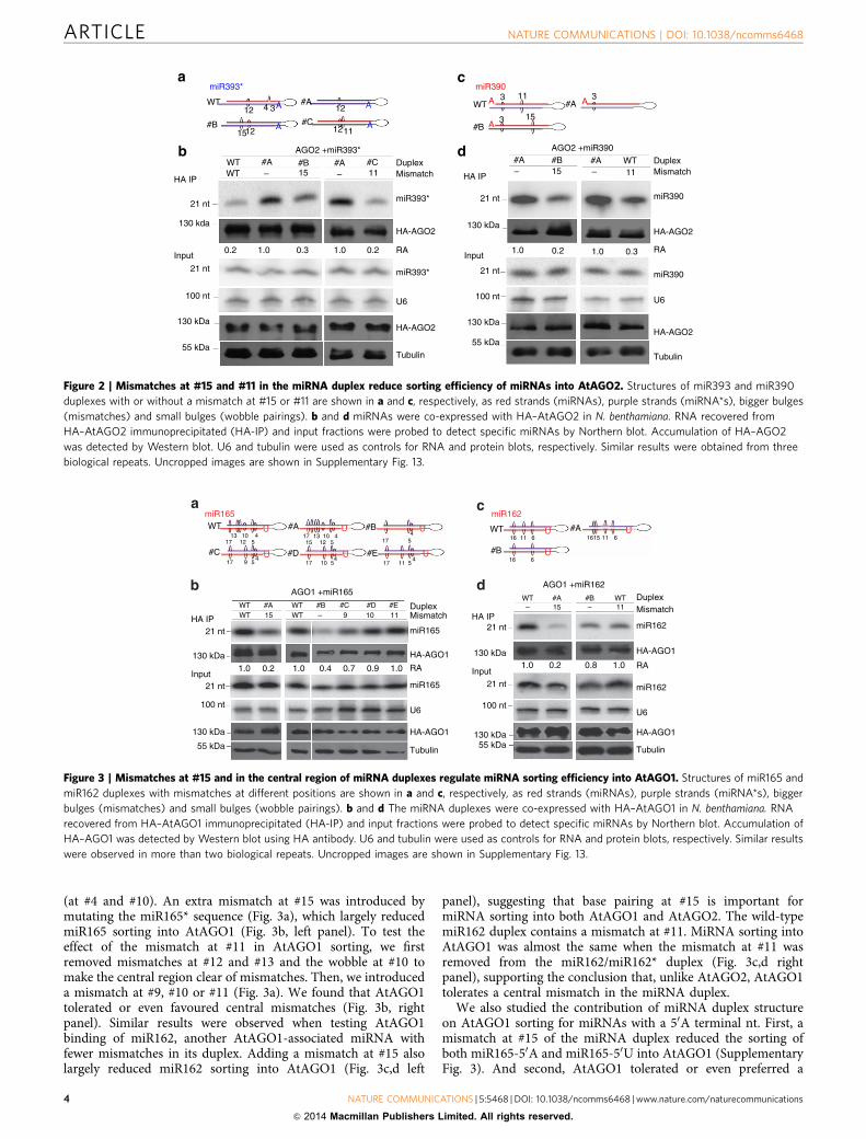
(at #4 and #10). An extra mismatch at #15 was introduced bymutating the miR165* sequence (Fig. 3a), which largely reducedmiR165 sorting into AtAGO1 (Fig. 3b, left panel). To test theeffect of the mismatch at #11 in AtAGO1 sorting, we firstremoved mismatches at #12 and #13 and the wobble at #10 tomake the central region clear of mismatches. Then, we introduceda mismatch at #9, #10 or #11 (Fig. 3a). We found that AtAGO1tolerated or even favoured central mismatches (Fig. 3b, rightpanel). Similar results were observed when testing AtAGO1binding of miR162, another AtAGO1-associated miRNA withfewer mismatches in its duplex. Adding a mismatch at #15 alsolargely reduced miR162 sorting into AtAGO1 (Fig. 3c,d left
panel), suggesting that base pairing at #15 is important formiRNA sorting into both AtAGO1 and AtAGO2. The wild-typemiR162 duplex contains a mismatch at #11. MiRNA sorting intoAtAGO1 was almost the same when the mismatch at #11 wasremoved from the miR162/miR162* duplex (Fig. 3c,d rightpanel), supporting the conclusion that, unlike AtAGO2, AtAGO1tolerates a central mismatch in the miRNA duplex.
We also studied the contribution of miRNA duplex structureon AtAGO1 sorting for miRNAs with a 50A terminal nt. First, amismatch at #15 of the miRNA duplex reduced the sorting ofboth miR165-50A and miR165-50U into AtAGO1 (SupplementaryFig. 3). And second, AtAGO1 tolerated or even preferred a
miR393* miR390
WT
WTAGO2 +miR393*
DuplexMismatch
DuplexMismatch
miR393*
HA-AGO2
HA IP
21 nt
130 kDa
21 nt
100 nt
130 kDa
55 kDa
InputRA
miR393*
U6
HA-AGO2
Tubulin
miR390
HA-AGO2
RA
miR390
U6
HA-AGO2
Tubulin
WTHA IP
21 nt
130 kda
0.2 1.0 0.3 1.0 0.2 0.21.0 0.31.0Input
21 nt
100 nt
130 kDa
55 kDa
#A #B #A #C #A– 15 11–
#B #A WT
– 15 – 11
#B
12 4 3
1512
WT
#B
3 3
3
11
15
#A#A
#C
12
1211
A
A
A
A
A A
A
AGO2 +miR390
Figure 2 | Mismatches at #15 and #11 in the miRNA duplex reduce sorting efficiency of miRNAs into AtAGO2. Structures of miR393 and miR390
duplexes with or without a mismatch at #15 or #11 are shown in a and c, respectively, as red strands (miRNAs), purple strands (miRNA*s), bigger bulges
(mismatches) and small bulges (wobble pairings). b and d miRNAs were co-expressed with HA–AtAGO2 in N. benthamiana. RNA recovered from
HA–AtAGO2 immunoprecipitated (HA-IP) and input fractions were probed to detect specific miRNAs by Northern blot. Accumulation of HA–AGO2
was detected by Western blot. U6 and tubulin were used as controls for RNA and protein blots, respectively. Similar results were obtained from three
biological repeats. Uncropped images are shown in Supplementary Fig. 13.
miR165 miR162
U U
U
U
U U
U
U
U
WTWT
21 nt
21 nt
HA IP
100 nt
Input21 nt
21 nt
Input
130 kDa
1.0 0.2 1.0 0.4 0.7 0.9 1.0
55 kDa
130 kDa
#A15
WTWT
#B #C #D #E DuplexMismatch
miR165
HA-AGO1
RA
miR165
U6
HA-AGO1
Tubulin
1110– 9
AGO1 +miR165WT–
HA IP
100 nt
130 kDa1.0 0.2 0.8 1.0 RA
miR162
HA-AGO155 kDa
130 kDa
#A15 11
WT#B
miR162
U6
HA-AGO1
Tubulin
DuplexMismatch–
AGO1 +miR162
WT13 1310 104
4
#A
#C
#B WT #A
#B#E#D
4
4 4
4
1717
5
5917 510 1117
17
17 16
16 15 11 61611 6
6
5 5121512
5
Figure 3 | Mismatches at #15 and in the central region of miRNA duplexes regulate miRNA sorting efficiency into AtAGO1. Structures of miR165 and
miR162 duplexes with mismatches at different positions are shown in a and c, respectively, as red strands (miRNAs), purple strands (miRNA*s), bigger
bulges (mismatches) and small bulges (wobble pairings). b and d The miRNA duplexes were co-expressed with HA–AtAGO1 in N. benthamiana. RNA
recovered from HA–AtAGO1 immunoprecipitated (HA-IP) and input fractions were probed to detect specific miRNAs by Northern blot. Accumulation of
HA–AGO1 was detected by Western blot using HA antibody. U6 and tubulin were used as controls for RNA and protein blots, respectively. Similar results
were observed in more than two biological repeats. Uncropped images are shown in Supplementary Fig. 13.
ARTICLE NATURE COMMUNICATIONS | DOI: 10.1038/ncomms6468
4 NATURE COMMUNICATIONS | 5:5468 | DOI: 10.1038/ncomms6468 | www.nature.com/naturecommunications
& 2014 Macmillan Publishers Limited. All rights reserved.
mismatch at #11 of miR165-50U, miR165-50A, miR396*-50U andmiR396*-50A (Supplementary Fig. 4). Therefore, both the 50-terminal nt and the secondary structure of the miRNA/miRNA*clearly contribute to the sorting of miRNA/miRNA*s intoAtAGO1.
Finally, to further validate the contribution of the miRNAduplex structure on the sorting of miRNAs into AtAGOs, weanalyzed published high throughput sequencing data sets fromuntreated Arabidopsis, including AtAGO1- and AtAGO2-associated sRNAs24. Out of the 15 miRNAs or miRNA*s thatare at least twofold enriched in AtAGO2 IP fraction comparedwith the input sample (total RNA), only one (6.6%) wasderived from miRNA duplexes containing a mismatch at #15(Supplementary Data 1a). Similarly, in AtAGO1 IP fraction,only one out of the 35 (2.9%) enriched miRNAs or miRNA*swas derived from duplexes containing a mismatch at #15(Supplementary Data 1b). On the other hand, 12 out of the 15(80.0%) miRNAs or miRNA*s enriched in AtAGO2 IP fractionwere derived from duplexes with no central mismatches (#9–#12)(Supplementary Data 1a), whereas 23 out of the 35 (65.7%)miRNAs or miRNA*s enriched in AtAGO1 IP fraction originatedfrom duplexes with central mismatches (Supplementary Data 1b).These data support our experimental results that (i) basepairing at #15 is essential for sRNA sorting into both AtAGO1and AtAGO2, and (ii) AtAGO2 favours sRNA duplexes withno central mismatches, whereas AtAGO1 can tolerate centralmismatches.
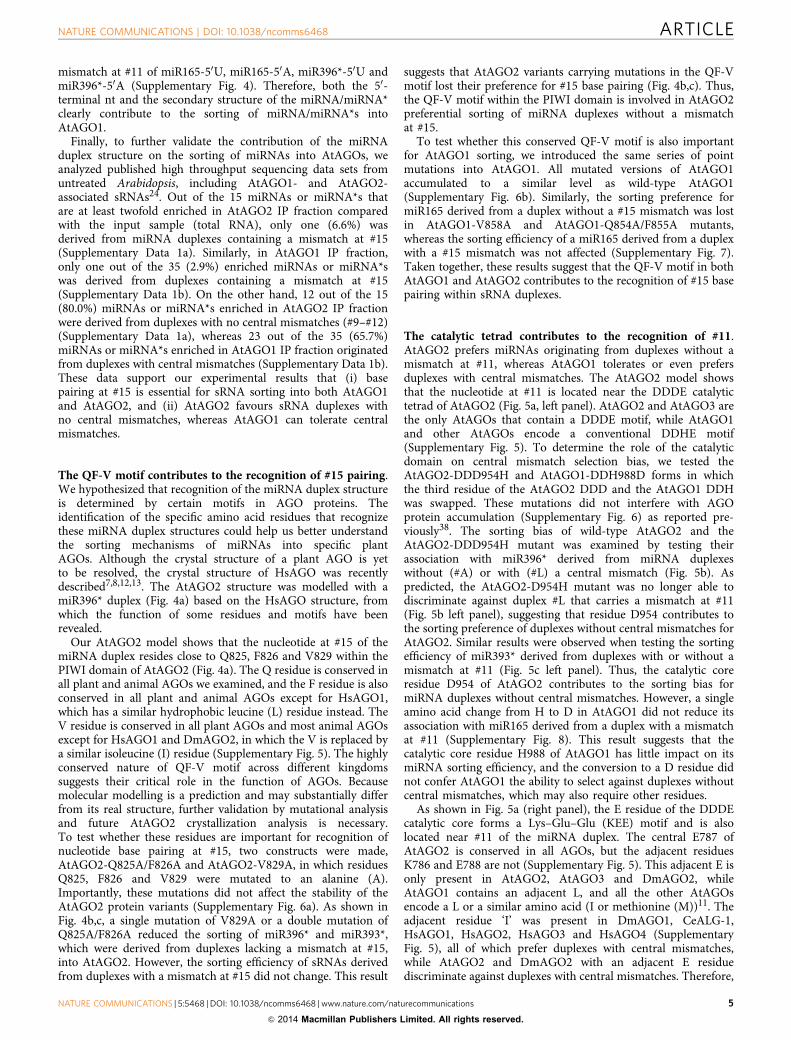
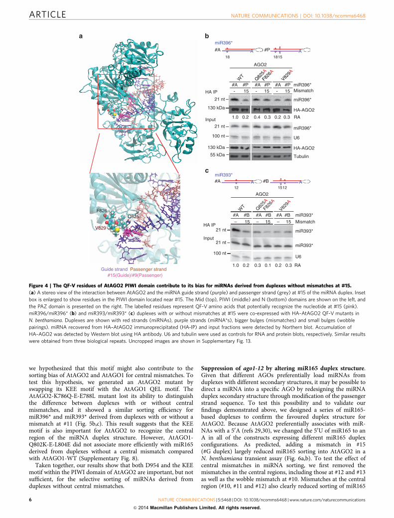
The QF-V motif contributes to the recognition of #15 pairing.We hypothesized that recognition of the miRNA duplex structureis determined by certain motifs in AGO proteins. Theidentification of the specific amino acid residues that recognizethese miRNA duplex structures could help us better understandthe sorting mechanisms of miRNAs into specific plantAGOs. Although the crystal structure of a plant AGO is yetto be resolved, the crystal structure of HsAGO was recentlydescribed7,8,12,13. The AtAGO2 structure was modelled with amiR396* duplex (Fig. 4a) based on the HsAGO structure, fromwhich the function of some residues and motifs have beenrevealed.
Our AtAGO2 model shows that the nucleotide at #15 of themiRNA duplex resides close to Q825, F826 and V829 within thePIWI domain of AtAGO2 (Fig. 4a). The Q residue is conserved inall plant and animal AGOs we examined, and the F residue is alsoconserved in all plant and animal AGOs except for HsAGO1,which has a similar hydrophobic leucine (L) residue instead. TheV residue is conserved in all plant AGOs and most animal AGOsexcept for HsAGO1 and DmAGO2, in which the V is replaced bya similar isoleucine (I) residue (Supplementary Fig. 5). The highlyconserved nature of QF-V motif across different kingdomssuggests their critical role in the function of AGOs. Becausemolecular modelling is a prediction and may substantially differfrom its real structure, further validation by mutational analysisand future AtAGO2 crystallization analysis is necessary.To test whether these residues are important for recognition ofnucleotide base pairing at #15, two constructs were made,AtAGO2-Q825A/F826A and AtAGO2-V829A, in which residuesQ825, F826 and V829 were mutated to an alanine (A).Importantly, these mutations did not affect the stability of theAtAGO2 protein variants (Supplementary Fig. 6a). As shown inFig. 4b,c, a single mutation of V829A or a double mutation ofQ825A/F826A reduced the sorting of miR396* and miR393*,which were derived from duplexes lacking a mismatch at #15,into AtAGO2. However, the sorting efficiency of sRNAs derivedfrom duplexes with a mismatch at #15 did not change. This result
suggests that AtAGO2 variants carrying mutations in the QF-Vmotif lost their preference for #15 base pairing (Fig. 4b,c). Thus,the QF-V motif within the PIWI domain is involved in AtAGO2preferential sorting of miRNA duplexes without a mismatchat #15.
To test whether this conserved QF-V motif is also importantfor AtAGO1 sorting, we introduced the same series of pointmutations into AtAGO1. All mutated versions of AtAGO1accumulated to a similar level as wild-type AtAGO1(Supplementary Fig. 6b). Similarly, the sorting preference formiR165 derived from a duplex without a #15 mismatch was lostin AtAGO1-V858A and AtAGO1-Q854A/F855A mutants,whereas the sorting efficiency of a miR165 derived from a duplexwith a #15 mismatch was not affected (Supplementary Fig. 7).Taken together, these results suggest that the QF-V motif in bothAtAGO1 and AtAGO2 contributes to the recognition of #15 basepairing within sRNA duplexes.
The catalytic tetrad contributes to the recognition of #11.AtAGO2 prefers miRNAs originating from duplexes without amismatch at #11, whereas AtAGO1 tolerates or even prefersduplexes with central mismatches. The AtAGO2 model showsthat the nucleotide at #11 is located near the DDDE catalytictetrad of AtAGO2 (Fig. 5a, left panel). AtAGO2 and AtAGO3 arethe only AtAGOs that contain a DDDE motif, while AtAGO1and other AtAGOs encode a conventional DDHE motif(Supplementary Fig. 5). To determine the role of the catalyticdomain on central mismatch selection bias, we tested theAtAGO2-DDD954H and AtAGO1-DDH988D forms in whichthe third residue of the AtAGO2 DDD and the AtAGO1 DDHwas swapped. These mutations did not interfere with AGOprotein accumulation (Supplementary Fig. 6) as reported pre-viously38. The sorting bias of wild-type AtAGO2 and theAtAGO2-DDD954H mutant was examined by testing theirassociation with miR396* derived from miRNA duplexeswithout (#A) or with (#L) a central mismatch (Fig. 5b). Aspredicted, the AtAGO2-D954H mutant was no longer able todiscriminate against duplex #L that carries a mismatch at #11(Fig. 5b left panel), suggesting that residue D954 contributes tothe sorting preference of duplexes without central mismatches forAtAGO2. Similar results were observed when testing the sortingefficiency of miR393* derived from duplexes with or without amismatch at #11 (Fig. 5c left panel). Thus, the catalytic coreresidue D954 of AtAGO2 contributes to the sorting bias formiRNA duplexes without central mismatches. However, a singleamino acid change from H to D in AtAGO1 did not reduce itsassociation with miR165 derived from a duplex with a mismatchat #11 (Supplementary Fig. 8). This result suggests that thecatalytic core residue H988 of AtAGO1 has little impact on itsmiRNA sorting efficiency, and the conversion to a D residue didnot confer AtAGO1 the ability to select against duplexes withoutcentral mismatches, which may also require other residues.
As shown in Fig. 5a (right panel), the E residue of the DDDEcatalytic core forms a Lys–Glu–Glu (KEE) motif and is alsolocated near #11 of the miRNA duplex. The central E787 ofAtAGO2 is conserved in all AGOs, but the adjacent residuesK786 and E788 are not (Supplementary Fig. 5). This adjacent E isonly present in AtAGO2, AtAGO3 and DmAGO2, whileAtAGO1 contains an adjacent L, and all the other AtAGOsencode a L or a similar amino acid (I or methionine (M))11. Theadjacent residue ‘I’ was present in DmAGO1, CeALG-1,HsAGO1, HsAGO2, HsAGO3 and HsAGO4 (SupplementaryFig. 5), all of which prefer duplexes with central mismatches,while AtAGO2 and DmAGO2 with an adjacent E residuediscriminate against duplexes with central mismatches. Therefore,
NATURE COMMUNICATIONS | DOI: 10.1038/ncomms6468 ARTICLE
NATURE COMMUNICATIONS | 5:5468 | DOI: 10.1038/ncomms6468 | www.nature.com/naturecommunications 5
& 2014 Macmillan Publishers Limited. All rights reserved.
we hypothesized that this motif might also contribute to thesorting bias of AtAGO2 and AtAGO1 for central mismatches. Totest this hypothesis, we generated an AtAGO2 mutant byswapping its KEE motif with the AtAGO1 QEL motif. TheAtAGO2-K786Q-E-E788L mutant lost its ability to distinguishthe difference between duplexes with or without centralmismatches, and it showed a similar sorting efficiency formiR396* and miR393* derived from duplexes with or without amismatch at #11 (Fig. 5b,c). This result suggests that the KEEmotif is also important for AtAGO2 to recognize the centralregion of the miRNA duplex structure. However, AtAGO1-Q802K-E-L804E did not associate more efficiently with miR165derived from duplexes without a central mismatch comparedwith AtAGO1-WT (Supplementary Fig. 8).
Taken together, our results show that both D954 and the KEEmotif within the PIWI domain of AtAGO2 are important, but notsufficient, for the selective sorting of miRNAs derived fromduplexes without central mismatches.
Suppression of ago1-12 by altering miR165 duplex structure.Given that different AGOs preferentially load miRNAs fromduplexes with different secondary structures, it may be possible todirect a miRNA into a specific AGO by redesigning the miRNAduplex secondary structure through modification of the passengerstrand sequence. To test this possibility and to validate ourfindings demonstrated above, we designed a series of miR165-based duplexes to confirm the favoured duplex structure forAtAGO2. Because AtAGO2 preferentially associates with miR-NAs with a 50A (refs 29,30), we changed the 50U of miR165 to anA in all of the constructs expressing different miR165 duplexconfigurations. As predicted, adding a mismatch in #15(#G duplex) largely reduced miR165 sorting into AtAGO2 in aN. benthamiana transient assay (Fig. 6a,b). To test the effect ofcentral mismatches in miRNA sorting, we first removed themismatches in the central regions, including those at #12 and #13as well as the wobble mismatch at #10. Mismatches at the centralregion (#10, #11 and #12) also clearly reduced sorting of miR165
miR396*
MismatchmiR396*
V829A
F826A
U6
miR396*
HA-AGO2
Tubulin
miR393*
AGO2
miR393*
HA IP
HA IP
Input
100 nt
21 nt
21 nt
Input
100 nt
21 nt
21 nt
130 kDa
55 kDa
130 kDa
U6
miR393*
V829A
F826A
Q825A
WT
– 15 – 15 – 15 #A #B #A #B #A #B miR393*
Mismatch
1.0 0.2 0.3 0.1 0.2 0.3 RA
12 1512
#A #BA A
Q825A
WT
HA-AGO2
#A #P #A #P #A #P
1.0 0.2 0.4 0.3 0.2 0.3 RA
AGO2
- 15 - 15 - 15
miR396*
18 1815
#A #PA A
Q825
Guide strand Passenger strand #15(Guide)/#9(Passenger)
F826
V829
Figure 4 | The QF-V residues of AtAGO2 PIWI domain contribute to its bias for miRNAs derived from duplexes without mismatches at #15.
(a) A stereo view of the interaction between AtAGO2 and the miRNA guide strand (purple) and passenger strand (grey) at #15 of the miRNA duplex. Inset
box is enlarged to show residues in the PIWI domain located near #15. The Mid (top), PIWI (middle) and N (bottom) domains are shown on the left, and
the PAZ domain is presented on the right. The labelled residues represent QF-V amino acids that potentially recognize the nucleotide at #15 (pink).
miR396/miR396* (b) and miR393/miR393* (c) duplexes with or without mismatches at #15 were co-expressed with HA–AtAGO2 QF-V mutants in
N. benthamiana. Duplexes are shown with red strands (miRNAs), purple strands (miRNA*s), bigger bulges (mismatches) and small bulges (wobble
pairings). miRNA recovered from HA–AtAGO2 immunoprecipitated (HA-IP) and input fractions were detected by Northern blot. Accumulation of
HA–AGO2 was detected by Western blot using HA antibody. U6 and tubulin were used as controls for RNA and protein blots, respectively. Similar results
were obtained from three biological repeats. Uncropped images are shown in Supplementary Fig. 13.
ARTICLE NATURE COMMUNICATIONS | DOI: 10.1038/ncomms6468
6 NATURE COMMUNICATIONS | 5:5468 | DOI: 10.1038/ncomms6468 | www.nature.com/naturecommunications
& 2014 Macmillan Publishers Limited. All rights reserved.

into AtAGO2 (Fig. 6a,b). Furthermore, duplex configuration ‘M’,in which all the mismatches between #4 and #17 were removedand only the mismatches at #4 and #17 were maintained, had thehighest sorting efficiency into AtAGO2 (Fig. 6a,b). To test whe-ther the miR165 generated from the duplex ‘M’ is functional,miR165 target PHABULOSA (PHB) was co-expressed in N.benthamiana with the miR165 duplex ‘M’ and AtAGO2. Asshown in Fig. 6c,d, the accumulation of both PHB messengerRNA (mRNA) and protein was significantly reduced when co-expressed with the miR165 duplex ‘M’ than with the miR165duplex ‘J’, which contains a mismatch at #11. Thus, the miR165generated from the duplex ‘M’ configuration is functional in vivo.
The Arabidopsis ago1-12 mutant (Ler background) carries theH765L mutation in the PIWI domain that lies near the nucleotideat #11 of the miRNA duplex (Supplementary Figs 5 and 9).Similar to what was described for the H977 residue in an AGO ofK. polysporus, the mutation of H765 to L or A did not disturbmiRNA sorting into AtAGO1 but did abolish AtAGO1 mediatedsilencing of PHB (Supplementary Fig. 9)5,11. The ago1-12 mutant
has adaxialized trumpet-shaped leaves that curl upwards(Fig. 6e)39, which largely resemble the phenotype of gain-of-function mutant phb-1d or a miR165-resistant PHBoverexpression plant40–42. This suggests that the phenotype ofago1-12 mutant is largely due to the loss-of-function of miR165,which leads to over-accumulation of its targets—PHB familyproteins. If we could redirect sorting of miR165 into AtAGO2 byaltering its passenger strand sequences, we would expect that itcould suppress the adaxialized phenotype of ago1-12 caused bythe loss-of-function of miR165.
To this purpose, we generated Arabidopsis transgenic plantsoverexpressing miR165 from the duplex ‘M’ configuration(miR165M), previously shown to be functional in N. benthami-ana (Fig. 6c,d), in the ago1-12 mutant as well as in wild-type Lerplants. MiR165 accumulation in the miR165M overexpression(miR165M-OE) plants was analyzed in leaves by sRNA Northernblot to identify highly-expressed lines (Supplementary Fig. 10).The miR165M-OE lines in the wild-type background displayedstrong abaxialized phenotypes with leaves curling downward or
D746
Guide strand Pasenger strand #15(Guide)/#9(Passenger)
D819
D954
E787
E788
K786
AGO2 IP miR396*
A A#A #L18
AGO2
WT
D954H
K786Q
E788L
1118
#A #L #L #L#A #A miR396*
111111- - - Mismatch
miR396*21 nt
21 nt
100 nt
130 kDa
130 kDa
55 kDa
U6
miR396*
Tubulin
HA-AGO2
HA-AGO2
RA
HA IP
Input1.0 1.00.2 1.1 0.8 0.7
AGO2 IP miR393*
A A#A #C
AGO2
WT
D954H
K786Q
E788L
121112
#A #C #A #C #A #C miR393*
111111- - - Mismatch
miR393*
miR393*
21 nt
21 nt
100 nt U6
RA
HA IP
Input
1.0 0.70.1 0.8 0.8 0.7
Figure 5 | The catalytic domain and the KEE motif determine the AtAGO2 sorting bias. (a) The stereo views (top panel) and enlarged detailed views of
the interaction between AtAGO2 protein (cyan) with the guide strand (blue) and passenger strand (grey). Structures of AtAGO2 displaying
DDD (left) and AtAGO2 KEE (right) were predicted by homology modelling. The selected DDD catalytic domain and KEE motif are shown as coloured
atoms. Nucleotides #11 of the guide strand and #9 of the passenger strand are indicated in pink. miR396/miR396* (b) and miR393/miR393* (c) duplexes
with varying mismatches were co-expressed with HA–AtAGO2 in N. benthamiana. Duplexes are shown as red strands (miRNAs), purple strands
(miRNA*s), bigger bulges (mismatches) and small bulges (wobble pairings). miRNA recovered from HA–AtAGO2 immunoprecipitated (HA-IP) and input
fractions were detected by Northern blot. Accumulation of HA–AGO2 was detected by Western blot using HA antibody. U6 and tubulin were used as
controls for RNA and protein blots, respectively. Similar results were obtained from three biological repeats. Uncropped images are shown in
Supplementary Fig. 13.
NATURE COMMUNICATIONS | DOI: 10.1038/ncomms6468 ARTICLE
NATURE COMMUNICATIONS | 5:5468 | DOI: 10.1038/ncomms6468 | www.nature.com/naturecommunications 7
& 2014 Macmillan Publishers Limited. All rights reserved.

with even pinhead-shaped leaves (Fig. 6e,f). Similarly, ago1-12plants overexpressing the miR165M duplex also displayed anabaxialized phenotype that resembled the wild-type Ler over-expression lines (Fig. 6e,f). In the leaves from the miR165M-OElines, miR165 associated with AtAGO2 (Fig. 6f), and miR165
target genes (PHB, PHAVOLUTA (PHV), and REVOLUTA(REV)), were downregulated (Fig. 6g).
To rule out the possibility that miR165M may be sorted intoAtAGOs other than AtAGO2 to suppress PHB expression inthese miR165M-OE lines, we examined (1) miR165M sorting
miR165
#F
#I
#L
#G #H
#K#J
#M
13 10 4A
A
A A
A A
A A
5
#F #G #F #H
wt
#J #K #L #M Duplex
Mismatch
miR165
miR165
U6
U6
RA
4131211109
#I
wt
HA IP
Input21 nt
21 nt
100 nt
HA IP
Input21 nt
100 nt
21 nt
95 kDa
55 kDa
AGO2 IP
130 kDa
Input
100 nt
21 nt
21 nt
130 kDa
55 kDa
Ler WT
Ler WT
Ler WT
#7 #2#31
#4 #57#43
miR165#M OE in Ler WT
miR165#M OE in ago1–12
1.0 1.0 0.7 0.3 0.4 0.7 1.80.2
AGO2+AtPHB+miR165
#M #J Duplex AGO2+miR165+AtPHB
1.200
1.000
0.800
0.600
165#M 165#J
Rel
ativ
em
RN
A le
vel o
f AtP
HB
0.400
0.200
0.000
miR165
miR165
PHB-myc
Tubulin
0.2
15
5
12
4
17
17 10
54
17 13
1315
5
5
4
4
10 45
5
12
124
917 17
1717 11
417
AGO2 + miR165
ago1–12
ago1–12
ago1–12
CK– CK–# 7 #31 #2 #57
miR165
6.00
AtPHB
AtPHV
AtREV
5.00
4.00
Rel
ativ
e m
RN
A le
vel
3.00
#7
165#M OE 165#M OE
#31 #2 #4 #43 #57
2.00
1.00
0.00
miR165
AGO2
AGO2
Tubulin
U6
#43#4
miR165#M OE miR165#M OE
ARTICLE NATURE COMMUNICATIONS | DOI: 10.1038/ncomms6468
8 NATURE COMMUNICATIONS | 5:5468 | DOI: 10.1038/ncomms6468 | www.nature.com/naturecommunications
& 2014 Macmillan Publishers Limited. All rights reserved.

efficiency in all the nine AtAGOs known to be expressed inArabidopsis (AtAGO2—AtAGO7 and AtAGO9—AtAGO10;AtAGO8 was not included because it was annotated as apseudogene), and (2) the accumulation of its target PHB. AGO1-H765L was used as a control. As expected, miR165M wasefficiently loaded into AtAGO2, but surprisingly also intoAtAGO4, AtAGO6 and AtAGO9, three AGOs known to load24-nt sRNAs and be involved in RNA-directed DNA methylationpathway (Supplementary Fig. 11a). MiR165M was also sortedrather poorly into AtAGO3 and AtAGO7, two AGOs from thesame subgroup as AtAGO2 (ref. 36; Supplementary Fig. 11a).A trace amount of miR165 was also found in AtAGO5 andAtAGO10. These AtAGOs were functional in terms of sRNAsorting, as shown by efficient sorting of miR390, miR390*(Supplementary Fig. 11a) and miR165WT (SupplementaryFig. 11b) into AtAGO7, AtAGO5, and AtAGO1 or AtAGO10,respectively.
The accumulation of the miR165M target PHB was also co-expressed and analyzed to evaluate the function of each AtAGO/miR165M complex. PHB protein accumulation was reduced inthe presence of AtAGO2, and also in AtAGO3 and AtAGO5 to alesser degree. However, PHB protein accumulation was notchanged when AtAGO4, AtAGO6, AtAGO9, AtAGO7 andAtAGO10 were co-expressed. Note that although AtAGO5 didnot associate efficiently with miR165M, PHB accumulation wasalso slightly reduced when AtAGO5 was co-expressed, most likelydue to the sorting of endogenous miR165WT (SupplementaryFig. 11b). Efficient sorting of miR165WT into AtAGO4, AtAGO6and AtAGO10 was also observed (Supplementary Fig. 11b),providing additional evidence that neither AtAGO4, AtAGO5,AtAGO6 nor AtAGO10 could rescue the ago1-12 phenotype;otherwise, we would have only seen the ago1-12 phenotype inago1-12 double mutants with these AGOs.
Finally, expression analysis of AtAGO2, AtAGO3 and AtAGO5mRNAs in different tissues showed that only AtAGO2 mRNA isaccumulated in ago1-12 leaves, while AtAGO5 is mainlyaccumulated in flowers and siliques and AtAGO3 is mostlyexpressed in roots, flowers and siliques (Supplementary Fig. 12),in agreement with previous microarray data31. Taken together,these results strongly support that miR165 generated frommiR165M duplexes was functional in vivo through AtAGO2.
DiscussionThe sorting of sRNAs into AGO proteins is a key step in RNAsilencing pathways. First, we have found that the secondarystructure of miRNA duplexes plays a significant role in AGOsorting. miRNAs with the same 50-terminal nt but derived from
different secondary structures were sorted into AtAGOs withdifferent efficiencies. Second, using structure modelling andmutational analysis, we identified functional motifs within thePIWI domain of AGOs that are responsible for structuralrecognition of the miRNA duplex.
A mismatch at #15 in miRNA duplexes reduces miRNAsorting efficiency in both AtAGO2 and AtAGO1. The QF-Vmotif that resides near the nucleotide at #15 is involved in therecognition of the base pairing at this position. AGOs withmutations at QF or V lost their preference for the base pairing at#15. The Q residue is conserved in all analyzed AGOs fromArabidopsis, humans, Drosophila and Neurospora crassa(Supplementary Fig. 5). The F and V residues are conserved inall Arabidopsis AGOs, as well as in HsAGO2, DmAGO1 and N.crassa QDE2 (Supplementary Fig. 5). Such a high degree ofconservation strongly suggests that the QF-V motif is imperativeto AGO function. The HsAGO2 structure studies also suggestthat this motif may contribute to the binding of sRNA7,8,supporting its essential role in AGO function.
AtAGO2 associates preferentially with miRNAs derived fromduplexes without central mismatches, whereas AtAGO1 toleratesor prefers miRNAs derived from duplexes with central mis-matches. Although switching the catalytic core residue D954 to Hdoes not appear to change AGO2 slicing activity38, our structuremodelling and domain swap analysis demonstrated that theDDDE catalytic tetrad within the PIWI domain of AtAGO2contributes to the sorting preference for miRNAs derived fromduplexes without middle mismatches. AGO2 carrying D954H orK786Q-E-E788L mutations lost its preference for duplexeswithout central mismatches, and could tolerate duplexes withcentral mismatches. Similarly, DmAGO2 also preferentially bindsduplexes without central mismatches, while DmAGO1 has thesame preference as AtAGO1, which tolerates or prefers duplexeswith central mismatches16. DmAGO2 encodes a LEE motif,which is highly similar to the KEE motif in AtAGO2, supportingthe importance of this motif in preferential binding of duplexeswithout central mismatches. The catalytic core DDDE is onlypresent in eukaryotic AtAGO2, AtAGO3 and N. crassa QDE2and prokaryotic Thermus thermophilus AGO, while DDHE is themost common catalytic core for most AGOs, includingDmAGO2. However, AtAGO1 with a H988D mutation or withthe KEE motif cannot distinguish between duplexes with orwithout central mismatches as AtAGO2 does, indicating thatthese motifs are important but not sufficient to discriminateagainst duplexes with central mismatches. These results suggestthat other amino acids may be required in addition to theseresidues. Further analysis is required for identification of suchperipheral residues or motifs.
Figure 6 | An artificial miR165 designed to be sorted into AtAGO2 is functional in vivo. (a) Structures of miR165 duplexes with mismatches at
different positions with red strands (miRNAs), purple strands (miRNA*s), bigger bulges (mismatches) and small bulges (wobble pairings). (b) The
miR165M duplex has the highest sorting efficiency with AtAGO2 from the N. benthamiana transient expression assay. Mismatches at central positions or at
#15 of miR165 duplex decreased the sorting efficiency of miR165 into AtAGO2. miR165 recovered from HA–AtAGO2 immunoprecipitated (HA-IP) and
input fractions was detected by Northern blot. Similar results were obtained from three biological repeats. (c) miR165 derived from the miR165M duplex is
predicted to function through association with AtAGO2 when AtAGO2 was co-expressed with miR165 and 35S-PHB-myc in N. benthamiana. The PHB-myc
protein was detected by Western blot using myc antibody. Tubulin was used as a loading control. Similar results were obtained from four biological repeats.
(d) PHB mRNAs were coordinately downregulated when co-expressed with AtAGO2 and miR165. The expression of PHB genes in c was analyzed by
quantitative real-time PCR. Error bars represent s.d. from three technical repeats. N. benthamiana ubiquitin was used as an internal control. Similar results
were obtained from three biological repeats. (e) Transgenic Arabidopsis plants overexpressing the miR165M duplex suppressed the adaxialized phenotype
of ago1-12 (scale bars, 5 mm). Transgenic miR165M plants were generated in both Ler wild-type (WT) and ago1-12 backgrounds. Plants were grown under
12-h light, and pictures were taken of 4-week-old plants. (f) Transgenic miR165M plants with high accumulation of miR165 in Ler WT and ago1-12 plants
were selected. U6 and tubulin were used as controls for RNA and protein blots, respectively. Similar results were obtained from two generations. (g)
miR165 target RNAs—PHB/PHV/REV—were downregulated in miR165M overexpression (OE) plants. AtActin2 was used as an internal control for
quantitative real-time PCR. Error bars represent s.d. from three technical repeats. Similar results were obtained from three biological repeats. Uncropped
images are shown in Supplementary Fig. 13.
NATURE COMMUNICATIONS | DOI: 10.1038/ncomms6468 ARTICLE
NATURE COMMUNICATIONS | 5:5468 | DOI: 10.1038/ncomms6468 | www.nature.com/naturecommunications 9
& 2014 Macmillan Publishers Limited. All rights reserved.
Furthermore, in addition to AGO proteins, double-strandedRNA-binding proteins, such as DRB1, DRB2, DRB3, DRB4 andDRB5 in Arabidopsis, are believed to contribute to RNA silencing.In the drb1 mutant, most of the miRNAs have decreasedabundance and most of the miRNA*s have increased abundance,while in the drb2345 quadruple mutant, the opposite is true43. Asmost miRNAs are sorted into AtAGO1, while many miRNA*s aresorted into AtAGO2, these dsRBPs are also likely to interfere withthe sorting of sRNAs into different AtAGOs.
A better understanding of AGO sorting mechanisms willbenefit both the functional study as well as the application ofRNA silencing. Many organisms encode a large number of AGOs,which participate in distinct RNA silencing pathways. They mayhave evolved different mechanisms for sRNA sorting andfunction. The 50-terminal nt and the structure of sRNA duplexesplay an important role in AGO sorting. However, there are stillseveral AGO sorting phenomena and RNA silencing functionsthat cannot be fully explained by the 50-terminal nt or duplexstructure rules. Therefore, further functional studies are requiredto better elucidate the mechanisms of sRNA sorting in plants.
MethodsVector construction. The DNA fragments of miRNA precursors pre-miR396a*,pre-miR393b*, pre-miR390a, pre-miR390b, pre-miR165a and pre-miR162 werecloned from genomic DNA with corresponding oligos (Supplementary Data 2).The DNA fragment of amiRNA/amiRNA* was amplified by PCR using synthesizedlong oligos. These fragments were cloned into pENTR vector and then cloned intodestination vector pEarleyGate 100 (pEG100).
The genomic DNA fragment of 3HA–AtAGO2 and complementary DNAfragment of 3HA–AtAGO1 were amplified from pMDC32-AGO2/AGO1 (ref. 29)and cloned into pENTR vectors. Point mutations in AtAGO1 and AtAGO2 wereintroduced into pENTR-3HA–AGO1 and pENTR-3HA–AGO2, respectively, usingthe GENEART Site-Directed Mutagenesis System. These AGO fragments werethen cloned into pMDC32. 35S-HAAGO3, 35S-HAAGO4, 35S-HAAGO5,35S-HAAGO6, 35S-HA AGO7, 35S-HAAGO9, 35S-HAAGO10 and 35S-PHBwere previously described44,45.
Plant growth, transient expression and transformation.Arabidopsis and N. benthamiana plants were grown at 23 �C in a 12-h light/12-hdark photoperiod. Agrobacterium GV3101 cultures transformed with pEG100-miRNA or pMDC32-AGO were grown overnight at 28 �C with 20 mMacetosyringone. The Agrobacterium containing either AGO or a sRNA duplex wasadjusted to a concentration of OD600¼ 1.0 and then mixed in equal proportions.Three-week-old N. benthamiana plants were infiltrated with the mixed Agro-bacterium. The transient expression tissue was collected 3 days post inoculation.Agrobacterium containing pEG100-miRNAs was incubated similarly and trans-formed into genotyped ago1-12þ /� plants.
IP, RNA analysis and protein analysis. RNA IP of HA–AtAGO1/HA–AtAGO2was performed as previously described36. Briefly, 2 g N. benthamiana tissue or 0.5 gArabidopsis tissue was ground and dissolved in IP extraction buffer. After spinning,the lysates were used for RNA extraction using Trizol for the RNA input andprotein extraction for protein input. The same lysates were pre-cleaned withprotein A beads (Roche, 11134515001) and then immunoprecipitated withAtAGO2 antibody or HA-beads (Roche, 43400075). The washed beads were thenused for RNA and protein analysis. Total RNA (40 mg) was used for input control.Total and immunoprecipitated fractions are probed with HA antibody (Santa cruz,NC9929626), c-myc antibody (Santa cruz, SC-786), and tubulin antibody (Sigma-Aldrich, T6074). The uncropped blots are shown in Supplementary Fig. 13. Inputtotal RNA (5mg) was treated with DNase I and then used for reverse transcriptionand subsequent quantitative real-time PCR.
Structure prediction. The structure of HmAGO2 (PDB ID 4EI3; refs 7,8) andHmAGO1 (PDB ID 4KRF; refs 12,13) were used for the construction of AtAGO1and AtAGO2 models. First, multiple sequence alignments were performed usingVMD software and the T-Coffee program46,47. Different templates were used tobuild the initial homology models, and crystal structures 4EI3 (32% sequenceidentity to AtAGO2) and 4KRF (35% sequence identity to AtAGO1) were selectedas the template to build our models based on their sequence identity and thetorsion angle energy in the trail models. SWISS-MODEL (http://swissmodel.expasy.org/) Alignment Mode was used for the homology modelling48. The double-stranded mir396* RNA structure was built with VMD and VegaZZ programs49.The structure was then refined to ensure correct intramolecular contacts. The RNAwas then docked to apo AtAGO2 and the energy of the complex was minimized by
removing clashes and optimizing intermolecular interactions. All molecularmechanics calculations were performed with CHARMM force field using VMDand NAMD programs50,51.
Survey of sRNA duplex sorted into AGOs. The survey was performed withpublished high throughput sequencing data sets containing AtAGO1- andAtAGO2-sorted sRNAs30. A two-time enrichment was set as the cut-off. The smallRNAs generated from the different loci but containing the same structure andsequence were counted as 1 (for example, miR166a, miR166b and miR166c inAtAGO1 data set). The miR159b in AtAGO2 was not counted as the linker biasdue to its un-methylated ends.
References1. Zamore, P. D. & Haley, B. Ribo-gnome: The big world of small RNAs. Science
309, 1519–1524 (2005).2. Chapman, E. J. & Carrington, J. C. Specialization and evolution of endogenous
small RNA pathways. Nat. Rev. Genet. 8, 884–896 (2007).3. Martınez de Alba, A. E., Elvira-Matelot, E. & Vaucheret, H. Gene silencing in
plants: a diversity of pathways. Biochim. Biophys. Acta 1829, 1300–1308 (2013).4. Song, J. J., Smith, S. K., Hannon, G. J. & Joshua-Tor, L. Crystal structure of
argonaute and its implications for RISC slicer activity. Science 305, 1434–1437(2004).
5. Poulsen, C., Vaucheret, H. & Brodersen, P. Lessons on RNA silencingmechanisms in plants from eukaryotic argonaute structures. Plant Cell 25,22–37 (2013).
6. Wang, Y. et al. Structure of an argonaute silencing complex with a seed-containing guide DNA and target RNA duplex. Nature 456, 921–U72 (2008).
7. Schirle, N. T. & MacRae, I. J. The crystal structure of human Argonaute2.Science 336, 1037–1040 (2012).
8. Elkayam, E. et al. The structure of human Argonaute-2 in complex withmiR-20a. Cell 150, 100–110 (2012).
9. Boland, A., Huntzinger, E., Schmidt, S., Izaurralde, E. & Weichenrieder, O.Crystal structure of the MID-PIWI lobe of a eukaryotic Argonaute protein.Proc. Natl Acad. Sci. USA 108, 10466–10471 (2011).
10. Ma, J.-B., Ye, K. & Patel, D. J. Structural basis for overhang-specific smallinterfering RNA recognition by the PAZ domain. Nature 429, 318–322 (2004).
11. Nakanishi, K., Weinberg, D. E., Bartel, D. P. & Patel, D. J. Structure of yeastArgonaute with guide RNA. Nature 486, 368–374 (2012).
12. Faehnle, C. R., Elkayam, E., Haase, A. D., Hannon, G. J. & Joshua-Tor, L. Themaking of a slicer: activation of human argonaute-1. Cell Rep. 3, 1901–1909(2013).
13. Nakanishi, K. et al. Eukaryote-specific insertion elements control humanARGONAUTE slicer activity. Cell Rep. 3, 1893–1900 (2013).
14. Khvorova, A., Reynolds, A. & Jayasena, S. D. Functional siRNAs and miRNAsexhibit strand bias. Cell 115, 209–216 (2003).
15. Schwarz, D. S. et al. Asymmetry in the assembly of the RNAi enzyme complex.Cell 115, 199–208 (2003).
16. Tomari, Y., Du, T. & Zamore, P. D. Sorting of Drosophila small silencingRNAs. Cell 130, 299–308 (2007).
17. Ghildiyal, M. et al. Endogenous siRNAs derived from transposons and mRNAsin Drosophila somatic cells. Science 320, 1077–1081 (2008).
18. Ameres, S. L. et al. Target RNA–directed trimming and tailing of smallsilencing RNAs. Science 328, 1534–1539 (2010).
19. Kawamata, T., Seitz, H. & Tomari, Y. Structural determinants of miRNAs forRISC loading and slicer-independent unwinding. Nat. Struct. Mol. Biol. 16,953–U77 (2009).
20. Czech, B. et al. An endogenous small interfering RNA pathway in Drosophila.Nature 453, 798–U7 (2008).
21. Czech, B. et al. Hierarchical rules for argonaute loading in Drosophila. Mol. Cell36, 445–456 (2009).
22. Ghildiyal, M., Xu, J., Seitz, H., Weng, Z. P. & Zamore, P. D. Sorting ofDrosophila small silencing RNAs partitions microRNA* strands into the RNAinterference pathway. RNA 16, 43–56 (2010).
23. Ameres, S. L., Hung, J.-H., Xu, J., Weng, Z. & Zamore, P. D. TargetRNA-directed tailing and trimming purifies the sorting of endo-siRNAsbetween the two Drosophila Argonaute proteins. RNA 17, 54–63 (2011).
24. Forstemann, K., Horwich, M. D., Wee, L., Tomari, Y. & Zamore, P. D.Drosophila microRNAs are sorted into functionally distinct argonautecomplexes after production by Dicer-1. Cell 130, 287–297 (2007).
25. Steiner, F. A. et al. Structural features of small RNA precursors determineArgonaute loading in Caenorhabditis elegans. Nat. Struct. Mol. Biol. 14,927–933 (2007).
26. Frank, F., Sonenberg, N. & Nagar, B. Structural basis for 5’-nucleotide base-specific recognition of guide RNA by human AGO2. Nature 465, 818–822(2010).
27. Czech, B. & Hannon, G. J. Small RNA sorting: matchmaking for Argonautes.Nat. Rev. Genet. 12, 19–31 (2010).
ARTICLE NATURE COMMUNICATIONS | DOI: 10.1038/ncomms6468
10 NATURE COMMUNICATIONS | 5:5468 | DOI: 10.1038/ncomms6468 | www.nature.com/naturecommunications
& 2014 Macmillan Publishers Limited. All rights reserved.

28. Meister, G. Argonaute proteins: functional insights and emerging roles. Nat.Rev. Genet. 14, 447–459 (2013).
29. Montgomery, T. A. et al. Specificity of ARGONAUTE7-miR390 interaction anddual functionality in TAS3 trans-acting siRNA formation. Cell 133, 128–141(2008).
30. Mi, S. J. et al. Sorting of small RNAs into Arabidopsis argonaute complexes isdirected by the 5 ’ terminal nucleotide. Cell 133, 116–127 (2008).
31. Mallory, A. & Vaucheret, H. Form, function, and regulation of ARGONAUTEproteins. Plant Cell 22, 3879–3889 (2010).
32. Vaucheret, H. Plant ARGONAUTES. Trends Plant Sci. 13, 350–358 (2008).33. Zhu, H. L. et al. Arabidopsis Argonaute10 specifically sequesters miR166/165 to
regulate shoot apical meristem development. Cell 145, 242–256 (2011).34. Endo, Y., Iwakawa, H.-O. & Tomari, Y. Arabidopsis ARGONAUTE7 selects
miR390 through multiple checkpoints during RISC assembly. EMBO Rep. 14,652–658 (2013).
35. Maunoury, N. & Vaucheret, H. AGO1 and AGO2 act redundantly in miR408-mediated Plantacyanin regulation. PLoS ONE 6, e28729 (2011).
36. Zhang, X. M. et al. Arabidopsis Argonaute 2 regulates innate immunity viamiRNA393*-mediated silencing of a golgi-localized SNARE gene, MEMB12.Mol. Cell 42, 356–366 (2011).
37. Okamura, K., Liu, N. & Lai, E. C. Distinct mechanisms for microRNA strandselection by Drosophila Argonautes. Mol. Cell 36, 431–444 (2009).
38. Carbonell, A. et al. Functional analysis of three Arabidopsis ARGONAUTESusing slicer-defective mutants. Plant Cell 24, 3613–3629 (2012).
39. Kidner, C. A. & Martienssen, R. A. Spatially restricted microRNA directs leafpolarity through ARGONAUTE1. Nature 428, 81–84 (2004).
40. McConnell, J. R. et al. Role of PHABULOSA and PHAVOLUTA indetermining radial patterning in shoots. Nature 411, 709–713 (2001).
41. Mallory, A. C. et al. MicroRNA control of PHABULOSA in leaf development:importance of pairing to the microRNA 50 region. EMBO J. 23, 3356–3364(2004).
42. McConnell, J. R. & Barton, M. K. Leaf polarity and meristem formation inArabidopsis. Development 125, 2935–2942 (1998).
43. Eamens, A. L., Smith, N. A., Curtin, S. J., Wang, M. B. & Waterhouse, P. M. TheArabidopsis thaliana double-stranded RNA binding protein DRB1 directs guidestrand selection from microRNA duplexes. RNA 15, 2219–2235 (2009).
44. Takeda, A., Iwasaki, S., Watanabe, T., Utsumi, M. & Watanabe, Y. Themechanism selecting the guide strand from small RNA duplexes is differentamong argonaute proteins. Plant Cell Physiol. 49, 493–500 (2008).
45. Li, S. B. et al. MicroRNAs inhibit the translation of target mRNAs on theendoplasmic reticulum in Arabidopsis. Cell 153, 562–574 (2013).
46. Humphrey, W., Dalke, A. & Schulten, K. VMD: visual molecular dynamics.J. Mol. Graph. Model. 14, 33–38 (1996).
47. Notredame, C., Higgins, D. G. & Heringa, J. T-Coffee: A novel method for fastand accurate multiple sequence alignment. J. Mol. Biol. 302, 205–217 (2000).
48. Arnold, K., Bordoli, L., Kopp, J. & Schwede, T. The SWISS-MODEL workspace:a web-based environment for protein structure homology modeling.Bioinformatics 22, 195–201 (2006).
49. Pedretti, A., Villa, L. & Vistoli, G. VEGA—An open platform to developchemo-bio-informatics applications, using plug-in architecture and scriptprogramming. J. Comput. Aided Mol. Des. 18, 167–173 (2004).
50. Brooks, B. R. et al. CHARMM: the biomolecular simulation program.J. Comput. Chem. 30, 1545–1614 (2009).
51. Phillips, J. C. et al. Scalable molecular dynamics with NAMD. J. Comput. Chem.26, 1781–1802 (2005).
AcknowledgementsWe thank Yifan E. Lii, Patricia Springer and Hongwei Zhao for helpful comments; YijunQi, Xuemei Chen, David Baulcombe, Olivier Voinnet and Robert A. Martienssen forproviding antibodies, constructs and seeds. This work was supported by an NIH (R01GM093008), an NSF Career Award (MCB-0642843), an NSF Award (IOS-1257576)and an AES-CE Award (PPA-7517H) to H.J., and by NIH (AI043288) and NSF(MCB-0956526, MCB-1231726) grants to J.C.C. We thank Rizi Ai and Wanli Youfor constructing the protein and RNA complex structures.
Author contributionsH.J. conceived the idea and designed the project; X.Z., D.D.N., A.C. and A.W. performedthe experiments; C.A.C. performed the structure modelling analysis; A.L. and V.T. helpedprepare and characterize the plants; H.J., A.C., J.C.C., Z.W. and C.A.C. analyzed andinterpreted the data. H.J., X.Z., A.C., J.C.C. and C.A.C. wrote the manuscript. All authorsread and approved the final manuscript for publication.
Additional informationSupplementary Information accompanies this paper at http://www.nature.com/naturecommunications
Competing financial interests: The authors declare no competing financial interests.
Reprints and permission information is available online at http://npg.nature.com/reprintsandpermissions/
How to cite this article: Zhang, X. et al. ARGONAUTE PIWI domain and microRNAduplex structure regulate small RNA sorting in Arabidopsis. Nat. Commun. 5:5468doi: 10.1038/ncomms6468 (2014).
NATURE COMMUNICATIONS | DOI: 10.1038/ncomms6468 ARTICLE
NATURE COMMUNICATIONS | 5:5468 | DOI: 10.1038/ncomms6468 | www.nature.com/naturecommunications 11
& 2014 Macmillan Publishers Limited. All rights reserved.