Diaphragmatic Paralysis (Case Report)
15
Paediatr:ca Indonesiana 14 : 25- 25 32 Jan.- Feb. 1974. From the Department of Child Health, M·edical School, University of Indonesia, Jakarta. Diaphragmatic Paralysis (Case Report) by S. ZULKIFLI, H.E. MONINTJA ,and M. SUTAN ASSIN UnilateraJlJ paralysis of the diaph- ragm is a rare !disease in the newborn (Schaffer . and 'Avery, 1971). For the first time Naunyn (1902) published a case of diaphragmatic paralysis with homolateral Erb's palsy. Most phrenic nerve paralysis occur on the right sid!e, and about 75% are accompanied by homolateral Erb's palSy (Schaffer amd Avery, 1971). Most cases follow difficult breech de- liveries. Earllier ii.Jterature however struted that some cases ocoured in norm!lll vertex presentation or other presentations : as well (Schiffrin, 1952). Cavrot ' and Richard (1957) reviewed the whole literature and found 74 cases of dtaphragmrutic pa- ralysis, of which 21 were isolated ones. Since then there had been no other publication, until. Adams and (1971) reported one case which had been initially re- garded as a cyanotic congenital heart Received 25th October 1973. disease. Recently, Smith (1972) pu- blished a case of an isolated one. To the authors knowledge not one publ'ication on diaphragmatic paraly- sis in the newborn is availabl 1 e in the Indonesia-n literature. This pa- per communication is to report some cases of . diaphragmatic paralysis with Erb's palsy in the newborn and in early infancy detected in Jakarta. Case I: A male Indonesian baby M, was born on May 31, 1971, at the Obstet- dc Departmelllt of the Dr. Tjipto Ma- ngunkusumo GeneraJl: Hospitwl. He was the fkst child of a 24-year-old mother, whose pregnan.cy was une- ventful. The baby , was delivered by breech present!lltion with ,sUght diffi- culty in delivering the shoulder and the head. The Apgar score was 4 in the first minute. Cl'earing the air pas- sage a:nd oxygen ad/ministration im- proved the general condition slightJy.
-
Upload
khangminh22 -
Category
Documents
-
view
0 -
download
0
Transcript of Diaphragmatic Paralysis (Case Report)

Paediatr:ca Indonesiana 14 : 25- 25 32 Jan.- Feb. 1974.
From the Department of Child Health, M·edical School, University of Indonesia, Jakarta.
Diaphragmatic Paralysis (Case Report)
by
S. ZULKIFLI, H.E. MONINTJA ,and M. SUTAN ASSIN
UnilateraJlJ paralysis of the diaphragm is a rare !disease in the newborn (Schaffer . and 'Avery, 1971).
For the first time Naunyn (1902) published a case of diaphragmatic paralysis with homolateral Erb's palsy.
Most phrenic nerve paralysis occur on the right sid!e, and about 75% are accompanied by homolateral Erb's palSy (Schaffer amd Avery, 1971). Most cases follow difficult breech deliveries. Earllier ii.Jterature however struted that some cases ocoured in norm!lll vertex presentation or other presentations :as well (Schiffrin, 1952). Cavrot ' and Richard (1957) reviewed the whole literature and found 74 cases of dtaphragmrutic paralysis, of which 21 were isolated ones. Since then there had been no other publication, until. Adams and Gye~pes (1971) reported one case which had been initially regarded as a cyanotic congenital heart
Received 25th October 1973.
disease. Recently, Smith (1972) published a case of an isolated one.
To the authors knowledge not one publ'ication on diaphragmatic paralysis in the newborn is availabl1e in the Indonesia-n literature. This paper communication is to report some cases of . diaphragmatic paralysis with Erb's palsy in the newborn and in early infancy detected in Jakarta.
Case I:
A male Indonesian baby M, was born on May 31, 1971, at the Obstetdc Departmelllt of the Dr. Tjipto Mangunkusumo GeneraJl: Hospitwl. He was the fkst child of a 24-year-old mother, whose pregnan.cy was uneventful. The baby ,was delivered by breech present!lltion with ,sUght difficulty in delivering the shoulder and the head. The Apgar score was 4 in the first minute. Cl'earing the air passage a:nd oxygen ad/ministration improved the general condition slightJy.

26 S. ZULKIFLI ET AL.
The baby was transferred to the neon1atall1 ward in a lethargic conditi,on with a negative Mora's reflex. He was dy:spnoeic, cyamotic and grunting. The respiration rate was 60/ minute. The body weight wa;s 2550 gm and body length was 45 em. Heart and lungs were normal. The treatment consisted of in1travenoos fluid drip with a solution of glucose 10% and sodium bicarbonate 1.5% in a four to one
1 volume concentration.
.A:llltibiotics ( combi'!lation of Penicilaine and Kanamycin) and oxygen administratinn were given intermittently. The following day with the improvement of the motoric function, an Erb's paLsy of the right hand was apparent. The thorax photo showed no abnormall1ity (see fig. 1). At "!:.~:e age of 2 ;weeks 'the infant revealed a pansystolic murmur of grade III with the punctum maximum at the 3rd and 4th intercostal space on the; left -sternal boruer wh1ch was clinically very suggestive of V.S.D. The baby was discharged in a rather good condition with increased body weight and without apparent tachypnea.
At home the baby devruoped a persistent tachypnea although without cyanosts and lung •abnormalilties or, conventionaJl: clinical examination. At the age of 1% months ·a ·second JJOrax photo showed' evidently a right hcmidiaphragm palsy (ithe right hemiiliaiphragm was el!evated 3% ,spaces a.bove the l'eft and the mediastinum was . shifted to the left) (see fig. 2).
C.ase II :
R, a female Indonesian baby, was born 'to a 30-year-old mother on June 9, 1971, at the Obstetric Department of the Dr. TjiQltO Mangunkusrumo General Hospital. She was 'the third child and there were no abnormali~ ties during the previous pregnancie'3 and delivedes. In ~he third. trimester of this [1as1t pregnancy, externJal' version was tw1ce attempted to correct the breech position bUJt without success. ll'he baby was delivered by bre·ech extraction, with the un1bilical cord arou111d its neck. She did not cry instantly, the Apgar score was 2 in the fir.slt minute. ResuscitaJtion by suctioning, mouth to mouth respiration, oxygen administration, glucose 40% and sodium bicarbonate 7.5% into the umbhlical veiu were performed. The baby began to breath spontaneously but irregularly after 10 mtnutes of ,artificial resiPiration. She was transferred to the neonatal' ward sltill' im a lethargic and weak condition, with a body weight of 3530 gm and a body length of 50 em.
She was dyspnoeic, grunting and cyanotic; ·the ree1pi,ration rate was 60/m!nute. Moro's reflex and other physiologic reflexes were decreased to ne~ative. The thorax was symmetrical in form and movement, fine moist ral,e:s were detected over the base of both lungs. The thorax photo showed bronchopneumonia, the right hemidiaphragm was elevated for about 3 interspaces above the left
'

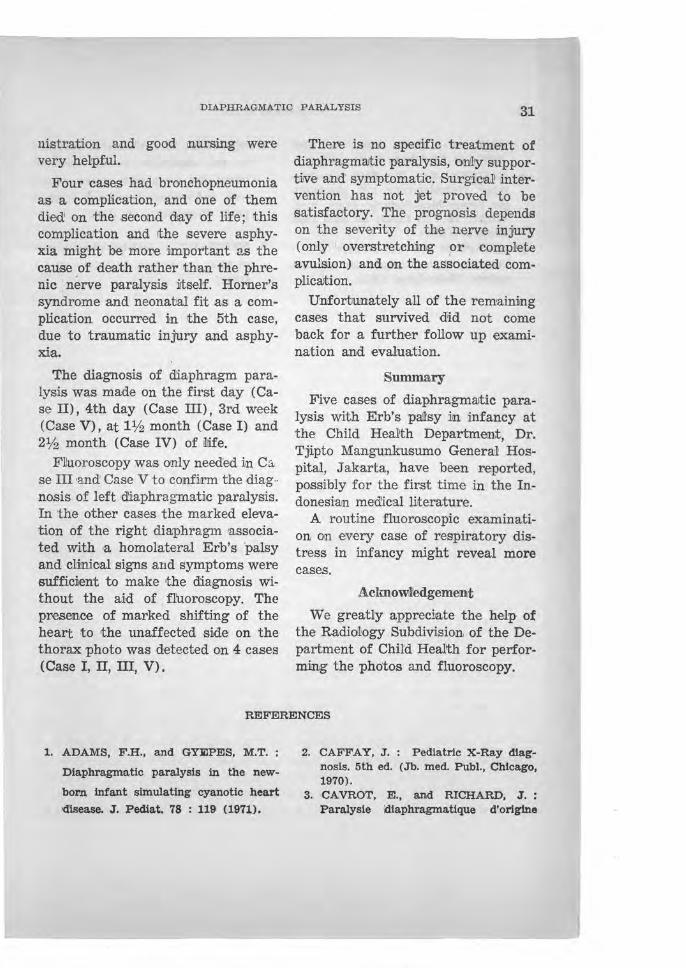
FIG. 1. Case I - Rontgenogram of the thoTax showing no abnormality.

' FIG. · 2. Case I - The second rontgenogram at 1 Yz month old showing right hemidiaphragm paralysis and marked shifting of the hoort to the left side.
I

FIG. 3. Case II- Rontgenogram of the thorax showing bronchopneumonia, rig1vt hemidiaphragm paralysis and shifting of the h ear t to the left side.

FIG. 4. Case III- Rontgenogram with Ba-contrast (orally) showing the left hemidiaphragm elevated slightly above >the right side and the m ediastinum was shifted to the right side. Fluoroscopical examination confirmed the diagnosis of lef t hemidiaphragm paralysis.

FIG. 5. Case IV- Rontgenogram of the thorax showing bronchopneumonia> right hemidiaphrogm
paralysis and shifting of the heart to the left side.

FIG . . 6. Case V - The fir&t rontgenogram of the thorax showing no abnormality except for the thymic gland enlargement.

FIG. 7. Case V - The second rontgenogram showing the left hemidiaphragm paralysis with the heart shifited to the right side. Further fluorosocpidal examination confirmed the diagnosis.

DIAPHRAGMATIC PARALYSIS 27
and the mediastinum seemed to be shifted to the left ;(see fig. 3). Intravenous fluid driiP by an indweliJiing umbilical catheter of a solutiJOn containing glucose 10% and sodtum bicarbonate 1.5% was given, besides antibiotics (PenidHine and Kanamycin) rund oxygen intermittently. Examination at the following days revealed Erb',s palsy of the right arm. Unfortunatelly the condibon became worse, and the infant died on the second day.
C,ase III :
P, a female Indonesian baby, was born on Jul'y 20, 1971, to a 35-yearold mother at the Obstetric Department of the Dr. Tjipto Mangunkusumo General Hospital. She was. the seventh child of a healthy mother. Pregnancy was uneventful. The delivery was at the begirnning attended by an j,ndige:nous midwife rut home. Because of the shoulder ;presentation with the left arm prola,psing the mother was sent to •the hospital. A Caesari·an Section was immediate1ly performed under ketal•ar and ether anaesthesia. The Apgar score wa,s two in 'the first minute and six in the fifth minute. Mouth to mouth respira;tion, oxygen and intravenous ,gl'ucose 40% and sodium bicarbonate 7.5% were given.
The baby was lethargic, dyspnoeic and cyano1tic. The Moro .refl'ex was negative. The body weight was. 2950 gm, the body length 50 em. The thorax was asymmetriJCal with a hematoma rund edema in the l,ef!t Ulpper hemitho-
rax and drudng inSIPiration this part moved more than the right hemithorax. Fine motslt rarles were heard over the base of both lll!llgs. The thorax photo revealed infiltrates suggestive of bronchopneumonia, without apparent abnormality of the diaphragm. Intravenous drip, antibiotics, oxygen rund Sipecial ruursing care were g1ven. On the fo['lowing days exami .. nation revealed the presence of a right hand palsy and persisting tachypnea without !apparent cyanosis.
.Ait the age of 4 days., the thorax photo wa.s re!Peated with Ba orally and showed the left hemidiaphragm elievated s1Eghtly above the right and the mediastinum shifted slight[~ to the right (see fig. 4). On fluoroscopy a paradox•al movement of the left hemidiaphragm was obvious, so the presence of a phrenic nerve injury wa.s confirmed. On the 9th day the baby was discharged by request of the parents; the tachypne1a disappeared and the baby thrived well.
Crase IV :
H, a male Indone.s1an infant of 2% months o]d, was admitted to the Depar1tment of Child Health, Dr. Tjipto Mangunkusumo General Hospita.l on August 14, 1972. He was born to a 24-year-o}id 'mother at a maternity hos!Pital wi1th a breech presentation. The birth weight was 3550 gm, the body length wa.s 49 em and mild asphyxi'a neonatorum was present. Erb's palsy of the righlt arm. and a slight tachypnea persisted when the

28 S. ZULKIFLI ET AL.
boy was discharged from the maternity hospital'. Three days rprior to admission, the boy beca~e more dyspnoeic foil'lowing a cough and a slight fever. The physical examination on admission revealed marked dyspnea (respiration rate was 68/minute) and marked Erb's pal'sy of the right hand. The right hemithorax was more flat a;n.d moved less than the left part. There was dullness With fine moist !'ales over the base of both lung.s. The thorax photo confirmed the diagnosis of bronchopneumonia. The right hemidiaphragm was el'evated about 3% interspaces above the left, and the heart was shilfted to the left (see fig. 5). By conservrutive treatment and antibiotics the tachypnea disappeared and by physiotherapy the arm pall'Sy improved. The boy was discharged after two weeks of hospitalization.
Case V :
N, a female Indonesian neonate was born on February 11, 1973, in the Obstetric Department of the Dr. Tjipto Mangunkusumo General Hospital. She was the first child of a 17-year-old mother with an uneventful pregnancy. The baby was delivered by fordpal extraction on the indication of toxemia parturientum, premature rupture of the membrane and persistent occiput posterior position of the head. She was asphyxiated, the Apgar score was 2 in the first minute and 4 in the fifth minute. Intermittent positive pressure ventila-
tion by endotracheal intubation was given, besides 10 ml sodium bicarbonate 7.5% and 4 ml glucose 40% intravenously i~1to the umbilical vein.
In the neonatal ward the baby remained weak with decreased Moro's and other physiologic reflexes. The body weight was 3220 gm, the body length 48 em and a large caput succedaneum was present on the head. She seemed to be dyspnoeic, slightly cyanotic with a respiration rate of 60 times per minute, but grunting and retraction were not detected. The photo of the thorax reveal1ed normal lungs and heart, and an en['argement of 'the thymic gland (see fig. 6). She was treated with antibiotics (Procaine Penicilline and Kanamycin), intravenous fluid drip of glucose 5% and sodium bicarbonate 1.5% in a four to one volume solution and oxygen.
The next day she developed a seizure of 5 minutes duration without fever. The cerebrospinal fluid and blood sugar content were within normar !limits. The neonatal fjt was treated successfully with va:ltium and dilantin. On the fifth day, with the improvement of motoric function, a left Erb's palsy was clearly seen accompanied by ptosis. and miosis of the left eye. The X-Ray of the head and neck revealed no abnormal'ity. On the lOth day of hospitali,zation she contracted bronchopneumonia and :lin acute infection of the midd:le ear; proper antibiotics were given

DIAPHRAGMATIC PARALYSIS 29
adequately. After clinical improvement of the bronchopneumonia rthe tachypnea still persislted 1and a second thorax photo was mad.e. The left hemidia:phragm was markedly more elevated than the right a:nd 1the heart was shifted to the right side of the chest (see f1g. 7). fThe fluoroscopic examination ·showed a paradoxa! mo· vement of the left hemidiaphragm and ,confirmed the diagnosis of phrenic nerve paralysis.
The general condition improved graduaJNy, the dyspnea disappeared, except for the persisting slight paresis of the [eft arm and the [~ft eye. The baby was discharged rut the age of 4 weeks. Physiotherapy was contitnued ambulattor.iJy.
Discussion
Phrenic ;nerve paralysis in run infant is usuany present in rthe first 24 hours of l'irfe. Most babies show lethargy and respiratory difficulties at birth. This poor reSipiratory condition may be due primariJy to the difficuiTt dellivery rather than to (the phrenic nerve paralysis itself. The respiratory dlistress varlites from '3
slight tachypnea to a severe respiratory distress syndrome which may even terminate in death. The affected hemithorax gives mostly a. decreased movement, bul. sometimes it shows flaring during inspj:ration which may be due to the pronounced effect of intercostal mus-
cles ~ontraction with diminished diaphragmatic corutraction. The tachypnea usually persists without any distimet abnormal finding. After several days or weeks the symptoms may disappear graduallly, but may also increase in severity. ,The latter may be due to !fue needs of more expanding llumg eapacity for the increasing necessity of oxygen consumption of the growing infant. This phrenic nerve paralysis may resemble any of the other [Causes of respiratory dj,stress such as diJ~phragmatic hernia, bronchopneumonia, atelectasis, massive aspiration ·syndrome, hyaline membrrune disease, congenital heart disease and cerebral injury.
Adams and Gyepes 1 (1971) reported a case of respiratory distress which was diagnosed iniUally as a cyanotic congenital: heart di~se,ase;
however heart ,crutheterisation exclud~ the !heart disease, and the thorax photo as confirmed by the t1luoroscopic examination reveal~ the diagnosis of phrenillc nerve palsy. lit is also interesting to ):lote that in their case the lright phrenic nerve paralysis was accompanied by the left Erb's palsy.
This contralateral combination has not been reported yet before. The thorax photo can help to diagnose phrenic nerve paralysis jf there is a marked elevation of one of the diaphragms ( usUial1y the right side) , and often wjth obvious shifting of the mediastinum to the unaffected side. N orm'ally, ,dn ~he first six

30 S. ZULKIFLI ET AL.
months of life, the left hemidiaphragm is Sllightly higher than the right one ;aJnd [n the second half year the
' I two hemidiaphragms are usually on the same level; after the first year of life the right hemidiaphragm is always hi1gher than the left (Caffey, 1970). But of rourse there are some exceptional case·s. If the hemidiaphragm is elhwated abnormalJ.Iy, especially if associated! w.i!th brachial palsy, then the diagnosis of phrenic nerve paralysis is justified even if only based on the thorax photo. But sometimes ithe elevation of the diaphra.gm is not qu.ilte so obvious, so that the diagnosis can only pe confirmed by fluoroscopic examination. We may see the see-saw or para:doxal movement of the affeCJted hemidiaphragm; it moves upwardly instead of downwardly d'urirng i!nspiration despite normal movement of the unaffected hemidiaphr.agm.
In our cases, 3 out of 5 cases ( Case I, II, IV) were del1ivered in breech presentation with some degree of difficulty in .shoulder or head delivery; but all of them were !3-Ssocia.ted wilth right Erb's palsy. The other 2 cases, one case (Case III) was in transverse (shoulder) . presel1Jtation and the other one (Case III) was in the vertex presen1tation. In the 3rd case, an operation was done because of a shoulder presentation with prolapse of the arm; this case had a contralaJteral parallysis of the right brachial nerve. This may be due to the hyperextension of the left :phrenic
nerve by the prolapse of the left arm, and the pullirng out !the right 'arm during srurgicall delivery of the body. The 5th case was deil!ivered by forcipal extraction, wi'th the complication of ~eft phrenic nerve paralysis, homolateral Erb's paJlisy and Horner's syndrome.
So 3 cases (Case I, II, IV) had homo1art::eraJ paralysis of the right side, one case (Case V) had homo-1ateral paralysis of the left side, and one case (Case III) had contralateral pamlysis of right brachial and Veft phrenic nerve. Adams amd Gyepes (1971) r8ported a similar case of such a contralateral paralysis.
Two j]ases of breech position (Case I, II), pne case (Case III) of transverse presentation amd one case (Case V) of persistent occiput position were delivered in the Obstetric Department of the Dr. Tjipto Mangunkusumo General Hospital, the other case (Case IV, breech presentation.) came from a maternilty hospital. In 1971 there were 3236 deliveries at the Obstetric Depal'itment, among wh:iJch : 275 cases (8.25°/o) were breech deliveries and 122 cases (3.13%) were !in the transverse presenltation; the daJbas of 1973 are not avaHable yet. No cases of phrenic nerve paralysis occurred m 1972.
In alft 5 cases, asphyxia neonatorum was ·encoun:tered. However, resuscjltatiOilll had saved their lives. Suction of the mucus in the airway passage, correction of the acidosis and caloric supply besides oxygen admi-
DIAPHRAGMATIC PARALYSIS 31
nistration and good nursing were very hel:pful.
Four cases had bronchopneumonia as a complication, and one of them died! on the .second day of life; this complication and !the severe asphyxia might be more important as the cause of death rather than the phrenic : nerve paralysis itself . . Horner's syndrome and neonatrai fit as a complication occurred iln the 5th case, due to traumatic injury and asphyxi,a.
The diagnosis of diaphragm paralysis was made on the first day ( Case II), 4th day (Case III), 3rd week (Case V) , at 1 liz month (.Case I) and 2l!z month (Case IV) of illife.
F]uoroscopy was only need'ed in Ci1 se III and' Case V to confi,rm the diag·· nosis of left dJ:aphragmatic paralysis. In the other cases the marked elevation of the right diaphragm associated with 'a homolateral Erb's 'palsy and clinioal signs and symptoms were sufficient to make the diagnosis without the aid of , fluoroscopy. The presence of marked shifting of the heart to the unaffected silde on the thorax photo was detected on 4 cases (Case I, II, III, V) .
There is no .specific treatment of diaphragmatic paralysis, on~y supportive and symptomatic. Surgical' intervention has not J~t proved to be satisfaCitory. The prognosis , depends on the severity of the nerve injury (only ' overstretching ,or complete avursion) and on the associated complicrution.
Unfortunately all of the remaining cases that survived did not come back for a further follow up examination and evaluation.
Summary
Five cases of diaphragmaJtic paralysis with Erb's pallsy in in£ancy at the Child Health Department, Dr. Tjipto Mangunkusumo General: Hospital, Jakarta, have been reported, possibly for the fi~st time in the Indonesi'am med'ical literature.
A routine fluoroscopic examination on every case of respiratory distress in infancy Inight reveal more cases.
Aclrno\dedgement
We greatly appredate the help of the Radidlogy Subdivision of the Department of Child Health for performing the photos and fluoroscopy.
REFERENCES
1. ADAMS, F .. H., and GY~PES, M.T. ;
Diaphragmatic paralysis in the new
born infant simulating cyanotic heart
disease. J. Pediat. 78 : 119 (1971).
2. CAFFAY, J. : Pediatric X-Ray diagnosis. 5th ed. (Jb. med. Publ., Chicago, 1970).
3. CA VROT, E., and RICHARD, J. : Paralysie lliaphragma.tique d'origine

32 S. ZULKIFLI ET AL.
obstetricale. Brux. ~med. , 3 : 1366 (1957) as cited from Smith (1972).
'· NAUNYN, B. : Ein Faile von Erb'scher Plexuslii.hmung mit gleichseitiger Sympathicu.slii.hmung. Dtsch. med. Wschr., 28 : 52 ~1902) ,as cited from Schifrin (1952) . '
5. SCHAFFER, A .J . and A VERY, M.E. : Diseases of the newborn, 3rd ed. (Saunders, Philadelphia, 1971).
6. SCHIFRIN, i N. : Unilateral paralysis
of the diaphragm in the newborn
infant due to phrenic nerve injury,
with and without , associated brachia]
palsy. PediiaJtrics : 69 (1952).
7. SMITH, B.T. : Isolated phrenic ner
ve palsy in newborn. Pediatrics 49 : 3
(1972) .