Bilateral Diaphragmatic Paralysis After Esophagectomy
6
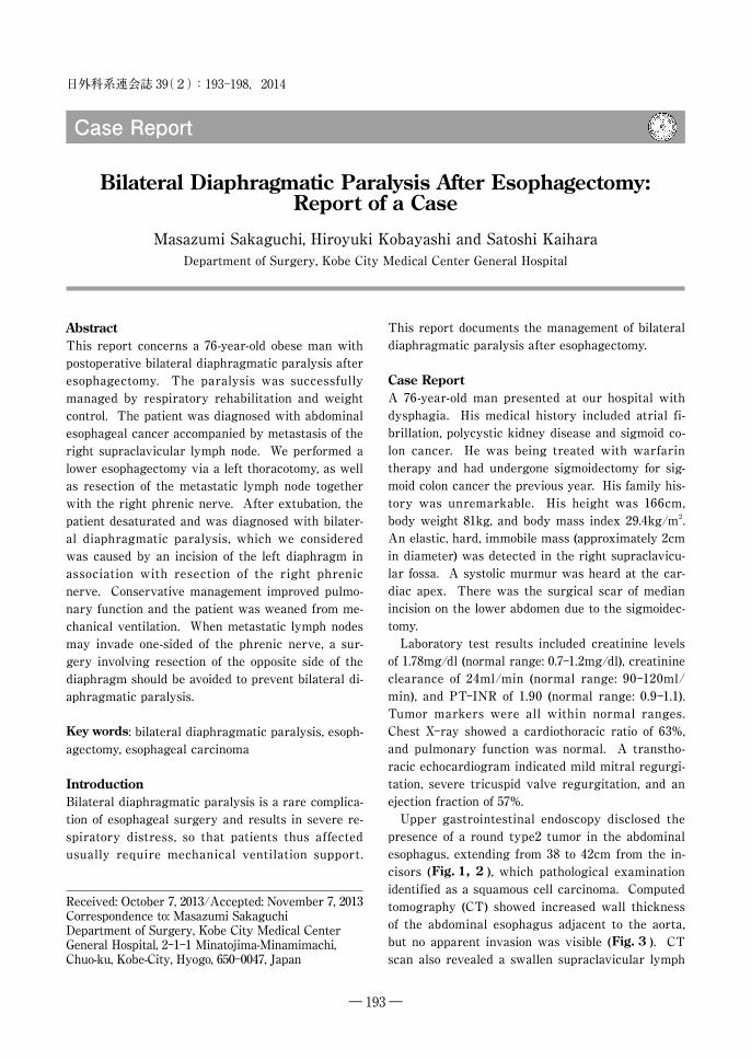
― 193 ― Abstract This report concerns a 76-year-old obese man with postoperative bilateral diaphragmatic paralysis after esophagectomy. The paralysis was successfully managed by respiratory rehabilitation and weight control. The patient was diagnosed with abdominal esophageal cancer accompanied by metastasis of the right supraclavicular lymph node. We performed a lower esophagectomy via a left thoracotomy, as well as resection of the metastatic lymph node together with the right phrenic nerve. After extubation, the patient desaturated and was diagnosed with bilater- al diaphragmatic paralysis, which we considered was caused by an incision of the left diaphragm in association with resection of the right phrenic nerve. Conservative management improved pulmo- nary function and the patient was weaned from me- chanical ventilation. When metastatic lymph nodes may invade one-sided of the phrenic nerve, a sur- gery involving resection of the opposite side of the diaphragm should be avoided to prevent bilateral di- aphragmatic paralysis. Key words: bilateral diaphragmatic paralysis, esoph- agectomy, esophageal carcinoma Introduction Bilateral diaphragmatic paralysis is a rare complica- tion of esophageal surgery and results in severe re- spiratory distress, so that patients thus affected usually require mechanical ventilation support. Received: October 7, 2013/Accepted: November 7, 2013 Correspondence to: Masazumi Sakaguchi Department of Surgery, Kobe City Medical Center General Hospital, 2-1-1 Minatojima-Minamimachi, Chuo-ku, Kobe-City, Hyogo, 650-0047, Japan This report documents the management of bilateral diaphragmatic paralysis after esophagectomy. Case Report A 76-year-old man presented at our hospital with dysphagia. His medical history included atrial fi- brillation, polycystic kidney disease and sigmoid co- lon cancer. He was being treated with warfarin therapy and had undergone sigmoidectomy for sig- moid colon cancer the previous year. His family his- tory was unremarkable. His height was 166cm, body weight 81kg, and body mass index 29.4kg/m 2 . An elastic, hard, immobile mass (approximately 2cm in diameter) was detected in the right supraclavicu- lar fossa. A systolic murmur was heard at the car- diac apex. There was the surgical scar of median incision on the lower abdomen due to the sigmoidec- tomy. Laboratory test results included creatinine levels of 1.78mg/dl (normal range: 0.7-1.2mg/dl), creatinine clearance of 24ml/min (normal range: 90-120ml/ min), and PT-INR of 1.90 (normal range: 0.9-1.1). Tumor markers were all within normal ranges. Chest X-ray showed a cardiothoracic ratio of 63%, and pulmonary function was normal. A transtho- racic echocardiogram indicated mild mitral regurgi- tation, severe tricuspid valve regurgitation, and an ejection fraction of 57%. Upper gastrointestinal endoscopy disclosed the presence of a round type2 tumor in the abdominal esophagus, extending from 38 to 42cm from the in- cisors ( Fig. 1, 2 ), which pathological examination identified as a squamous cell carcinoma. Computed tomography (CT) showed increased wall thickness of the abdominal esophagus adjacent to the aorta, but no apparent invasion was visible ( Fig. 3 ). CT scan also revealed a swallen supraclavicular lymph 日外科系連会誌 39 (2):193–198,2014 Case Report Bilateral Diaphragmatic Paralysis After Esophagectomy: Report of a Case Masazumi Sakaguchi, Hiroyuki Kobayashi and Satoshi Kaihara Department of Surgery, Kobe City Medical Center General Hospital
-
Upload
khangminh22 -
Category
Documents
-
view
1 -
download
0
Transcript of Bilateral Diaphragmatic Paralysis After Esophagectomy
― 193 ―
AbstractThis report concerns a 76-year-old obese man with postoperative bilateral diaphragmatic paralysis after esophagectomy. The paralysis was successfully managed by respiratory rehabilitation and weight control. The patient was diagnosed with abdominal esophageal cancer accompanied by metastasis of the right supraclavicular lymph node. We performed a lower esophagectomy via a left thoracotomy, as well as resection of the metastatic lymph node together with the right phrenic nerve. After extubation, the patient desaturated and was diagnosed with bilater-al diaphragmatic paralysis, which we considered was caused by an incision of the left diaphragm in association with resection of the right phrenic nerve. Conservative management improved pulmo-nary function and the patient was weaned from me-chanical ventilation. When metastatic lymph nodes may invade one-sided of the phrenic nerve, a sur-gery involving resection of the opposite side of the diaphragm should be avoided to prevent bilateral di-aphragmatic paralysis.
Key words: bilateral diaphragmatic paralysis, esoph-agectomy, esophageal carcinoma
IntroductionBilateral diaphragmatic paralysis is a rare complica-tion of esophageal surgery and results in severe re-spiratory distress, so that patients thus affected usually require mechanical ventilation support.
Received: October 7, 2013/Accepted: November 7, 2013 Correspondence to: Masazumi Sakaguchi Department of Surgery, Kobe City Medical Center General Hospital, 2-1-1 Minatojima-Minamimachi, Chuo-ku, Kobe-City, Hyogo, 650-0047, Japan
This report documents the management of bilateral diaphragmatic paralysis after esophagectomy.
Case ReportA 76-year-old man presented at our hospital with dysphagia. His medical history included atrial fi-brillation, polycystic kidney disease and sigmoid co-lon cancer. He was being treated with warfarin therapy and had undergone sigmoidectomy for sig-moid colon cancer the previous year. His family his-tory was unremarkable. His height was 166cm, body weight 81kg, and body mass index 29.4kg/m2. An elastic, hard, immobile mass (approximately 2cm in diameter) was detected in the right supraclavicu-lar fossa. A systolic murmur was heard at the car-diac apex. There was the surgical scar of median incision on the lower abdomen due to the sigmoidec-tomy.
Laboratory test results included creatinine levels of 1.78mg/dl (normal range: 0.7-1.2mg/dl), creatinine clearance of 24ml/min (normal range: 90-120ml/min), and PT-INR of 1.90 (normal range: 0.9-1.1). Tumor markers were all within normal ranges. Chest X-ray showed a cardiothoracic ratio of 63%, and pulmonary function was normal. A transtho-racic echocardiogram indicated mild mitral regurgi-tation, severe tricuspid valve regurgitation, and an ejection fraction of 57%.
Upper gastrointestinal endoscopy disclosed the presence of a round type2 tumor in the abdominal esophagus, extending from 38 to 42cm from the in-cisors (Fig. 1, 2 ), which pathological examination identified as a squamous cell carcinoma. Computed tomography (CT) showed increased wall thickness of the abdominal esophagus adjacent to the aorta, but no apparent invasion was visible (Fig. 3 ). CT scan also revealed a swallen supraclavicular lymph
日外科系連会誌 39(2):193–198,2014
Case Report
Bilateral Diaphragmatic Paralysis After Esophagectomy: Report of a Case
Masazumi Sakaguchi, Hiroyuki Kobayashi and Satoshi KaiharaDepartment of Surgery, Kobe City Medical Center General Hospital

日本外科系連合学会誌 第 39 巻 2 号
― 194 ―
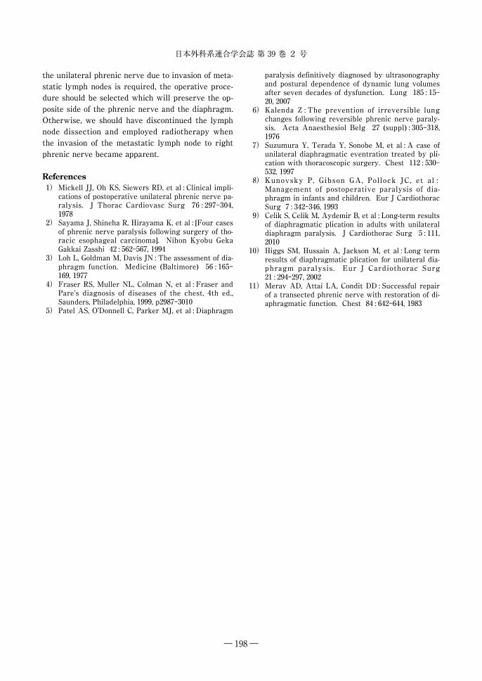
node (Fig. 4 ). 18F-Fluorodeoxyglucose-positron emission tomography showed an abnormal accumu-lation of the tracer in the abdominal esophagus and right supraclavicular lymph node (Fig. 5 ). The maximum standard uptake values of the esophagus and the lymph node were 15.40 and 7.80, respective-ly. Finally, there were no signs of recurrence of the
sigmoid colon cancer.The tumor was diagnosed as a cT3/N1/M1lym;
Stage Ⅳ esophageal cancer (TNM Classification, UICC, 7th edition, 2009). Although standard therapy was chemotherapy, the patient suffered from renal insufficiency due to polycystic kidney disease and heart failure caused by left ventricular dysfunction for the use of anticancer agents. Radiation therapy without chemotherapy was deemed to be insuffi-cient as a cure for the advanced carcinoma. Plus, the patient was suffered from malnutrition because of esophageal narrowing. He wished multidisci-plinary therapy giving oral intake his prior atten-tion. We therefore planned a lower esophagectomy to make oral food intake possible. Plus, we planned to excise the right supraclavicular lymph node sur-gically because the cervical metastatic lymph node was thought to cause severe symptoms. We also planned postoperative radiotherapy to the upper me-diastinum if possible.
We opened the abdomen via an upper median in-cision. The lower esophagus and cardia of the stom-ach were resected. We opened the left thorax by cutting the left diaphragm, continuing the abdomi-nal incision to make esophageal cutting surface can-cer-free, and dissecting the lower mediastinal lymph nodes. The oral esophagus was cut at the level of the left atrium, and we made a gastric tube and
Fig. 3 Enhanced computed tomogra-phy of the abdomen shows enhanced wall thickness of the abdominal esopha-gus adjacent to the aorta. Thin fat layer lying between tumor and aorta suggest-ed no aortic invasion of the cancer.
Fig. 2 Upper gastrointestinal series shows an irregular 4-cm-long defect in the abdominal esophagus.
Fig. 1 Upper gastrointestinal endoscopy shows a type 2 lesion in the abdominal esophagus at a distance of 38-42cm from the incisors.
Bilateral Diaphragmatic Paralysis After Esophagectomy
― 195 ―
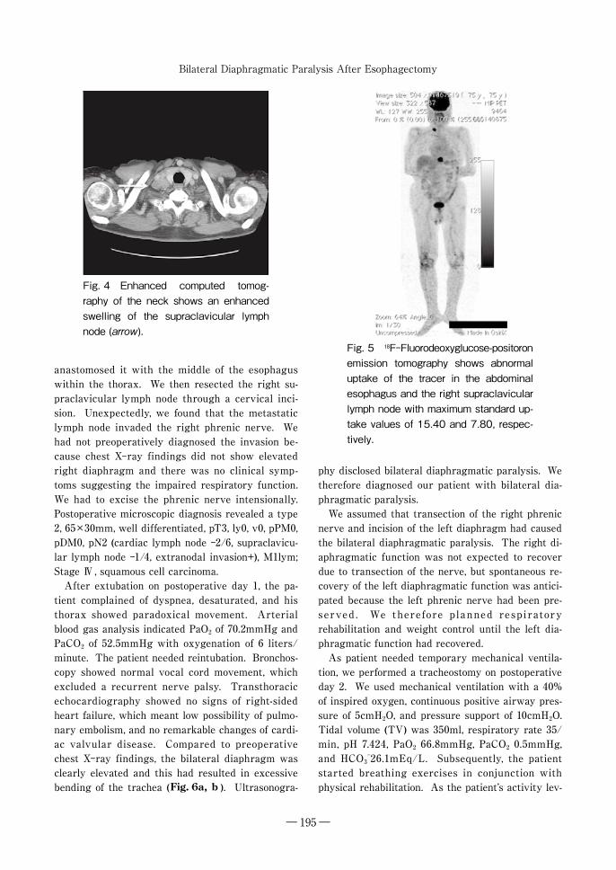
anastomosed it with the middle of the esophagus within the thorax. We then resected the right su-praclavicular lymph node through a cervical inci-sion. Unexpectedly, we found that the metastatic lymph node invaded the right phrenic nerve. We had not preoperatively diagnosed the invasion be-cause chest X-ray findings did not show elevated right diaphragm and there was no clinical symp-toms suggesting the impaired respiratory function. We had to excise the phrenic nerve intensionally. Postoperative microscopic diagnosis revealed a type 2, 65×30mm, well differentiated, pT3, ly0, v0, pPM0, pDM0, pN2 (cardiac lymph node -2/6, supraclavicu-lar lymph node -1/4, extranodal invasion+), M1lym; Stage Ⅳ , squamous cell carcinoma.
After extubation on postoperative day 1, the pa-tient complained of dyspnea, desaturated, and his thorax showed paradoxical movement. Arterial blood gas analysis indicated PaO2 of 70.2mmHg and PaCO2 of 52.5mmHg with oxygenation of 6 liters/minute. The patient needed reintubation. Bronchos-copy showed normal vocal cord movement, which excluded a recurrent nerve palsy. Transthoracic echocardiography showed no signs of right-sided heart failure, which meant low possibility of pulmo-nary embolism, and no remarkable changes of cardi-ac valvular disease. Compared to preoperative chest X-ray findings, the bilateral diaphragm was clearly elevated and this had resulted in excessive bending of the trachea (Fig. 6a, b ). Ultrasonogra-
phy disclosed bilateral diaphragmatic paralysis. We therefore diagnosed our patient with bilateral dia-phragmatic paralysis.
We assumed that transection of the right phrenic nerve and incision of the left diaphragm had caused the bilateral diaphragmatic paralysis. The right di-aphragmatic function was not expected to recover due to transection of the nerve, but spontaneous re-covery of the left diaphragmatic function was antici-pated because the left phrenic nerve had been pre-served . We therefore planned respiratory rehabilitation and weight control until the left dia-phragmatic function had recovered.
As patient needed temporary mechanical ventila-tion, we performed a tracheostomy on postoperative day 2. We used mechanical ventilation with a 40% of inspired oxygen, continuous positive airway pres-sure of 5cmH2O, and pressure support of 10cmH2O. Tidal volume (TV) was 350ml, respiratory rate 35/min, pH 7.424, PaO2 66.8mmHg, PaCO2 0.5mmHg, and HCO3
-26.1mEq/L. Subsequently, the patient started breathing exercises in conjunction with physical rehabilitation. As the patientʼs activity lev-
Fig. 5 18F-Fluorodeoxyglucose-positoron emission tomography shows abnormal uptake of the tracer in the abdominal esophagus and the right supraclavicular lymph node with maximum standard up-take values of 15.40 and 7.80, respec-tively.
Fig. 4 Enhanced computed tomog-raphy of the neck shows an enhanced swelling of the supraclavicular lymph node (arrow).

日本外科系連合学会誌 第 39 巻 2 号
― 196 ―
el increased, pulmonary function improved. During the recovery period, the patient tried to avoid the supine position and instead made every effort to as-sume sitting and prone positions. On postoperative day 15, respiratory rehabilitation without ventilator assistant was initiated. During training, the patient did not complain of dyspnea or desaturation. From postoperative day 41, he was able to undergo gait training without administration of oxygen, and from postoperative day 51, he required respiratory assis-tance only during the night. Finally, he was weaned from mechanical ventilation on postoperative day 90. His body weight had decreased from 81kg to 64kg (body mass index=23.4). On postoperative day 54, chest X-rays showed the bilateral diaphragms had moved downward one intercostal space in the inspiratory phase compared with in the expiratory phase (Fig. 6c, d ). On postoperative day 86, we initiated radiation therapy for the mediastinum (1.8
Gy×25 fraction, 50.4 Gy). The patient was dis-charged from the hospital on postoperative day 128 and he has remained healthy to date and, 10 months after esophagectomy, shows no signs of recurrences of esophageal carcinoma.
DiscussionThe phrenic nerve is important for breathing be-cause it passes motor information to and receives sensory information from the diaphragm. Most cas-es of acquired bilateral diaphragmatic paralysis are iatrogenic since the nerve is most frequently dam-aged during thoracic and cardiac operations1). Saya-ma et al. reported four cases of phrenic nerve paral-ysis following surgery of thoracic esophageal carcinoma and two cases of bilateral phrenic nerve paralysis2). To the best of our knowledge, this arti-cle is the only published report of bilateral diaphrag-matic paralysis after esophagectomy. According to
Fig. 6 Preoperative chest X-ray film shows no elevated diaphragm (a). The cardiotho-racic rate was 63%. After extubation, bilateral diaphragms became elevated (arrow), which led to excessive bending of the trachea (arrowhead) (b). On postoperative day 54, the bilateral diaphragm had moved downward one intercostal space during the inspiratory phase (arrow) (c), compared with the expiratory phase (d).

Bilateral Diaphragmatic Paralysis After Esophagectomy
― 197 ―
their report, the frequency of nerve paralysis was 1.9% for patients who underwent esphagectomy and 16% for patients who underwent extended lymph node dissection. They deemed that injury of the phrenic nerve was caused by lymph node dissection in neck and mediastinum and therefore concluded that it is important to pay special attention to the phrenic nerve during esophagectomy involving ex-tended lymph node dissection.
Unilateral paralysis may be asymptomatic, and in many cases the paralysis is detected during routine chest X-ray examination. Bilateral diaphragmatic paralysis causes a reduction in vital capacity and residual volume, which leads to hypoxia and hyper-capnia3). Diagnosis can be confirmed radiographi-cally by comparing the levels of both hemidia-phragms during inspiration and expiration on a standard chest X-ray. It is also useful to examine movement of the diaphragm using either ultrasonog-raphy or X-ray fluoroscopy4,5). In our case, we first suspected phrenic nerve paralysis because of clini-cal signs such as dyspnea and paradoxical respirato-ry movement of chest. The diagnosis of bilateral di-aphragmatic paralysis was established by means of ultrasonography.
The optimal management of bilateral diaphrag-matic paralysis remains controversial and varies ac-cording to the patientʼs physical condition, pulmo-nary function and cause of paralysis. If the phrenic nerve is preserved and spontaneous recovery can be expected, respiratory assistance and rehabilitation are indicated until phrenic nerve function recovers6). In cases where conservative management is not suc-cessful or the patient complains of dyspnea on ef-fort, diaphragm plication may be advisable7). The indication for, and optimal time of, intervention var-ies considerably8). Although a few articles have deal with the long-term results of diaphragmatic plica-tion for unilateral diaphragmatic paralysis, data on plication of both diagphragms for management of bi-lateral diaphragmatic paralysis are lacking9,10). If the phrenic nerve is transected, nerve repair has been recommended11).
In addition to bilateral diaphragmatic paralysis in our case, pain due to thoracotomy and laparotomy, pleural effusion, and obesity were thought to have led to worsening of pulmonary function. We fur-thermore believe that the immediate cause of the left diaphragmatic paralysis was not injury of the
phrenic nerve but a temporary dysfunction due to cutting of the left hemidiaphragm. Therefore, we anticipated spontaneous recovery of the left dia-phragmatic function and performed conservative management. While rehabilitation was continued, pain control and management of pleural effusion were employed. Because of the rehabilitation pro-gram to develop respiratory abdominal and back muscles, as well as bilateral diaphragmatic function-al recovery, our patientʼs pulmonary function im-proved and he could be weaned from mechanical ventilation. Moreover, the weight control program, which resulted in reduction of abdominal pressure, played an important role in improving pulmonary function.
As for therapeutic strategy of this patient, there might be a criticism that we should have chosen other indication instead of surgical operation. Basi-cally, standard therapy against Stage Ⅳ esophageal cancer should be chemotherapy. However, the pa-tient did not have enough renal and/or cardiac ca-pacity to endure the standard chemotherapy such as 5FU/cisplatin or 5FU/cisplatin/docetaxel. And his oral intake was not enough to continue treatment. We considered that oral intake should be retrieved from a viewpoint of quality of life. Plus, the mas-sive cervical metastatic lymph node could cause se-vere symptoms. Those were the reason we chosen surgical resection as the first therapeutic step of the multidisciplinary therapy. Although it resulted in unexpected complication, the patient could have an enriched quality of life for a certain period of time. Postoperatively we treated the patient with irradia-tion. We did the irradiation instead of mediastinal lymph nodes dissection which we should perform in the standard surgical operation against the esopha-geal cancer. We consider it might have some mean-ings for the cure of Stage Ⅳ esophageal cancer after macroscopic curative resection.
We consider that in our case the cause of bilateral diaphragmatic paralysis was the left thoracotomy which was performed in addition to transection of the right phrenic nerve due to invasion of the meta-static lymph node. As bilateral diaphragmatic pa-ralysis requires prolonged mechanical ventilation and can be a life-threatening complication, it should be avoided by all means. If we had chosen right in-stead of left thoracotomy, our patient would not have suffered such poor ventilation. When resection of
日本外科系連合学会誌 第 39 巻 2 号
― 198 ―
the unilateral phrenic nerve due to invasion of meta-static lymph nodes is required, the operative proce-dure should be selected which will preserve the op-posite side of the phrenic nerve and the diaphragm. Otherwise, we should have discontinued the lymph node dissection and employed radiotherapy when the invasion of the metastatic lymph node to right phrenic nerve became apparent.
References 1) Mickell JJ, Oh KS, Siewers RD, et al : Clinical impli-
cations of postoperative unilateral phrenic nerve pa-ralysis. J Thorac Cardiovasc Surg 76 : 297-304, 1978
2) Sayama J, Shineha R, Hirayama K, et al : [Four cases of phrenic nerve paralysis following surgery of tho-racic esophageal carcinoma]. Nihon Kyobu Geka Gakkai Zasshi 42 : 562-567, 1994
3) Loh L, Goldman M, Davis JN : The assessment of dia-phragm function. Medicine (Baltimore) 56 : 165-169, 1977
4) Fraser RS, Muller NL, Colman N, et al : Fraser and Pareʼs diagnosis of diseases of the chest, 4th ed., Saunders, Philadelphia, 1999, p2987-3010
5) Patel AS, OʼDonnell C, Parker MJ, et al : Diaphragm
paralysis definitively diagnosed by ultrasonography and postural dependence of dynamic lung volumes after seven decades of dysfunction. Lung 185 : 15-20, 2007
6) Kalenda Z : The prevention of irreversible lung changes following reversible phrenic nerve paraly-sis. Acta Anaesthesiol Belg 27 (suppl) : 305-318, 1976
7) Suzumura Y, Terada Y, Sonobe M, et al : A case of unilateral diaphragmatic eventration treated by pli-cation with thoracoscopic surgery. Chest 112 : 530-532, 1997
8) Kunovsky P, Gibson GA, Pollock JC, et al : Management of postoperative paralysis of dia-phragm in infants and children. Eur J Cardiothorac Surg 7 : 342-346, 1993
9) Celik S, Celik M, Aydemir B, et al : Long-term results of diaphragmatic plication in adults with unilateral diaphragm paralysis. J Cardiothorac Surg 5 : 111, 2010
10) Higgs SM, Hussain A, Jackson M, et al : Long term results of diaphragmatic plication for unilateral dia-phragm paralysis. Eur J Cardiothorac Surg 21 : 294-297, 2002
11) Merav AD, Attai LA, Condit DD : Successful repair of a transected phrenic nerve with restoration of di-aphragmatic function. Chest 84 : 642-644, 1983