Cold-induced disruption of Na+ channel slow inactivation underlies paralysis in highly...
10
J Physiol 587.8 (2009) pp 1705–1714 1705 Cold-induced disruption of Na + channel slow inactivation underlies paralysis in highly thermosensitive paramyotonia Thomas Carle 1 , Emmanuel Fournier 1,2 , Damien Sternberg 1,3 , Bertrand Fontaine 1,4 and Nacira Tabti 1 1 INSERM UMR 546, UPMC PVI, 2 F´ ed´ eration de Neurophysiologie Clinique, 3 D´ epartement de Biochimie and 4 F´ ed´ eration des Maladies du Syst` eme Nerveux, Groupe Piti´ e-Salpˆ etri` ere, Paris France The Q270K mutation of the skeletal muscle Na + channel α subunit (Nav1.4) causes atypical paramyotonia with a striking sensitivity to cold. Attacks of paralysis and a drop in the compound muscle action potential (CMAP) are exclusively observed at cold. To understand the pathogenic process, we studied the consequences of this mutation on channel gating at different temperatures. WT or Q270K recombinant Nav1.4 channels fused at their C-terminal end to the enhanced green fluorescent protein (EGFP) were expressed in HEK-293 cells. Whole-cell Na + currents were recorded using the patch clamp technique to examine channel gating at 30 ◦ C and after cooling the bathing solution to 20 ◦ C. Mutant channel fast inactivation was impaired at both temperatures. Cooling slowed the kinetics and enhanced steady-state fast inactivation of both mutant and WT channels. Mutant channel slow inactivation was fairly comparable to that of the WT at 30 ◦ C, but became clearly abnormal at 20 ◦ C. Cooling enhanced slow inactivation in the WT by shifting the voltage dependence toward hyperpolarization, but induced the opposite effect in the mutant. Destabilization of mutant channel slow inactivation in combination with defective fast inactivation is expected to increase the susceptibility to prolonged membrane depolarization, and can ultimately lead to membrane inexcitability and paralysis at cold. Thus, abnormal temperature sensitivity of slow inactivation can be a determinant pathogenic factor, and should therefore be more widely considered in thermosensitive Na + channelopathies. (Received 13 November 2008; accepted after revision 12 February 2009; first published online 16 February 2009) CorrespondingAuthor N. Tabti: D´ epartement de Physiologie, INSERM UMR 546, Facult´ edeM´ edecine Piti´ e-Salpˆ etri` ere, 91 Bd de l’H ˆ opital, 75013 Paris. Email: [email protected] Abbreviations PC: paramyotonia congenita; EMG: electromyography; CMAP: compound muscle action potential; EGFP: enhanced green fluorescence protein; WT: wild type; HEK: human embryonic kidney; TTP: time to peak. Mutations of the skeletal muscle Na + channel are responsible for a wide spectrum of excitability disorders, classically divided into two broad clinical groups, i.e. periodic paralyses and myotonias (Cannon, 1996). A large fraction of Nav1.4 mutations identified to date are associated with paramyotonia congenita, a disease mainly characterized by episodes of myotonia induced by cold and aggravated by exercise. Muscle weakness may also occur in response to exertion or cold. We recently reported a mutation of the Nav1.4 (Q270K), which causes a peculiar phenotype of paramyotonia with unusual sensitivity to cold (Fournier et al. 2006). Cold weather or direct contact with cold water induced muscle stiffness followed by severe attacks of paralysis lasting from minutes to hours. At warm temperatures, most clinical symptoms disappeared. This was corroborated by the unique electro- This paper has online supplemental material. myographic pattern evoked by exercise trials performed at room temperature or after cooling the muscles. In patients with typical PC mutations (i.e. T1313M), exercise trials induced a decrease in CMAP amplitude that was further pronounced with cooling. By contrast, in patients carrying the Q270K mutation, a sustained drop in CMAP, concomitant with muscle paralysis, only occurred when exercise was performed at cold (Fournier et al. 2006). The voltage-gated Na + channel is critical to skeletal muscle excitability. It is made of a large α subunit (Nav1.4) that forms by itself a functional pore, and a small regulatory β-subunit (Barchi, 1983; Catterall, 2000). The Q270K mutation is located in the outermost part of the S5 segment of domain I (DI-S5) of Nav1.4, which consists of four homologous domains, each comprising six transmembrane segments (Noda et al. 1984; George et al. 1992). Only a few disease-causing mutations have been identified in this domain (Takahashi & Cannon, 1999; Wu et al. 2001), among which is L266V, a nearby C 2009 The Authors. Journal compilation C 2009 The Physiological Society DOI: 10.1113/jphysiol.2008.165787
-
Upload
newcastle-au -
Category
Documents
-
view
4 -
download
0
Transcript of Cold-induced disruption of Na+ channel slow inactivation underlies paralysis in highly...

J Physiol 587.8 (2009) pp 1705–1714 1705
Cold-induced disruption of Na+ channel slow inactivationunderlies paralysis in highly thermosensitive paramyotonia
Thomas Carle1, Emmanuel Fournier1,2, Damien Sternberg1,3, Bertrand Fontaine1,4 and Nacira Tabti1
1INSERM UMR 546, UPMC PVI, 2Federation de Neurophysiologie Clinique, 3Departement de Biochimie and 4Federation des Maladies du SystemeNerveux, Groupe Pitie-Salpetriere, Paris France
The Q270K mutation of the skeletal muscle Na+ channel α subunit (Nav1.4) causes atypicalparamyotonia with a striking sensitivity to cold. Attacks of paralysis and a drop in thecompound muscle action potential (CMAP) are exclusively observed at cold. To understand thepathogenic process, we studied the consequences of this mutation on channel gating at differenttemperatures. WT or Q270K recombinant Nav1.4 channels fused at their C-terminal end to theenhanced green fluorescent protein (EGFP) were expressed in HEK-293 cells. Whole-cell Na+
currents were recorded using the patch clamp technique to examine channel gating at 30◦C andafter cooling the bathing solution to 20◦C. Mutant channel fast inactivation was impaired atboth temperatures. Cooling slowed the kinetics and enhanced steady-state fast inactivation ofboth mutant and WT channels. Mutant channel slow inactivation was fairly comparable to thatof the WT at 30◦C, but became clearly abnormal at 20◦C. Cooling enhanced slow inactivation inthe WT by shifting the voltage dependence toward hyperpolarization, but induced the oppositeeffect in the mutant. Destabilization of mutant channel slow inactivation in combination withdefective fast inactivation is expected to increase the susceptibility to prolonged membranedepolarization, and can ultimately lead to membrane inexcitability and paralysis at cold. Thus,abnormal temperature sensitivity of slow inactivation can be a determinant pathogenic factor,and should therefore be more widely considered in thermosensitive Na+ channelopathies.
(Received 13 November 2008; accepted after revision 12 February 2009; first published online 16 February 2009)Corresponding Author N. Tabti: Departement de Physiologie, INSERM UMR 546, Faculte de Medecine Pitie-Salpetriere,91 Bd de l’Hopital, 75013 Paris. Email: [email protected]
Abbreviations PC: paramyotonia congenita; EMG: electromyography; CMAP: compound muscle action potential;EGFP: enhanced green fluorescence protein; WT: wild type; HEK: human embryonic kidney; TTP: time to peak.
Mutations of the skeletal muscle Na+ channel areresponsible for a wide spectrum of excitability disorders,classically divided into two broad clinical groups, i.e.periodic paralyses and myotonias (Cannon, 1996). Alarge fraction of Nav1.4 mutations identified to date areassociated with paramyotonia congenita, a disease mainlycharacterized by episodes of myotonia induced by cold andaggravated by exercise. Muscle weakness may also occurin response to exertion or cold. We recently reported amutation of the Nav1.4 (Q270K), which causes a peculiarphenotype of paramyotonia with unusual sensitivity tocold (Fournier et al. 2006). Cold weather or directcontact with cold water induced muscle stiffness followedby severe attacks of paralysis lasting from minutes tohours. At warm temperatures, most clinical symptomsdisappeared. This was corroborated by the unique electro-
This paper has online supplemental material.
myographic pattern evoked by exercise trials performedat room temperature or after cooling the muscles. Inpatients with typical PC mutations (i.e. T1313M), exercisetrials induced a decrease in CMAP amplitude that wasfurther pronounced with cooling. By contrast, in patientscarrying the Q270K mutation, a sustained drop in CMAP,concomitant with muscle paralysis, only occurred whenexercise was performed at cold (Fournier et al. 2006).
The voltage-gated Na+ channel is critical to skeletalmuscle excitability. It is made of a large α subunit(Nav1.4) that forms by itself a functional pore, and asmall regulatory β-subunit (Barchi, 1983; Catterall, 2000).The Q270K mutation is located in the outermost part ofthe S5 segment of domain I (DI-S5) of Nav1.4, whichconsists of four homologous domains, each comprisingsix transmembrane segments (Noda et al. 1984; Georgeet al. 1992). Only a few disease-causing mutations havebeen identified in this domain (Takahashi & Cannon,1999; Wu et al. 2001), among which is L266V, a nearby
C© 2009 The Authors. Journal compilation C© 2009 The Physiological Society DOI: 10.1113/jphysiol.2008.165787

1706 T. Carle and others J Physiol 587.8
mutation characterized by cold-aggravated myotonia butno weakness (Wu et al. 2001).
One of the common defects of mutant channelsassociated with paramyotonia is the slowing of entry tofast inactivation. Cold exacerbates this slowing to a degreethat may represent the biophysical threshold for clinicalmyotonia (Bouhours et al. 2004, 2005). On the otherhand, a number of studies on Na+ channelopathies withpredominant muscle weakness point to a majorpathogenic contribution of altered channel slowinactivation to paralysis (Ruff, 1994; Cummins &Sigworth, 1996; Hayward et al. 1999; Ruff & Cannon,2000). To date, there are very few reports on temperaturesensitivity of slow inactivation (Ruff & Cannon,2000), although inherited Na+ channelopathies withvarious temperature-sensitive phenotypes are increasinglyacknowledged (Fournier et al. 2006; Rossignol et al. 2007;Webb & Cannon, 2008). To elucidate the mechanismbehind the peculiar phenotype caused by the Q270Kmutation, we investigated both fast and slow inactivationat different temperatures (30 and 20◦C). Our results showsimilar impairment of fast inactivation to that reportedfor the L266V mutation (Wu et al. 2001), but uniquetemperature-dependent alterations in slow inactivationthat can explain the occurrence of muscle paralysis in acold environment.
Methods
Plasmid construction, mutagenesis and celltransfection
The human SCN4A cDNA construct was subclonedinto the pEGFP-N2 vector (Clontech) for expression inmammalian cells. The DNA sequence for EGFP was inframe to the 3′-terminal end of the SCN4A sequenceimmediately before the stop codon. In the resulting fusionprotein, the C-terminus of Nav1.4 was fused to theN-terminus of EGFP. The Q270K mutation was insertedinto the SCN4A-EGFP cDNA construct by overlap PCR.The final cDNA product was entirely sequenced to verifythat the Q270K mutation was present, and that no othervariant had been introduced by accident during DNAamplification.
Human embryonic kidney cells (HEK 293) weretransfected with WT or Q270K SCN4A-EGFP constructsusing the calcium phosphate precipitation method. Cellswere routinely grown in a Dulbecco’s modified Eagle’smedium (DMEM) supplemented with 10% fetal calfserum (Bouhours et al. 2004, 2005). Na+ currents wererecorded from both transiently and stably expressing cells(at least 2 clones) in order to ensure that no bias wasintroduced by cell selection. The basic gating properties ofNav1.4 WT channels fused to EGFP were first comparedwith those of the non-fused channels to ascertain than
EGFP did not modify channel function (SupplementalFig. 1).
In vitro electrophysiology
Na+ currents were recorded in the whole cell configurationof the patch-clamp technique (Hamill et al. 1981), usingan EPC-7 amplifier (HEKA electronics). After breakinginto the cell, 10–15 min was allowed for Na+ currentstabilization. Standard pulse-evoked currents were filteredat 5 kHz through an 8-pole low-pass Bessel filter (M900,Frequency Devices, Corp., Sunnyvale, CA, USA), andusually digitized at a sampling rate of 20 μs using Digidata1332 and the pCLAMP software (Axon Instruments,Union City, CA, USA). P/N protocols (Armstrong &Bezanilla, 1977) from a holding potential (HP) of –120 mVwere applied to subtract residual leakage and capacitycurrents. Bath temperature was maintained at either 30 or20◦C by water flow through a Peltier-based thermal stage(HE-201, Dagan) connected to a temperature controller(HCC-100A). Pipettes were pulled from Drummondcapillary glass using a Sutter P-97 puller. All pipetteswere filled with a caesium-based solution (mM): 130 CsCl,2 MgCl2, 10 glucose and 10 hemi-Na-Hepes (pH 7.4),and had a resistance of 1.1–1.8 M�. The bath solutioncontained (mM): 100 NaCl, 42 choline-Cl, 3 KCl, 2 MgCl2,1 CaCl2, 10 glucose and 10 Hepes (pH 7.4). Data wereanalysed using Clampfit 9.2 (Axon Instruments), Micro-soft Excel, CorelDraw 12 and Origin 7.5 (OriginLabCorp., Northampton, MA, USA) software. All values areexpressed as means ± standard error of the mean (S.E.M.).Student’s unpaired t-test was used for statistical analysis.
The peak conductance G(V ) was computed fromthe peak current, and normalized conductance versusvoltage curves were fitted to the Bolzmann equation (seeFig. 1 legend). Steady-state inactivation curves were fittedto a two-state Boltzmann distribution (see Figs 3 and 4legends).
Results
Kinetics and voltage dependence of activation
Na+ currents were recorded from HEK cells expressingWT or Q270K channels fused with the green fluorescentprotein (EGFP) at 30 and 20◦C. Normalized I–V and G–Vcurves were plotted for each cell and then averaged forstatistical evaluation (Fig. 1). As can be seen from Fig. 1B,the Q270K mutation induced a very slight depolarizingshift in the voltage dependence of activation that wassomewhat better resolved at 30◦C (∼+3 mV) than at 20◦C(∼+1.5 mV). The slope factor (k) remained comparablebetween WT and mutant at both temperatures. Coolingfrom 30 to 20◦C did not significantly modify the voltage
C© 2009 The Authors. Journal compilation C© 2009 The Physiological Society

J Physiol 587.8 Human Nav.4 channel mutation disrupts slow inactivation at cold 1707
dependence of mutant or WT channel activation (seeTable 1).
Time to peak Na+ current (TTP: 10 to 90% rise time)was measured as a simple estimation of the activationkinetics from the crude data. As shown in Fig 1C, TTP wasslowed in Q270K mutant compared with WT, suggestinga decrease in the rate of channel activation. Note that thevoltage dependence of TTP remained unchanged.
At 30◦C, the mean values of TTP were 20 to 65%(average ∼45%) higher in Q270K mutant than in WT(voltage range −25 to +40 mV). Cooling from 30 to 20◦Csimilarly increased (by 30–35%) the TTP in both mutantand WT.
Kinetics and voltage dependence of fast inactivation
Fast inactivation is a process by which Na+ channelsbecome non-conducting following depolarization of theorder of milliseconds. It contributes thereby to therepolarizing phase of the action potential. Recoveryfrom fast inactivation terminates the refractory period,and enables the initiation of a new excitability cycle.Disruption of fast inactivation has been found in mostNav1.4 channelopathies in which myotonia predominates(Hayward et al. 1996).
To enable comparison between the Q270K andthe neighboring L266V mutation associated withcold-aggravated myotonia, a detailed analysis of thekinetics of fast inactivation was first performed at 20◦C(Fig. 2), a temperature at which the data for the L266Vwere obtained (Wu et al. 2001).
Recovery from fast inactivation was assessed by adouble-pulse protocol (see Fig. 2A). Q270K channelsrecovered faster from inactivation than the WT, asindicated by an average decrease of 46% in the timeconstant of recovery (τrecov) over the applied voltage range(Fig. 2D). The kinetics of entry to fast inactivation fromthe closed state (see Fig. 2B) were also accelerated by theQ270K mutation, as demonstrated by a 50% reductionin the time constant for the mutant compared with WT(Fig. 2D). The kinetics of entry to fast inactivation fromthe open state (Fig. 2C) were substantially slowed in themutant, as can be seen from the higher mean valuesof the inactivation time constant (τh) (Fig. 2D). TheQ270K mutation also reduced the voltage sensitivity ofτh, especially between −20 and 0 mV (Fig. 2D). All thesechanges were similar to those reported for the L266Vmutation (Wu et al. 2001). In particular, both mutationsinduced a crossover effect around −30 mV with fasterclosed state and slower open state inactivation kinetics ascompared to the WT. It is worth noting that the acceleratedclosed-state inactivation was not associated with ahyperpolarizing shift in the voltage dependence of steady-state inactivation. Instead, as shown below (Fig. 3B),
channel availability was increased for the Q270K mutantbetween −70 and −40 mV, suggesting a dominant effectof the accelerated rate of recovery from fast inactivation.
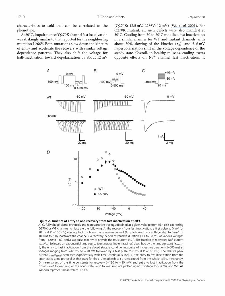
The effects of temperature were investigated on twomain characteristics of channel fast inactivation: the timeconstant τh, and the voltage dependence of steady-sateinactivation (Fig. 3).
At 30◦C, τh values obtained from the mutant were142–330% (average 247%) higher than those of the WT(voltage range −25 to +40 mV). Decreasing temperaturefrom 30 to 20◦C induced an increase in τh by 28–63%(average 44%) in the mutant compared with 37–78%(average 55%) in the WT. The difference in τh betweenmutant and WT at 20◦C ranged between 124 and 311%(average 224%), and was therefore slightly smaller thanthat found at 30◦C. Taken together these data suggest thattemperature sensitivity of the mutant is somewhat lowerthan that of the WT. Nevertheless, cooling enlarged theabsolute difference between mutant and WT as can beseen from Fig. 3A, and mean values of τh reached up to3.31 ms in the mutant as compared to 1.47 ms for theWT. Note that the τh/V relationship was not modified bycooling.
The voltage dependence of steady-sate fast inactivationwas assessed by a two-pulse protocol (see Fig. 3). At30◦C, steady-state inactivation curves (h∞) obtained fromthe Q270K mutant showed a shift of 13–14 mV towarddepolarization, and a reduced voltage sensitivity comparedwith WT (Fig. 3, see also Table 1). Decreasing temperaturefrom 30 to 20◦C caused a hyperpolarizing shift in h∞ to asimilar degree for both channel types (mutant: ∼−6 mV;WT: ∼−5 mV). Thus, the difference in V 0.5 between thetwo channels remained roughly the same upon cooling(Table 1). The slope factors obtained for mutant and WTwere not affected by this temperature change. Altogether,these data demonstrate an impairment of the voltagedependence of steady-state fast inactivation by the Q270Kmutation, but a normal response of this mutant channelcharacteristic to cooling.
Slow inactivation
Following prolonged depolarization Na+ channels shut offby a process that is distinct from fast inactivation, and isreferred to as slow inactivation (Rudy, 1978). In the skeletalmuscle, slow inactivation operates on a seconds time scaleand is thought to regulate the availability of excitable Na+
channels as a function of the resting membrane potential(Simoncini & Stuhmer, 1987; Ruff et al. 1988). It is nowwell established that human Nav1.4 channelopathies withaltered slow inactivation are associated with paralysisrather than myotonia (Hayward et al. 1997). In thepresent case, episodes of severe paralysis occur exclusivelyat cold. This led us to raise the hypothesis of an abnormal
C© 2009 The Authors. Journal compilation C© 2009 The Physiological Society

1708 T. Carle and others J Physiol 587.8
Figure 1. Voltage dependence of activation and effect of temperatureA, representative tracings of whole-cell Na+ currents recorded from HEK cells expressing either Q270K or WTchannels fused with EGFP. Recordings were performed at 30 or 20◦C using the voltage-clamp protocol shownat the top (HP −100 mV, voltage steps from −70 to +75 mV in 5 mV increments). B, normalized conductance(G/Gmax) versus voltage curves obtained for Q270K (30◦C (n = 8); 20◦C (n = 8)) and WT channels (30◦C (n = 7);20◦C (n = 9)). Na+ conductance was computed from the equation G(V ) = Ipeak/(V – ENa). The reversal potentialENa was determined by extrapolation of the regression line to zero current (voltages > 0 mV). The continuous
C© 2009 The Authors. Journal compilation C© 2009 The Physiological Society

J Physiol 587.8 Human Nav.4 channel mutation disrupts slow inactivation at cold 1709
Table 1. Boltzmann parameter estimates for Na+ channel activation and inactivation at 30 and 20◦C
30◦C 20◦C
WT Q270K WT Q270K
Activation V 0.5 −21.8 ± 0.3 −18.7 ± 0.5 −19.7 ± 0.5 −18.4 ± 0.2(mV) (7) (8) ∗∗∗ (9) (8) ∗
k 6.1 ± 0.1 5.8 ± 0.1 6.7 ± 0.2 6.3 ± 0.1(mV/e-fold) (7) (8)∗ (9) (8)∗
ENa (mV) +70.4 ± 1.4 +67.4 ± 0.7 +68.1 ± 0.9 +68.6 ± 0.6(7) (8) (9) (8)
Fast V 0.5 −60.6 ± 0.3 −46.8 ± 0.5 −65.3 ± 0.5 −52.8 ± 0.7inactivation (mV) (10) (8) ∗∗∗ (11)∗ (9) ∗∗∗
k 6.5 ± 0.1 8.2 ± 0.3 6.5 ± 0.1 8.9 ± 0.2(mV/e-fold) (10) (8) ∗∗∗ (11) (9)∗∗∗
Slow V 0.5 −49.4 ± 0.9 −47.8 ± 0.8 −53.8 ± 0.5 −42.7 ± 0.5inactivation (mV) (8) (12) (8) (8) ∗∗∗
k 11.6 ± 0.3 8.7 ± 0.5 11.2 ± 0.5 7.7 ± 0.2(mV/e-fold) (8) (12)∗∗∗ (8) (8)∗∗∗
Residual 7 ± 1 6 ± 1 6 ± 1 12 ± 2(%) (8) (12) (8) (8)∗∗
Values are mean ± S.E.M. (n). Statistical significance of the difference between Q270K and WT for eachtemperature: ∗P < 0.05, ∗∗P < 0.01, ∗∗∗P < 0.001.
slow inactivation gating in Q270K mutant channels atlow temperature. The sensitivity to temperature of Nav1.4slow inactivation has been addressed in a few studies,with discrepancies between reports that remain unsolved(Ruff, 1999, 2008; Webb & Cannon, 2008). Thus, besidesdisclosing the effects of Q270K mutation, the present studyprovides further information on temperature-dependentchanges of Nav1.4 slow inactivation.
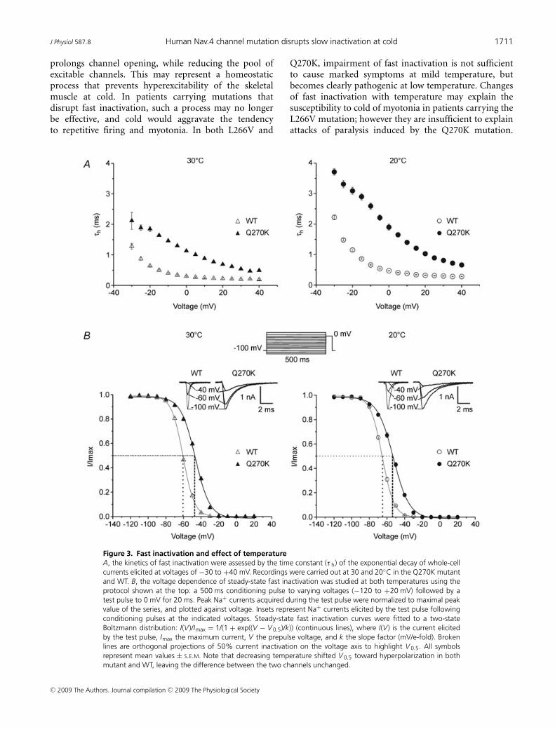
Steady-state slow inactivation was induced by a doublepulse protocol characterized by long prepulse potentials(50 s) (see Fig. 4). Steady-state slow inactivation curves(S∞) are depicted in Fig. 4. At 30◦C, mutant channelsshowed a minor depolarizing shift in V 0.5 (+1.6 mV) aswell as a decrease in the slope factor (of ∼3 mV/e-fold),i.e. steeper voltage sensitivity than the WT (Table 1). Theinteresting finding was the occurrence of a significantdifference of ∼11 mV between mutant and WT S∞ curveswhen temperature was decreased from 30 to 20◦C (Fig. 4).This apparent difference in the voltage dependence of slowinactivation between mutant and WT was the result of ahyperpolarizing shift of ∼−4.5 mV for the WT, combinedto a depolarizing shift of ∼+5 mV for the mutant. Thesteepness of both mutant and WT S∞ curves was not
lines represent the best fits to the Boltzmann function G(V )/Gmax = 1/(1 + exp(−(V − V 0.5)/k)), where Gmax isthe maximum conductance, V 0.5 the voltage for half-activation, and k the slope factor (mV/e-fold). C, kinetics ofchannel activation described by the time to peak (TTP). All symbols represent mean values ± S.E.M. Note the slightshift toward depolarization caused by the Q270K mutation. Cooling from 30 to 20◦C barely changed the voltagedependence of activation, and moderately slowed the activation kinetics in both mutant and WT.
significantly altered by cooling (Table 1). In addition,the fraction of Na+ channels that did not slow-inactivateincreased from 6 to 12% with cooling in the mutant, butdid not significantly change in the WT (7 and 6% at 30and 20◦C, respectively) (Table 1).
Discussion
Among skeletal muscle Na+ channelopathies,paramyotonia congenita is known for its sensitivity totemperature. Indeed, episodes of myotonia can be inducedor aggravated by cold. However, temperature-dependentsymptoms of either myotonia or paralysis have alsobeen observed in Nav1.4 channelopathies with atypicalphenotypes (Sugiura et al. 2000, 2003; Wu et al. 2001).For most cases, the mechanisms behind thermosensitivityof the symptoms remain unclear. One of the reasons isthat in vitro functional studies are usually performed atroom temperature (20–22◦C), and few data are availableon the temperature sensitivity of mutant Na+ channelgating. This is particularly true for slow inactivation. Ourstudy unveils Q270K mutant channel defects and providesevidence for specific responses of different channel gating
C© 2009 The Authors. Journal compilation C© 2009 The Physiological Society
1710 T. Carle and others J Physiol 587.8
characteristics to cold that can be correlated to thephenotype.
At 20◦C, impairment of Q270K channel fast inactivationwas strikingly similar to that reported for the neighboringmutation L266V. Both mutations slow down the kineticsof entry and accelerate the recovery with similar voltagedependence patterns. They also shift the voltage forhalf-inactivation toward depolarization by about 12 mV
Figure 2. Kinetics of entry to and recovery from fast inactivation at 20◦CA–C, full voltage-clamp protocols and representative tracings obtained at a given voltage from HEK cells expressingQ270K or WT channels to illustrate the following. A, the recovery from fast inactivation: a first pulse to 0 mV for20 ms (HP −100 mV) was applied to obtain the reference current (Iref), followed by a voltage step to 0 mV for100 ms to fully inactivate the channels, a recovery period of variable duration (0.1 to 38 ms) at various voltagesfrom −120 to −80, and a last pulse to 0 mV to provide the test current (Itest). The fraction of recovered Na+ current(Itest/Iref) followed an exponential time course (continuous line on tracings) described by the time constant (τ recov).B, the entry to fast inactivation from the closed state: a conditioning pulse of increasing duration (5–500 ms) atvoltages ranging from −40 mV to −70 mV followed by a test pulse to 0 mV (HP −100 mV). The relative peakcurrent (Itest/Icond) decreased exponentially with time (continuous line). C, the entry to fast inactivation from theopen state: same protocol as that used for the I–V relationship; τ h is measured from the whole-cell current decay.D, mean values of the time constants for recovery (−120 to −80 mV), and entry to fast inactivation from theclosed (−70 to −40 mV) or the open state (−30 to +40 mV) are plotted against voltage for Q270K and WT. Allsymbols represent mean values ± S.E.M.
(Q270K: 12.5 mV, L266V: 12 mV) (Wu et al. 2001). ForQ270K mutant, all such defects were also manifest at30◦C. Cooling from 30 to 20◦C modified fast inactivationin a similar manner for WT and mutant channels, withabout 50% slowing of the kinetics (τh), and 5–6 mVhyperpolarization shift in the voltage dependence of thesteady-state. Overall, in healthy muscles, cooling exertsopposite effects on Na+ channel fast inactivation: it
C© 2009 The Authors. Journal compilation C© 2009 The Physiological Society
J Physiol 587.8 Human Nav.4 channel mutation disrupts slow inactivation at cold 1711
prolongs channel opening, while reducing the pool ofexcitable channels. This may represent a homeostaticprocess that prevents hyperexcitability of the skeletalmuscle at cold. In patients carrying mutations thatdisrupt fast inactivation, such a process may no longerbe effective, and cold would aggravate the tendencyto repetitive firing and myotonia. In both L266V and
Figure 3. Fast inactivation and effect of temperatureA, the kinetics of fast inactivation were assessed by the time constant (τ h) of the exponential decay of whole-cellcurrents elicited at voltages of −30 to +40 mV. Recordings were carried out at 30 and 20◦C in the Q270K mutantand WT. B, the voltage dependence of steady-state fast inactivation was studied at both temperatures using theprotocol shown at the top: a 500 ms conditioning pulse to varying voltages (−120 to +20 mV) followed by atest pulse to 0 mV for 20 ms. Peak Na+ currents acquired during the test pulse were normalized to maximal peakvalue of the series, and plotted against voltage. Insets represent Na+ currents elicited by the test pulse followingconditioning pulses at the indicated voltages. Steady-state fast inactivation curves were fitted to a two-stateBoltzmann distribution: I(V )/Imax = 1/(1 + exp((V − V 0.5)/k)) (continuous lines), where I(V ) is the current elicitedby the test pulse, Imax the maximum current, V the prepulse voltage, and k the slope factor (mV/e-fold). Brokenlines are orthogonal projections of 50% current inactivation on the voltage axis to highlight V 0.5. All symbolsrepresent mean values ± S.E.M. Note that decreasing temperature shifted V 0.5 toward hyperpolarization in bothmutant and WT, leaving the difference between the two channels unchanged.
Q270K, impairment of fast inactivation is not sufficientto cause marked symptoms at mild temperature, butbecomes clearly pathogenic at low temperature. Changesof fast inactivation with temperature may explain thesusceptibility to cold of myotonia in patients carrying theL266V mutation; however they are insufficient to explainattacks of paralysis induced by the Q270K mutation.
C© 2009 The Authors. Journal compilation C© 2009 The Physiological Society
1712 T. Carle and others J Physiol 587.8
Interestingly, a major difference between these twomutations is their effect on slow inactivation. Indeed, at20◦C, slow inactivation of L266V mutant channels wasreported to be fairly normal, whereas that of Q270Kchannels is clearly impaired.
At 30◦C, the voltage at which half of the Q270K mutantchannels became slow inactivated was not significantlydifferent from that of the WT. However, when temperaturewas decreased to 20◦C, a difference of 11 mV in thevoltage for half-inactivation became visible between thetwo channel types. Cooling induced a hyperpolarizingshift of the voltage dependence of slow inactivation inthe WT but a depolarizing shift in the mutant.
Hyperpolarizing shift in S∞ curves of normal skeletalmuscle Na+ channels with cooling was previouslyobserved in rat muscle cells (Ruff, 1999). Similar responseto temperature seems to be shared by other TTX-sensitiveNa+ channels, such as those present in dorsal root ganglion(DRG) neurons (Zimmermann et al. 2007). However,in a recent study performed on HEK cells (Webb &Cannon, 2008), cooling was found to shift steady-stateslow inactivation of normal Nav1.4 channel in the oppositedirection. The discrepancy between these data and thoseobtained from muscle fibres remains to be clarified. Ourdata, which are in line with those of Ruff (1999), do notsupport the idea that this may be related to differences inexpression systems (Ruff, 2008; Webb & Cannon, 2008).Other experimental factors could be involved, such as the
Figure 4. Voltage-dependent steady-state slow inactivation and effect of temperatureSteady-sate slow inactivation curves obtained from the Q270K mutant and WT at 30 and 20◦C. Slow inactivationwas induced by the protocol shown at the top: 50 s prepulse potentials ranging from −120 to +10 mV werefollowed by a test pulse to 0 mV for 20 ms. A brief repolarizing step to −100 mV was applied for 20 ms priorto the test pulse to allow selective and full recovery from fast inactivation. Insets represent Na+ current tracingsevoked during the test pulse for different conditioning pulses as indicated. Data were fitted with a two-stateBoltzmann function with a non-zero pedestal I0: (I(V )/Imax = 1 − I0/1 + exp((V − V 0.5)/k)) + I0 (continuous lines,see Fig. 3 legend for equation terms). Symbols are mean values ± S.E.M. Note that a difference of about 10 mV inV 0.5 appears between mutant and WT when the temperature is decreased from 30 to 20◦C.
type of solutions used to study slow inactivation, and inparticular, the use of fluoride as a substitute for chloridein the internal pipette solution. Indeed, to improve sealstability during long protocols such as those required forNa+ channel slow inactivation, CsF is generally preferredto CsCl. Whether F− alters Na+ channel slow inactivationhas been subject to debate (Coste et al. 2004). Fluorideis known as a phosphatase inhibitor (Pinkse et al. 1999)which, by modifying the phosphorylation status of Na+
channels, may alter their gating (Cantrell & Catterall,2001; Chen et al. 2006). For these reasons, all experimentsassociated with the present study were performed usingCsCl-based intra-pipette solution.
Enhancement of Nav1.4 channel slow inactivation atcold may represent a protective factor against alterations ofmuscle excitability in conditions that generate long-lastingsarcolemma depolarization. In this respect, cold shiftsthe resting membrane potential toward depolarization byreducing Na+/K+ ATPase activity, and lengthens the actionpotential by slowing down the gating kinetics of both thevoltage-gated Na+ channels and the delayed rectifier K+
channels.If slow inactivation operates normally in mutant
channels with impaired fast inactivation, it will eventuallyshut off the defective channels and oppose the prolongedinflux of Na+. This would prevent long-lasting membranedepolarization that may lead to loss of excitabilityand paralysis (Ruff, 1994). Such process is particularly
C© 2009 The Authors. Journal compilation C© 2009 The Physiological Society

J Physiol 587.8 Human Nav.4 channel mutation disrupts slow inactivation at cold 1713
important at low temperatures where the abnormalNa+ influx is exacerbated, and the resting membranepotential depolarized. Accordingly, the L266V mutation,which preserves slow inactivation at cold, will mainlycause myotonia, whereas the Q270K, which does not,will cause paralysis at low temperatures. So far, onlyone other mutation of the human Nav1.4 (P1158S)has been reported to destabilize slow inactivation uponcooling. Consistent with the above reasoning, the latterwas also discerned by its unusual phenotype comprisingcold-induced attacks of paralysis (Webb & Cannon, 2008).
The molecular mechanism of Na+ channel slowinactivation is still unclear. Current hypotheses proposethat slow inactivation is linked to conformational changesof the pore region. Nonetheless, the contribution of theS5–S6 loops lining the outer pore (Balser et al. 1996;Vilin et al. 1999) versus the S6 transmembrane (TM)segments predicted to line the inner pore (Wang & Wang,1997; O’Reilly et al. 2001) is still under debate (Struyk &Cannon, 2002). The emerging consensus is that structuraldeterminants of slow inactivation involve both the outer(S5–S6 P-loops) and the inner vestibules (S5 and S6 TMsegments) of the ion conducting pore (Chancey et al. 2007;Tikhonov & Zhorov, 2007).
The Q270K mutation is located at the extracellular endof the S5 transmembrane segment of domain I, close tothe N-end of the S5–S6 pore loop. The functional roleof the amino-acids in this region remains to be clarified(Lipkind & Fozzard, 1997). Our results indicate thatthe Q270 residue contributes to temperature sensitivityof slow inactivation. Its replacement with lysine mayinterfere with conformational changes that promote orstabilize Na+ channel slow inactivation at cold. Sinceglutamine and lysine have similar hydrophobicity, theobserved alterations are likely to originate from thepositive charge and/or the larger size (an additional methylgroup) of lysine.
In conclusion, we have shown that the occurrence ofcold-induced attacks of paralysis, a phenotypic peculiarityof the Q270K mutation of Nav1.4, can be explained by anabnormal decrease of slow inactivation upon exposure tocold. More generally, our data provide further evidence foran enhancement of slow inactivation at low temperaturein normal Nav1.4 channels, and point out the need forexploring temperature sensitivity of slow inactivation inthermosensitive Nav1.4 channelopathies.
References
Armstrong CM & Bezanilla F (1977). Inactivation of thesodium channel. II. Gating current experiments. J GenPhysiol 70, 567–590.
Balser JR, Nuss HB, Chiamvimonvat N, Perez-Garcia MT,Marban E & Tomaselli GF (1996). External pore residuemediates slow inactivation in mu 1 rat skeletal musclesodium channels. J Physiol 494, 431–442.
Barchi RL (1983). Protein components of the purified sodiumchannel from rat skeletal muscle sarcolemma. J Neurochem40, 1377–1385.
Bouhours M, Luce S, Sternberg D, Willer JC, Fontaine B &Tabti N (2005). A1152D mutation of the Na+ channel causesparamyotonia congenita and emphasizes the role ofDIII/S4–S5 linker in fast inactivation. J Physiol 565,415–427.
Bouhours M, Sternberg D, Davoine CS, Ferrer X, Willer JC,Fontaine B & Tabti N (2004). Functional characterizationand cold sensitivity of T1313A, a new mutation of theskeletal muscle sodium channel causing paramyotoniacongenita in humans. J Physiol 554, 635–647.
Cannon SC (1996). Sodium channel defects in myotonia andperiodic paralysis. Annu Rev Neurosci 19, 141–164.
Cantrell AR & Catterall WA (2001). Neuromodulation of Na+channels: an unexpected form of cellular plasticity. Nat RevNeurosci 2, 397–407.
Catterall WA (2000). From ionic currents to molecularmechanisms: the structure and function of voltage-gatedsodium channels. Neuron 26, 13–25.
Chancey JH, Shockett PE & O’Reilly JP (2007). Relativeresistance to slow inactivation of human cardiac Na+ channelhNav1.5 is reversed by lysine or glutamine substitution atV930 in D2-S6. Am J Physiol Cell Physiol 293, C1895–1905.
Chen Y, Yu FH, Surmeier DJ, Scheuer T & Catterall WA (2006).Neuromodulation of Na+ channel slow inactivation viacAMP-dependent protein kinase and protein kinase C.Neuron 49, 409–420.
Coste B, Osorio N, Padilla F, Crest M & Delmas P (2004).Gating and modulation of presumptive NaV1.9 channels inenteric and spinal sensory neurons. Mol Cell Neurosci 26,123–134.
Cummins TR & Sigworth FJ (1996). Impaired slow inactivationin mutant sodium channels. Biophys J 71, 227–236.
Fournier E, Viala K, Gervais H, Sternberg D, Arzel-Hezode M,Laforet P, Eymard B, Tabti N, Willer JC, Vial C & Fontaine B(2006). Cold extends electromyography distinction betweenion channel mutations causing myotonia. Ann Neurol 60,356–365.
George AL Jr, Komisarof J, Kallen RG & Barchi RL (1992).Primary structure of the adult human skeletal musclevoltage-dependent sodium channel. Ann Neurol 31, 131–137.
Hamill OP, Marty A, Neher E, Sakmann B & Sigworth FJ(1981). Improved patch-clamp techniques forhigh-resolution current recording from cells and cell-freemembrane patches. Pflugers Arch 391, 85–100.
Hayward LJ, Brown RH Jr & Cannon SC (1996). Inactivationdefects caused by myotonia-associated mutations in thesodium channel III-IV linker. J Gen Physiol 107, 559–576.
Hayward LJ, Brown RH Jr & Cannon SC (1997). Slowinactivation differs among mutant Na channels associatedwith myotonia and periodic paralysis. Biophys J 72,1204–1219.
Hayward LJ, Sandoval GM & Cannon SC (1999). Defectiveslow inactivation of sodium channels contributes to familialperiodic paralysis. Neurology 52, 1447–1453.
Lipkind GM & Fozzard HA (1997). A model of scorpion toxinbinding to voltage-gated K+ channels. J Membr Biol 158,187–196.
C© 2009 The Authors. Journal compilation C© 2009 The Physiological Society

1714 T. Carle and others J Physiol 587.8
Noda M, Shimizu S, Tanabe T, Takai T, Kayano T, Ikeda T,Takahashi H, Nakayama H, Kanaoka Y, Minamino N et al.(1984). Primary structure of Electrophorus electricus sodiumchannel deduced from cDNA sequence. Nature 312,121–127.
O’Reilly JP, Wang SY & Wang GK (2001). Residue-specificeffects on slow inactivation at V787 in D2-S6 of Nav1.4sodium channels. Biophys J 81, 2100–2111.
Pinkse MW, Merkx M & Averill BA (1999). Fluoride inhibitionof bovine spleen purple acid phosphatase: characterizationof a ternary enzyme-phosphate-fluoride complex as a modelfor the active enzyme-substrate-hydroxide complex.Biochemistry 38, 9926–9936.
Rossignol E, Mathieu J, Thiffault I, Tetreault M, Dicaire MJ,Chrestian N, Dupre N, Puymirat J & Brais B (2007). A novelfounder SCN4A mutation causes painful cold-inducedmyotonia in French-Canadians. Neurology 69, 1937–1941.
Rudy B (1978). Slow inactivation of the sodium conductance insquid giant axons. Pronase resistance. J Physiol 283, 1–21.
Ruff RL (1994). Slow Na+ channel inactivation must bedisrupted to evoke prolonged depolarization-inducedparalysis. Biophys J 66, 542.
Ruff RL (1999). Effects of temperature on slow and fastinactivation of rat skeletal muscle Na+ channels. Am JPhysiol Cell Physiol 277, C937–947.
Ruff RL (2008). Slow inactivation: slow but not dull. Neurology70, 746–747.
Ruff RL & Cannon SC (2000). Defective slow inactivation ofsodium channels contributes to familial periodic paralysis.Neurology 54, 2190–2192.
Ruff RL, Simoncini L & Stuhmer W (1988). Slow sodiumchannel inactivation in mammalian muscle: a possible rolein regulating excitability. Muscle Nerve 11, 502–510.
Simoncini L & Stuhmer W (1987). Slow sodium channelinactivation in rat fast-twitch muscle. J Physiol 383, 327–337.
Struyk AF & Cannon SC (2002). Slow inactivation does notblock the aqueous accessibility to the outer pore ofvoltage-gated Na channels. J Gen Physiol 120, 509–516.
Sugiura Y, Aoki T, Sugiyama Y, Hida C, Ogata M & YamamotoT (2000). Temperature-sensitive sodium channelopathy withheat-induced myotonia and cold-induced paralysis.Neurology 54, 2179–2181.
Sugiura Y, Makita N, Li L, Noble PJ, Kimura J, Kumagai Y,Soeda T & Yamamoto T (2003). Cold induces shifts ofvoltage dependence in mutant SCN4A, causing hypokalemicperiodic paralysis. Neurology 61, 914–918.
Takahashi MP & Cannon SC (1999). Enhanced slowinactivation by V445M: a sodium channel mutationassociated with myotonia. Biophys J 76, 861–868.
Tikhonov DB & Zhorov BS (2007). Sodium channels: ionicmodel of slow inactivation and state-dependent drugbinding. Biophys J 93, 1557–1570.
Vilin YY, Makita N, George AL Jr & Ruben PC (1999).Structural determinants of slow inactivation in humancardiac and skeletal muscle sodium channels. Biophys J 77,1384–1393.
Wang SY & Wang GK (1997). A mutation in segment I-S6alters slow inactivation of sodium channels. Biophys J 72,1633–1640.
Webb J & Cannon SC (2008). Cold-induced defects of sodiumchannel gating in atypical periodic paralysis plus myotonia.Neurology 70, 755–761.
Wu FF, Takahashi MP, Pegoraro E, Angelini C, Colleselli P,Cannon SC & Hoffman EP (2001). A new mutation in afamily with cold-aggravated myotonia disrupts Na+ channelinactivation. Neurology 56, 878–884.
Zimmermann K, Leffler A, Babes A, Cendan CM, Carr RW,Kobayashi J, Nau C, Wood JN & Reeh PW (2007). Sensoryneuron sodium channel Nav1.8 is essential for pain at lowtemperatures. Nature 447, 855–858.
Author contributions
T.C. performed experiments and N.T. contributed to some;T.C. and N.T. analysed data; E.F. provided valuable EMGdata; B.F. and E.F. examined the patients; D.S. provided themolecular diagnosis; and N.T. conceived the study and wrotethe manuscript.
Acknowledgements
We thank Francine Moueza for her precious technical help, andmembers of Resocanaux network for their collaboration. Thiswork was supported by INSERM institutional grants and byAFM (Association Francaise contre les Myopathies) grants toN.T.
Disclosure
The authors report no conflicts of interest.
Supplemental material
Online supplemental material for this paper can be accessed at:http://jp.physoc.org/cgi/content/full/jphysiol.2008.165787/DC1
C© 2009 The Authors. Journal compilation C© 2009 The Physiological Society